who/emc/zoo/96.4 guidelines for speciation within...
TRANSCRIPT

WHO/EMC/ZOO/96.4
Guidelines for speciation within the Mycobacteriumtuberculosis complex. Second edition.John M. GrangeMalcolm D. Yatesand Isabel N. de Kantor
World Health OrganizationEmerging and other Communicable Diseases,Surveillance and Control
This document has been downloaded from the WHO/EMC Web site. Theoriginal cover pages and lists of participants are not included. Seehttp://www.who.int/emc for more information.

© World Health OrganizationThis document is not a formal publication of the World Health Organization(WHO), and all rights are reserved by the Organization. The document may,however, be freely reviewed, abstracted, reproduced and translated, in part orin whole, but not for sale nor for use in conjunction with commercial purposes.
The views expressed in documents by named authors are solely theresponsibility of those authors. The mention of specific companies or specificmanufacturers' products does no imply that they are endorsed orrecommended by the World Health Organization in preference to others of asimilar nature that are not mentioned.
WHO/EMC/ZOO/96.4
iii
Guidelines for speciation within the Mycobacterium tuberculosis complex
LIST OF CONTENTS
1. Introduction 1
2. Technical methods 3
2.1 Laboratory equipment and design 3
2.2 Specimen collection and transport 3
2.3 Initial inspection and decontamination of specimens 4
2.4 Culture media and their inoculation 5
2.5 Reading of cultures 6
3. Identification tests 7
3.1 Suspension bottles 7
3.2 Preparation of bacterial suspensions 7
3.3 Inoculation of the test media 7
3.4 Screening tests to confirm that an isolate is a member of theM. tuberculosis complex 7
4. Differentiation of M. bovis from othermembers of the M. tuberculosis complex 8
4.1 Nitratase test 8
4.2 Oxygen preference 9
4.3 Susceptibility to thiophen-2-carboxylic acid hydrazide (TCH) 9
4.4 Susceptibility to pyrazinamide (PZA) 10
4.5 Detection of pyrazinamidase activity (Wayne’s method) 10
4.6 The niacin test 11
Table 1: Speciation within the M. tuberculosis complex 11
Table 2: Differentiation of members of the M. tuberculosis complex fromother cultivable mycobacteria 12
WHO/EMC/ZOO/96.4
iv
Guidelines for speciation within the Mycobacterium tuberculosis complex
References 13
Appendix: Media 15
i) Löwenstein-Jensen medium 15
ii) Stonebrink’s medium 16
iii) Middlebrook 7H9 broth 16
iv) Middlebrook OADC (oleic acid-albumin-dextrose-catalase)enrichment 17
v) Kirchner medium 17
vi) Medium for pyrazinamide susceptibility tests 18
vii) Medium for detection of pyrazinamidase activity 18

WHO/EMC/ZOO/96.4
v
Guidelines for speciation within the Mycobacterium tuberculosis complex
Abstract
There is an increasing need forreliable and standardized techniquesfor the isolation of Mycobacteriumbovis from veterinary and humanclinical specimens and for itsdifferentiation from other members ofthe Mycobacterium tuberculosiscomplex; namely, M. tuberculosis andM. africanum. The techniques usedin veterinary laboratories differ fromthose used in medical laboratories,mainly because strains of M. bovisgrow poorly or not at all on theglycerol-based media used for thecultivation of M. tuberculosis.
Members of the M. tuberculosiscomplex are very closely relatedgenetically, and differentiationbetween them is not easy. Experiencehas shown that tests for nitratase
activity, oxygen requirements,susceptibility to the isoniazidanalogue thiophen-2-carboxylic acidhydrazide (TCH) and eithersusceptibility to pyrazinamide orpyrazinamidase activity provide areliable differentiation ofM. tuberculosis, M. bovis andM. africanum. Niacin production isthe basis of a simpler but less reliabletest.
These guidelines, based on themethods used routinely at the PublicHealth Laboratory ServicesMycobacterium Reference Unit,London, UK, and at the PanAmerican Institute for FoodProtection and Zoonosis, BuenosAires, Argentina, describe isolationmethods, identification tests andtheir interpretation, and the mediaand reagents required.

WHO/EMC/ZOO/96.4
1
Guidelines for speciation within the Mycobacterium tuberculosis complex
1. Introduction
In countries with a low prevalence ofanimal tuberculosis, where bovinetuberculosis eradication programmesare in progress or have beencompleted, detection of tuberculouslesions at slaughterhouse inspectionshould be followed by examination ofcattle at the farm of origin in order toidentify further cases.1 In some cases,epidemiological investigationsinclude wildlife sources (such as thehare in Argentina, the opossum inNew Zealand and the badger in theUK) and, on occasions, the humanattendants. Identification of theisolated organism confirms theprovisional diagnosis based onmacroscopic and microscopicexamination of the lesions.2 In areasof low incidence of animaltuberculosis, it is important todetermine whether isolates are M.bovis rather than M. tuberculosis orother pathogenic mycobacteria,particularly members of theM. avium complex.
The situation in countries with a highprevalence is different, as gross lesionssuggestive of tuberculosis are observedin a relatively high proportion ofanimals at slaughter. Laboratoryconfirmation in every new case wouldmean an overwhelming burden ofwork for the laboratory with no clearbenefit if no eradication campaign isunder way.1,3 Furthermore, in regionswith a high prevalence, mycobacteriaother than M. bovis are rarely the
cause of gross granulomatous lesionsin cattle. On the other hand, in suchcountries, examination of humanclinical specimens for M. bovis isrequired in order to demonstrate theextent of cattle-to-humantransmission of disease and therebyto provide incentives for theinstitution of control measures.4
Decisions to develop laboratoryfacilities must be based onconsiderations of cost, cost-effectiveness, complexity ofprocedures, safety, availability oftrained staff, continuous availabilityof reagents and power supply andmaintenance of equipment.
The Mycobacterium tuberculosiscomplex (the mammalian tuberclebacilli) contains four named species,although taxonomic studies based onphenotypic characteristics, therelatedness of soluble cytoplasmicantigens and DNA homology clearlyindicate that they are not merely veryclosely related but are, in fact,variants of a single species. Thus, thedefinition of named ‘species’ withinthis complex is somewhat artefactual.Certain DNA sequences identify thecomplex, but even if the rapidprogress in nucleic acid technologysoon provides probes capable ofreliably detecting specific DNAsequences of M. bovis, some timewould elapse before they be could beavailable for use in the clinicallaboratory.
The main species within this complexare M. bovis (the bovine tubercle

WHO/EMC/ZOO/96.4
2
Guidelines for speciation within the Mycobacterium tuberculosis complex
bacillus); M. tuberculosis (the humantubercle bacillus); M. africanum (asomewhat heterogeneous group ofstrains principally found in equatorialAfrica with properties intermediatebetween the former two) and M.microti (a rare pathogen of voles andother small mammals that has notbeen encountered in recent years).Bacille Calmette-Guérin (BCGvaccine) was derived from a strainpresumed to be M. bovis but it nowpossesses features that clearlydifferentiate it from strains of thatspecies. In addition, members of theM. tuberculosis complex not directlyconforming to the above species areoccasionally isolated. These includestrains isolated from water buffalo,seals, cats and the dassie or rockhyrax.
Isolation techniques for M. bovisdiffer from those for M. tuberculosis.The former grows poorly, or not at all,on standard glycerol-containingLöwenstein-Jensen medium as usedfor isolation of M. tuberculosis, but itsgrowth is stimulated by sodiumpyruvate.
The M. tuberculosis complex wasoriginally subdivided according tovirulence for the rabbit and growthcharacteristics on egg-based media.The former is expensive, impractical,and carries definite hazards to humanhealth; the latter is subjective andunreliable. Accordingly, four simplein vitro cultural and biochemical testshave been selected after carefulevaluation and used extensively in
epidemiological studies of human andanimal tuberculosis.5-8 These tests arenitratase activity, oxygen preference(aerobic or micro-aerophilic),susceptibility to the isoniazidanalogue thiophen-2-carboxylic acidhydrazide and susceptibility to theantituberculosis agent pyrazinamide(PZA) or the pyrazinamidase test.The test for PZA susceptibility,although technically demanding, wasincluded as it forms part of theexamination of isolates from patientsfor drug susceptibility.
Pyrazinamide susceptible strainshydrolyse pyrazinamide and tests forthis enzymatic activity have beenused to distinguish between the twospecies. Originally, this test wasincluded in Bönicke’s amide row,9 buta simpler and safer test wassubsequently introduced by Wayne.10
Although there appears to be a closerelationship between pyrazinamidaseactivity and susceptibility topyrazinamide, the enzyme test shouldnot be used as a substitute for theformal susceptibility test until furtherdata on such a correlation areavailable. On the other hand, fromthe point of view of differentiation,the pyrazinamidase test providessimilarly useful information as thepyrazinamide susceptibility test andis, moreover, swifter and simpler toperform. The enzymatic method ofWayne is described.
The tests described above divide theM. tuberculosis complex into six typesas shown in Table 1. One advantage

WHO/EMC/ZOO/96.4
3
Guidelines for speciation within the Mycobacterium tuberculosis complex
of this typing scheme is that thevariants most often isolated from manand animals (classical M. tuberculosisand M. bovis) differ in all the tests.Strains of M. africanum are divisibleinto two types. Type I phenotypicallyresembles M. bovis and is principallyfound in West Africa, while Type IIhas more in common withM. tuberculosis and occurs mainly inEast Africa.11,12 Omission of the testfor pyrazinamide susceptibility (orpyrazinamidase activity) could lead toa failure to discriminate betweenM. bovis and Type I strains ofM. africanum. The few availablestrains of M. microti are identical toM. africanum Type I.
2. Technical methods
2.1 Laboratory equipment anddesign
Ideally, the tuberculosislaboratory should be providedwith the following basicequipment: work bench, safetycabinet, incubator, centrifuge,vortex mixer, precision balance,pH meter, drying oven,refrigerator, shaker, microscope,staining rack, sink and a separatebasin for washing hands. Thesafety cabinet should be Class Ior II, depending on local policiesand regulations.13,14
An autoclave and an inspissator(coagulator) for preparation ofegg-based media are alsorequired, preferably in a separateroom. Purpose-built inspissators
are commercially available.Alternatively, water baths andovens may be adapted for thispurpose. If an oven is used, itshould be provided with an airconvection system and a sourceof humidity. In either case theequipment should bethermoregulated at 80-85oC(± 1oC) and provided with traysso that tubes can be placed in asloping position.
The laboratory design shouldtake into account the fact thattuberculosis is mainlytransmitted by the airborneroute. All work with tuberculousmaterial and cultures should beconducted in containmentlaboratories, physically separatedfrom other areas. The principleof the containment laboratory isthat, during working hours, theair is continually extracted to theoutside of the building by, forexample, an extractor fan in thewall or window. When the safetycabinet is in use, air is extractedthrough the cabinet - otherextractors must then be switchedoff.
For a review of the generalprinciples of laboratory designand safety procedures, seereferences 13-15.
2.2 Specimen collection andtransport
In veterinary practice, lymphnodes from the respiratory tract,lung and liver tissue, lymph

WHO/EMC/ZOO/96.4
4
Guidelines for speciation within the Mycobacterium tuberculosis complex
nodes from the gastrointestinaltract and pus or caseous materialfrom open tuberculous cavitiesare, in that order, the specimensmost frequently submitted forlaboratory examination. Theyare usually selected at veterinaryinspection in slaughterhouseswhen macroscopic lesionssuggestive of tuberculosis aredetected. In medical practice,the usual specimens are eithersputum samples or lymph nodebiopsies.
Place specimens in sterilized,wide-mouthed, hermeticallysealable plastic or heavy glasscontainers and label them clearlyand indelibly on the containeritself, not on the lid. Whenhistological examination is alsorequired, divide the specimen intwo and immediately put theportion designated for histologyin 10% formalin. It is importantto mark the two containersclearly so as to avoid the arrivalof formalin-preserved specimensin the bacteriology laboratory: anot infrequent occurrence.
Laboratories are usually situatedin urban areas, sometimes farfrom slaughterhouses and ruralclinics. If transport is notimmediately available,specimens should be kept cold byplacing them in a +4oCrefrigerator, a container with iceor (if available) a freezer at -20to -30oC. The latter prevents thegrowth of fungi. Alternatively,
specimens may be preserved byadding a 3% weight/volume(w/v) solution of sodium borateso that the container is two-thirds full. Such specimens maybe stored in the dark at roomtemperature for up to four daysbefore processing.
2.3 Initial inspection anddecontamination ofspecimens
Note: all operations involved ininspection, decontaminationand inoculation of culture mediashould be performed in a Class Ior II safety cabinet or under otherforms of protection according tolocal safety recommendationsand regulations. It is veryimportant that staff areadequately trained in generalsafety measures. Carefullyexamine tissue specimensmacroscopically and removesmall pieces of lesions by meansof sterilized scissors and forceps.Cut selected tissues asepticallyinto smaller pieces with scissorsand further homogenize them ina mortar and pestle or in a thickwalled ‘universal’ bottlecontaining glass beads (an equalamount of 1-2 mm and 3-4 mmbeads) which is then vortexed.(In some laboratories Griffithtubes are used. These shouldalways be held in a thick wad ofpaper tissue to avoid injury if thetube breaks.) Remove about 3 ml

WHO/EMC/ZOO/96.4
5
Guidelines for speciation within the Mycobacterium tuberculosis complex
of homogenised tissue by meansof a wide-bore pipette and addto an equal amount of autoclaved1N (4%) sodium hydroxide(NaOH) in a screw cap tube.Mix well on a vortex mixer, leaveat 37oC for 20 minutes, neutralizeby adding a 14% w/v solution ofmonopotassium phosphate(KH2PO4) containing enoughphenol red indicator, around 40mg/litre, to impart a yellowcolour (this turns red whenneutralization is complete) andcentrifuge for 20 minutes at2000-3000 G. Decant thesupernatant, and inoculate thedeposit on to suitable media.Treat sputum and pus in the samemanner. If an adequatecentrifuge is not available, standthe neutralised materialovernight at 4 oC and theninoculate media with thesediment in the bottom of thebottle. In this respect, the use ofcentrifuges which do not attain2000 G gives no better sensitivitythan methods not employingcentrifuges. An alternative‘softer’ decontamination methodis to add an equal volume of a23% solution of anhydroustrisodium phosphate (Na3PO4),leaving overnight at 37oC andeither inoculating the sedimentat the bottom of the bottle or acentrifuged deposit.
If contamination is a recurrentproblem, increase the duration ofthe NaOH treatment, or thetemperature at which the
treatment is carried out, or both;bearing in mind, however, thatsuch modifications will kill moreof the mycobacteria and thattreatment with 4% NaOH for 60minutes at 37oC will kill most ofthem. Oxalic acid (3%) insteadof NaOH is a usefuldecontaminating agent ifspecimens are heavilycontaminated with Pseudomonasspecies. Add 1 ml of specimento 3 ml of 3% oxalic acid in abottle. Mix well on a vortexmixer for 2 minutes and stand for10-15 minutes, but no longer.Add 16 ml of sterile distilledwater, centrifuge and culture thedeposit.
Mycobacteria may beconcentrated by the flocculationtechnique. Add a few drops ofthe flocculating solutioncomposed of 1.5% w/v calciumchloride (CaCl2.2H2O) and1.5% w/v barium chloride(BaCl2.2H2O), mixedimmediately before use, to10 ml amounts of the specimen/decontaminating solution andleave to stand overnight.Remove the flocculatedsediment with a pipette andinoculate the media.
2.4 Culture media and theirinoculation
Most laboratories use solid, egg-based media. Löwenstein-Jensen(LJ) medium, which contains

WHO/EMC/ZOO/96.4
6
Guidelines for speciation within the Mycobacterium tuberculosis complex
glycerol, is used for the isolationof M. tuberculosis but glycerol isinhibitory for most strains ofM. bovis. For this reason mediacontaining sodium pyruvateinstead of glycerol should be usedfor the isolation of M. bovis.These media are also better thanLJ for the isolation of M. avium.A suitable pyruvate-basedmedium is that of Stonebrink,the composition of which isshown in the Appendix.Alternatively, LJ medium withsodium pyruvate in place ofglycerol (LJP) may be used.
After homogenizing anddecontaminating the specimen,inoculate the media by means ofa pipette delivering around0.2 ml. It is recommended thatboth Stonebrink and LJ media beinoculated; ideally two slopes ofeach or, for veterinary work, atleast two of Stonebrink or LJPand one of LJ. Strains of M. bovisgrow best at slightly reducedoxygen tension: tightening thescrew caps of the culture bottleswill ensure that, as oxygen isconsumed, the atmospherebecomes microaerophilic.Incubate the slopes at 35-37oC.
2.5 Reading of cultures
Examine the slopes after48 hours incubation forcontamination by otherorganisms; this may be evident
as visible growth or as adiscolouration or softening of themedium. Discard any conta-minated slopes. Subsequently,examine the slopes weekly for upto 8 weeks. Examine anybacterial growth microscopicallyfor acid fastness by the Ziehl-Neelsen method or a relatedtechnique.
Visible growth of M. bovis is rarebefore 3 weeks and usually occursafter 4 or 5 weeks incubation.Colonies of tubercle bacilli areoff-white (buff) in colour. Theyare never yellow: such apigmentation indicates that theacid fast bacilli belong to anotherspecies. Some strains ofM. bovis grow exclusively onpyruvate media, some growbetter on pyruvate than onglycerol media, but strainsshowing no substrate preferenceare occasionally encountered.Thus differential growth on thetwo media may suggest, but notconfirm, that the isolate is M.bovis, especially as other species,e.g. members of the M. aviumcomplex, may also exhibit thisdifferential growth. If M. bovisgrows on LJ medium, thecolonies are small, flat andsmooth (‘dysgonic’ growth)while those of M. tuberculosis areusually rougher and moreheaped-up (‘eugonic’ growth),sometimes resembling bread-crumbs; too much relianceshould not, however, be placed

WHO/EMC/ZOO/96.4
7
Guidelines for speciation within the Mycobacterium tuberculosis complex
on gross appearance. Furthertests are required to identify theorganism to species level.
3. Identification tests
3.1 Suspension bottles
Take small iron nails, or piecesof stout stainless steel wire, about10 mm in length, wash them inacetone and then in ether toremove grease. Wash glass beads,3 mm in diameter, in weak(approx. 0.5%) hydrochloricacid (HCl) to remove soda.Place one nail and a few glassbeads in a small screw-cappedbottle containing 1 ml ofphosphate buffer pH 7.4(anhydrous disodium hydrogenphosphate (Na
2HPO
4) 6.6 g, and
monopotassium phosphate(KH
2PO
4) 1.75 g, in 1 litre of
distilled water; or prepare bufferfrom commercially availabletablets (‘Dulbecco A’ phosphatebuffered saline). Sterilise byautoclaving.
3.2 Preparation of bacterialsuspensions
Place a 10 µl loopful of growthfrom the primary culture into asuspension bottle. Place thebottle on a magnetic stirrer sothat the iron nail and the glassbeads break up the clumps ofbacteria. Allow the bottles tostand for 5 minutes to allow the
larger residual clumps of bacteriato settle. This suspensioncontains approximately 105
colony-forming units/ml.Alternatively, place the bottle ona vortex mixer for about10 seconds, and allow to standfor longer, 10 to 15 minutes, asmore aerosol is generated by thistechnique.
3.3 Inoculation of the testmedia
Media are inoculated either bymeans of a pipette delivering thestated amount of suspension orby use of a bacteriological loop.Disposable plastic loops arepreferred. Withdraw loops fromthe suspension edgewise so thatlarge convex drops are nottransferred.
3.4 Screening tests to confirmthat an isolate is a memberof the M. tuberculosiscomplex
Inoculate two slopes of LJmedium (containing glycerol orpyruvate, as indicated by growthof the primary isolate) and onetube of LJ medium containingp-nitrobenzoic acid (PNB),500 mg/litre. Incubate one LJslope and the PNB slope at 37°Cin an internally illuminatedincubator and the other LJ slopeat 25°C and examine at 3, 7, 14and 21 days. When growth is
WHO/EMC/ZOO/96.4
8
Guidelines for speciation within the Mycobacterium tuberculosis complex
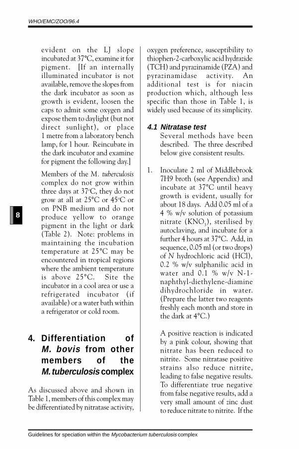
evident on the LJ slopeincubated at 37°C, examine it forpigment. [If an internallyilluminated incubator is notavailable, remove the slopes fromthe dark incubator as soon asgrowth is evident, loosen thecaps to admit some oxygen andexpose them to daylight (but notdirect sunlight), or place1 metre from a laboratory benchlamp, for 1 hour. Reincubate inthe dark incubator and examinefor pigment the following day.]
Members of the M. tuberculosiscomplex do not grow withinthree days at 37oC, they do notgrow at all at 25°C or 45oC oron PNB medium and do notproduce yellow to orangepigment in the light or dark(Table 2). Note: problems inmaintaining the incubationtemperature at 25°C may beencountered in tropical regionswhere the ambient temperatureis above 25°C. Site theincubator in a cool area or use arefrigerated incubator (ifavailable) or a water bath withina refrigerator or cold room.
4. Differentiation ofM. bovis from othermembers of theM. tuberculosis complex
As discussed above and shown inTable 1, members of this complex maybe differentiated by nitratase activity,
oxygen preference, susceptibility tothiophen-2-carboxylic acid hydrazide(TCH) and pyrazinamide (PZA) andpyrazinamidase activity. Anadditional test is for niacinproduction which, although lessspecific than those in Table 1, iswidely used because of its simplicity.
4.1 Nitratase testSeveral methods have beendescribed. The three describedbelow give consistent results.
1. Inoculate 2 ml of Middlebrook7H9 broth (see Appendix) andincubate at 37°C until heavygrowth is evident, usually forabout 18 days. Add 0.05 ml of a4 % w/v solution of potassiumnitrate (KNO3), sterilised byautoclaving, and incubate for afurther 4 hours at 37°C. Add, insequence, 0.05 ml (or two drops)of N hydrochloric acid (HCl),0.2 % w/v sulphanilic acid inwater and 0.1 % w/v N-1-naphthyl-diethylene-diaminedihydrochloride in water.(Prepare the latter two reagentsfreshly each month and store inthe dark at 4°C.)
A positive reaction is indicatedby a pink colour, showing thatnitrate has been reduced tonitrite. Some nitratase positivestrains also reduce nitrite,leading to false negative results.To differentiate true negativefrom false negative results, add avery small amount of zinc dustto reduce nitrate to nitrite. If the

WHO/EMC/ZOO/96.4
9
Guidelines for speciation within the Mycobacterium tuberculosis complex
strain is nitratase positive, andhas therefore already reduced allthe nitrate, the solution will notchange colour. If the strain isnitratase negative, nitrate will bereduced to nitrite and a pinkcolour will appear. Note: ‘falsepositive’ nitratase reactions mayoccur if the culture iscontaminated by certain otherbacteria.
2. Grow the organisms on a LJ slope(not containing pyruvate),transfer a few colonies to a 0.1%w/v solution of sodium nitrate,incubate at 37oC for 4 hours andthen proceed as above.
3. As an alternative to the reagentused in the two above methods,a powder, Lampe reagent, may beused and makes the test easier toperform while giving similarresults.16 Prepare the reagent byplacing one part of N-1-naphthyl-diethylene-diaminedihydrochloride, one part ofsulphanilic acid and 10 parts ofL(+) tartaric acid in a dark bottleand mix by shaking vigorously.The mixture is stable for at least6 months if stored in the dark.For the test, transfer a heavyloopful of a recently grownculture on solid egg media (LJG,LJP or Stonebrink) to a tubecontaining an aqueous 0.01M(0.085%) solution of sodiumnitrate and incubate for 3 hoursat 37oC. Add a small quantityof Lampe reagent (such as aspatula full: the quantity is not
critical). Read and interpret asin 1. above.
4.2 Oxygen preference
The medium is Middlebrook7H9 or Kirchner broth (seeAppendix) rendered semisolid bythe addition of 0.1% pure agarand dispensed in 10 ml amountsin screw-capped bottles. Pipette0.2 ml of the bacterial suspensionabout 1 cm below the surface ofthe medium and mix carefully,avoiding bubbles and aeration.Incubate at 37°C for 18 days.Aerobic strains grow at or nearthe surface, while microae-rophilic strains grow as a band10-20 mm below the surface,sometimes extending upwards.
4.3 Susceptibility to thiophen-2-carboxylic acid hydrazide(TCH)
Prepare slopes of LJ containingTCH, 5 mg/litre. Inoculate,incubate at 37°C for 2-3 weeksand compare growth with thaton TCH-free control medium.Note: 1) some strains ofM. bovis grow feebly or not at allon LJ containing glycerol andthus give false negative results ifthis medium is used. 2) Pyru-vate inhibits the activity ofisoniazid and could have a similareffect on TCH, which is ananalogue of isoniazid, and thusgive false positive results.17

WHO/EMC/ZOO/96.4
10
Guidelines for speciation within the Mycobacterium tuberculosis complex
Therefore those strains ofM. bovis that only grow on mediacontaining pyruvate may not betestable by this method. 3) Somestrains of M. tuberculosis aresusceptible to TCH: these areknown as the South Indian orAsian strains and are foundmainly in South India but alsoin Asian immigrantcommunities in other countries.Thus tests for susceptibility toTCH must be supplemented bythe other identification tests.
4.4 Susceptibility to pyrazina-mide (PZA)
The results are criticallydependent on pH. If the pH istoo high, the agent fails toinhibit bacterial growth and if itis too low the acidity inhibitsbacterial growth. Severalmethods of varying complexityhave been described but that ofYates gives consistent results.1,13
Four tubes of media are used, twocontrol and two containing PZA(see Appendix). Inoculate onetest and one control tube with20 µl of suspension and the otherpair of tubes with 20 µl of a 1:10dilution of the suspension.Incubate for 21 days at 37°C.PZA susceptibility is indicated bygrowth in the control tubes onlyor in the PZA tube receiving theheavier inoculum but not in thatreceiving the lighter inoculum,provided that there is growth inthe corresponding control tube.
Note: 1) Strains other thanM. bovis may acquire resistanceto pyrazinamide by mutation.2) Some recent isolates ofM. bovis do not grow in theacidified medium. Although thishas not been a problem in theUnited Kingdom, it could be soin other regions where use of thepyrazinamidase test (see nextsection) may be more suitable.
4.5 Detection of pyrazina-midase activity (Wayne’smethod) 10
Inoculate butts of Wayne’smedium (see Appendix) heavily,so that the inoculum is visible tothe naked eye. Incubate at 37°Cfor 7 days and add 1 ml of freshlyprepared ferrous ammoniumsulphate (Fe(NH4)2(SO4)2,6H2O), 1% w/v in distilled water.Refrigerate at 4°C for 4 hours. Apositive reaction is indicated bya pink band in the upper part ofthe agar butt. Include positiveand negative control slopesinoculated with M. tuberculosisand M. bovis respectively. A fewstrains of M. bovis produce a veryfaint pink band which is scoredas negative.
Pyrazinamidase is one of a seriesof enzymes detected in the amiderow of Bönicke,9 but thatmethod is more complex andhazardous as it involves washingby means of centrifugation andthe use of hazardous chemicals todetect ammonia production.
WHO/EMC/ZOO/96.4
11
Guidelines for speciation within the Mycobacterium tuberculosis complex
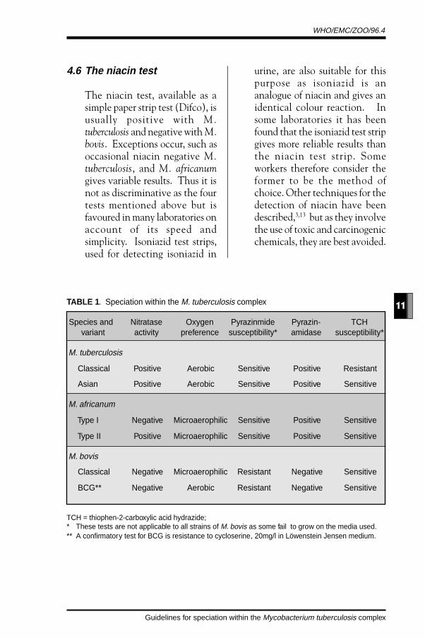
4.6 The niacin test
The niacin test, available as asimple paper strip test (Difco), isusually positive with M.tuberculosis and negative with M.bovis. Exceptions occur, such asoccasional niacin negative M.tuberculosis, and M. africanumgives variable results. Thus it isnot as discriminative as the fourtests mentioned above but isfavoured in many laboratories onaccount of its speed andsimplicity. Isoniazid test strips,used for detecting isoniazid in
urine, are also suitable for thispurpose as isoniazid is ananalogue of niacin and gives anidentical colour reaction. Insome laboratories it has beenfound that the isoniazid test stripgives more reliable results thanthe niacin test strip. Someworkers therefore consider theformer to be the method ofchoice. Other techniques for thedetection of niacin have beendescribed,3,13 but as they involvethe use of toxic and carcinogenicchemicals, they are best avoided.
TABLE 1 . Speciation within the M. tuberculosis complex
Species and Nitratase Oxygen Pyrazinmide Pyrazin- TCHvariant activity preference susceptibility* amidase susceptibility*
M. tuberculosis
Classical Positive Aerobic Sensitive Positive Resistant
Asian Positive Aerobic Sensitive Positive Sensitive
M. africanum
Type I Negative Microaerophilic Sensitive Positive Sensitive
Type II Positive Microaerophilic Sensitive Positive Sensitive
M. bovis
Classical Negative Microaerophilic Resistant Negative Sensitive
BCG** Negative Aerobic Resistant Negative Sensitive
TCH = thiophen-2-carboxylic acid hydrazide;* These tests are not applicable to all strains of M. bovis as some fail to grow on the media used.** A confirmatory test for BCG is resistance to cycloserine, 20mg/l in Löwenstein Jensen medium.

WHO/EMC/ZOO/96.4
12
Guidelines for speciation within the Mycobacterium tuberculosis complex
TABLE 2 . Differentiation of members of the M. tuberculosis complex from other cultivablemycobacteria.
Growth on Growth Growth Pigment IdentificationPNB Medium* at 25oC within production
3 days
- - - None M. tuberculosiscomplex
+ + - Light only Photochromogen
+ + - In dark Scotochromogenand light
+ + or - - None Nonchromogen
+ + + Variable Rapid grower
* Löwenstein-Jensen medium containing p-nitrobenzoic acid (PNB), 500 mg/litre.

WHO/EMC/ZOO/96.4
13
Guidelines for speciation within the Mycobacterium tuberculosis complex
References
1. Thoen CO, Steele JH. (Editors) Mycobacterium bovis infection in animalsand humans. Ames: Iowa State University Press. 1995.
2. US Department of Agriculture: Animal and Plant Inspection Service,Veterinary Services. Laboratory methods in veterinary mycobacteriology.Ames, Iowa: National Veterinary Services Laboratories. 1985.
3. Kantor IN. Bacteriología de la tuberculosis humana y animal, 2nd Edn. BuenosAires: CEPANZO. 1989.
4. Moda G, Daborn CJ, Grange JM, Cosivi O. The zoonotic importance ofMycobacterium bovis. Tubercle and Lung Disease 1996; 77: 103-108
5. Yates MD. The differentiation and epidemiology of the tubercle bacilliand a study into the identification of other mycobacteria. Master ofPhilosophy Thesis. University of London. 1984.
6. Collins CH, Yates MD, Grange JM. Subdivision of Mycobacteriumtuberculosis into five variants for epidemiological purposes: methods andnomenclature. Journal of Hygiene 1982; 89: 235-242.
7. Yates MD, Grange JM. Incidence and nature of human tuberculosis dueto bovine tubercle bacilli in South East England: 1977-1987. Epidemiologyand Infection 1988; 101: 225-229.
8. Grange JM, Collins JD, O’Reilly L, Costello E, Yates MD. Identificationand characteristics of Mycobacterium bovis isolated from cattle, badgersand deer in the Republic of Ireland. Irish Veterinary Journal 1990;43: 33-35.
9. Bönicke R. Report on identification of mycobacteria by biochemicalmethods. Bulletin of the International Union Against Tuberculosis 1962; 32:13-68.
10. Wayne LG. Simple pyrazinamidase and urease tests for routineidentification of mycobacteria. American Review of Respiratory Disease 1979;109: 147-151.

WHO/EMC/ZOO/96.4
14
Guidelines for speciation within the Mycobacterium tuberculosis complex
11. Rist N, Canetti G, Boisvert H, Le Lirzin M. L’antibiogramme du BCG.Valeur diagnostique de la résistance à la cycloserine. Revue de Tuberculoseet de Pneumonologie 1967; 31: 1060-1065.
12. Canetti G, Grosset J. Techniques et indications des examens bactériologiquesen tuberculose. Éditions de la Tourelle, St. Mandé. 1969: 140-143.
13. Collins CH, Grange JM, Yates MD. Organization and practice in tuberculosisbacteriology. London: Butterworths. 1985.
14. Kleeberg HH, Koornhof HJ, Palmhert H. Laboratory manual of tuberculosismethods, 2nd edn, revised. Pretoria: SAMRC Tuberculosis ResearchInstitute. 1980.
15. World Health Organization. Laboratory biosafety manual, 2nd edn. Geneva:World Health Organization. 1993.
16. Warren NG, Body BA, Dalton HP. An improved reagent for mycobacterialnitrate reductase tests. Journal of Clinical Microbiology 1983; 18: 546-549.
17. Boisvert H. Action d l’acide pyruvique sur la croissance et l’antibiogrammedes mycobactéries. Revue de Tuberculose (Paris) 1970; 34: 117.
WHO/EMC/ZOO/96.4
15
Guidelines for speciation within the Mycobacterium tuberculosis complex
Appendix - Media
(Powder for Middlebrook 7H9 broth and ready-for-use OADC enrichment arecommercially available. In some regions Löwenstein-Jensen medium iscommercially available.)
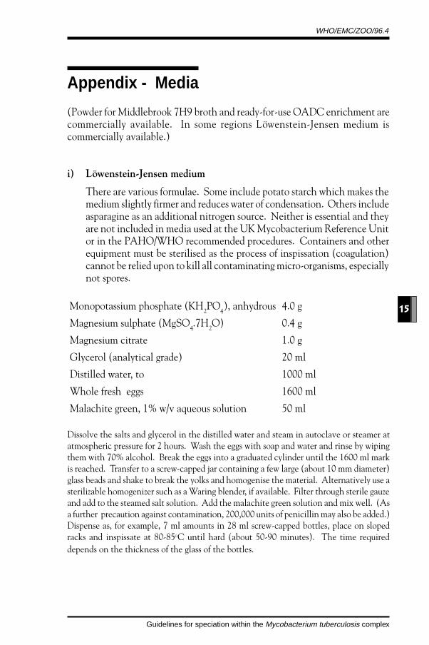
i) Löwenstein-Jensen medium
There are various formulae. Some include potato starch which makes themedium slightly firmer and reduces water of condensation. Others includeasparagine as an additional nitrogen source. Neither is essential and theyare not included in media used at the UK Mycobacterium Reference Unitor in the PAHO/WHO recommended procedures. Containers and otherequipment must be sterilised as the process of inspissation (coagulation)cannot be relied upon to kill all contaminating micro-organisms, especiallynot spores.
Monopotassium phosphate (KH2PO
4), anhydrous 4.0 g
Magnesium sulphate (MgSO4.7H
2O) 0.4 g
Magnesium citrate 1.0 g
Glycerol (analytical grade) 20 ml
Distilled water, to 1000 ml
Whole fresh eggs 1600 ml
Malachite green, 1% w/v aqueous solution 50 ml
Dissolve the salts and glycerol in the distilled water and steam in autoclave or steamer atatmospheric pressure for 2 hours. Wash the eggs with soap and water and rinse by wipingthem with 70% alcohol. Break the eggs into a graduated cylinder until the 1600 ml markis reached. Transfer to a screw-capped jar containing a few large (about 10 mm diameter)glass beads and shake to break the yolks and homogenise the material. Alternatively use asterilizable homogenizer such as a Waring blender, if available. Filter through sterile gauzeand add to the steamed salt solution. Add the malachite green solution and mix well. (Asa further precaution against contamination, 200,000 units of penicillin may also be added.)Dispense as, for example, 7 ml amounts in 28 ml screw-capped bottles, place on slopedracks and inspissate at 80-85oC until hard (about 50-90 minutes). The time requireddepends on the thickness of the glass of the bottles.

WHO/EMC/ZOO/96.4
16
Guidelines for speciation within the Mycobacterium tuberculosis complex
ii) Stonebrink’s medium
Monopotassium phosphate (KH2PO
4), anhydrous 3.5 g
Disodium phosphate (Na2HPO
4.12H
2O) 2.0 g
Sodium pyruvate 6.3 g
Distilled water, to 500 ml
Whole fresh eggs 1000 ml
Malachite green, 2% w/v aqueous solution 20 ml
The medium is prepared in the same manner as Löwenstein-Jensen medium. An alternativemedium is the Löwenstein-Jensen medium as described above but with 12 g of sodiumpyruvate instead of the glycerol.
iii) Middlebrook 7H9 broth
Ammonium sulphate ((NH4)
2SO
4) 0.5 g
Sodium glutamate 0.5 g
Disodium phosphate (Na2HPO
4.12H
20) 2.5 g
Monopotassium phosphate (KH2PO
4), anhydrous 1.0 g
Trisodium citrate 100 mg
Ferric ammonium citrate 40 mg
Magnesium sulphate (MgSO4.7H
2O) 50 mg
Zinc sulphate (ZnSO4.7H
2O) 1.0 mg
Cupric sulphate (CuSO4.5H
2O) 1.0 mg
Pyridoxine 1.0 mg
Calcium chloride (CaCl2) 0.5 mg
Biotin 0.5 mg
Tween 80 0.5 ml
Distilled water, to 900 ml
Dissolve by steaming. Distribute as required and autoclave at 115°C for 15 minutes. Whencool add 1 volume of Middlebrook OADC enrichment (see below), or 1 volume of sterilehorse serum, to 9 volumes of medium.
WHO/EMC/ZOO/96.4
17
Guidelines for speciation within the Mycobacterium tuberculosis complex
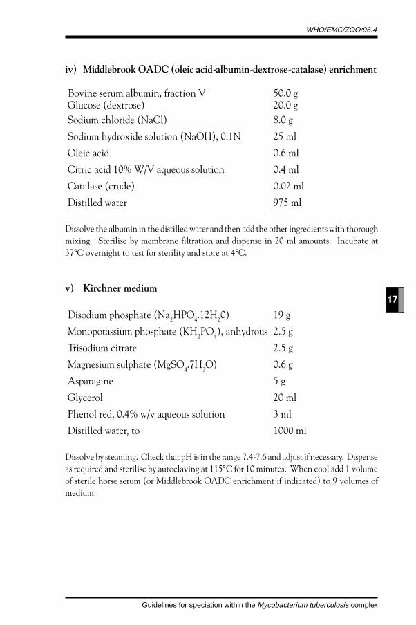
iv) Middlebrook OADC (oleic acid-albumin-dextrose-catalase) enrichment
Bovine serum albumin, fraction V 50.0 g Glucose (dextrose) 20.0 g Sodium chloride (NaCl) 8.0 g
Sodium hydroxide solution (NaOH), 0.1N 25 ml
Oleic acid 0.6 ml
Citric acid 10% W/V aqueous solution 0.4 ml
Catalase (crude) 0.02 ml
Distilled water 975 ml
Dissolve the albumin in the distilled water and then add the other ingredients with thoroughmixing. Sterilise by membrane filtration and dispense in 20 ml amounts. Incubate at37°C overnight to test for sterility and store at 4°C.
v) Kirchner medium
Disodium phosphate (Na2HPO
4.12H
20) 19 g
Monopotassium phosphate (KH2PO
4), anhydrous 2.5 g
Trisodium citrate 2.5 g
Magnesium sulphate (MgSO4.7H
2O) 0.6 g
Asparagine 5 g
Glycerol 20 ml
Phenol red, 0.4% w/v aqueous solution 3 ml
Distilled water, to 1000 ml
Dissolve by steaming. Check that pH is in the range 7.4-7.6 and adjust if necessary. Dispenseas required and sterilise by autoclaving at 115°C for 10 minutes. When cool add 1 volumeof sterile horse serum (or Middlebrook OADC enrichment if indicated) to 9 volumes ofmedium.

WHO/EMC/ZOO/96.4
18
Guidelines for speciation within the Mycobacterium tuberculosis complex
vi) Medium for pyrazinamide susceptibility tests
This is a two-phase medium, with solid and semisolid layers.
Solid phase. Prepare Löwenstein-Jensen medium with glycerol and adjustelectrometrically to pH 5.2 with N hydrochloric acid. (Use of a pH meter isessential for accuracy.) Add 9 ml of a 0.22% w/v solution of pyrazinamide,sterilised by membrane filtration, to 300 ml of medium. Add 9 ml of sterilewater to a further 300 ml of medium for the control. Place 1 ml amounts of themedia in small screw- capped bottles and inspissate (85°C for 1 hour) in avertical position.
Semisolid phase. Prepare 1 litre of Kirchner medium and add 1 g of agar and3 g of sodium pyruvate and adjust electrometrically to pH 5.2 withN hydrochloric acid. Dissolve agar by steaming. Divide into two 500 ml batchesand add 15 ml of the pyrazinamide solution (see above) to one and 15 ml ofsterile water to the other. Sterilise by autoclaving and, when cool, add 30 mlof Middlebrook OADC enrichment (see above) to both.
Final medium. Add 2 ml amounts of test and control semisolid medium to thecorresponding bottles containing the solid medium. Store at 4°C and use withinthree weeks of preparation.
vii) Medium for detection of pyrazinamidase activity
Middlebrook 7H9 broth (see above) 1000 ml Pyrazinamide 100 mg Sodium pyruvate 2 g Agar 15 g
Dissolve by steaming. Dispense in 5 ml amounts in screw-cap bottles and sterilise byautoclaving at 115°C for 15 minutes. Allow to cool in the upright position so that buttsrather than slopes are formed.