whole-body physiologically based pharmacokinetic model for
TRANSCRIPT
Whole-Body Physiologically Based Pharmacokinetic Model forNutlin-3a in Mice after Intravenous and Oral Administration□S
Fan Zhang,1 Michael Tagen,1 Stacy Throm, Jeremy Mallari, Laura Miller, R. Kiplin Guy,Michael A. Dyer, Richard T. Williams, Martine F. Roussel, Katie Nemeth, Fangyi Zhu,
Jiakun Zhang, Min Lu, John C. Panetta, Nidal Boulos, and Clinton F. Stewart
Departments of Pharmaceutical Sciences (F.Z., M.T., S.T., L.M., C.F.S., J.C.P.), Chemical Biology (J.M., R.K.G., F.Z., M.L.),Developmental Neurobiology (K.N., J.Z., M.A.D.), Oncology (R.T.W., N.B.), and Tumor Cell Biology (M.F.R.), St. Jude Children’s
Research Hospital, Memphis, Tennessee; and Department of Pharmaceutical Sciences (F.Z., C.F.S., J.C.P.), College ofPharmacy, University of Tennessee Health Science Center, Memphis, Tennessee
Received August 16, 2010; accepted October 13, 2010
ABSTRACT:
Nutlin-3a is an MDM2 inhibitor that is under investigation in pre-clinical models for a variety of pediatric malignancies, includingretinoblastoma, rhabdomyosarcoma, neuroblastoma, and leuke-mia. We used physiologically based pharmacokinetic (PBPK) mod-eling to characterize the disposition of nutlin-3a in the mouse.Plasma protein binding and blood partitioning were assessed by invitro studies. After intravenous (10 and 20 mg/kg) and oral (50, 100,and 200 mg/kg) dosing, tissue concentrations of nutlin-3a weredetermined in plasma, liver, spleen, intestine, muscle, lung, adi-pose, bone marrow, adrenal gland, brain, retina, and vitreous fluid.The PBPK model was simultaneously fit to all pharmacokineticdata using NONMEM. Nutlin-3a exhibited nonlinear binding to mu-
rine plasma proteins, with the unbound fraction ranging from 0.7 to11.8%. Nutlin-3a disposition was characterized by rapid absorp-tion with peak plasma concentrations at approximately 2 h andbiphasic elimination consistent with a saturable clearance pro-cess. The final PBPK model successfully described the plasma andtissue disposition of nutlin-3a. Simulations suggested high bio-availability, rapid attainment of steady state, and little accumula-tion when administered once or twice daily at dosages up to 400mg/kg. The final model was used to perform simulations of un-bound tissue concentrations to determine which dosing regi-mens are appropriate for preclinical models of several pediatricmalignancies.
Introduction
Nutlin-3a (2-piperazinone, 4-[[(4S,5R)-4,5-bis(4-chlorophenyl)-4,5-di-hydro-2-[4 methoxy-2-(1-methylethoxy)phenyl]-1H-imidazol-1-yl]car-bonyl]-) is currently undergoing preclinical investigation as a p53reactivation agent. Although many cancers and tumor types expressmutated forms of p53 (Hollstein et al., 1991), a subset of cancers, andparticularly pediatric tumors, retain wild-type p53 (Tweddle et al.,2003). In these cases, cancer cells frequently use other mechanisms toabrogate p53 function. One such mechanism is overexpression oramplification of the murine double minute (MDM2) protein. Thismolecule binds directly to p53 to accelerate its turnover and inhibitstranscription of downstream targets, including cell cycle and apoptoticgenes (Momand et al., 1992, 2000). Disruption of the MDM2-p53
interaction is proposed as a novel strategy for treatment of cancers thatdo not have p53 alterations (Shangary and Wang, 2008a,b).
Nutlins are a class of small molecules that target the p53-bindingpocket of MDM2 (Klein and Vassilev, 2004; Vassilev et al., 2004).Treatment of multiple types of cancer cells, including leukemias(Kojima et al., 2005; Gu et al., 2008), neuroblastoma (Barbieri et al.,2006b), rhabdomyosarcoma (Miyachi et al., 2009), and retinoblas-toma (Elison et al., 2006), with nutlin-3a induces p53-dependent cellcycle arrest and cell death, whereas in normal cells, nutlin-3a expo-sure leads to cell cycle arrest without cell death (Vassilev, 2005).Nutlin-3a has antitumor activity in a preclinical xenograft model ofneuroblastoma (Van Maerken et al., 2009) and was tested in severalother preclinical models of malignancies (Vassilev et al., 2004; Sarekand Ojala, 2007).
To date, the pharmacokinetics of nutlin-3a have not been reported.An understanding of the systemic disposition of nutlin-3a, as well asthe distribution to target tissue or tumor sites, will provide a rationalbasis for the selection of dosage regimens for preclinical models. Inaddition, because the in vitro tumor cell line sensitivities to nutlin-3ahave been determined, pharmacokinetic modeling can be used todetermine the dose and schedule necessary to achieve appropriateunbound nutlin-3a concentrations at the tumor site. One approach toanalyze these data is the use of whole-body physiologically based
This work was supported in part by the National Institutes of Health NationalCancer Institute [Grant N01-CM-42216]; and by the American Lebanese SyrianAssociated Charities.
1 F.Z. and M.T. contributed equally to this work.Article, publication date, and citation information can be found at
http://dmd.aspetjournals.org.doi:10.1124/dmd.110.035915.□S The online version of this article (available at http://dmd.aspetjournals.org)
contains supplemental material.
ABBREVIATIONS: MDM2, murine double minute 2; PBPK, physiologically based pharmacokinetic; ALL, acute lymphoblastic leukemia; PBS,phosphate-buffered saline; AUC, area under the curve; P-gp, P-glycoprotein.
0090-9556/11/3901-15–21$20.00DRUG METABOLISM AND DISPOSITION Vol. 39, No. 1Copyright © 2011 by The American Society for Pharmacology and Experimental Therapeutics 35915/3652940DMD 39:15–21, 2011 Printed in U.S.A.
15
pharmacokinetic model (PBPK) models, which are based on anatom-ical compartments and blood flow.
Thus, we performed pharmacokinetic studies to develop a PBPKmodel describing the disposition of nutlin-3a in plasma and tissues,including adipose, adrenal gland, bone marrow, brain, liver, lung,intestine, muscle, retina, spleen, and vitreous fluid. The PBPK modelwas used to perform simulations, which in combination with in vitrocell sensitivity data provided rationale for choosing dosing regimensfor mouse models of common childhood cancers, including retino-blastoma, neuroblastoma, rhabdomyosarcoma, and acute lymphoblas-tic leukemia (ALL).
Materials and Methods
Animals. Adult C57BL/6 mice were purchased from Charles River (BarHarbor, ME). Mice were housed in a temperature-controlled room on a normal12-h light/dark cycle, with free access to water and standard laboratory food.All procedures were approved by the St. Jude Institutional Animal Care andUse Committee and conducted in accordance with the National Institutes ofHealth guidelines for the care and use of laboratory animals (Institute ofLaboratory Animal Resources, 1996). The animal facility is accredited by theAmerican Association for Accreditation of Laboratory Animal Care.
Chemicals. cis-Nutlin-3a (98% purity, lot no. 08252008) was synthesizedand supplied by the Department of Chemical Biology and Therapeutics at St.Jude Children’s Research Hospital (Memphis, TN). The oral formulation(Vassilev et al., 2004) used in the pharmacokinetic studies was nutlin-3asuspended in 2% Klucel (Conservation Resources International, LLC, Spring-field, VA) and 0.5% Tween 80 (Sigma-Aldrich, St. Louis, MO), and theintravenous formulation used was nutlin-3a in 4% ethanol, 35% propyleneglycol (Fisher Scientific, Pittsburgh, PA), 10% PEG-400 (Sigma-Aldrich), and51% phosphate-buffered saline (PBS) (Mediatech Inc., Herndon, VA). Theinternal standard ketoconazole was purchased from Sigma-Aldrich. Blankmurine plasma was obtained from Hilltop Laboratory Animals, Inc. (Scotts-dale, PA). All other reagents were of analytical grade or higher.
Blood/Plasma Ratio. Pooled whole blood from healthy male C57BL/6mice was collected into heparin tubes. Nutlin-3a was spiked into aliquots ofwhole blood at final concentrations of 0.1, 1, 10, and 100 �M in triplicate.Samples were mixed and incubated at 37°C for 30 min with additional mixingevery 5 min. After incubation, 50 �l of whole blood was removed andimmediately placed on dry ice. The remainder of the sample was centrifugedat 16,000 rpm for 2 min, and a 50-�l plasma sample was removed andimmediately placed on dry ice. All samples were stored at �80°C untilanalysis. The blood/plasma concentration ratio was calculated using the fol-lowing equation:
Blood to plasma ratio �CWB
CP
where CP is the concentration in plasma and CWB is the concentration in wholeblood.
Nutlin-3a Protein Binding Studies. Equilibrium dialysis was performed ina 96-well dialysis plate with a 5-kDa cutoff membrane (Harvard Apparatus,Holliston, MA). For mouse plasma and cell culture media protein binding, 200�l of PBS buffer was added into the wells on one side of the membrane andan equivalent volume of male C57BL/6 plasma or cell culture media (RPMI1640 medium with 10% fetal bovine serum and 2 mM L-glutamine) containingvarying concentrations of nutlin-3a was added into the wells on the oppositeside. The plate was sealed and fixed onto a dual plate rotator (HarvardApparatus) at 37°C in a humidified incubator containing 5% CO2. Equilibriumdialysis was performed at 0.2, 20, and 100 �M nutlin-3a in triplicate for 24 h.For vitreous protein binding, 150 �l of rodent vitreous containing 0.5 �Mnutlin-3a was added to the sample side and equal volume of PBS buffer wasadded to the buffer side. Equilibrium dialysis was performed in triplicate for24 h. The samples were analyzed using the analytical method described below.The bound concentration was considered equal to the plasma side and the freeconcentration equal to the PBS side (Kariv et al., 2001; Wan and Rehngren,2006). Binding parameters were estimated with nonlinear regression using theLangmuir equation (Xu et al., 1993):
CP,b �Bmax � KA � CP,f
1 � KA � CP,f
where CP,b is the bound plasma concentration, Bmax is the quantity of plasmaprotein binding sites, KA is the binding association constant, and CP,f is theunbound plasma concentration.
Drug Administration and Sample Collection. Two pharmacokinetic stud-ies were conducted. For the first pharmacokinetic study, 145 adult C57BL/6 mice(128 male and 17 female) were divided into three groups: two oral dosage groups(100 and 200 mg/kg) and one intravenous dosage group (10 mg/kg). Nutlin-3a wasadministered as a single bolus dose by oral gavage or by intravenous tail veininjection. Each dosing group (n � 5 mice) and vehicle controls had nine collectiontime points (0.5, 1, 2, 4, 8, 12, 24, 36, and 48 h). At each time point, blood wascollected under isoflurane anesthesia via cardiac puncture. Whole-blood sampleswere centrifuged immediately at 12,000g for 5 min at 4°C to separate plasma.Tissue samples, including brain, vitreous, retina, liver, spleen, and bone marrow,were dissected simultaneously. Each sample was put on dry ice immediately aftercollection and stored at �80°C until analysis.
In the second pharmacokinetic study, 210 adult male C57BL/6 mice wereused. Two oral dosages (50 and 100 mg/kg) and two intravenous dosages (10and 20 mg/kg) were administered. Each dosing group (n � 5 for 10 mg/kgintravenous and 100 mg/kg oral dosages; n � 10 for 20 mg/kg intravenous and50 mg/kg oral dosages) and vehicle control had seven collection time points(0.5, 1, 2, 4, 8, 12, and 24 h for the intravenous dosing; 0.5, 1, 2, 4, 8, 12, and16 h for oral dosing). Serial plasma samples were collected from all mice.Tissue samples, including brain, lung, liver, spleen, kidney, adrenal gland,muscle, fat, and intestine from three mice per time point from the 20 mg/kgintravenous group, were collected. Each sample was put on dry ice immedi-ately after the sample collection and stored at �80°C until analysis.
Quantitative Analysis of Nutlin-3a in Mouse Tissues and PBS. Nutlin-3amouse plasma samples were analyzed based on our previously published liquidchromatography electrospray ionization tandem mass spectrometry analyticalmethod (Bai et al., 2009). For each sample type (cerebellum, brain, vitreous,retina, lung, heart, liver, gall bladder, spleen, kidney, adrenal gland, muscle,fat, bone marrow, intestine, whole blood, and PBS), standard curves andcontrols were generated using the corresponding untreated tissue or PBS toeliminate any matrix effect. For larger tissues, sections were cut, weighed, andstored on ice for further processing. Ten microliters of ice-cold homogenizationbuffer (5 mM HCOONH4, pH � 7) was added per milligram of tissue. For smallertissue samples, including vitreous, retina, adrenal gland, and gall bladder, theamount of homogenization buffer used was increased to a minimum volume of70�100 �l. Tissue samples were then sonicated on ice for 15 s, with 5-s intervals.The number of total sonications varied depending on the tissue types. Homoge-nated tissues and whole-blood samples were extracted and analyzed using proteinprecipitation, and the PBS samples were extracted using the liquid-liquid extrac-tion method as described previously (Bai et al., 2009).
Whole-Body PBPK Model Development. We developed a whole-bodyPBPK model for nutlin-3a based on in vitro blood cell partitioning, plasmaprotein binding, and pooled concentration-time data from all plasma and tissuesamples collected from both pharmacokinetic studies. This PBPK modelconsisted of a series of mass balance differential equations describing theconcentration of nutlin-3a in various tissues, which were connected by bloodflow. Physiological values for mouse organ size and blood flow are presentedin Table 1. A schematic representation of the model is shown in Fig. 1.
Plasma concentrations were converted to whole-blood concentrations basedon the in vitro blood partitioning experiment. Unbound plasma concentrationswere described by the following equation (Xu et al., 1993):
CP,u ���1 � Bmax � KA � CP� � ��1 � Bmax � KA � CP�
2 � 4 � KA � CP�1/ 2
2 � KA
where CP,u is the unbound plasma concentration, CP is the total plasmaconcentration, and Bmax and KA were determined from in vitro plasma proteinbinding studies. The unbound fraction (fub) was calculated by
fub �CP,u
CP
16 ZHANG ET AL.

Most organs fit well to a perfusion-limited model, and thus were describedby the following equation:
Vi �dAi
dt� Qi � �CART �
Ci
Ki�
where Vi is the volume of organ, Ai is the amount of drug in the organ, Ci isthe concentration in the organ, Ki is the partition coefficient, and CART is theconcentration of arterial plasma.
Liver blood flow (QLIV) was the sum of the blood flow to the hepatic artery,spleen, and liver, and the concentration of blood entering the liver (CBLO,LIV)was based on the arterial concentration and the venous outflow of the portalcirculation. The liver contained an elimination term (ke) for metabolism, basedon experiments showing that nutlin-3a is metabolized by mouse liver micro-somes (K. Guy, unpublished data):
VLIV �dALIV
dt� QLIV � �CBLO,LIV �
CLIV
KLIV�� ke � CART
The intestine was modeled with a separate lumen and tissue compartment.Absorption from the lumen was assumed to be linear based on an absorptionrate constant (ka):
VINT �dAINT
dt� ka � AINT
The eye was fit to a two-compartment model consisting of the retina andvitreous. Input into the vitreous was by diffusion from the retina. The follow-ing equations were used for the retina and vitreous:
VRET �dARET
dt� QRET � �CART �
CRET
KRET�� PAVIT � �CRET �
CVIT
KVIT�
VVIT �dAVIT
dt� PAVIT � �CRET �
CVIT
KVIT�
where PAVIT is the permeability-surface area product.All tissues that were not sampled were lumped together in a residual
compartment. Modeling this compartment as perfusion-limited did not ade-quately describe the multiexponential profile of nutlin-3a. Therefore, theresidual compartment was modeled as diffusion-limited, with the vascularspace assumed to be 5% of the residual volume. The equations for the residualvascular space and tissue are as follows:
VRES, BLO �dARES
dt� QRES � �CART � CRES, BLO� � PARES � �CRES �
CRES
KRES�
VRES �dARES
dt� PARES � �CRES �
CRES
KRES�
The input into the venous pool of blood was modeled as the sum of theoutput from all tissues except the lung. The volume of the venous pool wasfixed to 75% of the total blood volume. The lungs received all venous input,and the arterial input was the output from the lungs. The equation for the lungswas as follows:
VLUN �dALUN
dt� QBLO � �CVEN �
CLUN
KLUN�
The arterial concentrations were based on the output from the lungs andcontained an additional saturable elimination term:
VART �dAART
dt� QBLO � �CLUN
KLUN� CART��
Vmax � CART
Km � CART
Elimination terms in both the blood compartment and liver compartmentwere necessary for a good model fit to the data from both oral and intravenousadministration.
Simulations. After development of the PBPK model, tissue concentrationswere simulated with NONMEM after multiple oral and intravenous doses at
FIG. 1. Schematic diagram of PBPK model for nutlin-3a in mice. C,b, bound drugconcentration; C,f, free drug concentration. Arrows connecting compartments rep-resent blood flows from literature values.
TABLE 1
List of physiological parameters
Tissue Symbol Mass QB Reference
%b.wt. ml/h
Blood BLO 4.9 839 Brown et al., 1997Adipose ADI 6.8 58.7 Brown et al., 1997Adrenal gland ADR 0.048 2.52 Brown et al., 1997Bone marrow MRW 5.8 92.3 Brown et al., 1997Brain BRA 1.65 27.7 Brown et al., 1997Intestines INT 3.62 109 Brown et al., 1997Liver LIV 5.49 16.8 Brown et al., 1997Lung LUN 0.73 839 Brown et al., 1997Muscle MUS 38.4 133 Brown et al., 1997Retina RET 0.04 3.16 Experimental; Wright et al., 2009Spleen SPL 0.35 9.48 Brown et al., 1997Vitreous fluid VIT 0.035 0a ExperimentalRemainder RES 29.9 256.9
a Vitreous assumed to have no direct blood flow.
17PHYSIOLOGICALLY BASED PHARMACOKINETICS OF NUTLIN-3a
50, 100, 200, and 400 mg/kg given both once daily and twice daily. TheAUC0–24 at steady state was calculated with the log-linear trapezoidal methodapplied to the simulated data. Bioavailability was estimated using the ratio ofAUC0-�, IV/AUC0-�, PO with the simulated steady-state data.
Results
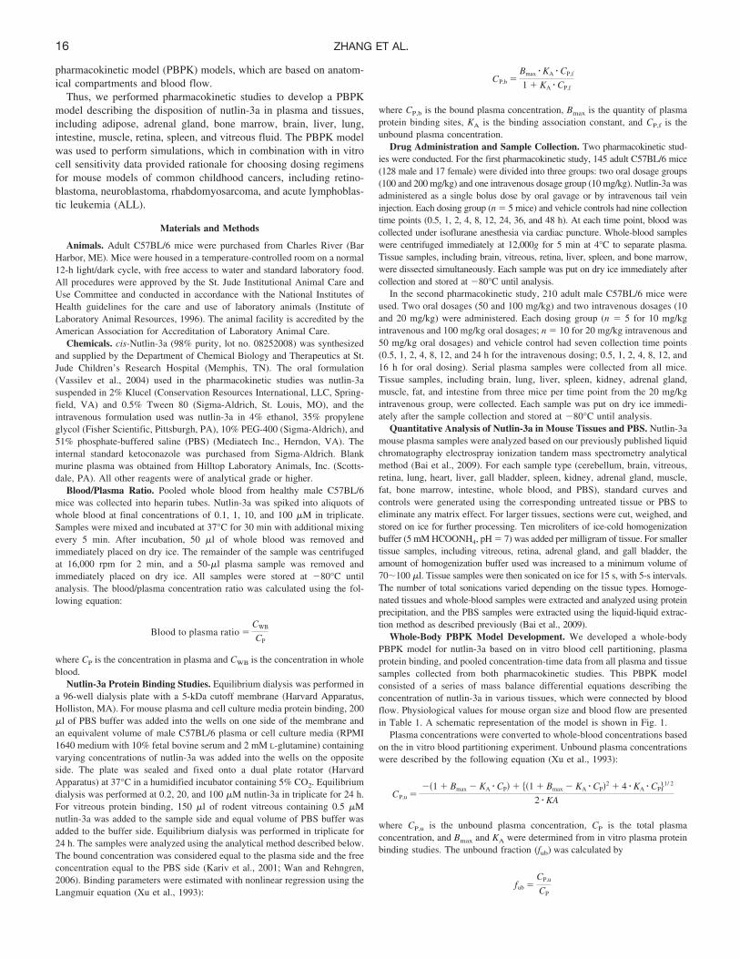
Blood-to-Plasma Partitioning and Plasma Protein Binding ofNutlin-3a. Blood-to-plasma partitioning showed an average blood/plasma concentration ratio of 0.70, indicating that 30% of nutlin-3apartitions to blood cells (Fig. 2A). Binding of nutlin-3a to mouseplasma proteins was nonlinear, with fub ranging from 0.007 at 0.1 �Mto 0.118 at 300 �M (Fig. 2B). Nonlinear regression of unbound versusbound plasma concentrations using the Langmuir equations resultedin a Bmax of 286 and a KA of 0.085 (Supplemental Fig. S1).
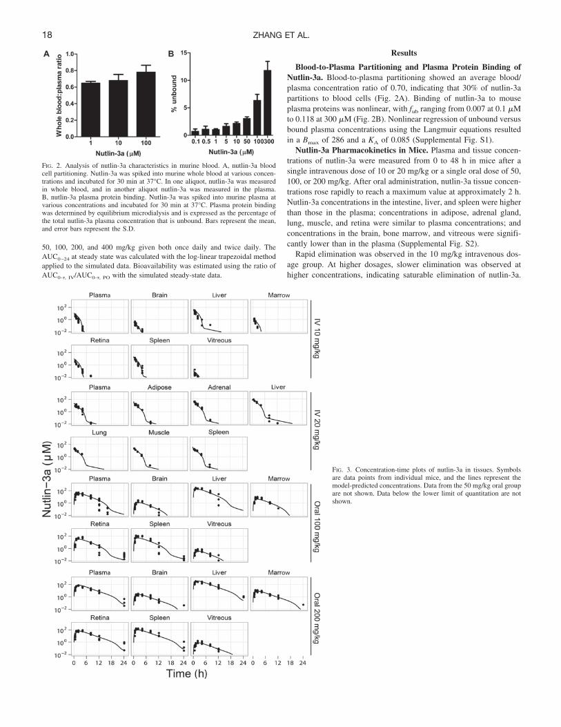
Nutlin-3a Pharmacokinetics in Mice. Plasma and tissue concen-trations of nutlin-3a were measured from 0 to 48 h in mice after asingle intravenous dose of 10 or 20 mg/kg or a single oral dose of 50,100, or 200 mg/kg. After oral administration, nutlin-3a tissue concen-trations rose rapidly to reach a maximum value at approximately 2 h.Nutlin-3a concentrations in the intestine, liver, and spleen were higherthan those in the plasma; concentrations in adipose, adrenal gland,lung, muscle, and retina were similar to plasma concentrations; andconcentrations in the brain, bone marrow, and vitreous were signifi-cantly lower than in the plasma (Supplemental Fig. S2).
Rapid elimination was observed in the 10 mg/kg intravenous dos-age group. At higher dosages, slower elimination was observed athigher concentrations, indicating saturable elimination of nutlin-3a.
FIG. 2. Analysis of nutlin-3a characteristics in murine blood. A, nutlin-3a bloodcell partitioning. Nutlin-3a was spiked into murine whole blood at various concen-trations and incubated for 30 min at 37°C. In one aliquot, nutlin-3a was measuredin whole blood, and in another aliquot nutlin-3a was measured in the plasma.B, nutlin-3a plasma protein binding. Nutlin-3a was spiked into murine plasma atvarious concentrations and incubated for 30 min at 37°C. Plasma protein bindingwas determined by equilibrium microdialysis and is expressed as the percentage ofthe total nutlin-3a plasma concentration that is unbound. Bars represent the mean,and error bars represent the S.D.
FIG. 3. Concentration-time plots of nutlin-3a in tissues. Symbolsare data points from individual mice, and the lines represent themodel-predicted concentrations. Data from the 50 mg/kg oral groupare not shown. Data below the lower limit of quantitation are notshown.
18 ZHANG ET AL.
After 24 h, all data were below the limit of quantitation of the assay.Models with linear elimination, Michaelis-Menten elimination, andcombined linear and Michaelis-Menten elimination were fit to thedata. Ultimately, the combination of both linear and Michaelis-Menten elimination had the best fit to the data. The concentration-timedata of nutlin-3a in all modeled tissues are plotted against the model-predicted concentrations in Fig. 3 (data not shown for oral 50 mg/kgdosage group, because only plasma was collected). The estimatedpharmacokinetic parameters are listed in Table 2.
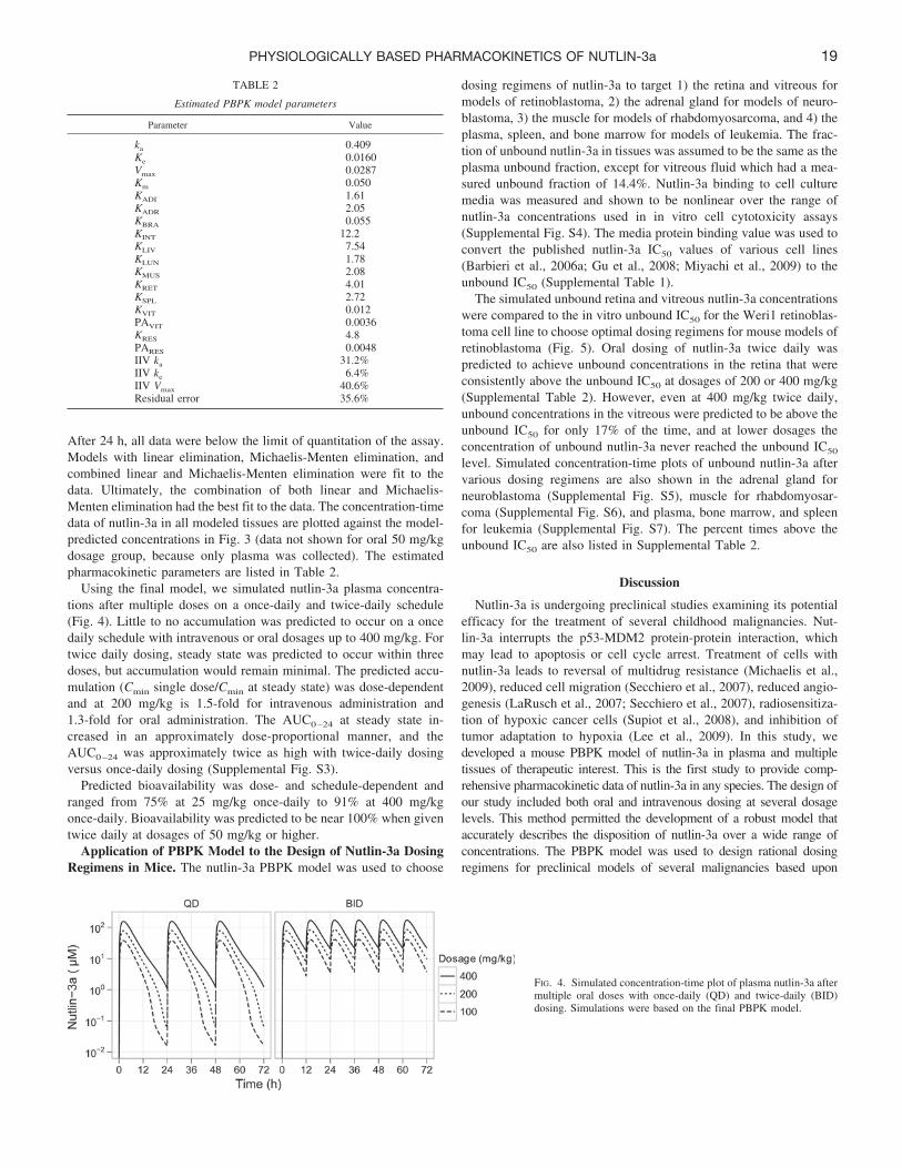
Using the final model, we simulated nutlin-3a plasma concentra-tions after multiple doses on a once-daily and twice-daily schedule(Fig. 4). Little to no accumulation was predicted to occur on a oncedaily schedule with intravenous or oral dosages up to 400 mg/kg. Fortwice daily dosing, steady state was predicted to occur within threedoses, but accumulation would remain minimal. The predicted accu-mulation (Cmin single dose/Cmin at steady state) was dose-dependentand at 200 mg/kg is 1.5-fold for intravenous administration and1.3-fold for oral administration. The AUC0–24 at steady state in-creased in an approximately dose-proportional manner, and theAUC0–24 was approximately twice as high with twice-daily dosingversus once-daily dosing (Supplemental Fig. S3).
Predicted bioavailability was dose- and schedule-dependent andranged from 75% at 25 mg/kg once-daily to 91% at 400 mg/kgonce-daily. Bioavailability was predicted to be near 100% when giventwice daily at dosages of 50 mg/kg or higher.
Application of PBPK Model to the Design of Nutlin-3a DosingRegimens in Mice. The nutlin-3a PBPK model was used to choose
dosing regimens of nutlin-3a to target 1) the retina and vitreous formodels of retinoblastoma, 2) the adrenal gland for models of neuro-blastoma, 3) the muscle for models of rhabdomyosarcoma, and 4) theplasma, spleen, and bone marrow for models of leukemia. The frac-tion of unbound nutlin-3a in tissues was assumed to be the same as theplasma unbound fraction, except for vitreous fluid which had a mea-sured unbound fraction of 14.4%. Nutlin-3a binding to cell culturemedia was measured and shown to be nonlinear over the range ofnutlin-3a concentrations used in in vitro cell cytotoxicity assays(Supplemental Fig. S4). The media protein binding value was used toconvert the published nutlin-3a IC50 values of various cell lines(Barbieri et al., 2006a; Gu et al., 2008; Miyachi et al., 2009) to theunbound IC50 (Supplemental Table 1).
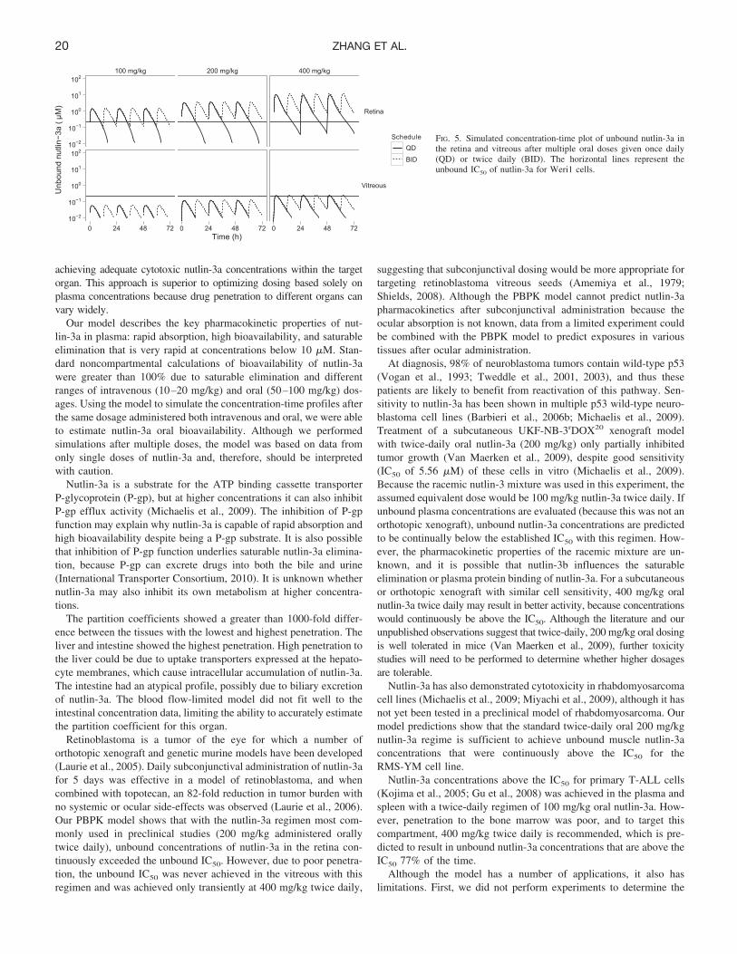
The simulated unbound retina and vitreous nutlin-3a concentrationswere compared to the in vitro unbound IC50 for the Weri1 retinoblas-toma cell line to choose optimal dosing regimens for mouse models ofretinoblastoma (Fig. 5). Oral dosing of nutlin-3a twice daily waspredicted to achieve unbound concentrations in the retina that wereconsistently above the unbound IC50 at dosages of 200 or 400 mg/kg(Supplemental Table 2). However, even at 400 mg/kg twice daily,unbound concentrations in the vitreous were predicted to be above theunbound IC50 for only 17% of the time, and at lower dosages theconcentration of unbound nutlin-3a never reached the unbound IC50
level. Simulated concentration-time plots of unbound nutlin-3a aftervarious dosing regimens are also shown in the adrenal gland forneuroblastoma (Supplemental Fig. S5), muscle for rhabdomyosar-coma (Supplemental Fig. S6), and plasma, bone marrow, and spleenfor leukemia (Supplemental Fig. S7). The percent times above theunbound IC50 are also listed in Supplemental Table 2.
Discussion
Nutlin-3a is undergoing preclinical studies examining its potentialefficacy for the treatment of several childhood malignancies. Nut-lin-3a interrupts the p53-MDM2 protein-protein interaction, whichmay lead to apoptosis or cell cycle arrest. Treatment of cells withnutlin-3a leads to reversal of multidrug resistance (Michaelis et al.,2009), reduced cell migration (Secchiero et al., 2007), reduced angio-genesis (LaRusch et al., 2007; Secchiero et al., 2007), radiosensitiza-tion of hypoxic cancer cells (Supiot et al., 2008), and inhibition oftumor adaptation to hypoxia (Lee et al., 2009). In this study, wedeveloped a mouse PBPK model of nutlin-3a in plasma and multipletissues of therapeutic interest. This is the first study to provide comp-rehensive pharmacokinetic data of nutlin-3a in any species. The design ofour study included both oral and intravenous dosing at several dosagelevels. This method permitted the development of a robust model thataccurately describes the disposition of nutlin-3a over a wide range ofconcentrations. The PBPK model was used to design rational dosingregimens for preclinical models of several malignancies based upon
FIG. 4. Simulated concentration-time plot of plasma nutlin-3a aftermultiple oral doses with once-daily (QD) and twice-daily (BID)dosing. Simulations were based on the final PBPK model.
TABLE 2
Estimated PBPK model parameters
Parameter Value
ka 0.409Ke 0.0160Vmax 0.0287Km 0.050KADI 1.61KADR 2.05KBRA 0.055KINT 12.2KLIV 7.54KLUN 1.78KMUS 2.08KRET 4.01KSPL 2.72KVIT 0.012PAVIT 0.0036KRES 4.8PARES 0.0048IIV ka 31.2%IIV ke 6.4%IIV Vmax 40.6%Residual error 35.6%
19PHYSIOLOGICALLY BASED PHARMACOKINETICS OF NUTLIN-3a
achieving adequate cytotoxic nutlin-3a concentrations within the targetorgan. This approach is superior to optimizing dosing based solely onplasma concentrations because drug penetration to different organs canvary widely.
Our model describes the key pharmacokinetic properties of nut-lin-3a in plasma: rapid absorption, high bioavailability, and saturableelimination that is very rapid at concentrations below 10 �M. Stan-dard noncompartmental calculations of bioavailability of nutlin-3awere greater than 100% due to saturable elimination and differentranges of intravenous (10–20 mg/kg) and oral (50–100 mg/kg) dos-ages. Using the model to simulate the concentration-time profiles afterthe same dosage administered both intravenous and oral, we were ableto estimate nutlin-3a oral bioavailability. Although we performedsimulations after multiple doses, the model was based on data fromonly single doses of nutlin-3a and, therefore, should be interpretedwith caution.
Nutlin-3a is a substrate for the ATP binding cassette transporterP-glycoprotein (P-gp), but at higher concentrations it can also inhibitP-gp efflux activity (Michaelis et al., 2009). The inhibition of P-gpfunction may explain why nutlin-3a is capable of rapid absorption andhigh bioavailability despite being a P-gp substrate. It is also possiblethat inhibition of P-gp function underlies saturable nutlin-3a elimina-tion, because P-gp can excrete drugs into both the bile and urine(International Transporter Consortium, 2010). It is unknown whethernutlin-3a may also inhibit its own metabolism at higher concentra-tions.
The partition coefficients showed a greater than 1000-fold differ-ence between the tissues with the lowest and highest penetration. Theliver and intestine showed the highest penetration. High penetration tothe liver could be due to uptake transporters expressed at the hepato-cyte membranes, which cause intracellular accumulation of nutlin-3a.The intestine had an atypical profile, possibly due to biliary excretionof nutlin-3a. The blood flow-limited model did not fit well to theintestinal concentration data, limiting the ability to accurately estimatethe partition coefficient for this organ.
Retinoblastoma is a tumor of the eye for which a number oforthotopic xenograft and genetic murine models have been developed(Laurie et al., 2005). Daily subconjunctival administration of nutlin-3afor 5 days was effective in a model of retinoblastoma, and whencombined with topotecan, an 82-fold reduction in tumor burden withno systemic or ocular side-effects was observed (Laurie et al., 2006).Our PBPK model shows that with the nutlin-3a regimen most com-monly used in preclinical studies (200 mg/kg administered orallytwice daily), unbound concentrations of nutlin-3a in the retina con-tinuously exceeded the unbound IC50. However, due to poor penetra-tion, the unbound IC50 was never achieved in the vitreous with thisregimen and was achieved only transiently at 400 mg/kg twice daily,
suggesting that subconjunctival dosing would be more appropriate fortargeting retinoblastoma vitreous seeds (Amemiya et al., 1979;Shields, 2008). Although the PBPK model cannot predict nutlin-3apharmacokinetics after subconjunctival administration because theocular absorption is not known, data from a limited experiment couldbe combined with the PBPK model to predict exposures in varioustissues after ocular administration.
At diagnosis, 98% of neuroblastoma tumors contain wild-type p53(Vogan et al., 1993; Tweddle et al., 2001, 2003), and thus thesepatients are likely to benefit from reactivation of this pathway. Sen-sitivity to nutlin-3a has been shown in multiple p53 wild-type neuro-blastoma cell lines (Barbieri et al., 2006b; Michaelis et al., 2009).Treatment of a subcutaneous UKF-NB-3rDOX20 xenograft modelwith twice-daily oral nutlin-3a (200 mg/kg) only partially inhibitedtumor growth (Van Maerken et al., 2009), despite good sensitivity(IC50 of 5.56 �M) of these cells in vitro (Michaelis et al., 2009).Because the racemic nutlin-3 mixture was used in this experiment, theassumed equivalent dose would be 100 mg/kg nutlin-3a twice daily. Ifunbound plasma concentrations are evaluated (because this was not anorthotopic xenograft), unbound nutlin-3a concentrations are predictedto be continually below the established IC50 with this regimen. How-ever, the pharmacokinetic properties of the racemic mixture are un-known, and it is possible that nutlin-3b influences the saturableelimination or plasma protein binding of nutlin-3a. For a subcutaneousor orthotopic xenograft with similar cell sensitivity, 400 mg/kg oralnutlin-3a twice daily may result in better activity, because concentrationswould continuously be above the IC50. Although the literature and ourunpublished observations suggest that twice-daily, 200 mg/kg oral dosingis well tolerated in mice (Van Maerken et al., 2009), further toxicitystudies will need to be performed to determine whether higher dosagesare tolerable.
Nutlin-3a has also demonstrated cytotoxicity in rhabdomyosarcomacell lines (Michaelis et al., 2009; Miyachi et al., 2009), although it hasnot yet been tested in a preclinical model of rhabdomyosarcoma. Ourmodel predictions show that the standard twice-daily oral 200 mg/kgnutlin-3a regime is sufficient to achieve unbound muscle nutlin-3aconcentrations that were continuously above the IC50 for theRMS-YM cell line.
Nutlin-3a concentrations above the IC50 for primary T-ALL cells(Kojima et al., 2005; Gu et al., 2008) was achieved in the plasma andspleen with a twice-daily regimen of 100 mg/kg oral nutlin-3a. How-ever, penetration to the bone marrow was poor, and to target thiscompartment, 400 mg/kg twice daily is recommended, which is pre-dicted to result in unbound nutlin-3a concentrations that are above theIC50 77% of the time.
Although the model has a number of applications, it also haslimitations. First, we did not perform experiments to determine the
FIG. 5. Simulated concentration-time plot of unbound nutlin-3a inthe retina and vitreous after multiple oral doses given once daily(QD) or twice daily (BID). The horizontal lines represent theunbound IC50 of nutlin-3a for Weri1 cells.
20 ZHANG ET AL.
route of elimination of nutlin-3a. Preliminary unpublished observa-tions (K. Guy, unpublished data) indicate that nutlin-3a is metabolizedby mouse liver microsomes, but a model with elimination only fromthe liver did not adequately fit the nutlin-3a plasma concentration-time data. Not modeling the elimination mechanistically could limitthe ability to extrapolate the PBPK model to other species. Second, weperformed all experiments in nontumor-bearing mice. Compared tonormal tissues, the altered environment in tumors (e.g., vasculature,pH, interstitial fluid pressure) may lead to different local disposition.Although it was not feasible to perform pharmacokinetic studies inmultiple preclinical models, data obtained from studies in tumor-bearing mice could be easily incorporated into this PBPK model.Another limitation is the assumption that the unbound fraction ofnutlin-3a in tissues was equivalent to the unbound fraction in plasma.We did directly measure nutlin-3a binding in vitreous fluid, which ismostly water but has a variety of proteins (Shires et al., 1993). Wealso performed simulations at dosages beyond those used to developthe model (i.e., 400 mg/kg). Although we were able to characterize thenonlinear elimination at higher plasma concentrations, it is possiblethat there are unknown nonlinear absorption or elimination pro-cesses occurring at this higher dosage, which would make thesemodel predictions inaccurate.
In summary, we performed extensive mouse pharmacokinetic stud-ies of nutlin-3a and developed a PBPK model, which was used todesign nutlin-3a dosing regimens for preclinical models of pediatricmalignancies. Although there are limitations to extrapolating in vitrocytotoxicity data, this analysis provides a starting point for furtherpharmacokinetic/pharmacodynamic studies in tumor-bearing mice.For models of retinoblastoma, the disposition of nutlin-3a after sub-conjunctival administration should be explored.
Acknowledgments
We thank Zaifang Huang, Mohamad Elmeliegy, Lei Yang, Shelly Wilker-son, and Frederique Zindy for help with dosing animals and collecting tissuesamples. We thank Daniel Groepper for helping with the protein binding studyin mouse plasma.
Authorship Contributions
Participated in research design: F. Zhang, Mallari, Miller, Guy, Dyer,Williams, Nemeth, Boulos, Panetta, and Stewart.
Conducted experiments: F. Zhang, Mallari, Miller, Dyer, Roussel, Nemeth,Zhu, Boulos, and J. Zhang.
Contributed new reagents or analytic tools: Lu.Performed data analysis: F. Zhang, Tagen, Panetta, and Stewart.Wrote or contributed to the writing of the manuscript: F. Zhang, Tagen,
Throm, Mallari, Guy, Dyer, Williams, Roussel, Boulos, Panetta, and Stewart.
References
Amemiya T, Yoshida H, and Ishigooka H (1979) Vitreous seeds in retinoblastoma, clinicalsignificance and ultrastructure. Albrecht Von Graefes Arch Klin Exp Ophthalmol 211:205–213.
Bai F, Zhu F, Tagen M, Miller L, Owens TS, Mallari J, Derrick E, Zhang F, and Stewart CF(2010) Determination of nutlin-3a in murine plasma by liquid chromatography electrosprayionization tandem mass spectrometry (LC-ESI-MS/MS). J Pharm Biomed Anal 51:915–920.
Barbieri A, Sabatini L, Indiveri P, Bonfiglioli R, Lodi V, and Violante FS (2006a) Simultaneousdetermination of low levels of methotrexate and cyclophosphamide in human urine by microliquid chromatography/electrospray ionization tandem mass spectrometry. Rapid CommunMass Spectrom 20:1889–1893.
Barbieri E, Mehta P, Chen Z, Zhang L, Slack A, Berg S, and Shohet JM (2006b) MDM2inhibition sensitizes neuroblastoma to chemotherapy-induced apoptotic cell death. Mol CancerTher 5:2358–2365.
Brown RP, Delp MD, Lindstedt SL, Rhomberg LR, and Beliles RP (1997) Physiologicalparameter values for physiologically based pharmacokinetic models. Toxicol Ind Health13:407–484.
Elison JR, Cobrinik D, Claros N, Abramson DH, and Lee TC (2006) Small molecule inhibition
of HDM2 leads to p53-mediated cell death in retinoblastoma cells. Arch Ophthalmol 124:1269–1275.
International Transporter Consortium, Giacomini KM, Huang SM, Tweedie DJ, Benet LZ,Brouwer KL, Chu X, Dahlin A, Evers R, Fischer V, et al. (2010) Membrane transporters indrug development. Nat Rev Drug Discov 9:215–236.
Gu L, Zhu N, Findley HW, and Zhou M (2008) MDM2 antagonist nutlin-3 is a potent inducerof apoptosis in pediatric acute lymphoblastic leukemia cells with wild-type p53 and overex-pression of MDM2. Leukemia 22:730–739.
Hollstein M, Sidransky D, Vogelstein B, and Harris CC (1991) p53 mutations in human cancers.Science 253:49–53.
Institute of Laboratory Animal Resources (1996) Guide for the Care and Use of LaboratoryAnimals 7th ed. Institute of Laboratory Animal Resources, Commission on Life Sciences,National Research Council, Washington DC.
Kariv I, Cao H, and Oldenburg KR (2001) Development of a high throughput equilibriumdialysis method. J Pharm Sci 90:580–587.
Klein C and Vassilev LT (2004) Targeting the p53-MDM2 interaction to treat cancer. Br JCancer 91:1415–1419.
Kojima K, Konopleva M, Samudio IJ, Shikami M, Cabreira-Hansen M, McQueen T, Ruvolo V,Tsao T, Zeng Z, Vassilev LT, et al. (2005) MDM2 antagonists induce p53-dependent apoptosisin AML: implications for leukemia therapy. Blood 106:3150–3159.
LaRusch GA, Jackson MW, Dunbar JD, Warren RS, Donner DB, and Mayo LD (2007) Nutlin3blocks vascular endothelial growth factor induction by preventing the interaction betweenhypoxia inducible factor 1alpha and Hdm2. Cancer Res 67:450–454.
Laurie NA, Donovan SL, Shih CS, Zhang J, Mills N, Fuller C, Teunisse A, Lam S, Ramos Y,Mohan A, et al. (2006) Inactivation of the p53 pathway in retinoblastoma. Nature 444:61–66.
Laurie NA, Gray JK, Zhang J, Leggas M, Relling M, Egorin M, Stewart C, and Dyer MA (2005)Topotecan combination chemotherapy in two new rodent models of retinoblastoma. ClinCancer Res 11:7569–7578.
Lee YM, Lim JH, Chun YS, Moon HE, Lee MK, Huang LE, and Park JW (2009) Nutlin-3, aHdm2 antagonist, inhibits tumor adaptation to hypoxia by stimulating the FIH-mediatedinactivation of HIF-1alpha. Carcinogenesis 30:1768–1775.
Michaelis M, Rothweiler F, Klassert D, von Deimling A, Weber K, Fehse B, Kammerer B, DoerrHW, and Cinatl J Jr (2009) Reversal of P-glycoprotein-mediated multidrug resistance by themurine double minute 2 antagonist nutlin-3. Cancer Res 69:416–421.
Miyachi M, Kakazu N, Yagyu S, Katsumi Y, Tsubai-Shimizu S, Kikuchi K, Tsuchiya K, IeharaT, and Hosoi H (2009) Restoration of p53 pathway by nutlin-3 induces cell cycle arrest andapoptosis in human rhabdomyosarcoma cells. Clin Cancer Res 15:4077–4084.
Momand J, Wu HH, and Dasgupta G (2000) MDM2–master regulator of the p53 tumorsuppressor protein. Gene 242:15–29.
Momand J, Zambetti GP, Olson DC, George D, and Levine AJ (1992) The mdm-2 oncogeneproduct forms a complex with the p53 protein and inhibits p53-mediated transactivation. Cell69:1237–1245.
Sarek G and Ojala PM (2007) p53 reactivation kills KSHV lymphomas efficiently in vitro andin vivo: new hope for treating aggressive viral lymphomas. Cell Cycle 6:2205–2209.
Secchiero P, Corallini F, Gonelli A, Dell’Eva R, Vitale M, Capitani S, Albini A, and Zauli G(2007) Antiangiogenic activity of the MDM2 antagonist nutlin-3. Circ Res 100:61–69.
Shangary S and Wang S (2008a) Small-molecule inhibitors of the MDM2–p53 protein-proteininteraction to reactivate p53 function: a novel approach for cancer therapy. Annu RevPharmacol Toxicol 49:223–241.
Shangary S and Wang S (2008b) Targeting the MDM2–p53 interaction for cancer therapy. ClinCancer Res 14:5318–5324.
Shields CL (2008) Forget-me-nots in the care of children with retinoblastoma. Semin Ophthalmol23:324–334.
Shires TK, Faeth JA, and Pulido JS (1993) Protein levels in the vitreous of rats with streptozo-tocin-induced diabetes mellitus. Brain Res Bull 30:85–90.
Supiot S, Hill RP, and Bristow RG (2008) Nutlin-3 radiosensitizes hypoxic prostate cancer cellsindependent of p53. Mol Cancer Ther 7:993–999.
Tweddle DA, Malcolm AJ, Bown N, Pearson AD, and Lunec J (2001) Evidence for thedevelopment of p53 mutations after cytotoxic therapy in a neuroblastoma cell line. Cancer Res61:8–13.
Tweddle DA, Pearson AD, Haber M, Norris MD, Xue C, Flemming C, and Lunec J (2003) Thep53 pathway and its inactivation in neuroblastoma. Cancer Lett 197:93–98.
Van Maerken T, Ferdinande L, Taildeman J, Lambertz I, Yigit N, Vercruysse L, Rihani A,Michaelis M, Cinatl J Jr, Cuvelier CA, et al. (2009) Antitumor activity of the selective MDM2antagonist nutlin-3 against chemoresistant neuroblastoma with wild-type p53. J Natl CancerInst 101:1562–1574.
Vassilev LT (2005) p53 activation by small molecules: application in oncology. J Med Chem48:4491–4499.
Vassilev LT, Vu BT, Graves B, Carvajal D, Podlaski F, Filipovic Z, Kong N, Kammlott U,Lukacs C, Klein C, et al. (2004) In vivo activation of the p53 pathway by small-moleculeantagonists of MDM2. Science 303:844–848.
Vogan K, Bernstein M, Leclerc JM, Brisson L, Brossard J, Brodeur GM, Pelletier J, and Gros P(1993) Absence of p53 gene mutations in primary neuroblastomas. Cancer Res 53:5269–5273.
Wan H and Rehngren M (2006) High-throughput screening of protein binding by equilibriumdialysis combined with liquid chromatography and mass spectrometry. J Chromatogr A1102:125–134.
Wright WS, Messina JE, and Harris NR (2009) Attenuation of diabetes-induced retinal vaso-constriction by a thromboxane receptor antagonist. Exp Eye Res 88:106–112.
Xu X, Selick P, and Pang KS (1993) Nonlinear protein binding and enzyme heterogeneity:effects on hepatic drug removal. J Pharmacokinet Biopharm 21:43–74.
Address correspondence to: Clinton F. Stewart, Department of Pharmaceu-tical Sciences, St. Jude Children’s Research Hospital, 262 Danny Thomas Place,Memphis, TN 38105. E-mail: [email protected]
21PHYSIOLOGICALLY BASED PHARMACOKINETICS OF NUTLIN-3a
Supplementary Data
Whole body physiologically-based pharmacokinetic model for nutlin-3a in mice after intravenous and
oral administration
Fan Zhang, Michael Tagen, Stacy Throm, Jeremy Mallari, Laura Miller, R. Kiplin Guy, Michael A. Dyer,
Richard T. Williams, Martine F. Roussel, Katie Nemeth, Fangyi Zhu, Jiakun Zhang, Min Lu, Clinton F.
Stewart
Drug Metabolism and Disposition
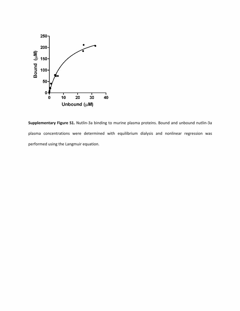
Supplementary Figure S1. Nutlin-3a binding to murine plasma proteins. Bound and unbound nutlin-3a
plasma concentrations were determined with equilibrium dialysis and nonlinear regression was
performed using the Langmuir equation.
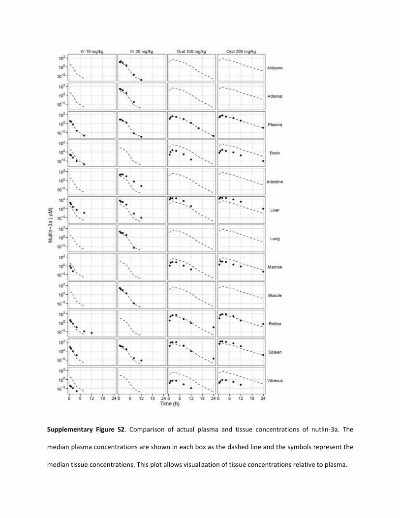
Supplementary Figure S2. Comparison of actual plasma and tissue concentrations of nutlin-3a. The
median plasma concentrations are shown in each box as the dashed line and the symbols represent the
median tissue concentrations. This plot allows visualization of tissue concentrations relative to plasma.
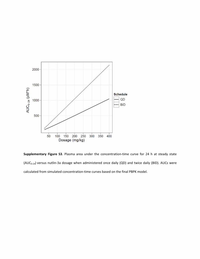
Supplementary Figure S3. Plasma area under the concentration-time curve for 24 h at steady state
(AUC0-24) versus nutlin-3a dosage when administered once daily (QD) and twice daily (BID). AUCs were
calculated from simulated concentration-time curves based on the final PBPK model.
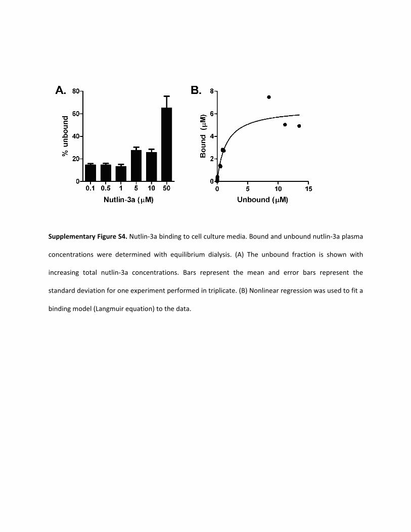
Supplementary Figure S4. Nutlin-3a binding to cell culture media. Bound and unbound nutlin-3a plasma
concentrations were determined with equilibrium dialysis. (A) The unbound fraction is shown with
increasing total nutlin-3a concentrations. Bars represent the mean and error bars represent the
standard deviation for one experiment performed in triplicate. (B) Nonlinear regression was used to fit a
binding model (Langmuir equation) to the data.
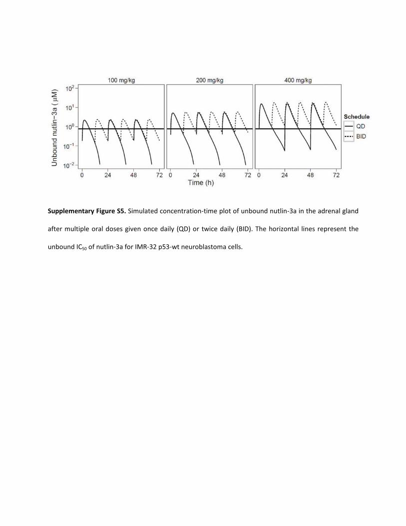
Supplementary Figure S5. Simulated concentration-time plot of unbound nutlin-3a in the adrenal gland
after multiple oral doses given once daily (QD) or twice daily (BID). The horizontal lines represent the
unbound IC50 of nutlin-3a for IMR-32 p53-wt neuroblastoma cells.
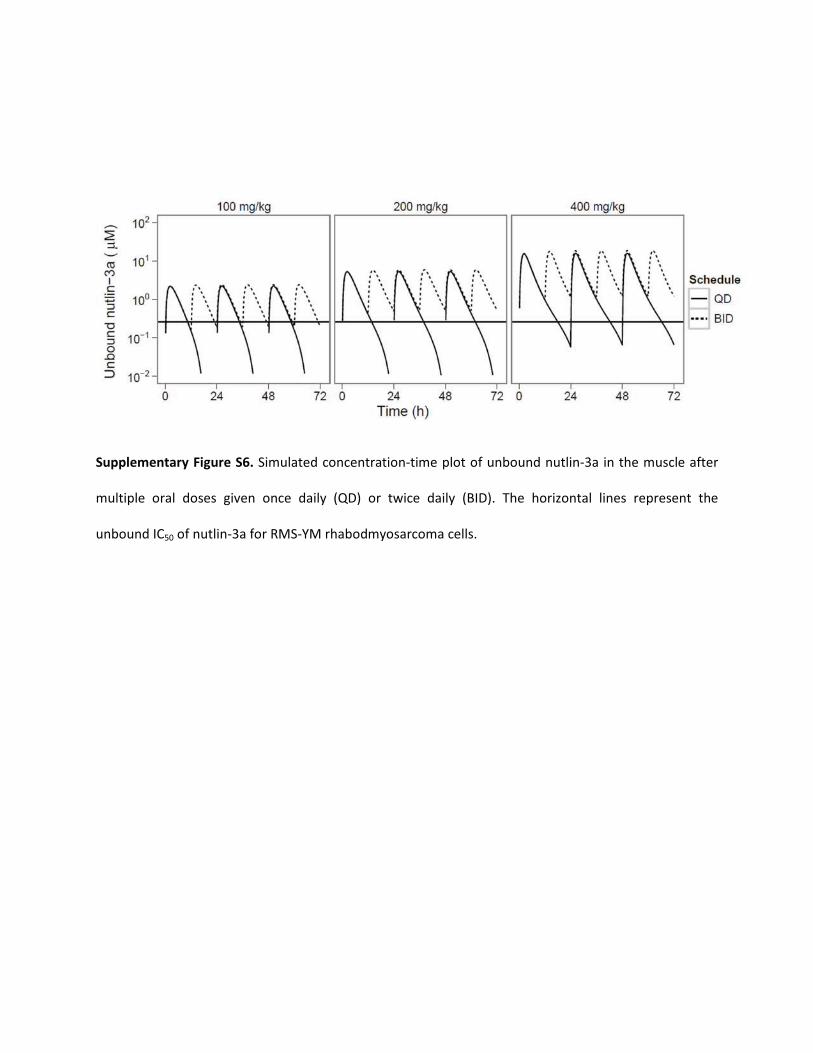
Supplementary Figure S6. Simulated concentration-time plot of unbound nutlin-3a in the muscle after
multiple oral doses given once daily (QD) or twice daily (BID). The horizontal lines represent the
unbound IC50 of nutlin-3a for RMS-YM rhabodmyosarcoma cells.
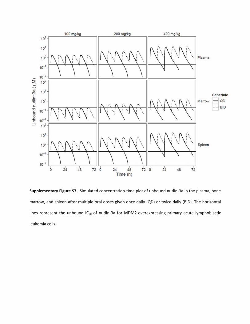
Supplementary Figure S7. Simulated concentration-time plot of unbound nutlin-3a in the plasma, bone
marrow, and spleen after multiple oral doses given once daily (QD) or twice daily (BID). The horizontal
lines represent the unbound IC50 of nutlin-3a for MDM2-overexpressing primary acute lymphoblastic
leukemia cells.
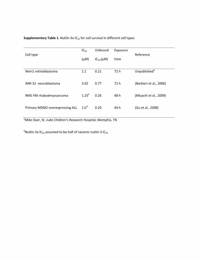
Supplementary Table 1. Nutlin-3a IC50 for cell survival in different cell types
Cell type IC50
(μM)
Unbound
IC50 (μM)
Exposure
time Reference
Weri1 retinoblastoma 1.1 0.21 72 h Unpublisheda
IMR-32 neuroblastoma 3.02 0.77 72 h (Barbieri et al., 2006)
RMS-YM rhabodmyosarcoma 1.25b 0.26 48 h (Miyachi et al., 2009)
Primary MDM2-overexpressing ALL 1.0 b 0.20 44 h (Gu et al., 2008)
aMike Dyer, St. Jude Children’s Research Hospital, Memphis, TN
bNutlin-3a IC50 assumed to be half of racemic nutlin-3 IC50

Supplementary Table 2. Percent time unbound tissue concentration is above unbound IC50
Cell line Tissue Time above IC50 (%)
QD
BID
100 200 400
100 200 400
Weri1 Retina 38 53 70
83 100 100
Vitreous 0 0 5
0 0 17
IMR32 Adrenal 27 39 54
58 85 100
RMS-YM Muscle 43 58 76
92 100 100
ALL Plasma 43 57 75
90 100 100
Spleen 46 61 80
90 100 100
Marrow 0 23 35
12 48 77