why bones bend but don’t break - iupui
TRANSCRIPT

270
Introduction
Most structures that sustain loads and bear stresses – bridges,airplane wings – eventually fail at loads below those they couldnormally sustain when they were new. Bone, however, sustainsmillions of loading cycles over the course of a lifetime andrarely breaks without a major traumatic event. It can sustainlarge deformations, sometimes at rapid rates, and yet emergesintact to function another day. It accomplishes this remarkablefeat both through structural means – the organization and com-position of the material itself – as well as through physiologicalmeans in its capacity as a self-repairing structure.
From an evolutionary perspective, an inability to prevent failure
from low energy events would exert a strong selective pressureagainst the organism. Although bones could adapt by simplyadding more material, increasing mass, there is a tipping point atwhich this would not be selectively advantageous. It is well knownthat bone can adapt by altering its geometry, providing thestrongest structure possible with a minimum amount of material.But strength is not always the end-goal, and strong bones are notalways those that are least at risk for fracture. For instance, the ca-pability to withstand high stresses, a measure of bone strength,may be accompanied by a smaller amount of energy absorption infracture, as in the case of osteopetrotic bone (Figure 1). Peoplewith osteopetrosis have very dense and very strong bones, but arealso at high risk for fracture because their bones are fragile as theresult of high mineralization. The mechanical strength of a bonedoes not necessarily equate to its resistance to fracture because abone that is very strong, defined mechanically as one that breaksat a high stress, may absorb less energy (the area under the stress-strain curve) before it breaks than a bone that breaks at a lowerstress (Figure 1). It is the amount of energy that can be absorbedbefore it breaks that defines a bone’s resistance to fracture. Mech-anisms by which our bones adapt at the tissue level, and the mannerin which they function physiologically, are designed to preventovert fractures from low energy loading. For fracture preventionin bone, as in airplane wings, maximum strength is not the design
J Musculoskelet Neuronal Interact 2011; 11(4):270-285
Why bones bend but don’t break
D.B. Burr
Department of Anatomy and Cell Biology, Indiana University School of Medicine and Department of Biomedical Engineering, Indiana University-Purdue University at Indianapolis, USA
Abstract
The musculoskeletal system is adept at dissipating potentially damaging energy that could accelerate fracture consequent tomultiple loading cycles. Microstructural damage reduces bone’s residual properties, but prevents high stresses within the materialby dissipating energy that can lead to eventual failure. Thus skeletal microdamage can be viewed as an adaptive process to preventbone failure by dissipating energy. Because a damaged bone has reduced strength and stiffness, it must be repaired, so bone hasevolved a system of self-repair that relies on microdamage-stimulated signaling mechanisms. When repair cannot occur quicklyenough, low energy stress fractures can occur. The regulating effects of muscle also prevent failure by controlling where highstresses occur. Acting synergistically, muscle forces dissipate energy by appropriately regulating accelerations and decelerationsof the limbs during movement. When muscles become fatigued, these functions are constrained, larger amounts of energy areimparted to bone, increasing the likelihood of microstructural damage and fracture. Thus, healthy bones are maintained by theability of the musculoskeletal system to dissipate the energy through synergistic muscular activity and through the maintenanceof microstructural and material properties that allow for crack initiation, but also for their repair.
Keywords: Microdamage, Energy, Fatigue, Muscle, Remodeling
Perspective Article Hylonome
The author has a grant from Amgen and Material Transfer agreementsfor drug from Eli Lilly, Pfizer, NephroGenex. He is also an advisorfor PharmaLegacy and for Wright Medical.
Corresponding author: David B. Burr, PhD, Dept of Anatomy and Cell Biology,MS 5035, Indiana University School of Medicine, 635 Barnhill Dr, Indianapolis,IN 46202, USA E-mail: [email protected]
Edited by: F. RauchAccepted 6 October 2011

D.B. Burr: Why bones bend but don’t break
271
goal; energy absorption that prevents or delays fracture is the goal.Evolutionarily, our musculoskeletal system has adapted mul-
tiple mechanisms to prevent fracture by dissipating energy thatcould cause the bone to break. Microscopic heterogeneity mayfacilitate formation of microcracks within the bone, releasingenergy that otherwise might build up and create an overt frac-ture. The heterogeneity of the system also prevents crackgrowth, providing time for the body to repair the damage. Thisrequires a signaling system, putatively provided by the osteo-cyte-canalicular system, that informs the bone when and wheredamage has occurred. The signaling system allows remodelingto be targeted efficiently to the locations of damage. Musclesalso dissipate energy that would otherwise be imparted to thebone. When muscles are ineffective at dissipating energy, highlevels of stress and strain can be generated in the bone that createlevels of microdamage that overwhelm the body’s ability to re-pair it quickly. This can lead to a degradation of bone’s mechan-ical properties over time, resulting in a stress fracture. Thesemechanisms of bone damage, repair and energy dissipation willbe explored in the sections below to provide a more holistic un-derstanding of why bones bend under load, but rarely break.
What do cross-species comparisons tell us aboutmechanisms bone uses to prevent failure?
Cross-species comparisons can be instructive in pointing ustowards the skeletal mechanisms that prevent fractures. Bonesare designed to withstand loading, and loading is primarily ex-erted by muscular and gravitational forces that are applied duringmovement. Therefore, one might expect that animals that movein different ways demonstrate different adaptive mechanisms.
At the tissue level, bone has developed a heterogeneousstructure in many different species as an adaptation for moreprolonged and impulsive loading1. Animals that do not live fora long time (decades), those that do not place large loads ontheir bones, and those that do not have a need for ballistic ac-tivity tend to have bone that is more homogeneous at the mi-croscopic level. This can be seen in rats and mice, for instance,short-lived animals that have fibrolamellar bone with unorgan-ized collagen, and which is devoid of structural topographywhen viewed under a microscope (Figure 2 A). Even in non-human primates, which typically have more lamellar bone andgreater organization in collagen structure, there is a wide rangeof microscopic morphology1, and the nature of the morphologyis largely related to the manner in which the animal moves (Fig-ure 2B,C). Arboreal and terrestrial quadrupeds that spend muchof their day trying to eat sufficient amounts of leaves, insects,fruits and nuts just to survive, are not known for rapid or sus-tained movement. These animals have low cortical bone re-modeling rates, and consequently do not develop cortical bonethat appears very heterogeneous2-5. Primates that use arm-swinging behaviors, however, subject their limbs to greater im-pulsive (e.g. high strain rate) behaviors, and these animalsremodel more rapidly and develop bone that is much more sim-ilar to human bone. From an evolutionary perspective, humanshave adapted to environments and diets in which it was neces-sary to travel long distances on two legs, sometimes at greatspeed. We, too, have developed the capacity to remodel rapidlywhen necessary, creating structures within our bone – second-ary osteons in cortical bone and hemiosteons in trabecular bone– that are heterogeneous with respect to lamellar organization,orientation, size and geometry (Figure 2D). These two featuresof our skeletons – the capacity to remodel, and the abilitythrough that remodeling mechanism to create a heterogeneoustissue that controls damage – are related to each other and func-tion in a complementary way to prevent bone failure. For thatto become apparent, however, it was necessary to demonstratethat microscopic damage occurs in bone, that osteons andhemiosteons are important in controlling crack growth, and thatthere is a mechanism in bone that can signal for their repair.
The mechanical role of microdamage
It is clear now, and more widely accepted than it was 20years ago, that microdamage in bone is a naturally occurringevent, and that it is probably important in some way for skele-tal health. The mere existence of microdamage was not always
Figure 1. Maximum yield stress defines the strength of bone, whilethe slope of the initial part of the curve represents elastic modulus, ortissue stiffness. The amount of energy the bone can absorb prior tofailure is defined by the area under the curve. Highly mineralized os-teopetrotic bone is stronger at yield and stiffer than healthy bone, butit breaks soon after it reaches its yield point; it does not absorb asmuch energy prior to fracture as healthy bone. Osteoporotic bone, onthe other hand, is neither as strong nor as stiff as healthy bone, andabsorbs less energy prior to fracture than healthy bone, but much morethan osteopetrotic bone. This is because it absorbs more energy afterthe yield point (the bend in the curve) than does osteopetrotic bone(i.e., it can deform more in the post-yield region). Bones that do notsustain much post-yield deformation/strain, are brittle. Therefore, al-though osteopetrotic bone is stronger and stiffer than osteoporotic ornormal bone, it is brittle, absorbs less energy, and breaks more easily.(Adapted from Ref 76).

D.B. Burr: Why bones bend but don’t break
272
accepted as a physiological feature of bone. There was sub-stantial controversy from the time that Harold Frost first in-troduced the concept in 19606 until at least the early 1990’sabout whether bones were subject to microscopic damage,even though it was well known by engineers that any cyclicallyloaded structural material would sustain damage over time.Over the past 20 years, because the phenomenon of microdam-age has been observed in multiple labs using different tech-niques, there is less controversy now that damage occurs7,although the debates about its mechanical and physiologicalimportance continue to rage on.
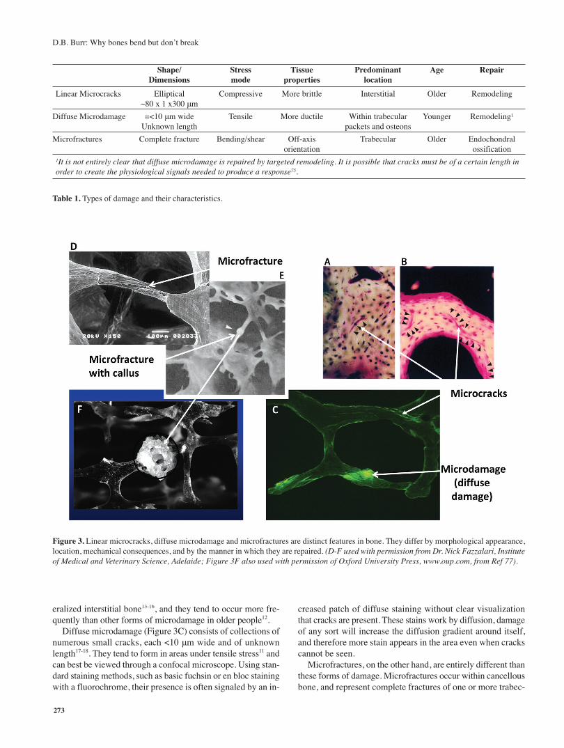
Microdamage is not all created equal, nor does all performthe same mechanical function. There are a at least three distinctvarieties of “microdamage,” which can be identified as linear
microcracks, diffuse microdamage, and microfractures8. These“types” are distinguished by the way they form and their mor-phology; the nature of the stimuli that cause them to form aswell as their location; and the manner in which they are repaired(Table 1).
Linear microcracks (Figure 3A-C) represent planes of sepa-ration within the bone tissue. They are three dimensional ellip-tical structures in that their dimensions will differ in each plane.In cross-section, they are about 40-100 microns long, and per-haps 1-2 microns wide, but can run longitudinally within thebone for 300-500 μm9,10. They tend to form preferentially in re-sponse to compressive stresses11, and are indicative of bone tis-sue that deforms more before it cracks12. Consequently, thepreponderance of them are found within the more highly min-
Figure 2. Photomicrographs taken under polarized light showing greater heterogeneity of structure in nonhuman primates than in mouse, andmore heterogeneity in human bone than in primates that use less impulsive movements, have shorter territorial ranges, or are shorter lived.These traits are all related to the capacity to form damage and the need to remodel.Mouse bone is fibrolamellar and has no osteonal bone. It is very homogeneous, which is not detrimental because mice do not place largestrains/strain rates on their bones, and do not live for a long time. They do not need the capacity to prevent cracks from growing, or to repairthem. The radial structures are vascular canals.The squirrel monkey (Saimiri sciureus) is an arboreal quadruped. It is a small primate (BW: 365-1135 g) which is classified as a quadrupedalclimber and seldom leaps. They do not use impulsive movements, and their bone is largely laminar, unremodeled, and quite homogeneous.Macaques (Shown here: Macaca mulatta) are terrestrial quadrupeds (although they also spend time arboreally) that do not often move quickly.They tend to have more osteonal bone in their cortex, but retain large amounts of primary lamellae. Their bone remodels more intracorticallyand is more heterogeneous than that in squirrel monkeys, but less so than in humans.Humans (H. sapiens) are larger bodied, longer-lived animals that tend to engage more often in high strain rate activities (walking long distancesand running). By virtue of their behaviors and lifespans, it is more important that they have osteons to prevent microcracks from growing tocritical size, and that they be able to repair microcracks with osteonal remodeling. Consequently, they have bone that is highly remodeled, withmany secondary Haversian systems. Interestingly, larger bodied, longer-lived chimpanzees (not shown) also have bone that is highly remodeledwith osteons intracortically.
A B
C D
D.B. Burr: Why bones bend but don’t break
273
eralized interstitial bone13-16, and they tend to occur more fre-quently than other forms of microdamage in older people12.
Diffuse microdamage (Figure 3C) consists of collections ofnumerous small cracks, each <10 μm wide and of unknownlength17-18. They tend to form in areas under tensile stress11 andcan best be viewed through a confocal microscope. Using stan-dard staining methods, such as basic fuchsin or en bloc stainingwith a fluorochrome, their presence is often signaled by an in-
creased patch of diffuse staining without clear visualizationthat cracks are present. These stains work by diffusion, damageof any sort will increase the diffusion gradient around itself,and therefore more stain appears in the area even when crackscannot be seen.
Microfractures, on the other hand, are entirely different thanthese forms of damage. Microfractures occur within cancellousbone, and represent complete fractures of one or more trabec-
Shape/ Stress Tissue Predominant Age RepairDimensions mode properties location
Linear Microcracks Elliptical Compressive More brittle Interstitial Older Remodeling~80 x 1 x300 μm
Diffuse Microdamage =<10 μm wide Tensile More ductile Within trabecular Younger Remodeling1
Unknown length packets and osteons
Microfractures Complete fracture Bending/shear Off-axis Trabecular Older Endochondral orientation ossification
1It is not entirely clear that diffuse microdamage is repaired by targeted remodeling. It is possible that cracks must be of a certain length inorder to create the physiological signals needed to produce a response75.
Table 1. Types of damage and their characteristics.
Figure 3. Linear microcracks, diffuse microdamage and microfractures are distinct features in bone. They differ by morphological appearance,location, mechanical consequences, and by the manner in which they are repaired. (D-F used with permission from Dr. Nick Fazzalari, Instituteof Medical and Veterinary Science, Adelaide; Figure 3F also used with permission of Oxford University Press, www.oup.com, from Ref 77).

D.B. Burr: Why bones bend but don’t break
274
ulae (Figure 3 D-F). Whether microfractures are the end-stageof linear or diffuse damage in cancellous bone is not known,and still somewhat controversial. However, these are full frac-tures, not cracks, and are clearly distinct from other forms ofdamage. The term should not be used as a general descriptorfor linear microcracks, although it often is.
These forms of damage are not only distinct in their mor-phology, but heal by completely different mechanisms. Linearmicrocracks and diffuse damage both repair through normalcoupled remodeling processes, although signaling for remod-eling may differ between them with linear microcracks havinggreater potential to initiate a repair response19-23. They are re-moved by resorption, and new bone is laid down where thedamage once was. Microfractures, on the other hand, are re-paired through normal fracture healing mechanisms which in-volve endochondral ossification (Figure 3F). A cartilage orwoven bone callus is formed over the broken area; these canbe readily observed through a dissecting microscope in nearlyany hemi-sectioned femoral head/neck from a middle-aged orolder person. The callus eventually remodels to re-establishthe normal lamellar structure of the trabecula.
The presence of microdamage within bone tissue has me-chanical effects on the residual properties of bone, and this isone reason that it is important to the skeleton to remove it andreplace it with undamaged bone. By definition, the residualstiffness of damaged bone tissue is less than in undamaged tis-sue. In fact, this is the definition – reduced stiffness – that en-gineers use to define damage in structural materials whichcannot be evaluated microscopically. Moreover, the introduc-tion of microdamage reduces the bone’s future capacity to ab-sorb energy prior to fracture, and in this sense deteriorates themechanical properties of bone. However, the paradox of thisis that the initiation and growth of microcracks in itself dissi-pates energy and delays a catastrophic complete fracture fromoccurring24,25. It is a truism that materials that perform wellunder cyclic loading conditions tend to be hereogeneous at themicroscopic level. These materials delay complete failure notby preventing the initiation of damage, but by reducing its abil-ity to grow to catastrophic size through microarchitectural or-ganizations that stop cracks from growing26,27. Thus, althoughwe typically think of microdamage as “bad,” it has a positiverole to play in preventing fracture. Particularly in a self-repair-ing structure like bone, any adaptation of the microarchitecturethat can stop a crack from growing long enough to allow forits repair is an adaptation that will promote survival of the in-dividual, and will tend to proliferate in the genome. This pre-sumes that the damage will be repaired in an efficient manner,before significantly more damage can be created. This requiresa signaling mechanism, and suggests a physiological role, notjust a mechanical one, for bone microdamage.
The physiological role of microdamage
There is a tendency to think of microdamage only as a me-chanical event, but it is possible that its role in mechanics isless important to skeletal health than its physiological role. Two
kinds of remodeling have been proposed: stochastic and tar-geted28-32. Stochastic remodeling functions primarily to main-tain mineral homeostasis, whereas targeted remodeling servesa mechanical function in removing microdamage from bone19-
23,33. Frost6 was the first to propose the concept that micrcracksin bone signal for their own repair, suggesting that damage tothe osteocyte’s canalicular network could be responsible.
To address this question more methodically, Burr et al.33
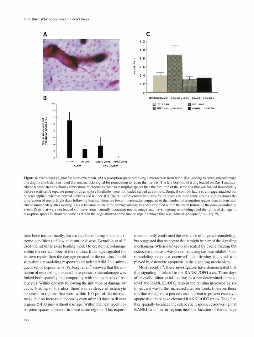
originally defined a probability function based on a comparisonbetween the number of cracks in association with resorptionspaces (nobs), compared to the maximum possible number ofcracks and resorption spaces (nmax). Targeted remodeling wasdefined by nobs/nmax>1.0, indicating that more cracks were ob-served associated with resorption spaces than expected bychance alone. In an experiment in which dog radii were loadedfor 10,000 cycles to generate microdamage in the bone, theyfound that nobs/nmax was between 6.2 and 44. This suggested thatmicrocracks were more likely to be found near resorptionspaces than not, leading to the conclusion that the damage waseliciting a remodeling response. However, that initial experi-ment was not able to separate those cases in which the crackspre-existed the resorption spaces, from those in which the re-sorption space preceded the crack. In other words, it still couldbe possible that the stress concentrations caused by active re-modeling sites caused the cracks to form at those locations, andthat the cracks were not eliciting the resorption response at all.
A second experiment was performed in which the right dogradius was loaded to create microdamage, as in the previous ex-periment, eight days were allowed to pass to permit the initiationof new remodeling, then the left radius of the dog was loaded,and the animal killed immediately after19. If the cracks were elic-iting the repair response, then the proportion of cracks found inassociation with remodeling sites (Figure 4A) should be greaterin the limb that was loaded first. Indeed, in this experiment,nobs/nmax=4.05 (Figure 4B), suggesting that following loading,cracks were found in association with resorption spaces fourtimes more often than expected under a model of stochastic re-modeling; this was not the case in the limb that was loaded im-mediately before sacrifice. In fact, the ratio of crack density(Cr.Dn) to resorption space density (Rs.Dn) was nearly the samein the limb that had been allowed to generate a repair responseas it was in control limbs of other dogs that were not loaded atall, whereas the ratio Cr.Dn/Rs.Dn was 3 times higher in thelimb that had not been allowed to mount a repair response (inother words, cracks were generated, but were not allowed to re-pair) (Figure 4C). This showed almost certainly that microcrackswere signaling for their own repair, and also suggested that therewas normally an equilibrium between the microdamage burdenand the activation of bone remodeling (This was determinedfrom a 2D analysis, and it is fair to point out that in three di-mensions it is possible that a different proportionality would befound. Subsequently, Martin32 made the mathematical argumentthat it is at least theoretically possible that all remodeling in cor-tical bone of the long bones is targeted remodeling).
A more definitive set of experiments was performed bySchaffler and his colleagues. Normally, rats do not remodel
D.B. Burr: Why bones bend but don’t break
275
their bone intracortically, but are capable of doing so under ex-treme conditions of low calcium or disuse. Bentolila et al.21
used the rat ulnar axial loading model to create microdamagewithin the cortical bone of the rat ulna. If damage signaled forits own repair, then the damage created in the rat ulna shouldstimulate a remodeling response; and indeed it did. In a subse-quent set of experiments, Verborgt et al.20 showed that the ini-tiation of remodeling mounted in response to microdamage waslinked both spatially and temporally with the apoptosis of os-teocytes. Within one day following the initiation of damage bycyclic loading of the ulna, there was evidence of osteocyteapoptosis in regions that were within 100 μm of the microc-racks, but no increased apoptosis even after 10 days in distantregions (>100 μm) without damage. Within the next week, re-sorption spaces appeared in these same regions. This experi-
ment not only confirmed the existence of targeted remodeling,but suggested that osteocyte death might be part of the signalingmechanism. When damage was created by cyclic loading butosteocyte apoptosis was prevented using caspase inhibitors, noremodeling response occurred22, confirming the vital roleplayed by osteocyte apoptosis in the signaling mechanism.
Most recently34, these investigators have demonstrated thatthis signaling is related to the RANKL/OPG axis. Three daysafter cyclic ulnar axial loading to a pre-determined damagelevel, the RANLKL/OPG ratio in the rat ulna increased by sixtimes , and was further increased after one week. However, thoserats that were given a pan-caspase inhibitor to prevent osteocyteapoptosis did not have elevated RANKL/OPG ratios. They fur-ther spatially localized the osteocytic response, discovering thatRANKL was low in regions near the location of the damage
Figure 4. Microcracks signal for their own repair. (A) A resorption space removing a microcrack from bone. (B) Loading to create microdamagein a dog forelimb demonstrated that microcracks signal for remodeling to repair themselves. The left forelimb of a dog loaded on Day 1 and sac-rificed 8 days later has about 4 times more microcracks close to resorption spaces than the forelimb of the same dog that was loaded immediatelybefore sacrifice. A separate group of dogs whose forelimbs were not loaded served as controls. Surgical controls had a strain gage attached butno load applied, whereas normal controls had neither. (C) The ratio of microcracks to resorption spaces in these same groups of dogs shows theprogression of repair. Eight days following loading, there are fewer microcracks compared to the number of resorption spaces than in dogs sac-rificed immediately after loading. This is because much of the damage already has been resorbed within the week following the damage-initiatingevent. Dogs that were not loaded still have some naturally occurring microdamage, and have ongoing remodeling, and the ratios of damage toresorption spaces is about the same as that in the dogs allowed some time to repair damage that was induced. (Adapted from Ref 19).

D.B. Burr: Why bones bend but don’t break
276
where osteocytes were apoptotic, but increased at distances of100-500 μm from the damage, where there were few apoptoticosteocytes. The response depends on the size of the microcrack,with larger cracks eliciting a greater increase in RANKL andgreater decrease in OPG than smaller cracks35. In combinationthese studies suggest that osteocyte apoptosis is the signal forthe initiation of bone remodeling, but that it is the healthy os-teocytes at a distance from the damage that provide the proteinnecessary for osteoclast differentiation and activation.
Summary of the role of microdamage in bone
We have learned from these studies over the past 30 yearsthat microdamage is not an artifact of histological preparation,but occurs naturally in bone, and that in the healthy skeletonthe production of microdamage is in equilibrium with its repairby bone remodeling. The presence of microdamage in bonereduces the bone’s residual strength, stiffness and energy tofracture. However, the paradox is that the initiation and growthof microdamage reduces the risk of fracture by releasing en-ergy that would otherwise cause the bone to fail. Thus, micro-damage prolongs bone’s integrity and is a selectivelyadvantageous evolutionary mechanism to preserve an animal’sfunctional capabilities. However, because it reduces residualproperties, it is important to repair the damage. Indeed, the pri-mary role and importance of microdamage in bone may be itsphysiologic function in stimulating the remodeling system torenew the bone matrix through bone turnover. It is absolutelyclear now that this important physiologic function of micro-damage in stimulating bone remodeling occurs mechanisticallyvia a cellular signal from dying osteocytes in the area of thebone damage, and that the activation of resorption is subse-quently caused by an increased osteocytic production ofRANKL, most likely by non-apoptotic osteocytes at some dis-tance from the damage.
Normally there is an equilibrium between damage and re-pair, and as long as that equilibrium is maintained bone con-tinues to function. However, bone does fail sometimes fromcyclic loading over long periods of time, resulting in nondis-placed fractures called stress fractures. Understanding whythese occur, ie whether they represent an increased microdam-age burden solely from overuse, or a failure of the repair sys-tem to remove damaged bone, is critical to understanding theirpathogenesis and ultimately their prevention.
The causes and prevention of stress fracture
It has never been shown definitively that the microdamageburden in human bone is sufficiently high under most conditionsto significantly reduce the mechanical properties of bone to theextent that it will increase the risk of fracture. Large amounts ofdamage can be induced in the laboratory, and at these levels canbe shown to degrade the mechanical properties of the bone.However, these levels of damage are rarely or never found invivo in bone. Yet the suspicion is that damage accumulation un-derlies at least some kinds of fractures, especially those that
occur spontaneously with low amounts of energy36,37.Cyclic loading studies of cow bone performed many years
ago showed that at what were considered to be physiologicalstrains (1200 με in tension) and strain rates (0.01-0.03/sec),bone could sustain literally millions of cycles without failure38.This experiment was flawed in that it was run under strain con-trol rather than load control so that as bone lost stiffness, thestresses also decreased. Even so, the inability to cause bone tobreak after 45 million cycles still did not seem consistent withthe observation that stress fractures do occur frequently, andcan occur in athletes and soldiers with many fewer cycles. Onethought was that the strains on bone were higher than thoseused in this experiment, at least on an occasional basis. Thisstimulated us to initiate a series of experiments in which straingages were place on human bone in vivo in regions at risk forstress fractures, and strains monitored during a series of activ-ities, including vigorous activities39-41. These studies showedthat strains were not particularly high in either the tibia or themetatarsus for most activities, ranging from 800 με in tensionto -1200 με in compression during walking, to approximately-1500 με or so during running on a flat surface. Strains on theorder of -2000 με could be achieved running up and downhill.Strains up to -5000 με occur in the metatarsus in landing froma jump about a meter high42,43, an activity that no one performsroutinely for very long.
Figure 5. If microdamage initiates the repair process, then the devel-opment of a stress fracture could be the result of positive feedbackbetween damage initiation, and the loss of bone mass caused by at-tempts at repair. Each time a crack is initiated, remodeling is acti-vated. The initial stage of remodeling is resorption, but as bone isremoved, there is less mass available to support loads, potentially ini-tiating a new cascade of damage. Because it takes much longer toform bone than to remove it, taking this to its logical conclusion sug-gests that continued loading might result in a stress fracture when thecritical threshold for stress or strain is reached. Inset: Biopsy of astress fracture showing the remodeling reaction to microdamage. (Ref78; Used with permission from CRC Press and Dr. Satoshi Mori).

D.B. Burr: Why bones bend but don’t break
277
Because these strains were not sufficiently high to causebone failure in a reasonable number of cycles, we hypothe-sized that the physiological response of the bone to microdam-age might be part of the pathophysiology for stress fractures.Knowing that microdamage initiation would activate the re-modeling system, and that this begins by active resorption ofbone, we hypothesized a positive feedback loop between dam-age production, loss of bone mass through initiation of resorp-tion and remodeling, and eventual stress fracture (Figure 5).The active involvement of remodeling activation in stress frac-ture physiology could be tested by suppressing bone turnoverin a well characterized group at high risk for developing stressfractures – soldiers in basic training – by giving an agent thatsuppresses remodeling and reduces the repair of damage that
occurs. If remodeling were a part of the pathophysiology ofthese fractures, then suppressing it should result in fewer stressfractures following the 14 week basic training program.
A blinded, randomized trial of Israeli soldiers in basic train-ing was performed in which 165 soldiers began to take a bis-phosphonate prior to basic training, while 159 soldiers weregiven a placebo44. Although there was substantial dropout dueto some negative publicity during the trial (52 and 56 soldiersin the treatment and placebo groups respectively, completedthe regimen), the results definitively showed, using both in-tention-to-treat and per-protocol analyses, that reducing the re-pair of microdamage did not significantly reduce the incidenceof stress fractures overall, or at specific risk sites including thetibia, femur and metatarsus (Figure 6). In fact, reducing repair
Figure 6. Reducing remodeling rate in soldiers during basic training in the military did not decrease the incidence of stress fractures at any site,suggesting that bone remodeling was not part of the pathophysiology for the development of a stress fracture. (Data from Ref 44).

D.B. Burr: Why bones bend but don’t break
278
of microdamage resulted in a greater incidence of stress frac-tures in those groups taking the bisphosphonate. This increasedincidence was not statistically significant, however, in part dueto the high dropout rate and resulting lower power. Neverthe-less, it was clear that interfering with the repair of damage wasnot going to improve the prognosis for developing a stressfracture.
Remodeling suppression increases microcrackaccumulation
Why might this be? It was known from previous pre-clinicalexperiments45-47 that high doses of bisphsophonates used to reduceremodeling in non-osteoporotic beagle dogs causes 2.5-7 fold in-creases in damage accumulation in the rib and spine. Subse-
quent experiments showed that using lower, more clinicallyrelevant doses, and even lower than clinical doses, also al-lowed significant damage accumulation48 caused both bygreater initiation and by reduced repair (Figure 7)49,50. The ac-cumulation of microdamage occurs in a nonlinear fashion andis inversely associated with activation frequency, a measure ofbone remodeling activity defined by the probability that a newremodeling unit will be started at a given location (Figure 8).This in turn is associated with ~20% reduction in energy tofracture when normalized to bone mineral density(BMD)45,46,50,51 meaning that for a given BMD, the quality ofthe bone tissue was impaired. This seemed to suggest that mi-crodamage, as most people expected, led inevitably to mechan-ical degradation of the bone, and that repair was essential tomaintain the health and quality of the bone tissue.
However, continued suppression of bone remodeling over
Figure 7. The lumbar vertebrae of dogs pre-treated for one year with one of several different anti-remodeling agents (the bisphosphonates al-endronate [ALN, 0.2 or 1.0 mg/kg/day] or risedronate [RIS, 0.1 or 0.5 mg/kg/day), or the selective estrogen receptor modulator (SERM) ralox-ifene [RAL, 0.5 mg/kg/day] were loaded in compression for 100,000 cycles at 5 Hz between 100-300% body weight. Vertebrae were staineden bloc in alizarin prior to loading. Following loading, the specimens were again stained en bloc in calcein to identify new microcracks createdby the loading. The data showed that vertebrae from dogs treated with alendronate were significantly more likely to initiate new microcracksunder loading than untreated dogs.

D.B. Burr: Why bones bend but don’t break
279
three years in this dog model did not result in significantlygreater accumulation of microdamage compared to controls,yet toughness, the strain energy required to cause microcrack-ing, continued to decline by a total of about 30% (Figure 9)51,52.This suggested that microdamage accumulation is not respon-sible, or at least not totally responsible, for the reduction inmechanical properties. Moreover, using regression analyses,no relationship (r2=0.02) could be found between microdam-age and bone toughness53,54, a result consistent with severalother studies55.
Subsequent studies have demonstrated that reduced boneremodeling allows the formation of additional collagen cross-links by non-enzymatic means, resulting in glycation and theaccumulation of advanced glycation end-products (AGEs) inthe bone tissue56. The accumulation of AGEs is directly relatedto the rate of bone turnover, estimated by activation frequency,and appears to be an inevitable result of the failure to renewthe tissue. Laboratory studies performed by Vashishth and hiscolleagues57,58 have clearly shown that bone glycation allowscracks in bone to grow more easily, therefore increasing theapparent microdamage burden measured as total crack surfacedensity, and has the added effect of reducing the post-yield de-formation of bone, making the bone tissue more brittle andmore likely to fracture. In vivo, in the canine tibia, there is asignificant non-linear reduction in post-yield energy to fracture(the area under the stress-strain curve after the yield point) as-sociated with reduced remodeling and AGE accumulation59,even though the bones are stronger and stiffer. Increasedstrength and stiffness of a bone without increased energy ab-sorption necessarily implies a more brittle structure.
Although the growth of microcracks in bone, which will in-
crease the apparent microdamage burden, is widely viewed asa negative effect on bone’s mechanical properties (and is), itactually delays or prevents the ultimate failure of the bone byreleasing energy that otherwise would lead to immediate bonefracture. Easier crack initiation in this case is an adaptation toprevent the early failure of more heavily glycated, and lessductile, bone. Thus, crack accumulation, whether caused by
Figure 8. Microdamage will accumulate naturally if not repaired. Theamount of damage that accumulates is nonlinearly related to the rateof bone remodeling, measured as activation frequency (Ac.f). Thisgraph combines data from dogs treated for one year with saline vehicle,or alendronate, risedronate or raloxifene at high, clinical, or low dosesto show the relationship between Ac.f and damage accumulation.
Figure 9. The modulus of toughness in vertebrae from dogs treatedfor one year with alendronate (ALN) or Risedronate (RIS) at the clin-ical dose declined significantly by 17% in the ALN treated group.Modulus of toughness in vertebrae from dogs following 3 years oftreatment with ALN at either the clinical dose or a dose five timeshigher than that continued to decline by about 30% compared to thatin untreated dogs. However there was no significant increase in mi-crodamage accumulation between 1 and 3 years, suggesting that thismechanical decline was not related to damage accumulation. Also,even though there was significantly greater damage accumulation atthe higher dose of ALN after three years of treatment51, there was nosignificant difference in toughness between the two doses, lendinggreater credence to the idea that damage accumulation was not re-sponsible for the decline in toughness.

D.B. Burr: Why bones bend but don’t break
280
pharmaceutical treatments that reduce remodeling, or causedby overuse during athletic and military exercises, is an adap-tive mechanism to dissipate energy and delay fracture. This isespecially true if crack growth can be constrained by the het-erogeneous microstructure of the bone, ie by interfaces suchas cement lines that will allow the crack to dissipate energy,but prevent it from growing to critical size.
The role of muscle in energy dissipation andcontrolled crack growth
There are other mechanisms that the body uses to controlcrack growth and prevent bone fracture; this is not just a re-sponsibility of bone structure. Muscles also serve this functionby regulating and dissipating the energy that is imparted to thebone, in part through eccentric contraction60-62 and in part byworking synergistically to control loads63. When a bone is bent,one surface is subject to compression and the opposite surfaceto tension. When muscles that span the tensile surface of abone contract appropriately, they limit the magnitude of ten-sion by adding compression to this surface, and reduce stresson the bone. Muscle forces are the greatest single forces placedon bones, and although we generally view muscles as creatingloads and strains on bones, when they are properly functioning
they can actually relieve loads on bone and reduce bone strain,or strain rate64,65. This is particularly true of tensile strain. Bonedamage can be initiated more easily at lower strains by tensileforces than by compressive forces66, and the nature of the mi-crodamage tends to be in the form of more damaging linearmicrocracks than less mechanically severe diffuse damage11.
Studies using dogs running on an inclined treadmill to fa-tigue showed that muscle fatigue was indeed associated withincreased bone strains, although strain only increased by about30%65. Whether this is sufficient to generate microdamage inbone is not known. More importantly, however, muscular fa-tigue caused an alteration in the distribution of strain so thatregions previously adapted to relatively low strains were sud-denly subjected to strains of were many times higher thanusual (Figure 10). Studies of soldiers undergoing basic train-ing67,68 carrying backpacks on long forced marches and whoare known to be at high risk for stress fractures verified thatmuscular fatigue increased tensile strain by more than 40%,both in the distal tibia and in the first metatarsal, both sites forincreased risk of stress fracture (Table 2). Still other studies69
have shown increased plantar pressures following fatiguingexercise, suggesting that metatarsal strains would be increasedunder such conditions (Figure 11).
More important is that strain rates, which may be more crit-
Figure 10. Strains were measured around the tibiae in dogs walked on an inclined treadmill to muscular fatigue. Following fatiguing exercise,the distribution of strain around the bone changed markedly, rotating by about 25% so that regions previously under low strain were undermuch greater strain following fatigue. (Adapted from Ref 65).

D.B. Burr: Why bones bend but don’t break
281
ical for the initiation of skeletal microdamage, increase by 10-20% following muscular fatigue67. In a separate study70 a pe-riod of fatiguing exercise in humans was associated with asignificant increase in strain rate on the tibia in those youngerthan 35 years. It is known that muscle is less capable of dissi-pating energy following fatigue because the initiation of con-traction does not begin as quickly and because the transfer ofmechanical energy from concentric to eccentric contractionmay be smaller (Figure 12)71,72. Consequently, the muscles areunable to absorb and dissipate as much energy at heelstrike ina fatigued condition as they could when rested73. Moreover,there is an acceleration of the lower limb joints during theswing phase of gait following muscular fatigue, which in-creases tibial accelerations by as much as 50%73,74 and causesgreater impact forces at heelstrike. All of these features resultin a 25% increase in ground reaction force, a fact likely to in-crease both strain and strain rate on bones of the lower limb.
Thus, the action of muscle is critical to preventing bone
fracture, at two different levels. The first is through its controlover limb acceleration and deceleration, and the consequencesthat has on ground reaction force and the dissipation of thatforce at heelstrike. Secondarily, however, muscles also controlthe formation and growth of cracks by contracting in syner-gistic patterns that regulate high levels of strain and high strainrates. These concerted actions may be as fundamental to bone’sintegrity as the mechanisms by which bone itself regulates itsown damage formation and repair.
Conclusion
Our musculoskeletal system is a finely tuned integratedmulti-organ system that functions to allow movement. Butfrom an evolutionary and selective standpoint, its more impor-tant role is to prevent bone fracture which, in less civilized set-tings, can mean certain death (a very strong selective pressureindeed!). If bones were very stiff and didn’t deform or bend at
Table 2. Mean tibial axial strains and strain rates increase significantly after vigorous exercise.
Figure 11. Foot pressures increase significantly at different locations on the sole of the foot following fatiguing exercise. (Adapted from Ref 69).

D.B. Burr: Why bones bend but don’t break
282
all when muscles pulled on them, they would likely break veryeasily. At the same time, large and impulsive muscular forcesimpart energy to the bone that is manifest in high stresses andrapidly applied strains that can be damaging, and could causebone failure through muscular contraction alone. So why don’tbones break? The answer to this conundrum lies in the abilityof the musculoskeletal system to dissipate energy at a varietyof levels: muscles dissipate the energy that is imparted to bone;microdamage dissipates energy caused by stresses generatedin part by forces applied to bone; and microstructural hetero-geneity dissipates energy by slowing down and eventually“trapping” cracks within its structure. It’s all about energy. Ourapplication of high strain rates through the normal impulsiveloading of our hindlimbs that is a requirement of bipedalismelicits adaptive responses over time within bone and to mus-cular response that effectively minimize damage, and preventor delay fracture from fatigue-related processes.
Acknowledgements
I am particularly grateful to the many students and fellows I have hadthe fortune to work with, and who each have helped to generate pieces ofthe puzzle that have led to the ideas expressed in this paper. For the workdescribed in this paper, these include Drs. Mitchell Schaffler, Satoshi Mori,Mark Forwood, Matt Allen, and Tamim Diab. I have also had the fortuneto have many fine collaborators who have challenged me and with whomI have spent many hours in happy and productive debate. These collabo-rators are co-authors on some of the papers cited here. I would also liketo thank Dr. Keith Condon for his help in preparing some of the histological
sections and photomicrographs included in the paper; Dr. Matt Allen forpreparation of some of the figures; Drs. Dennis Van Gerven and BertCovert for providing some of the nonhuman primate specimens; and MartheCadet for her help with several of the figures.
References
1. Schaffler MB, Burr DB. Primate cortical bone mi-crostructure: Relationship to locomotion. Am J Phys An-thropol 1984;65:191-7.
2. Burr DB, Nishikawa RY, Van Gerven D. Bone growth andremodeling in Cayo Santiago-derived Macaca mulatta.Puerto Rican Health Sciences Journal 1989;8:191-6.
3. Bouvier M, Hylander WL. Strain gradients, age, and lev-els of modeling and remodeling in the facial bones ofMacaca fascicularis. In: Biological Mechanisms of ToothMovement and Craniofacial Adaptation (Z Davidovitchand LA Norton, eds). Boston: Harvard Society for the Ad-vancement of Orthodontics; 1996; pp. 407-412.
4. Havill, LM. Osteon remodeling dynamics in the CayoSantiago Macaca mulatta: The effect of matriline. Am JPhys Anthropol 2003;121:354-60.
5. Stroup GR, Kumar S, Jerome CP. Treatment with a potentcathepsin K inhibitor preserves cortical and trabecularbone mass in ovariectomized monkeys. Calcif Tiss Int2009;85:344-55.
Figure 12. Time to initiate muscle activity in four muscle groups prior to and following fatigue. Times are in milliseconds (msec) prior to heelstrike. This graph shows that there is a significant or nearly significant delay in the time to initiate muscle activity following muscular fatigue.This impairs the muscle’s ability to slow the limb and damp energy prior to heel strike, increasing the potential for damaging loads on the lowerlimb. (Data from Ref 73).

D.B. Burr: Why bones bend but don’t break
283
6. Frost HM. Presence of microscopic cracks in vivo inbone. Henry Ford Hosp Med Bull 1960;8:25-35.
7. Donahue SW, Galley SA. Microdamage in bone: Impli-cations for fracture, repair, remodeling, and adaptation.Crit Rev Biomed Engng 2006;34:215-71.
8. Boyce TM, Fyhrie, DP, Glotkowski MC, Radin EL,Schaffler MB. Damage type and strain mode associationsin human compact bone bending fatigue. J Orthop Res1998;16:322-9.
9. Burr DB, Martin RB. Calculating the probability that mi-crocracks initiate resorption spaces. J Biomech 1993;26:613-6.
10. Taylor D, TC Lee. Measuring the shape and size of mi-crocracks in bone. J Biomech 1998;31:1177-80.
11. Diab T, Vashishth D. Effects of damage morphology oncortical bone fragility. Bone 2005;37:96-102.
12. Diab T, Condon KW, Burr DB, Vashishth D. Age-relatedchange in the damage morphology of human corticalbone and its role in bone fragility. Bone 2006;38:427-31.
13. Schaffler MB, Choi K, Milgrom C. Aging and matrix mi-crodamage accumulation in human compact bone. Bone1995;17:521-5.
14. Zioupos P, Currey JD. The extent of microcracking andthe morphology of microcracks in damaged bone. J MatSci 1994;29:978-86.
15. Norman TL, Wang Z. Microdaamge of human corticalbone: incidence and morphology in long bones. Bone1997;20:375-9.
16. Wasserman N, Yerramshetty J, Akkus O. Microcrackscolocalize within highly mineralized regions of corticalbone. Europ J Morphol 2005;42:43-51.
17. Fazzalari NL, Forwood MR, Mathey BA, Smith K, Kole-sik P. Three-dimensional confocal images of microdam-age in cancellous bone. Bone 1998;23:373-8.
18. Vashishth D, Koontz J, Qiu SJ, Lundin-Cannon D, YeniYN, Schaffler MB, Fyhrie DP. In vivo diffuse damage inhuman vertebral trabecular bone. Bone 2000;26:147-52.
19. Mori S, Burr DB. Increased intracortical remodeling fol-lowing fatigue damage. Bone 1993;14:103-9.
20. Verborgt O, Gibson GJ, Schaffler MB. Loss of osteocyteintegrity in association with microdamage and bone re-modeling after fatigue in vivo. J Bone Miner Res 2000;15:60-7.
21. Bentolila, V, Boyce TM, Fyhrie DP, Drumb R, SkerryTM, Schaffler MB. Intracortical remodeling in adult ratlong bones after fatigue loading. Bone 1998;23:275-81.
22. Cardoso L, Herman BC, Verborgt O, Laudier D, MajeskaRJ, Schaffler MB. Osteocyte apoptosis controls activationof intracortical resorption in response to bone fatigue. JBone Miner Res 2009;24:597-605.
23. Herman BC, Cardoso L, Majeska RJ, Jepsen KJ, Schaf-fler MB. Activation of some remodeling after fatigue:Differential response to linear microcracks and diffusedamage. Bone 2010;47:766-72.
24. Vashishth D, Behiri JC, Bonfield W. Crack growth resist-ance in cortical bone: Concept of microcrack toughening.
J Biomech 1997;30:763-9.25. Zimmerman EA, Schaible E, Bale H, Barth HD, Tang SY,
Reichert P, Busse B, Alliston T, Ager JW III, Ritchie RO.Age-related changes in the plasticity and toughness ofhuman cortical bone at multiple length scales. Proc NatAcad Sci doi/10.1073/pnas.1107966108.
26. Gordon JE. The New Science of Strong Materials or WhyYou Don’t Fall through the Floor. Princeton NJ: PrincetonUniversity Press; 1968.
27. Akkus O, Rimnac C. Cortical bone tissue resists fatiguefracture by deceleration and arrest of microcrack growth.J Biomech 2001;34:757-64.
28. Parfitt AM. Skeletal heterogeneity and the purposes ofbone remodeling: Implications for the understanding ofosteoporosis. In: Marcus R, Feldman D, Kelsey J, eds.Osteoporosis. San Diego, CA: Academic Press; 1996;315-329.
29. Parfitt AM, Mundy GR, Roodman, GD, Hughes DE,Boyce BF. A new model of regulation of bone resorptionwith particular reference to the effects of bisphospho-nates. J Bone Miner Res 1996;11:150-9.
30. Burr DB. Targeted and nontargeted remodeling. Bone2002;30:2-4.
31. Parfitt AM. Targeted and nontargeted bone remodeling:Relationship to basic multicellular unit origination andprogression. Bone 2002;30:5-7.
32. Martin RB. Is all cortical bone remodeling initiated bymicrodamage? Bone 2002;30:8-13.
33. Burr DB, Martin RB, Schaffler MB, Radin EL. Bone re-modeling in response to in vivo fatigue microdamage. JBiomech 1985;18:189-200.
34. Kennedy OD, Laudier D, Fealey D, Majeska RJ, Schaf-fler MB. Response to bone fatigue in vivo involves apop-tosis and active pro-osteoclastogenic signaling by distinctosteocyte cell populations. Trans Orthop Res Soc 2011;No. 461.
35. Mulcahy LE, Taylor D, Lee TC, Duffy GP. RANKL andOPG activity is regulated by injury size in networks ofosteocyte-like cells. Bone 2011;48:182-188.
36. Burr DB, Milgrom C. Musculoskeletal Fatigue and StressFractures. Boca Raton: CRC Press; 2001.
37. Shane E, Burr DB, Ebeling PR et al. Atypical sub-trochanteric and diaphyseal femoral fractures: Report ofa Task Force of the American Association for Bone andMineral Research. J Bone Miner Res 2010;25:2267-94.
38. Schaffler MB, Radin EL, Burr DB. Long-term fatigue be-havior of compact bone at low strain magnitude and rate.Bone 1990;11:321-26.
39. Burr DB, Milgrom C, Fyhrie D, Forwood M, Nyska M,Finestone A, Hoshaw S, Saiag E, Simkin A. In vivo meas-urement of human tibial strains during vigorous activity.Bone 1996;18:405-410.
40. Milgrom C, Finestone A, Simkin A, Ekenman I, Mendel-son S, Milgram M, Nyska M, Larsson E, Burr D. In vivostrain measurements to evaluate the strengthening poten-tial of exercises on the tibial bone. J Bone Jt Surg 2000;

D.B. Burr: Why bones bend but don’t break
284
82B:591-4.41. Milgrom C, Finestone A, Levi Y, Simkin A, Ekenman I,
Mendelson S, Milgram M, Nyska M, Benjuya N, Burr D.Do high impact exercises produce higher tibial strainsthan running? Br J Sports Med 2000;34:195-9.
42. Milgrom C, Milgram M, Simkin A, Burr D. Ekenman I,Finestone A. A home exercise program for tibial bonestrengthening based on in vivo strain measurements. AmJ Phys Med Rehab 2001;80:433-8.
43. Milgrom C, Finestone A, Sharkey N, Hamel A, MandesV, Burr D, Arndt A, Ekenman I. Metatarsal strains are suf-ficient to cause fatigue fracture during cyclic overloading.Foot and Ankle International 2002;23:230-5.
44. Milgrom C, Finestone A, Novack V, Pereg, D, Goldich Y,Kreiss Y, Zimlichman E, Kaufman S, Liebergall M, BurrD. The effect of prophylactic treatment with risedronateon stress fracture incidence among infantry recruits. Bone2004;35:418-24.
45. Mashiba T, Hirano T, Turner CH, Forwood MR, JohnstonCC, Burr DB. Suppressed bone turnover by bisphospho-nates increases microdamage accumulation and reducessome biomechanical properties in dog rib. J Bone MinerRes 2000;15:20.
46. Mashiba T, Turner CH, Hirano T, Forwood MR, JohnstonCC, Burr DB. Effects of suppressed bone turnover by bis-phosphonates on microdamage accumulation and biome-chanical properties in clinically relevant skeletal sites inbeagles. Bone 2001;28:31.
47. Komatsubara S, Mori S, Mashiba T, Li J, Nonaka K, Kaji Y,Akiyama T, Miyamoto K, Cao Y, Kawanishi J, NorimatsuH. Suppressed bone turnover by long-term bisphosphonatetreatment accumulates microdamage but maintains intrin-sic material properties in cortical bone of dog rib. J BoneMiner Res 2004;19:1005.
48. Allen MR, Iwata K, Phipps R, Burr DB. Alterations incanine vertebral bone turnover, microdamage accumula-tion, and biomechanical properties following 1-year treat-ment with clinical treatment doses of risedronate oralendronate. Bone 2006;39:9.
49. Iwata K, Allen MR, Phipps, R, Burr DB. Microcrack ini-tiation occurs more easily in vertebrae from beaglestreated with alendronate than with risedronate. Bone2006;38(Suppl.1):S42.
50. O’Neal JM, Diab T, Allen MR, Vidakovic B, Burr DB,Guldberg RE. One year of alendronate treatment lowersmicrostructural stresses associated with trabecular micro-damage initiation. Bone 2010;47:241-7.
51. Allen MR, Burr DB. Three years of alendronate treatmentresults in similar levels of vertebral microdamage as afterone year of treatment. J Bone Miner Res 2007;22:1759-65.
52. Allen MR, Burr DB. Changes in vertebral strength-den-sity and energy absorption-density relationships follow-ing bisphosphonate treatment in beagle dogs. OsteoporosInt 2008;19:95-9.
53. Allen MR, Burr DB. Alendronate reduces bone toughnessof ribs without significantly increasing microdamage ac-
cumulation in dogs following 3 years of daily treatment.Calcif Tiss Int 2008;m82:354-60.
54. Allen MR, Burr, DB. Skeletal microdamage: Less aboutbiomechanics and more about remodeling. Clinic RevBone Miner Metab 2008;6:24-30.
55. Follet H, Viguet-Carrin S, Burt-Pichat B, Dépalle B, BalaY, Gineyts E, Munoz F, Arlot M, Boivin G, Chapurlat RD,Delmas PD, Bouxsein ML. Effects of preexisting micro-damage, collagen cross-links, degree of mineralization,age, and architecture on compressive mechanical proper-ties of elderly human vertebral trabecular bone. J OrthopRes 2011;29:481-8.
56. Allen MR, Gineyts E, Leeming DJ, Burr DB, Delmas PD.Bisphosphonates alter trabecular bone collagen cross-linking and isomerization in beagle dog vertebrae. Osteo-poros Int 2008;19:329-37.
57. Vashishth D, Gibson GJ, Khoury JI, Schaffler MB,Kimura J, Fyhrie DP. Influence of nonenzymatic glyca-tion on biomechanical properties of cortical bone. Bone2001;28:195-201.
58. Vashishth D. Advanced glycation end-products and bonefractures. IBMS BoneKEy 2009;6:268-78.
59. Tang SY, Allen MR, Phipps R, Burr DB, Vashishth D.Changes in non-enzymatic glycation and its associationwith altered mechanical properties following 1-year treat-ment with risedronate or alendronate. Osteoporos Int2009;887-94.
60. Hill DB. Production and absorption of work by muscle.Science 1962;131:897-903.
61. McMahon T. Muscles, Reflexes and Locomotion. Prince-ton NJ: Princeton University Press; 1984.
62. Paul IL, Munro MB, Abernethy PJ, Simon SR, Radin EL,Rose RM. Musculo-skeletal shock absorption: Relativecontribution of bone and soft tissues at various frequen-cies. J Biomech 1978;11:237-9.
63. Mizrahi J, Verbitsky O, Isakov E. Fatigue-related loadingimbalance on the shank in running: A possible factor instress fractures. Ann Biomed Engng 2000;28:463-9.
64. Scott SH, Winter DA. Internal forces at chronic runninginjury sites. Med Sci Sports Exerc 1990;22:357-369.
65. Yoshikawa T, Mori S, Santiesteban AJ, Sun TC, HafstadE, Chen J, Burr DB. The effects of muscle fatigue on bonestrain. J Exp Biol 1994;188:217-33.
66. Pattin CA, Caler WE, Carter DR. Cyclic mechanicalproperty degradation during fatigue loading of corticalbone. J Biomech 1996;29;69-79.
67. Milgrom C, Radeva-Petrova DR, Finestone A, Nyska M,Mendelson S, Benjuya N, Simkin A, Burr D. The effectof muscle fatigue on in vivo tibial bone strains. J Biomech2007;40:845-50.
68. Arndt A, Ekenman I, Westblad P, Lundberg A. Effects offatigue and load variation on metatarsal deformationmeasured in vivo during barefoot walking. J Biomech2002;35:621-8.
69. Weist R, Eils E, Rosenbaum D. The influence of musclefatigue on electromyogram and plantar pressure patterns

D.B. Burr: Why bones bend but don’t break
285
as an explanation for the incidence of metatarsal stressfractures. Am J Sports Med 2004;32:1893-8.
70. Fyhrie DP, Milgrom C, Hoshaw SJ, Simkin A, Dar, S,Drumb D, Burr DB. Effect of fatiguing exercise on lon-gitudinal bone strain as related to stress fracture in hu-mans. Ann Biomed Engng 1998;26:660-5.
71. Gollhofer A, Moki PV, Miyashita M, Aura O. Fatigue dur-ing stretch-shortening cycle cxercise: Changes in me-chanical performance of human skeletal muscle. Int JSports Med 1987;8:71-8.
72. Schultz AB, Ashton-Miller JA, Alexander NB. Whatleads to age and gender differences in balance mainte-nance and recovery? Muscle and Nerve 1997;5(Suppl.):S60-S64.
73. Nyland JA, Shapiro R, Stine RL, Horn TS, Ireland ML.Relationship of fatigued run and rapid stop to ground re-action forces, lower extremity kinematics, and muscle ac-tivation. J Orthop Sports Phy Ther 1994;20:132-7.
74. Verbitsky O, Mizrahi J, Voloshin A, Treiger J, Isakov E.
Shock absorption and fatigue in humans. J Appl Biomech1998;14:300-11.
75. Sobelman OS, Gibeling JC, Stover SM, Hazelwood SJ,Yeh OC, Shelton DR, Martin RB. Do microcracks de-crease or increase fatigue resistance in cortical bone? JBiomech 2004;37:1295-303.
76. Burr DB, Turner CH. Biomechanics of bone. In: Primeron the Metabolic Bone Diseases and Disorders of MineralMetabolism. M Favus (ed). 5th Ed. Washington DC:American Society for Bone and Mineral Research; pp.58-64; 2003.
77. Burr DB. Subchondral bone in the pathogenesis of os-teoarthritis. Mechanical aspects. In: Osteoarthritis. 2nd
Edition. KD Brandt, M Doherty, LS Lohmander (eds).Oxford: University Press; p. 125-133; 2003.
78. Mori S, Li J, Kawaguchi Y. The histological appearanceof stress fractures. In: Musculoskeletal Fatigue and StressFractures. DB Burr, C Milgrom (eds). Boca Raton: CRCPress; pp. 151-159; 2001.