why is mitral regurgitation - eurovalve...
TRANSCRIPT

Why is MITRAL REGURGITATION so important in HEART FAILURE ?
And how do we fix it ?
Prof. Dr. R. Dion, ZOL – Genk, Belgium

Faculty disclosure Robert Dion
I disclose the following financial relationships:
Consultant for: - Sorin Biomedica - Edwards Lifesciences - St-Jude Medical - Johnson & Johnson

Why is MR so important ?


IMR: Regurgitant Volume and 5y Survival
M. Enriquez-Sarano
0 ml → 61% 1 – 19 ml → 47% > 30 ml → 21%


Lancellotti, P. et al. Long-term outcome of patients with heart failure and dynamic functional mitral regurgitation. Eur Heart J 2005 26:1528-1532
• The degree of IMR at baseline was not an independent predictor of morbidity • ERO diff>13mm2, LVEFdiff, LVEDV were independent factors of morbidity • 1/3of the patients with a large ERO diff who died had moderate MR at rest
Importance of exercise induced IMR

Methods and Results—Patients with ejection fraction ≤35% and coronary artery disease amenable to CABG were randomized at 99 sites worldwide to medical therapy with or without CABG. The decision to treat the mitral valve during CABG was left to the surgeon. The primary end point was mortality. Of 1212 randomized patients, 435 (36%) had none/trace MR, 554 (46%) had mild MR, 181 (15%) had moderate MR, and 39 (3%) had severe MR. In the medical arm, 70 deaths (32%) occurred in patients with none/trace MR, 114 (44%) in those with mild MR, and 58 (50%) in those with moderate to severe MR. In patients with moderate to severe MR, there were 29 deaths (53%) among 55 patients randomized to CABG who did not receive mitral surgery (hazard ratio versus medical therapy, 1.20; 95% confidence interval, 0.77–1.87) and 21 deaths (43%) among 49 patients who received mitral surgery (hazard ratio versus medical therapy, 0.62; 95% confidence interval, 0.35–1.08). After adjustment for baseline prognostic variables, the hazard ratio for CABG with mitral surgery versus CABG alone was 0.41 (95% confidence interval, 0.22– 0.77; P=0.006). Conclusion—Although these observational data suggest that adding mitral valve repair to CABG in patients with left ventricular dysfunction and moderate to severe MR may improve survival compared with CABG alone or medical therapy alone, a prospective randomized trial is necessary to confirm the validity of these observations.
Circulation. 2012;125:2639-2648
ZOL 10/12

Background—The role of mitral valve repair (MVR) during coronary artery bypass grafting (CABG) in patients with moderate ischemic mitral regurgitation (MR) is uncertain. We conducted a randomized, controlled trial to determine whether repairing the mitral valve during CABG may improve functional capacity and left ventricular reverse remodeling compared with CABG alone. Methods and Results—Seventy-three patients referred for CABG with moderate ischemic MR and an ejection fraction > 30% were randomized to receive CABG plus MVR (34 patients) or CABG only (39 patients). The study was stopped early after review of interim data. At 1 year, there was a greater improvement in the primary end point of peak oxygen consumption in the CABG plus MVR group compared with the CABG group (3.3 mL/kg/min versus 0.8 mL/kg/min; P<0.001). There was also a greater improvement in the secondary end points in the CABG plus MVR group compared with the CABG group: left ventricular end-systolic volume index, MR volume, and plasma B-type natriuretic peptide reduction of 22.2 mL/m², 28.2 mL/beat, and 557.4 pg/mL, respectively versus 4.4 mL/m² (P=0.002), 9.2 mL/beat (P=0.001), and 394.7 pg/mL (P=0.003), respectively. Operation duration, blood transfusion, intubation duration, and hospital stay duration were greater in the CABG plus MVR group. Deaths at 30 days and 1 year were similar in both groups: 3% and 9%, respectively in the CABG plus MVR group, versus 3% (P=1.00) and 5% (P=0.66), respectively in the CABG group. Conclusions—Adding mitral annuloplasty to CABG in patients with moderate ischemic MR may improve functional capacity, left ventricular reverse remodeling, MR severity, and B-type natriuretic peptide levels, compared with CABG alone. The impact of these benefits on longer term clinical outcomes remains to be defined.
Circulation. 2012;126:2502-2510


DISCUSSION The main finding of this study is that severe FMR, defined as RV >30 ml or ERO >0.2 cm2 or VC >0.4 cm, is associated with a twofold increased risk of adverse events after adjustment for LVEF and RMP in patients with HF due to DCM. Accordingly, FMR should not be considered just a mere consequence of ventricular remodelling but a major predictor for the outcome of patients with HF, suggesting that in patients with severe FMR all therapeutic options of pharmacological and non-pharmacological treatment should be considered. … … Finally, the demonstration of a clear and powerful association between FMR and prognosis might only suggest that treatment of FMR may improve outcome. However, particularly for the percutaneous approach to FMR, the effectiveness of these procedures can be demonstrated only by randomised trials. …
Heart 2011;97:1675-1680
ZOL 10/12

MRI Assessment of Reverse Left Ventricular Remodeling Late After Restrictive Mitral Annuloplasty in Dilated Cardiomyopathy
J Braun, JJM Westenberg, NR van de Veire, RJM Klautz,
MIM Versteegh, SD Roes, RJ van der Geest, A de Roos,
EE van der Wall, JHC Reiber, JJ Bax, RAE Dion
Departments of Cardio-Thoracic Surgery, Radiology and Cardiology
Leids Universitair Medisch Centrum, Leiden, The Netherlands
J Thorac Cardiovasc Surg. 2008 Jun; 135 (6): 1247-52; discussion 1252-3

Patient characteristics
22 selected patients (MRI)
Mild-moderate heart failure NYHA 2.2 ± 0.4
Severe MR mean grade 3.6 ± 0.5
Non-ischemic dilated cardiomyopathy LVEF 37 ± 5
LVEDV 215 ± 34 ml
AATS 05/08/2007

Imaging outcome pre-surgery follow-up p
MR grade 3.6 ± 0.5 0.6 ± 0.5 <0.01
Coaptation (mm) 3 ± 1 8 ± 3 <0.01
LAESV/BSA (ml/m2) 84 ± 20 68 ± 12 <0.01
LAEDV/BSA (ml/m2) 48 ± 16 44 ± 10 0.15
LVESV/BSA (ml/m2) 42 ± 14 31 ± 12 <0.01
LVEDV/BSA (ml/m2) 110 ± 18 80 ± 17 <0.01
LV Mass/BSA (g/m2) 76 ± 21 66 ± 12 0.03
LVEF (%) 37 ± 5 55 ± 10 <0.01

Imaging outcome
pre-surgery follow-up p
MR grade 3.6 ± 0.5 0.6 ± 0.5 <0.01
Coaptation (mm) 3 ± 1 8 ± 3 <0.01
LAESV/BSA (ml/m2) 84 ± 20 68 ± 12 <0.01
LAEDV/BSA (ml/m2) 48 ± 16 44 ± 10 0.15
LVESV/BSA (ml/m2) 42 ± 14 31 ± 12 <0.01
LVEDV/BSA (ml/m2) 110 ± 18 80 ± 17 <0.01
LV Mass/BSA (g/m2) 76 ± 21 66 ± 12 0.03
LVEF (%) 37 ± 5 55 ± 10 <0.01
Conclusion
MRI confirms that in early stages
non-ischemic DCM, stringent RMA alone resolves
functional MR
and induces reverse remodeling in the long term
AATS 05/08/2007

PRE POST
(note: MI jet) (note: restrictive ring)
Long-Term Durability after restrictive MVP

P.K. Smith et al N Engl J Med 2014;371:2178-88
Methods We randomly assigned 301 patients with moderate ischemic mitral regurgitation to CABG alone or CABG plus mitral-valve repair (combined procedure). The primary end point was the left ventricular end-systolic volume index (LVESVI), a measure of left ventricular remodeling, at 1 year. This end point was assessed with the use of a Wilcoxon rank-sum test in which deaths were categorized as the lowest LVESVI rank. Results … There were no significant between-group differences in major adverse cardiac or cerebrovascular events, deaths, readmissions, functional status, or quality of life at 1 year. Conclusions In patients with moderate ischemic mitral regurgitation, the addition of mitral-valve repair to CABG did not result in a higher degree of left ventricular reverse remodeling. Mitral-valve repair was associated with a reduced prevalence of moderate or severe mitral regurgitation but an increased number of untoward events. Thus, at 1 year, this trial did not show a clinically meaningful advantage of adding mitral-valve repair to CABG. Longer-term follow-up may determine whether the lower prevalence of mitral regurgitation translates into a net clinical benefit.

Randomized studies RMA vs ≠ RMA in MODERATE IMR
RIME SMITH
LVESI preop 78.4 57
∆- LVESI RMA 28% 9.4%
≠ RMA 6% 9.3%
Persistent MR ≠ RMA 50% 31%
NYHA at 1y RMA vs ≠ RMA I 76% vs 21% II 20% vs 64% III 4% vs 15% ~
NOT THE SAME PATIENTS !!!

How do we fix it ?

Before considering surgery in IMR
1) Optimal medical treatment 2) CAVE acute coronary syndrome ? PCI
3) CRT ?
ZOL 04/14


Restrictive Mitral Annuloplasty
= Complete (1/2) rigid ring
+ ≥ 8mm coaptation length
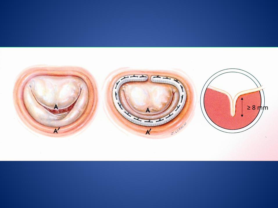
≥ 8 mm

0%
20%
40%
60%
80%
100%
40 60 80 100LVEDD (mm)
specificity sensitivity
89 %
65
LVEDD and Reverse Remodeling
Bax J et al. Circulation 2004; 110 (suppl II): II-103-II-108

Results: Mortality per LVEDD
0%
10%
20%
30%
40%
50%
60%
0 1 2 3 4 5 6
100 87 82 60 40 27 11 Patients at risk
Years since surgery
All-cause death
LVEDD >65
LVEDD ≤65
P-value 0.002 HR 3.4 and 95% CI 1.5-7.4
71 ± 8.5%
49 ± 11%
93 ± 3.0%
80 ± 5.2%
Braun J et al. Ann Thorac Surg 2008 Feb;85(2):430-6

Echo Results: Mitral Regurgitation
BASELINE 3.1 ± 0.5
EARLY 0.5 ± 0.7 INTERMEDIATE (18m) 0.7 ± 0.7 LATE (46m) 0.9 ± 0.8
P < 0.05
Braun J et al. Ann Thorac Surg 2008 Feb;85(2):430-6

Results: Echocardiography
baseline intermediate late
MV gradient 3.6 ± 1.5 3.9 ± 1.7
MV area (cm2)
2.8 ± 0.6 2.6 ± 0.6
Tenting area (cm2)
4.8 ± 1.4
1.4 ± 0.6
Coaptation Height (mm)
3 ± 1 8 ± 2 8 ± 2

Predictors of mitral regurgitation recurrence in patients with heart failure undergoing mitral valve annuloplasty. Agnieszka Ciarka et al
Am J Cardiol 2010;106:395-401
ZOL 09/10
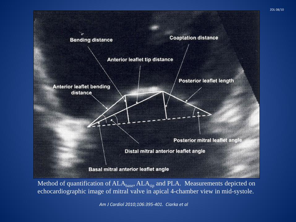
Method of quantification of ALAbase, ALAtip and PLA. Measurements depicted on echocardiographic image of mitral valve in apical 4-chamber view in mid-systole.
Am J Cardiol 2010;106:395-401. Ciarka et al
ZOL 08/10
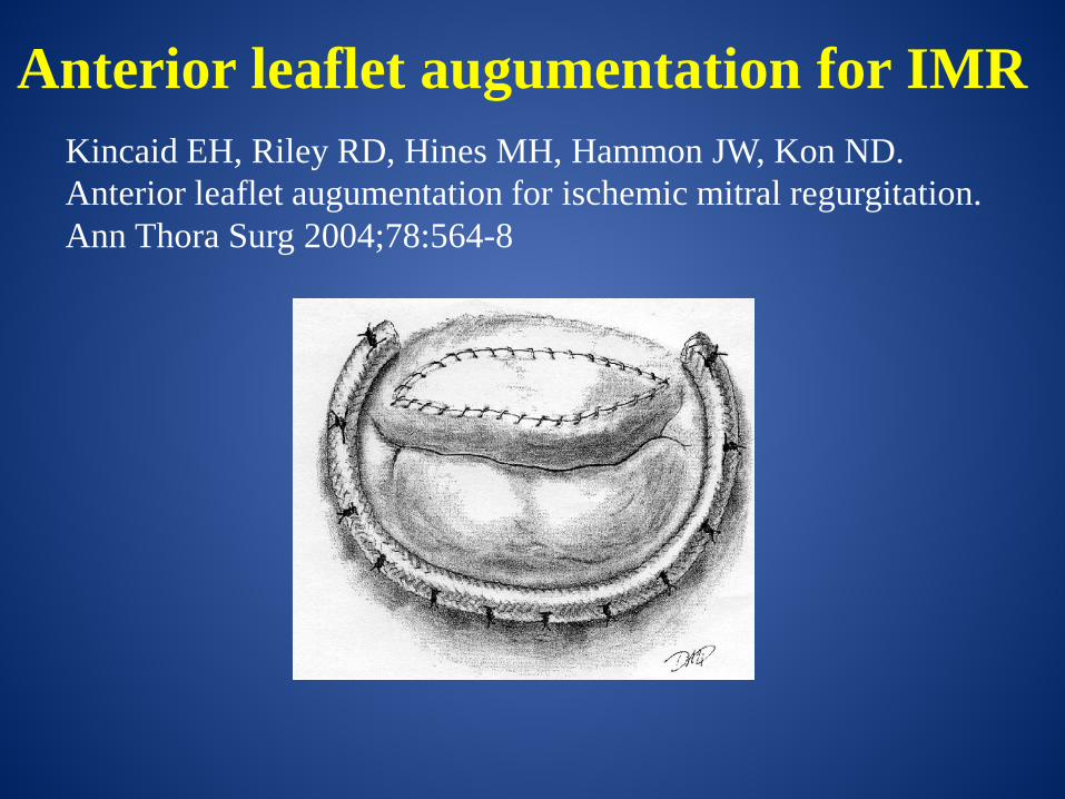
Anterior leaflet augumentation for IMR Kincaid EH, Riley RD, Hines MH, Hammon JW, Kon ND. Anterior leaflet augumentation for ischemic mitral regurgitation. Ann Thora Surg 2004;78:564-8

de Varennes, B. et al. Circulation 2009;119:2837-2843
The bovine pericardial patch extending the medial half of P2 and all of P3
Initial results of posterior leaflet extension for severe type IIIb ischemic mitral regurgitation.

Chordal cutting Messas E, Pouzet B, Touchot B, et al. Efficacy of chordal cutting to relieve chronic persistent ischemic mitral regurgitation. Circulation 2003;108[suppl II]:II-111-II-115

RING plus STRING: Papillary muscle repositioning as an adjunctive repair technique for ischemic mitral regurgitation
Langer F. JTCS 2007;133:247-9

Kron IL: Surgical relocation of the posterior papillary muscle in chronic ischemic mitral regurgitation. Ann Thorac Surg 2002;74:600-1
Relocation of the posterior papillary muscle (Kron)

Papillary muscles approximation + papillary muscles suspension
Papillary muscles approximation
CV4 for pulling up both PM
Hvass et al. J Thorac Cardiovasc Surg 2010 Feb; 139(2): 418-23 Shingu Y et al. Circ J. 2009 Nov;73(11):2061-7
Langer F et al. J Thorac Cardiovasc Surg 2011 May; 141(5): 1315-6

Aim of our study was to compare the results of combined approach papillary muscles relocation (PPMr) + mitral annuloplasty (MA) vs only restrictive annuloplasty (RA) in ischemic mitral regurgitation, guided by 3-dimensional (3D) echocardiography. Sixty-nine patients with severe ischemic mitral regurgitation who had PPMr + MA and coronary artery bypass grafting were matched 1:1 with patients who underwent isolated RA and coronary artery bypass grafting. A comprehensive pre- and postoperatory 2-dimensional and 3D transesophageal echocardiographic examination followed by a 3D offline assessment of the mitral valve apparatus was performed. Five-year freedom from cardiac-related event in the PPMr + MA group and isolated RA group was 83% ± 2.1% and 65.4% ± 1.2%, respectively (P < 0.001). Recurrent mitral regurgitation equal to or greater than moderate occurred in 2 (2.8%) and 8 (11.5%) in PPMr + MA group and RA group, respectively (P < 0.02). The PPMr promoted a significant reversal in left ventricle remodeling compared with the isolated RA. PPMr + MA reduce the tenting area and the coaptation depth with respect to RA, with less incidence of recurrent mitral regurgitation.
Semin Thoracic Surg 2012; 24:246-253 ZOL 11/13

Baseline and preop data
Follow-up data
PPMR + MA RA P
NYHA Class > II 22 (31.9%) 3 (4.3%) 4 (5.8%) 0.81
LVEDD (mm) 57 ± 8 51 ± 7 55 ± 8 0.02
LVESD (mm) 49 ± 1 41 ± 6 45 ± 5 0.02
Mean LVEF % 43 ± 8 46 ± 5 45 ± 9 0.21
Mean tenting area (cm²) 3.5 ± 0.9 1.2 ± 0.3 2.3 ± 0.4 <0.001
Mean coaptation depth 1.2 ± 0.1 0.6 ± 0.2 1 ± 0.2 <0.001
Recurrent MR ≥ moderate 2 (2.8%) 8 (11.5%) 0.02
The Role of Papillary Muscle Relocation in Ischemic Mitral Valve Regurgitation.
Fattouch Khalil et al. Semin Thoracic Surg 2012; 24:246-253
ZOL 11/13

CASE description
Fattouch et al, JTCVS, 2012

ZOL 04/14
Results At 12 months, the mean LVESVI among surviving patients was 54.6±25.0 ml per square meter of body-surface area in the repair group and 60.7±31.5 ml per square meter in the replacement group (mean change from baseline, −6.6 and −6.8 ml per square meter, respectively). The rate of death was 14.3% in the repair group and 17.6% in the replacement group (hazard ratio with repair, 0.79; 95% confidence interval, 0.42 to 1.47; P = 0.45 by the log-rank test). There was no significant between-group difference in LVESVI after adjustment for death (z score, 1.33; P = 0.18). The rate of moderate or severe recurrence of mitral regurgitation at 12 months was higher in the repair group than in the replacement group (32.6% vs. 2.3%, P<0.001). There were no significant between-group differences in the rate of a composite of major adverse cardiac or cerebrovascular events, in functional status, or in quality of life at 12 months.
2014;370:23-32

Acker et al N Engl J Med 2014;370:23-32
… In the repair group, the 12-month LVESVI was 64.1±23.9 ml per square meter in patients with recurrent mitral regurgitation versus 47.3±23.0 ml per square meter in those without recurrent mitral regurgitation (P<0.001). …
ZOL 04/14

PREDICTING RECURRENT MR FOLLOWING REPAIR FOR SEVERE IMR
I. L. Kron et al J. Thorac Cardiovasc Surg in press
Objective: … whether baseline echocardiographic and clinical characteristics could identify those who will develop moderate/severe recurrent mitral regurgitation or die. Results: 110 pts (116-6): over 2 years 34 (31%) perfect 53 MR rec 60% 30% at 1m 13 MR rec + † 10 † Conclusions: 10 variables model demonstrated good discrimination (AUC 0.82).
ZOL 02/15

Characteristic
No Recurrence
or Death (N=40)
Recurrence and/or Death
(N=76) P-value
No Recurrence
(N=44) Recurrence
(N=66) P-value
Age (y) 65.7 ± 12.5 70.6 ± 8.6 0.030 67.0 ± 12.6 69.6 ± 8.7 0.237
BMI (kg/m2) 28.1 ± 4.8 26.7 ± 4.3 0.106 27.9 ± 4.7 27.0 ± 4.4 0.300
Male 28 (70.0) 42 (55.3) 0.123 30 (68.2) 37 (56.1) 0.202
White 36 (90.0) 59 (77.6) 0.100 40 (90.9) 50 (75.8) 0.044
EROA (cm2) 0.4 ± 0.1 0.4 ± 0.2 0.219 0.4 ± 0.1 0.4 ± 0.2 0.128
Basal Dyskinesia/Aneurysm * 8 (20.0) 44 (57.9) <0.001 9 (20.5) 41 (62.1) <0.001
NYHA Class I+II 10 (25.0) 40 (52.6) 0.004
13 (29.5) 34 (51.5) 0.022
NYHA Class III+IV 30 (75.0) 36 (47.4) 31 (70.5) 32 (48.5)
History of CABG 4 (10.0) 15 (19.7) 0.178 4 (9.1) 13 (19.7) 0.132
History of PCI 13 (32.5) 34 (44.7) 0.202 15 (34.1) 30 (45.5) 0.235
History of Ventricular Arrhythmia 8 (20.0) 6 (7.9) 0.074 10 (22.7) 3 (4.5) 0.004
Baseline patient characteristics by patient outcomes
ZOL 06/14
J. Thorac Cardiovasc Surg in press. Kron et al * 50 pts

ZOL 06/14
preop


postop postop
ZOL 06/14
MVR in IMR ? - Mechanical or bioprosthesis ?
- Which size ?
- What if reverse remodeling and bioprosthesis ?


Results
Bertrand PB, Dion RA et al, J Am Coll Cardiol 2015;65:452-61
Anterior leaflet opening angle 1. EOA at peak exercise correlates well with anterior leaflet opening angle..
2. Higher increases in anterior leaflet angle results in higher increases in EOA during exercise.

Addendum
Bertrand PB, Dion RA et al, ACC.14, Washington
Comparison with mechanical mitral valve replacement (MVR): 16 MVR patients versus propensity-matched restrictive annuloplasty subgroup (n=16)
CONCLUSION: In contrast to restrictive mitral annuloplasty, the effective mitral valve area following mechanical MVR does not increase during exercise. Therefore, important hemodynamic difference exist during exercise, in favour of valve repair (if durable result is obtained)

Conclusions
Bertrand PB, Dion RA et al, J Am Coll Cardiol 2015;65:452-61
In RMA patients, effective mitral valve area increases during exercise, despite fixed annular size.
Diastolic AL tethering plays a key role in this dynamic process, with increasing AL opening during exercise being associated with higher exercise mitral valve area.
Indexed effective valve area at peak exercise is a strong and independent
predictor of exercise capacity and is associated with clinical outcome.
These findings stress the importance of maximizing AL opening by targeting the subvalvular apparatus in future repair algorithms.

CONCLUSIONS 1) Always consider repair BUT
NO RESIDUAL MR ! MVR ? 2) LVEDD 65 ≤ : RMA ± LV procedure LVESD 51
3) LVEDD 65 > : INVENTIVITY OR MVR ? LVESD 51
- Leaflet augmentation
- Constraint device (?)
- PM procedures
BUT
? ? Efficacy in truly dilated LV
ZOL 11/13


CorCap™
Braun J. et al. J Thorac Cardiovasc Surg 2011;142:e93-e100

ZOL 04/11
Evolution of LVEDV and LVESV at early postoperative follow-up, and at long-term follow-up
CSD + RMA + OMT
Braun et al, J Thorac Cardiovasc Surg 2011;142:e93-e100


J Thorac Cardiovasc Surg 2004;128:916-24
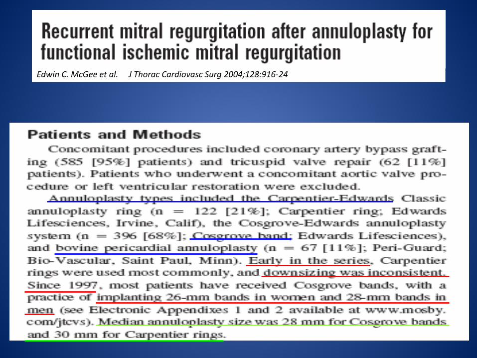
Edwin C. McGee et al. J Thorac Cardiovasc Surg 2004;128:916-24


Baseline patient characteristics by patient outcomes
Age (y) BMI (kg/m2) Male White EDV (ml) EF (%) EROA (cm2) ESV (ml) LVEDD Mid-Ventricle (cm) LVESD Mid-Ventricle (cm) MR Peak Velocity (cm/sec) Vena Contracta (cm) Angle (anterior-ap4) (°) Angle (posterior-ap4) (°) Sphericity index (ED) Sphericity index (ES) Tenting Area (cm2)
Tenting Height (cm) Basal Aneurysm NYHA Class I+II NYHA Class III+IV Planned Revascularization History of AF History of CABG Chronic Lung Disease (≥Moderate) Diabetes History of Heart Failure Hypertension History of MI History of PCI History of Renal Insufficiency Previous Valve Repair Previous Valve Replacement History of Ventricular Arrhythmia
ZOL 06/14
J. Thorac Cardiovasc Surg in press. Kron et al

Results
Bertrand PB, Dion RA et al, J Am Coll Cardiol 2015;65:452-61

Results
Bertrand PB, Dion RA et al, J Am Coll Cardiol 2015;65:452-61
Exercise capacity (VO2max)
EOA at peak exercise < 0.9cm2/m2 versus ≥ 0.9cm2/m2

Results
Bertrand PB, Dion RA et al, J Am Coll Cardiol 2015;65:452-61

preoperative images
MMode: LV diameter end-diastolic: 6,5 cm.
Parasternal long axis. Moderate LV function. Restrictive MV motion.

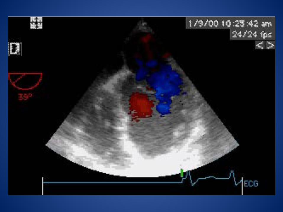
preoperative images
Apical four chamber view, color-Doppler encoded. Note the severe regurgitation.
Apical two chamber view. Note the restrictive closure of the mitral valve.

postoperative images
MMode: LV diameter end-diastolic: 4,6 cm. LVIDd showed 1,9 cm regression to normal value.
Parasternal short axis 3 years after repair. Note the improved systolic function.

Results
Exercise Doppler measurements Resting Peak exercise p-value
MV peak gradient, mmHg 11.4 ± 3.6 16.8 ± 6.1 <0.001
MV mean gradient, mmHg 4.4 ± 1.8
8.2 ± 4.2
<0.001
Systolic PAP, mmHg 43 ± 13
53 ± 20
0.012
Cardiac output, L/min 3.9 ± 0,8
5.8 ± 2.0
<0.001
MR vena contracta width, mm 1.3 ± 1.0
1.3 ± 1.2
NS
<5mmHg (n=14) ≥ 5mmHg (n=9)
Bertrand PB, Dion RA et al, J Thorac Cardiovasc Surg 2014;148:183-7

Results
Exercise Doppler measurements
Mean gradient <5mmHg (n=14)
Mean gradient >5mmHg (n=9) p-value
Variable Resting Exercise Resting Exercise Resting Exercise
MV peak gradient, mmHg 9.5 ± 2.7 13.1 ± 4.3 14.5 ± 2.6 22.6 ± 3.4 <0.001 <0.001
MV mean gradient, mmHg 3.3 ± 1 5.7 ± 1.6 6.2 ± 1 12.0 ± 4.0 <0.001 <0.001
Syst. PAP, mmHg 43 ± 15 46 ± 26 43 ± 11 50 ± 20 0.94 0.71
Cardiac output 3.6 ± 0.8 4.7 ± 1.3 4.4 ± 0.8 7.3 ± 2.0 0.03 0.001
MR VC width, mm 1.2 ± 1.2 1.2 ± 1.2 1.4 ± 0.7 1.3 ± 1.3 NS NS
Comparison of other variables:
LV EF,% 42 ± 16 53 ± 7 0.036
LV EDV, ml 138 ± 55 105 ± 30 0.076
LV ESV, ml 86 ± 49 50 ± 15 0.020
Maximal workload, Watt 38 ± 14 69 ± 23 <0.001
VO2max, ml/kg/min 12.3 ± 3.4 15.3 ± 2.6 0.035
Bertrand PB, Dion RA et al, J Thorac Cardiovasc Surg 2014;148:183-7

Conclusions
• Transmitral gradients following RMA are not merely determined by the degree of functional stenosis, but also depend of flow (cardiac output).
• Functional capacity (VO2max) following RMA is not necessarily worse in patients with a higher transmitral gradient.
a flow-independent measure should be validated for comparison of
postoperative results in this population.
Effective mitral valve area?
Bertrand PB, Dion RA et al, J Thorac Cardiovasc Surg 2014;148:183-7