why phosphoproteomics is still a challenge
TRANSCRIPT

This journal is©The Royal Society of Chemistry 2015 Mol. BioSyst., 2015, 11, 1487--1493 | 1487
Cite this:Mol. BioSyst., 2015,
11, 1487
Why phosphoproteomics is still a challenge
Fiorella A. Solari,†a Margherita Dell’Aica,†a Albert Sickmannabc and Rene P. Zahedi*a
Despite continuous improvements phosphoproteomics still faces challenges that are often neglected,
e.g. partially poor recovery of phosphopeptide enrichment, assessment of phosphorylation
stoichiometry, label-free quantification, poor behavior during chromatography, and general limitations of
peptide-centric proteomics. Here we critically discuss current limitations that need consideration in both
qualitative and quantitative studies.
Introduction
Protein activity is mainly modulated by dynamic reversible (andirreversible) post-translational modifications (PTMs).1 Phos-phorylation is a reversible PTM involved in numerous regulatorymechanisms in eukaryotic cells, such as cell division, apoptosis,response to extracellular signals and growth factor stimulation,2 andis regulated by the interplay of protein kinases and phosphatases.3,4
The fundamental importance of protein phosphorylationand the continuous development in the field of protein massspectrometry render phosphoproteomics a valuable tool incurrent life science, allowing the study of regulatory changesover time and/or between conditions. However, despite itsroutine use in many laboratories around the world severallimitations still have to be faced.5,6
Among those are (i) the analysis of low abundant proteins inthe light of the vast dynamic range of cells, (ii) the lowstoichiometry of phosphorylation, (iii) potentially impaireddigestion efficiency,7 (iv) phosphopeptide losses during samplepreparation and chromatography,8–11 (v) impaired ionizationefficiency of phosphopeptides, (vi) the peculiar behavior of thelabile phosphate group upon collision induced dissociation,often resulting in poor quality MS/MS spectra that can impairboth identification of the peptide sequence and the (vii) correctlocalization of the phosphorylation site(s),12–14 and (viii) thegeneral limitations of peptide-centric proteomics.15
Taken together, these issues can complicate the identifi-cation and quantification of phosphopeptides (and particularly
phosphorylation sites) compared to their non-phosphorylatedcounterparts, and can produce wrong, misleading and inaccurateidentification and quantification results.
Nevertheless, some issues can be (partially) addressed byimproved methods for sample preparation, phosphopeptideenrichment, MS analysis, and computational analysis ofMS-derived phosphoprotemics data.16
Here, we discuss current obstacles that can emerge inphosphoproteomic analysis and demonstrate why beside allthe advances achieved in recent years, phosphoproteomics isstill a challenge.
Sample amount, sample preparationand noise
Phosphorylation is a fast and dynamic process that, like anyenzyme-driven biochemical reaction, can be prone to errors.17
Consequently, it is conceivable that within a cell certain levelsof protein phosphorylation might occur without major functionalrelevance. However, for a given site the degree of phosphorylation,i.e. the stoichiometry that reflects how many copies of a proteinare phosphorylated at that amino acid at a given time point,might represent a threshold that may distinguish relevant fromnon-functional phosphorylation events. Many studies use largeamounts of protein starting material (often in the mg range) toenrich and identify as many phosphopeptides as possible, oftenleading to the identification of 10 000s of phosphorylation sites.As information about stoichiometry is almost always missing, itmay be argued how many of these phosphorylation sites reallyderive from specific phosphorylation events, and how many areindeed just ‘noise’ derived from more-or-less random phos-phorylation events, without functional consequence. Such randomevents however may be detected because of the extremely high
a Leibniz-Institut fur Analytische Wissenschaften – ISAS – e.V., Otto-Hahn-Str. 6b,
44227 Dortmund, Germany. E-mail: [email protected] Department of Chemistry, College of Physical Sciences, University of Aberdeen,
Aberdeen, Scotland, UKc Medizinische Fakultat, Ruhr Universitat Bochum, Bochum, Germany
† Equal contribution.
Received 10th January 2015,Accepted 13th March 2015
DOI: 10.1039/c5mb00024f
www.rsc.org/molecularbiosystems
MolecularBioSystems
OPINION
Ope
n A
cces
s A
rtic
le. P
ublis
hed
on 1
3 M
arch
201
5. D
ownl
oade
d on
11/
28/2
021
9:06
:43
AM
. T
his
artic
le is
lice
nsed
und
er a
Cre
ativ
e C
omm
ons
Attr
ibut
ion-
Non
Com
mer
cial
3.0
Unp
orte
d L
icen
ce.
View Article OnlineView Journal | View Issue

1488 | Mol. BioSyst., 2015, 11, 1487--1493 This journal is©The Royal Society of Chemistry 2015
sensitivity of modern mass spectrometers. If present on (relatively)high abundant proteins, these random events may interferewith the identification of phosphorylation sites on low abun-dant proteins.
Obtaining temporal profiles of protein phosphorylationupon specific treatment may be one strategy to dissect andinterpret the huge amount of data obtained using modernphosphoproteomics approaches.18,19
Moreover, sample preparation steps prior to LC-MS analysiscan have a huge impact on the final results. Recently, Mertinset al. investigated the proteome and the phosphoproteome ofhuman ovarian tumor and breast cancer tissue after definedischemic events. They demonstrated that, while the proteomeremains unchanged for 60 min after tissue removal from its bloodsupply, up to 24% of the phosphoproteome showed rapidphosphorylation changes, particularly in pathways relevantfor cancer. Thus, it is recommended to exhaustively examineand optimize protocols for sample collection before analyzingthe phosphoproteome.20
Popular sample preparation protocols such as Filter AidedSample Preparation (FASP) involve the use of urea, which is apotent chaotropic agent.21,22 In aqueous solution urea dissociatesover time, a reaction that is accelerated upon heating, such asoften done e.g. during the reduction of disulfide bonds. Notably,one of its degradation products, isocyanate reacts with primaryamines such as protein N-termini and e-amines of lysines,inducing in vitro carbamylation. Thus, if used without care urea(e.g. by using non-freshly prepared urea buffers or by heating ureabuffers) can easily induce artificial carbamylation of primaryamines, thereby reducing identification rates and interfering withseveral stable isotope labeling strategies that target Lys residues.23
Especially in case of low complexity, samples are sometimesseparated using SDS-PAGE followed by silver staining, in-geldigestion and LC-MS analysis in order to identify phosphoryla-tion sites. However, as demonstrated by Gharib et al. certainsilver staining protocols can induce artificial sulfation which isalmost isobaric to phosphorylation and thus can be easilymistaken for the latter.24
Generally, when looking for quantitative changes in proteinphosphorylation upon specific treatment/stimulation of samples,special attention has to be paid to the experimental design inorder to (i) guarantee maximal reproducibility, (ii) not inducealterations of phosphorylation profiles, (iii) reduce samplelosses, especially for low sample amounts, and (iv) not intro-duce artificial modifications.
Phosphorylation stoichiometry,digestion efficiency and peptide-centric phosphoproteomics
As mentioned before, during sample preparation in proteomicstudies there are initial and crucial steps that directly affect thereliability of the final results. Are the cells effectively lysed and isthe activity of endogenous kinases and particularly phosphatasesinhibited? Is the sample properly digested? The underlying steps
should be carefully controlled, as they are important for thesuccess of proteomics studies.16,25,26
For shotgun proteomics and phosphoproteomics trypsin isthe protease-of-choice due to its availability, high specificityand cleavage properties, resulting in peptides with homogenousproperties such as an average length of B14 amino acids.25,27
However, proteolytic cleavage can be strongly impaired inproximity of phosphoamino acids, leading to missed cleavagesites and complicating phosphoproteome analysis. We recentlyevaluated this effect in detail using synthetic phosphopeptides,demonstrating that it not only depends on the position of thephosphoamino acid, but on its very nature (Ser/Thr/Tyr). Thus,reduced digestion efficiency can be partially compensated byusing optimized digestion conditions, such as the addition oforganic solvents or simply by increasing the amount of protease.7
Indeed, it is recommended to make use of relative high con-centrations of trypsin up to 1 : 20 (enzyme : protein, wt : wt),whereas often used concentrations of 1 : 50 or even 1 : 100 canlead to a substantially reduced cleavage efficiency and thereforeloss of sensitivity.
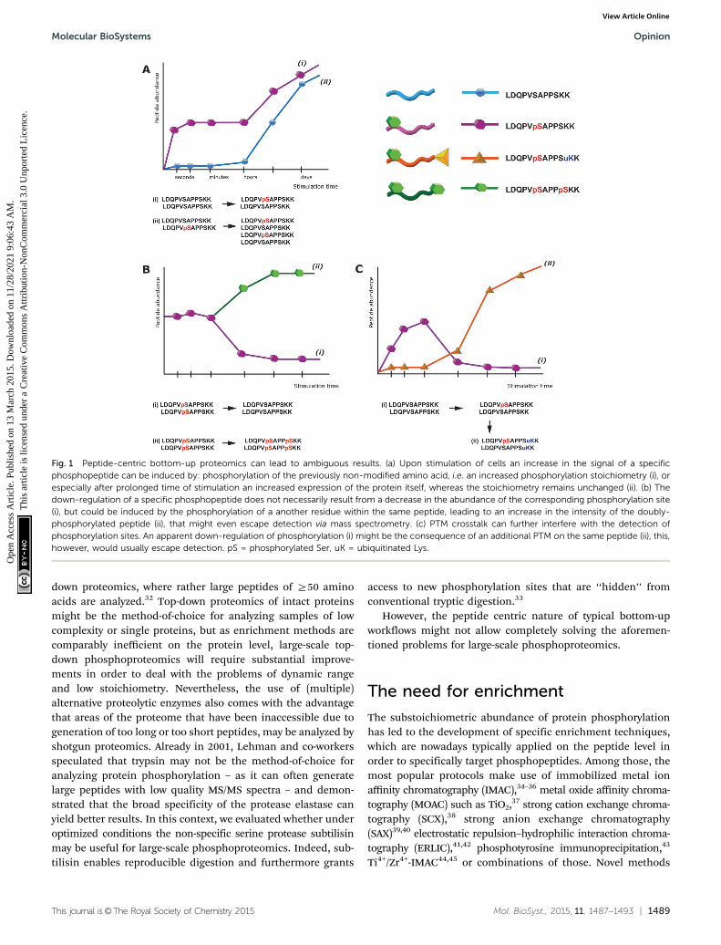
Notably, this interference of proteolytic digestion by proteinphosphorylation can strongly affect the determination of phos-phorylation stoichiometry. To determine, for a given protein,how many copies/which share is phosphorylated at a givenposition and time point is still one of the most challengingissues in the field of phosphoproteomics. One strategy to assessthe degree of phosphorylation is based on the usage of syntheticstable isotope labeled analogues of endogenous phosphopeptidesand their non-phosphorylated counterparts, in order to absolutelyquantify both versions and thus assess the phosphorylationdegree.28 However, the impairment of digestion can stronglyinterfere with this strategy. For affected phosphorylation sitesthe digestion can be up to 10 times more efficient in absence ofa phosphorylation, such that the mere comparison of phos-phorylated and non-phosphorylated fully tryptic peptides canlead to wrong results. In other strategies, the sample is split intwo aliquots, one of which is treated with phosphatase prior toLC-MS. The increase of the non-phosphorylated peptide signalupon treatment with phosphatase can then be used to deducethe degree of phosphorylation.29 But also here impaired digestionefficiency will affect the quantitative results and even moreimportantly, after phosphatase treatment different phospho-isoforms of the same peptide will contribute to the same non-phosphorylated signal and therefore cannot be distinguished.This shortcoming is a direct consequence of the generallimitations of peptide-centric shotgun proteomics. Althoughoften neglected, in phosphoproteomics peptides rather thanphosphorylation sites are quantified. As a consequence the up-regulation of a specific phosphopeptide does not necessarilycorrelate with an increase of the particular phosphorylationsite, as depicted in Fig. 1. Moreover, in conventional bottom-uppeptide-centric proteomics is it unclear how many of the differentphosphorylation sites/peptides often detected for a single proteinreally occur on the very same protein molecule.
One strategy to address these issues may be the use of(multiple) alternative proteases30,31 and so-called middle-
Opinion Molecular BioSystems
Ope
n A
cces
s A
rtic
le. P
ublis
hed
on 1
3 M
arch
201
5. D
ownl
oade
d on
11/
28/2
021
9:06
:43
AM
. T
his
artic
le is
lice
nsed
und
er a
Cre
ativ
e C
omm
ons
Attr
ibut
ion-
Non
Com
mer
cial
3.0
Unp
orte
d L
icen
ce.
View Article Online
This journal is©The Royal Society of Chemistry 2015 Mol. BioSyst., 2015, 11, 1487--1493 | 1489
down proteomics, where rather large peptides of Z50 aminoacids are analyzed.32 Top-down proteomics of intact proteinsmight be the method-of-choice for analyzing samples of lowcomplexity or single proteins, but as enrichment methods arecomparably inefficient on the protein level, large-scale top-down phosphoproteomics will require substantial improve-ments in order to deal with the problems of dynamic rangeand low stoichiometry. Nevertheless, the use of (multiple)alternative proteolytic enzymes also comes with the advantagethat areas of the proteome that have been inaccessible due togeneration of too long or too short peptides, may be analyzed byshotgun proteomics. Already in 2001, Lehman and co-workersspeculated that trypsin may not be the method-of-choice foranalyzing protein phosphorylation – as it can often generatelarge peptides with low quality MS/MS spectra – and demon-strated that the broad specificity of the protease elastase canyield better results. In this context, we evaluated whether underoptimized conditions the non-specific serine protease subtilisinmay be useful for large-scale phosphoproteomics. Indeed, sub-tilisin enables reproducible digestion and furthermore grants
access to new phosphorylation sites that are ‘‘hidden’’ fromconventional tryptic digestion.33
However, the peptide centric nature of typical bottom-upworkflows might not allow completely solving the aforemen-tioned problems for large-scale phosphoproteomics.
The need for enrichment
The substoichiometric abundance of protein phosphorylationhas led to the development of specific enrichment techniques,which are nowadays typically applied on the peptide level inorder to specifically target phosphopeptides. Among those, themost popular protocols make use of immobilized metal ionaffinity chromatography (IMAC),34–36 metal oxide affinity chroma-tography (MOAC) such as TiO2,37 strong cation exchange chroma-tography (SCX),38 strong anion exchange chromatography(SAX)39,40 electrostatic repulsion–hydrophilic interaction chroma-tography (ERLIC),41,42 phosphotyrosine immunoprecipitation,43
Ti4+/Zr4+-IMAC44,45 or combinations of those. Novel methods
Fig. 1 Peptide-centric bottom-up proteomics can lead to ambiguous results. (a) Upon stimulation of cells an increase in the signal of a specificphosphopeptide can be induced by: phosphorylation of the previously non-modified amino acid, i.e. an increased phosphorylation stoichiometry (i), orespecially after prolonged time of stimulation an increased expression of the protein itself, whereas the stoichiometry remains unchanged (ii). (b) Thedown-regulation of a specific phosphopeptide does not necessarily result from a decrease in the abundance of the corresponding phosphorylation site(i), but could be induced by the phosphorylation of a another residue within the same peptide, leading to an increase in the intensity of the doubly-phosphorylated peptide (ii), that might even escape detection via mass spectrometry. (c) PTM crosstalk can further interfere with the detection ofphosphorylation sites. An apparent down-regulation of phosphorylation (i) might be the consequence of an additional PTM on the same peptide (ii), this,however, would usually escape detection. pS = phosphorylated Ser, uK = ubiquitinated Lys.
Molecular BioSystems Opinion
Ope
n A
cces
s A
rtic
le. P
ublis
hed
on 1
3 M
arch
201
5. D
ownl
oade
d on
11/
28/2
021
9:06
:43
AM
. T
his
artic
le is
lice
nsed
und
er a
Cre
ativ
e C
omm
ons
Attr
ibut
ion-
Non
Com
mer
cial
3.0
Unp
orte
d L
icen
ce.
View Article Online

1490 | Mol. BioSyst., 2015, 11, 1487--1493 This journal is©The Royal Society of Chemistry 2015
and protocols for enrichment are frequently published, however,all these methods have in common that they work best whencombined with optimized and reasonable sample preparationand LC-MS analysis workflows. Then, in combination with state-of-the-art instrumentation, phosphopeptide yields in the rangeof 100 sites per 1 mg of sample can be obtained.42,46,47 In general,the aforementioned methods for enrichment seem to havedifferent preferences to more efficiently enrich for specific subsetsof the phosphoproteome and thus may be combined to yield ahigher coverage. Whereas phosphopeptide recovery varies betweenthe different methods and respective methodical variations,and furthermore depends on the sample and its complexity,even under optimal conditions recovery is clearly below 50% formost phosphopeptides, as can be derived from spike-in experi-ments with synthetic peptides. Particularly methods that primarilyenrich but not additionally fractionate, such as IMAC and MOACshould be combined with subsequent (or prior) fractionatione.g. by Hydrophilic Interaction Liquid Chromatography (HILIC),SCX18 or high pH RP48 to increase the number of identifiedphosphopeptides. Notably, ERLIC provides both, highly selectiveenrichment and fractionation (e.g. separating singly from multiplyphosphorylated peptides) at once.42
Generally, phosphopeptide enrichment is accompanied by theloss of information about the non-phosphorylated counterpart andtherefore cannot readily be used for determining stoichiometry.
Challenges in separation,fragmentation and identification ofphosphopeptides
Several properties of phosphopeptides complicate their analysisby LC-MS. Metal ions on the surfaces of the HPLC flow path,or within solvents can lead to losses due to the formation ofphosphopeptide–metal ion complexes, which can be partiallycircumvented by the addition of EDTA to the LC buffers.49 In this
context, Lehmann and co-workers investigated in detail theeffect of additives given to the sample before LC-MS analysis.They demonstrated that citrate can overcome the adsorption ofphosphopeptides to surfaces and is compatible with LC-MS.10
Especially multi-phosphorylated peptides can smear substan-tially during reversed phase chromatography, which in severecases can dramatically impair their identification (see Fig. 2).
Another frequently discussed issue is the ionization efficiencyof phosphopeptides. Using synthetic peptides Steen et al. showedthat lower ionization/detection efficiencies of phosphopeptidescompared to their counterparts could not be substantiated. InLC-MS some phosphopeptides showed better ionization/detectionthan their cognates.50 In contrast, Choi et al. demonstrated thationization/detection efficiency negatively correlates with thenumber of phosphorylation sites,11 which, however, could be aconsequence of poor chromatography. Indeed, both studies agreethat in complex mixtures with non-phosphorylated peptides,phosphopeptides show reduced ionization efficiencies.
Another issue that complicates phosphopeptide identificationis the labile character of the phosphoester bond which, dependingon the type of fragmentation used, can severely impair and alterthe fragmentation behavior when compared to non-modifiedpeptides.13 Still, the most popular technique to induce fragmenta-tion of peptide ions is collision-induced dissociation (CID),51 whichcan be categorized into two subclasses, ion trap CID and beam-typeCID,52 as conducted in quadrupoles. In ion traps, upon excitationpeptides experience hundreds of light collisions with an inert gas,each collision transferring internal energy to the peptide. As thephosphoester bond is highly labile, it tends to break first, leadingto a so-called neutral loss of phosphoric acid (98 Da) from theprecursor. Phosphopeptide CID MS/MS spectra are often domi-nated by this neutral loss signal and show only minor fragmenta-tion. Moreover, ion trap CID is often accompanied by additionalneutral losses of water. In beam-type CID (also called higher energyCID or HCD) an electrical potential is used to accelerate peptideions towards the inert gas, leading to fewer but higher energy
Fig. 2 Column performance affects detection of multiply phosphorylated peptides. A set of synthetic peptides, KVTQTIITLK, KVtQTIITLK, KVtQTIItLK,KVtQtIItLK (lower case letters indicate phosphorylation sites) was analyzed by LC-MS on the same instrument on a new main column (a), and after 1.5 monthsof use (b). Even with an optimal LC system (a), the full width at half maximum (FWHM) increases slightly with the number of phosphorylation sites. However,with column ageing (b), the FWHM of the triply-phosphorylated peptide is further increasing, resulting in a reduced maximum intensity and therefore loss-of-sensitivity. While the given example still shows a relatively good separation considering the number of phosphorylation sites, non-optimal LC conditions canlead to FWHM in the min range and consequently to a complete absence of multiphosphorylated peptide identifications in complex samples.
Opinion Molecular BioSystems
Ope
n A
cces
s A
rtic
le. P
ublis
hed
on 1
3 M
arch
201
5. D
ownl
oade
d on
11/
28/2
021
9:06
:43
AM
. T
his
artic
le is
lice
nsed
und
er a
Cre
ativ
e C
omm
ons
Attr
ibut
ion-
Non
Com
mer
cial
3.0
Unp
orte
d L
icen
ce.
View Article Online

This journal is©The Royal Society of Chemistry 2015 Mol. BioSyst., 2015, 11, 1487--1493 | 1491
collisions. Consequently, the presence of the neutral loss precursoris substantially reduced leading to richer fragmentation patterns.
Beside the fragmentation mode, the occurrence of the neutralloss precursor depends on peptide sequence and charge state(more prominent for +2 than +3 charge states), with phospho-tyrosine showing only minor neutral losses of 80 Da, correspondingto meta-phosphoric acid. Beside the neutral loss of the precursorion, fragment ions containing the phosphoamino acid can bepresent as intact (i.e. with phospho moiety) and neutral loss form,both in ion trap and beam-type CID. Notably, neutral losses ofphosphopeptides reduce spectrum quality, as the overall signalintensity is distributed over more fragment ions when compared tonon-phosphorylated peptides, therefore also complicating spec-trum identification. Indeed, for two peptides sharing the samesequence but having different phosphorylation sites, such as IsASand ISAs (lowercase indicating the phosphoamino acid), theoccurrence of neutral losses can complicate the exact localizationof the phospho moiety substantially, as demonstrated in Table 1.
In contrast, electron transfer dissociation (ETD) can preservelabile side chain modifications such as phosphorylation53 andcan therefore facilitate phosphospeptide identification. How-ever, ETD fragmentation works best with charge states above+2, is less efficient than CID and thus slightly less sensitive. Therecently introduced EThcD combines ETD and HCD to generateboth b/y and c/z ions and provides data-rich MS/MS spectra thatcan yield higher peptide sequence coverage and more confidentlocalization of phosphorylation sites.54
Localization of phosphoamino acids
Although fragmentation modes such as beam-type CID, ETD,and EThcD yield data-rich fragmentation spectra that facilitate
the localization of phosphoamino acids, common searchalgorithms still can mis-localize the phosphorylation site withina given peptide sequence. Thus, in the past decade a couple ofalgorithms12,18,55–57 that focus on a more reliable localizationhave been developed and have substantially improved the qualityof phosphopeptide identifications. But even with improvedalgorithms, peptide spectrum matches can be prone to falsepositive identifications. Thus, phosphorylation site localizationcan be wrong58 and – owing to differences in quality and signal/noise – even redundant MS/MS spectra of the same syntheticphosphopeptide can yield different phosphoamino acid locali-zations or probabilities. This issue particularly complicateslabel free quantitative phosphoproteomics, as discussed below.
Importantly, public databases with PTM information can stillcontain site localizations that either have not been validatedwith specific algorithms or have not reported certain probabilities,and thus might include a substantial amount of wrong or non-confident phosphorylation sites. With the introduction of guide-lines for uploading LC-MS raw data to open repositories59,60 formost proteomics journals, and very recently the possibility forautomated and quality controlled reanalysis of such large-scaledata sets,61 the quality of data in public databases shouldimprove considerably.
Phosphopeptide quantification
Quantitative phosphoproteomics allows elucidating changes inprotein phosphorylation between different samples. SeveralMS-based quantification methods have been used for this purpose,including chemical and metabolic stable-isotope labeling strategiessuch as iTRAQ,62 TMT,63 dimethyl or SILAC.64,65 Generally, SILACis well-suited for quantitative phosphoproteomics in cell cultureexperiments, however, in contrast to chemical labeling techniquesSILAC cannot be directly used for clinical samples such as bloodcells, biopsies or primary tissue.19,20
Label free quantification represents an increasingly usedapproach for quantification, which on the one hand has nogeneral limitations with regard to the number of samples orreplicates, but on the other hand demands a great deal of theexperimental design and quality control to ensure reproduci-bility and to avoid the introduction of biases. Primarily labelfree phosphoproteomics not only requires a reproducible samplepreparation and LC-MS analysis with minor deviations in reten-tion times and elution profiles, but furthermore has to deal withthe issues of confident phosphorylation site localization. Thisis particularly challenging, as different phospho-isoforms of thesame peptide can have highly similar retention times or even(partially) overlap in their elution profiles, rendering discrimi-nation across multiple runs extremely difficult.66 Moreover, theaforementioned varying and sometimes low recovery of phos-phopeptides might be a major issue, as slight variations inpeptide signals of already low intensity disproportionately impairquantification reproducibility. Thus, an important step towardsmore robust label free phosphoproteomics of limited sampleamounts might be to further improve phosphopeptide recovery.
Table 1 Under CID conditions, different phospho-isoforms can producethe same m/z for different b- and y-ions. The two phosphorylatedpeptides IsAS and ISAs can produce b- and y-ions that are isobaric buthave a different origin (labeled in bold), owing to the neutral losses ofphosphoric acid (H3PO4) and water (H2O). For instance, IsAS y1-H2O andISAs y1-H3PO4 at 88.0393 m/z are not distinguishable. Besides, bothpeptides also share isobaric b- and y-ions of the same origin (italicized).Theoretical fragment masses were calculated using MS Product (http://prospector.ucsf.edu/prospector/cgi-bin/msform.cgi?form=msproduct)
IsAS ISAs
m/z Fragment ion m/z Fragment ion
88.0393 y1-H2O 88.0393 y1-H3PO4
106.0499 y1 159.0764 y2-H3PO4
159.0764 y2-H2O 183.1128 b2-H2O177.0870 y2 186.0162 y1
183.1128 b2-H3PO4 201.1234 b2
246.1084 y3-H3PO4 246.1084 y3-H3PO4
254.1499 b3-H2O 254.1499 b3-H2O281.0897 b2 257.0533 y2
326.0748 y3-H2O 272.1605 b3
344.0853 y3 326.0748 y3-H2O352.1268 b3 344.0853 y3
359.1925 MH-H3PO4 359.1925 MH-H3PO4
439.1588 MH-H2O 439.1588 MH-H2O457.1694 MH 457.1694 MH
Molecular BioSystems Opinion
Ope
n A
cces
s A
rtic
le. P
ublis
hed
on 1
3 M
arch
201
5. D
ownl
oade
d on
11/
28/2
021
9:06
:43
AM
. T
his
artic
le is
lice
nsed
und
er a
Cre
ativ
e C
omm
ons
Attr
ibut
ion-
Non
Com
mer
cial
3.0
Unp
orte
d L
icen
ce.
View Article Online

1492 | Mol. BioSyst., 2015, 11, 1487--1493 This journal is©The Royal Society of Chemistry 2015
Recently, Graaf et al. performed label free phosphoproteomicsof 108 samples based on Ti+4-IMAC, analyzed the samples usingdecision tree-based ion-trap CID or ETD fragmentation to obtaindata-rich MS/MS spectra, and performed the data analysis usingMaxQuant67 to assess the phosphorylation site localization.68
As such studies demand a great deal of reproducibility andquality control, chemical labeling techniques that allow multi-plexing might facilitate the reliable quantification of phospho-peptides across samples.
Conclusion
Here, we described several issues that have to be faced in modernphosphoproteomics in order to generate robust and reliable data.Although some of those have been partially addressed and solvedin recent years, they still represent common pitfalls when doingquantitative phosphoproteomics. Owing to the large amount ofdata generated in current (and past) phosphoproteomics studiesand their impact on public protein databases and in the literature,it is imperative that researchers pay special attention to the qualityand reliability of the study design, experimental procedures andobtained data. Otherwise, even despite the use of state-of-the-artequipment, protocols, and software tools flawed data can be easilygenerated. This is particularly important with the advent of clinicalstudies that (i) are often limited in sample amount, (ii) generallyhave a much higher variation than the bulk of cell culture-basedphosphoproteomic studies, and (iii) consequently are much moredemanding in terms of robust protocols and data.
Considering the time, money, and effort spent for studyingjust a single phosphorylation site and its function, it is man-datory to constantly and critically assess the applied strategies.
Abbreviations
CID Collision induced dissociationHCD Higher collision induced dissociationETD Electron transfer dissociationSILAC Stable isotopic label by amino acids in cell cultureiTRAQ Isobaric tag for relative and absolute quantificationLC-MS Liquid chromatography online mass spectrometryIMAC Immobilized metal ion affinity chromatographyMOAC Metal oxide affinity chromatographyERLIC Electrostatic repulsion–hydrophilic interaction liquid
chromatographyHILIC Hydrophilic interaction liquid chromatographySCX Strong cation exchange chromatographyEDTA Ethylenediaminetetraacetic acidTMT Tandem mass tag
Acknowledgements
The financial support by the Ministerium fur Innovation,Wissenschaft und Forschung des Landes Nordrhein-Westfalenis gratefully acknowledged and by the ISAS Integrated ResearchProject ‘Monitoring Signaling Events’. We furthermore would like
thank the Italian Proteomics Association (ItPA) for the greatmeeting in June 2014. In addition, we also thank our collegesfor vivid daily discussions, in particular Stefan Loroch, OliverPagel, Clarissa Dickhut and Humberto Gonczarowska Jorge.
References
1 J. M. Burkhart, S. Gambaryan, S. P. Watson, K. Jurk, U. Walter,A. Sickmann, J. W. Heemskerk and R. P. Zahedi, Circ. Res.,2014, 114, 1204–1219.
2 R. H. Newman, J. Zhang and H. Zhu, Front. Genet., 2014, 5, 263.3 J. Reinders and A. Sickmann, Proteomics, 2005, 5, 4052–4061.4 E. Maes, K. Tirez, G. Baggerman, D. Valkenborg, L. Schoofs,
J. R. Encinar and I. Mertens, Mass Spectrom. Rev., 2014, DOI:10.1002/mas.21440.
5 J. Yang, Q. M. Zou, S. X. Cai, G. Guo and Y. H. Zhu, ShengwuGongcheng Xuebao, 2003, 19, 244–248.
6 N. Li, Methods Mol. Biol., 2012, 876, 17–32.7 C. Dickhut, I. Feldmann, J. Lambert and R. P. Zahedi,
J. Proteome Res., 2014, 13, 2761–2770.8 J. Kim, D. G. Camp 2nd and R. D. Smith, J. Mass Spectrom.,
2004, 39, 208–215.9 T. Nakamura, K. T. Myint and Y. Oda, J. Proteome Res., 2010,
9, 1385–1391.10 D. Winter, J. Seidler, Y. Ziv, Y. Shiloh and W. D. Lehmann,
J. Proteome Res., 2009, 8, 418–424.11 H. Choi, H. S. Lee and Z. Y. Park, Anal. Chem., 2008, 80,
3007–3015.12 S. A. Beausoleil, J. Villen, S. A. Gerber, J. Rush and S. P. Gygi,
Nat. Biotechnol., 2006, 24, 1285–1292.13 P. J. Boersema, S. Mohammed and A. J. Heck, J. Mass
Spectrom., 2009, 44, 861–878.14 A. M. Palumbo and G. E. Reid, Anal. Chem., 2008, 80,
9735–9747.15 A. S. Venne, L. Kollipara and R. P. Zahedi, Proteomics, 2014,
14, 513–524.16 S. Loroch, C. Dickhut, R. P. Zahedi and A. Sickmann,
Electrophoresis, 2013, 34, 1483–1492.17 J. A. Ubersax and J. E. Ferrell Jr., Nat. Rev. Mol. Cell Biol.,
2007, 8, 530–541.18 J. V. Olsen, B. Blagoev, F. Gnad, B. Macek, C. Kumar,
P. Mortensen and M. Mann, Cell, 2006, 127, 635–648.19 F. Beck, J. Geiger, S. Gambaryan, J. Veit, M. Vaudel,
P. Nollau, O. Kohlbacher, L. Martens, U. Walter,A. Sickmann and R. P. Zahedi, Blood, 2014, 123, e1–e10.
20 P. Mertins, F. Yang, T. Liu, D. R. Mani, V. A. Petyuk, M. A.Gillette, K. R. Clauser, J. W. Qiao, M. A. Gritsenko,R. J. Moore, D. A. Levine, R. Townsend, P. Erdmann-Gilmore, J. E. Snider, S. R. Davies, K. V. Ruggles, D. Fenyo,R. T. Kitchens, S. Li, N. Olvera, F. Dao, H. Rodriguez,D. W. Chan, D. Liebler, F. White, K. D. Rodland,G. B. Mills, R. D. Smith, A. G. Paulovich, M. Ellis andS. A. Carr, Mol. Cell. Proteomics, 2014, 13, 1690–1704.
21 J. R. Wisniewski, A. Zougman, N. Nagaraj and M. Mann,Nat. Methods, 2009, 6, 359–362.
Opinion Molecular BioSystems
Ope
n A
cces
s A
rtic
le. P
ublis
hed
on 1
3 M
arch
201
5. D
ownl
oade
d on
11/
28/2
021
9:06
:43
AM
. T
his
artic
le is
lice
nsed
und
er a
Cre
ativ
e C
omm
ons
Attr
ibut
ion-
Non
Com
mer
cial
3.0
Unp
orte
d L
icen
ce.
View Article Online

This journal is©The Royal Society of Chemistry 2015 Mol. BioSyst., 2015, 11, 1487--1493 | 1493
22 L. L. Manza, S. L. Stamer, A. J. Ham, S. G. Codreanu andD. C. Liebler, Proteomics, 2005, 5, 1742–1745.
23 L. Kollipara and R. P. Zahedi, Proteomics, 2013, 13, 941–944.24 M. Gharib, M. Marcantonio, S. G. Lehmann, M. Courcelles,
S. Meloche, A. Verreault and P. Thibault, Mol. Cell. Proteomics,2009, 8, 506–518.
25 J. M. Burkhart, C. Schumbrutzki, S. Wortelkamp, A. Sickmannand R. P. Zahedi, J. Proteomics, 2012, 75, 1454–1462.
26 S. Gallien, A. Bourmaud and B. Domon, J. Proteome Res.,2014, 13, 2688–2695.
27 L. Switzar, M. Giera and W. M. Niessen, J. Proteome Res.,2013, 12, 1067–1077.
28 A. N. Kettenbach, J. Rush and S. A. Gerber, Nat. Protoc.,2011, 6, 175–186.
29 D. Domanski, L. C. Murphy and C. H. Borchers, Anal. Chem.,2010, 82, 5610–5620.
30 N. Taouatas, A. F. Altelaar, M. M. Drugan, A. O. Helbig,S. Mohammed and A. J. Heck, Mol. Cell. Proteomics, 2009, 8,190–200.
31 J. M. Gilmore, A. N. Kettenbach and S. A. Gerber, Anal.Bioanal. Chem., 2012, 402, 711–720.
32 C. Wu, J. C. Tran, L. Zamdborg, K. R. Durbin, M. Li,D. R. Ahlf, B. P. Early, P. M. Thomas, J. V. Sweedler andN. L. Kelleher, Nat. Methods, 2012, 9, 822–824.
33 J. H. Gonczarowska, M. Dell’Aica, C. Dickhut andR. P. Zahedi, Methods Mol. Biol., in press.
34 S. B. Ficarro, M. L. McCleland, P. T. Stukenberg, D. J. Burke,M. M. Ross, J. Shabanowitz, D. F. Hunt and F. M. White,Nat. Biotechnol., 2002, 20, 301–305.
35 L. Andersson and J. Porath, Anal. Biochem., 1986, 154, 250–254.36 H. Zhou, M. Ye, J. Dong, E. Corradini, A. Cristobal,
A. J. Heck, H. Zou and S. Mohammed, Nat. Protoc., 2013,8, 461–480.
37 M. W. Pinkse, P. M. Uitto, M. J. Hilhorst, B. Ooms andA. J. Heck, Anal. Chem., 2004, 76, 3935–3943.
38 B. A. Ballif, J. Villen, S. A. Beausoleil, D. Schwartz andS. P. Gygi, Mol. Cell. Proteomics, 2004, 3, 1093–1101.
39 S. B. Ficarro, Y. Zhang, M. J. Carrasco-Alfonso, B. Garg,G. Adelmant, J. T. Webber, C. J. Luckey and J. A. Marto, Mol.Cell. Proteomics, 2011, 10, O111.011064, DOI: 10.1074/mcp.O111.01106.
40 M. Dong, M. Ye, K. Cheng, C. Song, Y. Pan, C. Wang, Y. Bianand H. Zou, J. Proteome Res., 2012, 11, 4673–4681.
41 A. J. Alpert, Anal. Chem., 2008, 80, 62–76.42 S. Loroch, R. P. Zahedi and A. Sickmann, Anal. Chem., 2015,
87, 1596–1604, DOI: 10.1021/ac502708m.43 J. Rush, A. Moritz, K. A. Lee, A. Guo, V. L. Goss, E. J. Spek,
H. Zhang, X. M. Zha, R. D. Polakiewicz and M. J. Comb, Nat.Biotechnol., 2005, 23, 94–101.
44 S. Feng, M. Ye, H. Zhou, X. Jiang, X. Jiang, H. Zou andB. Gong, Mol. Cell. Proteomics, 2007, 6, 1656–1665.
45 H. Zhou, M. Ye, J. Dong, G. Han, X. Jiang, R. Wu and H. Zou,J. Proteome Res., 2008, 7, 3957–3967.
46 S. B. Ficarro, Y. Zhang, M. J. Carrasco-Alfonso, B. Garg,G. Adelmant, J. T. Webber, C. J. Luckey and J. A. Marto, Mol.Cell. Proteomics, 2011, 10, O111 011064.
47 C. Dickhut, S. Radau and R. P. Zahedi, Methods Mol. Biol.,2014, 1156, 417–430.
48 C. Song, M. Ye, G. Han, X. Jiang, F. Wang, Z. Yu, R. Chenand H. Zou, Anal. Chem., 2010, 82, 53–56.
49 S. Liu, C. Zhang, J. L. Campbell, H. Zhang, K. K. Yeung,V. K. Han and G. A. Lajoie, Rapid Commun. Mass Spectrom.,2005, 19, 2747–2756.
50 H. Steen, J. A. Jebanathirajah, J. Rush, N. Morrice andM. W. Kirschner, Mol. Cell. Proteomics, 2006, 5, 172–181.
51 R. N. Hayes and M. L. Gross, Methods Enzymol., 1990, 193,237–263.
52 K. Biemann, Methods Enzymol., 1990, 193, 455–479.53 J. E. Syka, J. J. Coon, M. J. Schroeder, J. Shabanowitz and
D. F. Hunt, Proc. Natl. Acad. Sci. U. S. A., 2004, 101, 9528–9533.54 C. K. Frese, H. Zhou, T. Taus, A. F. Altelaar, K. Mechtler, A. J.
Heck and S. Mohammed, J. Proteome Res., 2013, 12, 1520–1525.55 M. M. Savitski, S. Lemeer, M. Boesche, M. Lang,
T. Mathieson, M. Bantscheff and B. Kuster, Mol. Cell.Proteomics, 2011, 10, M110 003830.
56 T. Taus, T. Kocher, P. Pichler, C. Paschke, A. Schmidt, C. Henrichand K. Mechtler, J. Proteome Res., 2011, 10, 5354–5362.
57 M. Vaudel, D. Breiter, F. Beck, J. Rahnenfuhrer, L. Martensand R. P. Zahedi, Proteomics, 2013, 13, 1036–1041.
58 H. Wiese, K. Kuhlmann, S. Wiese, N. S. Stoepel, M. Pawlas,H. E. Meyer, C. Stephan, M. Eisenacher, F. Drepper andB. Warscheid, J. Proteome Res., 2014, 13, 1128–1137.
59 J. A. Vizcaino, E. W. Deutsch, R. Wang, A. Csordas, F. Reisinger,D. Rios, J. A. Dianes, Z. Sun, T. Farrah, N. Bandeira, P. A. Binz,I. Xenarios, M. Eisenacher, G. Mayer, L. Gatto, A. Campos,R. J. Chalkley, H. J. Kraus, J. P. Albar, S. Martinez-Bartolome,R. Apweiler, G. S. Omenn, L. Martens, A. R. Jones andH. Hermjakob, Nat. Biotechnol., 2014, 32, 223–226.
60 L. Martens, H. Hermjakob, P. Jones, M. Adamski, C. Taylor,D. States, K. Gevaert, J. Vandekerckhove and R. Apweiler,Proteomics, 2005, 5, 3537–3545.
61 M. Vaudel, J. M. Burkhart, R. P. Zahedi, E. Oveland, F. S.Berven, A. Sickmann, L. Martens and H. Barsnes, Nat.Biotechnol., 2015, 33, 22–24, DOI: 10.1038/nbt.3109.
62 P. L. Ross, Y. N. Huang, J. N. Marchese, B. Williamson,K. Parker, S. Hattan, N. Khainovski, S. Pillai, S. Dey,S. Daniels, S. Purkayastha, P. Juhasz, S. Martin, M. Bartlet-Jones, F. He, A. Jacobson and D. J. Pappin, Mol. Cell.Proteomics, 2004, 3, 1154–1169.
63 A. Thompson, J. Schafer, K. Kuhn, S. Kienle, J. Schwarz,G. Schmidt, T. Neumann, R. Johnstone, A. K. Mohammedand C. Hamon, Anal. Chem., 2003, 75, 1895–1904.
64 H. Zhu, S. Pan, S. Gu, E. M. Bradbury and X. Chen, RapidCommun. Mass Spectrom., 2002, 16, 2115–2123.
65 S. E. Ong, B. Blagoev, I. Kratchmarova, D. B. Kristensen,H. Steen, A. Pandey and M. Mann, Mol. Cell. Proteomics,2002, 1, 376–386.
66 F. Beck, U. Lewandrowski, M. Wiltfang, I. Feldmann, J. Geiger,A. Sickmann and R. P. Zahedi, Proteomics, 2011, 11, 1099–1109.
67 J. Cox and M. Mann, Nat. Biotechnol., 2008, 26, 1367–1372.68 E. L. de Graaf, P. Giansanti, A. F. Altelaar and A. J. Heck,
Mol. Cell. Proteomics, 2014, 13, 2426–2434.
Molecular BioSystems Opinion
Ope
n A
cces
s A
rtic
le. P
ublis
hed
on 1
3 M
arch
201
5. D
ownl
oade
d on
11/
28/2
021
9:06
:43
AM
. T
his
artic
le is
lice
nsed
und
er a
Cre
ativ
e C
omm
ons
Attr
ibut
ion-
Non
Com
mer
cial
3.0
Unp
orte
d L
icen
ce.
View Article Online