will be visualized online metadata of the chapter that · division departamento de anatomía y...
TRANSCRIPT

Metadata of the chapter thatwill be visualized online
Series Title Methods in Molecular Biology
Chapter Title In Vitro Studies on Odontogenic Tumors
Chapter SubTitle
Copyright Year 2012
Copyright Holder Springer Science+Business Media, LLC
Family Name CatónParticleGiven Name Javier
Corresponding Author
SuffixDivision Departamento de Anatomía y Embriología Humana I, Faculty of
MedicineOrganization Universidad Complutense MadridAddress Madrid, SpainEmail [email protected]
Family Name MitsiadisParticleGiven Name Thimios A.
Author
SuffixDivision Department of Orofacial Development and Regeneration, Faculty of
MedicineOrganization Institute of Oral Biology, ZZM, University of ZurichAddress Zurich, SwitzerlandEmailFamily Name MorganParticleGiven Name Peter R.
Author
SuffixDivision Oral PathologyOrganization King’s College London Dental InstituteAddress London, UKEmail
Abstract Ameloblastomas are uncommon benign neoplasms of the jaws. They originate from dental epithelial cells,but they are not capable of mineralizing or forming enamel. The study of these tumors is limited to livetissue collected from patients during scheduled surgery. Ameloblastomas grow slowly in vivo and thisproperty is translated to their behavior in vitro. Here, we describe the methods to culture ameloblastomasin organotypic cultures, as well as to isolate stem/progenitor cells from these tumors.
Key words: (separated by'-')
Ameloblastomas - Odontogenic tumors - Enamel - Organotypic culture - Tumor stem cells - Cellcocultures

Chrissa Kioussi (ed.), Odontogenesis: Methods and Protocols, Methods in Molecular Biology, vol. 887,DOI 10.1007/978-1-61779-860-3_15, © Springer Science+Business Media New York 2012
Chapter 15
In Vitro Studies on Odontogenic Tumors
Javier Catón, Thimios A. Mitsiadis, and Peter R. Morgan
Abstract
Ameloblastomas are uncommon benign neoplasms of the jaws. They originate from dental epithelial cells, but they are not capable of mineralizing or forming enamel. The study of these tumors is limited to live tissue collected from patients during scheduled surgery. Ameloblastomas grow slowly in vivo and this property is translated to their behavior in vitro. Here, we describe the methods to culture ameloblastomas in organotypic cultures, as well as to isolate stem/progenitor cells from these tumors.
Key words: Ameloblastomas, Odontogenic tumors, Enamel, Organotypic culture, Tumor stem cells, Cell cocultures
Odontogenic tumors (OTs) present considerable challenges for any investigator willing to use cell and organotypic culture in stud-ies with human tissue as the starting material. These challenges could be summarized as follows:
1. The range and diversity of the tumors 2. The rarity of individual types of odontogenic tumors 3. The frequent, although not exclusive, intra-osseous location of
these tumors 4. The diverse tissue composition of the odontogenic tumors 5. The usually slow rate of the odontogenic tumors’ growth
If due consideration is paid to these drawbacks, it is possible to employ the tissue in experimental, not simply descriptive, investigations.
1. Introduction
1
2
3
4
5
6
7
8
9
10
11
12
13
14
15
16
17
18
19
20
21
22
23
24
25

J. Catón et al.
The classification of OTs most used currently is based on that published in 2005 by the World Health Organization (WHO) (1), although unfortunately it introduced several somewhat arbitrary changes in terminologies from those in common use. Broadly, the benign OTs are classified along embryological lines according to whether neoplastic odontogenic epithelium appears to reflect interac-tion with odontogenic ectomesenchyme or not. One subgroup appears to represent neoplastic growth of tissues derived from the ectomesenchyme itself. Malignant OTs are generally classified descrip-tively according to their similarity to their benign counterparts.
Benign OTs represent a range of growth disorders from unequivocal neoplasms (e.g., ameloblastomas) to unequivocal hamartomas (compound and complex odontomas) with some entities having an intermediate status (e.g., adenomatoid odonto-genic tumor). Parallels with normal tooth development break down with some tumors because these produce unique structures and/or cells not found in the developing teeth (e.g., ghost cells in the calcifying odontogenic cyst, now termed the calcifying cystic odontogenic tumor). These examples illustrate some of the range and diversity exhibited by OTs.
OTs are uncommon tumors. The most common unequivocal OT is the ameloblastoma, a locally aggressive benign neoplasm. Ameloblastomas represent less than 5% of head and neck neo-plasms. In the Afro-Caribbean ethnic group, they are more com-mon and in parts of Africa they represent a significant proportion of untreated neoplasms. Controversially, in 2005, the WHO included a cyst, the odontogenic keratocyst, among odontogenic neoplasms, based upon molecular genetic criteria and its propen-sity for recurrence. This instantly made this cystic lesion the most common odontogenic neoplasm in ethnic Caucasian and Asians, but the status of this entity is still not settled (2). Adenomatoid odontogenic tumors and the odontomes are the next common OTs, after ameloblastomas, both manifesting in a younger popula-tion, the second decade. Ameloblastomas and odontogenic kerato-cysts peak in the fourth or fifth decades. Most other odontogenic tumors are very rare indeed and, unfortunately for the experimen-tal scientist, usually inaccurately diagnosed preoperatively. The commonest malignant OT is the ameloblastic carcinoma, which is more rare than its benign counterpart. A compendium of incidence data for OTs is to be found in Reichart & Philipsen (3).
Most OTs are intra-osseous, a minority of them arising in the gingiva (i.e., peripheral OTs). This makes for difficulties in access-ing tumorigenic tissue for culture or banking fresh frozen samples. Band-saw slicing, appropriate for fixed hard tissues, is contraindi-cated in fresh specimens for reasons of infection control. However, the behavior of the tumors sometimes assists access. In those OTs that expand the jaws, typically large ameloblastomas, the normally dense cortical bone is thinned and can be prized open after slicing
[AU1]
[AU2]
26
27
28
29
30
31
32
33
34
35
36
37
38
39
40
41
42
43
44
45
46
47
48
49
50
51
52
53
54
55
56
57
58
59
60
61
62
63
64
65
66
67
68
69
70
71
72
73

15 In Vitro Studies on Ameloblastomas
with a scalpel to access the soft tumorigenic tissue. Samples taken in this way are less likely to be contaminated with oral microorgan-isms than those from tumors exposed to the mouth.
A feature of OTs that may pose a problem for diagnosis as well as constituting a disadvantage for cell studies is the extensive cystic change. This is well illustrated in most of the large ameloblasto-mas. As the neoplasm enlarges, multiple cystic spaces in the epithe-lial component (i.e., microcysts) and/or the delicate connective tissue (i.e., stromal cysts) expand and coalesce so that opening an expanded cortex reveals a space filled with straw-colored fluid. This is a frequent finding in the two commonest subtypes of ameloblas-tomas, the solid/multicystic (Fig. 1) and the unicystic variants. Incisional biopsies that include epithelium only from the expanded cyst wall hamper diagnosis, as it is thin and may not show classical features of the tumor and if this is the only material available for cell culture the epithelial cell yield is low.
Other OTs, such as the adenomatoid odontogenic tumor and calcifying odontogenic cyst, may present with a largely cystic expan-sion of the jaw. The odontogenic keratocyst, assuming we regard it as an OT, has the disadvantage from the perspective of cell culture of not expanding the jaw, or doing so only in juveniles or after a long period of neglect. On the other hand, odontogenic kerato-cysts are usually treated conservatively nowadays by the delicate detachment of the cyst wall from the inner surface of the jaw (i.e., endosteal), so experimental samples may be obtained direct from the surgeon or pathologist.
Apart from the rare and serendipitous presentation of many OTs, a further problem for cell culture and analytical studies is their heterogeneous nature. Particularly, the rarer OTs contain a mixture of hard and soft tissues. Where the hard tissue is a tooth or several discrete tooth-like elements, these may be removed before explanting or freezing the tissue, but some OTs have dispersed
Fig. 1. H&E sections of the two most common solid/multicystic ameloblastomas: Follicular and Plexiform.
74
75
76
77
78
79
80
81
82
83
84
85
86
87
88
89
90
91
92
93
94
95
96
97
98
99
100
101
102
103
104
105
J. Catón et al.
dentine- or enamel-like hard tissues from which odontogenic epithelium, or even mesenchymal stroma, may be impossible to sep-arate. Two of the most common tumors of this type are mature Pindborg tumors (i.e., calcifying epithelial odontogenic tumors) and the cementoblastoma, one category of OT of odontogenic ectomes-enchymal origin consisting almost solely of mineralized tissue.
In the following, we describe methods for the culture of amelo-blastoma explants taking into consideration the above difficulties for the collection of this tumor. We do not mention any ethical considerations on the understanding that each researcher will fol-low the protocols of the institution where the research will take place.
Tissue should arrive at the pathology department fresh. The tissue is divided for pathology, tissue bank, and research (see Note 1).
Liquid nitrogen is commercially available (see Note 2), and stored in liquid nitrogen dewars and liquid nitrogen storage containers. Cryo tubes, Cryo 1°C Freezing Container such as “Mr. Frosty,” and cryo-protectants such as glycerol or dimethyl sulfoxide (DMSO) for cellular cryopreservation.
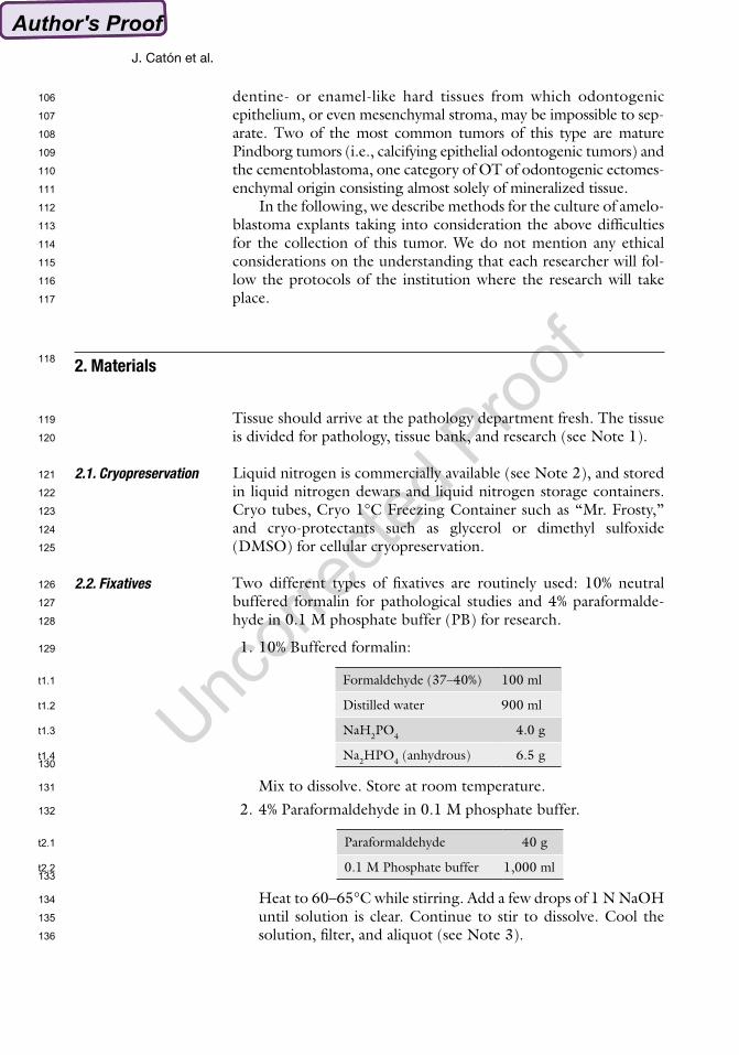
Two different types of fixatives are routinely used: 10% neutral buffered formalin for pathological studies and 4% paraformalde-hyde in 0.1 M phosphate buffer (PB) for research.
1. 10% Buffered formalin:
Formaldehyde (37–40%) 100 ml
Distilled water 900 ml
NaH2PO4 4.0 g
Na2HPO4 (anhydrous) 6.5 g
Mix to dissolve. Store at room temperature. 2. 4% Paraformaldehyde in 0.1 M phosphate buffer.
Paraformaldehyde 40 g
0.1 M Phosphate buffer 1,000 ml
Heat to 60–65°C while stirring. Add a few drops of 1 N NaOH until solution is clear. Continue to stir to dissolve. Cool the solution, filter, and aliquot (see Note 3).
2. Materials
2.1. Cryopreservation
2.2. Fixatives
106
107
108
109
110
111
112
113
114
115
116
117
118
119
120
121
122
123
124
125
126
127
128
129
130
131
132
133
134
135
136
t1.1
t1.2
t1.3
t1.4
t2.1
t2.2

15 In Vitro Studies on Ameloblastomas
1. 0.2 M Phosphate buffer, pH 7.4.
Na2HPO4 21.8 g
NaH2PO4 6.4 g
Distilled water up to 1,000 ml. 2. 1 M Tris–Cl (pH 7.4).
Tris base 121.1 g
HCl, concentrated 70 ml
Distilled water up to 1,000 ml. 3. 10× Tris/EDTA (TE).
1 M Tris–Cl 100 ml
0.5 M EDTA, pH 8.0 20 ml
Distilled water up to 1,000 ml (see Note 4). 4. HANKs (commercially available for cell culture). 5. PBS (commercially available for cell culture).
1. SuperFrost Plus glass slides or similar slides to increase adherence. 2. Microtome/cryostat and material related to their use. 3. Wax for embedding (purified paraffin/synthetic resin blend). 4. Xylene/Histoclear (see Note 5). 5. Alcohol gradient, 50, 70, 80, 90, and 100% (see Note 6). 6. Tissue-Tek CRYO-OCT Compound, Sucrose, cryo-embed-
ding molds, and −80°C freezer. 7. Routine H&E staining material.
1. Progenitor cell targeted to oral epithelium defined liquid cul-ture medium (CnT24).
2. DMEM supplemented with 1× penicillin/streptomycin antibi-otics for washing and tissue transport.
3. DMEM supplemented with 10% fetal calf serum (FCS) and 1× penicillin/streptomycin antibiotics for organotypic cultures.
4. Differentiation medium: BGJb medium supplemented with 10 mg/ml ascorbic acid, 2 mM of sodium ß-glycerophosphate, and 1× penicillin/streptomycin antibiotics.
5. 2.4 U/ml of Dispase. 6. 0.25% Trypsin. 7. Cell and organ culture dishes, metal grids, and support filters.
1. Glass dish. 2. Forceps, scalpels, blades, and needles.
2.3. Buffers
2.4. Tissue Sectioning and Staining
2.5. Culture Media and Materials
2.6. Tools for Dissection
137
138
139
140
141
142
143
144
145
146
147
148
149
150
151
152
153
154
155
156
157
158
159
160
161
162
163
164
165
166
167
168
169
t3.1
t3.2
t4.1
t4.2
t5.1
t5.2

J. Catón et al.
Once surgery of the tumor has been scheduled, it is important to follow up with the surgical team. Most hospitals will have a service to bring the tissue to the pathology department (see Note 7), but it is recommended to collect it personally―if possible―to mini-mize the time from surgery to the laboratory.
A pathologist familiar with ameloblastomas should select an area of the tumor that will be likely to be richer in tumor cells. As men-tioned in the introduction, some of these tumors could have very little starting material to work with. The tissue is then divided for pathology, tissue bank (see Note 8), and research. This methods chapter focuses only on the research portion.
The tissue selected for research should be divided:
1. Flash freeze This is achieved by submerging the sample in liquid nitrogen
or a mixture of dry ice and ethanol. This frozen tissue can be later used to extract nucleic acid for gene expression and genetic studies (qPCR, microarray, etc.).
2. Fixation Although some tissue will be fixed using 10% buffered forma-
lin (see Note 9), most tissue for research should be fixed imme-diately by submerging it in 4% PFA at 4°C overnight. The fixed tissue can be used for genetic studies (generally, we perform in situ hybridization, immunohistochemistry).
For in situ hybridization, cryosections are normally used. 3. Cryoprotection After fixing, the tissue should be rinsed at room temperature in
0.1 M phosphate buffer with 5% sucrose (this process will initi-ate the cryoprotection of the tissue). Continue with increasing concentrations of sucrose starting at 5% sucrose in phosphate buffer reaching 20% in 5% increments. Proceed for 30 min in each sucrose mixture at room temperature leaving it overnight at 4°C in fresh 20% sucrose/phosphate.
4. Infiltration The tissue is then placed into an infiltration mixture (2:1, 20%
sucrose phosphate buffer and O.C.T. embedding medium) for 30 min at room temperature before freezing (see Note 10).
5. Embedding and freezing Transfer the tissue to an embedding mold and fill the mold with
fresh infiltration mixture. Rapidly submerge the mold into Isopentane cooled with liquid nitrogen (see Note 11). After the material is frozen, wrap the block and store at −80°C.
3. Methods
3.1. Tissue Extraction/Preservation
3.2. Tissue Selection
3.3. Tissue for Research
3.3.1. Fresh Tissue for In Vivo Studies
170
171
172
173
174
175
176
177
178
179
180
181
182
183
184
185
186
187
188
189
190
191
192
193
194
195
196
197
198
199
200
201
202
203
204
205
206
207
208
209
210
211

15 In Vitro Studies on Ameloblastomas
6. Sectioning 3–5-mm sections are cut at −20°C in a cryostat. To achieve
ideal sections, it is critical to have the knife-edge as sharp as possible. Trim the block face to a diamond shape, with the long axis oriented vertically. This orientation helps to make removal of the sections from the knife-edge easier, and will minimize handling damage of the tissue. Use a small camel hairbrush to guide the section off the block face and transfer it to glass slides (see Note 12). Allow the section to dry on the slide at room temperature. Store slides at −80°C until needed.
Fresh tissue can be cultured in an organotypic form to maintain the architectural three dimensions of the tissue or in monolayer cell culture to isolate specific cells.
1. Organotypic cultures Tissue is cut into 2–5-mm cubes and placed into Trowell-type
culture dishes (4). These cultures can be maintained for approximately 15 days using DMEM medium supplemented with 10% FCS. The tissue can be induced using proteins and/or cell cocultures. The usage of beads for induction has the advantage of showing the effect in the precise site of applica-tion. Following are examples of induction beads and cell cocultures.
2. Induction beads Affi-gel agarose (75–150-mm diameter) or heparin acrylic
beads (100–200 mesh/100–250-mm diameter), depending on the type of proteins, are needed for induction. Ameloblastomas are induced with proteins involved in tooth development. Recombinants are diluted with 0.1% bovine serum albumin (BSA) in PBS, pH 7.4, to concentrations of 100–200 ng/ml. Recombinant BMP2, BMP4 (Fig. 2), and SHH (200 ng/ml) are used to preload affi-gel agarose beads and FGF2, FGF3, and FGF4 (100 ng/ml) are used to preload heparin acrylic beads. These preloaded beads are incubated for 30 min at room temperature and then washed for 5–15 min in culture media before being transferred with a mouth-controlled capillary pipette on top of the explants. As controls, beads loaded with 0.1% BSA in PBS are used. The explants with beads are cultured in serum-free medium for 20 h and processed for in situ hybrid-ization (5, 6), proliferation assays (7), and immunohistochemis-try (6, 8) as described in the references (see Note 13).
3. Cell cocultures The usage of cell cocultures for induction of ameloblastoma
tissues allows exposing the tumors to a collection of factor the cells express in vitro. We use murine embryonic odontogenic cells (pre-ameloblasts or pre-odontoblasts) to determine the effect of each specific cell type on the explant.
3.3.2. Fresh Tissue for In Vitro Studies
212
213
214
215
216
217
218
219
220
221
222
223
224
225
226
227
228
229
230
231
232
233
234
235
236
237
238
239
240
241
242
243
244
245
246
247
248
249
250
251
252
253
254
255
256
257

J. Catón et al.
Dissected first mandibular molars from E16.5 mouse (see Note 14) are placed in 2.4 U/ml of Dispase and incubated for 1 h at 4°C. The mesenchymal tissue is mechanically separated from the epithelium using tungsten needles. The two tissues are placed separately in 0.25% trypsin at 37°C for 30 min. Disaggregating is achieved by passing the cells after trypsin treatment through an 18-g needle. The cell suspension is cen-trifuged and the pellet washed in DMEN with 1× pen/strep. The cells are centrifuged once more and the pellet is placed using a pipette tip in the ameloblastoma explants (Fig. 3). These explants are cultured in differentiation medium for up to 10 days. The cocultured explants are fixed and treated for study as described before.
4. Monolayer cultures Cells are isolated from the fresh tissue using two methods:
Explants shedding allows cells to cast off from small tissue explants into a culture dish. The explants are cut in a similar way as the organotypic cultures. These explants are then placed in 10-cm cell culture dishes with CnT24 media just covering the bottom of the dishes. This allows the explants to have an optimum gas exchange. The explants are kept in this culture conditions for approximately 1 week or when epithelial-like cell colonies are observed attaching to the culture dish. The tissue can also be digested in collagenase IV (freshly prepared
[AU3]
[AU4]
Fig. 2. Preloaded affi-gel agarose beads (BMP4 beads) implanted on top of an ameloblas-toma (AB) organotypic culture.
258
259
260
261
262
263
264
265
266
267
268
269
270
271
272
273
274
275
276
277
278
279
280
281

15 In Vitro Studies on Ameloblastomas
500 CDU/ml) at 37°C for 30 min. The cells’ disaggregation is achieved by passing them through an 18-g needle. The cell suspension is centrifuged and the pellet washed in DMEN with 1× pen/strep. Centrifuge the cells once more and resuspend the pellet in CnT24 medium for plating in culture dishes (see Note 15).
Ameloblastoma cells can then be characterized for markers common with other epithelial stem cells and other tumor stem cells. These markers can be detected by immunohistochemis-try, in situ hybridization, or RT-PCR. For membrane-bound markers, the cells can be shorted using fluorescence activated cell sorting (FACS) (9). This allows separating living cells expressing the marker of interest from the rest.
1. Chemicals are purchased commercially from your choice of provider and solutions should be prepared nuclease free for studies with the need for RNA preservation.
4. Notes
Fig. 3. GFP expressing odontogenic cells (GFP) placed on top of ameloblastoma (AB) explants in organotypic cocultures.[AU5]
282
283
284
285
286
287
288
289
290
291
292
293
294
295
296
297
298

J. Catón et al.
2. Liq N2 should be handled with extreme care and protective clothing, gloves, and eye shield should be worn.
3. 4% PFA aliquots can be stored long term at −20°C. Avoid repeated temperature change cycles and bring to near room temperature before use. It is recommended to use phosphate buffer made with nuclease-free water.
4. We also purchase molecular biology-grade 100× TE for nucleic acid work.
5. Histoclear is less toxic and the results are similar. 6. Dehydration of tissue for nucleic acid work―in situ hybridiza-
tion―should be done with gradient alcohols made with nucle-ase-free water.
7. We normally received the tissue from the pathology depart-ment. They select a portion of the tumor and hand it to the research team on DMEM with Pen/Strep.
8. Most departments will have a tissue bank for storage of the tis-sue. This could be useful for in vivo studies.
9. The pathology department that provides the tissue will nor-mally process it in this manner for routine histological analysis. These preparations could be used for morphological study of the tissue.
10. The tissue should sink to the bottom of the container to indi-cate a correct infiltration.
11. Rapid freezing is recommended, although we have observed that simply placing the mold in dry ice will freeze the sample quickly enough without any adverse effect. It is also possible to use dry ice and ethanol mixture to accelerate the freezing process.
12. If the cryostat has auto-sectioning mode, then one should slow the speed of sectioning (approx. 5 mm/s) to ease the manipu-lation of the sections as they are coming off the block face. Do not use the anti-roll plate furnished with the cryostat; it com-presses the sections and results in poor tissue morphology.
13. In situ hybridization can be done on whole mount or cryosections.
14. We use mice expressing green fluorescent protein (GFP) in order to being able to distinguish the mouse cells from the tumor cells.
15. Epithelial cells are more likely to grow when plated in higher concentrations.
[AU6]
[AU7]
299
300
301
302
303
304
305
306
307
308
309
310
311
312
313
314
315
316
317
318
319
320
321
322
323
324
325
326
327
328
329
330
331
332
333
334
335
336
337
338

15 In Vitro Studies on Ameloblastomas
References
1. Barnes, L. et al. (2005) Pathology and Genetics of Head and Neck Tumours, IARC Press, Lyon.
2. Li, T. J. (2011) The odontogenic keratocyst: a cyst, or a cystic neoplasm? J Dent Res 90, 133–142.
3. Reichart, P. A., and Philipsen, H. P. (2003) [Revision of the 1992 edition of the WHO his-tological typing of odontogenic tumors. A sug-gestion], Mund Kiefer Gesichtschir 7, 88–93.
4. Trowell, O. A. (1954) A modified technique for organ culture in vitro, Exp Cell Res 6, 246–248.
5. Mitsiadis, T. A., Hirsinger, E., Lendahl, U., and Goridis, C. (1998) Delta-notch signaling in odontogenesis: correlation with cytodifferenti-ation and evidence for feedback regulation, Dev Biol 204, 420–431.
6. Mitsiadis, T. A., Salmivirta, M., Muramatsu, T., Muramatsu, H., Rauvala, H., Lehtonen, E., Jalkanen, M., and Thesleff, I. (1995) Expression of the heparin-binding cytokines, mid kine (MK) and HB-GAM (pleiotrophin) is
associated with epithelial-mesenchymal inter-actions during fetal development and organo-genesis, Development 121, 37–51.
7. Mitsiadis, T. A., Muramatsu, T., Muramatsu, H., and Thesleff, I. (1995) Midkine (MK), a heparin-binding growth/differentiation factor, is regulated by retinoic acid and epithelial- mesenchymal interactions in the developing mouse tooth, and affects cell proliferation and morphogenesis, J Cell Biol 129, 267–281.
8. Mitsiadis, T. A., Dicou, E., Joffre, A., and Magloire, H. (1992) Immunohistochemical localization of nerve growth factor (NGF) and NGF receptor (NGF-R) in the developing first molar tooth of the rat, Differentiation 49, 47–61.
9. Mekada, E., Yamaizumi, M., and Okada, Y. (1978) An attempt to separate mononuclear cells fused with human red blood cell-ghosts from a cell mixture treated with HVJ (Sendai virus) using a fluorescence activated cell sorter (FACS II), J Histochem Cytochem 26, 62–67.
339
340
341
342
343
344
345
346
347
348
349
350
351
352
353
354
355
356
357
358
359
360
361
362
363
364
365
366
367
368
369
370
371
372
373
374
375
376
377
378
379
380
381
382
383

Author QueriesChapter No.: 15 0001507499
Queries Details Required Author’s Response
AU1 Please check whether the edits made to the sentence “The classification of OTs most used…” are ok.
AU2 Please check whether it should be “pried” instead of “prized” in the sentence “In those OTs that expand the jaws…”.
AU3 Please check whether it should be “DMEM” instead of “DMEN” in the sentence “The cell suspension is centrifuged…”; comment applicable for all similar usages.
AU4 Please check whether the edits made to the sentence “These explants are then placed…” are ok.
AU5 Please check whether “(GFP)” should be deleted from the legend of the Fig. 3.
AU6 Please check whether the edits made to the sentence “We normally received the tissue…” are ok.
AU7 Whom does the pronoun “They” implies in the sentence “They select a portion of the tumor…”? Please check and make necessary changes to enhance understand-ability.