will we by the routine tests of full blood count including platelet … · · 2016-04-24the study...
TRANSCRIPT

1
Will we by the routine tests of full blood count including platelet count and determination of lactate dehydrogenase be able to
exclude severe falciparum malaria?
- a clinical study in a rural, malaria endemic area – Haydom, Tanzania
Principal investigators: Linn Nybråten Tjønsø and Kjersti Standal, medicalstudents, University in Oslo
Project responsible: Professor Johan N. Bruun MD PhD, Ullevål University Hospital, Oslo, Norway.
Assistant medical director and assistant medical offiser Isaack Malleyeck, Haydom Lutheran Hospital, Tanzania
The study is a collaboration study between Haydom Lutheran Hospital,
Tanzania and Ullevål University Hospital, Norway.

2
TABLE OF CONTENTS Will we by the routine tests of full blood count including platelet count and determination of lactate dehydrogenase be able to exclude severe falciparum malaria? .....................................1 ABSTRACT...........................................................................................................................3 ABBREVIATIONS................................................................................................................4 INTRODUCTION..................................................................................................................4 METHODS............................................................................................................................9
Recruitment......................................................................................................................10 Procedures........................................................................................................................11 Ethical approval ...............................................................................................................12 Data entry and analysis.....................................................................................................12
RESULTS............................................................................................................................13 Thrombocytopenia............................................................................................................15 Lactate Dehydrogenase.....................................................................................................16 Glucose ............................................................................................................................16 Anemia.............................................................................................................................17 White Blood Cells ............................................................................................................18 Symptoms ........................................................................................................................18 Treatment .........................................................................................................................19 Test results and discrepancies ...........................................................................................19
Blood smear: ................................................................................................................19 Rapid tests: ...................................................................................................................19
DISCUSSION......................................................................................................................20 Conclusion .......................................................................................................................24
APPENDIX: TABLES.........................................................................................................25
3
ABSTRACT
Background: Malaria disease causes enormous morbidity and mortality worldwide. More
than 2 million people die annually; the majority of these children under the age of five living
in Sub-Saharan Africa. Despite this high prevalence of disease, there is also a high degree of
overdiagnosis and overtreatment.
Objective: A clinical study in Tanzania, where the aim was to enhance the reliability of
malaria diagnostics by assessment of the sensitivity and specificity of thrombocytopenia and
raised lactate dehydrogenase (LDH) in malaria infected patients.
Methods: 179 patients with malaria symptoms were included. Blood slide, full blood count
(FBC), LDH, glucose and rapid tests (NOW and ASSURE) were performed.
Results: Thrombocytopenia was significantly more frequent in the malaria positive group
compared to the malaria negative. No such difference was found for LDH, most likely due to
technical circumstances at the study site. In addition, there were a higher amount of anemic
patients in the malaria positive group. There were few false negative blood slides. Out of the
malaria positive 96% had infection with P.falciparum, and 4% had P.vivax, P.malaria or
P.ovale.
Discussion: FBC is not routinely performed in this particular hospital, but from our findings
we believe that thrombocytes and hemoglobin (Hb) would be valuable tools in the diagnostic
workup for suspected malaria. A great number of patients with negative blood slides were
treated as if they had malaria, and other causes for their symptoms were often not pursued. As
false negatives were seldom, a negative blood slide should initiate investigation for other
diseases.

4
ABBREVIATIONS
P.falciparum = Plasmodium falciparum P.vivax = Plasmodium vivax P.ovale = Plasmodium ovale P.malariae = Plasmodium malariae LDH = lactate dehydrogenase RBC = red blood cells WBC = white blood cells Hb = hemoglobin FBC = full blood count OPD = out patient departement RCHC = reproductive and child health clinic HLH = Haydom Lutheran Hospital CO = clinical officer RES = reticuloendothelial system DIC = disseminated intravascular coagulation PPV = positive predictive value NPV = negative predictive value RCD = red cells deformability ROS = reactive oxygen species WHO = world health organization ARDS = acute respiratory distress syndrome
INTRODUCTION
The risk of being infected with malaria is present in 105 countries(1). There are four main
plasmodia that affect humans: P. falciparum, P. vivax, P. malariae and P. ovale, and their
distribution throughout the countries varies. P. falciparum is the malignant specie as it causes
more than 2 million deaths per year, and an even higher amount of morbidity (2). The
Anopheles mosquito transmits plasmodia to humans. Cyclic changes occur both in the
mosquito and in the human. After a bite of an anopheline mosquito the sporozoites enter
human bloodstream and are transported to the liver (3). Here they invade livercells and after a
while become tissue schizonts or hypnozoites (P. vivax and P. ovale). Each schizont produces
a large amount of merozoites that are subsequently released from the liver as the hepatocytes
lyse. Every single one of these merozoites is again capable of invading erythrocytes, where
they replicate and release new merozoites in a cyclic manner. Some of the intraerythrocytic
parasites develop into the sexual forms (gametocyte) necessary to complete the life cycle in

5
the anopheline vector. Cyclic fevers are the hallmark of malaria (3). Other clinical signs and
symptoms are tachycardia, hypotension, headache, backache, abdominal pain, vomiting,
diarrhea, joint pain and altered consciousness. Though many people suffer from malaria, it is
also highly overdiagnosed. Almost half of all hospital admissions in much of Africa are
attributed to the disease, though not all of these have laboratory evidence of disease at further
investigation (4). But this overdiagnosis of malaria and corresponding treatment often results
in lack of treatment of the real cause of disease. One study has shown that ⅔ of patients with
severe febrile illness and no malaria parasite load did not receive treatment with antibiotics
(3). Malaria treatment can also cause severe side effects, most common are gastrointestinal,
neurological, neuropsychiatrical and dermatological (5). We wanted to evaluate the possibility
of decreasing the amount of overdiagnosis and overtreatment by improving diagnostic
accuracy of malaria. This we would do by using some simple blood parameters; most
importantly platelets and LDH, but also Hb, glucose and white blood cells (WBC).
FBC can generally give an indication of severity of disease, and thrombocytopenia has been
reported to be associated with malaria (6) (7). There are many explanations for this, and it is
likely that different mechanisms play a role. Thrombocytopenia can be a result of either
reduced production or increased turn-over; the first is less likely because an increased number
of megakaryocytes in patients with acute malaria has been reported (8). There are different
hypotheses for the increased turn-over;
• Immunomediated destruction: Some studies suggest an immunological alteration of
the platelets (8), or an attachment of malaria antigens to the platelet surface, which in
turn activates immune mechanisms that favour destruction. Mohanty et al. found high
levels of anti-platelet antibodies in the serum of thrombocytopenic malaria patients
(9).
• Platelet activation: Some studies (9;10) have shown that the platelets undergo a
structural change during malaria infection, such as centralization of dense granules,
glycogen depletion and formation of pseudopods and microaggregates. These
structural changes may in turn lead to thrombocytopenia. There is also evidence of a
hypersensitivity of platelets in malariainfected patients (6;9) with platelet aggregation
as a response to different substances such as ADP (adenosine diphosphate), adrenalin,
collagen and ristocetin (an antibiotic).

6
• Removal by the reticuloendothelial system (RES): malarial infection activates the
RES and this leads to generalised RES hyperplasia and fast removal of damaged
platelets, and also reduces the lifespan of normal platelets (11).
• Consumption in different steps of coagulation: DIC was initially believed to be a
central mechanism in the development of thrombocytopenia, and experimental studies
done in the 1960s pointed in that direction when they found depletion of coagulation
factors (mostly in malariainfected monkeys). Later studies have shown that this is
quite rare (6;8;9), but appears more often in severe malaria.
• Intraplatelet parasites have been demonstrated in human and animal infections (12),
but in other studies this is not seen (9), and the contribution of this to the
thrombocytopenia is to our knowledge, not yet known.
Thrombocytopenia is strongly associated with severe malaria, and the extent of
thrombocytopenia correlates with the degree of parsitaemia (6;9). A study by Gérardin et al
(2002) (13) looked at the prognostic value of thrombocytopenia, and found that when
thrombocytopenia was present, it was predictive of fatal outcome regardless of clinical
presentation.
It is likely that LDH can be used as a measure of the severity of malaria infection. LDH is an
intracellular enzyme found in many tissues in the human body. It catalyses the oxidation of
lactate to pyruvate. The concentration is especially high in heart, liver, erythrocytes, skeletal
muscles and kidneys (14). What conditions can cause an increase? Diseases in the previous
mentioned organs, some types of cancer (small-celled lung cancer, nephroblastoma, metabolic
endocrine tumor), measles and cervical lymphadenitis among others. LDH is often increased
in malaria infected patients. It has been estimated that in approximately 80 % of the patients it
is higher than normal (1). The level of LDH seems to be normal until the parasite load
exceeds 100 000 trophozoites/µL, and once it exceeds this threshold, lactate level rises
exponentially (15). It is believed that the main reason for the increase in malaria is the parasite
invasion of hepatocytes and erythrocytes and the following hemolysis with release of
substances, among them LDH (14). A study by van Genderen et al (15) measured the LDH in
returning travellers with malaria. They found that it was significantly higher in those who
suffered from severe malaria than in those with uncomplicated malaria (sensitivity 0.67,
specificity 0.94), and that the association between the two where stronger the higher the level
of LDH. The pathogenesis of increased LDH in severe malaria is most likely multifactorial

7
(15), in addition to hemolysis; the parasite itself produces lactate, but this is believed to
constitute a small portion of the total LDH. The sequestration of parasites leads to increased
production of lactate locally. Also, sequestrated parasites release toxins that increase release
of proinflammatory cytokines – endothelial transport and cellular metabolism may then be
compromised (cytokine inhibition of oxidative metabolism). There is also evidence of
decreased liver and kidney removal of lactate in patients suffering from severe malaria.
Hypoglycemia is a common feature in malaria. If a non-diabetic in an endemic area presents
with hypoglycemia, malaria should be tested for (1). Decreased glucose level can alone be the
reason for altered level of consciousness. It can also be a complicating factor to cerebral
malaria. One study has shown that ⅓ of children with cerebral malaria had hypoglycemia on
admission (16). Causes include depletion of glycogen stores, inadequate intake and quinine-
induced hyperinsulinaemia. However, it looks like this is independent of the malaria process.
This is the conclusion Van Thien et al (17) draw in a study where they looked at glucose
metabolism in patients with cerebral malaria. They found that in cerebral malaria glucose
production is stimulated rather than repressed, and that the gluconeogenesis is increased
compared to what is the case in uncomplicated malaria.
Infection with malaria is often associated with anemia, the pathogenesis of which is not
completely understood, but is thought to be multifactorial in origin. It might be due to
hemolysis, decreased red blood cell (RBC) production, unmasking of borderline folic acid
deficiency, and genetic factors may also play a contributory role (18). The most profound
anemia is seen with severe acute malaria, but also chronic malaria infection can give rise to a
severe form of anemia. It is probable that in acute malaria the severity of anemia depends on
the degree of RBC destruction, whereas in chronic infection where the parasitemia is low, it is
thought that ineffective erythropoiesis and dyserythropoiesis might be of greater importance
(19). A study by Philips et al 1986 showed that the severity of anemia was proportional to the
degree of parasitemia, and it was also more severe in pregnant women and in patients who
had a concomitant bacterial infection (20). Several mechanisms may contribute to the
hemolysis seen with malaria infection.
• There is a nonspecific splenic and reticuloendothelial hyperactivity resulting in
increased phagocytosis of both infected and uninfected RBCs by activated
macrophages. It has been estimated that approximately 10 uninfected cells are cleared
from the circulation for every infected cell (2).

8
• There is a reduction in mean red cell deformability (RCD). This leads to increased
clearance of RBCs in the spleen (20). It is shown that the reduction in mean RCD
corresponds with the severity of the disease and with the severity of anemia (21).
• It is thought that the trophozoite induces changes in the red cell membrane
phospholipids distribution (22). This new distribution allows a flux of cations
(pathway having a higher permeability to K+ then Na+), which results in loss of cell
water and thereby a reduction of the cell volume (23). The new distribution will also
serve as a signal for recognition and removal by the RES (24).
• Exposure to reactive oxygen species (ROS) results in molecular changes in the outer
membrane of both infected and uninfected RBC which leads to both
IgG(immunoglobulin G)-dependent and -independent phagocytosis (20;25), The
malaria antigens attached to the RBC (of both infected and uninfected cells) might
also be the target of the hosts immunoglobulins (26).
• There is also an alteration in the expression of the complement regulatory proteins
CR1, CD55 and CD59 on the RBC surface (25). These proteins are important in
protection of the host cells from attack by complement, thereby reducing the risk of
malarial anemia. It has been shown that CR1 also has a different role, by acting as a
ligand in rosette formation (uninfected RBC bind to infected RBC, increasing the
vascular resistance and blocking the microvasculature (27)) seen in cerebral malaria
(25). A low level of CR1 would therefore be protective against severe malaria.
• Hemolysis might also be induced by drugs such as quinine, rimaquine, pamaquine and
chloroquine, and primaquine, in patients with glucose-6-phosphate deficiency (18).
Some of the anemia seen in malaria can be attributed to incomplete compensation of
hemolysis due to bone marrow dysfunction.
• Patients with malaria have reduced erythrocyte production (19). This is thought to be
due to decreased responsiveness of erythroid progenitor cells to erythropoietin or
relative impaired erythropoietin production, both mediated by inflammatory cytokines
(19).
• It is shown in cell culture that chloroquine has an inhibitory effect on erythropoietin
synthesis, and may therefore aggravate the defect in erythropoietin production (28).
WBC counts during infection with malaria are generally characterized as being low to normal.
This is thought to be due to flow of leukocytes away from the peripheral circulation and to the

9
spleen and other marginal pools, rather that actual depletion or stasis (29). There have been
some reports of leukocytosis, which may be associated with concurrent infections and/or poor
prognosis. A study by Ladhani et al from 2002 showed that children admitted with malaria
had a higher mean WBC than community controls, but not as high as the children admitted to
hospital for other medical conditions (6). This study also showed that in children with
malaria, leukocytosis was associated with both severity (prostration, coma, deep breathing,
hyperparasitaemia and severe anaemia) and mortality.
Blood smears are considered gold standard in diagnosing malaria, although it is noted in the
literature that its quality depends on how experienced the microscopist is (30). We used blood
smears and 2 different rapid tests as diagnostic tools. By using the two different rapid tests we
wanted to enhance the diagnostic accuracy and also be able to distinguish the relative amount
of malaria infections caused by P.falciparum in the study area. The rapid tests are developed
to detect different antigens that circulate in the blood of an infected person. The antigens
tested for are primarily histidine-rich protein 2 (HRP2), parasite-specific lactate
dehydrogenase (pLDH) and Plasmodium aldolase. ASSURE® tests specifically for
P.falciparum HRP2 (PfHRP2). NOW® tests for both PfHRP2 and plasmodium aldolase, the
latter present when infected with any of the four species. Clinical studies in endemic areas
have shown that the sensitivity and specificity of a rapid test for PfHRP2 are 92.7% (91-94.5)
and 99.2% (98.2-99.9) respectively(30). Two clinical studies using NOW® rapid test, have
shown a sensitivity for detecting pure P.falciparum infection of 96.4% and 100%, and a
specificity of 97% and 100%. The sensitivity for detecting non-falciparum infection was
found to be 66.7% and 90.7%, while the specificity was 100% and 93.1%(31;32).
METHODS
The study was conducted at Haydom Lutheran Hospital (HLH) in Mbulu which is situated at
approximately 1500-2000 m above sea level, in the Northern highland of Tanzania. It took
place in the period May to July 2007. Malaria is endemic in Tanzania, with some variations
between low-land and high-land. Transmission occurs throughout the year, with a seasonal
increase in intensity corresponding with the two rainy seasons in the country, the first from
March to May and the second from October to December. HLH was built by Norwegian

10
missionaries in 1953 on the request of the then existing British government in Tanzania. In
1963 the administration of the hospital was handed over to the local church, the Evangelical
Lutheran Church of Tanzania, (ELCT), Mbulu Synod. The hospital has been part of the
Tanzanian central health plan since 1967. It has been estimated that HLH serves a population
of about 450 000 residents (33). HLH has 450 beds distributed between seven adult medical
wards and one pediatric ward. HLH also has an Out Patient Department (OPD) and a
Reproductive and Child Health Clinic (RCHC) where adults and children respectively are
examined by clinical officers, blood samples are taken and they receive treatment, before
going home. Around 300 patients are treated here every day, 80 patients per clinical officer.
Some of which are admitted. People who are severely ill are taken directly to the reception at
the hospital, from which they are sent to different wards for treatment.
Recruitment
We collaborated with the Clinical Officers (CO) at the OPD, RCHC and in the reception in
the period May to July 2007. The COs assessed the patient and included them in the study, if
one or more of the following inclusion criteria were present:
A) Fever of unknown cause, > 38.5°C, or
B) Altered level of consciousness (confusion, coma etc.) with no known
cause, or
C) Patients considered by the CO to have malaria on admission, due to other
clinical features suspicious of malaria.
Exclusion criteria:
Patients who had received adequate treatment for their acute disease before being treated at
HLH were excluded from the study.
All patients (whatever age and gender) who fulfilled the before mentioned inclusion criteria
were recruited into the study after they themselves or their relatives had given their informed
oral consent. For unconscious patients, permission was given by their relatives or it was
sought when the patient regained consciousness. If the patient were included, the CO filled
out a form, where he or she registered the age, gender, ten cell leader, signs, symptoms and
temperature of the patient. For the patients who were admitted, the CO was also to register if
there were any signs of severe malaria (behavioural changes, prostration/extreme weakness,
coma, vomiting everything, inability to drink or breastfeed, circulatory collapse/shock,
11
pulmonary oedema, bleeding tendency/disseminated intravascular coagulation (DIC),
jaundice or acute renal failure). All included patients were asked to come back to the hospital
after 5 days for a follow up consultation. We were also to do a follow-up after 4-5 days on the
patients admitted to the hospital.
Our study took place in the dry season, when the prevalence of malaria is low. We chose to
include patients based on an already positive blood smear because we, as the study went
along, discovered that few of the patients included on basis of suspicion (judged by the CO)
tested positive on either smears or rapid tests. To be able to draw any conclusions regarding
our main questions (platelets and LDH), we needed a certain amount of malaria positive
patients.
Procedures
From the included patients forearm venous blood (10 ml) were drawn into ETDA
(ethylenediaminetetraacetic acid) glass for full blood count and blood sugar, and into empty
sterile test tubes for the measurement of serum LDH. Thick and thin blood smears are
considered gold standard in diagnosing malaria. Only thick blood smears are routinely
examined on HLH today. A thick blood smear was made from capillary blood sample in the
wards or in the OPD. The smears were made on regular glass slide and the name or number of
the patients was put on. They were then brought to the lab, where they were stained with
Giemsa solution and examined by laboratorian technicians. They counted the number of
P.falciparum parasites per 200 leucocytes. A slide was to be considered negative if no
parasites were found after 100 high power fields were scanned.
In addition we used two different rapid tests, NOW and ASSURE, to increase the
reliability of the result, and to differentiate between P.falciparum and non-falciparum malaria.
The procedure was conducted by the study principal investigators, and the result was also read
by them. We brought rapid tests from Norway, we only brought 138 of the NOW rapid test,
so many of the patients were only tested for P.falciparum. If there were discrepancy between
the different tests (blood smear/rapid tests or ASSURE/NOW), a new thick blood smear was
made and brought back to Norway where laboratory technicians at Ullevål University
Hospital, Oslo, looked at them.

12
Hb level, WBC and platelets were measured with a Sysmex kx-21 machine (sysmex
corpartion Kobe, Japan). Venous blood glucose was measured with Ascensia Entrust Blood
glucose meter (Bayer).LDH was measured from blood serum with a Visual (Bio Merrieux),
this was done by the study investigators.
We also recorded if any other diagnostic tests or procedures were done, and also what
treatment the patients received.
Ethical approval
The study was approved by COSTECH (Tanzania Commission for Science and Technology),
NIMR (National Institute for Medical Research) and REK Øst (Regional komité for
medisinsk og helsefaglig forskningsetikk). In addition permission was granted from HLH.
Oral consent was obtained from the patient or their relatives. Patients had the right to
withdraw from the study at any time without being excluded from further care and treatment.
They could also claim access to their registered data. All patients included in the study
received the best available treatment.
Data entry and analysis
Patient data were recorded in hospital files and study forms, and entered into Excel sheets in a
study computer. Data analyzed consisted of information from hospital record on demographic,
clinical and laboratory variables. All data was kept protected in the study computer, and
access was restricted to the study investigators. All clinical and laboratory data was entered in
SPSS (Statistical Package for the Social Sciences), and analysis was done. The group of
patients who were positive on both blood smear and rapid tests (group 1) was considered to
have true acute malaria. And we compared clinical and laboratory features between this group
and the rest (who were thought to have no acute malaria). This was also done for severe
versus uncomplicated malaria. Continuous variables with a normal distribution were
compared between groups by t-test. A p-value of <0.05 were considered significant.

13
RESULTS
In total there were 201 patients who fulfilled the inclusion criteria. Out of them, 38 had been
using antimalaria drugs, 9 of them were selected based upon positive blood smear, and are
therefore included in the study. 7 of the patients had used only one dose of antimalaria the
same day, and we do not believe this to have an impact on the disease or test results, so they
are therefore still included in the study. The remaining 22 were excluded from the study, and
we were therefore left with 179 included persons in our study. 8 had been taken unknown
drugs. For 80 patients the data about prehospital drug use is missing.
125 (69.8%) of the patients were from the OPD, out of which 16 got admitted to the hospital.
54 (30.2%) were included from the reception. 52 (29.1%) patients were included on the basis
of a positive bloodslide, 43 from the OPD and 9 from the reception. There were 81 (45.3%)
men and 98 (54.7%) women. Mean age at admission was 21.1 years, median 20 years and
range was 67.8 (0.2-68)
Out of the 109 who were treated as outpatients, only 9 (8%) returned for a follow-up
consultation. Of the 70 who were admitted, we were able to follow up 35 patients with blood
samples and a new examination. The rest (35 patients) were already discharged from the
hospital or dead (3 patients, none of which had malaria) when the follow-up was due. For 20
of them we were able to get some information about their symptoms and treatment from their
chart. The follow-up data is hence incomplete, and for 115 patients there is no data (64.2%).
We believe that this is due to the fact that normally at HLH a follow-up consultation is not
routine, and we were not able to implicate the importance of it to the COs and patients . One
other problem is that a lot of the patients who came to the HLH had travelled a long way to
get there, and although one of the exclusion criteria was that they lived far away, this did not
apply to the group that was selected based upon positive blood smear. As for the follow up of
the admitted patients, most were discharged after only 1-3 days, and therefore too early for the
next examination. Although some were asked to come back for a new consultation, this did
not occur.
We used the thick blood smear, rapid tests and clinical signs and symptoms to decide whether
or not acute malaria was the cause of admission. Several times there were discrepancy

14
between the different test, and we therefore divided the patients into different groups based on
the results and findings.
1. Malaria being the true cause of admission: (49 patients)
Positive blood smear and positive rapid test. No other cause for illness found.
Patients with malaria. (39 patients)
Patients with severe malaria. (10 patients)
2. Malaria is possibly the true cause of admission: (84 patients)
a) Negative blood smear and positive rapid test, where the patient has clinical
signs corresponding with malaria, and no other cause for illness found. (3
patients)
b) Negative blood smear and negative rapid test, where the patient has clinical
signs corresponding with malaria, and no other cause for illness found. (70
patients)
c) Positive blood smear and negative rapid test, where the patient has clinical
signs corresponding with malaria, and no other cause for illness found. (11
patients)
3. No acute malaria disease; chronic malaria: (2 patients)
Positive blood smear or positive rapid test, but further investigation reveals other
cause of acute disease.
4. No malaria: (15 patients)
Negative malaria tests
Other cause of acute disease revealed.
5. Malaria not likely to be the true cause of admission: (29 patients)
Negative malaria tests. Symptoms corresponding with other disease, but no other
disease diagnosed. Malaria treatment not given (or the treatment is unknown).
Totally there were 49 patients who had a positive blood smear and rapid test. 10 of them
fulfilled the World Health Organization (WHO) 2000 criteria for severe malaria (34).
(1) One of the following criteria; cerebral malaria (unrousable coma), severe normocytic
anemia (Hb<5 g/dL in the presence of parasitemia > 10,000 parasites per µL), renal failure,
pulmonary oedema, acute respiratory distress syndrome (ARDS), hypoglycemia (whole blood
glucose < 2.2mmol/L), circulatory collapse, shock, spontaneous bleeding, DIC, repeated
generalized seizures, acidemia or acidosis, hemoglobinuria.

15
(2) Asexual parasitemia with P.falciparum (although smear-negative cerebral malaria may
occur).
Additional criteria are: impaired consciousness, but rousable, prostration and extreme
weakness, hyperparasitemia, jaundice, hyperpyrexia, post-mortem evidence of severe malaria.
The 84 patients in group 2 got treated as if they had malaria or the treatment given is
unknown. There were 2 patients in group 3, they had positive blood smear, were negative on
ASSURE® and were positive for P. vivax, P.ovale or P.malariae on NOW®.
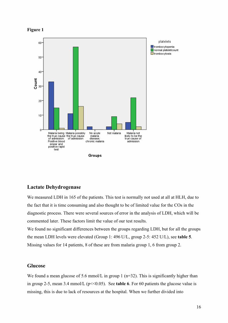
Thrombocytopenia
In the previously mentioned groups we found that the mean platelet count was low in group 1
(130 x 103/10-6L) compared to the other groups (274 x 103/10-6L), see table 1. The platelet
value is missing for one patient, this patients is in group 2. Further division of group 1 into
severe malaria and uncomplicated malaria gave a mean platelet value of 113 for the severe
and 136 for the uncomplicated, see table 2.
When looking at the sensitivity of thrombocytopenia (defined as < 150 *10^3 per 10 -6 L) we
have calculated it in two different ways:
• First, we separated group 1 from the others, regarding it as the true malaria group. We
then found the sensitivity of thrombocytopenia in group 1 to be 0.67, specificity 0.85.
The difference between these two groups proved to be significant (p<<0.05)
• When adding the first two groups as the groups believed by the clinical officers to
have malaria, the sensitivity was 0.33 and specificity 0.80.
The positive predictive value (PPV) in the first case is 0.62, while the negative predictive
value (NPV) is 0.87. For the second alternative PPV is 0.83 and NPV is 0.29.
Regarding platelet count and its ability to distinguish severe from uncomplicated malaria, the
sensitivity of thrombocytopenia in the severe group was 0.80, while the specificity was 0.36.
(PPV 0.24, NPV 0.88) The difference between these two groups was not significant (p=0.56)
Figure 1 and tables 3 and 4 shows the distribution of patients with thrombocytopenia, normal
platelet count and thrombocytosis in the different groups.
16
Figure 1
Lactate Dehydrogenase
We measured LDH in 165 of the patients. This test is normally not used at all at HLH, due to
the fact that it is time consuming and also thought to be of limited value for the COs in the
diagnostic process. There were several sources of error in the analysis of LDH, which will be
commented later. These factors limit the value of our test results.
We found no significant differences between the groups regarding LDH, but for all the groups
the mean LDH levels were elevated (Group 1: 496 U/L, group 2-5: 452 U/L), see table 5.
Missing values for 14 patients, 8 of these are from malaria group 1, 6 from group 2.
Glucose
We found a mean glucose of 5.6 mmol/L in group 1 (n=32). This is significantly higher than
in group 2-5, mean 3.4 mmol/L (p<<0.05). See table 6. For 60 patients the glucose value is
missing, this is due to lack of resources at the hospital. When we further divided into

17
uncomplicated (n=25) and severe malaria (n=8), we found that for the first group the mean
glucose was 5.3 mmol/L, and for the second it was 6.6 mmol/L. This was not a significant
difference (p=0.49). There is a tendency that the group with severe malaria has a higher mean
glucose than the uncomplicated, and it would be interesting to see if we by including more
patients in each group could have found a significant difference.
Anemia
In our study, we did a FBC of all the patients included in the study. We have then divided the
patients into different groups based on their age. We divided the persons in each age group
into two groups, anemic or not anemic, based on their level of hemoglobin, see table 7.
For the groups under the age of 12:
• In group 1, 21 out of 24 had anemia. And in the groups 2-5, 8 out of 37 had anemia.
This gives us a sensitivity and specificity for anemia of 0.88 and 0.78 respectively.
The PPV is 0.72 and the NPV is 0.91.
• As for the patients that the clinical officers regarded as positive for malaria (group 1
and 2), 28 out of 48 had anemia. In the other groups (3.4 and 5), 1 out of 13 was
anemic. The sensitivity and specificity is here 0.58 and 0.92.This gives us a PPV of
0.97 and a NPV of 0.38.
For all ages (table 8):
• In group 1, 40 out of 49 had anemia, and in the other groups (2-5), 35 out of 130 were
anemic. This gives us a sensitivity and specificity for anemia of 0.82 and 0.73, and a
PPV of 0.53 and a NPV of 0.91.
• As for the patients who the clinical officers regarded as malaria positive (group 1 and
2), 66 out of 133 had anemia. In the groups 3-5, 9 out of 46 were anemic. The
sensitivity and specificity is here 0.51 and 0.80 .This gives us a PPV of 0.88, and a
NPV of 0.36.
We divided the patients in group 1 into severe and uncomplicated malaria, and looked at all
the age groups as a whole. 9 out of 10 persons with severe malaria had anemia, and 31 out of
the 39 who had uncomplicated malaria were anemic, see table 9. This gives a sensitivity of
0.90. The specificity was 0.21. This gives us a PPV of 0.23 and a NPV of 0.89.

18
The severity categories are based upon WHO 2000 criteria for severe malaria. Although one
of the criteria is severe normocytic anemia (Hb <5 g/dL in the presence of parasitemia >
10,000 parasites per µL), only one of the patients in our study was included in this group
based on this criteria. We therefore do not think that this will have an effect on the result on
hemoglobin level.
White Blood Cells
We looked at the WBC count in all patients included in the study. When comparing the mean
WBC count in the group of malaria positive patients (group 1) with the malaria negative
patients (group 2-5), we found that malaria positive had a lower mean WBC than the group of
malaria negative patients (6.5x10³/10 -6L compared to 7.6x 10³/10 -6L, see table 10 and 11).
Although lower, it was not significant (p= 0.207). When comparing the two groups that the
clinical officers stated as positive (group 1 and 2) with the three other groups, we found a
mean WBC of 7.0x10³/10 -6L and 8.1x10³/10 -6L, neither this was significant (p=0.056).
Comparing the group of severe malaria with the group with uncomplicated malaria (patients
in group 1), we found a mean WBC of 8.5x10³/10 -6L and 6.0x10³/10 -6L, see table 12. This
was not significant (p=0.08). We do, on the other hand, see a tendency here, and it is possible
that we by increasing the number of patients in each group would be able to find a significant
result.
Symptoms
We wanted to register what signs and symptoms the patients included in our study presented
with. We made standard questionnaires for the COs to fill in, one for the OPD and one for the
inward, focusing on the serious complications of malaria. Examples of symptoms asked for
were headache, nausea, and general fatigue, examples of findings were splenomegaly and
temperature, and for the inward patients – level of consciousness, jaundice etc. Symptoms for
malaria are very little specific, and generally found in many diseases. The COs included
patients based upon suspicion of malaria and hence presence of these general symptoms.
Therefore the registrations made are not very good at comparing the symptoms in malaria
positive versus malaria negative patients. We have no control group never suspected to suffer
from malaria. In all groups the majority of patients reported to have fever, headache and
general malaise, while about half of the patients reported joint pain, nausea, vomiting and

19
poor appetite. The severe malaria patients generally reported to have more symptoms than the
others. The recordings of symptoms are relatively incomplete; information is missing for
many of the patients.
Treatment
Although treatment was one of the aspects we wanted to look at, it was not a question on the
form that the COs were to fill out. This led to some lack of recording. In total there are 40
patients for whom we have not registered treatment received. Of the 49 patients that were
positive for malaria (group 1), 38 (78%) received effective antimalaria treatment, 1 (2%) did
not get any effective treatment towards malaria, and for 10 persons (20%) the value is
missing. 11 patients received antibiotics in addition to the antimalaria treatment. The one who
did not receive effective antimalaria treatment, had uncomplicated malaria, and received only
antibiotics. There were 130 patients in our study who did not have malaria; out of them 93
persons (71.5%) got effective treatment towards malaria. 23 patients (18%) received
antibiotics in addition to the antimalaria treatment. Value is missing for 30 persons (23%).
3 out of the 130 patients received only antibiotics. Totally, 26 out of the 130 negative patients
(20%) received antibiotics. One person with severe malaria received blood transfusion. Four
persons who did not have malaria received diazepam, and one got treatment towards
tuberculosis.
Test results and discrepancies
Blood smear:
There were 61 positive blood smears, 114 negative, and 4 unknown. Some patients (n=52)
were included based on a positive blood smear, but most (n=126) were included based on
clinical signs and suspicion of malaria (in 4 of these we do not know the blood smear result).
For one of the patients it is not known if he were selected based on a positive blood smear or
not.
Rapid tests:
• Assure: There were 51 positive ASSURE tests, and 128 negative.

20
• Now: There were 49 positive NOW tests (40 for P.falciparum or mixed infection, 7
for only P. falciparum, 2 for P. vivax, ovale or malariae), and 77 negative. For 53
patients NOW is missing.
On the before mentioned discrepancy:
Several times there were discrepancies between the different tests. We then brought the slides
back to Norway, and laboratory technicians at Ullevål University Hospital looked at them. We
used their results as a gold standard, so when the different tests did not correspond, we
adjusted the result after what the technicians here in Norway came to. We then ended up with
51 patients with a malaria infection, 125 patients who did not have malaria and for three
patients we were not able to get a second opinion and the result for them are therefore still
unknown. The results of the different tests before and after second opinion evaluation are
shown in table 13. There were therefore a total of 10 false positive blood slides and 2 false
negative blood slides (one was positive for P.vivax, P. malaria or P.ovale, and the other were
positive for P.falciparum). For the rapid test there were 1 false positive (assure pos
P.falciparum and now positive for P.falciparum or mixed), there were no false negative. For
the three patients with discrepancy between the different tests, where we were not able to get
a second opinion here in Norway; two had positive blood slides and negative rapid tests, and
one had negative blood slide and positive assure (now is missing).
It is worth mentioning that out of the 51 patients that were positive for malaria, 2 patients
(4%) were positive for P.vivax, P.malariae or P.ovale. The rest, 49 (96%) had an infection
with P.falciparum (or mixed infection with P.falciparum and another plasmodium specie).
DISCUSSION
In our study the main aim was to see if we could make use of platelets count and LDH level to
enhance the diagnostic accuracy of malaria diagnosis. According to our protocol, we
separated the patients into different groups based upon the likelihood that malaria was the
cause of medical attention. There were four different groups. We added one more group
where the cause was unknown, where we found malaria to be very unlikely. Due to the high
prevalence of malaria in the area in Tanzania where the study was performed, COs often
assume that malaria is the cause of disease in many patients. But there are seasonal variations,
and therefore over-diagnosis and over-treatment occur, especially in the dry season (4). Many

21
patients with both negative blood smear and negative rapid test were therefore included in our
study based upon clinical signs corresponding with malaria. The majority of these patients
ended up in group 2, and many did most likely not have malaria. We therefore choose to
comment our results in the following discussion on the presumption that group 1 (with both
positive blood smear and rapid tests) contain the patients with the most accurate diagnosis.
While working with this study, we found several areas where errors may have occurred:
• Regarding inclusion: The COs included a large group of patients who were malaria
negative. Most likely these patients had symptoms corresponding with malaria, but
since the symptoms are not specific there were many included patients who did not
have malaria. Perhaps the inclusion criteria should have been narrower, in order to get
more malaria positive patients. Also contributing to the large amount of included
patients might be the COs limited time for each patient. They might not have had the
time to examine each patient well enough. For example although enlarged spleen was
one of the signs that we wanted to examine, this was not done in 79 of the patients
(44%).One other factor is that the clinical officers have only three years of education,
and therefore limited knowledge and skills in some areas.
• Handling of the slides: there was no consistency in how to handle the slides regarding
identification. Name and patient number were used interchangeably. There were not
any procedures for double checking the identity. Mistakes could very easily be made,
and we experienced both that the wrong patient number were put on, and sometimes
that the names or numbers were indistinguishable.
• About the equipment: When we measured the LDH, we had to dilute the reagents and
mix the different components ourselves, this gives a high risk of human errors. One
other problem regarding the LDH, was that we in the middle of the study ran out of
reagents, and the new ones were of a different brand. We are unsure of the effect of
this on the results. Measurement of the LDH was not blinded, since it was the study
investigators who did the test. Most of the time we already knew the result of the rapid
tests and blood smear before we measured the LDH. Another problem was that the test
tubes used for serum, were not sterile. We believe that they were sterilised after use,
but they were left in a non sterile environment. There was also little internal and
external control of the equipment used. There was internal control programmed into
the machine that measured the full blood count, but the LDH machine did not have
this.

22
• Blood slides: We found that the accuracy of the result of the blood smears, depended a
lot on how experienced the microscopist was, as is previously noted in the literature
(30). Although a slide was to be considered negative if no parasites were found after
100 high power fields were scanned, we have reasons to believe that many of the
microscopists at HLH did not examine that many fields before the slide was
considered negative.
• Rapid tests: It was not blinded; we often knew the result of the blood smear before we
did the rapid tests. In some cases we were in doubt if the result was positive or not (the
line was sometimes barely visible). We decided to read this as negative.
As previously mentioned, thrombocytopenia is often present in malaria infected patients.
Platelet count is normally not measured on patients at HLH. In our study we found that there
was a higher amount of patients who had thrombocytopenia in the group where malaria was
the true cause of admission (group 1), 34 out of 50 patients, compared with the other groups
(group 2-5). In these groups there were a total of 19 out of 130 patients who had
thrombocytopenia. The validity found by calculating the sensitivity and specificity for
thrombocytopenia as a diagnostic tool for the diagnosis of malaria, shows:
- If, in addition to clinical signs, thrombocytopenia is present, this increases the
likelihood of malaria (specificity 0.85).
- If there are clinical signs, but no thrombocytopenia is present, malaria can not be
excluded, but the likelihood of the symptoms being caused by other conditions
increase (sensitivity 0.67).
- If a patient with malaria has thrombocytopenia, this does not increase the risk of
having severe malaria (specificity 0.36).
- If a patient with malaria does not have thrombocytopenia the risk of having severe
malaria is lower (sensitivity 0.80, NPV 0.88).
As discussed earlier, infection with malaria is often associated with anemia. We looked at the
Hb level in different age groups, and registered amount of anemic patients in each group. We
found that for the patients in group 1 under the age of twelve, 21 out of 24 were anemic
compared to altogether 8 out of 37 in the four other groups. There were 40 out of 49 anemic
patients in group 1, and 35 out of 130 in the other groups. 9 out of the 10 patients with severe
malaria suffered from anemia.
- If a patient under the age of twelve with clinical signs corresponding with

23
malaria, does not have anemia, the likelihood of the symptoms being
caused by other conditions (and not malaria) increases. (sensitivity 0.88,
NPV 0.91)
- If a patient under the age of twelve with clinical signs corresponding with malaria
also has anemia, the cause can be a variety of diseases, but malaria should not be
excluded (specificity 0.78, PPV 0.72).
- If a patient of any age presents with clinical signs of malaria and does not have
anemia, other conditions than malaria are more likely the cause (sensitivity 0.82,
NPV 0.91).
- If a patient of any age presents with clinical signs of malaria and is anemic, it is
likely that it is caused by malaria (specificity 0.73, PPV 0.53), but other
conditions should be considered.
- Regarding the severity of malaria disease – if a patient with malaria does not have
anemia, it is not likely to be a severe form (sensitivity 0.90, NPV 0.89).
- If a patient with malaria is anemic, it is not possible to distinguish the severe cases
from the uncomplicated (specificity 0.21, PPV 0.23).
As previously mentioned LDH is normally not used at all at HLH. As commented earlier,
there was not any significant difference between the groups, but for all the groups the mean
LDH levels were elevated. Due to the many sources of error, we will not be able to make any
conclusions based on these findings. On the other hand, as things are at HLH today, we would
anyhow not recommend the use of LDH in malaria diagnostics there.
There have been reported different results regarding the WBC in malaria infected patients.
Most often they are found to range from low to normal, but on the other hand, leukocytosis is
found to be associated with higher severity and mortality. In our study the mean WBC was
within normal range (4-11 x103/10 -6L) in all five groups, and there was no significant
difference between the different groups.
Hypoglycemia is a common feature in malaria infections. Due to lack of resources in the
hospital, there are 60 patients for whom we do not have a glucose value. Other factors that
affect our results are that patients were not necessarily fasting when the blood was retrieved,
and in addition some may have received intravenous glucose beforehand. The mean glucose
values were within normal range in all the groups, but the mean was significantly higher in

24
group 1 compared to the other groups. In 3 patients we measured a blood glucose below 2.2
mmol/L, all of these were in group 2.
Conclusion
Our main goals with this study were to evaluate the use of platelet count and LDH in malaria
diagnostics. From the results for these different parameters we may conclude that platelet
count can be a valuable tool. In addition we found Hb to be of value. If thrombocytopenia
and/or anemia is found in a patient with malaria symptoms, it increases the likelihood of
malaria diagnosis. In contrast, the reliability of LDH as a tool in malaria diagnosis was low
due to technical difficulties and problems with reagents, and, as already mentioned, we do not
recommend the use of LDH. We found that most of the patients at HLH with the unspecific
symptoms of malaria are treated for malaria in spite of a negative blood slide. Out of the 84
patients in group 2, at least 80 patients did not have malaria, but were treated for it. Other
causes for their symptoms were often not sought. In our study we found very few false
negative slides, and we therefore recommend that other diagnosis than malaria should be
pursued when patients presents with negative blood slides. We would also recommend that an
FBC is done in all the patients who presents with malaria suspicious symptoms, since we
found platelet count and Hb to be of diagnostic value. Malaria cannot be excluded in patients
with normal platelet count and/or Hb, but levels within normal range increase the likelihood
of other illness causing the symptoms, when the blood slide is negative. In these cases other
diagnoses than malaria should be considered. It is worth mentioning that our study was done
in the dry season, when malaria prevalence is relatively low. We might have gotten other
results in the rainy season.
Acknowledgements:
We would like to thank the following for their assistance:
Professor Johan N. Bruun MD PhD.
Staff at Haydom Lutheran Hospital .
The laboratory personnel at Ullevål University Hospital.

25
APPENDIX: TABLES Table 1 Mean platelet count (*10^3 per 10 -6 L) for the different groups.
Table 2 Mean platelet count (*10^3 per 10 -6 L) in severe and uncomplicated malaria. Severity Mean N Std. Deviation Range Severe malaria 113.00 10 126.540 33-353 Uncomplicated malaria 135.85 39 101.774 22-432
Table 3 Number of patients with thrombocytopenia, normal platelet count and thrombocytosis respectively in the different groups. Thrombocytopenia
(<150 *10^3 per 10 -6 L)
normal plateletcount
Trombocytosis (>400 *10^3 per 10 -6 L) Total
Groups Count Count Count Count Group 1 33 15 1 49 Group 2 11 57 16 84 Group 3 2 0 0 2 Group 4 2 9 4 15 Group 5 5 22 2 29 Total 53 103 23 179
Groups Mean N Std.
Deviation Range Group 1 131.18 49 106.249 22-432 Group 2 274.53 83 136.931 28-704 Group 3 88.50 2 6.364 84-93 Group 4 343.93 15 267.495 82-1121 Group 5 246.90 29 94.941 71-412 Group 2-5 273.50 129 151.95 28-1121 Total 234.33 178 154.340 22-1121

26
Table 4 Number of thrombocytopenic, patients with normal plateletcount and patients with thrombocytosis respectively in severe and uncomplicated malaria.
Table 5 LDH (U/L) in group 1 and group 2-5. Groups Mean N Std. Deviation Range Group 1 495.83 41 248.295 91-1210 Group 2-5 452.23 124 341.584 123-2710 Total 463.07 165 320.786 91-2710 Table 6 Glucose (mmol/L) in group 1 and group 2-5.
Trombocytopenia (<150 *10^3 per
10 -6 L) normal
plateletcount
Trombocytosis (>400 *10^3 per 10 -6 L)
Total
Severity Count Count Count
Count
Severe malaria 8 2 0 10 Uncomplicated malaria 25 13 1 39
Total 33 15 1 49
Groups Mean N
Std.
Deviation Range
Group 1 5.634 32 2.9374 2.3-15.4
Group 2-5 3.377 87 1.7594 2.1-10.9
Total 3.984 119 2.3522 2.1-15.4

27
Table 7 Frequency of anemia patients in different age groups.
Anemia Age groups Anemic if Hb level below: Malaria positive
(group 1) Malaria negative
(Group 2-5) 0-2 years Hemoglobin < 10.0 100% (3/3) 29% (7/24) 2-12 years Hemoglobin < 11.0 86% (18/21) 8% (1/13) 12-18 years female Hemoglobin < 12.0 100% (1/1) 25% (2/8) 12-18 years male Hemoglobin < 13.0 100% (2/2) 0% (0/6) Above 18 years female
Hemoglobin < 12.0 86% (12/14) 36% (18/50)
Above 18 years male
Hemoglobin < 13.5 50% (4/8) 25% (7/28)
Unknown age Hemoglobin < 13.5 0 % (0/0) 0% (0/1) Total 40/49 35/130 Table 8 Number of anemic and not anemic patients in the different diagnostic groups. anemia no anemia Total Groups Count Count Count Group 1 40 9 49 Group 2 26 58 84 Group 3 0 2 2 Group 4 2 13 15 Group 5 7 22 29 Table 9 Number of anemic and not anemic patients in severe and. uncomplicated malaria. anemia no anemia Total Severity Count Count Count Severe malaria 9 1 10 Uncomplicated malaria 31 8 39
Total 40 9 49

28
Table 10 WBC pr 103/10 -6L in group 1 and groups 2-5
Table 11 WBC pr 103/10 -6L for the different groups.
Groups Mean N Std.
Deviation Range Group 1 6.506 49 3.5248 1.6- 19.7 Group 2 7.358 84 3.5534 1.0-18.9 Group 3 5.100 2 1.8385 3.8-6.4 Group 4 9.907 15 4.9265 1.9-18.1 Group 5 7.359 29 3.4970 3.1-14.4 Total 7.313 179 3.7272 1.0-19.7 Table 12 WBC pr 103/10 -6L for severe and uncomplicated malaria.
Groups Mean N Std.
Deviation Range Group 1 6.506 49 3.5248 1.6-19.7 Group 2-5 7.618 130 3.7690 1.0-18.9 Total 7.313 179 3.7272 1.0-19.7
Severity Mean N Std.
Deviation Range Severe malaria 8.460 10 4.5444 2.6-16.6 Uncomplicated malaria 6.005 39 3.0887 1.6-19.7

29
Table 13 Adjusted results of malaria diagnosis after second opinion Malaria vs not malaria after second opinion Malaria Not malaria Unknown Count Count Count
positive 49 10 2 negative 2 111 1 data missing 0 4 0
blood smear
Total 51 125 3 positive 49 1 1 negative 2 124 2
Rapid test Assure
Total 51 125 3 Positive p. Falciparum or mixed 39 1 0
Positive p. Falciparum 6 1 0 Postive p. Vivax, p. Ovale or p. Malariae 2 0 0
Negative 0 75 2 data missing 4 48 1
Rapid test Now
Total 51 125 3 . Table 14 Table showing the results for platelet count, LDH, glucose and WBC: Mean (Standard deviation)(Number). platelet count,
*10^3 per 10 -6 L
LDH U/L Glucose mmol/L WBC pr 103/10 -
6L
Group 1 131.2 (+/-106.2) (n= 49)
496 (+/- 258) (n=49)
5.6 (+/- 2.9) (n=32)
6.5 (+/-3.5) (n=49)
Group 2 274.53 (+/- 136.9) (n=83)
441 (+/- 330) (n=84)
3.4 (+/- 1.9) (n=53)
7.4 (+/-3.6) (n=84)
Group 3 88.5 (+/- 6.4) (n=2)
622 (+/-113) (n=2)
2.4 (+/-0.14) (n=2)
5.1 (+/-1.8) (n=2)
Group 4 343.9 (+/- 267.5) (n=15)
404 (+/-130) (n=15)
3.9 (+/- 2.1) (n=11)
9.9 (+/-4.9) (n=15)
Group 5 246.90 (+/- 94.9) (n=29)
496 (+/-446) (n=29)
3.1 (+/-1.2) (n=21)
7.4 (+/-3.5) (n=29)

30
Reference List
(1) Suh KN KJ. Malaria and babesiosis. In: Gorbach SL BJBN, editor. Infectious diseases. Lippincott Williams and Wilkins ed. Lippincott Williams and Wilkins; 2004. p. 2290-308.
(2) Moore DA, Jennings RM, Doherty TF, Lockwood DN, Chiodini PL, Wright SG, et al. Assessing the severity of malaria. BMJ 2003, 326:808-9.
(3) Fairhurst RM WT. Plasmodium species (malaria). In: Mandell GL BJDR (eds). Principles and practise of infectious diseases. Philadelphia Pennsylvania: Elsevier Churchill Livingstone; 2004. pp. 3121-44.
(4) Reyburn H, Mbatia R, Drakeley C, Carneiro I, Mwakasungula E, Mwerinde O, et al. Overdiagnosis of malaria in patients with severe febrile illness in Tanzania: a prospective study. BMJ 2004, 329:1212.
(5) Petersen J. Malariakemoprofylakse. Ugeskrift læger 2005, 167:3984-7.
(6) Ladhani S, Lowe B, Cole AO, Kowuondo K, Newton CR. Changes in white blood cells and platelets in children with falciparum malaria: relationship to disease outcome. Br J Haematol 2002, 119:839-47.
(7) Patel U, Gandhi G, Friedman S, Niranjan S. Thrombocytopenia in malaria. J Natl Med Assoc 2004, 96:1212-4.
(8) Beale PJ, Cormack JD, Oldrey TB. Thrombocytopenia in malaria with immunoglobulin (IgM) changes. Br Med J 1972, 1:345-9.
(9) Mohanty D, Marwaha N, Ghosh K, Sharma S, Garewal G, Shah S, et al. Functional and ultrastructural changes of platelets in malarial infection. Trans R Soc Trop Med Hyg 1988, 82:369-75.
(10) Essien EM, Ebhota MI. Platelet hypersensitivity in acute malaria (Plasmodium falciparum) infection in man. Thromb Haemost 1981, 46:547-9.
(11) Wilson JJ, Neame PB, Kelton JG. Infection-induced thrombocytopenia. Semin Thromb Hemost 1982, 8:217-33.
(12) Fajardo LF. The role of platelets in infections. I. Observations in human and murine malaria. Arch Pathol Lab Med 1979, 103:131-4.
(13) Gerardin P, Rogier C, Ka AS, Jouvencel P, Brousse V, Imbert P. Prognostic value of thrombocytopenia in African children with falciparum malaria. Am J Trop Med Hyg 2002, 66:686-91.
(14) Garba IH, Ubom GA. Total serum lactate dehydrogenase activity in acute Plasmodium falciparum malaria infection. Singapore Med J 2005, 46:632-4.
(15) van Genderen PJ, van dM, I, Consten J, Petit PL, van GT, Overbosch D. Evaluation of plasma lactate as a parameter for disease severity on admission in travelers with Plasmodium falciparum malaria. J Travel Med 2005, 12:261-4.

31
(16) Idro R, Jenkins NE, Newton CR. Pathogenesis, clinical features, and neurological outcome of cerebral malaria. Lancet Neurol 2005, 4:827-40.
(17) van TH, Ackermans MT, Dekker E, Thanh C, V, Le T, Endert E, et al. Glucose production and gluconeogenesis in adults with cerebral malaria. QJM 2001, 94:709-15.
(18) David JR. Anemia in malaria. Up To Date. Version 16.3. Updated 2008 Jun 11; cited 2009 Feb 3. Available from: http://www.uptodateonline.com/online/content/topic.do?topicKey=red_cell/22262&selectedTitle=1~150&source=search_result
(19) Ekvall H. Malaria and anemia. Curr Opin Hematol 2003, 10:108-14.
(20) Kai OK, Roberts DJ. The pathophysiology of malarial anaemia: where have all the red cells gone? BMC Med 2008, 6:24.
(21) Dondorp AM, Nyanoti M, Kager PA, Mithwani S, Vreeken J, Marsh K. The role of reduced red cell deformability in the pathogenesis of severe falciparum malaria and its restoration by blood transfusion. Trans R Soc Trop Med Hyg 2002, 96:282-6.
(22) Schwartz RS, Olson JA, Raventos-Suarez C, Yee M, Heath RH, Lubin B, et al. Altered plasma membrane phospholipid organization in Plasmodium falciparum-infected human erythrocytes. Blood 1987, 69:401-7.
(23) Kirk K, Horner HA. Novel anion dependence of induced cation transport in malaria-infected erythrocytes. J Biol Chem 1995, 270:24270-5.
(24) Allen TM, Williamson P, Schlegel RA. Phosphatidylserine as a determinant of reticuloendothelial recognition of liposome models of the erythrocyte surface. Proc Natl Acad Sci U S A 1988, 85:8067-71.
(25) Waitumbi JN, Opollo MO, Muga RO, Misore AO, Stoute JA. Red cell surface changes and erythrophagocytosis in children with severe plasmodium falciparum anemia. Blood 2000, 95:1481-6.
(26) Haldar K, Murphy SC, Milner DA, Taylor TE. Malaria: mechanisms of erythrocytic infection and pathological correlates of severe disease. Annu Rev Pathol 2007, 2:217-49.
(27) Kaul DK, Roth EF, Jr., Nagel RL, Howard RJ, Handunnetti SM. Rosetting of Plasmodium falciparum-infected red blood cells with uninfected red blood cells enhances microvascular obstruction under flow conditions. Blood 1991, 78:812-9.
(28) el Hassan AM, Saeed AM, Fandrey J, Jelkmann W. Decreased erythropoietin response in Plasmodium falciparum malaria-associated anaemia. Eur J Haematol 1997, 59:299-304.
(29) McKenzie FE, Prudhomme WA, Magill AJ, Forney JR, Permpanich B, Lucas C, et al. White blood cell counts and malaria. J Infect Dis 2005, 192:323-30.

32
(30) Ochola LB, Vounatsou P, Smith T, Mabaso ML, Newton CR. The reliability of diagnostic techniques in the diagnosis and management of malaria in the absence of a gold standard. Lancet Infect Dis 2006, 6:582-8.
(31) Durand F, Crassous B, Fricker-Hidalgo H, Carpentier F, Brion JP, Grillot R, et al. Performance of the Now Malaria rapid diagnostic test with returned travellers: a 2-year retrospective study in a French teaching hospital. Clin Microbiol Infect 2005, 11:903-7.
(32) Gatti S, Gramegna M, Bisoffi Z, Raglio A, Gulletta M, Klersy C, et al. A comparison of three diagnostic techniques for malaria: a rapid diagnostic test (NOW Malaria), PCR and microscopy. Ann Trop Med Parasitol 2007, 101:195-204.
(33) Nylehn P. Under en høyere himmel. TidsskrNorLægeforen 2004, 124:2386-8.
(34) WHO criteria for severe malaria. Institute of algorithmic Medicine under license to Medal Org, Ltd. Release 22.0, september 2008. Cited 2009 Feb 3. Available from: http://www.medal.org/visitor/www/Active/ch24/ch24.01/ch24.01.07.aspx