wireless, implantable catheter-type oximeter designed for
TRANSCRIPT

Wireless, implantable catheter-type oximeterdesigned for cardiac oxygen saturation25 February 2021, by Thamarasee Jeewandara
Implantable, wireless catheter oximeter for real-timemonitoring of cardiac physiology in the context ofsurgical procedures. (A) Schematic illustration of the useof an implanted device for wireless blood oximetry nearthe cardiac surface. The system consists of a catheter-type oximeter with sensing tip sutured onto the surfaceof the heart, interfaced to an electronic module thatattaches to the skin for signal collection and wirelessdata transmission through Bluetooth protocols. A customGUI displays and records the data on a computer andserves as a control interface to the device. (B and C)Exploded view schematic illustration of the devicedesign. (B) The electronic module contains five layers: abottom elastomeric substrate, a flexible PCB, acollection of electronic components, a lithium ion battery,and a top elastomeric encapsulation. (C) The enlargedimage shows the sensor probe, which consists of aflexible PCB, optical stimulation and sensingcomponents, and optical blocking modules. The probehas a diameter of 1.5 mm and is fully encapsulated with
transparent, biocompatible silicone. (D) Image of acatheter oximeter wrapped around a glass rod. (E) Imageof an electronic module without encapsulation. (F) Imageof a catheter-type oximetry sensor. (G) Schematic blockdiagram of the system. Photo credit: Wei Lu and WubinBai, Northwestern University. Credit: Science Advances,doi: 10.1126/sciadv.abe0579
The real-time monitoring of intravascular oxygenlevels is important to accurately track thecardiopulmonary health of patients aftercardiothoracic surgery. Existing methods useintravascular placement of glass fiber-opticcatheters that pose risks of blood vessel damage,thrombosis and infection. Physical tethers to powersupply systems can limit freedom of movement inthe intensive care unit. In a new report now on Science Advances, Wei Lu and a team ofinternational researchers in multidisciplinaryresearch across the U.S., China, the Republic ofKorea and Italy introduced a wireless, miniaturizedand implantable optoelectronic catheter system.The device included optical components on theprobe, encapsulated by soft biocompatiblematerials. The flexible, biocompatible constructionof the probe represented key defining features toform a high-performance, patient-friendly oximeterthat could monitor localized tissue oxygen, heartrate and respiratory activity in real time. Theplatform offered measurement accuracy andprecision similarity to existing chemical standards.
The cardiovascular system
The cardiovascular system delivers oxygen andnutrients to tissues and cells in the body andmaintains an adequate balance between oxygendelivery and consumption for cellular physiologicalfunction. The accurate and real-time monitoringprocess of specific intracardial and major vascularsaturations after open-heart surgery is critical totreat patients suffering from cyanotic congenital
1 / 6
heart defects. Wearable oximeters and clinical pulseoximeters can capture global oxygenation of thebody. In the intensive care unit (ICU) setting, thefiber-optic oximetric catheter can be used tomonitor blood oxygen saturation levelscontinuously. With existing fiber-optic catheteroximetry, clinicians incorporate hard glass fiberwaveguides to connect to a light source andsensing module to deliver light form an externalsource to the blood at the tip of the catheter inorder to transmit some fractions of thebackscattered light back to an external unit fordetection. The apparatus can be connected to anadditional interface containing a display monitorand controlling software. The platform introduced inthis work contained a thin, flexible catheter-type optoelectronic probe connecting to a small,wearable electronic module for wireless andcontinuous real-time measurements ofintravascular oxygen with clinical-grade accuracy.
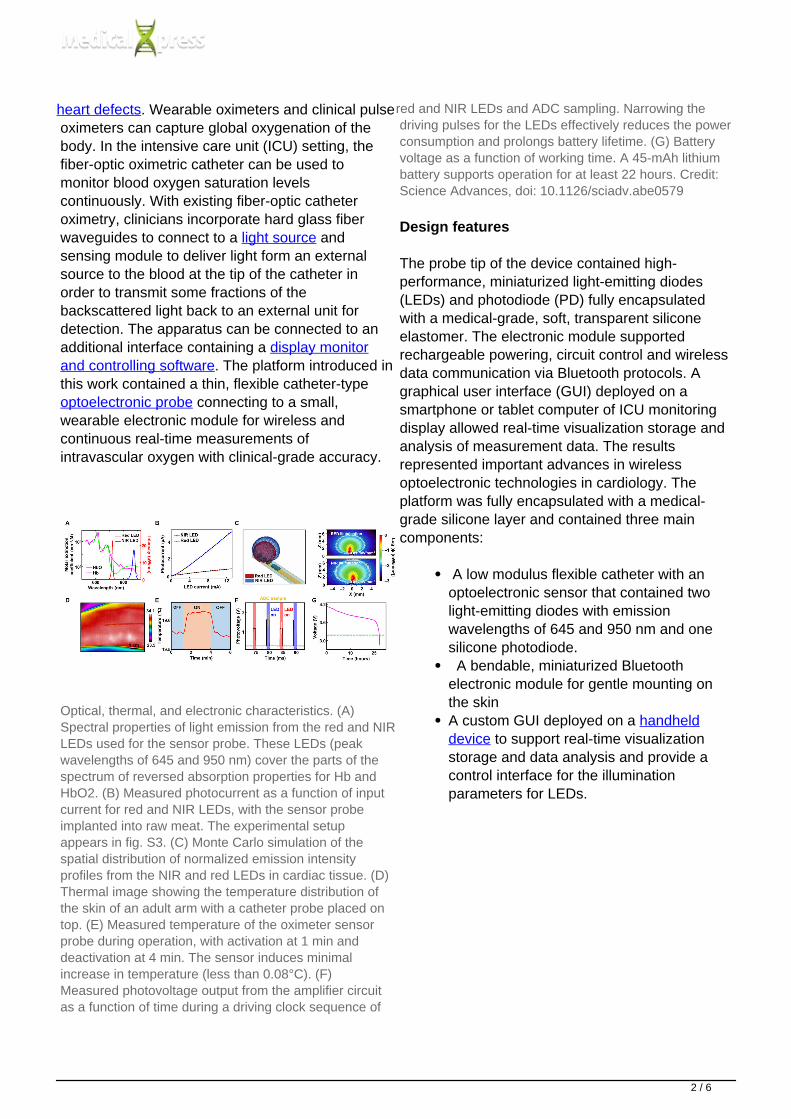
Optical, thermal, and electronic characteristics. (A)Spectral properties of light emission from the red and NIRLEDs used for the sensor probe. These LEDs (peakwavelengths of 645 and 950 nm) cover the parts of thespectrum of reversed absorption properties for Hb andHbO2. (B) Measured photocurrent as a function of inputcurrent for red and NIR LEDs, with the sensor probeimplanted into raw meat. The experimental setupappears in fig. S3. (C) Monte Carlo simulation of thespatial distribution of normalized emission intensityprofiles from the NIR and red LEDs in cardiac tissue. (D)Thermal image showing the temperature distribution ofthe skin of an adult arm with a catheter probe placed ontop. (E) Measured temperature of the oximeter sensorprobe during operation, with activation at 1 min anddeactivation at 4 min. The sensor induces minimalincrease in temperature (less than 0.08°C). (F)Measured photovoltage output from the amplifier circuitas a function of time during a driving clock sequence of
red and NIR LEDs and ADC sampling. Narrowing thedriving pulses for the LEDs effectively reduces the powerconsumption and prolongs battery lifetime. (G) Batteryvoltage as a function of working time. A 45-mAh lithiumbattery supports operation for at least 22 hours. Credit:Science Advances, doi: 10.1126/sciadv.abe0579
Design features
The probe tip of the device contained high-performance, miniaturized light-emitting diodes(LEDs) and photodiode (PD) fully encapsulatedwith a medical-grade, soft, transparent siliconeelastomer. The electronic module supportedrechargeable powering, circuit control and wirelessdata communication via Bluetooth protocols. Agraphical user interface (GUI) deployed on asmartphone or tablet computer of ICU monitoringdisplay allowed real-time visualization storage andanalysis of measurement data. The resultsrepresented important advances in wirelessoptoelectronic technologies in cardiology. Theplatform was fully encapsulated with a medical-grade silicone layer and contained three maincomponents:
A low modulus flexible catheter with anoptoelectronic sensor that contained twolight-emitting diodes with emissionwavelengths of 645 and 950 nm and onesilicone photodiode. A bendable, miniaturized Bluetoothelectronic module for gentle mounting onthe skinA custom GUI deployed on a handhelddevice to support real-time visualizationstorage and data analysis and provide acontrol interface for the illuminationparameters for LEDs.
2 / 6

Mechanical encapsulation and biocompatibilitycharacteristics. (A) Measured Young’s moduli for threecatheter probes (inset images; scale bar, 2 cm)encapsulated with three different biocompatible siliconeelastomers (labeled: MED-1040, MED-1000, andMED-1037, respectively). The Young’s moduli of thethree catheter probes range from 800 to 1700 kPa. (B)Measured bending stiffnesses for the three catheterprobes in (A), a catheter probe fabricated from relativelystiff copper wire encapsulated with MED-1000, and acommercial fiber-optic catheter (Swan Ganz 777F8,Edwards LifeSciences Inc.). The bending stiffnesses are1.6, 1.8, 2.3, 20, and 243 N/mm2, respectively. (C) Finiteelement modeling of the sensor probe and cathetersubjected to a bending radius of 22 and 27 mm,respectively. (D) Measured photovoltage from thecatheter probe as a function of cycles of compressionand bending. Experimental details appear in figs. S7 andS8. The photovoltage generated from the photodetectorcorresponds to operation of the two LEDs (peakwavelengths of 645 and 950 nm, respectively) at the tipof the catheter probe. a.u., arbitrary units. (E) Measuredphotovoltage as a function of immersion time in PBSsolution at 37°C. Experimental details appear in figs. S10and S11. The data indicate negligible change inperformance over 8 weeks. (F) CT image of the cathetersensor after 2 weeks of implantation. (G and H) Analysisof complete blood count (G) and blood chemistry (H) formice with an oximetry probe implanted subcutaneouslyfor 30 days (labeled as Experiment) and for mice withoutdevice implantation (labeled as Control). Credit: ScienceAdvances, doi: 10.1126/sciadv.abe0579
The team then wirelessly transferred data to a
personal computer using Bluetooth protocols.
Optical, thermal and electrical characterization
The scientists provided effective estimates of bloodoxygen saturation using well-known opticalapproaches to define the fraction of oxyhemoglobin(HbO2) relative to total hemoglobin (HbO2 +deoxyhemoglobin—Hb) by comparing the absorptionspectra of oxyhemoglobin and deoxyhemoglobin inthe visible and near-infrared spectral range. Usingthe large differences at 645 nm and 950 nm, thescientists established the basis of opticalmeasurements of blood oxygenation. Lu et al.measured the physics of light transport in biologicaltissues using the Monte Carlo method. The resultsprovided quantitative insights into the illuminationdistribution around the LEDs and into aspects oflight detection by the photodiode based on theoptical properties of human cardiac muscle tissue found in literature. While the device functionedwhen pressed against the skin of the fingertips, Luet al. obtained thermal images with an IR camera,which did not show apparent increase intemperature in the region. The scientists performedmeasurements every five minutes for a duration offive seconds.
3 / 6

Performance characteristics for oximetry measurements.(A) Comparisons of light emission profiles of acommercial catheter oximeter (Swan Ganz 777F8,Edwards LifeSciences Inc.) and the device introducedhere. (B) Comparisons of the commercial catheteroximeter and the device introduced here in measuringthe oxygen saturation in blood solutions with differentratios of HbO2 and Hb. The inset image shows acomparison of the wireless catheter probe and acommercial fiber-optic catheter (scale bar, 1 cm). (C)Measured pulse signals from the device placed on theindex finger of an adult. (D) Algorithm flow chart of thecalculation of pulse oximetry based on photovoltagesignals. (E and F) Measured SpO2 (E) and HR (F) duringa period of rest followed by a breath hold and thenanother period of rest. The results match those obtainedwith a commercial oximeter (General Electronic Inc). Theresults of additional experiments appear in fig. S17. (Gand H) Bland-Altman plots. (G) SpO2 from finger (four
subjects, 801 points). (H) HR from finger (4 subjects, 801points). Credit: Science Advances, doi:10.1126/sciadv.abe0579
Mechanical characterization and encapsulationperformance
The mechanical properties of the device minimizedthe mechanical forces on adjacent biologicaltissues for improved biocompatibility. Human skintypically maintains a Young's modulus or stiffnessbetween 400 and 800 kPa and human cardiactissue muscles have a Young's modulusapproximating 100 kPa. The Bluetooth modulecontained a biocompatible silicone forencapsulation with a Young's modulus in the rangeof human skin. The team used three catheteroximeter probes with biocompatible silicone withdiverse Young's modulus values. The resultingbending stiffness of the construct was 50 timesgreater than that of the probe. The probe couldeasily and conformally deform with the heartmuscle with negligible mechanical load and withoutassociated damage to the heart, although itsmodulus was greater. Lu et al. implanted the devicesubdermally on the back near the spine of a mousemodel and conducted computed tomography twoweeks later. The team then obtained completeblood count and blood chemistry for mice withimplants for 30 days to indicate no evidence oforgan damage or injury and without detrimentaleffects on electrolyte or enzyme balance.
Bench tests and in vivo studies
4 / 6
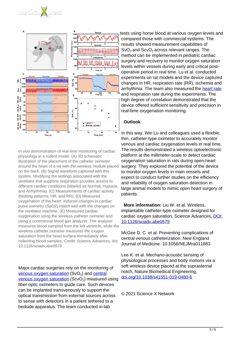
In vivo demonstration of real-time monitoring of cardiacphysiology in a rodent model. (A) 3D schematicillustration of the placement of the catheter oximeteraround the heart of a rat with the wireless module placedon the back. (B) Signal waveform captured with thissystem. Modifying the settings associated with theventilator that supports respiration provides access todifferent cardiac conditions (labeled as Normal, Hypoxia,and Arrhythmia). (C) Measurements of cardiac activity(beating patterns, HR, and RR). (D) Measuredoxygenation of the heart. Induced changes in cardiacpulse oximetry (SpO2) match well with the changes onthe ventilator machine. (E) Measured cardiacoxygenation using the wireless catheter oximeter andusing a commercial blood gas analyzer. The analyzermeasures blood sampled from the left ventricle, while thewireless catheter oximeter measures the oxygensaturation from the heart surface immediately aftercollecting blood samples. Credit: Science Advances, doi:10.1126/sciadv.abe0579
Major cardiac surgeries rely on the monitoring of venous oxygen saturation (SvO2) and centralvenous oxygen saturation (ScvO2) measured usingfiber-optic oximeters to guide care. Such devicescan be implanted transvenously to support theoptical transmission from external sources acrossto sense with detectors in a patient tethered to abedside apparatus. The team conducted in-lab
tests using horse blood at various oxygen levels andcompared those with commercial systems. Theresults showed measurement capabilities ofSvO2 and ScvO2 across relevant ranges. Themethod can be implemented in pediatric cardiacsurgery and recovery to monitor oxygen saturationlevels within vessels during early and critical post-operative period in real time. Lu et al. conductedexperiments on rat models and the device capturedchanges in HR, respiration rate (RR), ischemia andarrhythmia. The team also measured the heart rateand respiration rate during the experiments. Thehigh degree of correlation demonstrated that thedevice offered sufficient sensitivity and precision inreal-time oxygenation monitoring.
Outlook
In this way, Wei Lu and colleagues used a flexible,thin, catheter-type oximeter to accurately monitorvenous and cardiac oxygenation levels in real time.The results demonstrated a wireless optoelectronicplatform at the millimeter-scale to detect cardiacoxygenation saturation in rats during open-heartsurgery. They explored the potential of the deviceto monitor oxygen levels in main vessels andexpect to conduct further studies on the efficiencyand reliability of oxygen saturation detection inlarge animal models to mimic open heart surgery ofpatients.
More information: Liu W. et al. Wireless,implantable catheter-type oximeter designed forcardiac oxygen saturation, Science Advances, DOI:10.1126/sciadv.abe0579
McGee D. C. et al. Preventing complications ofcentral venous catheterization. New EnglandJournal of Medicine. 10.1056/NEJMra011883
Lee K. et al. Mechano-acoustic sensing ofphysiological processes and body motions via asoft wireless device placed at the suprasternalnotch, Nature Biomedical Engineering, doi.org/10.1038/s41551-019-0480-6
© 2021 Science X Network
5 / 6

APA citation: Wireless, implantable catheter-type oximeter designed for cardiac oxygen saturation (2021,February 25) retrieved 17 March 2022 from https://medicalxpress.com/news/2021-02-wireless-implantable-catheter-type-oximeter-cardiac.html
This document is subject to copyright. Apart from any fair dealing for the purpose of private study or research, nopart may be reproduced without the written permission. The content is provided for information purposes only.
Powered by TCPDF (www.tcpdf.org)
6 / 6