wnt-mediated downregulation of sp1 target genes by a ...1 wnt-mediated downregulation of sp1 target...
TRANSCRIPT
1
Wnt-mediated downregulation of Sp1 target genes by a transcriptional repressor Sp5
Naoko Fujimura‡, Tomas Vacik‡, Ondrej Machon‡, Cestmir Vlcek‡, Simone Scalabrin
#, Martin
Speth§, Dzung Diep §, Stefan Krauss § and Zbynek Kozmik‡¶
‡Institute of Molecular Genetics, Academy of Sciences of the Czech Republic, Prague, Czech
Republic #University of Udine, via delle Scienze 206, I-33100 Udine, Italy
§Institute for Microbiology, Rikshospitalet, Gaustadalleen 21, 0349 Oslo, Norway
Running title:Wnt-mediated downregulation of Sp1 target genes by a transcriptional repressor Sp5.
¶To whom correspondence should be addressed: Zbynek Kozmik, Institute of Molecular Genetics, Videnska 1083, 142 20 Prague 4, Czech Republic; FAX: +420-241062110; Phone: +420-241062146; Email: [email protected] Wnt/β-catenin signaling regulates many processes during vertebrate development. To study transcriptional targets of canonical Wnt signaling, we used the conditional Cre/loxP system in mouse to ectopically activate β-catenin during central nervous system development. We show that the activation of Wnt/β-catenin signaling in the embryonic mouse telencephalon results in the upregulation of Sp5 gene which encodes a member of the Sp1 transcription factor family. A proximal promoter of Sp5 gene is highly evolutionarily conserved and contains five TCF/LEF binding sites that mediate direct regulation of Sp5 expression by canonical Wnt signaling. We provide evidence that Sp5 works as a transcriptional repressor and has three independent repressor domains, called R1, R2 and R3, respectively. Furthermore, we show that the repression activity of R1 domain is mediated through direct interaction with a transcriptional corepressor mSin3a. Finally, our data strongly suggest that Sp5 has the same DNA binding specificity as Sp1 and represses Sp1 target genes such as p21. We conclude that Sp5 transcription factor mediates the downstream responses to Wnt/β-catenin signaling by directly repressing Sp1 target genes.
Wnt/β-catenin signaling plays important
roles in multiple developmental processes and has a profound effect on cell proliferation, cell polarity and cell fate determination (1). Wnt molecules are secreted glycoproteins which work as signaling molecules. Wnt molecules bind with Frizzled receptors and low density lipoprotein receptor-related protein (LRP) coreceptors at the cell surface to initiate the signaling. In the absence of Wnt/β-catenin signaling, the level of cytoplasmic β-catenin, the key mediator of Wnt/β-catenin signaling, is kept low. β-catenin is recruited to a destruction complex containing the tumor suppressors adenomatous polyposis coli (APC), Axin, Casein kinase 1 (CK1) and Glycogen Synthase Kinase 3β (GSK3β), respectively, and is constitutively phosphorylated. The phosphorylated β-catenin protein is degraded by the ubiquitin pathway. Members of the TCF/LEF transcription factor family bind corepressor Groucho and repress Wnt target genes in the nucleus. The binding of Wnt molecules to the receptors and the coreceptors results in the inactivation of the kinase activity of the destruction complex. As a consequence, β-catenin protein is not phosphorylated, begins to accumulate in the cytoplasm and is then translocated to the nucleus where it binds to TCF/LEF transcription factors. The binding
http://www.jbc.org/cgi/doi/10.1074/jbc.M605851200The latest version is at JBC Papers in Press. Published on November 6, 2006 as Manuscript M605851200
Copyright 2006 by The American Society for Biochemistry and Molecular Biology, Inc.
by guest on March 1, 2020
http://ww
w.jbc.org/
Dow
nloaded from

2
converts TCF/LEF into an activator that initiates the transcription of Wnt target genes including c-Myc , Axin2 and Lef1 (2-4).
During central nervous system (CNS) development, multiple Wnt genes are expressed including Wnt3a, Wnt7a, Wnt7b and Wnt8b (5). Transgenic mice which express a stabilized form of β-catenin in neural progenitor cells develop enlarged brains (6). In Wnt3a mutant mice as well as in Lef1 mutant mice, the hippocampus is missing (5,7). These reports indicate critical roles of canonical Wnt signaling in CNS development.
To study targets of Wnt/β-catenin signaling, we used the conditional Cre/loxP system in mouse to ectopically activate Wnt/β-catenin signaling during CNS development. Activation of Wnt/β-catenin signaling is achieved by a deletion of exon 3 of the β-catenin gene that encodes phosphorylation sites necessary for β-catenin degradation (8). To activate canonical Wnt signaling during CNS development, Nes11Cre mice were crossed to Catnblox(ex3) mice. Mutant animals Nes11Cre/Catnb lox(ex3) display hyperplasia in the telelencephalon that resembles the phenotype of the mouse mutants in which activated β-catenin is directly coupled to the nestin enhancer (6). We show that the constitutive activation of Wnt/β-catenin signaling results in the upregulation of the Sp5 gene in the mouse telencephalon. The Sp5 gene encodes a member of Sp1 transcription factor family (9). The proximal promoter of the Sp5 gene is highly evolutionarily conserved and has five TCF/LEF binding sites that mediate direct regulation of Sp5 expression by Wnt/β-catenin signaling. Sp5 appears to work as a transcriptional repressor at least in part by directly interacting with a corepressor mSin3a. We show that Sp5 has the same DNA binding specificity as Sp1 and represses Sp1 target genes such as p21. In conclusion, our report suggests that the Sp5 transcription factor mediates the downstream responses to Wnt/beta-catenin signaling by directly repressing Sp1 target genes.
EXPERIMENTAL PROCEDURES Mouse Lines - Analysis of Cre-mediated recombination pattern in Nes11-Cre (10) was
performed by mating to the ROSA26R reporter line as described (11). The ROSA26R mice (stock #003309) and Nes11-Cre mice (stock # 003771) were purchased from Jackson Lab. D6Cre transgenic mice express Cre recombinase under the control of Dach1 enhancer which is active in the telencephalon (12). Mice with a conditional “floxed” allele of β-catenin , Catnblox(ex3), were kindly provided by Dr. M.M. Taketo (8). Plasmids – The mouse Sp5 promoter and truncated promoters were amplified by PCR using C57BL/6J mouse genomic DNA (kindly provided by J. Forejt) as a template. PCR products were cloned to pCR4-TOPO (Invitrogen) and sequenced. The resulting plasmids were digested with EcoRI (New England Biolab), blunted with T4 DNA polymerase (New England Biolab), and cloned into SmaI digested pGL3 basic (Promega) vector. Mouse Sp5 enhancers were amplified by PCR using primers with XbaI recognition sites. PCR products were digested by XbaI and cloned into a NheI site upstream of the minimal TK promoter cloned in the pGL3 vector. For Gal4-Sp5, the full-length mouse Sp5 cDNA was excised from pBS-KX-Sp5 (kindly provided by D. Houzelstein) and cloned into a Gal4 expression plasmid. To generate Gal4 fusion constructs with individual domains of Sp5, the corresponding regions of mouse Sp5 cDNA were amplified by PCR and cloned into the Gal4 expression plasmid. To generate 6xHis-Sp5, the coding sequence of Sp5 was cloned into the procaryotic expression vector pETH2α. For Sp5-Flag, the Sp5 cDNA was amplified by PCR and cloned into pKW-Flag in frame with the Flag coding sequence located at the N-terminus. For retroviral infection of neurosphere cultures, Sp5 cDNA was inserted into pNIT retroviral vector (provided by F. Gage). To generate GST fusions with a Sp5 R1 domain, the corresponding region was amplified by PCR and cloned into pET42a(+) (Novagen). For GST-Sp5R1A3P and Gal4-Sp5A3P, the R1 region was amplified by PCR using primers which contained the corresponding point mutation and cloned into pET42a(+) or Gal4 expression plasmid. All constructs were verified by sequencing. A luciferase reporter plasmid containing the p21 promoter (p21-Luc) was kindly provided by E. Sancho. For p21GC-Luc, the p21-Luc plasmid was
by guest on March 1, 2020
http://ww
w.jbc.org/
Dow
nloaded from
3
digested with PstI and BglII (-198/+12), blunted, and cloned into pGL3 basic. For p21ΔGC-Luc, a p21 promoter fragment (-2326/-197) was cut with PstI/HindIII and fused to the minimal p21 promoter (-30/+12) located in pGL3 basic. Microarray experiment - RNA was isolated from the dissected telencephalon of E13.5 mouse embryos (Nes11-Cre/ Catnblox(ex3) or Catnblox(ex3) ) using an Ambion kit and subjected to hybridization on Affymetrix MOE 430A. Neurospheres were cultured in neurobasal-A medium with B27 supplement (both Gibco) and with EGF (20 ng/ml) and bFGF (8 ng/ml) (both R&D Systems). Cells were passaged every three days. The Sp5 retrovirus was produced in a Phoenix packaging cell line (provided by G. Nollan) by transient transfection of pNIT-Sp5 and neurosphere cells were infected as described in (13). Three days after infection, selection with G418 antibiotics was started (250 μg/ml) and pools of cell clones were maintained in the selection media. RNA was isolated from three separate plates of Sp5 virus or mock infected neurospheres using an Ambion kit and used for hybridization on Affymetrix MOE 430A. Microarray data were analyzed by Affymetrix Suite 5.1 software. Cell Culture, Transient Transfection and Luciferase Reporter Assay -293T cells were cultured in Dulbecco’s modified Eagle’s medium (SIGMA) supplemented with 10% Fetal bovine serum (PAA laboratories), 2mM L-glutamine, 100units/ml penicillin, and 0.1mg/ml streptomycin (SIGMA). Neurospheres were cultured in neurobasal-A medium with B27 supplement (both Gibco), EGF (20 ng/ml), and bFGF (8 ng/ml) (both R&D Systems). Cells were passaged every three days and maintained at 37 °C in an atmosphere of humidified air with 5% CO2. Transient transfection of 293T cells was performed using Fugene 6 (Roche) according to the manufacturer’s protocol. Cells were plated in 24-well plates 24 hours prior to transfection. Typically, the total amount of DNA transfected per well was 300ng and was adjusted with pUC18 when necessary. A β-Galactosidase expression plasmid was cotransfected to normalize the
transfection efficiency. Triplicate assays were performed to obtain standard deviations. Two days after transfection, the cells were lysed in 100ul of 1× passive lysis buffer (Promega). Luciferase reporter assays were performed using Luciferase Reporter assay kit (Promega). β-Galactosidase was detected with Galacto-Star system (Applied Biosystems). Chromatin Immunoprecipitation Assay (ChIP) - ChIP assay was performed according to the manufacturer's protocol (Upstate Biotech) with modifications. The cortical parts of D6Cre/ Catnblox(ex3) brains were harvested at E18.5, homogenized in 1% formaldehyde in PBS and crosslinked at 37 °C for 15 minutes. Crosslinking was stopped by adding glycine (0.125M) and incubating at RT for 5 minutes. Crosslinked cells were washed twice with cold PBS containing fresh protease inhibitors, pelleted and resuspended in 2 ml of SDS lysis buffer (1% SDS, 10 mM EDTA, 50 mM Tris-HCl, pH 8.0) with protease inhibitors. Samples were incubated on ice for 10 minutes and lysates were sonicated on ice-water bath to produce 150-500 bp DNA fragments. Cell debris was removed by centifugation for 10 minutes at 14000 rpm at 4 °C and the supernatant was diluted ten times with dilution buffer (0.01% SDS, 1.1% Triton X-100, 1.2 mM EDTA, 16.7 mM Tris-HCl, pH 8.0, 167 mM NaCl) containing protease inhibitors. 30 µg of sonicated chromatin was precleared with 50 µl of proteinA(G)/Agarose slurry (Upstate Biotech) for one hour at 4 °C. Beads were pelleted by centrifugation for 5 minutes at 3000 rpm at 4C. The supernatant was incubated either with 5 µg of antibody or with no antibody (no antibody control) O/N at 4 °C. The following antibodies were used: anti-β-catenin (E-5, sc-7963, Santa Cruz Biotechnology), anti-Lef1 (N-17, sc-8591, Santa Cruz Biotechnology), anti-Tcf4 (a gift from V.Korinek). 30 µl of proteinA(G)/agarose slurry (Upstate Biotech) was added and samples were rocked at 4ºC for 1 hour. After washing for 5 minutes at 4 °C twice in low salt buffer (0.1% SDS, 1%Triton X-100, 2 mM EDTA, 20 mM Tris-HCl, pH 8.0, 150 mM NaCl), twice in high salt buffer (0.1% SDS, 1% Triton X-100, 2 mM EDTA, 20 mM Tris-HCl, pH 8.0,500 mM NaCl), four times in LiCl buffer (0.25 M LiCl, 1% Nonidet P-40, 1% deoxycholate,1 mM
by guest on March 1, 2020
http://ww
w.jbc.org/
Dow
nloaded from

4
EDTA, 10 mM Tris-HCl, pH 8.0), and twice in TE buffer (10mM Tris-HCl, 1 mM EDTA, pH 8.0), immunocomplexes were eluted twice with 100 µl of elution buffer (0.1M NaHCO3, 1%SDS) for 15 minutes at RT. Immunoprecipitated DNA was decrosslinked O/N at 65 °C in the presence of proteinase K (0.06U/µl, Roche Applied Science) and 250 mM NaCl. Samples were purified using MinElute Reaction Cleanup kit (Qiagen) and 1/20 of eluate was used for PCR. PCR was performed as follows: 95 °C 2 minutes for 1 cycle, 95 °C 30 s, 60 °C 30 s, 72 °C 30 s for 40 cycles, 72 °C 5 minutes. The primers used were as follows: Sp5D-H_F CCTAGAGATAACAAAGACACT, Sp5D-H_R AGTCAGAGGAAAGATTTATGG, Sp5-2kb_F TGGCTGCTTAATTGCCTAAAGAG, Sp5-2kb_R CAGGGGTTTGAGTGCTGTGGA, Sp5+6kb_F AACGGAAGCTGAGTGTAAATTAG, Sp5+6kb_R GTAACTAAGACAGACGCCTAAAC Electrophoretic Mobility Shift Assay (EMSA) - The following double-stranded oligonucleotides derived from the Sp5 promoter were used in EMSA (only top strand is shown for simplicity): Sp5A ATTGAAGAAACAAAGTTTGATCT, Sp5B CACTCATCAACAAAGGAAAGCCC, Sp5C GGATACCTCTTTGAACTGACCCC, Sp5D CTAGAGATAACAAAGACACTTTG, Sp5E AAGGCCCCCTTTGATCAGGAAAA, Sp5F TTTGTGGATTCAAAGGATTTGCT, Sp5G CCGCTATTCTTTGATGATTGGGT, Sp5H CGGCAAACTTCAAAGCCATAAAT. The following double-stranded oligonucleotides derived from the p21 promoter were used: I+II GAATTCTGAGGCGGGCCCGGGCGGGGCGGTTGGAATTC, III+IV GAATTCCGAGCGCGGGTCCCGCCTCCGAATTC, V+VI GAATTCGGAGGGCGGTCCCGGGCGGCGCGAATTC. The following double-stranded oligonucleotides representing consensus (wt) and multiple versions of the Sp1 binding site were used: WT ATTCGATCGGGGCGGGGCGAGC, M1 ATTCGATCGGTTCGGGGCGAGC, M2 ATTCGATCGGGGAGGGGCGAGC, M3
ATTCGATCGGGGTGGGGCGAGC, M4 ATTCGATCGAGGCGGGGCGAGC, M5 ATTCGATCGGGGCGGAGCGAGC, SA1 GTGCGGAGGCGTGGTTAGAG, AX2 CGGGCGGCGGGGGAGGCGGGGTC, XN2 CGGCGGGGAGGTGGGGCGAGGAGAG, BTE AGCTTGAGAAGGAGGCGTGGCCAACGCATG. Double-stranded oligonucleotides containing Tcf/Lef or Sp1/Sp5 binding sites were radioactively labeled at the 5’ends with γ32PdATP using polynucleotide kinase (Boehringer Manheim) and purified on microspin columns (Amersham Biosciences). 32P-labeled oligonucleotides were incubated with in vitro-synthesized LEF1 (TNT Quick, Promega), bacterially-purified 6xHis-Sp5 (Qiagen), or Sp1 (Promega) in binding buffer (10 mM HEPES at pH 7.7, 75 mM KCl, 2.5 mM MgCl2,0.1 mM EDTA, 1 mM DTT, 20% glycerol, 0.5% BSA, and 0.1mg/mL poly-dIdC) on ice for 15 minutes. For supershifts, 32P-labeled oligonucleotides were preincubated on ice for 10 min with 1 µg anti-Lef antibody. Samples were analysed by 6 % polyacrylamide gel electrophoresis and autoradiography. Immunoprecipitation and Western Blotting - 293T cells were plated in 10cm dishes 24 hours prior to transfection. Myc-mSin3a plasmid (2μg; kindly provided by C. Laherty) was cotransfected with Flag-Sp5 expression plasmid (3μg) or empty Flag expression plasmid (3μg) into 293T cells. Two days after transfection, 293T cells were washed with PBS and lysed in lysis buffer (50mM Tris HCl, pH7.4, with 150mM NaCl, 1mM EDTA, 1% TRITON X-100 and 0.1mM PMSF) for 30 minutes on ice. Cell debris was pelleted by centrifugation at 12000×g for 10min. An aliquot of this whole cell lysate was boiled with 2 × SDS sample buffer for 5min. For immunoprecipitation, 500ml of the whole cell lysate was incubated with 40μl of anti-Flag M2 affinity beads (Sigma) overnight at 4°C. The beads were washed with 1×Wash buffer (50mM Tris HCl, pH7.4, with 150mM NaCl, 1mM EDTA) five times and boiled with 2 × SDS sample buffer for 5min. Samples were separated by 8% or 12% SDS-PAGE and transferred to nitrocellulose
by guest on March 1, 2020
http://ww
w.jbc.org/
Dow
nloaded from

5
membranes for Western blotting. Myc-tagged mSin3a was detected by anti-Myc antibody (Roche) and Flag-tagged Sp5 was detected by anti-Flag M2 (Sigma). Detection was performed using polyclonal rabbit anti-mouse immunoglobulins / HRP (DAKO Cytomation) and SuperSignal West Pico Chemiluminescent Substrate (PIERCE). GST-Pull Down Assay - Myc-tagged mSin3a was prepared by TNT Quick Coupled Transcription/Translation Systems according to the manufacturer’s protocol (Promega). GST fusion expression plasmids were transformed into BL21 CodonPlus (DE3)-RIPL cells (Stratagene). A single colony from the transformation was cultured in 2ml LB medium containing 50μg/ml of chloramphenicol and 30ng/ml of kanamycin overnight at 37°C. The cultures were transferred to 100ml of LB without antibiotics. The expression of the fusion construct was induced by adding IPTG to a final concentration of 2mM for 2 hours. The cells were harvested by centrifugation and resuspended in 5ml of NETN buffer (20mM Tris pH8.0, 100mM NaCl, 1mM EDTA, 0.5% NP40). Lysozyme was added to a final concentration of 0.1mg/ml. The lysates were incubated on ice for 20min, sonicated and centrifuged to remove the cell debris. The supernatant was incubated with 200μl of glutathione-Sepharose beads slurry (BD Bioscience) for 1 hour at 4°C. The beads were washed three times by 5ml of Binding buffer (20mM Tris pH8.0, 100mM KCl, 5mM MgCl2, 0.1mM EDTA, 20% Glycerol) containing 0.1% NP40. GST fusion proteins bound to the beads were checked by SDS-PAGE. Beads containing normalized amounts of fusion proteins were blocked by Binding buffer containing 0.05% of NP40 and 5mg/ml of BSA for 2 hours at 4°C and resuspended in 150μl of Binding buffer containing 0.05% NP40, 1mg/ml BSA and 100μg/ml Ethidium Bromide. The beads were incubated overnight at 4°C with 3μl of Myc-tagged mSin3a produced by TNT. The beads were washed three times with 500μl of Binding buffer containing 0.05% NP40 and boiled with SDS sample buffer. Myc-tagged mSin3a was detected by Western blotting using an anti-Myc antibody.
In Situ Hybridization. - In situ hybridization on cryosections was carried out as described previously (14). Plasmids carrying mouse Sp5, Axin2, and Nkd1 cDNA were linearized with an appropriate restriction enzyme and an antisense riboprobe was synthesized using the DIG RNA labeling kit (Roche).
RESULTS Sp5 is a target gene of Wnt/β-catenin signaling.
To identify target genes of Wnt/β-catenin signaling during CNS development, two lines of mice were interbred to activate Wnt/β-catenin signaling. The Catnblox(ex3), in which exon 3 of β-catenin gene is floxed by loxP sites (8), was mated to Nes11Cre, a transgenic mouse line expressing Cre recombinase under the control of nestin regulatory elements in neural progenitor cells (10). Exon 3 of β-catenin gene encodes phosphorylation sites necessary for β-catenin degradation (15). Cre recombinase-mediated deletion of exon 3 of β-catenin gene results in the expression of a stabilized form of β-catenin which leads to the constitutive activation of Wnt/β-catenin signaling. To map the area in which Cre recombinase is active in the Nes11Cre mice, Nes11Cre mice were crossed with a reporter mouse line, ROSA26R (R26R) (11). Within the telencephalon, Cre recombinase activity was detected in the neural progenitor cells of the pallium and the subpallium (Fig. 1A). To activate canonical Wnt signaling during CNS development, Nes11Cre mice were crossed to Catnblox(ex3) mice. Mutant animals Nes11Cre/Catnb lox(ex3) displayed hyperplasia in the telelencephalon that resembles the phenotype of the mouse mutants in which activated β-catenin is directly coupled to the nestin enhancer (6). Further, the dorso-ventral patterning in the mutant telencephalon is impaired such that genes normally expressed in the dorsal pallium expand into the ventral areas while ventrally-expressed genes are downregulated (69). To identify target genes of Wnt/β-catenin signaling, RNA was isolated from the telencephalon at E13.5 and overall gene expression was analyzed by Affymetrix microarray. We noticed that the expression levels of several known targets of Wnt/β-catenin were upregulated, such as Axin2
by guest on March 1, 2020
http://ww
w.jbc.org/
Dow
nloaded from

6
3.4x, Nkd1 9.5x, Dkk1 5x and Pitx2 7x (3,16,17). On the other hand, ventrally-expressed genes such as Dlx2, Dlx1, Lhx6 or Mash1 were downregulated 10.5-, 5.1-, 15.1- and 7.2-fold, respectively. The expression of several genes was verified by in situ hybridization on coronal sections of Nes11Cre / Catnblox(ex3) mice and wild type mice at E13.5 (Fig. 1D-G, see also ref. 69). Interestingly, we found that Sp5, a member of Sp1 family, was upregulated 32-fold in the Affymetrix data and strong gene activation was confirmed by in situ hybridization (Fig. 1B-C). In wild type mice, Sp5 is expressed weakly in the hippocampal primordium (Fig. 1B). In Nes11Cre / Catnblox(ex3) mice, Sp5 is strongly expressed in the pallium and the subpallium i.e. in the area of Cre-mediated recombination (Fig. 1C). These results suggest that Wnt/β-catenin signaling positively regulates Sp5. Sp5 is a direct target gene of Wnt/β-catenin signaling.
We next examined whether Sp5 is regulated by Wnt/β-catenin signaling directly. To find important transcriptional regulatory elements, we compared the upstream sequences of Sp5 of Mus musculus, Homo sapiens, Gallus gallus, Danio rerio and Xenopus tropicalis since the important transcriptional regulatory elements are often evolutionarily conserved. We found three evolutionarily conserved regions containing TCF/LEF consensus sites located at positions -200bp / +200bp, -2.9kbp / -2.7kbp and -3.9kbp / -3.4kbp, refered to as proximal promoter, ECR2, and ECR1, respectively. ECR2 contains two conserved TCF/LEF consensus sites named B and C. ECR1 contains one conserved TCF/LEF consensus site named A. The Sp5 proximal promoter contains five TCF/LEF consensus sites, named D, E, F, G and H, respectively. Sites E, G and H in the Sp5 promoter were evolutionarily conserved among all five vertebrate species. Site F was not conserved in Danio rerio and site D was conserved only between Mus musculus and Homo sapiens (Fig. 2A).
To examine if the Sp5 promoter is responsive to Wnt/β-catenin signaling, a mouse Sp5 promoter (-1536 / +200) was cloned into the luciferase reporter plasmid and transiently transfected into 293T cells. Cotransfection of the
promoter with Lef1 and N-terminally truncated β -catenin (β-cateninΔN), which is constitutively stabilized and able to bind with TCF/LEF transcription factors (18), stimulated reporter gene expression about 15-fold. Conversely, cotransfection with N-terminally truncated TCF4 (dnTCF4), which does not bind to β-catenin and acts as a potent inhibitor of the β-catenin/TCF complexes (19), repressed the activity of the promoter construct 4.9-fold (Fig. 2B). These results suggest that the Sp5 promoter is directly responsive to Wnt/β-catenin signaling.
To identify functional TCF/LEF elements within the Sp5 promoter, three reporter plasmids containing different regions of the promoter cloned upstream of the luciferase reporter gene were constructed (Fig. 2B). Luciferase reporter plasmids containing -206/+200, -27/+200 and -1536/+3 of the Sp5 promoter were named D1, D2 and D3, respectively. Each plasmid was cotransfected in 293T cells with β-cateninΔN/Lef1 or dnTCF4. D1 and D2 were stimulated 10-fold and 6-fold by β-cateninΔN/Lef1, respectively, and repressed 2.9-fold and 9.4-fold by dnTCF4, respectively. In contrast, D3 was not affected by either β-cateninΔN/Lef 1 or dnTCF4 (Fig. 2B). These results suggest that sites F, G, H play a critical role in mediating Wnt/β-catenin signaling and site E supports site F, G and H to give further activation.
To examine whether ECR1 and ECR2 are also responsive to Wnt/β-catenin signaling, ECR1 and ECR2 were cloned upstream of the minimal TK promoter driving luciferase reporter gene expression. Each of the constructs was cotransfected in 293T cells with β-cateninΔN/Lef1 or dnTCF4. ECR1, ECR2 and TK were stimulated 1.4- fold, 4.0- fold, and 1.8- fold by β-cateninΔN/Lef1, respectively, and repressed 2.4- fold, 2.3- fold and 1.5- fold by dnTCF4 (Fig. 2C). These results suggest that ECR2 is an additional Wnt-responsive regulatory element.
To examine whether TCF/LEF binds putative A-H binding sites within the Sp5 proximal promoter, ECR1, and ECR2 (Fig. 3B), EMSA was performed. Oligonucleotides containing sites A - H were incubated with in vitro translated LEF1 and were analyzed by electrophoresis. As shown in Fig. 3A, all sites
by guest on March 1, 2020
http://ww
w.jbc.org/
Dow
nloaded from

7
were bound by LEF1. The identity of the LEF1 protein in the complex was verified by supershifts using LEF1 antibody. This result suggests that TCF/LEF can bind with TCF/LEF binding sites within the Sp5 proximal promoter, ECR1, and ECR2.
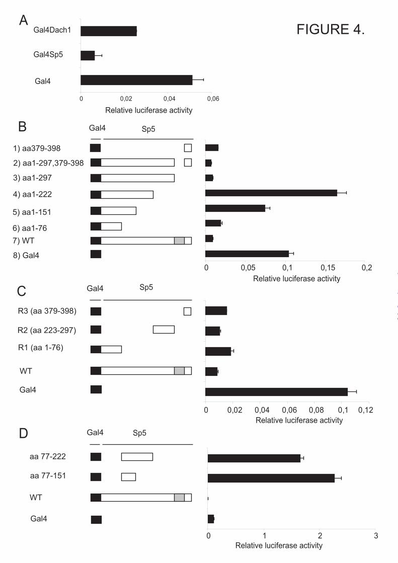
We next examined whether LEF/TCF transcription factors and β-catenin are associated with the Sp5 promoter in vivo. Chromatin immunoprecipitation (ChIP) was performed using antibodies against LEF1, TCF4 and β-catenin using cortical part of brain from D6Cre/Catnblox(ex3) mice at E18.5. D6Cre is a transgenic line expressing Cre recombinase in the telencephalon using Dach1 enhancer (12). ChIP data show that LEF/TCF/β-catenin complexes are present on the proximal Sp5 promoter (Fig. 3C). This result suggests that LEF/TCF/β-catenin complex binds the Sp5 promoter in vivo to regulate transcription. We therefore conclude that Sp5 is a direct target gene of Wnt/β-catenin signaling. Sp5 is a potent transcriptional repressor To our surprise, many genes were downregulated in the telencephalon of Nes11Cre/Catnblox(ex3) mice as compared to control mice. We hypothesized that downregulation of at least some of the genes could be mediated by Sp5 since Sp5 itself is highly induced in Nes11Cre/Catnblox(ex3) mice and several Sp1 family members are known to act as repressors (20). To examine the transcriptional properties of Sp5, a Gal4 reporter assay was employed. Plasmids encoding Gal4, Gal4 fusion with Sp5 (Gal4-Sp5), or Gal4 fusion with Dach1 (Gal4-Dach1), a known repressor (21), were cotransfected with a Gal4-dependent reporter plasmid driving luciferase gene expression. Both Gal4-Sp5 and Gal4-Dach1 repressed transcription 7.8-fold and 1.9-fold, respectively (Fig. 4A). This result indicates that Sp5 acts as a transcriptional repressor.
To identify functional domains within Sp5 which mediate transcriptional activity, Gal4 fusion constructs with different regions of Sp5 were cotransfected together with the Gal4 reporter plasmid. The Gal4 fusion proteins containing amino acids (aa) 1-76, 1-297, 1-297 plus 379-398 and 379-398 of Sp5 repressed 5.5, 11-fold, 16-fold and 7.1-fold, respectively. However, the Gal4
fusion proteins containing aa 1-151 and 1-222 of Sp5 did not exert any significant effect on transcription (Fig. 4B). These results suggest that aa 1-76, 223-297 and 379-398 of Sp5 contain repressor domains and that the region between aa 77-222 of Sp5 might contain an activation domain. To examine our hypothesis, Gal4 fusions with aa 223-297, 379-398, 77-222 or 152-222 were cotransfected with a Gal4 reporter plasmid. Gal4 fusions with aa 223-297 and 379-398 repressed 10-fold and 7.1-fold, respectively (Fig. 4C). Gal4 fusions with aa 77-222 and 77-151 activated 21-fold and 15-fold respectively (Fig. 4D). These results suggest that, overall, Sp5 acts as a repressor and has three separable and independent repressor domains located within aa 1-76, 223-297 and 379-398. In addition, there is a potential transcriptional activation domain located within residues 152-222 of Sp5. In the following text, we refer to the repressor domains located within aa 1-76, 223-297 and 379-398 as R1, R2 and R3, respectively. Corepressor mSin3a interacts with the R1 domain of Sp5 and regulates its transcriptional activity. We next examined the mechanism(s) that control the transcriptional properties of the repressor domains. We found a core mSin3a interacting domain (SID), AA/VXXL (22), within the R1 domain of Sp5. Corepressor mSin3a is known to interact with Class I histone deacetylases and a number of transcription factors containing α-helical structure harboring SID (23,24). We found that the R1 domain is predicted to form α-helical structure. We therefore examined whether the putative SID within Sp5 is responsible for the transcriptional repression function of the R1 domain. The Gal4 fusion constructs with the R1 domain containing wild type (Gal4-Sp5R1) or a mutated SID (Gal4-Sp5R1A3P), in which alanine is changed to proline to disrupt the formation of α-helical structure, were cotransfected with the Gal4 reporter plasmid. Interestingly, in contrast to the wild type Gal4-Sp5R1 which acts as a potent repressor, the Gal4-Sp5R1A3P acted as an activator (Fig. 5A). In addition, another Gal4 fusion construct with the R1 domain lacking a SID (Gal4-Sp5R1Δ3-7) also worked as an activator (N.F. and Z.K., data not shown). Combined, these
by guest on March 1, 2020
http://ww
w.jbc.org/
Dow
nloaded from

8
results suggest that the SID is crucial for the repressive activity of the R1 domain.
To examine whether Sp5 interacts with mSin3a directly through a SID, GST pull down assays were performed with the wild type Sp5 R1 domain (GST-R1) and the SID mutated R1 domain (GST-R1A3P). GST-R1 pulled down in vitro translated mSin3a. In contrast, neither GST nor GST-R1A3P were able to interact with mSin3a (Fig. 5B). In accordance with the fact that we have not been able to detect any potential SID motifs within R2 and R2, GST-Sp5R2 and GST-Sp5R3 domain fusions did not pulled down in vitro translated mSin3a (data not shown). These results suggest that mSin3a interacts with Sp5 directly through the SID located within R1. To provide further evidence that mSin3a interacts with Sp5 in vivo, coimmunoprecipitation was performed. Flag-tagged Sp5 expression plasmid (Sp5-Flag) was cotransfected with Myc-tagged mSin3a expression plasmid (Myc-mSin3a) into 293T cells and the total cell lysate was precipitated using Flag antibody beads. We found that Sp5-Flag was immunoprecipitated with Myc-mSin3a (Fig. 5C) providing evidence that Sp5 can interact with mSin3a in vivo. In summary, our results suggest that the transcriptional repression activity of R1 domain is mediated through the interaction with mSin3a corepressor. Sp5 binds Sp1 target sequences and attenuates Sp1 regulated transcription.
Sp5 belongs to the large family of Sp1-like transcription factors. Intrigued by the fact that the founding member, Sp1, acts as an activator whereas Sp5 acts as a repressor we next examined whether Sp5 downregulates Sp1 target genes. The zinc finger domain of Sp1 family members conforms to the Cys2His2 zinc finger consensus sequence. The similarity of the zinc finger between Sp1 and Sp5 is 92.6% (20). The amino acids predicted to make contact with the DNA are conserved between Sp5 and Sp1. Furthermore, it is shown that Sp5 binds to canonical Sp1 consensus site (GGGCGG) in vitro by EMSA (9). To examine whether Sp5 has the same DNA binding specificity as Sp1, EMSA was performed using bacterially-purified Sp5 and Sp1 proteins on a large panel of binding sites. Oligonucleotides containing canonical Sp1 binding site (WT),
mutated Sp1 binding sites (M1, M2, M3, M4 and M5), Sp1 binding regions within the proximal promoter of TGF-βRI gene (SA1, AX2 and XN2), or BTE site (BTE), a well characterized GC-rich element (25,26) were tested. Binding site M1 has a mutation which abolishes Sp1 binding (27). As shown in Fig. 6A, Sp5 and Sp1 bound to WT, M2, M3, M4, M5, XN2, SA1, AX2 and BTE with similar affinities. Consistent with Sp1 data, Sp5 did not bind to M1 (Fig. 6A). This result suggests that Sp5 has a very similar if not identical DNA binding specificity as Sp1. We next examined whether Sp5 represses Sp1 target genes. First of all, we have investigated p21 as a well characterized Sp1 target gene. It was shown previously that the proximal promoter of p21 gene contains six Sp1 binding sites (I-VI) and that it is positively regulated by Sp1 through these binding sites (28). To examine whether Sp5 binds to the Sp1 binding sites within the p21 promoter, EMSA was performed using bacterially-purified Sp5 or Sp1 and oligonucleotides containing sites I + II, III + IV and V + VI. As shown in Fig. 6B, Sp5 bound strongly with the oligonucleotides in the same manner as did Sp1. To examine whether Sp5 has the ability to repress p21 gene promoter, the luciferase reporter assay was performed. Reporter genes containing 2.3kb of the p21 promoter (p21-Luc), the proximal p21 promoter (p21GC-Luc) or the promoter lacking the six Sp1 binding sites (p21ΔGC-Luc) were cotransfected with or without the Sp5 expression plasmid into 293T cells. As shown in Fig. 6C, both p21-Luc and p21GC-Luc were repressed by Sp5 (13-fold and 3.3-fold, respectively). We were unable to see any effect of Sp5 on p21ΔGC-Luc since the basal level of p21ΔGC-Luc was even lower than that of the parental plasmid pGL3. Our results suggest that Sp5 can repress p21 promoter, most likely due to its ability to compete with Sp1 (or with related activator, Sp3) for promoter binding. Since full length p21 promoter and truncated p21 promoter were repressed 13-fold and 3.3-fold, respectively, there may be additional Sp5-responsive elements upstream of the proximal p21 promoter. These results suggest that Sp5 binds p21 gene regulatory elements and represses its promoter.
To obtain further evidence that Sp5 represses Sp1 target genes in vivo, and to identify additional Sp5 targets in neural cells, we have
by guest on March 1, 2020
http://ww
w.jbc.org/
Dow
nloaded from
9
established primary neurosphere cultures overexpressing Sp5. Neurospheres represent cultured neural stem cells that divide in vitro and yield major neural lineages upon differentiation. pNIT retroviral vector carrying Sp5 coding sequence as well as G418 resistance was used to infect neurosphere cells isolated from the mouse telencephalon at E12.5. Neurospheres were grown in medium containing G418 and the pool of G418-resistant clones was used for isolation of RNA. Real-time RT-PCR revealed that neurosphere cells infected with the Sp5 retrovirus manifested 107.6-fold induction of Sp5 mRNA as compared to mock infected cells (T.V., data not shown). We then profiled gene expression of Sp5 overexpressing neurospheres by Affymetrix microarray analysis. We found that 107 genes were downregulated more than two-fold in Sp5-infected neurospheres. Notably, 90 genes were Sp1 target genes or genes which contain canonical Sp1 binding sites in the proximal promoter (-500/+1) and 5’ untranslated region (Table 1) (29-52). Of a special interest is the gene encoding solute carrier family 12, member 2 (scl12a2, NKCC1) which contains canonical Sp1 binding sites and becomes downregulated by Wnt/β-catenin signaling (53). In conclusion, our results show that Sp5 represses Sp1 target genes.
DISCUSSION
In this study we have shown that the Sp5 gene is a direct target of Wnt/β-catenin signaling and that Sp5 acts as a transcriptional repressor and represses Sp1 regulated target genes. Since the induction of Sp5 by Wnt/β-catenin signaling is very high, Sp5 might be useful as a new marker for Wnt/β-catenin signaling. It is known that Wnt/β-catenin signaling represses the transcription of several genes (54-56). Our report may give some insight into Wnt/β-catenin signaling-dependent repression.
We have shown that Sp5 is regulated by Wnt/β-catenin signaling directly and predominantly through the proximal promoter which has evolutionarily conserved TCF/LEF binding sites. We have shown that Sp5 promoter and ECRs are evolutionarily conserved between mouse and zebrafish. A recent report has shown
elevated expression of Sp5 in colon cancer tissues in which Wnt/β-catenin signaling is constitutively active (57). Previous reports and results presented here suggest that Sp5 is directly regulated by Wnt/β-catenin signaling. In zebrafish embryos, Sp5 expression is induced by Wnt8 and repressed by dominant-negative TCF (58). Both ECR1 and ECR2 identified in our study are highly evolutionarily conserved and have TCF/LEF binding sites. Although neither ECR1 nor ECR2 mediated a strong response to Wnt/β-catenin signaling in our cell transfection assays, we can not rule out the possibility that ECR1 and ECR2 represent genuine regulatory elements under Wnt/β-catenin control. Interestingly, FGF8 which activates MEF2 and ATF1 transcription factor (59,60) can also induce Sp5 expression in zebrafish (61). Interestingly, ECR1 and ECR2 contain putative AFT1 binding sites and ECR2 contains a putative MEF2 binding site. ECR1 and ECR2 may thus be responsive to FGF8. Further, we have been unable to fully recapitulate the expression pattern of endogenous Sp5 in medaka using a transgene containing the mouse proximal Sp5 promoter fused to EGFP reporter despite extremely high sequence conservation between mouse and fish, suggesting requirement for additional regulatory elements (J. Ruzickova and Z.K., unpublished data). We show here that Sp5 is a potent transcriptional repressor and has three autonomous repressor domains. Since mSin3a interacts with Sp5 and deletion and mutation of SID made R1 domain an activator, the transcriptional activity of the R1 domain is regulated by the interaction with corepressor mSin3a. We also examined the mechanisms which control the transcriptional activity of R2 and R3 domains. R2 domain has a polyalanine tract which is often found associated with repressor domains (62). However, a deletion of the polyalanine tract did not change the transcriptional activity of the R2 domain (N.F. and Z.K., data not shown). The R3 domain has an evolutionarily conserved SUMOylation consensus ψKXE (63). SUMOylation consensus is often found in repressor domains and inhibitory domains of activators and SUMOylation facilitates transcriptional repression activity (64,65). One of the consequences of SUMOylation is to promote
by guest on March 1, 2020
http://ww
w.jbc.org/
Dow
nloaded from

10
the interaction of transcription factors with corepressors (66). Mutation in SUMOylation consensus within the R3 domain of Sp5 had only a modest effect on the repression activity of R3 in our transient reporter assays in 293T cells (N.F. and Z.K., data not shown). However, we can not exclude the possibility that the transcriptional activity of R3 might be affected by SUMOylation in vivo. In order to identify correpressors mediating the activity of R2 and R3 domain we also tested the effect of common corepressors, CtBP and Groucho, on the transcriptional activity of Sp5. Examining Sp5 protein sequence did not reveal a well-defined binding motif for either CtBP or Groucho (67,68). In accordance with this, cotransfection with either CtBP or Groucho did not have any effect on the transcriptional property of R2 and R3 (N.F. and Z.K., data not shown). Combined, our results indicate that the transcriptional property of R1 is mediated by mSin3a whereas the repressor domains R2 and R3 interact with an, as yet, unidentified corepressor.
Wnt/β-catenin signaling is active in the pallium and is important for dorso-ventral specification of the telencephalon (69). In the telencephalon of Nes11Cre / Catnblox(ex3) mice, the expression level of subpallial markers, Nkx2.1, Mash1, Gsh2, Olig2 and Dlx2 were significantly decreased. Interestingly Nkx2.1 is regulated by Sp1 and Sp3 directly (70). In the telencephalon of Nes11Cre / Catnblox(ex3) mice, the expression level of Nkx2.1 is significantly reduced in the subpallium where Sp5 is ectopically expressed (69). In addition, other subpallial markers have putative canonical Sp1 binding sites and its related sequence in their proximal promoters. Furthermore we noticed that Mash1 was downregulated in Sp5 infected neurospheres by microarray and Real-time RT-PCR (O.M. and T.V., data not shown). Our results and previous reports suggest that Wnt/β-catenin signaling induces Sp5 and represses subpallial markers to establish dorso-ventral specification. Wnt/β-catenin signaling is also essential for the maintenance of proliferation of neural progenitors (6). Wnt/β-catenin signaling induces Cyclin D1 and c-Myc which affects cell proliferation (2,71). It has been shown that Sp1 and other Sp1 family members have an effect on proliferation and apoptosis (20). In addition, the
expression level of several genes which affect cell proliferation, differentiation and apoptosis were changed in Sp5-infected neurosphere culture and in MCF7 transformed cell line (table1) (72). Since Sp5 gene is induced by Wnt/β-catenin signaling, the maintenance of proliferation of neural precursors might be partially regulated by Sp5.
We attempted to correlate a profound CNS phenotype observed in vivo (6; this study) with any discernable phenotype in neurospheres cultured in vitro. To this end, we have isolated neurospheres from Nes11Cre/Catnb lox(ex3) telencephalon and compared them to Sp5-infected and mock-infected neurospheres. However, neurospheres overexpressing Sp5 or activated b-catenin (isolated from Nes11Cre/Catnb lox(ex3) telencephalons) did not show any significant changes in cell growth and differentiation into various neural lineages when compared to wild type neurospheres (O.M. , S.K. and Z.K., unpublished data). This is most likely due to the dominant effect of EGF/bFGF growth factors necessary to propagate neurospheres in vitro. We have recently shown that the original dorsal telencephalon cell fate is lost in neurosphere cultures grown in the presence of EGF/bFGF and that the expression profile is specifically changed in cultured cells in just three passages (77).
A more than 100-fold upregulation of Sp5 expression in Sp5-infected neurospheres lead to only a 2-3 fold downregulation of most known Sp1 target genes. There are at least two potential reasons to explain this apparent discrepancy. First of all, the level of Sp5 protein as a repressor has to reach the level at which it can overcome the activator function of ubiquitously expressed Sp1-family members such as Sp1 and Sp3. Therefore, even 100-fold upregulation of Sp5 mRNA may not represent a sufficient amount of Sp5 protein to observe stronger repression of known Sp1 target genes in our experimental system. In addition, Sp5 expression was determined across the whole neurosphere population at the mRNA level using quantitative RT-PCR. In order to achieve a wide-spread overexpression of Sp5, the infected cells carrying the Sp5 retrovirus were selected using G418. Nevertheless, the selected cell pools might contain some proportion of G418-resistent cells
by guest on March 1, 2020
http://ww
w.jbc.org/
Dow
nloaded from

11
not expressing Sp5 protein. No commercial Sp5 antibodies are currently available to allow analysis of Sp5 protein expression at the single-cell level.
Intriguingly, it was argued that zebrafish Sp5 might work as a transcriptional activator. First of all, Sp5 partially rescued Drosophila embryos mutated in buttonhead (Btd), one of Drosophila Sp1 homologues known to act as an activator(61). Furthermore, zebrafish Sp5 induced pax2.1 expression in midbrain - hindbrain boundary (61). However, this latter result could be explained by an indirect effect: by Sp5 repressing a repressor of pax2.1. In fact all three repressor domains R1, R2 and R3 are highly conserved between mouse and zebrafish, 71%, 63% and 56% respectively. In addition mSin3a core consensus site and SUMO modification site are evolutionarily conserved. According to the high sequence similarity within the repressor domains, zebrafish Sp5 has the potential to act as a repressor. Although in the full-length context Sp5 acts as a potent repressor, Sp5 has a cryptic transactivation domain between amino acids 77 and 151. Furthermore, the R1 domain with a mutation in the SID that abrogates mSin3a binding acted as transactivation domain. The phosphorylation of TIEG2 at Thr/Ser adjacent to SID by Erk2 results in the disruption of TIEG2-mSin3a interaction (73). Sp5 also contains an Erk2
consensus site, S/T-P (74), adjacent to the SID. Previous reports and our results indicate that Sp5 may act as an activator in some contexts.
Sp8 is another member of Sp1 family (75). The sequence similarity of Zinc finger domain between Sp5 and Sp8 is 93.8%. The expression pattern of Sp8 and Sp5 in mouse is quite similar, for example during CNS development (76). Sp8 knockout mice die at birth and manifest severe phenotypes in the CNS (76). On the other hand, Sp5 knockout mice show no obvious phenotype (9). Furthermore, Wnt/β-catenin signaling induces Sp8 although it is not known whether Sp8 is directly regulated by TCF/LEF/β-catenin transcription complex (75,76). Sp8 acts as an activator during limb development (75). However, Sp8 has the potential to act as a repressor since Sp8 has several mSin3a core consensus sequences and polyalanine tracts (N.F. and Z.K., unpublished data). Sp8 is likely to bind to most Sp1 binding sites due to the high sequence similarity of its zinc finger domain to that of Sp1. It may be Sp8 which compensates for Sp5 in the Sp5 knockout mice. Our results and previous reports suggest that Wnt/β-catenin signaling regulates Sp1 target genes by inducing Sp5 and potentially Sp8.
REFERENCE
1. Moon, R. T., Kohn, A. D., De Ferrari, G. V., and Kaykas, A. (2004) Nat Rev Genet 5(9),
691-701 2. He, T. C., Sparks, A. B., Rago, C., Hermeking, H., Zawel, L., da Costa, L. T., Morin, P.
J., Vogelstein, B., and Kinzler, K. W. (1998) Science 281(5382), 1509-1512 3. Jho, E. H., Zhang, T., Domon, C., Joo, C. K., Freund, J. N., and Costantini, F. (2002) Mol
Cell Biol 22(4), 1172-1183 4. Hovanes, K., Li, T. W., Munguia, J. E., Truong, T., Milovanovic, T., Lawrence Marsh, J.,
Holcombe, R. F., and Waterman, M. L. (2001) Nat Genet 28(1), 53-57 5. Lee, S. M., Tole, S., Grove, E., and McMahon, A. P. (2000) Development 127(3), 457-
467 6. Chenn, A., and Walsh, C. A. (2002) Science 297(5580), 365-369 7. Galceran, J., Miyashita-Lin, E. M., Devaney, E., Rubenstein, J. L., and Grosschedl, R.
(2000) Development 127(3), 469-482 8. Harada, N., Tamai, Y., Ishikawa, T., Sauer, B., Takaku, K., Oshima, M., and Taketo, M.
M. (1999) Embo J 18(21), 5931-5942 9. Harrison, S. M., Houzelstein, D., Dunwoodie, S. L., and Beddington, R. S. (2000) Dev
Biol 227(2), 358-372
by guest on March 1, 2020
http://ww
w.jbc.org/
Dow
nloaded from

10. Tronche, F., Kellendonk, C., Kretz, O., Gass, P., Anlag, K., Orban, P. C., Bock, R., Klein, R., and Schutz, G. (1999) Nat Genet 23(1), 99-103
11. Soriano, P. (1999) Nat Genet 21(1), 70-71 12. van den Bout, C. J., Machon, O., Rosok, O., Backman, M., and Krauss, S. (2002) Mech
Dev 110(1-2), 179-182 13. Rappa, G., Kunke, D., Holter, J., Diep, D. B., Meyer, J., Baum, C., Fodstad, O., Krauss,
S., and Lorico, A. (2004) Neuroscience 124(4), 823-830 14. Machon, O., van den Bout, C. J., Backman, M., Rosok, O., Caubit, X., Fromm, S. H.,
Geronimo, B., and Krauss, S. (2002) Neuroscience 112(4), 951-966 15. Liu, C., Li, Y., Semenov, M., Han, C., Baeg, G. H., Tan, Y., Zhang, Z., Lin, X., and He,
X. (2002) Cell 108(6), 837-847 16. Kioussi, C., Briata, P., Baek, S. H., Rose, D. W., Hamblet, N. S., Herman, T., Ohgi, K.
A., Lin, C., Gleiberman, A., Wang, J., Brault, V., Ruiz-Lozano, P., Nguyen, H. D., Kemler, R., Glass, C. K., Wynshaw-Boris, A., and Rosenfeld, M. G. (2002) Cell 111(5), 673-685
17. Niida, A., Hiroko, T., Kasai, M., Furukawa, Y., Nakamura, Y., Suzuki, Y., Sugano, S., and Akiyama, T. (2004) Oncogene 23(52), 8520-8526
18. Gat, U., DasGupta, R., Degenstein, L., and Fuchs, E. (1998) Cell 95(5), 605-614 19. van de Wetering, M., Sancho, E., Verweij, C., de Lau, W., Oving, I., Hurlstone, A., van
der Horn, K., Batlle, E., Coudreuse, D., Haramis, A. P., Tjon-Pon-Fong, M., Moerer, P., van den Born, M., Soete, G., Pals, S., Eilers, M., Medema, R., and Clevers, H. (2002) Cell 111(2), 241-250
20. Kaczynski, J., Cook, T., and Urrutia, R. (2003) Genome Biol 4(2), 206 21. Li, X., Perissi, V., Liu, F., Rose, D. W., and Rosenfeld, M. G. (2002) Science 297(5584),
1180-1183 22. Zhang, J. S., Moncrieffe, M. C., Kaczynski, J., Ellenrieder, V., Prendergast, F. G., and
Urrutia, R. (2001) Mol Cell Biol 21(15), 5041-5049 23. Knoepfler, P. S., and Eisenman, R. N. (1999) Cell 99(5), 447-450 24. Cook, T., Gebelein, B., Belal, M., Mesa, K., and Urrutia, R. (1999) J Biol Chem 274(41),
29500-29504 25. Ji, C., Casinghino, S., McCarthy, T. L., and Centrella, M. (1997) J Biol Chem 272(34),
21260-21267 26. Kaczynski, J., Zhang, J. S., Ellenrieder, V., Conley, A., Duenes, T., Kester, H., van Der
Burg, B., and Urrutia, R. (2001) J Biol Chem 276(39), 36749-36756 27. Baker, D. L., Dave, V., Reed, T., and Periasamy, M. (1996) J Biol Chem 271(10), 5921-
5928 28. Koutsodontis, G., Moustakas, A., and Kardassis, D. (2002) Biochemistry 41(42), 12771-
12784 29. Ahn, J., Ko, M., Lee, K., Oh, J., Jeon, S. H., and Seong, R. H. (2005) Biochem Biophys
Res Commun 338(3), 1435-1446 30. Aino, H., Hashimoto, H., Ogawa, N., Nishino, A., Yamamoto, K., Nogi, H., Nagata, S.,
and Baba, A. (1995) Gene 164(2), 301-304 31. Avila, J., Alvarez de la Rosa, D., Gonzalez-Martinez, L. M., Lecuona, E., and Martin-
Vasallo, P. (1998) Gene 208(2), 221-227 32. Belanger, C., Peri, K. G., and MacKenzie, R. E. (1991) Nucleic Acids Res 19(16), 4341-
4345 33. Bond, G. L., Hu, W., Bond, E. E., Robins, H., Lutzker, S. G., Arva, N. C., Bargonetti, J.,
Bartel, F., Taubert, H., Wuerl, P., Onel, K., Yip, L., Hwang, S. J., Strong, L. C., Lozano, G., and Levine, A. J. (2004) Cell 119(5), 591-602
34. Bros, M., Ross, X. L., Pautz, A., Reske-Kunz, A. B., and Ross, R. (2003) J Immunol 171(4), 1825-1834
12
by guest on March 1, 2020
http://ww
w.jbc.org/
Dow
nloaded from

35. Cheng, L., Jin, Z., Liu, L., Yan, Y., Li, T., Zhu, X., and Jing, N. (2004) FEBS Lett 565(1-3), 195-202
36. Collins, M., and Bornstein, P. (1996) Nucleic Acids Res 24(19), 3661-3669 37. Ding, W., Bellusci, S., Shi, W., and Warburton, D. (2004) Am J Physiol Lung Cell Mol
Physiol 287(1), L52-59 38. Imaki, H., Nakayama, K., Delehouzee, S., Handa, H., Kitagawa, M., Kamura, T., and
Nakayama, K. I. (2003) Cancer Res 63(15), 4607-4613 39. Iwata, Y., Nakayama, A., Murakami, H., Iida, K., Iwashita, T., Asai, N., and Takahashi,
M. (1999) Biochem Biophys Res Commun 261(2), 381-384 40. Klenova, E. M., Fagerlie, S., Filippova, G. N., Kretzner, L., Goodwin, G. H., Loring, G.,
Neiman, P. E., and Lobanenkov, V. V. (1998) J Biol Chem 273(41), 26571-26579 41. Martino, A., Holmes, J. H. t., Lord, J. D., Moon, J. J., and Nelson, B. H. (2001) J
Immunol 166(3), 1723-1729 42. Miskimins, W. K., McClelland, A., Roberts, M. P., and Ruddle, F. H. (1986) J Cell Biol
103(5), 1781-1788 43. Nagata, D., Suzuki, E., Nishimatsu, H., Satonaka, H., Goto, A., Omata, M., and Hirata,
Y. (2001) J Biol Chem 276(1), 662-669 44. Nakajima, T., Iwaki, K., Kodama, T., Inazawa, J., and Emi, M. (2000) J Hum Genet
45(4), 212-217 45. Ntambi, J. M., Buhrow, S. A., Kaestner, K. H., Christy, R. J., Sibley, E., Kelly, T. J., Jr.,
and Lane, M. D. (1988) J Biol Chem 263(33), 17291-17300 46. Randall, J., Thorne, T., and Delpire, E. (1997) Am J Physiol 273(4 Pt 1), C1267-1277 47. Soccio, R. E., Adams, R. M., Maxwell, K. N., and Breslow, J. L. (2005) J Biol Chem
280(19), 19410-19418 48. Spatuzza, C., Renna, M., Faraonio, R., Cardinali, G., Martire, G., Bonatti, S., and
Remondelli, P. (2004) J Biol Chem 279(41), 42535-42544 49. Uetsuki, T., Takagi, K., Sugiura, H., and Yoshikawa, K. (1996) J Biol Chem 271(2), 918-
924 50. Weidenfeld, J., Shu, W., Zhang, L., Millar, S. E., and Morrisey, E. E. (2002) J Biol Chem
277(23), 21061-21070 51. Wenger, R. H., Rochelle, J. M., Seldin, M. F., Kohler, G., and Nielsen, P. J. (1993) J Biol
Chem 268(31), 23345-23352 52. Wilgenbus, K. K., Hsieh, C. L., Lankes, W. T., Milatovich, A., Francke, U., and
Furthmayr, H. (1994) Genomics 19(2), 326-333 53. Bierie, B., Nozawa, M., Renou, J. P., Shillingford, J. M., Morgan, F., Oka, T., Taketo, M.
M., Cardiff, R. D., Miyoshi, K., Wagner, K. U., Robinson, G. W., and Hennighausen, L. (2003) Oncogene 22(25), 3875-3887
54. Ziegler, S., Rohrs, S., Tickenbrock, L., Moroy, T., Klein-Hitpass, L., Vetter, I. R., and Muller, O. (2005) Febs J 272(7), 1600-1615
55. Okubo, T., and Hogan, B. L. (2004) J Biol 3(3), 11 56. Morkel, M., Huelsken, J., Wakamiya, M., Ding, J., van de Wetering, M., Clevers, H.,
Taketo, M. M., Behringer, R. R., Shen, M. M., and Birchmeier, W. (2003) Development 130(25), 6283-6294
57. Takahashi, M., Nakamura, Y., Obama, K., and Furukawa, Y. (2005) Int J Oncol 27(6), 1483-1487
58. Weidinger, G., Thorpe, C. J., Wuennenberg-Stapleton, K., Ngai, J., and Moon, R. T. (2005) Curr Biol 15(6), 489-500
59. Tan, Y., Rouse, J., Zhang, A., Cariati, S., Cohen, P., and Comb, M. J. (1996) Embo J 15(17), 4629-4642
60. Yang, S. H., Galanis, A., and Sharrocks, A. D. (1999) Mol Cell Biol 19(6), 4028-4038
13
by guest on March 1, 2020
http://ww
w.jbc.org/
Dow
nloaded from
61. Tallafuss, A., Wilm, T. P., Crozatier, M., Pfeffer, P., Wassef, M., and Bally-Cuif, L. (2001) Development 128(20), 4021-4034
62. Hanna-Rose, W., and Hansen, U. (1996) Trends Genet 12(6), 229-234 63. Verger, A., Perdomo, J., and Crossley, M. (2003) EMBO Rep 4(2), 137-142 64. Perdomo, J., Verger, A., Turner, J., and Crossley, M. (2005) Mol Cell Biol 25(4), 1549-
1559 65. Ross, S., Best, J. L., Zon, L. I., and Gill, G. (2002) Mol Cell 10(4), 831-842 66. Gill, G. (2005) Curr Opin Genet Dev 15(5), 536-541 67. Aronson, B. D., Fisher, A. L., Blechman, K., Caudy, M., and Gergen, J. P. (1997) Mol
Cell Biol 17(9), 5581-5587 68. Turner, J., and Crossley, M. (1998) Embo J 17(17), 5129-5140 69. Backman, M., Machon, O., Mygland, L., van den Bout, C. J., Zhong, W., Taketo, M. M.,
and Krauss, S. (2005) Dev Biol 279(1), 155-168 70. Li, C., Ling, X., Yuan, B., and Minoo, P. (2000) Biochim Biophys Acta 1490(3), 213-224 71. Tetsu, O., and McCormick, F. (1999) Nature 398(6726), 422-426 72. Chen, Y., Guo, Y., Ge, X., Itoh, H., Watanabe, A., Fujiwara, T., Kodama, T., and
Aburatani, H. (2006) Biochem Biophys Res Commun 340(3), 758-766 73. Ellenrieder, V., Zhang, J. S., Kaczynski, J., and Urrutia, R. (2002) Embo J 21(10), 2451-
2460 74. Davis, R. J. (1993) J Biol Chem 268(20), 14553-14556 75. Kawakami, Y., Esteban, C. R., Matsui, T., Rodriguez-Leon, J., Kato, S., and Belmonte, J.
C. (2004) Development 131(19), 4763-4774 76. Treichel, D., Schock, F., Jackle, H., Gruss, P., and Mansouri, A. (2003) Genes Dev
17(21), 2630-2635 77. Machon, O., Backman, M., Krauss, S., and Kozmik, Z. (2005) Mol Cell Neurosci 30 (3),
388-397.
FOOTNOTES This work was supported by grants from Grant Agency of the Czech Republic (204/04/1358), Academy of Sciences of the Czech Republic (AVZ50520514) and The Advanced Research Program of the Norwegian Research Council. We thank Drs. F. Gage, R. Eisenman, E. Sancho, D. Houzelstein, C. Laherty, G. Nollan, J. Forejt, V. Korinek for reagents and M. M. Taketo for Catnblox(ex3) mice. We thank Janine Davis (LMDB, NEI, NIH) for valuable comments on the manuscript.
FIGURE LEGENDS FIGURE 1. Sp5 is regulated by Wnt/β-catenin signaling. A, X-gal staining was performed on coronal brain sections of Nes11Cre / ROSA26R mice at E12.5. B-G, in-situ hybridization was performed on coronal sections of wild type and Nes11Cre / Catnblox(ex3) mice using Sp5 (B-C), Nkd1 (E-D), or Axin2 (F-G) specific probes. FIGURE 2. Sp5 is a direct target of Wnt/β-catenin signaling. A, localization of putative TCF/LEF binding sites, A-H, within the regulatory region of the mouse Sp5 gene. Sp5 promoter sequences from Mus musculus, Homo sapiens, Gallus gallus, Danio rerio and Xenopus tropicalis were compared. Evolutionarily conserved, putative TCF/LEF binding sites are boxed. B-C, the indicated regions of the Sp5 promoter and enhancers were cloned into the pGL3 plasmid. The luciferase reporter plasmids
14
by guest on March 1, 2020
http://ww
w.jbc.org/
Dow
nloaded from

15
(100ng) were cotransfected with N-terminally truncated β-catenin (β-cateninΔN) and LEF1 (50ng each) or N-terminally truncated TCF4 (dnTCF4) (100ng) into 293T cells. β-galactosidase expression plasmid (5ng) was cotransfected to normalize for transfection efficiency. Luciferase reporter assay and β-galactosidase assay were performed as described in materials and methods. FIGURE 3. TCF and LEF proteins bind to Sp5 regulatory sequences. A, Electrophoretic mobility shift assays (EMSA) demonstrated in vitro binding of LEF1 protein to putative binding sites A-H in the Sp5 locus as depicted in B. LEF1 protein binds all of the sites (- lanes) and its binding specificity is demonstrated by the addition of anti-Lef1 antibody (+ lanes) that results in the formation of a supershifted complex (asterisk). B, the map of Sp5 locus with putative TCF/LEF binding sites highlighted (black circles). C, chromatin immunoprecipitation (ChIP) assay was used to detect the presence of TCF/LEF/β-catenin complex on Sp5 regulatory elements. TCF/LEF/β-catenin associated DNA in D6Cre / Catnblox(ex3) cortical parts (E18.5) was analyzed by PCR with primers spanning sites D-H. The downstream (+6kb) region was used as a negative control. FIGURE 4. Mapping of transcriptional regulatory domains within Sp5. A, the expression plasmids encoding Gal4, Gal4-Sp5 or Gal4-Dach1 (100ng) were cotransfected with the Gal4 reporter plasmid (100ng) into 293T cells. A β-galactosidase expression plasmid (5ng) was cotransfected to normalize for transfection efficiency. Luciferase reporter assays were performed as described in materials and methods. B-D, the expression plasmids encoding Gal4 fusions with various regions of Sp5 (100ng) were cotransfected with the Gal4 reporter plasmid (100ng) into 293T cells. The β-galactosidase expression plasmid (5ng) was cotransfected to normalize for transfection efficiency. FIGURE 5. Sp5 interacts with corepressor mSin3a. A, the expression plasmids encoding Gal4 fusion with the R1 domain (Gal4Sp5R1) or mutated R1 domains (Gal4Sp5R1A3P) (100ng) were cotransfected with the Gal4 reporter plasmid (100ng). The β-galactosidase expression plasmid (5ng) was cotransfected to normalize for transfection efficiency. B, GST-Pull down assays were performed with GST, GST-Sp5R1, GST-Sp5R1Δ3-7 and GST-Sp5R1A3P. An in vitro translated, Myc-tagged mSin3a was incubated with the indicated GST fusions bound to the glutathione-Sepharose beads. Western blotting was performed with an anti-Myc antibody to detect Myc-tagged mSin3a (upper panel). The normalized amounts of the GST proteins used in the pull-down assay are shown by Coomassie stained gel (bottom panel). C, Myc-tagged mSin3a expression plasmid (Myc-mSin3a) was cotransfected with Flag-tagged Sp5 expression plasmid (Sp5-Flag) or empty expression plasmid into 293T cells. Cells were harvested two days later. Immunoprecipitation was performed with an anti-Flag M2 affinity beads and western blotting was performed using anti-Flag or anti-Myc antibodies. FIGURE 6. Sp5 binds the Sp1 recognition sequences and regulates the Sp1 target gene, p21. A, EMSA was performed using a consensus Sp1 binding site (WT) and various modifications (mutants M1-M5). SA1, AX2 and XN2 represent Sp1 regulatory elements from the TGF-βRI gene (25). The BTE binding site is a target sequence of the closely related transcription factor BTEB3 (26). Sequences of the oligonuleotides are described in experimental procedures. B, Sp5 binds Sp1 regulatory elements from the human p21 gene promoter in EMSA. C, mapping of the Sp5 responsive elements in p21 promoter. An expression plasmid encoding Sp5 or an empty expression plasmid (100ng) were cotransfected with the indicated luciferase reporter plasmids (100ng) into 293T cells. β-galactosidase expression plasmid (5ng) was cotransfected to normalize for transfection efficiency. TABLE 1. List of genes downregulated in Sp5-overexpressing primary neurospheres. From a total of 107 genes downregulated >2-fold, only those containing Sp1 binding sites in their regulatory regions (-500/+1 and 5’ UTR) are shown. References indicate previous studies of the genes with respect to Sp1 regulation.
by guest on March 1, 2020
http://ww
w.jbc.org/
Dow
nloaded from

wild typeSp5B
Nes11Cre/ROSA26RA
Nkd1 Nkd1E Dwild type
Nes11Cre/Catnb(ex3)
Fwi
Figure 1
Nes11Cre/Catnb(ex3)Sp5C
Axin2 Axin2Gld type
Nes11Cre/Catnb(ex3)
by guest on March 1, 2020
http://ww
w.jbc.org/
Dow
nloaded from

A-222 -104 +50 +99 +114
5' UTR-2725-2781-3706
A B D E F G HC
FIGURE 2.
D1
D3
D2
FL
-2996 -2352Luciferase
-2725-2781TKECR2
-3911 -3463
-3706LuciferaseTKECR1
LuciferaseTK
Relative luciferase activity
LuciferasepGL3
TK
Relative luciferase activity
B
C0 0,4 0,8 1,2 1,6
β-catenin, Lef1dnTCF Control
0 1 2 3
β-catenin, Lef1dnTCF4Control
-206Luciferase
E F G H-27
LuciferaseF G H
Luciferase-1536 +200
F G HD E
B C
A
Luciferase+3
D E
-1536
by guest on March 1, 2020
http://ww
w.jbc.org/
Dow
nloaded from

Lef1*
free probe
+1
Sp5A
-3.6kb
B C D E F G
-2.7kb
A
B CH
A B C D E F G H
- + - + - + - + - + - + - + - +
TCF/LEFconsensus CTTTGTT
AA
CTTTGTTCTTTGAACTTTGTTCTTTGAT
CTTTGTT
CTTTGAACTTTGATCTTTGAA
A
F
HG
EDCB
FIGURE 3.
D-H
+ Ab
No A
b
Wat
er
Inpu
t
+6kb
D
by guest on March 1, 2020
http://ww
w.jbc.org/
Dow
nloaded from
0 0,05 0,1 0,15 0,2
1) aa379-398
2) aa1-297,379-398
3) aa1-297
4) aa1-222
5) aa1-151
6) aa1-767) WT
8) Gal4
0 0,02 0,04 0,06
Gal4
Gal4Sp5
Gal4Dach1A
BRelative luciferase activity
Relative luciferase activity
Gal4 Sp5
0 0,02 0,04 0,06 0,08 0,1 0,12
WT
R1 (aa 1-76)
R2 (aa 223-297)
R3 (aa 379-398)
C
Relative luciferase activity
Gal4 Sp5
Gal4
0 1 2 3
WT
aa 77-151
aa 77-222
D
Relative luciferase activity
Gal4 Sp5
Gal4
FIGURE 4.
by guest on March 1, 2020
http://ww
w.jbc.org/
Dow
nloaded from

merge
0 0,05 0,1 0,15 0,2 0,25
3 7
Gal4
Gal4Sp5 (WT)
Gal4Sp5 R1
Zn finger
Gal4Sp5 R1
(AVAVL)
(PVAVL)
Relative luciferase activity
GSTGST-R
1A3P
GST-R1
Input
Myc-mSin3a
GST-R1/R1A3P
GST
Pull DownWB: α-Myc
Coomassie
Sp5-Flag
Myc-mSin3a
+
-
- -
+ +
IP: α-FlagWB: α-Myc
WB: α-Myc
WB: α-FlagCell Lysate
Cell Lysate
B
A
C
FIGURE 5.
A3P
by guest on March 1, 2020
http://ww
w.jbc.org/
Dow
nloaded from

A B
Sp1
Sp5
WT
M1
M3
M2
M4
M5
SA
1A
X2
XN
2B
TE
freeprobe
p21
Sp1 binding sites
IIIIIIIVVVI
freeprobe
I+II
III+IV
V+VI
p21
Sp1
Sp5
Luciferase
Luciferase
Luciferase
p21
p21GC
p21∆GC
-2326 +12Sp1 binding sites
IIIIIIIVVVI
IIIIIIIVVVI
C
0
0,4
0,8
1,2
1,6
pGL3 p21 p21GC p21∆GC
Sp5-Sp5+
-198
FIGURE 6.
-197 -30
by guest on March 1, 2020
http://ww
w.jbc.org/
Dow
nloaded from

Gene Symbol Gene Fold change (Sp5+ / Sp5-) reference
Rpl14 ribosomal protein L14 0,38Gpiap1 GPI-anchored membrane protein 1 0,42Ccnd1 cyclin D1 0,47 43Sc4mol sterol-C4-methyl oxidase-like 0,52610209M04Rik RIKEN cDNA 2610209M04 gene 0,33Cd24a CD24a antigen 0,43 51Poldip3 polymerase (DNA-directed), delta interacting protein 3 0,45Trim27 tripartite motif protein 27 0,44 39Rab8a RAB8A, member RAS oncogene family 0,46Mtap2 Microtubule-associated protein 2 0,37Msn moesin 0,26 52Ranbp9 RAN binding protein 9 0,45Hnrpab heterogeneous nuclear ribonucleoprotein A/B 0,46Tde1 Tumor differentially expressed 1 0,48Hip1 huntingtin interacting protein 1 0,44Atp1b2 ATPase, Na+/K+ transporting, beta 2 polypeptide 0,35 31Klf3 Kruppel-like factor 3 (basic) 0,32Mtx1 metaxin 1 0,5 361110001A07Rik RIKEN cDNA 1110001A07 gene 0,5Tfrc transferrin receptor 0,46 42Fscn1 fascin homolog 1, actin bundling protein (Strongylocentrotus) purpuratus) 0,4 34Eif2s3x eukaryotic translation initiation factor 2, subunit 3, structural gene X-linked 0,48Pdlim5 PDZ and LIM domain 5 0,41Vps35 vacuolar protein sorting 35 0,48Git2 G protein-coupled receptor kinase-interactor 2 0,47Ptprs protein tyrosine phosphatase, receptor type, S 0,23Stard4 StAR-related lipid transfer (START) domain containing 4 0,48 47Nes nestin 0,38 35Pafah1b2 platelet-activating factor acetylhydrolase, isoform 1b, alpha2 subunit 0,46Cbx5 chromobox homolog 5 (Drosophila HP1a) 0,45Tagln2 transgelin 2 0,47Scd1 stearoyl-Coenzyme A desaturase 1 0,32 45Kif2a kinesin family member 2A 0,36Myo10 myosin X 0,22Slc41a3 solute carrier family 41, member 3 0,35Slc12a2 solute carrier family 12, member 2 0,29 46Ctcf CCCTC-binding factor 0,37 40Epb4.1l4a erythrocyte protein band 4.1-like 4a 0,48Ctdsp2 CTD (carboxy-terminal domain, RNA polymerase II, polypeptide A) small phosphatase 2 0,45Kif11 kinesin family member 11 0,39Coro1c coronin, actin binding protein 1C 0,5Skp2 S-phase kinase-associated protein 2 (p45) 0,38 38Ndn necdin 0,48 49Map2k4 mitogen activated protein kinase kinase 4 0,32Eif4g1 eukaryotic translation initiation factor 4, gamma 1 0,15D2Ertd435e DNA segment, Chr 2, ERATO Doi 435, expressed 0,43Syncrip synaptotagmin binding, cytoplasmic RNA interacting protein 0,37Fzd2 frizzled homolog 2 (Drosophila) 0,47Trip4 thyroid hormone receptor interactor 4 0,3Ddx6 DEAD (Asp-Glu-Ala-Asp) box polypeptide 6 0,4Mthfd2 methylenetetrahydrofolate dehydrogenase (NAD+ dependent), methenyltetrahydrofolate cyclohydrolase 0,39 32Smarcc1 SWI/SNF related, matrix associated, actin dependent regulator of chromatin, subfamily c, member 1 0,45 29Trp53inp1 transformation related protein 53 inducible nuclear protein 1 0,4Clcn6 chloride channel 6 0,49AW557805 expressed sequence AW557805 0,41Mdm2 transformed mouse 3T3 cell double minute 2 0,48 33Brd3 bromodomain containing 3 0,46Wnt7b wingless-related MMTV integration site 7B 0,49 50Spry4* sprouty homolog 4 (Drosophila) 0,38 37Pik3r1 phosphatidylinositol 3-kinase, regulatory subunit, polypeptide 1 (p85 alpha) 0,34Csen calsenilin, presenilin binding protein, EF hand transcription factor 0,49Baz1b bromodomain adjacent to zinc finger domain, 1B 0,42Usp1 ubiquitin specific protease 1 0,45Sort1 sortilin 1 0,44D030056L22 hypothetical protein D030056L22 0,3Shoc2 soc-2 (suppressor of clear) homolog (C. elegans) 0,44Lman1 lectin, mannose-binding, 1 0,44 48Kif1b kinesin family member 1B 0,41Ccnd2* cyclin D2 0,4 41Rab3b RAB3B, member RAS oncogene family 0,42Adcyap1r1 adenylate cyclase activating polypeptide 1 receptor 1 0,34 30Rrbp1 Ribosome binding protein 1 0,49Arhgef1 Rho guanine nucleotide exchange factor (GEF) 1 0,482610507B11Rik RIKEN cDNA 2610507B11 gene 0,44Ugcg UDP-glucose ceramide glucosyltransferase 0,36Adcyap1r1 adenylate cyclase activating polypeptide 1 receptor 1 0,4Xpr1 xenotropic and polytropic retrovirus receptor 1 0,42BC010304 cDNA sequence BC010304 0,23Smad5 MAD homolog 5 (Drosophila) 0,33Tbl1xr1 transducin (beta)-like 1X-linked receptor 1 0,46Kif1a kinesin family member 1A 0,48Narg1 NMDA receptor-regulated gene 1 0,5Mbtps1 membrane-bound transcription factor protease, site 1 0,48 44C1galt1 core 1 UDP-galactose:N-acetylgalactosamine-alpha-R beta 1,3-galactosyltransferase 0,42610005L07Rik RIKEN cDNA 2610005L07 gene 0,2Dia1 diaphorase 1 (NADH) 0,17Rev3l REV3-like, catalytic subunit of DNA polymerase zeta RAD54 like (S. cerevisiae) 0,34Fmr1 fragile X mental retardation syndrome 1 homolog 0,33Smc1l1 SMC (structural maintenance of chromosomes 1)-like 1 (S. cerevisiae) 0,37
TABLE 1.
by guest on March 1, 2020
http://ww
w.jbc.org/
Dow
nloaded from
Speth, Dzung Diep, Stefan Krauss and Zbynek KozmikNaoko Fujimura, Tomas Vacik, Ondrej Machon, Cestmir Vlcek, Simone Scalabrin, MartinWnt-mediated downregulation of Sp1 target genes by a transcriptional repressor Sp5
published online November 6, 2006J. Biol. Chem.
10.1074/jbc.M605851200Access the most updated version of this article at doi:
Alerts:
When a correction for this article is posted•
When this article is cited•
to choose from all of JBC's e-mail alertsClick here
by guest on March 1, 2020
http://ww
w.jbc.org/
Dow
nloaded from