world journal of - microsoft · world journal of rheumatology (world j rheumatol, wjr, online issn...
TRANSCRIPT

Published by Baishideng Publishing Group Inc
World Journal of RheumatologyWorld J Rheumatol 2015 March 12; 5(1): 1-49
ISSN 2220-3214 (online)

World Journal of RheumatologyW J R
EDITOR-IN-CHIEFJörg HW Distler, Erlangen
GUEST EDITORIAL BOARD MEMBERSYih-Hsin Chang, TaichungJing-Long Huang, TaoyuanPi-Chang Lee, TaipeiChin-San Liu, ChanghuaKo-Hsiu Lu, TaichungFuu-Jen Tsai, TaichungChih-Shung Wong, TaipeiJeng-Hsien Yen, Kaohsiung
MEMBERS OF THE EDITORIAL BOARD
Argentina
Javier Alberto Cavallasca, Santa FeEnrique Roberto Soriano, Buenos Aires
Australia
Chang-Hai Ding, MelbourneDavinder Singh-Grewal, SydneyGethin Thomas, BrisbaneYin Xiao, Brisbane
Belgium
Olivier Bruyère, LiègeNijs Jo, BrusselsJean-Yves Reginster, Liège
Brazil
Simone Appenzeller, Cidade UniversitariaMittermayer Santiago, Nazaré SalvadorSamuel K Shinjo, São paulo
Canada
Hong-Yu Luo, MontrealGuang-Ju Zhai, St John’s
Chile
Iván Palomo, Maule
China
Jun-Min Chen, FuzhouSheng-Ming Dai, ShanghaiAi-Ping Lu, BeijingChi Chiu Mok, Hong KongLing Qin, Hong KongHan-Shi Xu, GuangzhouQing-Yu Zeng, ShantouPeng Zhang, Shenzhen
Egypt
Yasser Emad, Cairo
Finland
Yrjö T Konttinen, Helsinki
Rahman Shiri, Helsinki
France
Didier Attaix, TheixFrancis Berenbaum, ParisMichel Jacques de Bandt, Aulnay sous BoisPascal Laugier, ParisPierre Miossec, LyonM Djavad Mossalayi, BordeauxLuc Mouthon, ParisAleth Perdriger, RennesAlain Saraux, Brest
Germany
Magali Cucchiarini, HomburgThomas Jax, NeussFriedrich Paul Paulsen, ErlangenMed H H Peter, Freiburg
Greece
Andrew P Andonopoulos, RionDimitrios Daoussis, PatrasKosmas I Paraskevas, AthensGrigorios Sakellariou, ThessalonikiLazaros I Sakkas, LarissaMichael Voulgarelis, Athens
Hungary
Laszlo Czirjak, PecsAndrás Komócsi, Pecs
I
Editorial Board2011-2015
The World Journal of Rheumatology Editorial Board consists of 191 members, representing a team of worldwide experts in rheumatology. They are from 38 countries, including Argentina (2), Australia (4), Belgium (3), Brazil (3), Canada (2), Chile (1), China (16), Egypt (1), Finland (2), France (9), Germany (5), Greece (6), Hungary (2), India (3), Iran (2), Israel (6), Italy (11), Japan (2), Kuwait (1), Mexico (4), Morocco (2), Netherlands (3), Peru (1), Poland (1), Portugal (2), Qatar (1), Saudi Arabia (2), Slovakia (1), South Korea (4), Spain (7), Sweden (2), Switzerland (2), Thailand (1), Tunisia (1), Turkey (14), United Arab Emirates (1), United Kingdom (13), and United States (48).
March 12, 2013WJR|www.wjgnet.com

India
Vikas Agarwal, LucknowSrikantiah Chandrashekara, BangaloreRajesh Vijayvergiya, Chandigarh
Iran
Nima Rezaei, TehranZahra Rezaieyazdi, Mashhad
Israel
Boaz Amichai, Ramat GanGeorge S Habib, Nazareth IllitLeonid Kalichman, Beer ShevaIgal Leibovitch, Tel-AvivAmi Schattner, RehovotElias Toubi, Haifa
Italy
Silvano Adami, VeronaGiuseppe Barbaro, RomeMauro Cellini, BolognaNicola Giordano, SienaEstrella Garcia Gonzalez, SienaGiovanni La Montagna, NapoliClaudio Lunardi, VeronaFrancesco Oliva, RomeDonato Rigante, RomeDario Roccatello, TurinMaurizio Turiel, Milano
Japan
Yoshiya Tanaka, KitakyushuTakashi Usui, Kyoto
Kuwait
Adel M A Alawadhi, Kuwait
Mexico
Carlos Abud-Mendoza, San Luis PotosiMonica Vazquez-Del Mercado, GuadalajaraJosé F Muñoz-Valle, ZapopanJosé Alvarez Nemegyei, Mérida
Morocco
Zoubida Tazi Mezalek, RabatFaissal Tarrass, Larache
Netherlands
Esmeralda Blaney Davidson, NijmegenTimothy Ruben Radstake, Nijmegen
Nico M Wulffraat, Utrecht
Peru
Claudia Selene Mora-Trujillo, Lima
Poland
Przemyslaw Kotyla, Katowice
Portugal
Elizabeth Benito-Garcia, OeirasAlexandrina Ferreira Mendes, Coimbra
Qatar
Mohammed Hammoudeh, Doha
Saudi Arabia
Almoallim Hani Mohammad, JeddahMohammed Tikly, Johannesburg
Slovakia
Ivica Lazúrová, Košice
South Korea
Dae-Hyun Hahm, SeoulYoung Mo Kang, DaeguMyeong Soo Lee, DaejeonChang-Hee Suh, Suwon
Spain
Pedro Carpintero Benítez, CordobaFrancisco J Blanco, CoruñaVicente Giner Galvañ, AlcoySegundo Gonzalez, OviedoNarcis Gusi, CaceresLuis Martinez-Lostao, ZaragozaGusi Narcis, Caceres
Sweden
Aladdin Mohammad, LundRonald van Vollenhoven, Stockholm
Switzerland
Daniel Aeberli, BernHossein Hemmatazad, Zurich
Thailand
Prachya Kongtawelert, Chiang Mai
Tunisia
Ghazi Chabchoub, Sfax
Turkey
Aynur Akay, İzmirDeniz Evcik, AnkaraSibel Eyigor, IzmirOzgur Kasapcopur, IstanbulSuleyman Serdar Koca, ElazigUgur Musabak, AnkaraDemet Ofluoglu, IstanbulSalih Ozgocmen, KayseriCagatay Ozturk, IstanbulMehmet Akif Ozturk, AnkaraIsmail Sari, IzmirMehmet Soy, BoluYavuz Yakut, AnkaraSerap Yalın, Mersin
United Arab Emirates
Ashok Kumar, Dubai
United Kingdom
Ade O Adebajo, SheffieldKhalid Binymin, MersysideDimitrios P Bogdanos, LondonDavid D’Cruz, LondonMagdalena Dziadzio, LondonEdzard Ernst, ExeterElena A Jones, LeedsJoseph G McVeigh, BelfastSanjay Mehta, LondonJonathan Rees, LondonAnita Williams, SalfordHazem M Youssef, AberdeenWei-Ya Zhang, Nottingham
United States
Cynthia Aranow, ManhassetJoseph R Berger, LexingtonVance Berger, RockvilleDaniel Bikle, San FranciscoMarc R Blackman, WashingtonGalina S Bogatkevich, CharlestonCharles R Brown, ColumbiaLeigh F Callahan, Chapel HillHamid Chalian, ChicagoMajid Chalian, BaltimoreSean Patrick Curtis, RahwayBarbara A Eberhard, New Hyde ParkLuis R Espinoza, New OrleansShu -Man Fu, CharlottesvilleDaniel E Furst, Los AngelesReda Ebeid Girgis, BaltimoreAlexei A Grom, CincinnatiSimon Helfgott, BostonHoward J Hillstrom, New YorkGary S Hoffman, ClevelandSeung Jae Hong, Chicago
II March 12, 2013WJR|www.wjgnet.com

III March 12, 2013WJR|www.wjgnet.com
Meenakshi Jolly, ChicagoM Firoze Khan, GalvestonIrving Kushner, Shaker HeightsAntonio La Cava, Los AngelesYi Li, GainesvilleChuan-Ju Liu, New YorkCharles J Malemud, ClevelandMahnaz Momeni, WashingtonSwapan K Nath, Oklahoma
Ewa Olech, OklahomaAlicia Rodríguez Pla, DallasChaim Putterman, BronxRobert James Quinet, New OrleansAllison B Reiss, MineolaLisa Georgianne Rider, BethesdaBruce M Rothschild, LawrenceHee-Jeong Im Sampen, ChicagoNaomi Schlesinger, New Brunswick
H Ralph Schumacher, PhiladelphiaJasvinder A Singh, BirminghamJianxun (Jim) Song, HersheyYu-Bo Sun, CharlotteThomas H Taylor, NorwichGeorge C Tsokos, BostonYu-Cheng Yao, Los AngelesPing Zhang, IndianapolisXiao-Dong Zhou, Houston

W J REVIEW1 Interstitiallungdiseaseinrheumatoidarthritis:Currentconceptsinpathogenesis,diagnosisand
therapeutics
Olivas-Flores EM, Bonilla-Lara D, Gamez-Nava JI, Rocha-Muñoz AD, Gonzalez-Lopez L
23 Osteoporosisinrheumaticdiseases
Gao LX, Jin HT, Xue XM, Wang J, Liu DG
MINIREVIEWS36 Classification,diagnosisandtreatmentofANCA-associatedvasculitis
Moiseev SV, Novikov PI
45 Orofacialpainandfibromyalgiapain:Beingawareofcomorbidconditions
Alpaslan C
Contents Four-monthly Volume 5 Number 1 March 12, 2015
IWJR|www.wjgnet.com March 12, 2015|Volume 5|Issue 1|
World Journal of RheumatologyR

ContentsWorld Journal of Rheumatology
Volume 5 Number 1 March 12, 2015
IIWJR|www.wjgnet.com
ABOUT COVER
AIM AND SCOPE
March 12, 2015|Volume 5|Issue 1|
EditorialBoardMemberofWorldJournalofRheumatology,FaissalTarrass,MD,DepartmentofHemodialysis,HospitalPrincessLalaMeryem,92000Larache,Morocco
World Journal of Rheumatology (World J Rheumatol, WJR, online ISSN 2220-3214, DOI: 10.5499) is a peer-reviewed open access academic journal that aims to guide clinical practice and improve diagnostic and therapeutic skills of clinicians.
WJR covers topics concerning osteoarthritis, metabolic bone disease, connective tissue diseases, antiphospholipid antibody-associated diseases, spondyloarthropathies, acute inflammatory arthritis, fibromyalgia, polymyalgia rheumatica, vasculitis syndromes, periarticular rheumatic disease, pediatric rheumatic disease, miscellaneous rheumatic diseases, and rheumatology-related therapy, pain management, rehabilitation.
We encourage authors to submit their manuscripts to WJR. We will give priority to manuscripts that are supported by major national and international foundations and those that are of great basic and clinical significance.
World Journal of Rheumatology is now indexed in Digital Object Identifier.
EDITORS FOR THIS ISSUE
Responsible Assistant Editor: Xiang Li Responsible Science Editor: Yue-Li TianResponsible Electronic Editor: Huan-Liang Wu Proofing Editorial Office Director: Xiu-Xia SongProofing Editor-in-Chief: Lian-Sheng Ma
Room 903, Building D, Ocean International Center, No. 62 Dongsihuan Zhonglu, Chaoyang District, Beijing 100025, ChinaTelephone: +86-10-59080039Fax: +86-10-85381893E-mail: [email protected] Desk: http://www.wjgnet.com/esps/helpdesk.aspxhttp://www.wjgnet.com
PUBLISHERBaishideng Publishing Group Inc8226 Regency Drive, Pleasanton, CA 94588, USATelephone: +1-925-223-8242Fax: +1-925-223-8243E-mail: [email protected] Desk: http://www.wjgnet.com/esps/helpdesk.aspxhttp://www.wjgnet.com
PUBLICATIONDATEMarch 12, 2015
COPYRIGHT© 2015 Baishideng Publishing Group Inc. Articles pub-lished by this Open-Access journal are distributed under the terms of the Creative Commons Attribution Non-commercial License, which permits use, distribution, and reproduction in any medium, provided the original work is properly cited, the use is non commercial and is other-wise in compliance with the license.
SPECIALSTATEMENTAll articles published in journals owned by the Baishideng Publishing Group (BPG) represent the views and opinions of their authors, and not the views, opinions or policies of the BPG, except where other-wise explicitly indicated.
INSTRUCTIONSTOAUTHORSFull instructions are available online at http://www.wjg-net.com/2220-3214/g_info_20100722180909.htm
ONLINESUBMISSIONhttp://www.wjgnet.com/esps/
NAMEOFJOURNALWorld Journal of Rheumatology
ISSNISSN 2220-3214 (online)
LAUNCHDATEDecember 31, 2011
FREQUENCYFour-monthly
EDITOR-IN-CHIEFJörg HW Distler, MD, Department of Internal Medicine 3, University of Erlangen-Nuremberg, Uni-versitätsstr, 29, 91054 Erlangen, Germany
EDITORIALOFFICEJin-Lei Wang, DirectorXiu-Xia Song, Vice DirectorWorld Journal of Rheumatology
FLYLEAF I-III EditorialBoard
INDEXING/ABSTRACTING

Interstitial lung disease in rheumatoid arthritis: Current concepts in pathogenesis, diagnosis and therapeutics
Eva M Olivas-Flores, David Bonilla-Lara, Jorge I Gamez-Nava, Alberto D Rocha-Muñoz, Laura Gonzalez-Lopez
Eva M Olivas-Flores, Hospital General Regional 180 Instituto Mexicano del Seguro Social (IMSS), Tlajomulco 45640, MexicoDavid Bonilla-Lara, Jorge I Gamez-Nava, Alberto D Rocha-Muñoz, Laura Gonzalez-Lopez, Centro Universitario de Ciencias de la Salud (CUCS), Universidad de Guadalajara, Guadalajara, Jalisco 44280, MexicoJorge I Gamez-Nava, Hospital de Especialidades Centro Medico Nacional de Occidente, Instituto Mexicano del Seguro Social (IMSS), Guadalajara, Jalisco 44340, Mexico Laura Gonzalez-Lopez, Hospital General Regional 110 Departamento de Medicina Interna-Reumatologia, Instituto Mexicano del Seguro Social (IMSS), Guadalajara, Jalisco 48520, Mexico Author contributions: All the authors equally contributed to this work.Open-Access: This article is an open-access article which was selected by an in-house editor and fully peer-reviewed by external reviewers. It is distributed in accordance with the Creative Commons Attribution Non Commercial (CC BY-NC 4.0) license, which permits others to distribute, remix, adapt, build upon this work non-commercially, and license their derivative works on different terms, provided the original work is properly cited and the use is non-commercial. See: http://creativecommons.org/licenses/by-nc/4.0/Correspondence to: Laura Gonzalez-Lopez, MD, MSc, PhD, Hospital General Regional 110 Departamento de Medicina Interna-Reumatologia, Instituto Mexicano del Seguro Social (IMSS), Guadalajara, Jalisco 48520, Mexico. [email protected]: +52-33-38541369Received: July 21, 2014Peer-review started: July 22, 2014First decision: July 22, 2014Revised: September 27, 2014Accepted: December 10, 2014Article in press: December 10, 2014Published online: March 12, 2015
AbstractRheumatoid arthritis (RA) is the most common chronic autoimmune inflammatory joint disease. RA-associated interstitial lung disease (RA-ILD) is a major extra-
articular complication and causes symptoms that lead to a deterioration in the quality of life, high utilization of health resources, and an increased risk of earlier mortality. Early in the course of RA-ILD, symptoms are highly variable, making the diagnosis difficult. Therefore, a rational diagnostic strategy that combines an adequate clinical assessment with the appropriate use of clinical tests, including pulmonary function tests and high-resolution computed tomography, should be used. In special cases, lung biopsy or bronchioalveolar lavage should be performed to achieve an early diag-nosis. Several distinct histopathological subtypes of RA-ILD are currently recognized. These subtypes also have different clinical presentations, which vary in therapeutic response and prognosis. This article reviews current evidence about the epidemiology of RA-ILD and discusses the varying prevalence rates observed in different studies. Additionally, aspects of RA-ILD pathogenesis, including the role of cytokines and other molecules such as autoantibodies, as well as the evidence linking several drugs used to treat RA with lung damage will be discussed. Some aspects of the clinical characteristics of RA-ILD are noted, and diagnostic strategies are reviewed. Finally, this article analyzes current treatments for RA-ILD, including immunosuppressive therapies and biologic agents, as well as other therapeutic modalities. The prognosis of this severe complication of RA is discussed. Additionally, this paper examines updated evidence from studies identifying an association between drugs used for the treatment of RA and the development of ILD.
Key words: Rheumatoid arthritis; Interstitial lung disease; Pathogenesis; Diagnosis; Therapeutic
© The Author(s) 2015. Published by Baishideng Publishing Group Inc. All rights reserved.
Core tip: This review analyzes current evidence regar-ding the epidemiology, pathogenesis, diagnosis and treatment of interstitial lung disease associated with
REVIEW
Submit a Manuscript: http://www.wjgnet.com/esps/Help Desk: http://www.wjgnet.com/esps/helpdesk.aspxDOI: 10.5499/wjr.v5.i1.1
World J Rheumatol 2015 March 12; 5(1): 1-22ISSN 2220-3214 (online)
© 2015 Baishideng Publishing Group Inc. All rights reserved.
March 12, 2015|Volume 5|Issue 1|WJR|www.wjgnet.com
World Journal of RheumatologyW J R
1
rheumatoid arthritis (RA-ILD). Data regarding differences in the prevalence of RA-ILD in different populations are presented. Updates regarding the pathogenesis of RA-ILD, including genetics, environmental factors, cytokines and autoantibodies, are presented. The paper also reviews the different tests used to diagnose RA-ILD, describes RA-ILD treatment, and discusses studies that were designed to identify a therapeutic response to immunosuppressive drugs or biological agents.
Olivas-Flores EM, Bonilla-Lara D, Gamez-Nava JI, Rocha-Muñoz AD, Gonzalez-Lopez L. Interstitial lung disease in rheu-matoid arthritis: Current concepts in pathogenesis, diagnosis and therapeutics. World J Rheumatol 2015; 5(1): 1-22 Available from: URL: http://www.wjgnet.com/2220-3214/full/v5/i1/1.htm DOI: http://dx.doi.org/10.5499/wjr.v5.i1.1
INTRODUCTIONRheumatoid arthritis (RA) is a chronic systemic inflammatory disease that involves synovial joints and extraarticular organs. Worldwide, the prevalence of RA has small variations. In Mexico, PeláezBallestas et al[1] reported a prevalence of RA of 1.6% (95%CI: 1.41.8). Extraarticular manifestations in RA (ExRA) are a frequent complication, affecting approximately 40% of patients with RA[2]. Pulmonary involvement is an important ExRA manifestation, as it is associated with a decrease in survival rates[3]. Pulmonary involvement can present in a number of ways, such as pleural disease, pulmonary nodules, Caplan’s syndrome, bronchiectasis, bronchiolitis, and airway or interstitial disease[4]. Of these presentations, interstitial lung disease (ILD) is the most relevant pulmonary complication in terms of morbidity, impairment in quality of life (QoL), and mortality. ILD heterogeneously affects the lung parenchyma; its clinical spectrum ranges from an incidental subclinical finding of diffuse inflammation to a rapidly progressive, lifethreatening, endstage pulmonary fibrosis (PF). Therefore, ILD is a complex extraarticular complication that is classified according to specific clinical, serological, radiological, and histopathological features[5].
EPIDEMIOLOGYThe prevalence of ILD in RA varies widely and is affected by factors such as country, race, clinical setting, study design, and intensity of assessment. In their study, Bongartz et al[6] reported a lifetime risk for the development of ILD of 7.7%. Detection of RAassociated ILD (RAILD) in the disease’s early stages can be difficult and requires a high level of diagnostic suspicion, as well as a systematic strategy for patient evaluation. One diagnostic problem is that in its early stages, RAILD can be asymptomatic or have nonspecific symptoms, rendering suspicion of this entity
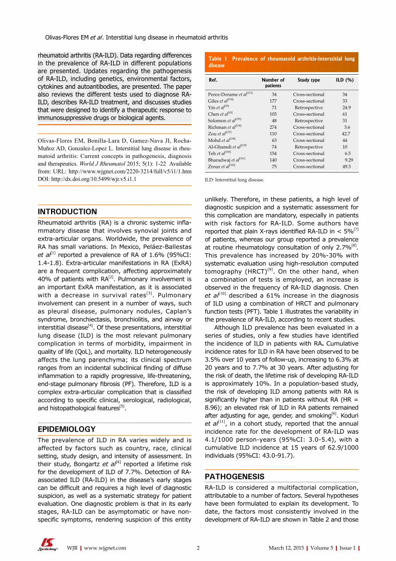
unlikely. Therefore, in these patients, a high level of diagnostic suspicion and a systematic assessment for this complication are mandatory, especially in patients with risk factors for RAILD. Some authors have reported that plain Xrays identified RAILD in < 5%[7] of patients, whereas our group reported a prevalence at routine rheumatology consultation of only 2.7%[8]. This prevalence has increased by 20%30% with systematic evaluation using highresolution computed tomography (HRCT)[9]. On the other hand, when a combination of tests is employed, an increase is observed in the frequency of RAILD diagnosis. Chen et al[10] described a 61% increase in the diagnosis of ILD using a combination of HRCT and pulmonary function tests (PFT). Table 1 illustrates the variability in the prevalence of RAILD, according to recent studies.
Although ILD prevalence has been evaluated in a series of studies, only a few studies have identified the incidence of ILD in patients with RA. Cumulative incidence rates for ILD in RA have been observed to be 3.5% over 10 years of followup, increasing to 6.3% at 20 years and to 7.7% at 30 years. After adjusting for the risk of death, the lifetime risk of developing RAILD is approximately 10%. In a populationbased study, the risk of developing ILD among patients with RA is significantly higher than in patients without RA (HR = 8.96); an elevated risk of ILD in RA patients remained after adjusting for age, gender, and smoking[6]. Koduri et al[11], in a cohort study, reported that the annual incidence rate for the development of RAILD was 4.1/1000 personyears (95%CI: 3.05.4), with a cumulative ILD incidence at 15 years of 62.9/1000 individuals (95%CI: 43.091.7).
PATHOGENESISRAILD is considered a multifactorial complication, attributable to a number of factors. Several hypotheses have been formulated to explain its development. To date, the factors most consistently involved in the development of RAILD are shown in Table 2 and those
Olivas-Flores EM et al . Interstitial lung disease in rheumatoid arthritis
2 March 12, 2015|Volume 5|Issue 1|WJR|www.wjgnet.com
Ref. Number of patients
Study type ILD (%)
Perez-Dorame et al[111] 34 Cross-sectional 34 Giles et al[154] 177 Cross-sectional 33 Yin et al[29] 71 Retrospective 24.9 Chen et al[10] 103 Cross-sectional 61 Solomon et al[155] 48 Retrospective 31 Richman et al[156] 274 Cross-sectional 3.6 Zou et al[157] 110 Cross-sectional 42.7 Mohd et al[158] 63 Cross-sectional 44 Al-Ghamdi et al[159] 74 Retrospective 10 Teh et al[160] 154 Cross-sectional 6.5 Bharadwaj et al[161] 140 Cross-sectional 9.29 Zrour et al[162] 75 Cross-sectional 49.3
Table 1 Prevalence of rheumatoid arthritis-interstitial lung disease
ILD: Interstitial lung disease.

can be classified as follows: (1) environmental; (2) genetic; (3) autoimmune (cytokines, autoantibodies); and (4) drugrelated[12].
Environmental factorsEpidemiological factors associated with ILD in RA include aging, smoking, and RA duration. Mori et al[12], in a prospective cohort study, observed a 4.58fold increase in the risk for development of ILD in patients aged ≥ 65 years (P = 0.003); additionally, the risk of ILD was higher in males than in females (50% vs 23.2%, respectively; OR = 3.31, P = 0.004). A relationship between smoking and an increase in the prevalence of ILD has been identified in several studies. Miyake et al[13] observed, in a casecontrol study, that smoking increases the risk for ILD 2.21fold. Saag et al[14] found a relationship between smoking and ILD, reporting an approximately 3.8fold increase in the risk for ILD among patients with a smoking history of ≥ 25 packyears. Baumgartner et al[15] reported, in a casecontrol study, that patients with a history of ever smoking or former smoking have 1.6 and 1.9fold increases in the risk of ILD, respectively. Occupational exposure, such as silica inhalation, contributes to the development of chronic lung inflammationrelated ILD[16].
Genetic factorsCoultas et al[17] reported that the prevalence of ILD is approximately 20% higher in males than in females. Aubart et al[18] observed that male gender increases the risk for ILD in RA by 3.29fold (P = 0.0013).
Several alleles are associated with an increased susceptibility for RAILD; susceptibility to RAILD can be triggered by environmental factors, leading to the development of ILD. Mori et al[12], in a prospective cohort study, observed that patients with RA who were carriers of the HLADRB1*1501 and *1502 alleles had an increased risk for ILD. Michalski et al[19] observed that α1antitrypsinvariant phenotypes, particularly nonM1M1 α1antitrypsin, are significantly associated
with PF in patients with RA. Charles et al[20] found an association between
antigen HLAB40 and pulmonary involvement of RA. The authors observed an enhanced risk of approximately 40.54fold in pulmonary involvement, compared with other ExRA manifestations. Sugiyama et al[21] reported an increase in the frequency of HLAB54 (63.2%) and HLADR4 (60%) polymorphisms in patients with ILDRA compared with controls (11.4% and 37.9%, respectively).
Cytokines and autoantibodies related to ILD in RA: Several cytokines have been linked to ILD. Chaudhary et al[22] observed, in an experimental model of PF, the profibrotic effects of plateletderived growth factor (PDGF), vascular endothelial growth factor (VEGF), and transforming growth factorbeta (TGF)β. The authors observed that targeting these molecules leads to an attenuation of lung fibrosis, suggesting that these cytokines may constitute a possible target for novel therapeutic approaches. Gochuico et al[23] quantified concentrations of TGFβ1, TGFβ2, PDGFAA, PDGFAB, PDGFBB, and interferon gamma (IFN)γ in fluids obtained by bronchoalveolar lavage (BAL) from 3 different group of patients: (1) RA without lung involvement; (2) RA with pulmonary fibrosis (RAPF); and (3) RA with preclinical ILD (RA preclinicalILD). They observed significantly higher concentrations of PDGFAB and PDGFBB in patients with RAILD compared with RA patients without PF, suggesting a profibrotic effect of the alveolar microenvironment in RA preclinicalILD. Interestingly, when the RAILD group was subcategorized into RA with progressive preclinical ILD and RA with stable preclinical ILD, the authors observed significantly higher concentrations of TGFβ1 and IFNγ in patients with RA with progressive preclinical ILD vs patients with RA with stable lung disease (P = 0.038 and P = 0.044, respectively). TGFβ is one of the strongest profibrotic cytokines; it triggers lung fibrosis, interacting with connective tissue growth factor (CTGF) to increase the fibrotic process. Ponticos et al[24] demonstrated, in an experimental model, that CTGF exerts a direct profibrotic effect on the development of PF through transcriptional activation of collagen gene type 1 α2 (Col1α2). Proinflammatory cells, such as macrophages and mononuclear cells, also contribute to the activation of fibrosis by means of interleukin (IL)4 and IL13, inducing TGFβ production. Jakubzick et al[25] observed, in an experimental model of PF, that IL4 and IL13 expression was increased in macrophages and mononuclear cells in regions of active fibrosis. Monocyte chemotactic protein (MCP)1 is also a profibrotic cytokine that exerts its action through chemokine receptor type 2 (CCR2). Moore et al[26] observed increased levels of MCP1 in the CCR2/ model compared with the wildtype (P = 0.004) after induction of PF in the wildtype and the CCR2/ experimental models. Furthermore, the authors reported that a lack of CCR2 is a protective
3 March 12, 2015|Volume 5|Issue 1|WJR|www.wjgnet.com
Factors
Environmental Cigarette smokingOccupational exposure (silica)
Demographic Male sexAge (≥ 65 yr)
Genetic HLA-DRB1 alleles Clinical RA duration
Anti-CCP (high titers)RF (high titers)
Medications Methotrexate Leflunomide Sulfasalazine
TNF-α inhibitors
Table 2 Risk factors for interstitial lung disease in rheumatoid arthritis
RF: Rheumatoid factor; HLA-DRB1: Human leukocyte antigen-DRB-1; Anti-CCP: Anti-cyclic citrullinated peptide; Anti-TNF: Anti-tumor necro-sis factor; RA: Rheumatoid arthritis.
Olivas-Flores EM et al . Interstitial lung disease in rheumatoid arthritis

association between MTX and the development of RAILD. Conway et al. reported, in a metaanalysis of randomized controlled trials from 19902013 that included 22 studies, that MTX treatment is a risk factor for the development of pneumonitis (RR = 7.81; 95%CI: 1.7634.72)[33]. Bongartz et al[6] also reported that treatment with MTX confers a 2.3fold risk for ILD development. However, Sathi et al[34] reported, in a prospective study of 223 patients, that the incidence of MTXinduced pneumonitis after 2 years of followup was only ~1%, suggesting that pneumonitis is an uncommon complication. Assessing the actual incidence of MTXinduced ILD is difficult because ILD can be observed in patients with RA independently of MTX treatment; furthermore, MTX is frequently used with other drugs that can also be associated with ILD. Therefore, the most useful data regarding MTXinduced ILD come from studies that evaluated this drug as monotherapy. In a systematic review, Salliot et al[35] examined the longterm safety of MTX as monotherapy in 21 prospective studies and reported that only 15 of 3463 patients developed pneumonitis, yielding a frequency of 0.43%. Criteria have been proposed for the diagnosis of MTXinduced pneumonitis. In 1987, Searles et al[36] proposed the following 9 criteria for the diagnosis of MTXinduced pneumonitis, which include: (1) acute onset of dyspnea; (2) fever > 38 ℃; (3) tachypnea; (4) radiological evidence of pulmonary interstitial or alveolar infiltrates; (5) white blood cell count < 15000/cu mm, with or without eosinophilia; (6) negative blood and sputum cultures (mandatory); (7) the finding of a restrictive pattern and decreased diffusing capacity of the lung for carbon monoxide (DLCO) on PFT; (8) PO2 < 60 mmHg on room air; and (9) histopathology consistent with bronchiolitis or interstitial pneumonitis with giant cells and without evidence of infection. MTXinduced pneumonitis is considered definite if ≥ 6 criteria are present, probable if 5 of 9 criteria are present, and possible if 4 of 9 criteria are present. Subsequently, new guidelines have been developed that include 3 major criteria, which are: (1) hypersensitivity pneumonitis by histopathology without evidence of a pathogenic organism; (2) radiologic evidence of pulmonary interstitial or alveolar infiltrates; and (3) negative blood and initial sputum cultures. The guidelines also include 5 minor criteria, which are (1) shortness of breath for less than 8 wk; (2) nonproductive cough; (3) oxygen saturation ≤ 90% on room air at the initial evaluation; (4) DLCO ≤ 70% of predicted for age; and (5) leukocyte count ≤ 15000 cells/cu mm3. The diagnosis is considered certain when a patient meets the first major criterion and at least 3 of the minor criteria, or when a patient meets major criteria 2 and 3 as well as 3 minor criteria. In this system, the diagnosis is considered probable when a patient meets only the major criteria 2 and 3 and 2 of the minor criteria[37]. Sidhu et al[4] reported that chest Xray findings included diffuse, bilateral, basal interstitial, or
factor against PF. Wilson et al[27] described enhanced levels of IL17 and IL1β in the BALF of patients with PF, suggesting that these cytokines play a profibrotic role in the lung fibrosis pathway. On the other hand, IL10, a wellrecognized antiinflammatory cytokine with immunosuppressive effects, has also been related to the induction of PF. Sun et al[28] observed, in an experimental model, that overexpression of IL10 in lung tissue promoted collagen production and induced recruitment of fibrocytes into the lung, leading to the development of PF in mice.
Antibodies to cyclic citrullinated peptides (antiCCP) and rheumatoid factor (RF) have also been associated with ILD. Yin et al[29], in a retrospective study, observed that serum levels of antiCCP2 and RF were significantly enhanced in RAILD patients compared with RA patients (P < 0.001 and P = 0.02, respectively). Kelly et al[30] identified positive titers for antiCCP and RF in 94% and 89%, respectively, of RAILD patients compared with RA patients (55%, P = 0.006; 58%, P = 0.01). Furthermore, they reported that antiCCP and RF act as predictors of ILD in patients with RA (P < 0.003 and P < 0.008, respectively). Citrullinated proteins are not only restricted to synovial tissue; they have also been detected at extraarticular sites in patients with RA. Bongartz et al[31] observed that citrullination occurs inside mononuclear cells in lung tissue in openlung biopsy specimens from patients with RAassociated interstitial pneumonia. The authors also reported that despite the high specificity of antiCCP for RA, citrullination was also found in lung tissue from patients with idiopathic interstitial pneumonia. It remains unclear whether distinct citrullinated RAspecific proteins play a key role in the pathophysiological process in RAILD.
Pharmacological agents as risk factors for RA-ILD: Presently, there is controversy regarding the actual effects of some medications on the development of ILD in patients with RA. Druginduced ILD can develop within days of treatment initiation or many years after treatment. The major drugs that have been strongly associated with the induction of ILD are methotrexate (MTX), leflunomide (LFN), sulfasalazine (SFZ), and tumor necrosis factorα (TNFα) inhibitors, such as etanercept, infliximab, and adalimumab. However, other drugs, including dpenicillamine and gold compounds, are also associated with lung damage. There have been recent case reports of the induction or exacerbation of ILD by the newer antiTNF agents, as well as other biologic agents that act by different mechanisms. This part of the review attempts to highlight the evidence linking these drugs to lung damage, primarily ILD.
Methotrexate and ILDMTX is considered by the European League Against Rheumatism to be part of the firstline treatment of RA[32], and several studies have reported an
4 March 12, 2015|Volume 5|Issue 1|WJR|www.wjgnet.com
Olivas-Flores EM et al . Interstitial lung disease in rheumatoid arthritis

alveolar infiltrates. The authors also observed that the most frequent radiographic pattern shown on HRCT of RAILD is the nonspecific interstitial pneumonia (NSIP) pattern. MTXassociated pneumonitis is described as a typeⅣ delayedhypersensitivity pneumonitis dominated by lymphocytic proliferation and alveolitis[38]; it is associated with a specific cellular immune response involving the release of cytokines[39]. Chikura et al[40], in a retrospective study, observed that the following two forms of lLD have been attributed to MTX. Type 1 MTXrelated ILD appears shortly after treatment initiation (< 6 mo) and is characterized by neutrophil infiltration, lung fibrosis, lower time of MTX to lowdose exposure, and a high mortality rate. Type 2 MTXrelated ILD occurs later in MTX treatment (> 6 mo) and is associated with lymphocytedominated infiltrates, low levels of lung fibrosis, a higher MTX dose exposure, and a low mortality rate. Type II pneumocyte hyperplasia and fibroblast proliferation have been reported as being suggestive of, but not pathognomonic for, MTXinduced lung toxicity[37]. A combination of a recent history of MTX initiation, clinical characteristics such as dyspnea, cough and fever, plus the findings of patchy groundglass opacities on HRCT, increased lymphocytes and eosinophils in the BAL, and (if available) a lung biopsy showing interstitial pneumonitis with nonnecrotizing granulomas and eosinophils, supports the diagnosis of MTXinduced ILD. To date, the optimal costeffective strategy for detecting ILD changes in patients who are beginning MTX treatment has not been identified. Khadadah et al[41] have suggested that periodic monitoring with PFT in patients with RA starting MTX therapy could be a rational strategy. Nevertheless, the findings of other authors do not support these recommendations. For example, Dawson et al[42] did not observe differences in PFT or HRCT findings between patients with RA who had been treated with MTX versus other drugs, concluding that serial PFT in patients receiving MTX has no significant advantages. Therefore, there is presently no conclusive information about whether to perform PFT or HRCT in patients who are receiving MTX and do not have clinical symptoms or signs suggesting lung toxicity. However, the use of MTX in patients with preexisting RAILD constitutes a significant risk factor for the development of pulmonary toxicity. Therefore, we recommend avoiding, if possible, the use of this drug in patients with a previous diagnosis of ILD. Other factors related to MTXinduced lung toxicity include elderly age, diabetes mellitus, and hypoalbuminemia, among others[43]. Once MTXinduced ILD is suspected, the treatment must include the immediate suspension of MTX and corticosteroid treatment, with the corticosteroid dosage depending on the severity of the lung involvement and other relevant clinical characteristics. In severe cases, supplementary oxygen, antibiotics or assisted ventilation should be considered. Once the patient is stabilized, MTX must be avoided, and alternative agents that do not increase the risk of
developing subsequent episodes of ILD should be considered. Among these options are the antimalarials. The prognosis of MTXinduced lung toxicity is usually good for the majority of patients, although a mortality rate of 13% has been reported in a review of MTXinduced pneumonitis in patients with a variety of different diseases (approximately 50% of whom had RA)[44].
LFN and lung damageEstablishing a clear association between LFN treatment and the development of ILD has been difficult, as LFN is frequently used after MTX failure; it can therefore be difficult to distinguish whether the development of ILD was secondary to LFN, MTX, or both drugs. Sawada et al[45] analyzed the results of a cohort of 5054 Japanese RA patients and observed the development of ILD in 1.2% of patients. Suissa et al[46] reported that LFN may enhance the risk of ILD by 1.9fold. Chikura et al[47] described, in a systematic review, that LFNinduced interstitial pneumonia occurs within the first 20 wk of LFN treatment initiation. Additionally, the authors reported a 19% mortality rate in patients with LFNassociated ILD. The factors associated with LFNinduced ILD were also analyzed by Sawada et al[45] and included the use of LFN in patients with preexisting ILD (OR = 8.17, 95%CI: 4.6314.41, P < 0.001), the use of an LFN loading dose (OR = 3.97, 95%CI: 1.2212.92, P = 0.02), cigarette smoking (OR = 3.12, 95%CI: 1.735.597, P = 0.001) and low body weight < 40 kg (OR = 2.91, 95%CI: 1.157.37, P = 0.02). Sato et al[48], in a retrospective study of patients with LFNinduced pulmonary injury, observed that an oxygen saturation level of < 90% is a marker for greater mortality in RAILD patients. The authors also found that serum Creactive protein level were higher (P = 0.03) and that the albumin level decreased (P = 0.03) at the outset of lung injury in patients with fatal outcomes in comparison with patients who recovered. It is relevant to highlight that the main histopathological finding reported in this study in the two autopsied patients was diffuse alveolar damage (DAD), in contrast to the alveolitis with lymphocyte infiltration observed in patients who recovered. The mechanism of the development of ILD in patients exposed to LFN could be related to the effects of the active metabolite A771726, which induces the transition of lung epithelial cells to myofibroblasts[49]. In addition to other established therapeutic strategies for ILD, such as corticosteroids and (if required) mechanical ventilation, some authors recommend the immediate suspension of LFN and the addition of cholestyramine as washout therapy, constituting a rational intervention for these patients[50].
Sulfasalazine and lung damageNumerous case reports have been published associating sulfasalazine (SSZ) with lung toxicity; a review of 50 cases[51] reported that most cases occurred
5 March 12, 2015|Volume 5|Issue 1|WJR|www.wjgnet.com
Olivas-Flores EM et al . Interstitial lung disease in rheumatoid arthritis

in patients with ulcerative colitis, although some cases were reported in RA patients. These authors noted that the clinical characteristics of SSZinduced lung toxicity include dyspnea of recent onset that is associated with lung infiltrates and, in more than half of cases, with peripheral eosinophilia and a variable spectrum of pathological findings; the most common pathologic findings were eosinophilic pneumonia and interstitial inflammation in some patients with lung fibrosis. To date, it is not conclusively known which drug component is primarily responsible for lung toxicity, although it is believed that the major culprit is sulfapyridine. Once SSZ lung toxicity is suspected, the drug should be withdrawn immediately. Stopping the drug is followed by the rapid improvement of symptoms and signs of lung toxicity in most cases, although some patients with SSZinduced lung toxicity may die, mainly if the drug is not withdrawn. Although a number of patients with SSZinduced lung toxicity have been managed with corticosteroids, evidence for the benefit of corticosteroids in this setting is not definitive, and further studies are required.
Azathioprine and lung damageLung toxicity associated with azathioprine (AZA) has been observed primarily in patients with kidney transplants and may result from both allergic and dosedependent toxicities. To date, there is only limited, case reportbased information suggesting that AZA may induce lung toxicity in patients with previous ILD. Ishida et al[52] reported the case of a male patient who developed interstitial pneumonia, was subsequently treated with AZA, and suffered worsening symptoms. The patient developed lung infiltrate and groundglass opacities on lung HRCT after only 6 d of treatment with AZA. These pulmonary infiltrates resolved after the suspension of AZA treatment. As a small proportion of patients may die, physicians should be aware of this complication in patients who have initiated treatment with AZA and have a recent onset of cough, fever and dyspnea.
Other synthetic disease-modifying antirheumatic drugs and lung damageCurrently, gold salts and dpenicillamine are infrequently used to treat RA. Goldinduced lung damage is a challenging diagnosis in RA. Tomioka et al[53] performed a review of published information regarding the clinical features and prognosis of goldinduced pulmonary disease in RA, identifying 140 cases of patients treated with gold, 81% of whom had RA. These authors reported that patients with goldinduced pulmonary damage frequently have other side effects associated with toxicity to gold salts, such as skin rash (38%), peripheral eosinophilia (38%), proteinuria (22%) and liver dysfunction (15%). In this review, factors frequently associated with goldinduced pulmonary disease included female sex, fever, skin rash, absence of rheumatoid nodules, low titers of rheumatoid factor,
lymphocytosis in BAL, and alveolar opacities along the bronchovascular bundles visualized on chest computed tomography. Patients generally improve after withdrawal of the gold salts and may require treatment with corticosteroids. Currently, dpenicillamine is rarely used. Chakravarty et al[54] reported that after 2 years of followup, 21% of their patients treated with dpenicillamine developed a restrictive pattern on PFT. Nevertheless, the incidence of severe pulmonary adverse reactions to dpenicillamine is relatively rare. Grove et al[55] evaluated common adverse reactions to synthetic diseasemodifying antirheumatic drugs (DMARDs) in 2170 patients with RA, who were followed for a total of 9378 treatmentyears. Of these, 582 patients were exposed to dpenicillamine during a total of 1889 monitored treatment years. Although this was an important series of patients treated with dpenicillamine, the authors were able to find only one patient who stopped dpenicillamine due to a severe pulmonary reaction.
Regarding synthetic DMARDinduced ILD, it is important to take into account the following points: (1) the ageadjusted incidence of MTXinduced pneumonitis is approximately 3.78 cases per 1000 patients treated with MTX[56]; (2) factors associated with MTXinduced ILD include: male gender, impairment in functioning, and elevated ESR[56]; (3) the initiation of MTX treatment, along with clinical manifestations including dyspnea, cough, fever, and patchy groundglass opacities on HRCT may suggest the diagnosis of MTXinduced ILD; (4) if MTXinduced ILD is suspected, the drug must be immediately discontinued; (5) the use of LFN increases the risk of ILD, which usually occurs within the first 20 wk after beginning this therapy[46,47]; (6) a relevant marker for mortality in LFNinduced ILD patients is a < 90% oxygen saturation level[48]; (7) patients with LFNinduced ILD must immediately stop treatment with LFN, begin corticosteroids, undergo mechanical ventilation if required, and receive cholestyramine washout treatment[50]; and (8) similar guidelines can be used to manage ILD induced by other synthetic DMARDs.
Biologic agents and lung damageTNFα inhibitors are commonly used for the treatment of RA and offer a good alternative in patients who have failed treatment with MTX or other synthetic DMARDs with high response rates. There are two major concerns with the use of antiTNF agents and RAILD: (1) the possible association between the use of antiTNF agents and the new onset of clinically significant ILD; and (2) the possibility of exacerbating preexisting ILD when an antiTNF agent is used for controlling disease activity in RA (despite reports that treatment with antiTNF agents may stabilize or improve ILD in some patients). The paradoxical effects of antiTNF agents in ILD are interesting, and further studies are required to identify why some patients improve while others develop worsening disease. We will briefly review some of the evidence regarding ILD related to
6 March 12, 2015|Volume 5|Issue 1|WJR|www.wjgnet.com
Olivas-Flores EM et al . Interstitial lung disease in rheumatoid arthritis

the use of antiTNF agents in RA. Presently, there is increasing evidence suggesting
that the use of TNFα inhibitors is associated with the development of ILD. RamosCasals et al[57], in a case series of 233 patients treated with antiTNF agents (71% of whom had RA), observed that 10% of patients developed ILD after initiation of antiTNF therapy; the mean time for developing ILD after receiving antiTNF drugs was 42 wk, and mortality was reported in 32% of patients with ILD.
There are a significant number of studies reporting the development of new cases of ILD or the worsening of preexisting ILD following the use of antiTNF agents, including infliximab[5860], etanercept[6164], and adalimumab[6568], as well as the newer antiTNF agents such as golimumab[69] and certolizumab pegol[7072].
The development of ILD with etanercept treatment has been described in approximately 0.6% of patients (77 cases from 13894 patients treated with etanercept)[73]. For infliximab, one study[74] reported an incidence of 0.5% for ILD (25 cases of ILD from 5000 patients treated). In another study, the incidence of ILD in patients receiving tocilizumab was 0.48%[75]. However, for abatacept the incidence in one study has been reported to range from 0.1% (shortterm) to 0.3% (longterm)[76].
PerezAlvarez et al[77] analyzed 122 cases of new onset or exacerbated ILD secondary to biologic agents. Of these, 58 cases were observed in patients receiving etanercept and 56 cases in those treated with infliximab. The majority of these patients had RA. ILD developed at a mean of 26 wk after initiation of the biologic agent. Fiftytwo patients had detailed followup; 29% died, 70% of these during the first weeks after the initiation of biologic agents.
Several mechanisms may explain the development of ILD associated with antiTNF agents. It is unclear whether TNF blockers can potentiate the pulmonary toxicity of MTX[78]. However, some of these agents, such as infliximab, bind to TNF that is expressed on the surface of macrophages and CD4+ and T cells, resulting in cell lysis[79]. It is thus conceivable that the local release of macrophagederived proteolytic enzymes may contribute to MTX toxicity. Other potential mechanisms for the development or progression of ILD and lung fibrosis in some patients receiving antiTNF agents may involve the downregulation of TNFα (due to TNF blockade), which causes the upregulation of antiinflammatory cytokines including transforming growth factor β, leading to a profibrotic state[80].
In the study by PerezAlvarez[77], patients with antecedents of ILD before being treated with biologic agents had a high mortality rate, which was associated with worsening ILD after the initiation of biologic therapy. Other factors associated with mortality were age > 65 years, later onset of ILD, and use of immunosuppressive drugs.
RamosCasals et al[81] analyzed 379 cases of autoimmune diseases secondary to antiTNF agents.
Using data obtained from the BIOGEAS project (www.biogeas.org), a study with the aim of collecting data on the use of biological agents in patients with systemic autoimmune diseases, RamosCasals reported cases of ILD induced by biological agents. These authors described 34 patients who developed ILD after the initiation of antiTNF agents, 30 of whom had RA. The most commonly used antiTNF agents were infliximab in 20 cases (59%), etanercept in 11 cases (32%) and adalimumab in 3 cases (9%). Interestingly, although the majority of the patients had received MTX, 11/31 patients (35%) of these patients had no history of MTX use. The use of antiTNF agents, particularly in the lung, has poor efficacy in controlling collagenosisassociated ILD and can lead to other complications, such as reactivation of mycobacterial and fungal infections, as well as to sarcoidosis and other ILD[81].
Most recently, the rate of mortality has been evaluated in patients with RA who had ILD before beginning treatment with antiTNF agents. The British Society for Rheumatology Biologics Register[82] followed 299 patients with preexisting RAILD who were treated with antiTNF agents, as well as 68 patients who were treated with synthetic DMARDs. In this cohort, 70/299 patients with preexisting ILD who were treated with antiTNF agents died, with RAILD being the underlying cause of death in 15/70 (21%) patients. However, 14/68 patients treated with synthetic DMARDs died; in only one patient (7%) was the cause of death related to ILD. Although the proportion of deaths attributable to RAILD in this study was higher in patients receiving antiTNF agents, the authors recognized the possibility of reporting bias that may have influenced the validity of their results.
Other biologic agents associated with ILDTo date, there has been one case report of a patient with RA who was treated with abatacept and developed worsening ILD[83]. Weinblatt et al[76] analyzed the data from 8 clinical trials of abatacept in RA and observed a rate of 0.1% (2 cases of 3173 patients analyzed) for the development of ILD in the shortterm period (≤ 12 mo). This rate increased to 0.3% (11 cases of 4149 abatacepttreated patients) in the pooled longterm period.
Some isolated cases of new ILD or exacerbations of preexisting ILD have been associated with the use of tocilizumab (TCZ). Kawashiri et al[84] described an exacerbation of preexisting ILD in a 68yearman with RA after 10 mo of treatment with TCZ. This patient died despite treatment with pulseddose steroids and antibiotics. The main pharmacological agents related to ILD in RA patients are summarized in Table 3.
Some points to remember in ILDassociated biologic agents include the following: (1) the incidence of newonset ILD with antiTNF agents is low, and in some studies probably does not differ from the incidence observed with MTX[85]; (2) although a higher incidence of newonset ILD is expected in RA patients
7 March 12, 2015|Volume 5|Issue 1|WJR|www.wjgnet.com
Olivas-Flores EM et al . Interstitial lung disease in rheumatoid arthritis

treated with antiTNF agents (compared with other CTD that are also treated with antiTNF therapy), this rate is approximately 7 times higher in RA compared with other diseases such as ankylosing spondylitis or psoriatic arthritis; (3) most reported cases of newonset or worsening ILD with antiTNF therapies are secondary to etanercept or infliximab[77]; (4) always suspect a worsening of ILD in patients with previous ILD who develop cough, dyspnea and fever; (5) most reported cases of newonset ILD or worsening of a previous ILD appear in the first year after initiation of biologics; in one report, the mean was 26 wk[77]; (6) in patients with baseline (before treatment initiation) ILD, the mortality attributable to ILD in patients treated with antiTNF agents is higher than those treated with synthetic DMARDs[82]; (7) characteristics supporting an association between ILD and treatment with biologics include recent initiation of therapy with a biologic agent, usually in elderly patients; most such patients show clinical improvement after the suspension of biologic agents and the addition of steroids; and (8) Treatment for patients with a suspicion for ILD induced or worsened by synthetic or biologic DMARDs should include the following elements: if there is a suspicion of druginduced pulmonary damage, the agent must be rapidly discontinued; the use of other drugs that may potentially be implicated in lung damage should be avoided; smokers should stop smoking; patients may receive supportive therapy, such as supplementary oxygen, treatment of concurrent respiratory infection with antibiotics or mechanical ventilation, as indicated; and corticosteroids are the most commonly used drug for the management of druginduced pulmonary damage and can be administered orally or intravenously at variable dosages. (In severe cases, prednisone should be administered at a dosage of 1 mg/kg. Other corticosteroids can be given at equivalent dosages, and, if required, a steroid pulse can be
used, particularly intravenous methylprednisolone at dosages of 1 g/d over 3 to 5 d). In patients with acute episodes, a clinical and symptomatic response can be observed around 2448 h after withdrawal of the offending drugs. However, in cases of chronic damage, this response can be delayed.
One study[77] described response rates in 52 cases of biologicassociated ILD: complete resolution was achieved in 40%, improvement or partial resolution in 25%, and no resolution in 35%. In this study, 29% of patients died during followup, with 70% of deaths occurring during the first 5 wk after the development or worsening of a previous biologicassociated ILD.
Importance of hepatitis C virus and lung damage in RA: Maillefert et al[86] observed that the prevalence of hepatitis C virus (HCV) in patients with RA was approximately 0.65% (taking into account both history of HCV or active infection) and did not differ from the prevalence of HCV infection in the general population. Nevertheless, HCV infection is relevant because patients with concurrent HCV and RA may have an increased prevalence of lung damage. Aliannejad et al[87] in a review, observed a discrepancy between studies evaluating the frequency of HCV in idiopathic pulmonary fibrosis (IPF) patients, which might be attributed to geographical differences for the prevalence of HCV infection or selection bias in choosing the control group. HCV infection is associated with increased counts of lymphocytes and neutrophils in BAL fluid. These studies have shown that HCV infection is associated with nonspecific pulmonary inflammatory reactions that lead in some patients to pulmonary fibrosis. The treatment of HCV infection, especially with interferon therapy, has also been implicated in the development of lung damage in HCV patients. Complications associated with INF therapy include interstitial pneumonia and pulmonary sarcoidosis. Ueda et al[88] reported a higher prevalence of HCV antibodies in patients with IPF (28.8%) compared with that observed in agematched control subjects (3.6%). Ferri et al[89], in a cohort of 300 HCVpositive patients, observed eight patients with interstitial lung involvement. In 6 patients, the presence of lung involvement was suspected on the basis of dyspnea with dry cough or digital clubbing. Different degrees of reduction in DLCO were observed; spirometric abnormalities, consistent with a global restrictive pattern, were found less frequently. The presence of parenchymal radiotracer uptake on G67 scan and an increased percentage of neutrophils and lymphocytes on BAL suggested active lung involvement. The treatment of HCV infection is associated with decreased pulmonary function. Foster et al[90] reported the results of a controlled clinical trial of 391 patients with HCV infection who received 24 wk of treatment with albIFNα2b or pegylated IFNα2a (pegIFNα2a) and ribavirin. Patients were followed over 6 mo with spirometry, DLCO, and chest
8 March 12, 2015|Volume 5|Issue 1|WJR|www.wjgnet.com
Pharmacological agent Relevant information
DMARDs MTX Long-term frequency of MTX-induced ILD is
0.43%[35]
Incidence is 3.78/1000 patients[56]
Risk factor for ILD in RA patients (RR = 7.81)[33]
LFN Increases the risk of developing ILD[46]
Mortality of 19% in patients with LFN-induced ILD[47]
AZA Complication of interstitial pneumonia after treatment with AZA[52]
TNF-α inhibitors Mortality is 32% in patients with ILD treated with TNF-α inhibitors[57]
Etanercept Incidence of etanercept-induced ILD is 0.6%[73]
Infliximab Incidence of infliximab-induced ILD is 0.5%[74]
Table 3 Pharmacological agents implicated in the development of interstitial lung disease in rheumatoid arthritis patients
DMARDs: Disease-modifying antirheumatic drugs; MTX: Methotrexate; ILD: Interstitial lung disease; RA: Rheumatoid arthritis; RR: Relative risk; LFN: Leflunomide; AZA: Azathioprine; TNF-α: Tumor necrosis factor-α.
Olivas-Flores EM et al . Interstitial lung disease in rheumatoid arthritis

Xray. During followup, DLCO declines of < 15% were observed in 173 (48%) of patients, whereas one patient developed new interstitial chest Xray abnormalities. The underlying mechanisms for this decline in pulmonary function in patient’s treatment with albIFNα2b or pegylated IFNα2a require further investigation.
BIOMARKERS FOR RA-ILDTo date, the use of RF and antiCCP as predictive biomarkers for ILD development in patients with RA remains controversial. Some evidence indicates that there is a clear association between high RF and antiCCP titer levels and RAILD[29]. However, other authors have not identified an association between antiCCP and RAILD[31].
In serum from patients with RAILD, Harlow et al[91] identified citrullinated heat shock proteins (Hsp) 90α and Hsp90β as potential biomarkers for ILD in patients with RA (Sensitivity, 0.29; Specificity, 0.96). Serum ferritin has been proposed as a prognostic marker in sclerodermaILD based on the finding that patients with higher ferritin levels at baseline (> 1500 μg/L) had a significantly increased risk of fatal outcomes[92]. To date, there has been a lack of information about serum ferritin in RAILD. However, in a crosssectional study, Rosas et al[93] observed significantly increased matrix metalloproteases (MMP)7 and MMP1 concentrations in the serum of patients with IPF (P = 0.01 and P < 0.001, respectively). Additionally, the authors reported that a combination of enhanced concentrations of MMP7 and MMP1 could discriminate IPF from hypersensitivity pneumonitis, with a sensitivity of 96.3% and a specificity of 87.2%[93]. Further studies of these metalloproteases in RAILD are required.
Ascherman et al[94] reviewed potential biomarkers implicated in RAILD. To date, the following cytokines have been considered as potential biomarkers of ILD: platelet derived growth factor isoforms AB and BB, interferonalpha, and profibrotic cytokine transforming growth factorB1. Elevated levels of these cytokines have been observed in BAL. High levels of Krebs von den Lungen6 protein (KL6) have been identified
in serum, reflecting alveolar damage. KL6 protein levels have demonstrated a correlation with the severity of ILD, as evaluated by HRCT[95]. The role of other potential biomarkers, such as surfactant proteinD (SPD), surfactant proteinA (SPA), and YKL40 chitinase3like protein 1, or cytokines such as chemokine motif ligand 18, which have been identified in other CTD complicated by lung involvement, should be evaluated in RAILD[96].
HISTOPATHOLOGYFive main histological patterns of ILD have been characterized, including NSIP, usual interstitial pneumonia (UIP), DAD, organizing pneumonia (OP), and lymphocytic interstitial pneumonia (LIP)[97]. The histological patterns of ILD and their relationship to clinical and radiological features are summarized in Table 4. The most frequent histological pattern of RAILD is UIP, followed by NSIP. In terms of severity, Kim et al[98] reported in 2010 that the UIP pattern in RAILD was associated with worse survival than the nonUIP pattern. In patients with UIP, the mean survival was 3.2 years; in patients with the nonUIP pattern, mean survival time was 6.6 years (P = 0.04). The severity and high mortality of the DAD pattern has been recognized. Tsuchiya et al[99] reported that patients with the DAD histological pattern of RAILD had the highest mortality, with a median survival time of 0.2 years.
DIAGNOSISClinical featuresThe clinical symptoms of RAILD are nonspecific. Dyspnea on exertion is the most frequent symptom, and cough, sputum production, wheezing, and chest pain have also been reported[100]. However, dyspnea and physical limitations may not be apparent in the early stages of disease.
Core set of domains in clinical trialsUsing Delphi and nominal group techniques, a group of experts recently proposed a preliminary core set of outcome measures in connective tissue diseaseassociated ILD (CTDILD) and idiopathic pulmonary fibrosis for use in clinical trials[101]. The results of this study included identification of the following domains to be measured in clinical trials: (1) dyspnea; (2) healthrelated quality of Life (HRQoL); (3) lung imaging; (4) lung physiology/function; (5) survival; and (6) medications.
The instruments accepted for each domain were derived from the Delphi Technique and are depicted in Figure 1[101]. Selection of this core of domains and instruments is very useful in diverse contexts in order to standardize the assessment of clinical responses across studies, rendering these results useful for systematic reviews or metaanalyses, and to facilitate
9 March 12, 2015|Volume 5|Issue 1|WJR|www.wjgnet.com
Histologic pattern Clinical-Radiological-Pathological Diagnosis
Usual interstitial pneumonia
Idiopathic pulmonary fibrosis/COP
NSIP NSIP Organizing pneumonia COP Diffuse alveolar damage Acute interstitial pneumonia LIP LIP
Table 4 Histological and clinical classification of idiopathic interstitial pneumonias
From: ref.[107], American Thoracic Society; European Respiratory Society. Am J Respir Crit Care Med 2002; 165: 277-304. COP: Cryptogenic fibrosing alveolitis; NSIP: Non-specific interstitial pneumonia; LIP: Lymphoid inter-stitial pneumonia.
Olivas-Flores EM et al . Interstitial lung disease in rheumatoid arthritis

the selection of outcome measures in multicenter randomized controlled trials.
The treatment of RAILD can be classified into supportive measures and treatment against the inflammatory processes that are responsible for ILD. To date, there is no specific treatment for RAILD. The best therapeutic strategy is believed to be a multidisciplinary approach that evaluates the severity of lung involvement, the type of pneumonitis, concomitant organs involved, and associated comorbidities. At our center, this therapeutic approach is performed by a rheumatologist, a pulmonologist, and a specialist in internal medicine. Included among supportive measures are supplementary therapy with oxygen, pulmonary rehabilitation, antireflux therapy, and treatment of comorbidities[102]. Many patients may have coexisting infections, and appropriate antimicrobial agents should be considered in such cases.
Six-minute walk test The sixminute walk test (6MWT) measures the distance that a patient can walk quickly on a flat, hard surface over a period of 6 min (6MWD). It evaluates the global and integrated responses of all of the systems involved in exercise, including the pulmonary and cardiovascular systems, systemic circulation, peripheral circulation, blood, neuromuscular units,
and muscle metabolism. It does not provide specific information on the function of each of the different organs and systems involved in exercise or on the mechanism of exercise limitation, as is possible with maximal cardiopulmonary exercise testing[103]. Changes in 6MWD after therapeutic interventions correlate with subjective improvements in dyspnea[104].
St. George’s Respiratory Questionnaire The St. George’s Respiratory Questionnaire was originally developed to assess the health status of patients with chronic obstructive pulmonary disease and asthma[105]. It has also been used for patients with other diseases, such as bronchiectasis and ILD[106]. Chang et al[106] observed that forced vital capacity (FVC)% was more strongly correlated with activity score than with symptom score. Similarly, on the chronic respiratory questionnaire, the dyspnea score was significantly correlated with FVC%, whereas the fatigue and emotional scores were not correlated.
PFTPatients with RAILD usually demonstrate a restrictive pattern on PFT with reduced total lung capacity (TLC), or a diminished FVC with a normal or increased forced expiratory volume at 1 second/forced vital capacity (FEV1/FVC) ratio and/or impaired gas exchange, which is characterized by an increased P (Aa) O2 (Alveolar
10 March 12, 2015|Volume 5|Issue 1|WJR|www.wjgnet.com
Dyspnea
Assessment instruments: Borg Dyspnea Index Dyspnea scale Borg dyspnea index pre and post-exercise
Survival
Issues to evaluate: Time to decline in FVC Progression-free survival
Medication
Issues to evaluate: Increase or decrease in glucocorticoids Increase or decrease in concomitant immunosuppressive agents
HRQoL
Assessment instruments: Medical Outcomes Trust Short form 36 (SF-36) Health survey Visual analog scale of patient Ability to carry out Activities of daily living Health assessment Questionnaire-disability index
CTD-ILD
Lung imaging
Issues to evaluate: Extent of honeycombing on HRCT Extent of ground-glass opacities on HRCT Overall extent of ILD on HRCT
Lung physiology/function
Issues to evaluate: Supplemental oxygen requirement FVC on spirometry Diffusion capacity of lung for carbon monoxide 6MWT with maximal desaturation on pulse oximetry
Figure 1 Suggested instruments to assess connective tissue disease associated interstitial lung disease, based on the Delphi Technique[101]. CTD-ILD: Connective tissue disease associated-interstitial lung disease; FVC: Forced vital capacity; HRQoL: Health-related quality of life; HRCT: High-resolution computed to-mography.
Olivas-Flores EM et al . Interstitial lung disease in rheumatoid arthritis

arterial pressure difference for O2), decreased PaO2 at rest or exertion, or decreases in the DLCO[107]. Chen et al[10] observed, in a crosssectional study of patients with RAILD, the presence of severe respiratory impairment [lower percent predicted FVC (74.9 ± 12.2 vs 86.9 ± 11.3; P < 0.001), TLC (87.8 ± 15.7 vs 98.4 ± 11.3; P = 0.001), FEV1 (74.1 ± 14.6 vs 88.0 ± 12.9; P < 0.001), and DLCO (68.1 ± 19.5 vs 96.2 ± 17.7; P < 0.001)] compared to RA patients without ILD. Saag et al[14], in a crosssectional study, found that worse functioning as evaluated by the Health Assessment Questionnaire DisabilityIndex (HAQDI), was a risk factor for declines in both the DLCO and FVC. However, Kim et al[98], in a retrospective study, observed that variables associated with a decrease in survival time in patients with RAILD included baseline FVC (HR = 0.98; P = 0.01), baseline DLCO (HR = 0.97; P = 0.002), and the presence of a UIP pattern on HRCT (HR = 2.09; P = 0.04).
Radiological findingsRadiographically, changes observed in RAILD are indistinguishable from those observed in IPF or ILD associated with other connectivetissue diseases. Plain chest Xrays mainly demonstrate reticular and fine nodular opacities. These findings are commonly concentrated in the lower lung zones. Early on, these changes may appear as a patchy, alveolarfilling infiltrate. Disease progression results in a more reticulonodular pattern. Plain chest Xray is an insensitive means for identifying ILD, which has a prevalence rate of only 6%[9]. Progression to nodular, patchy infiltrates may develop. Rarely, lymphadenopathy, rheumatoid nodules, and pleural effusions may be present[107]. Gabbay et al[9], in a crosssectional study, observed the prevalence of RAILD (14%) by employing a number of sensitive techniques in patients with RA for < 2 years.
High resolution computed tomography and histological correlationOne of the varied manifestations of ILD is asymptomatic disease that is detected by HRCT of the chest and PFT[108]. The American Thoracic Society and the European Respiratory Society (ATS/ERS), in collaboration with the Japanese Respiratory Society (JRS), and Latin American Thoracic Association (ALAT), published HRCT criteria for the diagnosis of UIP. The following are the main criteria for UIP in HRCT (all four features must be present): subpleural, basal predominance; reticular abnormality; honeycombing with or without traction bronchiectasis, and the absence of features listed as inconsistent with the UIP pattern. The criteria for possible UIP pattern include all features for the UIP pattern listed above, except for honeycombing. Inconsistent with the UIP pattern are any of the following seven features: upper or midlung predominance; peribronchovascular predominance; extensive groundglass abnormality (extent >
reticular abnormality); profuse micronodules (bilateral, predominantly upper lobes); discrete cysts (multiple, bilateral, at a distance from areas of honeycombing); diffuse mosaic attenuation/airtrapping (bilateral, in three or more lobes); and consolidation in bronchopulmonary segment(s)/lobe(s)[109].
Assayag et al[110] compared, in a cohort of 69 patients with RAILD, the usefulness of two computed tomography (CT) criteria and their correspondence with histopathologic patterns. Using the strict criteria, a definite UIP pattern on a CT scan had 96% specificity with histopathological findings and a positive predictive value of 95%. However, the sensitivity of the UIP pattern on CT scan was 45%, and when the broad criteria were used, the sensitivity of CT scan increased to 81%, with a decrease in specificity to 85%. Kim et al[98], in a retrospective study that included bivariate survival analysis of specific HRCT features in patients with RAILD, found that reticulation, traction bronchiectasis, and honeycombing were significantly associated with worse survival time. Cox regression modeling found that the presence and extent of traction bronchiectasis were significant independent predictors of worse survival time, with a hazard ratio (HR) 2.6; honeycombing had a HR for death of 2.1.
PérezDórame et al[111] observed, in a crosssectional study, the likelihood of NSIP being the most prevalent pattern on HRCT scans (29%). UIP patterns were observed in 13% of the patients. However, there was considerable overlap among tomographic patterns: 42% of patients had two ILD tomographic patterns, and 20% of patients also had small airway disease, defined as the presence of mosaic attenuation and airtrapping images.
Correlation between PFT and HRCTMcDonagh et al[112], in a crosssectional study, calculated the sensitivity and specificity of PFT, using HRCT as the gold standard. These authors observed that reduced FEV and low total lung capacity (TLC) [both > 1 Standard (SD) deviation below that predicted] were highly sensitive markers for of the presence of ILD on HRCT (88% and 90%, respectively). However, the specificity of each was relatively low (59% and 71%, respectively). The most sensitive test appeared to be measurement of residual volume (RV). A reduction of > 1 SD below the predicted RV was 83% specific for ILD.
Figure 2 describes a diagnosis strategy for patients with suspicion of RAILD. This strategy is based on the findings of clinical features and/or presence of risk factors for ILD in patients with a recognized RA. A recommendation is to perform a systematic assessment of the arterial blood gas, PFT and chest radiograph. If there is evidence in any of these tests that justify further investigation, we recommend a HRCT as the next step. HCRT may exclude or confirm the diagnosis of ILD, nevertheless in case of a reasonable suspicion justified by the clinical findings
11 March 12, 2015|Volume 5|Issue 1|WJR|www.wjgnet.com
Olivas-Flores EM et al . Interstitial lung disease in rheumatoid arthritis

with a HRCT that is not conclusive, probably invasive approaches, such as BAL or open lung biopsy should be considered.
Positron emission tomography and interstitial lung diseaseHRCT is an exclusively structural imaging technique from which only indirect inferences in relation to metabolism can be made. Recent technologic advances have led to the integration of positron emission
tomography (PET) with CT, allowing molecular imaging to be combined with the fine structural detail of CT. PET/CT has profoundly affected the management of cancer[113]. However, to date, PET/CT has not been used in patients with IPF and ILD[114]. PET with [18F]Fluorodeoxyglucose ([18F]FDG) can be used to quantify pulmonary inflammation. [18F]FDG, a glucose analog, is taken up by the same transporters that take up glucose into the cell; therefore, [18F]FDG uptake tracks cellular glucose transport, which
12 March 12, 2015|Volume 5|Issue 1|WJR|www.wjgnet.com
Rheumatoid arthritis
Clinical features
Bibasal inspiratory cracklesInsidious onset of unexplained dyspnea on exertion by more than 3 mo illness
Risk factors
Age > 65 yrCurrent smokingRheumatoid factorAnti-CCP antibodiesMTX or Anti-TNF-α treatment
Chest radiograph
Bilateral hilar lymphadenopathyNodulesGround-glass or reticular opacityPatchy bilateral consolidationBasal-predominant reticular abnormality with volume loss
Lung function test
DLCO < 70%FEV < 80%Reduced Total lung capacity Reduced Vital capacity Normal or increased FEV1/FVC ratio
Arterial blood gas analysisDLCO >70%
Increased P (A-a) O2 (alveolar- arterial pressure difference for CO2)Decreased PaO2 with exercise Other causes of
dyspnea
BAL or TBBx
Increased cellularity: lymphocytes and neutrophils
Non-diagnosis yet?
Open lung biopsy
Typical HRCT findings
UIPReticular, honeycombing Traction bronchiectasis/bronchiolectasis; architectural distortion. Focal ground glassNSIPGround glass attenuation Irregular lines. ConsolidationOP Patchy consolidation and/or nodulesDAD Consolidation and ground glass opacity, often with lobular sparing Traction bronchiectasis laterLIP Centrilobular nodules, ground glass attenuation, septal and bronchovascular thickening, thin-walled cysts
HRCTNon-diagnosis
Figure 2 Recommendations for the diagnosis of interstitial lung disease in rheumatoid arthritis patients. Anti-CCP: Anti-cyclic citrullinated peptide; MTX: methotrexate; Anti-TNF-α: Anti-tumor necrosis factor-α; DLCO: Diffusing capacity of the lung for carbon monoxide; FEV: Forced expiratory volume; FVC: Forced vital capacity; BAL: Bronchoalveolar lavage; TBBx: Transbronchial lung biopsy; HRCT: High-resolution computed tomography; UIP: Usual interstitial pneumonia; NSIP: Non-specific interstitial pneumonia; OP: Organizing pneumonia; DAD: Diffuse alveolar damage; LIP: Lymphocytic interstitial pneumonia.
Olivas-Flores EM et al . Interstitial lung disease in rheumatoid arthritis

is highly correlated with the rate of cellular glucose metabolism[115]. Increased pulmonary [18F]FDG metabolism in all patients with IPF and other forms of diffuse parenchymal lung disease was observed. Pulmonary 18FFDG uptake predicts measurements of health and lung physiology in these patients. 18FFDG metabolism was higher when the site of maximal uptake corresponded to areas of reticulation/honeycombing on HRCT, rather than to areas with groundglass patterns. To date, there are, to our knowledge, no studies evaluating lung metabolism in patients with RAILD, and longitudinal studies evaluating treatment based on pulmonary metabolism are required.
PULMONARY ARTERIAL HYPERTENSION AND RA-ILD Pulmonary arterial hypertension (PAH) may be an extraarticular manifestation of RA or may be associated with RAILD[116]. PAH in patients with RAILD who have either dyspnea or lung dysfunction [reduced carbon monoxide transfer factor (TLCO) or desaturation on exercise] can appear disproportionate to the extent of parenchymal lung disease. Transthoracic echocardiography is a suitable screening tool for detection of pulmonary hypertension in patients with ILD[102], and PAH can be confirmed with cardiac catheterization.
PHARMACOLOGICAL TREATMENTThere is only limited information derived from welldesigned clinical trials or prospective cohort studies regarding the efficacy of immunosuppressive or biological therapy for RAILD. Current understanding suggests that the therapeutic response depends on several factors, such as early detection of involvement, the radiologicalhistological subset (with lower rates of therapeutic response in fibrotic UIP compared with Bronchiolitis obliterans organizing pneumonia and nonfibrotic NSIP), and other comorbidities such as renal failure. There are several common clinical scenarios. The first scenario is an asymptomatic patient in whom ILD is discovered incidentally. In this patient, the decision to start treatment is not always easy, because ILD may remain stable in some of these patients for years, and aggressive therapy may cause severe, lifethreatening side effects. On the other hand, an incidental finding of ILD represents a window of opportunity for initiating treatment prior to clinical worsening. In this scenario, patients should initially be closely monitored monthly, and thereafter, at 36 mo intervals with PFT and 6MWT; in case of deterioration, immunosuppressive therapy should be considered. The second scenario is that of a patient with symptoms and clinical signs of ILD and a confirmed diagnosis based on PFT and HRCT. In these patients, immunosuppressive therapy against the inflammatory
process should be initiated. The third scenario involves a patient who has failed treatment with immunosuppressive drugs, has severe lung fibrosis, and has very few or absent signs of inflammation on HRCT. These patients generally do not benefit greatly from immunosuppressive therapy. If, after a course of corticosteroids and immunosuppressive drugs, such patients suffer rapid deterioration of FVC, diffuse PaO2 capacity of the lung for carbon dioxide (DLCO2), or clinical parameters, other therapies including lung transplantation should be considered (see later). In patients with moderate or severe symptoms and who have rapid progression of ILD (as reflected by a rapid deterioration of FVC and DLCO2 with an increase in dyspnea), corticosteroids are considered firstline treatment.
However, there is a lack of evidence from controlled studies regarding the effect size of corticosteroid treatment on the therapeutic response in RAILD. This lack of clinical trials is explained because ILD is a lifethreatening complication and ethically is not suitable for evaluation in placebocontrolled trials. One of the most recent studies evaluating the effect of corticosteroids on the therapeutic response was performed by RojasSerrano et al[117]. These authors, in a retrospective cohort design of 40 patients with RAILD treated with prednisone 1 mg/kg per day for 6 wk followed by tapering of 10 mg/d for approximately 6–8 mo, observed significant improvement in FVC at the final evaluation (compared with baseline values). However, the lack of a comparison group and the fact that the majority of these 40 patients with ILD concomitantly received MTX, AZA, or LFN limit the study’s usefulness in understanding the true effect of corticosteroids in these patients.
Ineffective agents Some medications have been used in CTDILD but have not demonstrated significant efficacy. These drugs include dpenicillamine and colchicine, which have been tested in systemic sclerosis but not in RAILD[118,119]. In an original study, Steen et al[118] evaluated the effects of dpenicillamine in 44 patients with systemic sclerosis compared with patients who did not receive this drug; while patients who received dpenicillamine had no further progression of dyspnea or fibrosis in chest Xrays during followup, there were no significant modifications in vital capacity (VC). In an open trial, van der Schee et al[120] evaluated the effects of dpenicillamine (750 mg/d) in seven patients with ILDRA. Patients also received prednisone 60 mg/d during month 1 with a gradual taper; VC and CO diffusion were measured prior to treatment, at 1 mo, and annually. Anecdotal reports have described some cases of patients with ILDRA who exhibited improvement after receiving cyclosporine[121].
Immunosuppressive agentsAzathioprine: Since the late 1970s, azathioprine and
13 March 12, 2015|Volume 5|Issue 1|WJR|www.wjgnet.com
Olivas-Flores EM et al . Interstitial lung disease in rheumatoid arthritis

corticosteroid therapy have been used for the treatment of RAILD in order to improve functional parameters and to stabilize lung inflammation. Cohen et al[122] published one of the first case reports, which discussed a patient with RAILD who had been treated for 5 years with azathioprine; improvements in pulmonary function and clinical symptoms were observed. Interestingly, there is also a lack of evidence from controlled studies regarding the efficacy of azathioprine in RAILD.
Cyclophosphamide: Cyclophosphamide (CYC) is an immunosuppressive drug commonly used to treat patients with ILD. A recent study[123] evaluated the effects of CYC on serum and bronchoalveolar lavage (BALF), TNFα, TGFβ1, and MMP9 levels, as well as TNFα and TGFβ1 messenger RNA (mRNA) levels, in the peripheral blood of patients with primary Sjögren’s syndrome with ILD. The results of this study showed that TNFα, TGFβ1, and MMP9 levels decreased significantly after CYC treatment.
The majority of evidence published on CYC in ILD has been derived from patients with systemic sclerosis who were treated with CYC. Although CYC is the “gold standard” immunosuppressant for the treatment of CTDILD, a metaanalysis[124] evaluating the evidence of three randomized clinical trials and six prospective cohorts evaluating the effect of CYC on systemic sclerosis and ILD did not observe significant changes in the FVC or DLCO after 12 mo of therapy, concluding that CYC treatment did not result in a clinically significant improvement of pulmonary function in these patients. However, when the individual studies are examined, there was wide variability in CYC doses and administration, with some studies evaluating oral CYC whereas others employed intravenous (iv) administration. The studies also differ in concurrent interventions; patients in some studies also received high doses of corticosteroids, others low corticosteroid doses, and in one study, corticosteroids were not used. Therefore, new studies with similar designs, inclusion criteria, and concurrent interventions are required to support the results of this metaanalysis. CYC therapy has also been used in patients with suspected druginduced ILD. In a case report, an RA patient with MTXinduced pneumonitis was considered resistant to withdrawal of MTX, oxygen administration, and pulsedose corticosteroids. This patient was treated with an iv CYC pulse, resulting in a substantial improvement in hypoxemia and Xray findings. The authors suggest that CYC should be considered in patients with MTXinduced pneumonitis without response to corticosteroids[125].
Mycophenolate mofetil: Mycophenolate mofetil (MMF) has been studied in patients with CTDILD. In a case series[126], 10 patients with autoimmune disorders complicated by ILD (three of whom had RA) received MMF. Symptomatic improvement was observed in 10/11 patients, and 4/5 discontinued oxygen. There
was stabilization or improvement in HRCT lesions in 8/8 patients, only 1/9 had worsening PFT, and patients were able to significantly decrease the dose of prednisone. The authors concluded that MMF is probably safer and more effective than CYC and should be considered as a firstline agent or a maintenance therapy after CYC treatment. However, these data are very preliminary and require corroboration in a controlled study that compares CYC vs MMF in ILDRA.
Combined therapy with methylprednisolone pulses: Combined therapy with methylprednisolone and CYC has been evaluated mainly in patients with systemic sclerosisassociated ILD. Yiannopoulos et al[127] evaluated 13 patients with systemic sclerosisassociated ILD, observing that 66.6% had stable or improved pulmonary function parameters. However, ILD worsened in some individuals after stopping treatment. The authors concluded that this combination is effective and welltolerated and helps to stabilize respiratory function in ILD. Airò et al[128] described the results of an observational study evaluating the results of the combination of CYC and 6methylprednisolone in 13 patients with systemic sclerosis and active alveolitis, observing an increase in FVC (P = 0.005) at 6 mo compared to baseline.
Biologic agents Tocilizumab: Tocilizumab is an interleukin (IL)6 receptor blocker useful in the treatment of joint symptoms and some systemic manifestations in RA. Excessive production of IL6 is associated with fibrosis in ILD; therefore, IL6 constitutes a potential target in the treatment of RAILD. Gallelli et al[129] have observed, in an in vitro study that used primary cultures of normal and fibrotic human lung fibroblasts, that the proliferative mechanisms induced by TGFβ1 are in part mediated by an increased release of IL6, leading to phosphorylationdependent mitogenactivated protein kinase (MAPK) activation. These findings help to understand the effects of therapies that are based on IL6 inhibition and their effects on lung fibroblasts. Mohr et al[130] described the results of tocilizumab in one patient with ILDRA, observing an improvement in alveolitis and groundglass opacities. Although the existing evidence is clearly insufficient to establish strong conclusions, it indicates the necessity of performing controlled studies to evaluate the efficacy of tocilizumab in these patients.
Anti-TNF agents and ILD: Only few case reports and case series have been published regarding patients with RAILD who may have benefited from antiTNF treatment. Bargagli et al[131] described the case of one patient with RA and pulmonary fibrosis, refractory to corticosteroids and azathioprine, who was treated with infliximab. These authors observed an improvement in vital capacity, TLCO and FEV1 after 15 mo of infliximab therapy. Similarly, Vassallo et al[132]
14 March 12, 2015|Volume 5|Issue 1|WJR|www.wjgnet.com
Olivas-Flores EM et al . Interstitial lung disease in rheumatoid arthritis

described a response to infliximab in a patient with RA and pulmonary fibrosis refractory to corticosteroids. After 12 mo of infliximab treatment, this patient had symptomatic improvement with stabilization of PFT. Additionally, Antoniou et al[133] identified responses to infliximab in a case series of 4 patients with CTDassociated pulmonary fibrosis (3 with RA and 1 with systemic sclerosis). The authors observed a stabilization of pulmonary fibrosis in terms of PFT results and HRCT findings after at least 12 mo of treatment. Etanercept is another antiTNF agent where a therapeutic response in ILD has been observed. Schultz et al[134] described a girl with juvenile chronic arthritis and pulmonary interstitial and intraalveolar cholesterol granulomas, in whom treatment with etanercept improved symptoms and physical capacities. Wang et al[135] described a therapeutic response to etanercept in a 52yearold woman with RAILD that was refractory to corticosteroids and azathioprine. These authors observed a sustained improvement in symptoms, PFT results, and HRCT findings.
There is controversy concerning whether antiTNF agents are associated with an increase in the prevalence of RAILD, and several case reports have been published on the development of RA in patients receiving antiTNF agents[65,66,7072,136]. In addition, cases have also been reported of patients with RAILD experiencing exacerbations of lung disease after receiving antiTNF therapy[61]. PerezAlvarez et al[77] analyzed 122 cases of newonset ILD or exacerbation of ILD in connective tissue diseases after administration of biological agents. Among these, 108 (89%) patients had RA. The drugs that were most frequently associated with ILD were etanercept (58 patients) and infliximab (56 patients); ILD developed at a mean of 26 wk after starting biological agents.
Rituximab: B cells are probably involved in the pathogenesis of RAILD. Atkins et al[137] have observed the presence of follicular Bhyperplasia and infiltration of the interstitium with plasma cells in patients with interstitial pneumonia. Observational and open uncontrolled studies have described the effects of rituximab (RTX) in patients with RAILD. Matteson et al[138] described the effects of RTX (1000 mg given on day 1 and day 15 and again after 24 and 26 wk) on 10 patients with RAILD who were evaluated in a 48wk, open clinical trial. At the end of the study, only 7/10 patients were assessed for therapeutic response. Among these patients, DLCO2 increased > 15% of baseline in 2/7 patients, remained stable in 4/7 patients, and worsened in 1/7 patients. However, the FVC increased by at least 10% in 2/7 patients, was stable in 4/7 patients, and declined in 1/7 patients. In the six patients who had a followup HRCT, findings remained unchanged in 5/6 and improved in 1/6. These preliminary data suggest that RTX benefits only some patients with RAILD; nevertheless, further controlled studies are required to identify the possible
effects of RTX on patients with established RAILD. Dass et al[139] described the safety of RTX among 67 patients with RA and lung involvement; of these, 48 patients (71.6%) had ILD. The authors observed 3 deaths (2 patients with ILD and 1 patient with chronic obstructive pulmonary disease), one of which was secondary to pneumonia and acute progression of ILD observed in the 4 wk after the first cycle of RTX. These authors conclude that treatment with RTX in patients with RA and lung involvement apparently does not increase the rate of expected severe side effects.
Romero et al[140] described the safety of RTX in a series of 14 patients with CTDILD, 29 of whom had RAILD. They observed a decreased incidence of ILD relapse during rituximab therapy (0.745/100 patientmonths) compared to 5.56/100 patientmonths during the pretreatment period. Only 12 patients had PFT results available during followup, demonstrating an increase in FVC and DLCO. Radiographic studies were available in 6 patients and demonstrated stabilization of ILD in 5/6 and improvement in 1/6. These authors conclude that RTX was safe in the sample studied, although there was 1 death secondary to neutropenia and a disseminated fungal infection during followup. Becerra et al[141] described the results of treatment with RTX in 38 patients with RA and lung involvement, 19 of whom had ILD. They observed that lung disease remained stable, although one patient with severe UIP developed progressive lung disease. Interestingly, 66% of the patients had respiratory infections, 2 of which required hospitalization. There were 2 deaths in this series, neither of which was related to RTX treatment. These authors conclude that RTX is a relatively safe therapy in patients with RA and lung involvement; however, there is no significant evidence to demonstrate improvement in lung disease.
Abatacept: Abatacept is a promising biologic agent for RA; nevertheless, there is a lack of studies evaluating the safety of abatacept in RAILD, and most information about this medication has been obtained from observational studies, particularly case reports. In a mice model of hypersensitivity pneumonitis characterized by T cellmediated lung inflammation, the administration of abatacept significantly decreased the extent of lung damage and decreased the number of inflammatory cells in the BAL[142]. Wada et al[83] reported the case of a 55yearold man with RA and interstitial pneumonia who deteriorated early after the administration of abatacept. This patient had a rapid clinical and radiographic deterioration of ILD that improved after abatacept was stopped. Nevertheless, other causes of ILD besides the abatacept should be considered, and additional information is required before establishing definite conclusions about the safety of abatacept in patients with RAILD.
Lung transplantation and RA-ILDSeveral studies have demonstrated that patients with
15 March 12, 2015|Volume 5|Issue 1|WJR|www.wjgnet.com
Olivas-Flores EM et al . Interstitial lung disease in rheumatoid arthritis

systemic sclerosis had similar rates of survival after lung transplantation compared with patients who had idiopathic pulmonary fibrosis or idiopathic pulmonary arterial hypertension[143]. Nevertheless, there are only few studies evaluating outcomes in patients with RAILD who underwent lung transplantation. Yazdani et al[144] performed a retrospective study to examine survival in 10 patients with RAILD who received a lung transplant, compared with 53 patients with IPF and 17 with systemic sclerosisILD (SScILD). The authors reported similar cumulative survival rates in RAILD compared to IPF (67% vs 69%, respectively), although the cumulative survival rate was higher in SScILD (82%). These data suggest that RAILD patients have a similar cumulative survival rate compared to other recipient of lung transplan, and therefore lung transplant should be considered in patients with refractory ILD who have not responded to other therapeutic strategies.
Other treatmentsSome treatments used for idiopathic pulmonary fibrosis have been infrequently investigated in patients with rheumatic disorders associated ILD. These treatments include (1) pirfenidone; (2) bosentan and sildenafil; (3) imatinib; and (4) warfarin. Pirfenidone is an antifibrotic drug that inhibits fibroblast proliferation and collagen synthesis and clinically is used for IPF. In an openlabel trial, Nagai et al[145] evaluated the effects of one year of treatment with oral pirfenidone (40 mg/kg body weight) in patients with advanced pulmonary fibrosis secondary to systemic sclerosis without observing a survival benefit, although these patients had no significant deterioration in chest radiographic findings or arterial oxygen pressure. To date, there have been no studies evaluating pirfenidone in RAILD. Therefore, new evidence derived from such studies is required. Bosentan is an endothelin1 antagonist used in patients with pulmonary arterial hypertension. However, most of the information of bosentan’s effects on CTDILD is derived from patients with systemic sclerosis. Mittoo et al[146] performed a retrospective assessment of 13 patients with CTDILD and pulmonary hypertension. Only 2/13 of these patients had RAILD. These patients received bosentan alone, sildenafil alone or bosentan plus sildenafil. This study found that the drugs used to treat pulmonary hypertension were well tolerated, with higher mortality rates among patients with systemic sclerosis compared with other CTD. New studies evaluating bosentan in RAILD are required to draw definite conclusions. Imatinib mesylate inhibits the activation of the PDGF receptor, as well as the cAbl, BcrAbl and cKit tyrosine kinases. Consequently, imatinib mesylate suppresses the activation and proliferation of fibroblasts, requiring this drug to be evaluated in RAILD[147]. Warfarin has been only evaluated in retrospective studies. Watanabe et al[148] performed a retrospective analysis of 20 patients with
rapidly progressive interstitial pneumonia, 11 cases of which were secondary to CTD (2/11 were due to rheumatoid arthritis). These authors classified the patients into 2 groups: group A, which included 11 patients treated with anticoagulant therapy (warfarin or dalteparin), and group B, which included 9 patients who did not receive anticoagulation. At the end of the study, patients treated with anticoagulation had a better survival rate compared with the nonanticoagulated group (P = 0.038). Nevertheless, this evidence is too weak to recommend the use of warfarin in patients with RAILD. Nacetylcysteine is an antioxidant, acts as a scavenger for free radicals and has antiinflammatory properties. This agent also suppresses the production of TNFα and TGFβ by alveolar macrophages in patients with idiopathic pulmonary fibrosis[149]. Nacetylcysteine is an interesting drug in idiopathic pulmonary fibrosis, where it is widely used as an adjuvant therapy, although recent data did not demonstrate significant differences between Nacetylcysteine vs placebo in terms of FVC, frequency of exacerbations or mortality rates[150]. To date, limited information exists about the effects of Nacetylcysteine in CTDILD. Rosato et al[151] evaluated, in a retrospective study, the effects of intravenous Nacetylcysteine in patients with systemic sclerosis, observing a decrease in the rate of deterioration of DLCO, VC and TLC. Nevertheless, to date, no studies have reported the effects of Nacetylcysteine in RAILD. Evidence against the use of this drug has appeared in one study that demonstrated an increased risk of death and hospitalization in patients with idiopathic pulmonary fibrosis who received a combination of prednisone, azathioprine and Nacetylcysteine compared with patients who received placebo.
PROGNOSISPredictors of mortality include older age, male sex, lower socioeconomic status, decreased lung function, the presence of fibrosis, the extent of disease, the presence of a lunginjury pattern of usual interstitial pneumonia, higher disease activity scores, higher erythrocyte sedimentation rates, higher lactate dehydrogenase levels, greater baseline pain, and worse health assessment questionnaire scores[152]. Average survival in patients with RA is 1011 years shorter than that of the general population. Lung disease is especially common in RA and is directly responsible for 10%20% of all RAassociated mortality[153]. A retrospective study by Kelly et al[30] demonstrated that mortality rates were related to the subtype of lung disease; patients with a UIP/OS pattern had an RR of death from any cause of 3.9 compared with patients who had a pattern of NSIP/cryptogenic organizing pneumonia (COP). These authors observed during followup that, compared with limited disease, extensive disease was associated with an RR of death from any cause of 2.17.
16 March 12, 2015|Volume 5|Issue 1|WJR|www.wjgnet.com
Olivas-Flores EM et al . Interstitial lung disease in rheumatoid arthritis

Gochuico et al[23] examined the differences between progressive RAILD and stable RAILD. Higher alveolar concentrations of IFNγ and TGFβ2 were observed in patients with progressive RAILD versus stable RAILD. Additionally, patients with progressive RAILD were more likely to be treated with MTX, suggesting that treatment with this agent may constitute a risk factor for progression of preclinical RAILD. Assayag et al[152] performed a systematic review evaluating predictors of mortality in RAILD. Factors associated with higher mortality rates were older age, male gender, lower DLCO, extent of fibrosis and UIP pattern. Nevertheless, the authors recognized that the review was limited by the low quality of some of the included studies; therefore, larger, welldesigned, multicenter studies evaluating prognostic factors in RAILD are still required.
CONCLUSIONRecent evidence indicates that ILD is presently observed more frequently in RA than was the case a decade ago. Establishing an early diagnosis of this complication depends on the level of clinical suspicion, as well as the strategy used to assess patients at risk of ILD. The adequate assessment of patients with suspected ILD should be based on a combination of tests, including clinical assessments, PFT, HRCT, and in some cases BAL or lung biopsy. Currently, distinct clinical subtypes of RAILD are recognized that may differ importantly in terms of prognosis and therapeutic response. Efforts to identify the subtype of RAILD should be made in order to design a therapeutic strategy that will be of the greatest benefit to a particular patient. In terms of treatment, recently identified therapeutic targets have produced new drugs for evaluation. Nevertheless, most of the information about these treatments is derived from observational or uncontrolled open studies. Therefore, evidence about the effectiveness of these agents is too weak to establish definite conclusions in patients with RAILD. New welldesigned, randomized, multicenter, doubleblinded clinical trials are needed to evaluate the use of novel therapeutic agents in RAILD. This represents an important opportunity for future research.
REFERENCES1 Peláez-Ballestas I, Sanin LH, Moreno-Montoya J, Alvarez-Neme-
gyei J, Burgos-Vargas R, Garza-Elizondo M, Rodríguez-Amado J, Goycochea-Robles MV, Madariaga M, Zamudio J, Santana N, Cardiel MH. Epidemiology of the rheumatic diseases in Mexico. A study of 5 regions based on the COPCORD methodology. J Rheumatol Suppl 2011; 86: 3-8 [PMID: 21196592 DOI: 10.3899/jrheum.100951]
2 Turesson C, O’Fallon WM, Crowson CS, Gabriel SE, Matteson EL. Extra-articular disease manifestations in rheumatoid arthritis: incidence trends and risk factors over 46 years. Ann Rheum Dis 2003; 62: 722-727 [PMID: 12860726 DOI: 10.1136/ard.62.8.722]
3 Olson AL, Swigris JJ, Sprunger DB, Fischer A, Fernandez-Perez
ER, Solomon J, Murphy J, Cohen M, Raghu G, Brown KK. Rheu-matoid arthritis-interstitial lung disease-associated mortality. Am J Respir Crit Care Med 2011; 183: 372-378 [PMID: 20851924 DOI: 10.1164/rccm.201004-0622OC]
4 Sidhu HS, Bhatnagar G, Bhogal P, Riordan R. Imaging features of the pleuropulmonary manifestations of rheumatoid arthritis: pearls and pitfalls. J Clin Imaging Sci 2011; 1: 32 [PMID: 21966629 DOI: 10.4103/2156-7514.82244]
5 AmericanThoracicSociety.Idiopathicpulmonaryfibrosis:diagno-sis and treatment. International consensus statement. American Tho-racic Society (ATS), and the European Respiratory Society (ERS). Am J Respir Crit Care Med 2000; 161: 646-664 [PMID: 10673212 DOI: 10.1164/ajrccm.161.2.ats3-00]
6 Bongartz T, Nannini C, Medina-Velasquez YF, Achenbach SJ, Crowson CS, Ryu JH, Vassallo R, Gabriel SE, Matteson EL. Inci-dence and mortality of interstitial lung disease in rheumatoid arthri-tis: a population-based study. Arthritis Rheum 2010; 62: 1583-1591 [PMID: 20155830 DOI: 10.1002/art.27405]
7 Morrison SC, Mody GM, Benatar SR, Meyers OL. The lungs in rheumatoid arthritis--a clinical, radiographic and pulmonary func-tion study. S Afr Med J 1996; 86: 829-833 [PMID: 8764910]
8 Gonzalez-Lopez L, Rocha-Muñoz AD, Ponce-Guarneros M, Flores-Chavez A, Salazar-Paramo M, Nava A, Cardona-Muñoz EG, Fajardo-Robledo NS, Zavaleta-Muñiz SA, Garcia-Cobian T, Gamez-Nava JI. Anti-cyclic citrullinated peptide (anti-CCP) and anti-mutated citrullinated vimentin (anti-MCV) relation with extra-articular manifestations in rheumatoid arthritis. J Immunol Res 2014; 2014: 536050 [PMID: 24804270 DOI: 10.1155/2014/536050]
9 Gabbay E, Tarala R, Will R, Carroll G, Adler B, Cameron D, Lake FR. Interstitial lung disease in recent onset rheumatoid arthritis. Am J Respir Crit Care Med 1997; 156: 528-535 [PMID: 9279235 DOI: 10.1164/ajrccm.156.2.9609016]
10 Chen J, Shi Y, Wang X, Huang H, Ascherman D. Asymptomatic preclinical rheumatoid arthritis-associated interstitial lung disease. Clin Dev Immunol 2013; 2013: 406927 [PMID: 23983768 DOI: 10.1155/2013/406927]
11 Koduri G, Norton S, Young A, Cox N, Davies P, Devlin J, Dixey J, GoughA,ProuseP,WinfieldJ,WilliamsP.Interstitiallungdiseasehas a poor prognosis in rheumatoid arthritis: results from an incep-tion cohort. Rheumatology (Oxford) 2010; 49: 1483-1489 [PMID: 20223814 DOI: 10.1093/rheumatology/keq035]
12 Mori S, Koga Y, Sugimoto M. Different risk factors between interstitial lung disease and airway disease in rheumatoid arthri-tis. Respir Med 2012; 106: 1591-1599 [PMID: 22867979 DOI: 10.1016/j.rmed.2012.07.006]
13 Miyake Y, Sasaki S, Yokoyama T, Chida K, Azuma A, Suda T, Kudoh S, Sakamoto N, Okamoto K, Kobashi G, Washio M, Inaba Y, Tanaka H. Occupational and environmental factors and idiopathic pulmonary fibrosis in Japan. Ann Occup Hyg 2005; 49: 259-265 [PMID: 15640309 DOI: 10.1093/annhyg/meh090]
14 Saag KG, Kolluri S, Koehnke RK, Georgou TA, Rachow JW, Hun-ninghake GW, Schwartz DA. Rheumatoid arthritis lung disease. Determinants of radiographic and physiologic abnormalities. Ar-thritis Rheum 1996; 39: 1711-1719 [PMID: 8843862 DOI: 10.1002/art.1780391014]
15 Baumgartner KB, Samet JM, Stidley CA, Colby TV, Waldron JA. Cigarettesmoking:ariskfactorforidiopathicpulmonaryfibrosis.Am J Respir Crit Care Med 1997; 155: 242-248 [PMID: 9001319 DOI: 10.1164/ajrccm.155.1.9001319]
16 Cavagna L,MontiS,GrossoV,BoffiniN,ScorlettiE,CrepaldiG,Caporali R. The multifaceted aspects of interstitial lung disease in rheumatoid arthritis. Biomed Res Int 2013; 2013: 759760 [PMID: 24205507 DOI: 10.1155/2013/759760]
17 Coultas DB, Zumwalt RE, Black WC, Sobonya RE. The epi-demiology of interstitial lung diseases. Am J Respir Crit Care Med 1994; 150: 967-972 [PMID: 7921471 DOI: 10.1164/ajrc-cm.150.4.7921471]
18 Aubart F, Crestani B, Nicaise-Roland P, Tubach F, Bollet C, Da-widowicz K, Quintin E, Hayem G, Palazzo E, Meyer O, Chollet-Martin S, Dieudé P. High levels of anti-cyclic citrullinated peptide
17 March 12, 2015|Volume 5|Issue 1|WJR|www.wjgnet.com
Olivas-Flores EM et al . Interstitial lung disease in rheumatoid arthritis

autoantibodies are associated with co-occurrence of pulmonary diseases with rheumatoid arthritis. J Rheumatol 2011; 38: 979-982 [PMID: 21362759 DOI: 10.3899/jrheum.101261]
19 Michalski JP, McCombs CC, Scopelitis E, Biundo JJ, Medsger TA. Alpha 1-antitrypsin phenotypes, including M subtypes, in pul-monary disease associated with rheumatoid arthritis and systemic sclerosis. Arthritis Rheum 1986; 29: 586-591 [PMID: 3487321 DOI: 10.1002/art.1780290502]
20 Charles PJ, Sweatman MC, Markwick JR, Maini RN. HLA-B40: a marker for susceptibility to lung disease in rheumatoid arthritis. Dis Markers 1991; 9: 97-101 [PMID: 1782749]
21 Sugiyama Y, Ohno S, Kano S, Maeda H, Kitamura S. Diffuse pan-bronchiolitis and rheumatoid arthritis: a possible correlation with HLA-B54. Intern Med 1994; 33: 612-614 [PMID: 7827377 DOI: 10.2169/internalmedicine.33.612]
22 Chaudhary NI, Roth GJ, Hilberg F, Müller-Quernheim J, Prasse A, Zissel G, Schnapp A, Park JE. Inhibition of PDGF, VEGF and FGFsignallingattenuatesfibrosis.Eur Respir J 2007; 29: 976-985 [PMID: 17301095 DOI: 10.1183/09031936.00152106]
23 Gochuico BR, Avila NA, Chow CK, Novero LJ, Wu HP, Ren P, MacDonald SD, Travis WD, Stylianou MP, Rosas IO. Progressive preclinical interstitial lung disease in rheumatoid arthritis. Arch Intern Med 2008; 168: 159-166 [PMID: 18227362 DOI: 10.1001/archinternmed.2007.59]
24 Ponticos M, Holmes AM, Shi-wen X, Leoni P, Khan K, Rajkumar VS, Hoyles RK, Bou-Gharios G, Black CM, Denton CP, Abraham DJ, Leask A, Lindahl GE. Pivotal role of connective tissue growth factorinlungfibrosis:MAPK-dependenttranscriptionalactivationof type I collagen. Arthritis Rheum 2009; 60: 2142-2155 [PMID: 19565505 DOI: 10.1002/art.24620]
25 Jakubzick C, Kunkel SL, Puri RK, Hogaboam CM. Therapeutic targeting of IL-4- and IL-13-responsive cells in pulmonary fibrosis. Immunol Res 2004; 30: 339-349 [PMID: 15531774 DOI: 10.1385/ir]
26 Moore BB, Paine R, Christensen PJ, Moore TA, Sitterding S, Ngan R, Wilke CA, Kuziel WA, Toews GB. Protection from pulmonary fibrosisintheabsenceofCCR2signaling.J Immunol 2001; 167: 4368-4377 [PMID: 11591761 DOI: 10.4049/jimmunol.167.8.4368]
27 Wilson MS, Madala SK, Ramalingam TR, Gochuico BR, Rosas IO, Cheever AW, Wynn TA. Bleomycin and IL-1beta-mediated pulmo-naryfibrosisisIL-17Adependent.J Exp Med 2010; 207: 535-552 [PMID: 20176803 DOI: 10.1084/jem.20092121]
28 Sun L, Louie MC, Vannella KM, Wilke CA, LeVine AM, Moore BB, Shanley TP. New concepts of IL-10-induced lung fibrosis: fibrocyterecruitmentandM2activationinaCCL2/CCR2axis.Am J Physiol Lung Cell Mol Physiol 2011; 300: L341-L353 [PMID: 21131395 DOI: 10.1152/ajplung.00122.2010]
29 Yin Y, Liang D, Zhao L, Li Y, Liu W, Ren Y, Li Y, Zeng X, Zhang F, Tang F, Shan G, Zhang X. Anti-cyclic citrullinated Peptide antibody is associated with interstitial lung disease in patients with rheuma-toid arthritis. PLoS One 2014; 9: e92449 [PMID: 24743261 DOI: 10.1371/journal.pone.0092449]
30 Kelly CA, Saravanan V, Nisar M, Arthanari S, Woodhead FA, Price-Forbes AN, Dawson J, Sathi N, Ahmad Y, Koduri G, Young A. Rheumatoid arthritis-related interstitial lung disease: associations, prognostic factors and physiological and radiological characteristics--a large multicentre UK study. Rheumatology (Oxford) 2014; 53: 1676-1682 [PMID: 24758887 DOI: 10.1093/rheumatology/keu165]
31 Bongartz T, Cantaert T, Atkins SR, Harle P, Myers JL, Turesson C, Ryu JH, Baeten D, Matteson EL. Citrullination in extra-articular manifestations of rheumatoid arthritis. Rheumatology (Oxford) 2007; 46: 70-75 [PMID: 16782731 DOI: 10.1093/rheumatology/kel202]
32 Smolen JS, Landewé R, Breedveld FC, Buch M, Burmester G, Dougados M, Emery P, Gaujoux-Viala C, Gossec L, Nam J, Ramiro S, Winthrop K, de Wit M, Aletaha D, Betteridge N, Bijlsma JW, Boers M, Buttgereit F, Combe B, Cutolo M, Damjanov N, Hazes JM, Kouloumas M, Kvien TK, Mariette X, Pavelka K, van Riel PL, Rubbert-Roth A, Scholte-Voshaar M, Scott DL, Sokka-Isler T, Wong JB, van der Heijde D. EULAR recommendations for the management of rheumatoid arthritis with synthetic and biological
disease-modifying antirheumatic drugs: 2013 update. Ann Rheum Dis 2014; 73: 492-509 [PMID: 24161836 DOI: 10.1136/annrheum-dis-2013-204573]
33 Conway R, Low C, Coughlan RJ, O’Donnell MJ, Carey JJ. Metho-trexate and lung disease in rheumatoid arthritis: a meta-analysis of randomized controlled trials. Arthritis Rheumatol 2014; 66: 803-812 [PMID: 24757133 DOI: 10.1002/art.38322]
34 Sathi N, Chikura B, Kaushik VV, Wiswell R, Dawson JK. How common is methotrexate pneumonitis? A large prospective study investigates. Clin Rheumatol 2012; 31: 79-83 [PMID: 21638023 DOI: 10.1007/s10067-011-1758-6]
35 Salliot C, van der Heijde D. Long-term safety of methotrexate monotherapy in patients with rheumatoid arthritis: a systematic literature research. Ann Rheum Dis 2009; 68: 1100-1104 [PMID: 19060002 DOI: 10.1136/ard.2008.093690]
36 Searles G, McKendry RJ. Methotrexate pneumonitis in rheumatoid arthritis: potential risk factors. Four case reports and a review of the literature. J Rheumatol 1987; 14: 1164-1171 [PMID: 3325643]
37 Kremer JM, Alarcón GS, Weinblatt ME, Kaymakcian MV, Ma-caluso M, Cannon GW, Palmer WR, Sundy JS, St Clair EW, Alex-ander RW, Smith GJ, Axiotis CA. Clinical, laboratory, radiographic, and histopathologic features of methotrexate-associated lung injury in patients with rheumatoid arthritis: a multicenter study with litera-ture review. Arthritis Rheum 1997; 40: 1829-1837 [PMID: 9336418 DOI: 10.1002/1529-0131(199710)40]
38 Mornex JF, Cordier G, Pages J, Vergnon JM, Lefebvre R, Brune J, Revillard JP. Activated lung lymphocytes in hypersensitivity pneumonitis. J Allergy Clin Immunol 1984; 74: 719-727 [PMID: 6209321 DOI: 10.1016/0091-6749(84)90236-7]
39 Akoun GM, Gauthier-Rahman S, Mayaud CM, Touboul JL, De-nis MF. Leukocyte migration inhibition in methotrexate-induced pneumonitis. Evidence for an immunologic cell-mediated mecha-nism. Chest 1987; 91: 96-99 [PMID: 3539547 DOI: 10.1378/chest.91.1.96]
40 Chikura B, Sathi N, Lane S, Dawson JK. Variation of immunologi-cal response in methotrexate-induced pneumonitis. Rheumatology (Oxford) 2008; 47: 1647-1650 [PMID: 18812430 DOI: 10.1093/rheumatology/ken356]
41 Khadadah ME, Jayakrishnan B, Al-Gorair S, Al-Mutairi M, Al-Maradni N, Onadeko B, Malaviya AN. Effect of methotrexate on pulmonary function in patients with rheumatoid arthritis--a prospec-tive study. Rheumatol Int 2002; 22: 204-207 [PMID: 12215867 DOI: 10.1007/s00296-002-0227-6]
42 Dawson JK, Graham DR, Desmond J, Fewins HE, Lynch MP. Investigation of the chronic pulmonary effects of low-dose oral methotrexate in patients with rheumatoid arthritis: a prospective study incorporating HRCT scanning and pulmonary function tests. Rheumatology (Oxford) 2002; 41: 262-267 [PMID: 11934961]
43 Roubille C, Haraoui B. Interstitial lung diseases induced or exac-erbated by DMARDS and biologic agents in rheumatoid arthritis: a systematic literature review. Semin Arthritis Rheum 2014; 43: 613-626 [PMID: 24231065 DOI: 10.1016/j.semarthrit.2013.09.005]
44 Imokawa S, Colby TV, Leslie KO, Helmers RA. Methotrexate pneumonitis:reviewoftheliteratureandhistopathologicalfindingsin nine patients. Eur Respir J 2000; 15: 373-381 [PMID: 10706507]
45 Sawada T, Inokuma S, Sato T, Otsuka T, Saeki Y, Takeuchi T, Mat-suda T, Takemura T, Sagawa A. Leflunomide-induced interstitial lung disease: prevalence and risk factors in Japanese patients with rheumatoid arthritis. Rheumatology (Oxford) 2009; 48: 1069-1072 [PMID: 19321513 DOI: 10.1093/rheumatology/kep052]
46 Suissa S,HudsonM,ErnstP.Leflunomideuseandtheriskofin-terstitial lung disease in rheumatoid arthritis. Arthritis Rheum 2006; 54: 1435-1439 [PMID: 16645972 DOI: 10.1002/art.21806]
47 Chikura B, Lane S, Dawson JK. Clinical expression of leflun-omide-induced pneumonitis. Rheumatology (Oxford) 2009; 48: 1065-1068 [PMID: 19321511 DOI: 10.1093/rheumatology/kep050]
48 Sato T, Inokuma S, Sagawa A, Matsuda T, Takemura T, Otsuka T, Saeki Y, Takeuchi T, Sawada T. Factors associated with fatal out-comeofleflunomide-inducedlunginjuryinJapanesepatientswithrheumatoid arthritis. Rheumatology (Oxford) 2009; 48: 1265-1268
18 March 12, 2015|Volume 5|Issue 1|WJR|www.wjgnet.com
Olivas-Flores EM et al . Interstitial lung disease in rheumatoid arthritis

[PMID: 19651883 DOI: 10.1093/rheumatology/kep227]49 Namba T, Tanaka KI, Ito Y, Hoshino T, Matoyama M, Yamakawa N,
Isohama Y, Azuma A, Mizushima T. Induction of EMT-like pheno-typesbyanactivemetaboliteofleflunomideanditscontributiontopulmonaryfibrosis.Cell Death Differ 2010; 17: 1882-1895 [PMID: 20489727 DOI: 10.1038/cdd.2010.64]
50 Wong SP, Chu CM, Kan CH, Tsui HS, Ng WL. Successful treat-ment of leflunomide-induced acute pneumonitis with cholestyr-amine wash-out therapy. J Clin Rheumatol 2009; 15: 389-392 [PMID: 19955995 DOI: 10.1097/RHU.0b013e3181c3f87e]
51 Parry SD, Barbatzas C, Peel ET, Barton JR. Sulphasalazine and lung toxicity. Eur Respir J 2002; 19: 756-764 [PMID: 11999006]
52 Ishida T, Kotani T, Takeuchi T, Makino S. Pulmonary toxicity after initiation of azathioprine for treatment of interstitial pneumonia in a patient with rheumatoid arthritis. J Rheumatol 2012; 39: 1104-1105 [PMID: 22550011 DOI: 10.3899/jrheum.111415]
53 Tomioka R, King TE. Gold-induced pulmonary disease: clinical features, outcome, and differentiation from rheumatoid lung disease. Am J Respir Crit Care Med 1997; 155: 1011-1020 [PMID: 9116980 DOI: 10.1164/ajrccm.155.3.9116980]
54 Chakravarty K, Webley M. A longitudinal study of pulmonary function in patients with rheumatoid arthritis treated with gold and D-penicillamine. Br J Rheumatol 1992; 31: 829-833 [PMID: 1458289]
55 Grove ML, Hassell AB, Hay EM, Shadforth MF. Adverse reactions to disease-modifying anti-rheumatic drugs in clinical practice. QJM 2001; 94: 309-319 [PMID: 11391029]
56 Shidara K, Hoshi D, Inoue E, Yamada T, Nakajima A, Taniguchi A, Hara M, Momohara S, Kamatani N, Yamanaka H. Incidence of and risk factors for interstitial pneumonia in patients with rheuma-toid arthritis in a large Japanese observational cohort, IORRA. Mod Rheumatol 2010; 20: 280-286 [PMID: 20217173 DOI: 10.1007/s10165-010-0280-z]
57 Ramos-Casals M, Brito-Zerón P, Muñoz S, Soria N, Galiana D, Bertolaccini L, Cuadrado MJ, Khamashta MA. Autoimmune dis-eases induced by TNF-targeted therapies: analysis of 233 cases. Medicine (Baltimore) 2007; 86: 242-251 [PMID: 17632266 DOI: 10.1097/MD.0b013e3181441a68]
58 Mori S, Imamura F, Kiyofuji C, Sugimoto M. Development of interstitial pneumonia in a rheumatoid arthritis patient treated with infliximab, an anti-tumor necrosis factor alpha-neutralizing anti-body. Mod Rheumatol 2006; 16: 251-255 [PMID: 16906378 DOI: 10.1007/s10165-006-0491-5]
59 Ostör AJ, Chilvers ER, Somerville MF, Lim AY, Lane SE, Crisp AJ, Scott DG. Pulmonary complications of infliximab therapy in patients with rheumatoid arthritis. J Rheumatol 2006; 33: 622-628 [PMID: 16511933]
60 Taki H, Kawagishi Y, Shinoda K, Hounoki H, Ogawa R, Sugiyama E, Tobe K. Interstitial pneumonitis associated with infliximab therapy without methotrexate treatment. Rheumatol Int 2009; 30: 275-276 [PMID: 19373466 DOI: 10.1007/s00296-009-0931-6]
61 Hagiwara K, Sato T, Takagi-Kobayashi S, Hasegawa S, Shigihara N, Akiyama O. Acute exacerbation of preexisting interstitial lung disease after administration of etanercept for rheumatoid arthritis. J Rheumatol 2007; 34: 1151-1154 [PMID: 17444583]
62 Horai Y, Miyamura T, Shimada K, Takahama S, Minami R, Yama-moto M, Suematsu E. Eternacept for the treatment of patients with rheumatoid arthritis and concurrent interstitial lung disease. J Clin Pharm Ther 2012; 37: 117-121 [PMID: 21128990 DOI: 10.1111/j.1365-2710.2010.01234.x]
63 Lindsay K, Melsom R, Jacob BK, Mestry N. Acute progression of interstitial lung disease: a complication of etanercept particularly in the presence of rheumatoid lung and methotrexate treatment. Rheu-matology (Oxford) 2006; 45: 1048-1049 [PMID: 16760195 DOI: 10.1093/rheumatology/kel090]
64 Tournadre A, Ledoux-Eberst J, Poujol D, Dubost JJ, Ristori JM, Soubrier M. Exacerbation of interstitial lung disease during etaner-cept therapy: Two cases. Joint Bone Spine 2008; 75: 215-218 [PMID: 17977770 DOI: 10.1016/j.jbspin.2007.04.028]
65 Dascalu C, Mrejen-Shakin K, Bandagi S. Adalimumab-induced
acute pneumonitis in a patient with rheumatoid arthritis. J Clin Rheumatol 2010; 16: 172-174 [PMID: 20511978 DOI: 10.1097/RHU.0b013e3181df8361]
66 Dias OM, Pereira DA, Baldi BG, Costa AN, Athanazio RA, Kairalla RA, Carvalho CR. Adalimumab-induced acute inter-stitial lung disease in a patient with rheumatoid arthritis. J Bras Pneumol 2014; 40: 77-81 [PMID: 24626274 DOI: 10.1590/s1806-37132014000100012]
67 Schoe A, van der Laan-Baalbergen NE, Huizinga TW, Breedveld FC, van Laar JM. Pulmonary fibrosis in a patient with rheuma-toid arthritis treated with adalimumab. Arthritis Rheum 2006; 55: 157-159 [PMID: 16463430 DOI: 10.1002/art.21716]
68 Yamazaki H, Isogai S, Sakurai T, Nagasaka K. A case of adali-mumab-associated interstitial pneumonia with rheumatoid arthri-tis. Mod Rheumatol 2010; 20: 518-521 [PMID: 20467775 DOI: 10.1007/s10165-010-0308-4]
69 Hadjinicolaou AV, Nisar MK, Bhagat S, Parfrey H, Chilvers ER, Ostör AJ. Non-infectious pulmonary complications of newer bio-logical agents for rheumatic diseases--a systematic literature review. Rheumatology (Oxford) 2011; 50: 2297-2305 [PMID: 22019799 DOI: 10.1093/rheumatology/ker289]
70 Glaspole IN, Hoy RF, Ryan PF. A case of certolizumab-induced interstitial lung disease in a patient with rheumatoid arthritis. Rheu-matology (Oxford) 2013; 52: 2302-2304 [PMID: 23661426 DOI: 10.1093/rheumatology/ket175]
71 Lager J, Hilberg O, Løkke A, Bendstrup E. Severe interstitial lung disease following treatment with certolizumab pegol: a case report. Eur Respir Rev 2013; 22: 414-416 [PMID: 23997067 DOI: 10.1183/09059180.00002013]
72 Pearce F, Johnson SR, Courtney P. Interstitial lung disease follow-ing certolizumab pegol. Rheumatology (Oxford) 2012; 51: 578-580 [PMID: 22116998 DOI: 10.1093/rheumatology/ker309]
73 Koike T, Harigai M, Inokuma S, Ishiguro N, Ryu J, Takeuchi T, Tanaka Y, Yamanaka H, Fujii K, Yoshinaga T, Freundlich B, Su-zukawa M. Postmarketing surveillance of safety and effectiveness of etanercept in Japanese patients with rheumatoid arthritis. Mod Rheumatol 2011; 21: 343-351 [PMID: 21264488 DOI: 10.1007/s10165-010-0406-3]
74 Takeuchi T, Tatsuki Y, Nogami Y, Ishiguro N, Tanaka Y, Yamanaka H, Kamatani N, Harigai M, Ryu J, Inoue K, Kondo H, Inokuma S, OchiT,KoikeT.Postmarketingsurveillanceofthesafetyprofileofinfliximabin5000Japanesepatientswithrheumatoidarthritis.Ann Rheum Dis 2008; 67: 189-194 [PMID: 17644554 DOI: 10.1136/ard.2007.072967]
75 Koike T, Harigai M, Inokuma S, Ishiguro N, Ryu J, Takeuchi T, Takei S, Tanaka Y, Sano Y, Yaguramaki H, Yamanaka H. Effective-ness and safety of tocilizumab: postmarketing surveillance of 7901 patients with rheumatoid arthritis in Japan. J Rheumatol 2014; 41: 15-23 [PMID: 24187110 DOI: 10.3899/jrheum.130466]
76 Weinblatt ME, Moreland LW, Westhovens R, Cohen RB, Kelly SM, Khan N, Pappu R, Delaet I, Luo A, Gujrathi S, Hochberg MC. Safety of abatacept administered intravenously in treatment of rheu-matoid arthritis: integrated analyses of up to 8 years of treatment from the abatacept clinical trial program. J Rheumatol 2013; 40: 787-797 [PMID: 23588946 DOI: 10.3899/jrheum.120906]
77 Perez-Alvarez R, Perez-de-Lis M, Diaz-Lagares C, Pego-Reigosa JM, Retamozo S, Bove A, Brito-Zeron P, Bosch X, Ramos-Casals M. Interstitial lung disease induced or exacerbated by TNF-targeted therapies: analysis of 122 cases. Semin Arthritis Rheum 2011; 41: 256-264 [PMID: 21277618 DOI: 10.1016/j.semarthrit.2010.11.002]
78 Su X, Zhou T, Yang P, Edwards CK, Mountz JD. Reduction of arthritis and pneumonitis in motheaten mice by soluble tumor ne-crosis factor receptor. Arthritis Rheum 1998; 41: 139-149 [PMID: 9433879 DOI: 10.1002/1529-0131(199801)41]
79 Scallon BJ, Moore MA, Trinh H, Knight DM, Ghrayeb J. Chime-ric anti-TNF-alpha monoclonal antibody cA2 binds recombinant transmembrane TNF-alpha and activates immune effector func-tions. Cytokine 1995; 7: 251-259 [PMID: 7640345 DOI: 10.1006/cyto.1995.0029]
80 Wynn TA. Fibrotic disease and the T(H)1/T(H)2 paradigm. Nat
19 March 12, 2015|Volume 5|Issue 1|WJR|www.wjgnet.com
Olivas-Flores EM et al . Interstitial lung disease in rheumatoid arthritis

Rev Immunol 2004; 4: 583-594 [PMID: 15286725 DOI: 10.1038/nri1412]
81 Ramos-Casals M, Brito-Zerón P, Soto MJ, Cuadrado MJ, Khamashta MA. Autoimmune diseases induced by TNF-targeted therapies. Best Pract Res Clin Rheumatol 2008; 22: 847-861 [PMID: 19028367 DOI: 10.1016/j.berh.2008.09.008]
82 Dixon WG,HyrichKL,WatsonKD,LuntM,SymmonsDP.Influ-ence of anti-TNF therapy on mortality in patients with rheumatoid arthritis-associated interstitial lung disease: results from the British Society for Rheumatology Biologics Register. Ann Rheum Dis 2010; 69: 1086-1091 [PMID: 20444754 DOI: 10.1136/ard.2009.120626]
83 Wada T, Akiyama Y, Yokota K, Sato K, Funakubo Y, Mimura T. [A case of rheumatoid arthritis complicated with deteriorated intersti-tial pneumonia after the administration of abatacept]. Nihon Rinsho Meneki Gakkai Kaishi 2012; 35: 433-438 [PMID: 23124086 DOI: 10.2177/jsci.35.433]
84 Kawashiri SY, Kawakami A, Sakamoto N, Ishimatsu Y, Eguchi K. A fatal case of acute exacerbation of interstitial lung disease in a patient with rheumatoid arthritis during treatment with tocilizumab. Rheumatol Int 2012; 32: 4023-4026 [PMID: 20480164 DOI: 10.1007/s00296-010-1525-z]
85 Herrinton LJ, Harrold LR, Liu L, Raebel MA, Taharka A, Win-throp KL, Solomon DH, Curtis JR, Lewis JD, Saag KG. Association betweenanti-TNF-αtherapyandinterstitiallungdisease.Pharma-coepidemiol Drug Saf 2013; 22: 394-402 [PMID: 23359391 DOI: 10.1002/pds.3409]
86 Maillefert JF, Muller G, Falgarone G, Bour JB, Ratovohery D, Dougados M, Tavernier C, Breban M. Prevalence of hepatitis C virus infection in patients with rheumatoid arthritis. Ann Rheum Dis 2002; 61: 635-637 [PMID: 12079907]
87 Aliannejad R,GhaneiM.HepatitisCandpulmonaryfibrosis:Hep-atitisCandpulmonaryfibrosis.Hepat Mon 2011; 11: 71-73 [PMID: 22087122]
88 Ueda T, Ohta K, Suzuki N, Yamaguchi M, Hirai K, Horiuchi T, WatanabeJ,MiyamotoT,ItoK.Idiopathicpulmonaryfibrosisandhigh prevalence of serum antibodies to hepatitis C virus. Am Rev Respir Dis 1992; 146: 266-268 [PMID: 1320820 DOI: 10.1164/ajrccm/146.1.266]
89 Ferri C, La Civita L, Fazzi P, Solfanelli S, Lombardini F, Begliomi-ni E, Monti M, Longombardo G, Pasero G, Zignego AL. Interstitial lungfibrosisandrheumaticdisordersinpatientswithhepatitisCvi-rus infection. Br J Rheumatol 1997; 36: 360-365 [PMID: 9133969]
90 Foster GR, Zeuzem S, Pianko S, Sarin SK, Piratvisuth T, Shah S, An-dreone P, Sood A, Chuang WL, Lee CM, George J, Gould M, Flisiak R, Jacobson IM, Komolmit P, Thongsawat S, Tanwandee T, Rasenack J, Sola R, Messina I, Yin Y, Cammarata S, Feutren G, Brown KK. De-cline in pulmonary function during chronic hepatitis C virus therapy withmodifiedinterferonalfaandribavirin.J Viral Hepat 2013; 20: e115-e123 [PMID: 23490379 DOI: 10.1111/jvh.12020]
91 Harlow L, Rosas IO, Gochuico BR, Mikuls TR, Dellaripa PF, Odd-isCV,AschermanDP.Identificationofcitrullinatedhsp90isoformsas novel autoantigens in rheumatoid arthritis-associated interstitial lung disease. Arthritis Rheum 2013; 65: 869-879 [PMID: 23400887 DOI: 10.1002/art.37881]
92 Gono T, Kawaguchi Y, Hara M, Masuda I, Katsumata Y, Shino-zaki M, Ota Y, Ozeki E, Yamanaka H. Increased ferritin predicts development and severity of acute interstitial lung disease as a com-plication of dermatomyositis. Rheumatology (Oxford) 2010; 49: 1354-1360 [PMID: 20385617 DOI: 10.1093/rheumatology/keq073]
93 Rosas IO, Richards TJ, Konishi K, Zhang Y, Gibson K, Lokshin AE, Lindell KO, Cisneros J, Macdonald SD, Pardo A, Sciurba F, Dauber J, Selman M, Gochuico BR, Kaminski N. MMP1 and MMP7 as potential peripheral blood biomarkers in idiopathic pul-monaryfibrosis.PLoS Med 2008; 5: e93 [PMID: 18447576 DOI: 10.1371/journal.pmed.0050093]
94 Ascherman DP. Interstitial lung disease in rheumatoid arthritis. Curr Rheumatol Rep 2010; 12: 363-369 [PMID: 20628839 DOI: 10.1007/s11926-010-0116-z]
95 Kinoshita F, Hamano H, Harada H, Kinoshita T, Igishi T, Hagino H, Ogawa T. Role of KL-6 in evaluating the disease severity of rheu-
matoid lung disease: comparison with HRCT. Respir Med 2004; 98: 1131-1137 [PMID: 15526815 DOI: 10.1016/j.rmed.2004.04.003]
96 Bonella F, Costabel U. Biomarkers in connective tissue disease-as-sociated interstitial lung disease. Semin Respir Crit Care Med 2014; 35: 181-200 [PMID: 24668534 DOI: 10.1055/s-0034-1371527]
97 de Lauretis A, Veeraraghavan S, Renzoni E. Review series: Aspects of interstitial lung disease: connective tissue disease-associated in-terstitial lung disease: how does it differ from IPF? How should the clinical approach differ? Chron Respir Dis 2011; 8: 53-82 [PMID: 21339375 DOI: 10.1177/1479972310393758]
98 Kim EJ, Elicker BM, Maldonado F, Webb WR, Ryu JH, Van Uden JH, Lee JS, King TE, Collard HR. Usual interstitial pneumonia in rheumatoid arthritis-associated interstitial lung disease. Eur Respir J 2010; 35: 1322-1328 [PMID: 19996193 DOI: 10.1183/09031936.00092309]
99 Tsuchiya Y, Takayanagi N, Sugiura H, Miyahara Y, Tokunaga D, Kawabata Y, Sugita Y. Lung diseases directly associated with rheumatoid arthritis and their relationship to outcome. Eur Respir J 2011; 37: 1411-1417 [PMID: 20884744 DOI: 10.1183/09031936.00019210]
100 Skare TL, Nakano I, Escuissiato DL, Batistetti R, Rodrigues Tde O, Silva MB. Pulmonary changes on high-resolution computed to-mography of patients with rheumatoid arthritis and their association with clinical, demographic, serological and therapeutic variables. Rev Bras Reumatol 2011; 51: 325-30, 336-7 [PMID: 21779709 DOI: 10.1590/S0482-50042011000400005]
101 Saketkoo LA, Mittoo S, Huscher D, Khanna D, Dellaripa PF, Dis-tler O, Flaherty KR, Frankel S, Oddis CV, Denton CP, Fischer A, Kowal-Bielecka OM, LeSage D, Merkel PA, Phillips K, Pittrow D, Swigris J, Antoniou K, Baughman RP, Castelino FV, Christmann RB, Christopher-Stine L, Collard HR, Cottin V, Danoff S, Highland KB, Hummers L, Shah AA, Kim DS, Lynch DA, Miller FW, Proud-man SM, Richeldi L, Ryu JH, Sandorfi N, Sarver C, Wells AU, Strand V, Matteson EL, Brown KK, Seibold JR. Connective tissue disease related interstitial lung diseases and idiopathic pulmonary fibrosis:provisionalcoresetsofdomainsandinstrumentsforuseinclinical trials. Thorax 2014; 69: 428-436 [PMID: 24368713 DOI: 10.1136/thoraxjnl-2013-204202]
102 Bradley B, Branley HM, Egan JJ, Greaves MS, Hansell DM, Har-rison NK, Hirani N, Hubbard R, Lake F, Millar AB, Wallace WA, Wells AU, Whyte MK, Wilsher ML. Interstitial lung disease guide-line: the British Thoracic Society in collaboration with the Tho-racic Society of Australia and New Zealand and the Irish Thoracic Society. Thorax 2008; 63 Suppl 5: v1-58 [PMID: 18757459 DOI: 10.1136/thx.2008.101691]
103 ATS Committee on Proficiency Standards for Clinical Pul-monary Function Laboratories. ATS statement: guidelines for the six-minute walk test. Am J Respir Crit Care Med 2002; 166: 111-117 [PMID: 12091180 DOI: 10.1164/ajrccm.166.1.at1102]
104 Guyatt GH, Townsend M, Keller J, Singer J, Nogradi S. Mea-suring functional status in chronic lung disease: conclusions from a randomized control trial. Respir Med 1991; 85 Suppl B: 17-21; discussion 33-37 [PMID: 1759016 DOI: 10.1016/S0954-6111(06)80164-2]
105 Jones PW, Quirk FH, Baveystock CM, Littlejohns P. A self-com-pletemeasureofhealthstatusforchronicairflowlimitation.TheSt.George’s Respiratory Questionnaire. Am Rev Respir Dis 1992; 145: 1321-1327 [PMID: 1595997 DOI: 10.1164/ajrccm/145.6.1321]
106 Chang JA, Curtis JR, Patrick DL, Raghu G. Assessment of health-related quality of life in patients with interstitial lung disease. Chest 1999; 116: 1175-1182 [PMID: 10559073 DOI: 10.1378/chest.116.5.1175]
107 American Thoracic Society; European Respiratory Society. American Thoracic Society/European Respiratory Society Interna-tionalMultidisciplinaryConsensusClassificationoftheIdiopathicInterstitial Pneumonias. This joint statement of the American Thoracic Society (ATS), and the European Respiratory Society (ERS) was adopted by the ATS board of directors, June 2001 and by the ERS Executive Committee, June 2001. Am J Respir Crit Care Med 2002; 165: 277-304 [PMID: 11790668 DOI: 10.1164/
20 March 12, 2015|Volume 5|Issue 1|WJR|www.wjgnet.com
Olivas-Flores EM et al . Interstitial lung disease in rheumatoid arthritis

ajrccm.165.2.ats01]108 Tanaka N, Kim JS, Newell JD, Brown KK, Cool CD, Meehan R,
Emoto T, Matsumoto T, Lynch DA. Rheumatoid arthritis-related lung diseases: CT findings. Radiology 2004; 232: 81-91 [PMID: 15166329 DOI: 10.1148/radiol.2321030174]
109 Raghu G, Collard HR, Egan JJ, Martinez FJ, Behr J, Brown KK, Colby TV, Cordier JF, Flaherty KR, Lasky JA, Lynch DA, Ryu JH, Swigris JJ, Wells AU, Ancochea J, Bouros D, Carvalho C, Costabel U, Ebina M, Hansell DM, Johkoh T, Kim DS, King TE, Kondoh Y, Myers J, Müller NL, Nicholson AG, Richeldi L, Selman M, Dudden RF,GrissBS,ProtzkoSL,SchünemannHJ.AnofficialATS/ERS/JRS/ALAT statement: idiopathic pulmonary fibrosis: evidence-based guidelines for diagnosis and management. Am J Respir Crit Care Med 2011; 183: 788-824 [PMID: 21471066 DOI: 10.1164/rccm.2009-040GL]
110 Assayag D, Elicker BM, Urbania TH, Colby TV, Kang BH, Ryu JH, King TE, Collard HR, Kim DS, Lee JS. Rheumatoid arthritis-associated interstitial lung disease: radiologic identification of usual interstitial pneumonia pattern. Radiology 2014; 270: 583-588 [PMID: 24126367 DOI: 10.1148/radiol.13130187]
111 Pérez-Dórame R, Mejía M, Mateos-Toledo H, Rojas-Serrano J. Rheumatoid arthritis-associated interstitial lung disease: Lung inflammation evaluated with high resolution computed tomog-raphy scan is correlated to rheumatoid arthritis disease activity. Reumatol Clin 2015; 11: 12-16 [PMID: 24913966 DOI: 10.1016/j.reuma.2014.02.007]
112 McDonagh J, Greaves M, Wright AR, Heycock C, Owen JP, Kelly C. High resolution computed tomography of the lungs in patients with rheumatoid arthritis and interstitial lung disease. Br J Rheuma-tol 1994; 33: 118-122 [PMID: 8162474 DOI: 10.1093/rheumatol-ogy/33.2.118]
113 Lardinois D, Weder W, Hany TF, Kamel EM, Korom S, Seifert B, von Schulthess GK, Steinert HC. Staging of non-small-cell lung cancer with integrated positron-emission tomography and com-puted tomography. N Engl J Med 2003; 348: 2500-2507 [PMID: 12815135 DOI: 10.1056/NEJMoa022136]
114 Groves AM, Win T, Screaton NJ, Berovic M, Endozo R, Booth H, Kayani I, Menezes LJ, Dickson JC, Ell PJ. Idiopathic pulmonary fibrosis and diffuse parenchymal lung disease: implications from initial experience with 18F-FDG PET/CT. J Nucl Med 2009; 50: 538-545 [PMID: 19289428 DOI: 10.2967/jnumed.108.057901]
115 Chen DL, Schuster DP. Imaging pulmonary inflammation with positron emission tomography: a biomarker for drug development. Mol Pharm 2006; 3: 488-495 [PMID: 17009847 DOI: 10.1021/mp060050w]
116 Gaine S. Pulmonary hypertension. JAMA 2000; 284: 3160-3168 [PMID: 11135781 DOI: 10.1001/jama.284.24.3160]
117 Rojas-Serrano J, González-Velásquez E, Mejía M, Sánchez-Ro-dríguez A, Carrillo G. Interstitial lung disease related to rheumatoid arthritis: evolution after treatment. Reumatol Clin 2012; 8: 68-71 [PMID: 22341526 DOI: 10.1016/j.reuma.2011.12.008]
118 Steen VD, Owens GR, Redmond C, Rodnan GP, Medsger TA. The effectofD-penicillamineonpulmonaryfindingsinsystemicscle-rosis. Arthritis Rheum 1985; 28: 882-888 [PMID: 4026885 DOI: 10.1002/art.1780280807]
119 Steen VD, Medsger TA, Rodnan GP. D-Penicillamine therapy in progressive systemic sclerosis (scleroderma): a retrospective analysis. Ann Intern Med 1982; 97: 652-659 [PMID: 7137731 DOI: 10.7326/0003-4819-97-5-652]
120 van der Schee AC, Dinkla BA, Festen JJ. Penicillamine for inter-stitial lung disease in rheumatoid arthritis. Respiration 1989; 56: 134-136 [PMID: 2602667 DOI: 10.1159/000195788]
121 Chang HK, Park W, Ryu DS. Successful treatment of progressive rheumatoid interstitial lung disease with cyclosporine: a case report. J Korean Med Sci 2002; 17: 270-273 [PMID: 11961317]
122 Cohen JM, Miller A, Spiera H. Interstitial pneumonitis complicat-ing rheumatoid arthritis. Sustained remission with azathioprine therapy. Chest 1977; 72: 521-524 [PMID: 908223 DOI: 10.1378/chest.72.4.521]
123 Zhang L, Mo H, Zhu M, Wang L. Effect of cyclophosphamide on
cytokines in patients with primary Sjögren’s syndrome-associated interstitial lung disease in South China. Rheumatol Int 2013; 33: 1403-1407 [PMID: 23143555 DOI: 10.1007/s00296-012-2561-7]
124 Nannini C, West CP, Erwin PJ, Matteson EL. Effects of cyclophos-phamide on pulmonary function in patients with scleroderma and interstitial lung disease: a systematic review and meta-analysis of randomized controlled trials and observational prospective cohort studies. Arthritis Res Ther 2008; 10: R124 [PMID: 18937831 DOI: 10.1186/ar2534]
125 Suwa A, Hirakata M, Satoh S, Mimori T, Utsumi K, Inada S. Rheu-matoid arthritis associated with methotrexate-induced pneumonitis: improvement with i.v. cyclophosphamide therapy. Clin Exp Rheu-matol 1999; 17: 355-358 [PMID: 10410272]
126 Saketkoo LA, Espinoza LR. Experience of mycophenolate mofetil in 10 patients with autoimmune-related interstitial lung disease demonstrates promising effects. Am J Med Sci 2009; 337: 329-335 [PMID: 19295413 DOI: 10.1097/MAJ.0b013e31818d094b]
127 Yiannopoulos G, Pastromas V, Antonopoulos I, Katsiberis G, Kalliolias G, Liossis SN, Andonopoulos AP. Combination of in-travenous pulses of cyclophosphamide and methylprednizolone in patients with systemic sclerosis and interstitial lung disease. Rheumatol Int 2007; 27: 357-361 [PMID: 17021713 DOI: 10.1007/s00296-006-0217-1]
128 Airò P, Danieli E, Rossi M, Frassi M, Cavazzana I, Scarsi M, Grot-tolo A, Franceschini F, Zambruni A. Intravenous cyclophosphamide for interstitial lung disease associated to systemic sclerosis: results with an 18-month long protocol including a maintenance phase. Clin Exp Rheumatol 2007; 25: 293-296 [PMID: 17543156]
129 Gallelli L, Falcone D, Pelaia G, Renda T, Terracciano R, Malara N, Vatrella A, Sanduzzi A, D’Agostino B, Rossi F, Vancheri C, Maselli R, Marsico SA, Savino R. Interleukin-6 receptor superantagonist Sant7 inhibits TGF-beta-induced proliferation of human lung fi-broblasts. Cell Prolif 2008; 41: 393-407 [PMID: 18435790 DOI: 10.1111/j.1365-2184.2008.00538.x]
130 Mohr M, Jacobi AM. Interstitial lung disease in rheumatoid ar-thritis: response to IL-6R blockade. Scand J Rheumatol 2011; 40: 400-401 [PMID: 21961704 DOI: 10.3109/03009742.2011.599072]
131 Bargagli E,GaleazziM,RottoliP.Infliximabtreatmentinapatientwith rheumatoid arthritis and pulmonary fibrosis. Eur Respir J 2004; 24: 708 [PMID: 15459153 DOI: 10.1183/09031936.04.00076904]
132 Vassallo R, Matteson E, Thomas CF. Clinical response of rheuma-toidarthritis-associatedpulmonaryfibrosistotumornecrosisfactor-alpha inhibition. Chest 2002; 122: 1093-1096 [PMID: 12226061 DOI: 10.1378/chest.122.3.1093]
133 Antoniou KM, Mamoulaki M, Malagari K, Kritikos HD, Bouros D, SiafakasNM,BoumpasDT.Infliximabtherapyinpulmonaryfibro-sis associated with collagen vascular disease. Clin Exp Rheumatol 2007; 25: 23-28 [PMID: 17417986]
134 Schultz R, Mattila J, Gappa M, Verronen P. Development of pro-gressive pulmonary interstitial and intra-alveolar cholesterol granu-lomas (PICG) associated with therapy-resistant chronic systemic ju-venile arthritis (CJA). Pediatr Pulmonol 2001; 32: 397-402 [PMID: 11596165 DOI: 10.1002/ppul.1149]
135 Wang Y, Xu SQ, Xu JH, Ding C. Treatment with etanercept in a patient with rheumatoid arthritis-associated interstitial lung disease. Clin Med Insights Case Rep 2011; 4: 49-52 [PMID: 22084614 DOI: 10.4137/CCRep.S8150]
136 Sakaida H, Komase Y, Takemura T. Organizing pneumonia in a patient with rheumatoid arthritis treated with etanercept. Mod Rheumatol 2010; 20: 611-616 [PMID: 20585825 DOI: 10.1007/s10165-010-0327-1]
137 Atkins SR, Turesson C, Myers JL, Tazelaar HD, Ryu JH, Matteson EL, Bongartz T. Morphologic and quantitative assessment of CD20+ Bcellinfiltratesinrheumatoidarthritis-associatednonspecificinter-stitial pneumonia and usual interstitial pneumonia. Arthritis Rheum 2006; 54: 635-641 [PMID: 16447242 DOI: 10.1002/art.21758]
138 Matteson EL, Bongartz T, Ryu JH , Crowson CS, Hartman TE, Dellaripa PF. Open-Label, Pilot Study of the Safety and Clinical Ef-fects of Rituximab in Patients with Rheumatoid Arthritis-Associated
21 March 12, 2015|Volume 5|Issue 1|WJR|www.wjgnet.com
Olivas-Flores EM et al . Interstitial lung disease in rheumatoid arthritis

Interstitial Pneumonia. Open J Rheumatol Autoimmune Dis 2012; 2: 53-58 [DOI: 10.4236/ojra.2012.23011]
139 Dass SF, Atzeni F, Vital E, Bingham S, Buch M, Beirne P, Emery P. Safety of rituximab in patients with rheumatoid arthritis and con-comitant lung disease. Ann Rheum Dis 2011; 70 Suppl 3: 71
140 Romero FI, Recuero S, Gomez-Seco J, Rodriguez-Nieto MJ, Presa T, Mahillo I, Serrano C, Herrero-Beaumont G, Sánchez-Pernaute O. Safety and clinical response to rituximab in patients with connec-tive tissue disease-associated interstitial lung disease: Preliminary results. Ann Rheum Dis 2013; 71 Suppl 3: 684
141 Becerra E,CambridgeG,LeandroMJ.Safetyandefficacyofritux-imab in patients with rheumatoid arthritis and lung involvement. [Abstract]. Ann Rheum Dis 2012; 64: Suppl 10: 502 [DOI: 10.1002/art.38237]
142 Jiménez-Alvarez L, Arreola JL, Ramírez-Martínez G, Ortiz-Quin-tero B, Gaxiola M, Reynoso-Robles R, Avila-Moreno F, Urrea F, Pardo A, Selman M, Zuñiga J. The effect of CTLA-4Ig, a CD28/B7 antagonist,onthelunginflammationandTcellsubsetprofiledur-ing murine hypersensitivity pneumonitis. Exp Mol Pathol 2011; 91: 718-722 [PMID: 21945736 DOI: 10.1016/j.yexmp.2011.09.010]
143 Schachna L, Medsger TA, Dauber JH, Wigley FM, Braunstein NA, White B, Steen VD, Conte JV, Yang SC, McCurry KR, Borja MC, Plaskon DE, Orens JB, Gelber AC. Lung transplantation in sclerodermacomparedwithidiopathicpulmonaryfibrosisandidio-pathic pulmonary arterial hypertension. Arthritis Rheum 2006; 54: 3954-3961 [PMID: 17133609 DOI: 10.1002/art.22264]
144 Yazdani A, Singer LG, Strand V, Gelber AC, Williams L, Mittoo S. Survival and quality of life in rheumatoid arthritis-associated interstitial lung disease after lung transplantation. J Heart Lung Transplant 2014; 33: 514-520 [PMID: 24630861 DOI: 10.1016/j.healun.2014.01.858]
145 Nagai S, Hamada K, Shigematsu M, Taniyama M, Yamauchi S, Izumi T. Open-label compassionate use one year-treatment with pirfenidonetopatientswithchronicpulmonaryfibrosis.Intern Med 2002; 41: 1118-1123 [PMID: 12521199 DOI: 10.2169/internalmedi-cine.41.1118]
146 Mittoo S, Jacob T, Craig A, Bshouty Z. Treatment of pulmonary hy-pertension in patients with connective tissue disease and interstitial lung disease. Can Respir J 2010; 17: 282-286 [PMID: 21165350]
147 Kameda H, Suzuki M, Takeuchi T. Platelet-derived growth factor as a therapeutic target for systemic autoimmune diseases. Drug Target Insights 2007; 2: 239-247 [PMID: 21901078 DOI: 10.4137/DTI.S0]
148 Watanabe K, Tajima S, Tanaka J, Moriyama H, Nakayama H, Terada M, Takada T, Suzuki E, Narita I. Effects of anticoagulant therapy for rapidly progressive interstitial pneumonias. Nihon Kokyuki Gakkai Zasshi 2011; 49: 407-412 [PMID: 21735740]
149 Cu A, Ye Q, Sarria R, Nakamura S, Guzman J, Costabel U. N-ace-tylcysteine inhibits TNF-alpha, sTNFR, and TGF-beta1 release byalveolarmacrophagesinidiopathicpulmonaryfibrosisinvitro.Sarcoidosis Vasc Diffuse Lung Dis 2009; 26: 147-154 [PMID: 20560295]
150 Martinez FJ, de Andrade JA, Anstrom KJ, King TE, Raghu G. Randomizedtrialofacetylcysteineinidiopathicpulmonaryfibro-sis. N Engl J Med 2014; 370: 2093-2101 [PMID: 24836309 DOI:
10.1056/NEJMoa1401739]151 Rosato E, Rossi C, Molinaro I, Giovannetti A, Pisarri S, Salsano F.
Long-term N-acetylcysteine therapy in systemic sclerosis interstitial lung disease: a retrospective study. Int J Immunopathol Pharmacol 2011; 24: 727-733 [PMID: 21978705]
152 Assayag D, Lubin M, Lee JS, King TE, Collard HR, Ryerson CJ. Predictors of mortality in rheumatoid arthritis-related interstitial lung disease. Respirology 2014; 19: 493-500 [PMID: 24372981 DOI: 10.1111/resp.12234]
153 Brown KK. Rheumatoid lung disease. Proc Am Thorac Soc 2007; 4: 443-448 [PMID: 17684286 DOI: 10.1513/pats.200703-045MS]
154 Giles JT, Danoff SK, Sokolove J, Wagner CA, Winchester R, Pappas DA, Siegelman S, Connors G, Robinson WH, Bathon JM. Associationoffinespecificityandrepertoireexpansionofanticitrul-linated peptide antibodies with rheumatoid arthritis associated inter-stitial lung disease. Ann Rheum Dis 2014; 73: 1487-1494 [PMID: 23716070 DOI: 10.1136/annrheumdis-2012-203160]
155 Solomon JJ, Ryu JH, Tazelaar HD, Myers JL, Tuder R, Cool CD, Curran-Everett D, Fischer A, Swigris JJ, Brown KK. Fibrosing interstitial pneumonia predicts survival in patients with rheuma-toid arthritis-associated interstitial lung disease (RA-ILD). Respir Med 2013; 107: 1247-1252 [PMID: 23791462 DOI: 10.1016/j.rmed.2013.05.002]
156 Richman NC, Yazdany J, Graf J, Chernitskiy V, Imboden JB. Ex-traarticular manifestations of rheumatoid arthritis in a multiethnic cohort of predominantly Hispanic and Asian patients. Medicine (Baltimore) 2013; 92: 92-97 [PMID: 23429352 DOI: 10.1097/MD.0b013e318289ce01]
157 Zou YQ,LiYS,DingXN,YingZH.TheclinicalsignificanceofHRCT in evaluation of patients with rheumatoid arthritis-associated interstitial lung disease: a report from China. Rheumatol Int 2012; 32: 669-673 [PMID: 21132550 DOI: 10.1007/s00296-010-1665-1]
158 Mohd Noor N, Mohd Shahrir MS, Shahid MS, Abdul Manap R, Shahizon Azura AM, Azhar Shah S. Clinical and high resolution computed tomography characteristics of patients with rheumatoid arthritis lung disease. Int J Rheum Dis 2009; 12: 136-144 [PMID: 20374331 DOI: 10.1111/j.1756-185X.2009.01376.x]
159 Al-Ghamdi A, Attar SM. Extra-articular manifestations of rheu-matoid arthritis: a hospital-based study. Ann Saudi Med 2009; 29: 189-193 [PMID: 19448378]
160 Teh CL, Wong JS. The pattern and clinical manifestations of rheumatoid arthritis in Sarawak General Hospital. Clin Rheu-matol 2008; 27: 1437-1440 [PMID: 18773254 DOI: 10.1007/s10067-008-0945-6]
161 Bharadwaj A, Haroon N. Interstitial lung disease and neuropa-thy as predominant extra-articular manifestations in patients with rheumatoid arthritis: a prospective study. Med Sci Monit 2005; 11: CR498-CR502 [PMID: 16192902]
162 Zrour SH, Touzi M, Bejia I, Golli M, Rouatbi N, Sakly N, Younes M, Tabka Z, Bergaoui N. Correlations between high-resolution computed tomography of the chest and clinical function in patients with rheumatoid arthritis. Prospective study in 75 patients. Joint Bone Spine 2005; 72: 41-47 [PMID: 15681247 DOI: 10.1016/j.jbspin.2004.02.001]
P- Reviewer: La Montagna G, Nas Kemal, Martinez-Lostao L, Saviola G, Turiel M, Xue ML S- Editor: Tian YL L- Editor: A
E- Editor: Wu HL
22 March 12, 2015|Volume 5|Issue 1|WJR|www.wjgnet.com
Olivas-Flores EM et al . Interstitial lung disease in rheumatoid arthritis

Osteoporosis in rheumatic diseases
Li-Xia Gao, Hong-Tao Jin, Xiao-Mei Xue, Jia Wang, Dong-Gang Liu
Li-Xia Gao, Hong-Tao Jin, Department of Rheumatology, the Second Hospital of Heibei Medical University, Shijiazhuang 050011, Hebei Province, ChinaXiao-Mei Xue, Jia Wang, Dong-Gang Liu, Biomedical En-gineering Center of Hebei Medical University, Shijiazhuang 050011, Hebei Province, ChinaAuthor contributions: Gao LX wrote the paper and searched the literature; Jin HT performed literature retrieval and analysis; Xue XM and Wang J revised the paper; Liu DG designed the re-view. Open-Access: This article is an open-access article which was selected by an in-house editor and fully peer-reviewed by exter-nal reviewers. It is distributed in accordance with the Creative Commons Attribution Non Commercial (CC BY-NC 4.0) license, which permits others to distribute, remix, adapt, build upon this work non-commercially, and license their derivative works on different terms, provided the original work is properly cited and the use is non-commercial. See: http://creativecommons.org/li-censes/by-nc/4.0/Correspondence to: Dong-Gang Liu, PhD, Professor, Bio-medical Engineering Center of Hebei Medical University, No. 9 Tiyu North Street, Shijiazhuang 050011, Hebei Province, China. [email protected]: +86-311-85917868Fax: +86-311-85917868Received: June 24, 2014 Peer-review started: June 25, 2014First decision: August 28, 2014Revised: September 19, 2014Accepted: October 28, 2014Article in press: October 29, 2014Published online: March 12, 2015
AbstractRheumatic diseases, characterized by chronic inflam-mation and damage to various organs and systems, include systemic lupus erythematosus, rheumatoid arthritis, ankylosing spondylitis and other connective tissue diseases. Bone is a target in many inflammatory rheumatic diseases. In recent years, the survival of patients with rheumatic diseases has increased markedly and the relationship between rheumatic diseases and osteoporosis (OP) has become more prominent. OP and related fragility fractures increase the morbidity
and mortality of rheumatic disease. The cause of OP in rheumatic diseases is complex. The pathogenesis of OP in rheumatic diseases is multifactorial, including disease and treatment-related factors. Osteoimmunology, a crosstalk between inflammatory and bone cells, provides some insight into the pathogenesis of bone loss in systematic inflammatory diseases. The aim of this article is to review different risk factors in rheumatic diseases. Several factors play a role, such as chronic inflammation, immunological factors, traditional factors, metabolism and drug factors. Chronic inflammation is the most important risk factor and drug treatment is complex in patients with OP and rheumatic disease. Attention should be paid to bone loss in rheumatic disease. Optimal treatment of the underlying rheumatic disease is the first step towards prevention of OP and fractures. Apart from that, a healthy lifestyle is important as well as calcium and vitamin D supplementation. Bisphosphonates or denosumab might be necessary for patients with a low T score.
Key words: Rheumatic diseases; Osteoporosis; Syste-mic lupus erythematosus; Rheumatoid arthritis; Spond-yloarthritis; Chronic inflammation
© The Author(s) 2015. Published by Baishideng Publishing Group Inc. All rights reserved.
Core tip: Osteoporosis (OP) and related fractures are one of important complications for patients with rheumatic diseases. The pathogenesis of OP in rheumatic diseases is multifactorial, including disease and treatment-related factors. Chronic inflammation is the most important risk factor and drug treatment is complex in patients with OP and rheumatic disease. Controlling rheumatic disease effectively is an important way to prevent OP.
Gao LX, Jin HT, Xue XM, Wang J, Liu DG. Osteoporosis in rheumatic diseases. World J Rheumatol 2015; 5(1): 23-35 Avail-able from: URL: http://www.wjgnet.com/2220-3214/full/v5/i1/23.htm DOI: http://dx.doi.org/10.5499/wjr.v5.i1.23
REVIEW
Submit a Manuscript: http://www.wjgnet.com/esps/Help Desk: http://www.wjgnet.com/esps/helpdesk.aspxDOI: 10.5499/wjr.v5.i1.23
World J Rheumatol 2015 March 12; 5(1): 23-35ISSN 2220-3214 (online)
© 2015 Baishideng Publishing Group Inc. All rights reserved.
March 12, 2015|Volume 5|Issue 1|WJR|www.wjgnet.com
World Journal of RheumatologyW J R
23

INTRODUCTIONRheumatic diseases include rheumatoid arthritis (RA), systemic lupus erythematosus (SLE), systemic sclerosis (SSc), ankylosing spondylitis (AS) and other connective tissue diseases. The characteristics of rheumatic disease are chronic inflammation and damage to various organs and systems. Rheumatic diseases can affect bone, muscle, periarticular attachment and soft tissue. Osteoporosis (OP) is a systemic bone disease characterized by low bone mass and disruption of bone microstructure, increasing skeletal fragility and resulting in fractures occurring easily. Bone mineral density (BMD) is commonly detected by dual-energy X-ray absorptiometry (DEXA). OP is defined by a T score of -2.5 or lower, that is, > 2.5 SD below the average density of a young normal adult.
The survival of patients with rheumatic diseases has increased dramatically during the past few decades. Patients with rheumatic diseases have an increased prevalence of long-term complications, such as cardiovascular diseases and OP[1,2]. Bone is always involved in many inflammatory rheumatic diseases. OP and related fractures are one of the most important complications for patients with rheumatic diseases. Osteoporotic fractures and osteonecrosis increase the morbidity and mortality of rheumatic diseases[3]. The pathogenesis of OP in rheumatic diseases is multifactorial and includes disease and treatment-related factors. Rheumatic diseases could result in bone loss through several mechanisms: inflammation, traditional risk factors and drug-induced factors[4]. OP significantly decreases the quality of life of the person with rheumatic disease but the clinical manifestations of OP are not typical. Glucocorticoid-induced osteoporosis (GIOP) accounts for 5.0%-61.9% of adult rheumatic disease[5]. Although OP has a high rate of prevalence among rheumatic disease patients, most patients do not receive adequate diagnostic evaluation and drug therapy. This article focuses on the relationship between rheumatic diseases and OP.
DATA SOURCES AND SEARCHESA systematic search of the literature (from January 1, 1990 to August 31, 2014) was performed using the PubMed and Cochrane databases. We also searched for previously published systematic literature reviews.
The following keywords were used for the search: “rheumatic disease” and “bone mineral mass” or “osteoporosis”; “rheumatoid arthritis” and “bone mineral mass” or “osteoporosis”; “systemic lupus erythematosus” and “bone mineral mass” or ”osteoporosis”; “ankylosing spondylitis” and “bone mineral mass” or ”osteoporosis”; ”systemic sclerosis“ and “bone mineral mass” or ”osteoporosis”.
Study inclusion/exclusion criteria The literature search was performed independently
by two of the authors and a consensus reached. The inclusion criteria for papers were as follows: (1) studies in English; (2) full text of articles available; (3) human patients with rheumatic disease; (4) randomized controlled clinical trials; and (5) diagnostic criteria of different rheumatic diseases met respective international diagnostic criteria. The exclusion criteria were as follows: (1) case reports; (2) reader comments; (3) duplicate publications; (4) literature without original data; and (5) studies with < 20 patients.
OP IN SLEPrevalence and sexSLE is characterized by a variety of clinical mani-festations, a spectrum of autoantibodies and a multisystem involvement. There are debates about OP in SLE. The main controversies are about the prevalence of OP and the secondary debate is the dependence of glucocorticoids (GCs). However, all studies have demonstrated that bone loss is more common in patients with SLE than in the healthy human. Several cross-sectional studies have evaluated BMD and the prevalence of OP in SLE patients. There was a difference in the prevalence of OP in these studies but the results suggest a generalized reduction in BMD[6]. The reported prevalence of osteopenia is 25%-74%, while that of OP is 1.4%-68%[7]. SLE influences mainly reproductive females and is affected by the change of sex hormones. For women, osteopenia was found in 40% of patients, while OP was found in only 5%. Low body mass index (BMI), long-term disease damage and corticosteroid treatment were risk factors for low BMD in premenopausal SLE patients. Lumbar and femoral BMD of premenopausal patients with SLE was decreased and related to disease damage and long-term corticosteroid therapy[8-13]. For postmenopausal SLE patients with long-term GC treatment, OP is always a common and terrible problem. The prevalence of lumbar spine OP is as high as 48%[14,15]. Recently, a cross-sectional study investigated BMD in 67 women with SLE in a Mediterranean region and reported that the prevalence of osteopenia was 28%-46% and OP was 3%-9%[16]. For men, although a few studies of OP in SLE have been reported, they have come to different conclusions. Two studies showed that bone mass in men with SLE was not decreased despite corticosteroid therapy[17,18]. Another study reported different results, that low BMD and low body mass were prevalent for males with SLE. When SLE patients were compared with healthy controls, the Z scores of BMD at the femoral neck and spine were significantly lower in SLE[19]. A recent cross-sectional and longitudinal study indicated that juvenile SLE patients had low bone mass and a decreased peak bone mass and juvenile-onset SLE had a high risk of OP in early adulthood[20]. Another longitudinal study of OP in juvenile SLE
Gao LX et al . Osteoporosis in rheumatic diseases
24 March 12, 2015|Volume 5|Issue 1|WJR|www.wjgnet.com

indicated that BMD had a significant inverse correlation with the cumulative dose of corticosteroids[21].
Risk factorsThe reason for OP in SLE is considered to be multi-factorial and includes inflammation, immune-mediated mechanisms, traditional OP risk factors, metabolic factors, serological factors and drug-induced adverse effects (Figure 1)[7].
The inflammation associated with active disease contributes to the development of OP in SLE. Recent literature has affirmed an association between low BMD and the inflammatory feature of SLE. Several inflammation markers, such as tumor necrosis factor (TNF)-α, interleukin (IL)-1 and IL-6, can induce osteoclastogenesis which promotes the proliferation of precursor osteoclastic cells or activation of differ-entiated osteoclasts[22]. Different studies have provided different views of the pathogenesis of inflammatory bone loss but now it is considered that the key osteoclastogenic cytokine, receptor activator of nuclear factor-ĸB ligand (RANKL), plays an important role in the balance of osteoclasts and osteoblasts. Osteoprotegerin (OPG) is the physiological decoy receptor that moderates the biological activity of RANKL[23]. SLE is a systemic autoimmune inflammatory disorder with increasing serum TNF-α, IL-1 and IL-6. These cytokines can increase and induce RANKL expression[23,24]. The serum level of oxidized low-density lipoprotein (LDL) is increased in SLE[25].
Oxidized lipids can induce increased production of RANKL and TNF by activating T cells. Both RANKL and TNF increase the activity and maturation of osteoclasts[26]. A 5 year follow up study demonstrated that SLE patients had significant BMD loss in the femoral neck and hip. Disease activity and new organ damage could result in bone loss and new organ involvement was an independent predictor of bone loss at the femoral neck[27].
Immune-mediated mechanisms are associated with OP of SLE and SLE per se contributes to the
deterioration in bone density, cortical microstructure and bone strength. SLE patients without GC treatment have a significantly lower real BMD at the femoral neck and hip and diminished radial total volumetric BMD and cortical area and thickness when compared with controls[28]. SLE is marked by both humoral and cellular abnormalities, including multiple autoantibodies that may participate in the disease. The absence of anti-SSA and presence of anti-Sm were associated with higher BMD in the lumbar spine. The patients with positive anti-SSA were generally advised to avoid sun exposure, which may explain the relationship between the absence of anti-SSA and lower bone loss[14]. Higher serum anti-double-stranded DNA level was an independent predictor of a higher 10 year probability of hip fracture and this reinforced the concept that the inflammatory state as reflected by high SLE disease activity might be an important driver for bone loss[29]. Although clinical studies could not make a conclusion about an association between disease activity score and low BMD, low C4 levels could predict low spine BMD in SLE[30]. The relationship between organ damage and bone loss was reported by several studies, organ damage resulted in bone loss at both the femoral and the lumbar level, and the relationship between cumulative disease damage and reduced BMD is independent of corticosteroid use[31,32].
Metabolic factors are also risk factors for OP. Vitamin D deficiency, hyperhomocysteinemia and low thyroid activities are metabolic conditions that can induce bone loss in SLE. Vitamin D is a secosteroid hormone that regulates calcium homeostasis, bone mineralization and remodeling, as well as neuromuscular function. Many studies in the past decade have reported increased frequency of vitamin D deficiency among patients with SLE[33-37]. The prevalence of vitamin D insufficiency in SLE patients ranged from 16% to 96% and the prevalence of vitamin D deficiency ranged from 4% to 54%[38]. A number of factors contributed, such as avoidance of sunshine as a result of photosensitivity, dark skin
25 March 12, 2015|Volume 5|Issue 1|WJR|www.wjgnet.com
Inflammation IL-6, TNF, CRP
RANKLOxidized LD
Immunological factorsAnti-SmAnti-SSA
Anti-dsDNAReduced C4
Metabolic factorsVitamin D deficiency
High homocysteine level?Low thyroid activity?
Osteoporosis in SLE
Traditional factorsAge, weight,
Menopausal status BMI
Life-style, ethnicity
Hormonal factorsLow androgenic stateHigh estrogenic state
MedicationsGlucocorticoids
CyclophosphamideAnticoagulants
Figure 1 Risk factors of osteoporosis in systemic lupus erythematosus. SLE: Systemic lupus erythematosus; IL-6: Interleukin-6; TNF: Tumor necrosis factor; CRP: C-reactive protein; RANKL: Receptor activator of nuclear factor-ĸB ligand; LDL: Low-density lipoprotein; BMI: Body mass index; Anti-Sm: Anti-Smith; Anti-dsDNA: Anti-double-stranded DNA.
Gao LX et al . Osteoporosis in rheumatic diseases

leading to impairment of the normal physiological effects of vitamin D[51,52]. Other drugs used for SLE that play a role in bone loss are methotrexate (MTX), cyclophosphamide, anticonvulsants, oral anticoagulants and heparin[53].
OP IN RAPrevalence and sexRA is a chronic inflammatory disorder in which an erosive, symmetric joint disorder maintains the center stage accompanied by a variable, but at times prominent, degree of extra-articular involvement. The inflammatory synovitis and damage of cartilage and bone is characteristic of RA patients. Bone involvement includes three types: periarticular osteopenia, bone erosion and systemic OP. There are two types of OP in RA: localized, occurring near to the site of inflamed joints, or generalized, involving the systemic bone. Local or periarticular bone loss is the typical radiographic sign in early RA. Systemic OP is prevalent in RA. So far, the use of biological therapy has not decreased the prevalence of OP in RA. RA patients have a lower bone mass in the appendicular and axial skeleton when compared with healthy controls, according to the conclusion of 10 cross-sectional studies[54]. There was a twofold increase in RA in women aged 20-70 years. The prevalence of OP was 16.8% in the lumbar spine and 14.7% in the femur. It reached 31.5% in the lumbar spine and 28.6% in the femur for women aged 60-70 years[55]. A multicenter cross-sectional study of RA and BMD indicated that the frequency of OP as assessed by DEXA was 28.8% at the lumbar spine and 36.2% at the femoral neck[56]. A longitudinal study indicated that BMD loss was lower in men[57]. A large study with 94 male RA patients concluded that a modest reduction in BMD was found only in patients aged 60-70 years. The percentage of BMD reduction in the femoral neck was 5.2% and the reduction in the hip was 6.9%, with no change in the spine. Despite a moderately low BMD, this report showed that the ratio of reduced BMD in men with RA was nearly twofold higher than in the control group[58].
Risk factorsThe reason for OP in RA patients is also multifactorial: factors related to the disease itself, antirheumatic drug use and traditional factors, such as low BMI, menopausal status, age and lack of physical exercise (Figure 2).
Local periarticular OP really reflects disease activity in early RA because the acute phase reac-tants are closely related to this phenomenon, but once periarticular OP appears, it is no longer a sign of disease activity[59]. Generalized OP is a feature of established RA. Some literatures have shown an association between OP and proinflammatory cytokines, such as TNF-α, IL-1 and IL-6[60], and these cytokines were independent risk factors of
pigment, sun screen precautions, disease activity, renal failure, use of drugs, such as GCs, antimalarial and antiepileptic agents, and anti-vitamin D antibodies. Homocysteine (Hcy) modulates bone remodeling via several mechanisms, such as increased osteoclast activity, decreased osteoblast activity and direct action on the bone matrix[39]. SLE patients have an increased level of plasma Hcy[40,41] but no studies demonstrated an association between hyperhomocysteinemia and OP in SLE[42].
The traditional factors, including age, low body weight and postmenopausal status, are all independent risk factors for OP in SLE. It is unclear if sex and ethnicity have an effect on bone loss in SLE; African-American women have lower hip and lumbar spine BMD compared with white women with SLE[43]. The prevalence of OP in Chinese SLE patients with corticosteroids is 4%-6%, less than that reported in Caucasians (12%-18%)[11]. Two studies have shown that white and non-African Caribbean races were a risk factor for OP in SLE patients[44,45]. Daily dietary calcium intake did not correlate with BMD in premenopausal women with SLE[46]. Smoking and alcohol have not been reported as risk factors for OP in lupus[13,47] but alcohol use was associated with low BMD in Hong Kong men with lupus[19].
Hormonal factors, for example, include the signi-ficant positive relationship between serum dehyd-roepiandrosterone sulfate and low BMD[48].
The last factor is drug-induced adverse effects in SLE therapy. GCs are widely used for the therapy of SLE exacerbations and complications. GCs are a double-edged sword with respect to bone loss, are associated with the development of OP and fracture and can trigger significant bone loss. At the same time, they have good effects by controlling disease activity and systemic inflammation in bone[7]. A dose-dependent relationship has been demonstrated between GC use and spinal bone loss in SLE. Signi-ficant bone loss was observed in the lumbar spine for SLE patients with a mean prednisolone dose of > 7.5 mg/d, but this phenomenon was not found in the hip[12]. Hydroxychloroquine (HCQ) may act by inhibiting the change of 25-hydroxyvitamin D into 1,25-dihydroxyvitamin D. An earlier study found that patients with SLE treated with HCQ had lower 1,25-dihydroxyvitamin D levels, although there were no differences in circulating 25-hydroxyvitamin D levels between treated and untreated patients[34]. In contrast, some studies have shown that HCQ is a protecting factor for OP[49]. It has been demonstrated that the treatment of HCQ is related to higher levels of 25-hydroxyvitamin D, which was probably a spur-ious effect of the drug at the expense of reducing the metabolically active form 1,25-dihydroxyvitamin D[50]. Calcineurin inhibitors, such as cyclosporine A and tacrolimus, have been increasingly used in patients with SLE. The use of the calcineurin inhibitors may potentially lead to a vitamin D resistant state,
26 March 12, 2015|Volume 5|Issue 1|WJR|www.wjgnet.com
Gao LX et al . Osteoporosis in rheumatic diseases

disease activity. IL-1 and IL-6, secreted by activated macrophages, synovial fibroblasts and T cells, result in synovial inflammation, bone damage and systemic manifestations of RA[61,62]. Inflammation has an uncoupling effect on bone resorption and formation. In patients with active compared to inactive RA, bone resorption was increased, whereas bone formation was decreased. These cytokines are closely associated with osteoclast physiology as they extend survival and improve the activity of mature osteoclasts, mainly through RANKL-mediated and Wnt-signaling pathways[63]. Anti-inflammatory treatment, especially with biological agents, in early RA reduces the rate of bone loss[64]. However, there are some debates about the relationship between inflammation and bone loss in RA and recent data show that bone loss starts before inflammation and clinical disease[65].
Osteoimmunology has attracted increased research attention and RA is also an autoimmune disease. There are many autoantibodies in RA, such as those against citrullinated proteins antibody (ACPA) and rheumatoid factor (RF). There are many data supporting the role of autoimmunity in bone destruction in RA. In RA, ACPA is an important prognostic factor. ACPA has a direct and independent stimulating effect on osteoclasts and induces elevated bone resorption[66]. Bone loss occurs in RA patients displaying ACPA without signs of inflammation[67]. ACPA-positive patients generally have not only higher disease activity and disability, but also more radiological damage[67,68]. Healthy individuals with ACPA have low BMD compared with controls without ACPA and the thickness of cortical bone is significantly lower in healthy individuals positive for ACPA[67]. The frequency of OP and lower BMD is higher in RF-positive patients[55,69] and patients with high C-reactive protein (CRP) levels (> 20 mg/dL) are more likely to have a low BMD in the spine and hips[57]. Immobility related to joint pain or damage aggravates bone loss[70]. Disease-related disability, assessed by Health Assessment Questionnaire (HAQ) score, has nothing to do with BMD in the lumbar spine but is inversely related to BMD in the femoral neck and whole body[71,72]. Disease activity and duration are also risk factors in RA and disease activity is the only reason for BMD loss in the lumbar spine. When active RA lasting more than 2
years is compared to inactive RA, mean bone loss in the former is higher.
The use of antirheumatic drugs plays an important role in OP in RA. There are some debates about the role of GCs in RA: on the one hand, low-dose corticosteroid therapy is associated with increased bone loss and fracture risk, but on the other hand, it effectively controls systemic inflammation. GIOP is the most common form of secondary OP. GCs can affect bone by several direct and indirect ways and affect both bone formation and resorption. Although high doses of GCs are related to bone loss, it is well known that GCs have a strong anti-inflammatory effect and low-dose GCs reduce localized bone loss in the hands. In RA patients with low doses of GCs or with rapidly tapered high-to-moderate dose induction therapy, the direct adverse effect of GCs on bone is counteracted by strong suppression of inflammation by GCs[73-75]. The accumulative dose of steroids is more important for OP and there is no threshold dose. Disease-modifying antirheumatic drugs (DMARDs) have anti-inflammatory and structure-modifying properties, leading to better disease control in RA. Traditional DMARDs include MTX, leflunomide (LEF), sulfasalazine (SSZ), HCQ and gold agents. MTX is considered to be the cornerstone of RA treatment and is the most widely used agent. Osteopathy is reported in patients with malignant diseases treated with high-dose MTX; mostly reported in children with long-term maintenance therapy of MTX for acute leukemia[76]. For postmenopausal women, MTX may be associated with OP because bone biopsy samples are consistent with osteoblast inhibition as a consequence of MTX action on the bone cells in RA patients when given at low doses for prolonged periods[77]. Recently, more and more studies have shown no association between low-dose MTX and bone loss and multivariate covariance analysis has shown that reduced BMD is due to disease severity and activity and not to a direct negative effect of MTX on bone[78-80]. The use of low-dose MTX was not associated with any change in BMD in patients without corticosteroid treatment[81]. MTX seems to have some direct effects on bone metabolism and its anti-inflammatory effects reduce the negative effect of RA on bone. LEF is an isoxazole derivative that
27 March 12, 2015|Volume 5|Issue 1|WJR|www.wjgnet.com
Anti-rheumatic drugs Glucocorticoid
Biological agentsDMARDs
Traditional factorsLow BMI
Menopausal status Physical inactivity
Life styleVitamin D
Disease activityInflammatory cytokines
Disease durationCRP
ACCP, RF
Osteoporosis in RA
Figure 2 Risk factors for osteoporosis in rheumatoid arthritis. RA: Rheumatoid arthritis; BMI: Body mass index; DMARDs: Disease-modifying antirheumatic drugs; CRP: C-reactive protein; ACCP: Anti-cyclic citrullinated peptide; RF: Rheumatoid factor.
Gao LX et al . Osteoporosis in rheumatic diseases

inhibits the mitochondrial enzyme dihydroorotate dehydrogenase and prevents bone loss by its active metabolite that can inhibit osteoclastogenesis and osteoclast function[82]. LEF can slow radiographic progression, both in terms of erosion and joint space narrowing. In vitro, SSZ inhibited osteoclastogenesis by acting on osteoclast precursor cells and regulating the RANKL-RANK-OPG interaction, primarily by reducing expression of RANKL on synovial fibroblasts and increasing expression of OPG[83]. No studies have investigated whether LEF and SSZ have a sparing effect on BMD or bone strength in RA. Biological DMARDs dramatically improve inflammatory arthritis treatment and prognosis. Biological agents include TNF-α blockers (infliximab, adalimumab, etanercept, certolizumab and golimumab), agents counteracting B cell activity (rituximab) and T cell activation (abatacept), and anti-IL-6 agents (tocilizumab). All TNF-α blockers reduce the progression and formation of joint erosion and joint space narrowing. Infliximab[84,85], adalimumab[86-88], etanercept[89-91] and rituximab[92] can counteract local bone erosion and generalized bone loss. Tocilizumab has a positive, corrective effect on bone balance. It induces a significant decrease in bone resorption, rebalances bone turnover and increases the BMD of RA patients who have osteopenia at baseline[93,94]. Fundamental studies have elucidated that inflammatory cytokines induce osteoclastogenesis through upre-gulation of RANKL, with subsequent activation of osteoclastogenesis which plays key role in bone loss in RA. Biological agents improve bone formation and reduce bone resorption by controlling active disease and inflammatory cytokine production[95].
Traditional factors result in osteoporosis in RA. Both postmenopausal women and men with RA have a prevalence of OP. The percentage of OP in postmenopausal women is 55.7% and 50.5% in men, with the prevalence of OP higher than in premen-opausal women (18%). OP risk factors are strongly dependent on gender and menopausal state[96]. Female sex, increasing age, years since menopause, low weight, familial OP and low BMI are risk factors for osteopenia in patients with RA[57,97-99].
OP IN SPONDYLOARTHRITISSpondyloarthritis (SpA) comprises a group of inflam-matory diseases, such as psoriatic arthritis (PsA), reactive arthritis (Reiter’s syndrome), enteropathic arthritis, undifferentiated spondyloarthropathy and AS. These diseases have some common characteristics, including imaging features, clinical manifestation and laboratory findings. Both sacroiliac joints and spine can be involved and to different degrees, peripheral joints.
Prevalence and sexAS is the prototype of SpA and the most frequent subtype. AS mainly involves the axial joints, especially the sacroiliac joints. The spine, peripheral joints
and enthuses can be affected to various degrees. Extraosseous new bone formation is considered a hallmark of AS. Although bone formation may affect the detection of BMD, OP always occurs in the early period of AS. The reported prevalence of OP varies from 19% to 62% in AS maybe because new bone formation and age distribution of the study cohorts make detecting BMD difficult[100]. It was reported that the prevalence of OP in patients with early AS within 10 years after diagnosis was unexpected; 13% in the femoral neck and 16% in the lumbar spine[101]. AS mainly affects young men and men with AS have had an annual total bone mass loss of 2.2% in longitudinal studies[102]. Male patients with AS have decreased BMD in their lumbar spine and femoral neck, and femoral neck BMD in male AS patients is 10% lower than in age-matched male controls[103,104].
In women, one study found a slight reduction in BMD in premenopausal women with early AS, but the difference was not significant[105]. Another study to assess BMD of the hip and spine by DEXA and calcaneal quantitative ultrasound in women with AS showed that women with AS had reduced hip BMD and significantly fewer markers of bone formation than controls[106]. In summary, OP is a significant complication in AS and significant OP can occur even in early disease. The spine is more likely to be damaged than the femur, with the spine still the most important site to diagnose OP in AS.
Risk factorsThere are several reasons for OP in AS, such as proinflammatory cytokines, acute phase reactants, immobility, vitamin D, sex hormones, age and disease duration.
The systemic inflammatory cytokines are the core of OP during AS. Maybe inflammation of the entheses and synovium increase secretion of proinflammatory cytokines. Cytokines are a link between local and systemic inflammation on the one hand and result in bone resorption and BMD reduction on the other hand. IL-6, IL-1 and TNF-α are well-known osteoclast activators and play an important role in inflammation in AS. The RANK-RANKL system and its natural inhibitor OPG may be the key in bone-cytokine in-terrelationships. There is a strong correlation bet-ween bone turnover, proinflammatory cytokines and acute-phase reactants, for example erythrocyte sedimentation rate (ESR)[107]. Low BMD is related to high biochemical markers of bone resorption, inflammatory activity and low OPG serum levels in AS patients[108]. Longer disease duration, Bath AS Functional Index (BASFI) and Bath AS Disease Activity Index are also factors associated with OP[100]. Low hip BMD was related to low BMI and high BASFI and Bath AS Radiology Index-total (BASRI-t) score and low lateral spinal BMD was associated with BASRI-t score[109]. In addition, high disease burden, immobility and syndesmophyte formation increased the risk of
28 March 12, 2015|Volume 5|Issue 1|WJR|www.wjgnet.com
Gao LX et al . Osteoporosis in rheumatic diseases

OP[110].Genetic factors and FokI genotypes of the vitamin
D receptor gene were significantly associated with the spine as independent predictors of low BMD, which was also affected by BMI, inflammatory level and degree of pain. CRP and ESR values were also closely related to FokI genotypes in male AS patients[111].
Metabolic factors are also risk factors for OP in AS. Bone loss is correlated with low serum sex steroid hormone levels in AS[107,108,112]. Bone loss in AS is associated with endocrine mechanisms such as parathyroid hormone, impaired calcium and vitamin D absorption[113].
In AS, traditional risk factors including a positive family history, older age, low BMI, Caucasian race, postmenopausal status in women or low androgen levels in men, low dietary calcium intake and vitamin D deficiency are risk factors for OP[107].
Therapy for AS mainly involves nonsteroidal anti-inflammatory drugs (NSAIDs), corticosteroids, biological agents including infliximab, etanercept, adalimumab and anakinra, and conventional DMARDs, including MTX, SSZ and LEF. Some NSAIDs inhibit prostaglandin synthesis which has anabolic effects on bone and is thought to be related to higher BMD in men and women[16,114]. However, 16 patients continued NSAIDs during the 24 mo follow-up period and data were inconclusive on the effect of NSAID use on BMD[115]. Studies have shown that TNF-α blockers prevent systemic inflammation-induced bone loss in AS or SpA[116-119]. In a larger cohort of 106 patients with SpA receiving infliximab or etanercept, patients had great improvement in the spine and hip BMD scores; the mean BMD scores in the lumbar spine reached 5.8% and increased by 2.3% in the total femur after 2 years follow-up[120]. A recent study included seven longitudinal studies and one randomized control trial and studied the effect of TNF inhibitors on BMD in 568 AS patients with a minimum follow-up period of 1 year. They found that lumbar spine BMD increased by 5.1% and total hip BMD increased by 1.8% after 1 year of treatment with TNF inhibitors and lumbar spine BMD increased by 8.6% and total hip BMD increased by 2.5% after 2 years. So, they concluded that TNF inhibitors maintain femoral neck BMD homeostasis and increase BMD in the lumbar spine and hip for up to 2 years[121]. Corticosteroids are used less often in AS so there have been few studies about GCs and OP in AS. We have not searched the literature about change of BMD in AS after using DMARDs treatment until now. Drug-related factors play an important role in OP for AS.
OP in other types of SpAThere are only a few studies about bone loss in other forms of SpA such as PsA in OP. The involvement of bone in PsA affects not only mechanisms of bone loss but also bone formation. Periarticular bone loss and
general bone loss are present. PsA patients were found to have periarticular bone loss in early disease but overall BMD values are higher than in RA patients[122]. There are conflicting data about bone loss and bone turnover markers in patients with PsA, with some studies showing evidence of association with low BMD and some not, especially systemic OP[123]. Some clinical studies conclude that BMD in patients with PsA has no significant decrease[124-126], but bone biopsies suggest a latent high osteopathy[127]. Recently, some literatures have indicated that the prevalence of OP increased in PsA patients, especially those with longer disease duration and disability[128,129]. When PsA patients were compared with age-matched controls, BMD in the femoral neck and lumbar spine was found to be reduced[130]. Bone demineralization occurs in 11% of young women, 47% of postmenopausal women and 29% men with non-axial PsA. OP is related to HAQ score, reflecting articular function[131].
OP IN OTHER RHEUMATIC DISEASESSSc, dermatomyositis (DM)/polymyositis (PM) and Behcet’s disease (BD) are also rheumatic diseases associated with OP.
OP in SScSSc is characterized by skin thickening and fibrosis. SSc can be classified into two subsets: diffuse and limited cutaneous SSc. Before 2004, a review concluded that it was unknown if patients with scleroderma have an increased prevalence of OP[132]. Recently, another review analyzed 19 studies about BMD in SSc. Fifteen studies found that the prevalence of BMD was 27%-53.3% and that of OP was 3%-51.1% in SSc patients compared to controls. Ten studies reported a lower BMD in SSc patients and two studies suggested no difference. It was concluded that SSc patients had a risk of low BMD and fracture. The cause of OP was complex, involving traditional factors, SSc-specific risk factors and drug-related factors[133]. After 2012, a Chinese study indicated that the whole body BMD of SSc patients was much lower than controls and there was no association between BMD and the severity of involvement of the skin and other systems, while advanced age, sex, menopause and low BMI were independently correlated with bone loss in the spine or hip in SSc patients[134]. An Italian study reported that the BMD of SSc patients was significantly lower than controls in the lumbar spine, femoral neck and total femur and serum 25 hydroxyvitamin D3 was significantly lower. In scleroderma patients, serum levels of 25 hydroxyvitamin D3 were greatly associated with parathyroid hormone levels, BMD, stiffness index and bone turnover markers[135]. A study about Spanish SSc patients showed that the prevalence of OP/osteopenia was high, reaching 77% in SSc patients, but there was no relationship between vitamin D and
29 March 12, 2015|Volume 5|Issue 1|WJR|www.wjgnet.com
Gao LX et al . Osteoporosis in rheumatic diseases

low BMD[136]. A cross-sectional study suggested that the prevalence of OP was 30% and fractures was 35% in SSc patients, they were higher than healthy controls (11% and 10%) and the degree was very similar to RA (32% and 33%). Age and vitamin D deficiency were thought to be risk factors for fracture in SSc[137]. So far, SSc patients have a high risk of OP but the risk factors need further study.
OP in DM/PMDM/PM are uncommon idiopathic and autoimmune myopathies with characteristic clinical symptoms of proximal symmetric muscle weakness, rashes and fatigue. OP/fracture is found in about one quarter of adult DM/PM patients. This bone alteration was correlated with lower BMI[138]. Most studies support decreased bone density in juvenile DM patients[139,140]. Low lean body mass and GC pulse treatment were the important factors for low hip BMD in juvenile DM patients[141] and the RANKL/OPG ratio is elevated in children with juvenile DM[142].
OP in BDBD is a multisystem vasculitis. BD may be a risk factor for OP because the BMD in the lumbar spine is lower than in healthy controls. The serum levels of cytokines such as IL-1, IL-6, IL-2 and TNF-α are increased in BD and there is a negative correlation between IL-1 levels and femur neck BMD[143]. On the contrary, two studies showed no significant BMD reduction in the lumbar spine and hip of BD patients[144,145].
CONCLUSIONIn summary, inflammatory rheumatic diseases are always accompanied by elevated bone loss and increased fracture rates. Attention should be paid to bone loss in rheumatic disease. OP in rheumatic disease is complex. Several factors take part in this process, such as the disease itself and traditional, metabolic and drug-related factors. Osteoimmunology, a crosstalk between inflammatory and skeletal component cells, has given some perceptions to the pathogenic mechanism of OP in systemic inflammatory diseases. Chronic inflammation plays a key role in OP for rheumatic disease and inflammatory cytokines regulate the homeostasis between bone formation and resorption. Fundamental studies have demonstrated that the RANKL-OPG system plays a major role in bone loss and inflammatory cytokines upregulate RANKL, which further activates osteoclastogenesis, resulting in OP. Clinical studies have shown that effective immunosuppressive therapy prevents bone loss. Thus, the first step to prevent OP and fractures is to control primary rheumatic disease activity. Apart from that, a healthy lifestyle is important with calcium and vitamin D supplementation and prevention of falls. Bisphosphonates or denosumab might be necessary
for patients with a low T score.
REFERENCES1 Dougados M, Soubrier M, Antunez A, Balint P, Balsa A, Buch
MH, Casado G, Detert J, El-Zorkany B, Emery P, Hajjaj-Hassouni N, Harigai M, Luo SF, Kurucz R, Maciel G, Mola EM, Montecucco CM, McInnes I, Radner H, Smolen JS, Song YW, Vonkeman HE, Winthrop K, Kay J. Prevalence of comorbidities in rheumatoid arthritis and evaluation of their monitoring: results of an international, cross-sectional study (COMORA). Ann Rheum Dis 2014; 73: 62-68 [PMID: 24095940 DOI: 10.1136/annrheumdis-2013-204223]
2 Sineglazova AV. Coronary atherosclerosis and osteoporosis in rheumatoid arthritis. Vestn Rentgenol Radiol 2013; (1): 25-28 [PMID: 23700922]
3 Gladman DD, Chaudhry-Ahluwalia V, Ibañez D, Bogoch E, Urowitz MB. Outcomes of symptomatic osteonecrosis in 95 patients with systemic lupus erythematosus. J Rheumatol 2001; 28: 2226-2229 [PMID: 11669161]
4 Bultink IE, Vis M, van der Horst-Bruinsma IE, Lems WF. Inflammatory rheumatic disorders and bone. Curr Rheumatol Rep 2012; 14: 224-230 [PMID: 22477520 DOI: 10.1007/s11926-012-0252-8]
5 Pereira RM, Carvalho JF, Canalis E. Glucocorticoid-induced osteop-orosis in rheumatic diseases. Clinics (Sao Paulo) 2010; 65: 1197-1205 [PMID: 21243296 DOI: 10.1590/S1807-59322010001100024]
6 García-Carrasco M, Mendoza-Pinto C, Escárcega RO, Jiménez-Hernández M, Etchegaray Morales I, Munguía Realpozo P, Rebollo-Vázquez J, Soto-Vega E, Delezé M, Cervera R. Osteoporosis in patients with systemic lupus erythematosus. Isr Med Assoc J 2009; 11: 486-491 [PMID: 19891237]
7 Bultink IE. Osteoporosis and fractures in systemic lupus erythematosus. Arthritis Care Res (Hoboken) 2012; 64: 2-8 [PMID: 22213721 DOI: 10.1002/acr.20568]
8 Pons F, Peris P, Guañabens N, Font J, Huguet M, Espinosa G, Ingelmo M, Muñoz-Gomez J, Setoain J. The effect of systemic lupus erythematosus and long-term steroid therapy on bone mass in pre-menopausal women. Br J Rheumatol 1995; 34: 742-746 [PMID: 7551659 DOI: 10.1093/rheumatology/34.8.742]
9 Kipen Y, Buchbinder R, Forbes A, Strauss B, Littlejohn G, Morand E. Prevalence of reduced bone mineral density in systemic lupus erythematosus and the role of steroids. J Rheumatol 1997; 24: 1922-1929 [PMID: 9330933]
10 Formiga F, Moga I, Nolla JM, Pac M, Mitjavila F, Roig-Escofet D. Loss of bone mineral density in premenopausal women with systemic lupus erythematosus. Ann Rheum Dis 1995; 54: 274-276 [PMID: 7763104 DOI: 10.1136/ard.54.4.274]
11 Li EK, Tam LS, Young RP, Ko GT, Li M, Lau EM. Loss of bone mineral density in Chinese pre-menopausal women with systemic lupus erythematosus treated with corticosteroids. Br J Rheumatol 1998; 37: 405-410 [PMID: 9619891 DOI: 10.1093/rheumatology/37.4.405]
12 Jardinet D, Lefèbvre C, Depresseux G, Lambert M, Devogelaer JP, Houssiau FA. Longitudinal analysis of bone mineral density in pre-menopausal female systemic lupus erythematosus patients: deleterious role of glucocorticoid therapy at the lumbar spine. Rheumatology (Oxford) 2000; 39: 389-392 [PMID: 10817771 DOI: 10.1093/rheumatology/39.4.389]
13 Mendoza-Pinto C, García-Carrasco M, Sandoval-Cruz H, Escárcega RO, Jiménez-Hernández M, Etchegaray-Morales I, Soto-Vega E, Muñoz-Guarneros M, López-Colombo A, Delezé-Hinojosa M, Cervera R. Risks factors for low bone mineral density in pre-menopausal Mexican women with systemic lupus erythematosus. Clin Rheumatol 2009; 28: 65-70 [PMID: 18670734 DOI: 10.1007/s10067-008-0984-z]
14 Mok CC, Mak A, Ma KM. Bone mineral density in postmenopausal Chinese patients with systemic lupus erythematosus. Lupus 2005; 14: 106-112 [PMID: 15751814 DOI: 10.1191/0961203305lu2039oa]
30 March 12, 2015|Volume 5|Issue 1|WJR|www.wjgnet.com
Gao LX et al . Osteoporosis in rheumatic diseases

15 Bhattoa HP, Bettembuk P, Balogh A, Szegedi G, Kiss E. Bone mineral density in women with systemic lupus erythematosus. Clin Rheumatol 2002; 21: 135-141 [PMID: 12086164 DOI: 10.1007/s10067-002-8272-9]
16 Salman-Monte TC, Torrente-Segarra V, Muñoz-Ortego J, Mojal S, Carbonell-Abelló J. Prevalence and predictors of low bone density and fragility fractures in women with systemic lupus erythematosus in a Mediterranean region. Rheumatol Int 2015; 35: 509-515 [PMID: 25030324 DOI: 10.1007/s00296-014-3087-y]
17 Formiga F, Nolla JM, Mitjavila F, Bonnin R, Navarro MA, Moga I. Bone mineral density and hormonal status in men with systemic lupus erythematosus. Lupus 1996; 5: 623-626 [PMID: 9116708 DOI: 10.1177/096120339600500612]
18 Bhattoa HP, Kiss E, Bettembuk P, Balogh A. Bone mineral density, biochemical markers of bone turnover, and hormonal status in men with systemic lupus erythematosus. Rheumatol Int 2001; 21: 97-102 [PMID: 11765229 DOI: 10.1007/s00296-001-0149-8]
19 Mok CC, Ying SK, To CH, Ma KM. Bone mineral density and body composition in men with systemic lupus erythematosus: a case control study. Bone 2008; 43: 327-331 [PMID: 18515206 DOI: 10.1016/j.bone.2008.04.003]
20 Stagi S, Cavalli L, Bertini F, Matucci Cerinic M, Luisa Brandi M, Falcini F. Cross-sectional and longitudinal evaluation of bone mass and quality in children and young adults with juvenile onset systemic lupus erythematosus (JSLE): role of bone mass determinants analyzed by DXA, PQCT and QUS. Lupus 2014; 23: 57-68 [PMID: 24218395 DOI: 10.1177/0961203313511679]
21 Trapani S, Civinini R, Ermini M, Paci E, Falcini F. Osteoporosis in juvenile systemic lupus erythematosus: a longitudinal study on the effect of steroids on bone mineral density. Rheumatol Int 1998; 18: 45-49 [PMID: 9782532 DOI: 10.1007/s002960050056]
22 Manolagas SC, Jilka RL. Bone marrow, cytokines, and bone remodeling. Emerging insights into the pathophysiology of osteoporosis. N Engl J Med 1995; 332: 305-311 [PMID: 7816067 DOI: 10.1056/NEJM199502023320506]
23 Weitzmann MN. The Role of Inflammatory Cytokines, the RANKL/OPG Axis, and the Immunoskeletal Interface in Physiological Bone Turnover and Osteoporosis. Scientifica (Cairo) 2013; 2013: 125705 [PMID: 24278766 DOI: 10.1155/2013/125705]
24 Robak E, Sysa-Jedrzejewska A, Dziankowska B, Torzecka D, Chojnowski K, Robak T. Association of interferon gamma, tumor necrosis factor alpha and interleukin 6 serum levels with systemic lupus erythematosus activity. Arch Immunol Ther Exp (Warsz) 1998; 46: 375-380 [PMID: 9883317]
25 Frostegård J, Svenungsson E, Wu R, Gunnarsson I, Lundberg IE, Klareskog L, Hörkkö S, Witztum JL. Lipid peroxidation is enhanced in patients with systemic lupus erythematosus and is associated with arterial and renal disease manifestations. Arthritis Rheum 2005; 52: 192-200 [PMID: 15641060 DOI: 10.1002/art.20780]
26 Svenungsson E, Fei GZ, Jensen-Urstad K, de Faire U, Hamsten A, Frostegard J. TNF-alpha: a link between hypertriglyceridaemia and inflammation in SLE patients with cardiovascular disease. Lupus 2003; 12: 454-461 [PMID: 12873047 DOI: 10.1191/0961203303lu412oa]
27 Qian BP, Qiu Y, Wang B, Zhu ZZ, Wang WJ, Ma WW. Unusual association of ankylosing spondylitis with congenital spinal deformity. Spine (Phila Pa 1976) 2010; 35: E1512-E1515 [PMID: 21102281 DOI: 10.1097/BRS.0b013e3181f3cf63]
28 Tang XL, Griffith JF, Qin L, Hung VW, Kwok AW, Zhu TY, Kun EW, Leung PC, Li EK, Tam LS. SLE disease per se contributes to deterioration in bone mineral density, microstructure and bone strength. Lupus 2013; 22: 1162-1168 [PMID: 23884986 DOI: 10.1177/0961203313498802]
29 Mak A, Lim JQ, Liu Y, Cheak AA, Ho RC. Significantly higher estimated 10-year probability of fracture in lupus patients with bone mineral density comparable to that of healthy individuals. Rheumatol Int 2013; 33: 299-307 [PMID: 22441963 DOI: 10.1007/s00296-012-2389-1]
30 Petri M. Musculoskeletal complications of systemic lupus erythematosus in the Hopkins Lupus Cohort: an update. Arthritis
Care Res 1995; 8: 137-145 [PMID: 7654797 DOI: 10.1002/art.1790080305]
31 Lee C, Almagor O, Dunlop DD, Manzi S, Spies S, Chadha AB, Ramsey-Goldman R. Disease damage and low bone mineral density: an analysis of women with systemic lupus erythematosus ever and never receiving corticosteroids. Rheumatology (Oxford) 2006; 45: 53-60 [PMID: 16278288 DOI: 10.1093/rheumatology/kei079]
32 Mendoza-Pinto C, García-Carrasco M, Sandoval-Cruz H, Muñoz-Guarneros M, Escárcega RO, Jiménez-Hernández M, Munguía-Realpozo P, Sandoval-Cruz M, Delezé-Hinojosa M, López-Colombo A, Cervera R. Risk factors of vertebral fractures in women with systemic lupus erythematosus. Clin Rheumatol 2009; 28: 579-585 [PMID: 19224131 DOI: 10.1007/s10067-009-1105-3]
33 Borba VZ, Vieira JG, Kasamatsu T, Radominski SC, Sato EI, Lazaretti-Castro M. Vitamin D deficiency in patients with active systemic lupus erythematosus. Osteoporos Int 2009; 20: 427-433 [PMID: 18600287 DOI: 10.1007/s00198-008-0676-1]
34 Huisman AM, White KP, Algra A, Harth M, Vieth R, Jacobs JW, Bijlsma JW, Bell DA. Vitamin D levels in women with systemic lupus erythematosus and fibromyalgia. J Rheumatol 2001; 28: 2535-2539 [PMID: 11708429]
35 Toloza SM, Cole DE, Gladman DD, Ibañez D, Urowitz MB. Vitamin D insufficiency in a large female SLE cohort. Lupus 2010; 19: 13-19 [PMID: 19897520]
36 Yeap SS, Othman AZ, Zain AA, Chan SP. Vitamin D levels: its relationship to bone mineral density response and disease activity in premenopausal Malaysian systemic lupus erythematosus patients on corticosteroids. Int J Rheum Dis 2012; 15: 17-24 [PMID: 22324943 DOI: 10.1111/j.1756-185X.2011]
37 Sumethkul K, Boonyaratavej S, Kitumnuaypong T, Angthararuk S, Cheewasat P, Manadee N, Sumethkul V. The predictive factors of low serum 25-hydroxyvitamin D and vitamin D deficiency in patients with systemic lupus erythematosus. Rheumatol Int 2013; 33: 1461-1467 [PMID: 23179257 DOI: 10.1007/s00296-012-2537-7]
38 Mok CC. Vitamin D and systemic lupus erythematosus: an update. Expert Rev Clin Immunol 2013; 9: 453-463 [PMID: 23634739 DOI: 10.1586/eci.13.19]
39 Vacek TP, Kalani A, Voor MJ, Tyagi SC, Tyagi N. The role of homocysteine in bone remodeling. Clin Chem Lab Med 2013; 51: 579-590 [PMID: 23449525 DOI: 10.1515/cclm-2012-0605]
40 Sabio JM, Vargas-Hitos JA, Martinez-Bordonado J, Navarrete-Navarrete N, Díaz-Chamorro A, Olvera-Porcel C, Zamora-Pasadas M, Jiménez-Alonso J. Relationship between homocysteine levels and hypertension in systemic lupus erythematosus. Arthritis Care Res (Hoboken) 2014; 66: 1528-1535 [PMID: 24692389 DOI: 10.1002/acr.22340]
41 Lazzerini PE, Capecchi PL, Selvi E, Lorenzini S, Bisogno S, Galeazzi M, Laghi Pasini F. Hyperhomocysteinemia: a cardiovascular risk factor in autoimmune diseases? Lupus 2007; 16: 852-862 [PMID: 17971357 DOI: 10.1177/0961203307084176]
42 Rhew EY, Lee C, Eksarko P, Dyer AR, Tily H, Spies S, Pope RM, Ramsey-Goldman R. Homocysteine, bone mineral density, and fracture risk over 2 years of followup in women with and without systemic lupus erythematosus. J Rheumatol 2008; 35: 230-236 [PMID: 18203323]
43 Lee C, Almagor O, Dunlop DD, Chadha AB, Manzi S, Spies S, Ramsey-Goldman R. Association between African American race/ethnicity and low bone mineral density in women with systemic lupus erythematosus. Arthritis Rheum 2007; 57: 585-592 [PMID: 17471525 DOI: 10.1002/art.22668]
44 Lakshminarayanan S, Walsh S, Mohanraj M, Rothfield N. Factors associated with low bone mineral density in female patients with systemic lupus erythematosus. J Rheumatol 2001; 28: 102-108 [PMID: 11196509]
45 Yee CS, Crabtree N, Skan J, Amft N, Bowman S, Situnayake D, Gordon C. Prevalence and predictors of fragility fractures in systemic lupus erythematosus. Ann Rheum Dis 2005; 64: 111-113 [PMID: 15608308 DOI: 10.1136/ard.2003.018127]
31 March 12, 2015|Volume 5|Issue 1|WJR|www.wjgnet.com
Gao LX et al . Osteoporosis in rheumatic diseases

46 Chong HC, Chee SS, Goh EM, Chow SK, Yeap SS. Dietary calcium and bone mineral density in premenopausal women with systemic lupus erythematosus. Clin Rheumatol 2007; 26: 182-185 [PMID: 16565892 DOI: 10.1007/s10067-006-0258-6]
47 Almehed K, Forsblad d’Elia H, Kvist G, Ohlsson C, Carlsten H. Prevalence and risk factors of osteoporosis in female SLE patients-extended report. Rheumatology (Oxford) 2007; 46: 1185-1190 [PMID: 17500075 DOI: 10.1093/rheumatology/kem105]
48 Formiga F, Moga I, Nolla JM, Navarro MA, Bonnin R, Roig-Escofet D. The association of dehydroepiandrosterone sulphate levels with bone mineral density in systemic lupus erythematosus. Clin Exp Rheumatol 1997; 15: 387-392 [PMID: 9272299]
49 Boyanov M, Robeva R, Popivanov P. Bone mineral density changes in women with systemic lupus erythematosus. Clin Rheumatol 2003; 22: 318-323 [PMID: 14579164 DOI: 10.1007/s10067-003-0743-0]
50 Ruiz-Irastorza G, Egurbide MV, Olivares N, Martinez-Berriotxoa A, Aguirre C. Vitamin D deficiency in systemic lupus erythematosus: prevalence, predictors and clinical consequences. Rheumatology (Oxford) 2008; 47: 920-923 [PMID: 18411213 DOI: 10.1093/rheumatology/ken121]
51 Lee CT, Ng HY, Lien YH, Lai LW, Wu MS, Lin CR, Chen HC. Effects of cyclosporine, tacrolimus and rapamycin on renal calcium transport and vitamin D metabolism. Am J Nephrol 2011; 34: 87-94 [PMID: 21691056 DOI: 10.1159/000328874]
52 Grenet O, Bobadilla M, Chibout SD, Steiner S. Evidence for the impairment of the vitamin D activation pathway by cyclosporine A. Biochem Pharmacol 2000; 59: 267-272 [PMID: 10609555 DOI: 10.1016/S0006-2952(99)00321-4]
53 Di Munno O, Mazzantini M, Delle Sedie A, Mosca M, Bombardieri S. Risk factors for osteoporosis in female patients with systemic lupus erythematosus. Lupus 2004; 13: 724-730 [PMID: 15485112 DOI: 10.1191/0961203303lu1097oa]
54 Deodhar AA, Woolf AD. Bone mass measurement and bone metabolism in rheumatoid arthritis: a review. Br J Rheumatol 1996; 35: 309-322 [PMID: 8624634 DOI: 10.1093/rheumatology/35.4.309]
55 Haugeberg G, Uhlig T, Falch JA, Halse JI, Kvien TK. Bone mineral density and frequency of osteoporosis in female patients with rheumatoid arthritis: results from 394 patients in the Oslo County Rheumatoid Arthritis register. Arthritis Rheum 2000; 43: 522-530 [PMID: 10728744 DOI: 10.1002/1529-0131(200003)43]
56 Sinigaglia L, Nervetti A, Mela Q, Bianchi G, Del Puente A, Di Munno O, Frediani B, Cantatore F, Pellerito R, Bartolone S, La Montagna G, Adami S. A multicenter cross sectional study on bone mineral density in rheumatoid arthritis. Italian Study Group on Bone Mass in Rheumatoid Arthritis. J Rheumatol 2000; 27: 2582-2589 [PMID: 11093437]
57 Gough AK, Lilley J, Eyre S, Holder RL, Emery P. Generalised bone loss in patients with early rheumatoid arthritis. Lancet 1994; 344: 23-27 [PMID: 7912297 DOI: 10.1016/S0140-6736(94)91049-9]
58 Haugeberg G, Uhlig T, Falch JA, Halse JI, Kvien TK. Reduced bone mineral density in male rheumatoid arthritis patients: frequencies and associations with demographic and disease variables in ninety-four patients in the Oslo County Rheumatoid Arthritis Register. Arthritis Rheum 2000; 43: 2776-2784 [PMID: 11145036 DOI: 10.1002/1529-0131(200012)43]
59 Dequeker J , Maenaut K, Verwilghen J, Westhovens R. Osteoporosis in rheumatoid arthritis. Clin Exp Rheumatol 1995; 13 Suppl 12: S21-S26 [PMID: 8846540]
60 Dischereit G, Lange U. Osteoporosis - inflammatory effects on bone metabolism and fracture risk. Z Orthop Unfall 2014; 152: 170-176 [PMID: 24760457 DOI: 10.1055/s-0034-1368247]
61 Abdel Meguid MH, Hamad YH, Swilam RS, Barakat MS. Relation of interleukin-6 in rheumatoid arthritis patients to systemic bone loss and structural bone damage. Rheumatol Int 2013; 33: 697-703 [PMID: 22531887 DOI: 10.1007/s00296-012-2375-7]
62 Abramson SB, Amin A. Blocking the effects of IL-1 in rheumatoid arthritis protects bone and cartilage. Rheumatology (Oxford) 2002; 41: 972-980 [PMID: 12209029]
63 Maeda K, Takahashi N, Kobayashi Y. Roles of Wnt signals in
bone resorption during physiological and pathological states. J Mol Med (Berl) 2013; 91: 15-23 [PMID: 23111637 DOI: 10.1007/s00109-012-0974-0]
64 Haugeberg G, Helgetveit KB, Førre Ø, Garen T, Sommerseth H, Prøven A. Generalized bone loss in early rheumatoid arthritis patients followed for ten years in the biologic treatment era. BMC Musculoskelet Disord 2014; 15: 289 [PMID: 25182527]
65 Kleyer A, Schett G. Arthritis and bone loss: a hen and egg story. Curr Opin Rheumatol 2014; 26: 80-84 [PMID: 24276089 DOI: 10.1097/BOR.0000000000000007]
66 Mustila A, Korpela M, Haapala AM, Kautiainen H, Laasonen L, Möttönen T, Leirisalo-Repo M, Ilonen J, Järvenpää S, Luukkainen R, Hannonen P. Anti-citrullinated peptide antibodies and the progression of radiographic joint erosions in patients with early rheumatoid arthritis treated with FIN-RACo combination and single disease-modifying antirheumatic drug strategies. Clin Exp Rheumatol 2011; 29: 500-505 [PMID: 21640044]
67 Kleyer A, Finzel S, Rech J, Manger B, Krieter M, Faustini F, Araujo E, Hueber AJ, Harre U, Engelke K, Schett G. Bone loss before the clinical onset of rheumatoid arthritis in subjects with anticitrullinated protein antibodies. Ann Rheum Dis 2014; 73: 854-860 [PMID: 23520034 DOI: 10.1136/annrheumdis-2012-202958]
68 Syversen SW, Goll GL, van der Heijde D, Landewé R, Lie BA, Odegård S, Uhlig T, Gaarder PI, Kvien TK. Prediction of radiographic progression in rheumatoid arthritis and the role of antibodies against mutated citrullinated vimentin: results from a 10-year prospective study. Ann Rheum Dis 2010; 69: 345-351 [PMID: 19648126 DOI: 10.1136/ard.2009.113092]
69 Güler-Yüksel M, Bijsterbosch J, Goekoop-Ruiterman YP, de Vries-Bouwstra JK, Ronday HK, Peeters AJ, de Jonge-Bok JM, Breedveld FC, Dijkmans BA, Allaart CF, Lems WF. Bone mineral density in patients with recently diagnosed, active rheumatoid arthritis. Ann Rheum Dis 2007; 66: 1508-1512 [PMID: 17456523 DOI: 10.1136/ard.2007.070839]
70 Laan RF, Buijs WC, Verbeek AL, Draad MP, Corstens FH, van de Putte LB, van Riel PL. Bone mineral density in patients with recent onset rheumatoid arthritis: influence of disease activity and functional capacity. Ann Rheum Dis 1993; 52: 21-26 [PMID: 8427509 DOI: 10.1136/ard.52.1.21]
71 Dao HH, Do QT, Sakamoto J. Bone mineral density and frequency of osteoporosis among Vietnamese women with early rheumatoid arthritis. Clin Rheumatol 2011; 30: 1353-1361 [PMID: 21547438 DOI: 10.1007/s10067-011-1762-x]
72 Book C, Karlsson M, Akesson K, Jacobsson L. Disease activity and disability but probably not glucocorticoid treatment predicts loss in bone mineral density in women with early rheumatoid arthritis. Scand J Rheumatol 2008; 37: 248-254 [PMID: 18612924 DOI: 10.1080/03009740801998747]
73 Boers M, Verhoeven AC, Markusse HM, van de Laar MA, Westhovens R, van Denderen JC, van Zeben D, Dijkmans BA, Peeters AJ, Jacobs P, van den Brink HR, Schouten HJ, van der Heijde DM, Boonen A, van der Linden S. Randomised comparison of combined step-down prednisolone, methotrexate and sulphasalazine with sulphasalazine alone in early rheumatoid arthritis. Lancet 1997; 350: 309-318 [PMID: 9251634 DOI: 10.1016/S0140-6736(97)01300-7]
74 Haugeberg G, Morton S, Emery P, Conaghan PG. Effect of intra-articular corticosteroid injections and inflammation on periarticular and generalised bone loss in early rheumatoid arthritis. Ann Rheum Dis 2011; 70: 184-187 [PMID: 20805297 DOI: 10.1136/ard.2009.128124]
75 Haugeberg G, Strand A, Kvien TK, Kirwan JR. Reduced loss of hand bone density with prednisolone in early rheumatoid arthritis: results from a randomized placebo-controlled trial. Arch Intern Med 2005; 165: 1293-1297 [PMID: 15956010 DOI: 10.1001/archinte.165.11.1293]
76 Ragab AH, Frech RS, Vietti TJ. Osteoporotic fractures secondary to methotrexate therapy of acute leukemia in remission. Cancer 1970; 25: 580-585 [PMID: 5264439 DOI: 10.1002/1097-0142(197003)25]
32 March 12, 2015|Volume 5|Issue 1|WJR|www.wjgnet.com
Gao LX et al . Osteoporosis in rheumatic diseases

77 Preston SJ, Diamond T, Scott A, Laurent MR. Methotrexate osteopathy in rheumatic disease. Ann Rheum Dis 1993; 52: 582-585 [PMID: 8215620 DOI: 10.1136/ard.52.8.582]
78 Cranney AB, McKendry RJ, Wells GA, Ooi DS, Kanigsberg ND, Kraag GR, Smith CD. The effect of low dose methotrexate on bone density. J Rheumatol 2001; 28: 2395-2399 [PMID: 11708409]
79 di Munno O, Mazzantini M, Sinigaglia L, Bianchi G, Minisola G, Muratore M, la Corte R, di Matteo L, Canesi B, Caminiti M, Broggini M, Adami S. Effect of low dose methotrexate on bone density in women with rheumatoid arthritis: results from a multicenter cross-sectional study. J Rheumatol 2004; 31: 1305-1309 [PMID: 15229948]
80 Kita K, Sierakowski S. The effect of low dose methotrexate treatment on bone mineral density in patients with rheumatoid arthritis. Pol Merkur Lekarski 2002; 12: 122-125 [PMID: 11995248]
81 Buckley LM, Leib ES, Cartularo KS, Vacek PM, Cooper SM. Effects of low dose methotrexate on the bone mineral density of patients with rheumatoid arthritis. J Rheumatol 1997; 24: 1489-1494 [PMID: 9263140]
82 Kobayashi Y, Ueyama S, Arai Y, Yoshida Y, Kaneda T, Sato T, Shin K, Kumegawa M, Hakeda Y. The active metabolite of leflunomide, A771726, inhibits both the generation of and the bone-resorbing activity of osteoclasts by acting directly on cells of the osteoclast lineage. J Bone Miner Metab 2004; 22: 318-328 [PMID: 15221489 DOI: 10.1007/s00774-003-0489-4]
83 Lee CK, Lee EY, Chung SM, Mun SH, Yoo B, Moon HB. Effects of disease-modifying antirheumatic drugs and antiinflammatory cytokines on human osteoclastogenesis through interaction with receptor activator of nuclear factor kappaB, osteoprotegerin, and receptor activator of nuclear factor kappaB ligand. Arthritis Rheum 2004; 50: 3831-3843 [PMID: 15593184 DOI: 10.1002/art.20637]
84 Korczowska I, Lacki JK, Hrycaj P. Influence of infliximab on cytokines network and markers of bone remodeling in rheumatoid arthritis patients. Yonsei Med J 2013; 54: 183-188 [PMID: 23225817 DOI: 10.3349/ymj.2013.54.1.183]
85 Musacchio E, Valvason C, Botsios C, Ostuni F, Furlan A, Ramonda R, Modesti V, Sartori L, Punzi L. The tumor necrosis factor-{alpha}-blocking agent infliximab inhibits interleukin 1beta (IL-1beta) and IL-6 gene expression in human osteoblastic cells. J Rheumatol 2009; 36: 1575-1579 [PMID: 19567627 DOI: 10.3899/jrheum.081321]
86 Krieckaert CL, Nurmohamed MT, Wolbink G, Lems WF. Changes in bone mineral density during long-term treatment with adalimumab in patients with rheumatoid arthritis: a cohort study. Rheumatology (Oxford) 2013; 52: 547-553 [PMID: 23221326 DOI: 10.1093/rheumatology/kes320]
87 Hoff M, Kvien TK, Kälvesten J, Elden A, Haugeberg G. Adalimumab therapy reduces hand bone loss in early rheumatoid arthritis: explorative analyses from the PREMIER study. Ann Rheum Dis 2009; 68: 1171-1176 [PMID: 18801760 DOI: 10.1136/ard.2008.091264]
88 Wijbrandts CA, Klaasen R, Dijkgraaf MG, Gerlag DM, van Eck-Smit BL, Tak PP. Bone mineral density in rheumatoid arthritis patients 1 year after adalimumab therapy: arrest of bone loss. Ann Rheum Dis 2009; 68: 373-376 [PMID: 18408246 DOI: 10.1136/ard.2008.091611]
89 Tanida A, Kishimoto Y, Okano T, Hagino H. Etanercept Promotes Bone Formation via Suppression of Dickkopf-1 Expression in Rats with Collagen-Induced Arthritis. Yonago Acta Med 2013; 56: 13-19 [PMID: 24031147]
90 Lisbona M, Maymó J, Solano A, Almirall M, Navallas M, Ares J, Carbonell J. Repair of erosions in patients with rheumatoid arthritis treated with etanercept: magnetic resonance imaging findings after 1 year of follow-up. Scand J Rheumatol 2013; 42: 437-444 [PMID: 23607571 DOI: 10.3109/03009742.2013.776104]
91 Ozaki T, Hashizume K, Nakahara R, Nishida K. Radiographic remodeling of the shoulder joint in a patient with rheumatoid arthritis after 4 years of treatment with etanercept. Mod Rheumatol 2012; 22: 635-637 [PMID: 22318335 DOI: 10.1007/
s10165-012-0599-8]92 Hein G, Eidner T, Oelzner P, Rose M, Wilke A, Wolf G, Franke S.
Influence of Rituximab on markers of bone remodeling in patients with rheumatoid arthritis: a prospective open-label pilot study. Rheumatol Int 2011; 31: 269-272 [PMID: 20661741 DOI: 10.1007/s00296-010-1560-9]
93 Kume K, Amano K, Yamada S, Kanazawa T, Ohta H, Hatta K, Amano K, Kuwaba N. The effect of tocilizumab on bone mineral density in patients with methotrexate-resistant active rheumatoid arthritis. Rheumatology (Oxford) 2014; 53: 900-903 [PMID: 24441151 DOI: 10.1093/rheumatology/ket468]
94 Karsdal MA, Schett G, Emery P, Harari O, Byrjalsen I, Kenwright A, Bay-Jensen AC, Platt A. IL-6 receptor inhibition positively modulates bone balance in rheumatoid arthritis patients with an inadequate response to anti-tumor necrosis factor therapy: biochemical marker analysis of bone metabolism in the tocilizumab RADIATE study (NCT00106522). Semin Arthritis Rheum 2012; 42: 131-139 [PMID: 22397953 DOI: 10.1016/j.semarthrit.2012.01.004]
95 Vis M, Güler-Yüksel M, Lems WF. Can bone loss in rheumatoid arthritis be prevented? Osteoporos Int 2013; 24: 2541-2553 [PMID: 23775419]
96 Oelzner P, Schwabe A, Lehmann G, Eidner T, Franke S, Wolf G, Hein G. Significance of risk factors for osteoporosis is dependent on gender and menopause in rheumatoid arthritis. Rheumatol Int 2008; 28: 1143-1150 [PMID: 18446340 DOI: 10.1007/s00296-008-0576-x]
97 Tourinho TF, Stein A, Castro JA, Brenol JC. Rheumatoid arthritis: evidence for bone loss in premenopausal women. J Rheumatol 2005; 32: 1020-1025 [PMID: 15940761]
98 Ørstavik RE, Haugeberg G, Uhlig T, Mowinckel P, Falch JA, Halse JI, Kvien TK. Self reported non-vertebral fractures in rheumatoid arthritis and population based controls: incidence and relationship with bone mineral density and clinical variables. Ann Rheum Dis 2004; 63: 177-182 [PMID: 14722207 DOI: 10.1136/ard.2003.005850]
99 Lodder MC, de Jong Z, Kostense PJ, Molenaar ET, Staal K, Voskuyl AE, Hazes JM, Dijkmans BA, Lems WF. Bone mineral density in patients with rheumatoid arthritis: relation between disease severity and low bone mineral density. Ann Rheum Dis 2004; 63: 1576-1580 [PMID: 15547081 DOI: 10.1136/ard.2003.016253]
100 Ghozlani I, Ghazi M, Nouijai A, Mounach A, Rezqi A, Achemlal L, Bezza A, El Maghraoui A. Prevalence and risk factors of osteoporosis and vertebral fractures in patients with ankylosing spondylitis. Bone 2009; 44: 772-776 [PMID: 19442629 DOI: 10.1016/j.bone.2008.12.028]
101 van der Weijden MA, Claushuis TA, Nazari T, Lems WF, Dijkmans BA, van der Horst-Bruinsma IE. High prevalence of low bone mineral density in patients within 10 years of onset of ankylosing spondylitis: a systematic review. Clin Rheumatol 2012; 31: 1529-1535 [PMID: 22706444 DOI: 10.1007/s10067-012-2018-0]
102 Bronson WD, Walker SE, Hillman LS, Keisler D, Hoyt T, Allen SH. Bone mineral density and biochemical markers of bone metabolism in ankylosing spondylitis. J Rheumatol 1998; 25: 929-935 [PMID: 9598894]
103 Will R, Palmer R, Bhalla AK, Ring F, Calin A. Osteoporosis in early ankylosing spondylitis: a primary pathological event? Lancet 1989; 2: 1483-1485 [PMID: 2574769 DOI: 10.1016/S0140-6736(89)92932-2]
104 Jun JB, Joo KB, Her MY, Kim TH, Bae SC, Yoo DH, Kim SK. Femoral bone mineral density is associated with vertebral fractures in patients with ankylosing spondylitis: a cross-sectional study. J Rheumatol 2006; 33: 1637-1641 [PMID: 16881119]
105 Juanola X, Mateo L, Nolla JM, Roig-Vilaseca D, Campoy E, Roig-Escofet D. Bone mineral density in women with ankylosing spondylitis. J Rheumatol 2000; 27: 1028-1031 [PMID: 10782832]
106 Speden DJ, Calin AI, Ring FJ, Bhalla AK. Bone mineral density, calcaneal ultrasound, and bone turnover markers in women with ankylosing spondylitis. J Rheumatol 2002; 29: 516-521 [PMID: 11908565]
33 March 12, 2015|Volume 5|Issue 1|WJR|www.wjgnet.com
Gao LX et al . Osteoporosis in rheumatic diseases

107 Magrey M, Khan MA. Osteoporosis in ankylosing spondylitis. Curr Rheumatol Rep 2010; 12: 332-336 [PMID: 20680529 DOI: 10.1007/s11926-010-0122-1]
108 Franck H, Meurer T, Hofbauer LC. Evaluation of bone mineral density, hormones, biochemical markers of bone metabolism, and osteoprotegerin serum levels in patients with ankylosing spondylitis. J Rheumatol 2004; 31: 2236-2241 [PMID: 15517638]
109 Ulu MA, Batmaz İ, Dilek B, Çevik R. Prevalence of osteoporosis and vertebral fractures and related factors in patients with ankylosing spondylitis. Chin Med J (Engl) 2014; 127: 2740-2747 [PMID: 25146606]
110 Klingberg E, Lorentzon M, Mellström D, Geijer M, Göthlin J, Hilme E, Hedberg M, Carlsten H, Forsblad-d’Elia H. Osteoporosis in ankylosing spondylitis - prevalence, risk factors and methods of assessment. Arthritis Res Ther 2012; 14: R108 [PMID: 22569245 DOI: 10.1186/ar3833]
111 Obermayer-Pietsch BM, Lange U, Tauber G, Frühauf G, Fahrleitner A, Dobnig H, Hermann J, Aglas F, Teichmann J, Neeck G, Leb G. Vitamin D receptor initiation codon polymorphism, bone density and inflammatory activity of patients with ankylosing spondylitis. Osteoporos Int 2003; 14: 995-1000 [PMID: 14530911 DOI: 10.1007/s00198-003-1501-5]
112 Aydin T, Karacan I, Demir SE, Sahin Z. Bone loss in males with ankylosing spondylitis: its relation to sex hormone levels. Clin Endocrinol (Oxf) 2005; 63: 467-469 [PMID: 16181241 DOI: 10.1111/j.1365-2265.2005.02369.x]
113 Lange U, Jung O, Teichmann J, Neeck G. Relationship between disease activity and serum levels of vitamin D metabolites and parathyroid hormone in ankylosing spondylitis. Osteoporos Int 2001; 12: 1031-1035 [PMID: 11846329 DOI: 10.1007/s001980170013]
114 Morton DJ, Barrett-Connor EL, Schneider DL. Nonsteroidal anti-inflammatory drugs and bone mineral density in older women: the Rancho Bernardo study. J Bone Miner Res 1998; 13: 1924-1931 [PMID: 9844111 DOI: 10.1359/jbmr.1998.13.12.1924]
115 Kaya A, Ozgocmen S, Kamanli A, Ardicoglu O. Bone loss in ankylosing spondylitis: does syndesmophyte formation have an influence on bone density changes? Med Princ Pract 2009; 18: 470-476 [PMID: 19797924 DOI: 10.1159/000235897]
116 Visvanathan S, van der Heijde D, Deodhar A, Wagner C, Baker DG, Han J, Braun J. Effects of infliximab on markers of inflammation and bone turnover and associations with bone mineral density in patients with ankylosing spondylitis. Ann Rheum Dis 2009; 68: 175-182 [PMID: 18495735 DOI: 10.1136/ard.2007.084426]
117 Briot K, Garnero P, Le Henanff A, Dougados M, Roux C. Body weight, body composition, and bone turnover changes in patients with spondyloarthropathy receiving anti-tumour necrosis factor {alpha} treatment. Ann Rheum Dis 2005; 64: 1137-1140 [PMID: 15642695 DOI: 10.1136/ard.2004.028670]
118 Allali F, Breban M, Porcher R, Maillefert JF, Dougados M, Roux C. Increase in bone mineral density of patients with spondyloarthropathy treated with anti-tumour necrosis factor alpha. Ann Rheum Dis 2003; 62: 347-349 [PMID: 12634235 DOI: 10.1136/ard.62.4.347]
119 Brandt J, Haibel H, Cornely D, Golder W, Gonzalez J, Reddig J, Thriene W, Sieper J, Braun J. Successful treatment of active ankylosing spondylitis with the anti-tumor necrosis factor alpha monoclonal antibody infliximab. Arthritis Rheum 2000; 43: 1346-1352 [PMID: 10857793 DOI: 10.1002/1529-0131(200006)43]
120 Briot K, Gossec L, Kolta S, Dougados M, Roux C. Prospective assessment of body weight, body composition, and bone density changes in patients with spondyloarthropathy receiving anti-tumor necrosis factor-alpha treatment. J Rheumatol 2008; 35: 855-861 [PMID: 18381782]
121 Haroon NN, Sriganthan J, Al Ghanim N, Inman RD, Cheung AM. Effect of TNF-alpha inhibitor treatment on bone mineral density in patients with ankylosing spondylitis: a systematic review and meta-analysis. Semin Arthritis Rheum 2014; 44: 155-161 [PMID: 24909809 DOI: 10.1016/j.semarthrit.2014.05.008]
122 Harrison BJ, Hutchinson CE, Adams J, Bruce IN, Herrick AL. Assessing periarticular bone mineral density in patients with early
psoriatic arthritis or rheumatoid arthritis. Ann Rheum Dis 2002; 61: 1007-1011 [PMID: 12379525 DOI: 10.1136/ard.61.11.1007]
123 Husni ME, Mease PJ. Managing comorbid disease in patients with psoriatic arthritis. Curr Rheumatol Rep 2010; 12: 281-287 [PMID: 20589485 DOI: 10.1007/s11926-010-0112-3]
124 Dheda K, Cassim B, Patel N, Mody GM. A comparison of bone mineral density in Indians with psoriatic polyarthritis and healthy Indian volunteers. Clin Rheumatol 2004; 23: 89 [PMID: 14749995 DOI: 10.1007/s10067-003-0818-y]
125 Grisar J, Bernecker PM, Aringer M, Redlich K, Sedlak M, Wolozcszuk W, Spitzauer S, Grampp S, Kainberger F, Ebner W, Smolen JS, Pietschmann P. Ankylosing spondylitis, psoriatic arthritis, and reactive arthritis show increased bone resorption, but differ with regard to bone formation. J Rheumatol 2002; 29: 1430-1436 [PMID: 12136902]
126 Nolla JM, Fiter J, Rozadilla A, Gomez-Vaquero C, Mateo L, Rodriguez-Moreno J, Roig-Escofet D. Bone mineral density in patients with peripheral psoriatic arthritis. Rev Rhum Engl Ed 1999; 66: 457-461 [PMID: 10567973]
127 Hein G, Abendroth K, Müller A, Wessel G. Studies on psoriatic osteopathy. Clin Rheumatol 1991; 10: 13-17 [PMID: 2065500 DOI: 10.1007/BF02208026]
128 Grazio S, Cvijetić S, Vlak T, Grubišić F, Matijević V, Nemčić T, Punda M, Kusić Z. Osteoporosis in psoriatic arthritis: is there any? Wien Klin Wochenschr 2011; 123: 743-750 [PMID: 22127468 DOI: 10.1007/s00508-011-0095-8]
129 Pedreira PG, Pinheiro MM, Szejnfeld VL. Bone mineral density and body composition in postmenopausal women with psoriasis and psoriatic arthritis. Arthritis Res Ther 2011; 13: R16 [PMID: 21299865 DOI: 10.1186/ar3240]
130 Borman P, Babaoğlu S, Gur G, Bingol S, Bodur H. Bone mineral density and bone turnover in patients with psoriatic arthritis. Clin Rheumatol 2008; 27: 443-447 [PMID: 17876648 DOI: 10.1007/s10067-007-0725-8]
131 Frediani B, Allegri A, Falsetti P, Storri L, Bisogno S, Baldi F, Filipponi P, Marcolongo R. Bone mineral density in patients with psoriatic arthritis. J Rheumatol 2001; 28: 138-143 [PMID: 11196516]
132 Loucks J, Pope JE. Osteoporosis in scleroderma. Semin Arthritis Rheum 2005; 34: 678-682 [PMID: 15692961 DOI: 10.1016/j.semarthrit.2004.08.006]
133 Omair MA, Pagnoux C, McDonald-Blumer H, Johnson SR. Low bone density in systemic sclerosis. A systematic review. J Rheumatol 2013; 40: 1881-1890 [PMID: 24037552 DOI: 10.3899/jrheum.130032]
134 Mok CC, Chan PT, Chan KL, Ma KM. Prevalence and risk factors of low bone mineral density in Chinese patients with systemic sclerosis: a case-control study. Rheumatology (Oxford) 2013; 52: 296-303 [PMID: 23006511 DOI: 10.1093/rheumatology/kes240]
135 Atteritano M, Sorbara S, Bagnato G, Miceli G, Sangari D, Morgante S, Visalli E, Bagnato G. Bone mineral density, bone turnover markers and fractures in patients with systemic sclerosis: a case control study. PLoS One 2013; 8: e66991 [PMID: 23818972 DOI: 10.1371/journal.pone.0066991]
136 Rios-Fernández R, Callejas-Rubio JL, Fernández-Roldán C, Simeón-Aznar CP, García-Hernández F, Castillo-García MJ, Fonollosa Pla V, Barnosi Marín AC, González-Gay MÁ, Ortego-Centeno N. Bone mass and vitamin D in patients with systemic sclerosis from two Spanish regions. Clin Exp Rheumatol 2012; 30: 905-911 [PMID: 22935485]
137 Avouac J, Koumakis E, Toth E, Meunier M, Maury E, Kahan A, Cormier C, Allanore Y. Increased risk of osteoporosis and fracture in women with systemic sclerosis: a comparative study with rheumatoid arthritis. Arthritis Care Res (Hoboken) 2012; 64: 1871-1878 [PMID: 22730393 DOI: 10.1002/acr.21761]
138 de Andrade DC, de Magalhães Souza SC, de Carvalho JF, Takayama L, Borges CT, Aldrighi JM, Pereira RM. High frequency of osteoporosis and fractures in women with dermatomyositis/polymyositis. Rheumatol Int 2012; 32: 1549-1553 [PMID: 21327426 DOI: 10.1007/s00296-011-1821-2]
34 March 12, 2015|Volume 5|Issue 1|WJR|www.wjgnet.com
Gao LX et al . Osteoporosis in rheumatic diseases

139 Alsufyani KA, Ortiz-Alvarez O, Cabral DA, Tucker LB, Petty RE, Nadel H, Malleson PN. Bone mineral density in children and adolescents with systemic lupus erythematosus, juvenile dermatomyositis, and systemic vasculitis: relationship to disease duration, cumulative corticosteroid dose, calcium intake, and exercise. J Rheumatol 2005; 32: 729-733 [PMID: 15801032]
140 Castro TC, Terreri MT, Szejnfeld VL, Len C, Fonseca AS, Hilário MO. Bone mineral density of Brazilian girls with juvenile dermatomyositis. Braz J Med Biol Res 2005; 38: 309-313 [PMID: 15785843 DOI: 10.1590/S0100-879X2005000200020]
141 Santiago RA, Silva CA, Caparbo VF, Sallum AM, Pereira RM. Bone mineral apparent density in juvenile dermatomyositis: the role of lean body mass and glucocorticoid use. Scand J Rheumatol 2008; 37: 40-47 [PMID: 18189194 DOI: 10.1080/03009740701687226]
142 Rouster-Stevens KA, Langman CB, Price HE, Seshadri R,
Shore RM, Abbott K, Pachman LM. RANKL: osteoprotegerin ratio and bone mineral density in children with untreated juvenile dermatomyositis. Arthritis Rheum 2007; 56: 977-983 [PMID: 17328075 DOI: 10.1002/art.22433]
143 Kirnap M, Calis M, Kaya N, Muhtaroglu S. Is the Behcet’s disease a risk factor for osteoporosis and is relation to cytokines? Bratisl Lek Listy 2010; 111: 340-344 [PMID: 20635679]
144 Tekin NS, Ozdolap S, Sarikaya S, Esturk E, Gumustas S. Bone mineral density and bone turnover markers of patients with Behçet’s disease. J Eur Acad Dermatol Venereol 2007; 21: 25-29 [PMID: 17207163 DOI: 10.1111/j.1468-3083.2006.01845.x]
145 Bicer A, Tursen U, Kaya TI, Ozer C, Camdeviren H, Ikizoglu G, Erdogan C. Bone mineral density in patients with Behçet’s disease. Rheumatol Int 2004; 24: 355-358 [PMID: 14556035 DOI: 10.1007/s00296-003-0381-5]
P- Reviewer: Korovessis P, Luo XH S- Editor: Tian YL L- Editor: Roemmele A E- Editor: Wu HL
35 March 12, 2015|Volume 5|Issue 1|WJR|www.wjgnet.com
Gao LX et al . Osteoporosis in rheumatic diseases

Classification, diagnosis and treatment of ANCA-associated vasculitis
Sergey V Moiseev, Pavel I Novikov
Sergey V Moiseev, Pavel I Novikov, Clinic of Nephrology, Internal and Occupational Diseases, The Sechenov First Moscow State Medical University, Moscow 119435, RussiaAuthor contributions: Both the authors contributed to this paper.Open-Access: This article is an open-access article which was selected by an in-house editor and fully peer-reviewed by exter-nal reviewers. It is distributed in accordance with the Creative Commons Attribution Non Commercial (CC BY-NC 4.0) license, which permits others to distribute, remix, adapt, build upon this work non-commercially, and license their derivative works on different terms, provided the original work is properly cited and the use is non-commercial. See: http://creativecommons.org/li-censes/by-nc/4.0/Correspondence to: Sergey V Moiseev, MD, Professor, Clinic of Nephrology, Internal and Occupational Diseases, The Sechenov First Moscow State Medical University, Rossolimo, 11/5, Moscow 119435, Russia. [email protected]: +7-495-2482544 Fax: +7-901-5904491Received: June 25, 2014 Peer-review started: June 26, 2014First decision: August 14, 2014Revised: October 29, 2014 Accepted: November 7, 2014Article in press: November 10, 2014Published online: March 12, 2015
AbstractDiagnosis of anti-neutrophil cytoplasmic antibodies (ANCA)-associated vasculitis is usually not difficult in patient with systemic disease, including lung and kidneys involvement, and laboratory signs of inflammation. The presence of ANCA and the results of histological investigation confirm diagnosis of ANCA-associated vasculitis. Cyclophosphamide/azathioprine in combination with high dose steroids are used to induce and maintain remission of systemic vasculitis. The clinical trials also showed efficacy of rituximab that induces depletion of B-cells. Our understanding and management of ANCA-associated vasculitis improved significantly over the last decades but there is still a
lot of debate over its classification, diagnostic criteria, assessment of activity and optimum treatment.
Key words: Systemic vasculitis; Anti-neutrophil cyto-plasmic antibodies; Granulomatosis with polyangiitis; Microscopic polyangiitis
© The Author(s) 2015. Published by Baishideng Publishing Group Inc. All rights reserved.
Core tip: The diagnosis and treatment of anti-neutro-phil cytoplasmic antibodies-associated vasculitis are a challenge for physicians. This article presents an updated information about these uncommon diseases.
Moiseev SV, Novikov PI. Classification, diagnosis and treatment of ANCA-associated vasculitis. World J Rheumatol 2015; 5(1): 36-44 Available from: URL: http://www.wjgnet.com/2220-3214/full/v5/i1/36.htm DOI: http://dx.doi.org/10.5499/wjr.v5.i1.36
INTRODUCTIONSystemic vasculitides associated with anti-neutrophil cytoplasmic autoantibodies (ANCA) include granu-lomatosis with polyangiitis (Wegener’s; GPA), microscopic polyangiitis (MPA) and eosinophilic granulomatosis with polyangiitis (Churg-Strauss; EGPA). The annual incidence of ANCA-associated vasculitides is 10 to 20 cases per 1000000 of the general population[1,2]. The relative incidence depends on the geographic region, e.g., in Europe GPA is more prevalent than MPA while the opposite is true in Japan[3]. The circulation of ANCA is the distinctive feature of all three ANCA-associated vasculitides though these autoantibodies are present only in part of patients, are not obligatory classification criterium and may be detected in patients with other diseases, including infective endocarditis[4]. In vitro
MINIREVIEWS
Submit a Manuscript: http://www.wjgnet.com/esps/Help Desk: http://www.wjgnet.com/esps/helpdesk.aspxDOI: 10.5499/wjr.v5.i1.36
World J Rheumatol 2015 March 12; 5(1): 36-44ISSN 2220-3214 (online)
© 2015 Baishideng Publishing Group Inc. All rights reserved.
March 12, 2015|Volume 5|Issue 1|WJR|www.wjgnet.com
World Journal of RheumatologyW J R
36

and in vivo studies suggest that ANCA are essential fo the development of ANCA-associated vasculitis. Interaction of autoantibodies with antigens expressed by neutrophils (and mononuclear cells) induces activation of cells and inflammatory response that ultimately leads to necrotic changes in vascular walls and surrounding tissues[5]. In these review we focused on GPA and MPA that have many common features and do not discuss EGPA.
CLASSIFICATION AND NOMENCLATURE OF ANCA-ASSOCIATED VASCULITIDESThe modern nomenclature of systemic vasculitides was developed in 2012 at the consensus conference in Chapel-Hill (United States)[6]. According to the current definition ANCA-associated vasculitis is predominantly small-vessel necrotizing vasculitis associated with autoantibodies for myeloperoxidase (MPO) or proteinase-3 (PR3)[7]. In patients with MPA inflammation involves practically exclusively vessels walls, mainly in kidneys and lungs, while in GPA (as is in EGPA) vasculitis is associated with extravas-cular necrotizing granulomatous inflammation of tissues, e.g., of upper and/or lower respiratory tract. Necrotizing glomerulonephritis is common in patients with both ANCA-associated vasculitides, especially in MPA.
The current classification of ANCA-associated vasculitides may be revised in the future. Lionaki et al[7] showed in 502 patients with ANCA-associated vasculitides that ANCA-specificity predicted the risk of relapse while the clinical phenotype had lower predictive value[7]. In patients with PR3-ANCA the risk of relapse was almost twice higher than in patients with MPO-ANCA (OR = 1.89; 95%CI: 1.33-2.69) though ANCA-specificity did not predict the resistance to standard treatment or the risk of end-stage renal failure and death. These data suggest that ANCA-specificity may be a valuable classification criterium, e.g., PR3-ANCA- and MPO-ANCA-associated vasculitis, though the obvious limitation of such approach is the absence of autoantibodies in significant number of patients.
The genetic studies confirmed the possible signi-ficance of ANCA-specificity for MPO and PR3 in disease recognition and prognosis. Lyons et al[8] in a large-scale study in 2687 patients with GPA or MPA and 7550 control patients have detected close association of PR3-ANCA with HLA-DP and genes that coded α1-antitrypsin (SERPINA1) and PR3 (PRTN3), while MPO-ANCA were associated with HLA-DQ[8]. Meanwhile the association of clinical syndromes with genetic factors was less significant.
Recently the researchers from the French Vasculitis Study Group (FVSG) and the European Vasculitis Society (EUVAS) have performed cluster analysis in 673 subjects with GPA (59%) and MPA (41%)[9].
Five partially redundant clusters were founed, e.g., “renal vasculitis with PR3-ANCA” (40% of subjects), “renal vasculitis without PR3-ANCA” (32%), “nonrenal vasculitis” (12%), “cardiovascular vasculitis” (9%) and “gastrointestinal vasculitis” (7%). The five phenotypes had distinct relapse rates and mortality. The non-renal ANCA-associated vasculitis class (this group predominantly consisted of patients with GPA) was characterized by the lowest risk of death and the highest risk of relapse and was chosen as the reference group. Kidney disease was associated with 2 to 4-fold lower relapse risk compared to reference group while the death risk was increased significantly only in patients with renal vasculitis without PR3-ANCA. Cardiovascular disease had unfavorable prognosis and was associated with the highest risk of death and the relapse rate comparable to that in non-renal ANCA-associated vasculitis. The authors suggested that a classification based on kidney involvement and ANCA specificity, and perhaps also gastrointestinal and cardiovascular diseases, may lead to more accurate stratification of patients into homogeneous disease groups though the clinical relevance of this approach requires further validation.
DIAGNOSTIC CRITERIAThere are no accepted criteria for the diagnosis of ANCA-associated vasculitis. The criteria developed by the American College of Rheumatology (ACR) in 1990[10] can be used for classification of systemic vasculitides, while the caterories that were defined in Chapel-Hill represent the nomenclature of these systemic diseases[6]. The ACR criteria performed badly in 198 patients who have been referred to rheumatologists with probable systemic vasculitis[11]. Moreover ACR classification did not include MPA. The Diagnostic and Classification Criteria for Vasculitis (DCVAS) study is a multinational observational study that was designed to develop diagnostic criteria for primary systemic vasculitis according to the guidelines of the ACR and the European League against Rheumatism (EULAR)[12]. The researchers anticipate to recruit > 2000 patients with primary systemic vasculitis and 1500 patients with other conditions that can mimic vasculitis. The study incorporates detailed clinical data, evaluation of ANCA and other laboratory parameters, biopsy and imaging data. As of April 2014 more than 115 medical centers in Europe, North America, Russia, Asia, Australasia, and South America were contributing data to this study.
Though diagnostic criteria for systemic vasculitis are not established, ANCA-associated vasculitis can be usually suspected in patients with typical clinical manifestations, e.g., fever, joint pain, disease of upper and lower respiratory tract, kidney and other organs, and laboratory signs of inflammation (high ESR and C-reactive protein)[13]. GPA and MPA have overlapping features but show certain differences, e.g.,
Moiseev SV et al . ANCA-associated vasculitis
37 March 12, 2015|Volume 5|Issue 1|WJR|www.wjgnet.com

ear, nose and throat involvement is more common in GPA than in MPA. In addition, patients with GPA frequently present with extravascular granulomatous lesions that are not seen in MPA. Not all patients with ANCA-associated vasculitides will have biopsy, while the results of histological examination may be difficult to interpretate. Thus, the clinical equivalents of granulomatous inflammation should be taken into account, e.g., the following ones[14,15]: (1) lower respiratory tract and lung disease: persisting infiltrates, nodules and cavities, stenosis of bronchi; (2) upper respiratory tract disease: necrotising rhinitis with nasal bleedings and crusting, saddle nose deformity, chronic sinusitis (> 3 mo) and radiological damage, otitis media and mastoiditis; subglottic stenosis of trachea; and (3) orbital inflammatory pseudotumour.
ANCA-specificity has no decisive diagnostic value though PR3-ANCA are usually detected in GPA patients while MPO-ANCA are more common in MPA. In clinical practice it may be difficult to differentiate GPA and MPA but it is worth noting that nosological form, especially at the time of diagnosis, is less important for treatment decisions than the extent and severity of target organs damage.
Diagnosis is usually more complicated in patients with localised GPA (up to 25% of cases) that involves ear, nose and throat, eyes and/or ears, especially if imaging methods show the presence of pseudotumour with destruction of orbital and nasal sinuses walls. In patients with isolated orbital mass that is not associated with systemic manifestations the diagnosis of GPA may be established only with biopsy or after resection of “tumour”. The presence of ANCA that can be detected with immunofluorescence method or ELISA contributes significantly to the diagnosis of ANCA-associated vasculitis[16] though these autoantibodies can be negative or disappear during immunosuppressive treatment. Biopsy (nose, lung, kidney, etc.) can be used to confirm the diagnosis of systemic vasculitis but histological study is not necessary for all patients.
ASSESSMENT OF ACTIVITY AND PROGNOSISThe detection of ANCA is a valuable diagnostic test but their role as a marker of activity has not been established. Birck et al[17] in meta-analysis of 22 studies in 950 patients with ANCA-associated vasculitides failed to confirm the value of serial ANCA titers for evaluation of activity[17]. In the cohort study PR3-ANCA levels also did not correlate with disease activity in 156 patients with GPA[18]. Nevertheless, the results of several studies suggest that detection of ANCA can predict relapse in patients with ANCA-associated vasculitis. In 87 patients with GPA or MPA and PR3-antibodies ANCA-positivity at 18 and 24 mo of immunosuppressive treatment was associated with 2.7 (95%CI: 1.1-4.3) and 4.6 (95%CI: 1.2-6.3)-fold
increased risk of relapse during 5-year follow-up[19]. Tomasson et al[20] evaluated the predictive value of ANCA detection in meta-analysis of 18 trials that measured the levels of autoantibodies during follow-up of patients. The persistence of ANCA-positivity increased the risk of relapse 2.84-fold (95%CI: 1.65-4.90) while increase in ANCA level during treatment was associated with 1.97-fold (95%CI: 1.43- 2.70) higher relapse rate. These data suggest that ANCA detection during immunosuppressive treatment may predict the relapse of ANCA-associated vasculitis though predictive power of a rise in or a persistence of ANCA is probably modest[4]. In at least 25% of patients there is no correlation between clinical signs of vasculitis and immunological parameters[21]. Thus treatment decisions cannot be based only on ANCA titers[21].
Monach et al[22] measured 28 serum proteins, including cytokines, soluble receptors, chemokines, markers of tissue damage and inflammation, at baseline and at 6 mo in 186 patients with active ANCA-associated vasculitis who received immunosuppressive agents in RAVE study. At 6 mo 137 patients have achieved remission of vasculitis and showed signi-ficant declines in 24 of the 28 studied biomarkers. ROC analysis suggested that CXCL13 (BCA-1), matrix metalloproteinase-3 and tissue inhibitor of metalloproteinases-1 levels best discriminated active vasculitis from remission (AUC > 0.8) and from healthy controls (AUC > 0.9). These proteins are the promising candidates for the future studies that would probably identify more reliable markers of activity and predictors of relapse of ANCA-associated vasculitis. Poor correlation of these markers with ESR or C-reactive protein (CRP) confirmed the low predictive value of the latters. Nevertheless the changes in ESR and CRP level during treatment should be taken into account especially if patients present with clinical signs of vasculitis relapse.
In 1994, Luqmani et al[23] in a study of 213 consecutive patients with different forms of vasculitis have devised the Birmingham Vasculitis Activity Score (BVAS) as the clinical index of activity in systemic necrotizing vasculitis[23]. BVAS is widely used in clinical studies in patients with ANCA-associated vasculitides[24]. BVAS 3.0 includes 56 clinical signs and symptoms in nine separate organ systems[25]. Disease signs and symptoms are scored only when they are attributable to active systemic vasculitis and to other causes, such as infection, hypertension, toxic effects of treatment, and when they are new or deteriorate in the previous 28 d. BVAS 3.0 was recently validated in 238 patients from 7 European countries[26]. Higher BVAS value reflects activity and severity of systemic vasculitis and indicates unfavorable prognosis[27].
Vasculitis damage index (VDI) was developed to assess the irreversible tissue damage in systemic vasculitis and to account for the consequences of
38 March 12, 2015|Volume 5|Issue 1|WJR|www.wjgnet.com
Moiseev SV et al . ANCA-associated vasculitis

infections (20%). Multivariable analysis showed an end-stage kidney disease, advancing age and higher BVAS were negative prognostic factors for patient survival.
In spite of considerable advances in treatment there is a high need in new immunosuppressive regimens as a significant proportion of patients are refractory to current therapies and around 50% develop a relapse within 5 years while more than 90% of patients accumulate irreversible damage associated with both vasculitis and prolonged immunosuppression[33].
The aim of treatment for ANCA-associated vascu-litis is to induce (usually within 3 to 6 mo) and to maintain remission. Maintenance treatment should be continued for at least 2 years or frequently life-long. The choice of the immunosuppressive regimen depends on activity, extent of damage and severity of visceral manifestations (e.g., kidney or lung disease) that can be fatal or disabling. It worth noting that patients with localised GPA can also require intensive immunosuppressive treatment taking into account the risk of serious outcomes (e.g., loss of vision or hearing, destruction of tissues) and/or generalization of vasculitis. In patients with active ANCA-associated vasculitis the current standard of care is cyclophosphamide oral (2 mg/kg daily) or intravenous (15 mg/kg every 2 wk for the first three doses and thereafter every 3 wk) administration in combination with high-dose glucocorticoids (0.5 to 1 mg/kg orally ± one to three intravenous pulses of up to 1000 mg). Cyclophosphamide dose should be reduced by up to 25% in the elderly and in patients with renal impairment. Following induction of remission glucocorticoids should be tapered or discontinued while cyclophosphamide can be replaced with azathioprine or other immunosuppresive agents, e.g., methotrexate or more rarely leflunomide or mycophenolate mofetil. Co-trimoxazole 960 mg three times per week is frequently administered for prevention of Pneumocystis jiroveci infections that can induce relapse of systemic vasculitis.
The efficacy of a sequential cyclophosphamide and azathioprine (2 mg/kg per day) treatment as an alternative to prolonged cyclophosphamide admi-nistration was established in the CYCAZAREM study[34]. In this trial, 155 randomized patients received either oral cyclophosphamide for 1 year or 3 to 6 mo of oral cyclophosphamide followed by azathioprine. At 18 mo the relapse rates was not significantly different between the two regimens. The randomised CYCLOPS study showed similar efficacy (the time to remission and the rate of remission at 9 mo) of intravenous or oral cyclophosphamide in 149 patients with generalised ANCA-associated vasculitis. However, long-term follow-up (median 4.3 years) showed higher relapse rate in patients who were treated with pulsed intravenous cyclophosphamide[35,36]. The potential advantages of intravenous cyclophosphamide included reduced exposure (8.2 g compared to 15.9 g with
immunosuppressive treatment (e.g., osteoporosis, diabetes, hypertension etc.) and other factors such as atherosclerosis[28]. Each feature is scored only if it persists for more than 3 mo. Patients with at least five items of damage on the VDI score had 7- to 11-fold higher risk of death, as compared with those with lower VDI score[29]. Irreversible damage develops in 80% to 90% of patients with ANCA-associated vasculitis and usually progresses with time. In 302 patients who were followed in four European Vasculitis Study group trials at 7.3 years post-diagnosis the most frequent items of vasculitis damage were proteinuria, impaired glomerular filtration rate, hypertension, nasal crusting, hearing loss and peripheral neuropathy while the most commonly reported items of treatment-related damage included hypertension (41.5%), osteoporosis (14.1%), malignancy (12.6%), and diabetes (10.4%)[30]. At long-term follow-up around one-third of patients had ≥ 5 items of damage. VDI does not measure functional disability related to systemic vasculitis or its treatment. For example, in patient with chronic nasal discharge and mild arterial hypertension VDI will be higher (2 items) than in disabled patient with persistent palsy associated with transverse myelitis (1 item) or end stage renal failure requiring dialysis (1 item).
Five-factor score (FFS) was developed by the French Vasculitis Study Group in 1996 as a prognostic index[31]. FFS was revised in 2009 in a study in 1108 consecutive patients with 4 systemic necrotizing vasculitides (GPA, polyarteritis nodosa, MPA and EGPA)[32]. This score is based on five factors that were associated with higher 5-year death rate, e.g., age (> 65 years), heart disease, gastrointestinal involvement and renal failure (creatinine level ≥ 150 µmol/L) and an additional criteria for GPA and EGPA-the absence of ENT symptoms. In patients with FFS of 0, 1 and ≥ 2 the 5-year mortality was 9%, 21% and 40%, respectively.
CURRENT TREATMENTWithout treatment majority of patients with ANCA-associated vasculitis die within two years after diagnosis. Treatment with corticosteroids and cyclo-phosphamide significantly increased patients survival but also induced the changes in the causes of death, e.g., increased the risk of cardiovascular outcomes and the complications of prolonged immunosuppression. In 535 patients with GPA and MPA who had been enrolled at the time of diagnosis to four randomised controlled trials in 1995-2002 overall survival at five years of follow-up was 75%[27]. Compared with an age- and sex-matched general population there was a mortality ratio of 2.6 (95%CI: 2.2-3.1). Within the first year of follow-up patients mainly died from infection (48%) and active vasculitis (19%) while later the death was more frequently attributed to cardiovascular disease (26%) and malignancy (22%) and more rarely to
39 March 12, 2015|Volume 5|Issue 1|WJR|www.wjgnet.com
Moiseev SV et al . ANCA-associated vasculitis

oral administration) and the lower rate of leucopenia though the latter was not associated with reduced risk of infectious complications.
In the NORAM study methotrexate 25 mg weekly was not inferior to oral cyclophosphamide at inducing remission in 100 patients with early GPA (e.g., without serious visceral manifestations) but showed slower effect in patients with pulmonary disease[37]. Metho-trexate administration was associated with lower risk of leucopenia, but higher rate of liver impairment and relapse of systemic vasculitis. In the long-term first-line treatment with methotrexate was associated with less effective disease control than cyclophoshamide induction therapy[38].
In the WEGENT study maintenance treatment with methotrexate was at least as effective as azathioprine in 126 patients with remission of ANCA-associated vasculitis that was induced by cyclophosphamide[39]. Thus, methotrexate can be used as an alternative to azathioprine in patients with normal kidney func-tion who do not tolerate the latter. The IMPROVE randomised study showed increased risk of relapses and shorter time to relapse in patients treated with mycophenolate mofetil after cyclophosphamide induction compared to those with azathioprine[40], while efficacy of leflunomide for maintenance treatment remains uncertain. In the multicentre, randomized controlled clinical trial, 54 patients with generalized GPA were treated either with oral leflunomide 30 mg/d or oral methotrexate (7.5 to 20 mg/wk) for 2 years following induction of remission with cyclophosphamide[41]. The rate of major relapses was significantly higher in the methotrexate group (P = 0.037), and the study was terminated prematurely. However, treatment with leflunomide was associated with an increased frequency of adverse events. Mycophenolate mofetil and leflunomide should not be used as a first-line treatment.
Rituximab was first studied in relapsing and refractory ANCA-associated vasculitis. It’s efficacy for induction of remission in patients with GPA and MPA was shown in two randomised trials (RITUXVAS and RAVE)[42,43] and numerous case series and uncontrolled studies[44-46]. In the RITUXVAS study 44 patients with ANCA-associated renal vasculitis were randomised to a standard glucocorticoid regimen plus either rituximab at a dose of 375 mg/m2 per week for 4 wk, with two intravenous cyclophosphamide pulses, or 3 to 6 mo intravenous cyclophosphamide[42]. Following induction of remission at 3 to 6 mo patients in the conrol group continued treatment with azathioprine, while in the rituximab group patients recieved only glucocorticoids for maintenance treatment. Sustained remission at 12 mo was achieved in 76% and 82% of patients in rituximab and control groups, respectively. The safety of two regimens was also comparable. Thus, a rituximab-based regimen was not inferior to intravenous cyclophosphamide in patients with severe ANCA-associated vasculitis. The use of rituximab
permitted to avoid of maintenance immunosuppression but was not associated with reduced rate of infectious complications.
In a multicenter, randomized, double-blind, nonin-feriority RAVE study rituximab (375 mg/m2 once a week for 4 wk) followed by placebo was compared to cyclophosphamide for 3 to 6 mo followed by aza-thioprine for 12 to 15 mo in 197 patients with severe ANCA-associated vasculitis[47]. Severe disease was defined as vital organ involvement that posed an immediate threat to the function of that organ or the patient’s life. By 5 mo all patients who had a remission without disease flares had discontinued glucocorticoids. The primary end point was remission of disease without the use of prednisone at 6 mo. Primary end point was reached in 63 patients in the rituximab group (64%) and 52 patients in the control group (53%). Non-Inferiority was confirmed in this study (P < 0.001). Rituximab was more efficacious than cyclophosphamide for inducing remission in relapse of vasculitis: the primary end point was reached in 67% of patients in the rituximab group and in 42% of patients in the control group (P = 0.01). Rituximab was also as effective as cyclophosphamide in the treatment of patients with renal involvement or alveolar hemorrhage and in patients with both GPA and MPA. The rate of adverse events was not different between the two groups. The long-term follow-up of patients confirmed the comparable efficacy of the rituximab- and cyclophosphamide-based regimens[47]. At 12 and 18 mo, the complete remissions was maintained in 48% and 39% of patients, respectively, in the rituximab group and 39% and 33% of patients in the control group. The duration of complete remi-ssion and the frequency or severity of relapses did not differ significantly between the two groups. In patients with relapsing disease, rituximab was superior to cyclophosphamide-based treatment at 12 mo (P = 0.009) but not at 18 mo (P = 0.06). At the latter point the majority of patients in the rituximab group had reconstituted B cells. The overall incidence of adverse events was not different between the two groups, with the exception of leukopaenia and pneumonia that were less common in the rituximab group. Thus, at 18 mo a single course of rituximab was as effective as a standard immunosuppressive therapy for the induction and maintenance of remissions in patients with severe ANCA-associated vasculitis. Rituximab may be superior to conventional immunosuppresive regimen in relapsing disease.
The high efficacy of rituximab was also shown in retrospective studies in patients with ANCA-associated vasculitis refractory to standard treatment. Rituximab may be less effective for induction of remission in patients with predominant granulomatous lesions, e.g., orbital pseudotumour. In one uncontrolled study in 59 patients with refractory GPA complete remission or improvement following rituximab treatment were achieved in 89.2% of patients with kidney disease and
40 March 12, 2015|Volume 5|Issue 1|WJR|www.wjgnet.com
Moiseev SV et al . ANCA-associated vasculitis

in only 44.4% of patients with orbital pseudotumor (P = 0.003)[48]. The efficacy of rituximab for maintenance therapy was established in the retrospective studies[46]. It is currently being evaluated in prospective, randomized MAINRITSAN trial, comparing 500 mg of rituximab every 6 mo for 18 mo vs azathioprine for 22 mo. Preliminary results indicate significantly fewer relapses in the rituximab arm[49].
Recently the Recommendations Committee of
the FVSG developed guidelines for rituximab use to induce and maintain remission of ANCA-associated vasculitis[50]. The main statements of these guidelines are summarised below: (1) rituximab is not inferior to conventional treatment and may be used to induce remission of GPA and MPA for the same indications as cyclophosphamide; (2) rituximab should preferentially be prescribed to women of childbearing age, especially when they are over 30 years old, taking into account
41 March 12, 2015|Volume 5|Issue 1|WJR|www.wjgnet.com
Trial (n ) Inclusion criteria Treatment groups (dose) Primary end-points
Outcome
Induction of remission NORAM (100) New diagnosis of GPA or MPA,
and creatinine < 150 μmol/LMethotrexate (0.3 mg/kg once weekly)
vs daily oral cyclophosphamideRemission
Time to relapseMethotrexate not inferior to
cyclophosphamideTime to relapse shorter with
methotrexate CYCLOPS (149) New diagnosis of GPA, MPA, or
relapse with renal involvement, creatinine 150-500 μmol/L
Intravenous pulse cyclophosphamide (15 mg/kg) vs daily oral
cyclophosphamide (2 mg/kg)
RemissionTime to relapse
Pulse cyclophosphamide not inferior to oral cyclophosphamide
Less leucopenia and trend towards more relapses with pulse
cyclophosphamide RITUXVAS (44) New diagnosis of AAV and
severe renal involvementRituximab (four 375 mg/m² infusions)
plus two intravenous pulses of cyclophosphamide, vs intravenous pulse
cyclophosphamide only
Sustained remission
Rituximab not inferior to pulse cyclophosphamide
RAVE (198) New or relapsing GPA or MPA Rituximab (4 × 375 mg/m² infusions) vs daily oral cyclophosphamide
Complete remission and
cessation of glucocorticoids
at 6 mo
Rituximab not inferior to oral cyclophosphamide
Rituximab better in patients with relapse than after first diagnosis
MEPEX (137) New diagnosis of GPA or MPA and creatinine > 500 μmol/L
Plasma exchange and oral cyclophosphamide vs 3 × intravenous
methylprednisolone pulse and oral cyclophosphamide
Renal survival at 3 mo
Better renal survival with plasma exchange
24% risk reduction for ESRD with plasma exchange
MYCYC (140) New diagnosis of GPA, MPA and major organ involvement
Mycophenolate mofetil (2-3 g daily) vs intravenous pulse cyclophosphamide
(15 mg/kg)
Remission at 6 mo
Relapse
Preliminary data: noninferiority not proven for mycophenolate mofetil
vs pulse cyclophosphamide CORTAGE (104) New diagnosis of MPA, GPA,
EGPA, PAN and age > 65 yrRapid glucocorticoid tapering and reduced-dose intravenous pulse
cyclophosphamide (500 mg) vs standard intravenous pulse cyclophosphamide
(500 mg/m²)
Severe adverse events
Preliminary data: less severe adverse events with reduced
immunosuppression, no difference in remission and relapse rates
Maintenance of remission CYCAZAREM (144) GPA, MPA or relapse and renal
or vital organ involvementOral azathioprine (2 mg/kg) vs oral
cyclophosphamide (1.5 mg/kg daily)Relapse
Adverse eventsNo difference in relapse
IMPROVE (165) New diagnosis of GPA or MPA Oral mycophenolate mofetil (2 g daily) vs oral azathioprine (2 mg/kg)
Time without relapse
Adverse events
More relapses with mycophenolate mofetil than azathioprine, trend
towards more adverse events with azathioprine
WEGENT (126) GPA or MPA and renal or multiorgan involvement
Methotrexate (0.3 mg/kg once weekly) vs azathioprine (2 mg/kg)
Adverse events with consecutive
treatment cessation or
death
No difference between groups in primary end point and relapses
LEM (54) Generalized GPA and creatinine < 1.3 mg/dL
Leflunomide (30 mg daily) vs methotrexate (up to 20 mg per week)
Relapse More relapses with methotrexate than leflunomide, trend towards
more adverse events with leflunomide
WGET (174) GPA and BVAS > 3 Etanercept and methotrexate or cyclophosphamide vs placebo and
methotrexate or cyclophosphamide
Sustained remission for
> 6 mo
No benefit with etanercept, more cancers in etanercept group
Table 1 Randomized controlled trials in patients with antineutrophil cytoplasmic antibody-associated vasculitis[53]
AAV: Antineutrophil cytoplasmic antibody-associated vasculitis; BVAS: Birmingham vasculitis activity score; EGPA: Eosinophilic granulomatosis with polyangiitis; ESRD: End-stage renal disease; GPA: Granulomatosis with polyangiitis; MPA: Microscopic polyangiitis; PAN: Polyarteriitis nodosa.
Moiseev SV et al . ANCA-associated vasculitis

the risk of infertility with cyclophosphamide; (3) rituximab should not be administered as a first-line treatment in patients with predominant granu-lomatous lesions, e.g., orbital pseudotumors, ENT manifestations, tracheal and bronchial stenosis; (4) rituximab should preferentially be chosen for patients with relapsing GPA or MPA who have received previously at least one full cyclophosphamide cycle; (5) rituximab is recommended to prescribe for failure or incomplete response to intravenous cyclophosphamide and in patients intolerant of cyclophosphamide or who developed complication(s) resulting from prior cyclophosphamide exposure (e.g., hemorrhagic cystitis); (6) rituximab should not be combined with conventional immunosuppressive treatments (except glucocorticoids) though such option is possible in patients not responding or responding incompletely to immunosuppressant(s) or rituximab alone; and (7) rituximab can be prescribed for maintenance treatment.
The other promising biologic agents for the treatment of ANCA-associated vasculitis include ocrelizumab, afatumumab, epratumumab, belimumab, abatacept, C5a complement inhibitor. The efficacy of belimumab for maintenance treatment is currently being studied in the placebo-controlled BREVAS study (NCT01663623) that plans to enroll around 300 patients with GPA and MPA who have achieved remission with oral or intravenous cyclophosphamide.
The results of main randomized controlled trials in patients with ANCA-associated vasculitides are summarized in Table 1[51].
CONCLUSIONThe diagnosis and treatment of ANCA-associated vasculitis were always the challenge for physicians. The criteria of activity and approaches to classification also remain the subject for discussion. The conventional immunosuppressive treatment allows to achieve and to maintain remission in the majority of patients with ANCA-associated vasculitis. Nevertheless, there is a need for more effective therapies for patients who are refractory or intolerant to current immunosuppressive regimens, and for those who have a relapsing systemic vasculitis. Biologic agents may have advantages over conventional immunosuppressive agents for efficacy and/or safety. The controlled and uncontrolled studies showed that rituximab can be used for induction of remission in patients with GPA and MPA and is the treatment of choice in patients with refractory ANCA-associated vasculitis and in those who had incomplete response to or were intolerant of cyclophosphamide.
REFERENCES1 Ntatsaki E, Watts RA, Scott DG. Epidemiology of ANCA-associat-
ed vasculitis. Rheum Dis Clin North Am 2010; 36: 447-461 [PMID: 20688243 DOI: 10.1016/j.rdc.2010.04.002]
2 Watts RA, Mooney J, Skinner J, Scott DG, Macgregor AJ. The
contrasting epidemiology of granulomatosis with polyangiitis (Wegener’s) and microscopic polyangiitis. Rheumatology (Oxford) 2012; 51: 926-931 [PMID: 22258386]
3 Fujimoto S, Watts RA, Kobayashi S, Suzuki K, Jayne DR, Scott DG, Hashimoto H, Nunoi H. Comparison of the epidemiology of anti-neutrophil cytoplasmic antibody-associated vasculitis between Japan and the U.K. Rheumatology (Oxford) 2011; 50: 1916-1920 [PMID: 21798892 DOI: 10.1093/rheumatology/ker454]
4 Millet A, Pederzoli-Ribeil M, Guillevin L, Witko-Sarsat V, Mouthon L. Antineutrophil cytoplasmic antibody-associated vasculitides: is it time to split up the group? Ann Rheum Dis 2013; 72: 1273-1279 [PMID: 23606701 DOI: 10.1136/annrheumdis-2013-203255]
5 Jennette JC, Falk RJ, Hu P, Xiao H. Pathogenesis of antineutrophil cytoplasmic autoantibody-associated small-vessel vasculitis. Annu Rev Pathol 2013; 8: 139-160 [PMID: 23347350 DOI: 10.1146/annurev-pathol-011811-132453]
6 Jennette JC, Falk RJ, Bacon PA, Basu N, Cid MC, Ferrario F, Flores-Suarez LF, Gross WL, Guillevin L, Hagen EC, Hoffman GS, Jayne DR, Kallenberg CG, Lamprecht P, Langford CA, Luqmani RA, Mahr AD, Matteson EL, Merkel PA, Ozen S, Pusey CD, Ras-mussen N, Rees AJ, Scott DG, Specks U, Stone JH, Takahashi K, Watts RA. 2012 revised International Chapel Hill Consensus Con-ference Nomenclature of Vasculitides. Arthritis Rheum 2013; 65: 1-11 [PMID: 23045170 DOI: 10.1002/art.37715]
7 Lionaki S, Blyth ER, Hogan SL, Hu Y, Senior BA, Jennette CE, Nachman PH, Jennette JC, Falk RJ. Classification of antineutrophil cytoplasmic autoantibody vasculitides: the role of antineutrophil cy-toplasmic autoantibody specificity for myeloperoxidase or protein-ase 3 in disease recognition and prognosis. Arthritis Rheum 2012; 64: 3452-3462 [PMID: 23023777 DOI: 10.1002/art.34562]
8 Lyons PA, Rayner TF, Trivedi S, Holle JU, Watts RA, Jayne DR, Baslund B, Brenchley P, Bruchfeld A, Chaudhry AN, Cohen Ter-vaert JW, Deloukas P, Feighery C, Gross WL, Guillevin L, Gunnars-son I, Harper L, Hrušková Z, Little MA, Martorana D, Neumann T, Ohlsson S, Padmanabhan S, Pusey CD, Salama AD, Sanders JS, Savage CO, Segelmark M, Stegeman CA, Tesař V, Vaglio A, Wiec-zorek S, Wilde B, Zwerina J, Rees AJ, Clayton DG, Smith KG. Genetically distinct subsets within ANCA-associated vasculitis. N Engl J Med 2012; 367: 214-223 [PMID: 22808956 DOI: 10.1056/NEJMoa1108735]
9 Mahr A, Katsahian S, Varet H, Guillevin L, Hagen EC, Höglund P, Merkel PA, Pagnoux C, Rasmussen N, Westman K, Jayne DR. Re-visiting the classification of clinical phenotypes of anti-neutrophil cytoplasmic antibody-associated vasculitis: a cluster analysis. Ann Rheum Dis 2013; 72: 1003-1010 [PMID: 22962314 DOI: 10.1136/annrheumdis-2012-201750]
10 Leavitt RY, Fauci AS, Bloch DA, Michel BA, Hunder GG, Arend WP, Calabrese LH, Fries JF, Lie JT, Lightfoot RW. The American College of Rheumatology 1990 criteria for the classification of Wegener’s granulomatosis. Arthritis Rheum 1990; 33: 1101-1107 [PMID: 2202308]
11 Rao JK, Allen NB, Pincus T. Limitations of the 1990 American College of Rheumatology classification criteria in the diagnosis of vasculitis. Ann Intern Med 1998; 129: 345-352 [PMID: 9735061]
12 Craven A, Robson J, Ponte C, Grayson PC, Suppiah R, Judge A, Watts R, Merkel PA, Luqmani RA. ACR/EULAR-endorsed study to develop Diagnostic and Classification Criteria for Vasculitis (DC-VAS). Clin Exp Nephrol 2013; 17: 619-621 [PMID: 23996327 DOI: 10.1007/s10157-013-0854-0]
13 Jennette JC, Falk RJ. Small-vessel vasculitis. N Engl J Med 1997; 337: 1512-1523 [PMID: 9366584]
14 Watts R, Lane S, Hanslik T, Hauser T, Hellmich B, Koldingsnes W, Mahr A, Segelmark M, Cohen-Tervaert JW, Scott D. Development and validation of a consensus methodology for the classification of the ANCA-associated vasculitides and polyarteritis nodosa for epidemiological studies. Ann Rheum Dis 2007; 66: 222-227 [PMID: 16901958]
15 Mueller A, Holl-Ulrich K, Gross WL. Granuloma in ANCA-associated vasculitides: another reason to distinguish between syndromes? Curr Rheumatol Rep 2013; 15: 376 [PMID: 24078103
42 March 12, 2015|Volume 5|Issue 1|WJR|www.wjgnet.com
Moiseev SV et al . ANCA-associated vasculitis

DOI: 10.1007/s11926-013-0376-5]16 Schmitt WH, van der Woude FJ. Clinical applications of antineu-
trophil cytoplasmic antibody testing. Curr Opin Rheumatol 2004; 16: 9-17 [PMID: 14673383]
17 Birck R, Schmitt WH, Kaelsch IA, van der Woude FJ. Serial ANCA determinations for monitoring disease activity in patients with ANCA-associated vasculitis: systematic review. Am J Kidney Dis 2006; 47: 15-23 [PMID: 16377381]
18 Finkielman JD, Merkel PA, Schroeder D, Hoffman GS, Spiera R, St Clair EW, Davis JC, McCune WJ, Lears AK, Ytterberg SR, Hummel AM, Viss MA, Peikert T, Stone JH, Specks U. Antipro-teinase 3 antineutrophil cytoplasmic antibodies and disease activity in Wegener granulomatosis. Ann Intern Med 2007; 147: 611-619 [PMID: 17975183]
19 Sanders JS, Huitma MG, Kallenberg CG, Stegeman CA. Predic-tion of relapses in PR3-ANCA-associated vasculitis by assessing re-sponses of ANCA titres to treatment. Rheumatology (Oxford) 2006; 45: 724-729 [PMID: 16399845]
20 Tomasson G, Grayson PC, Mahr AD, Lavalley M, Merkel PA. Value of ANCA measurements during remission to predict a relapse of ANCA-associated vasculitis--a meta-analysis. Rheumatology (Oxford) 2012; 51: 100-109 [PMID: 22039267 DOI: 10.1093/rheu-matology/ker280]
21 Thai LH, Charles P, Resche-Rigon M, Desseaux K, Guillevin L. Are anti-proteinase-3 ANCA a useful marker of granulomatosis with polyangiitis (Wegener’s) relapses? Results of a retrospective study on 126 patients. Autoimmun Rev 2014; 13: 313-318 [PMID: 24225075 DOI: 10.1016/j.autrev.2013.11.003]
22 Monach PA, Warner RL, Tomasson G, Specks U, Stone JH, Ding L, Fervenza FC, Fessler BJ, Hoffman GS, Iklé D, Kallenberg CG, Krischer J, Langford CA, Mueller M, Seo P, St Clair EW, Spiera R, Tchao N, Ytterberg SR, Johnson KJ, Merkel PA. Serum proteins reflecting inflammation, injury and repair as biomarkers of disease activity in ANCA-associated vasculitis. Ann Rheum Dis 2013; 72: 1342-1350 [PMID: 22975753]
23 Luqmani RA, Bacon PA, Moots RJ, Janssen BA, Pall A, Emery P, Savage C, Adu D. Birmingham Vasculitis Activity Score (BVAS) in systemic necrotizing vasculitis. QJM 1994; 87: 671-678 [PMID: 7820541 DOI: 10.1136/annrheumdis-2012-201981]
24 Merkel PA, Aydin SZ, Boers M, Direskeneli H, Herlyn K, Seo P, Suppiah R, Tomasson G, Luqmani RA. The OMERACT core set of outcome measures for use in clinical trials of ANCA-associated vasculitis. J Rheumatol 2011; 38: 1480-1486 [PMID: 21724720 DOI: 10.3899/jrheum.110276]
25 Mukhtyar C, Lee R, Brown D, Carruthers D, Dasgupta B, Dubey S, Flossmann O, Hall C, Hollywood J, Jayne D, Jones R, Lanyon P, Muir A, Scott D, Young L, Luqmani RA. Modification and valida-tion of the Birmingham Vasculitis Activity Score (version 3). Ann Rheum Dis 2009; 68: 1827-1832 [PMID: 19054820]
26 Suppiah R, Mukhtyar C, Flossmann O, Alberici F, Baslund B, Batra R, Brown D, Holle J, Hruskova Z, Jayne DR, Judge A, Little MA, Palmisano A, Stegeman C, Tesar V, Vaglio A, Westman K, Luqmani R. A cross-sectional study of the Birmingham Vasculitis Activity Score version 3 in systemic vasculitis. Rheumatology (Oxford) 2011; 50: 899-905 [PMID: 21156667 DOI: 10.1136/ard.2008.101279]
27 Flossmann O, Berden A, de Groot K, Hagen C, Harper L, Heijl C, Höglund P, Jayne D, Luqmani R, Mahr A, Mukhtyar C, Pusey C, Rasmussen N, Stegeman C, Walsh M, Westman K. Long-term pa-tient survival in ANCA-associated vasculitis. Ann Rheum Dis 2011; 70: 488-494 [PMID: 21109517 DOI: 10.1136/ard.2010.137778]
28 Exley AR, Bacon PA, Luqmani RA, Kitas GD, Gordon C, Savage CO, Adu D. Development and initial validation of the Vasculitis Damage Index for the standardized clinical assessment of damage in the systemic vasculitides. Arthritis Rheum 1997; 40: 371-380 [PMID: 9041949]
29 Bhamra K, Luqmani R. Damage assessment in ANCA-associ-ated vasculitis. Curr Rheumatol Rep 2012; 14: 494-500 [PMID: 22983618 DOI: 10.1007/s11926-012-0291-1]
30 Robson J, Doll H, Suppiah R, Flossmann O, Harper L, Höglund P,
Jayne D, Mahr A, Westman K, Luqmani R. Damage in the anca-asso-ciated vasculitides: long-term data from the European vasculitis study group (EUVAS) therapeutic trials. Ann Rheum Dis 2015; 74: 177-184 [PMID: 24243925 DOI: 10.1136/annrheumdis-2013-203927]
31 Guillevin L, Lhote F, Gayraud M, Cohen P, Jarrousse B, Lortholary O, Thibult N, Casassus P. Prognostic factors in polyarteritis nodosa and Churg-Strauss syndrome. A prospective study in 342 patients. Medicine (Baltimore) 1996; 75: 17-28 [PMID: 8569467]
32 Guillevin L, Pagnoux C, Seror R, Mahr A, Mouthon L, Le Tou-melin P. The Five-Factor Score revisited: assessment of prognoses of systemic necrotizing vasculitides based on the French Vasculitis Study Group (FVSG) cohort. Medicine (Baltimore) 2011; 90: 19-27 [PMID: 21200183 DOI: 10.1097/MD.0b013e318205a4c6]
33 Smith RM, Jones RB, Jayne DR. Progress in treatment of ANCA-associated vasculitis. Arthritis Res Ther 2012; 14: 210 [PMID: 22569190 DOI: 10.1186/ar3797]
34 Jayne D, Rasmussen N, Andrassy K, Bacon P, Tervaert JW, Dadon-iené J, Ekstrand A, Gaskin G, Gregorini G, de Groot K, Gross W, Hagen EC, Mirapeix E, Pettersson E, Siegert C, Sinico A, Tesar V, Westman K, Pusey C. A randomized trial of maintenance therapy for vasculitis associated with antineutrophil cytoplasmic autoanti-bodies. N Engl J Med 2003; 349: 36-44 [PMID: 12840090]
35 de Groot K, Harper L, Jayne DR, Flores Suarez LF, Gregorini G, Gross WL, Luqmani R, Pusey CD, Rasmussen N, Sinico RA, Tesar V, Vanhille P, Westman K, Savage CO. Pulse versus daily oral cyclophosphamide for induction of remission in antineutrophil cytoplasmic antibody-associated vasculitis: a randomized trial. Ann Intern Med 2009; 150: 670-680 [PMID: 19451574]
36 Harper L, Morgan MD, Walsh M, Hoglund P, Westman K, Floss-mann O, Tesar V, Vanhille P, de Groot K, Luqmani R, Flores-Suarez LF, Watts R, Pusey C, Bruchfeld A, Rasmussen N, Blockmans D, Savage CO, Jayne D. Pulse versus daily oral cyclophosphamide for induction of remission in ANCA-associated vasculitis: long-term follow-up. Ann Rheum Dis 2012; 71: 955-960 [PMID: 22128076 DOI: 10.1136/annrheumdis-2011-200477]
37 De Groot K, Rasmussen N, Bacon PA, Tervaert JW, Feighery C, Gregorini G, Gross WL, Luqmani R, Jayne DR. Randomized trial of cyclophosphamide versus methotrexate for induction of remission in early systemic antineutrophil cytoplasmic antibody-associated vasculitis. Arthritis Rheum 2005; 52: 2461-2469 [PMID: 16052573]
38 Faurschou M, Westman K, Rasmussen N, de Groot K, Flossmann O, Höglund P, Jayne DR. Brief Report: long-term outcome of a randomized clinical trial comparing methotrexate to cyclophospha-mide for remission induction in early systemic antineutrophil cyto-plasmic antibody-associated vasculitis. Arthritis Rheum 2012; 64: 3472-3477 [PMID: 22614882 DOI: 10.1002/art.34547]
39 Pagnoux C, Mahr A, Hamidou MA, Boffa JJ, Ruivard M, Ducroix JP, Kyndt X, Lifermann F, Papo T, Lambert M, Le Noach J, Khellaf M, Merrien D, Puéchal X, Vinzio S, Cohen P, Mouthon L, Cordier JF, Guillevin L. Azathioprine or methotrexate maintenance for ANCA-associated vasculitis. N Engl J Med 2008; 359: 2790-2803 [PMID: 19109574 DOI: 10.1056/NEJMoa0802311]
40 Hiemstra TF, Walsh M, Mahr A, Savage CO, de Groot K, Harper L, Hauser T, Neumann I, Tesar V, Wissing KM, Pagnoux C, Schmitt W, Jayne DR. Mycophenolate mofetil vs azathioprine for remission maintenance in antineutrophil cytoplasmic antibody-associated vas-culitis: a randomized controlled trial. JAMA 2010; 304: 2381-2388 [PMID: 21060104 DOI: 10.1001/jama.2010.1658]
41 Metzler C, Miehle N, Manger K, Iking-Konert C, de Groot K, Hell-mich B, Gross WL, Reinhold-Keller E. Elevated relapse rate under oral methotrexate versus leflunomide for maintenance of remission in Wegener’s granulomatosis. Rheumatology (Oxford) 2007; 46: 1087-1091 [PMID: 17519271]
42 Jones RB, Tervaert JW, Hauser T, Luqmani R, Morgan MD, Peh CA, Savage CO, Segelmark M, Tesar V, van Paassen P, Walsh D, Walsh M, Westman K, Jayne DR. Rituximab versus cyclophospha-mide in ANCA-associated renal vasculitis. N Engl J Med 2010; 363: 211-220 [PMID: 20647198 DOI: 10.1056/NEJMoa0909169]
43 Stone JH, Merkel PA, Spiera R, Seo P, Langford CA, Hoffman
43 March 12, 2015|Volume 5|Issue 1|WJR|www.wjgnet.com
Moiseev SV et al . ANCA-associated vasculitis

GS, Kallenberg CG, St Clair EW, Turkiewicz A, Tchao NK, Web-ber L, Ding L, Sejismundo LP, Mieras K, Weitzenkamp D, Ikle D, Seyfert-Margolis V, Mueller M, Brunetta P, Allen NB, Fervenza FC, Geetha D, Keogh KA, Kissin EY, Monach PA, Peikert T, Stegeman C, Ytterberg SR, Specks U. Rituximab versus cyclophosphamide for ANCA-associated vasculitis. N Engl J Med 2010; 363: 221-232 [PMID: 20647199 DOI: 10.1056/NEJMoa0909905]
44 Charles P, Néel A, Tieulié N, Hot A, Pugnet G, Decaux O, Marie I, Khellaf M, Kahn JE, Karras A, Ziza JM, Deligny C, Tchérakian C, Guillevin L. Rituximab for induction and maintenance treatment of ANCA-associated vasculitides: a multicentre retrospective study on 80 patients. Rheumatology (Oxford) 2014; 53: 532-539 [PMID: 24282319 DOI: 10.1093/rheumatology/ket381]
45 Besada E, Koldingsnes W, Nossent JC. Long-term efficacy and safety of pre-emptive maintenance therapy with rituximab in granu-lomatosis with polyangiitis: results from a single centre. Rheu-matology (Oxford) 2013; 52: 2041-2047 [PMID: 23934313 DOI: 10.1093/rheumatology/ket257]
46 Cartin-Ceba R, Golbin JM, Keogh KA, Peikert T, Sánchez-Menéndez M, Ytterberg SR, Fervenza FC, Specks U. Rituximab for remission induction and maintenance in refractory granulomatosis with polyangiitis (Wegener’s): ten-year experience at a single cen-ter. Arthritis Rheum 2012; 64: 3770-3778 [PMID: 22730028 DOI: 10.1002/art.34584]
47 Specks U, Merkel PA, Seo P, Spiera R, Langford CA, Hoffman GS, Kallenberg CG, St Clair EW, Fessler BJ, Ding L, Viviano L, Tchao NK, Phippard DJ, Asare AL, Lim N, Ikle D, Jepson B, Brunetta P, Allen NB, Fervenza FC, Geetha D, Keogh K, Kissin EY, Monach
PA, Peikert T, Stegeman C, Ytterberg SR, Mueller M, Sejismundo LP, Mieras K, Stone JH. Efficacy of remission-induction regimens for ANCA-associated vasculitis. N Engl J Med 2013; 369: 417-427 [PMID: 23902481 DOI: 10.1056/NEJMoa1213277]
48 Holle JU, Dubrau C, Herlyn K, Heller M, Ambrosch P, Noelle B, Reinhold-Keller E, Gross WL. Rituximab for refractory granulo-matosis with polyangiitis (Wegener’s granulomatosis): comparison of efficacy in granulomatous versus vasculitic manifestations. Ann Rheum Dis 2012; 71: 327-333 [PMID: 22021864 DOI: 10.1136/ard.2011.153601]
49 Guillevin L, Pagnoux C, Karras A, Khouatra C, Aumaitre O, Cohen P, Decaux O, Desmurs-Clavel H, Gobert P, Quemeneur T, Blanchard-Delaunay C, Godmer P, Puechal X, Carron PL, Hatron PY, Limal N, Hamidou M, Bonnotte B, Ravaud P, Mouthon L. Rituximab versus azathioprine for maintenance in ANCA-associat-ed vasculitis. A prospective study in 117 patients. Presse Med 2013; 42: 679 [DOI: 10.1016/j.lpm.2013.02.068]
50 Charles P, Bienvenu B, Bonnotte B, Gobert P, Godmer P, Hachulla É, Hamidou M, Harlé JR, Karras A, Lega JC, Le Quellec A, Mahr AD, Mouthon L, Papo T, Puéchal X, Pugnet G, Samson M, Sibilia J, Terrier B, Vandergheynst F, Guillevin L. Rituximab: Recommenda-tions of the French Vasculitis Study Group (FVSG) for induction and maintenance treatments of adult, antineutrophil cytoplasm antibody-associated necrotizing vasculitides. Presse Med 2013; 42: 1317-1330 [PMID: 24095054 DOI: 10.1016/j.lpm.2013.08.003]
51 Schönermarck U, Gross WL, de Groot K. Treatment of ANCA-associated vasculitis. Nat Rev Nephrol 2014; 10: 25-36 [PMID: 24189648 DOI: 10.1038/nrneph.2013.225]
P- Reviewer: Cavallasca JA, Espinoza LR, Khan S S- Editor: Ji FF L- Editor: A E- Editor: Wu HL
44 March 12, 2015|Volume 5|Issue 1|WJR|www.wjgnet.com
Moiseev SV et al . ANCA-associated vasculitis

Orofacial pain and fibromyalgia pain: Being aware of comorbid conditions
Cansu Alpaslan
Cansu Alpaslan, Department of Oral and Maxillofacial Surgery, Faculty of Dentistry, Gazi University, 06510 Ankara, TurkeyAuthor contributions: Alpaslan C solely contributed to this manuscript.Conflict-of-interest: The author confirms that the manuscript has no conflict of interest.Open-Access: This article is an open-access article which was selected by an in-house editor and fully peer-reviewed by external reviewers. It is distributed in accordance with the Creative Commons Attribution Non Commercial (CC BY-NC 4.0) license, which permits others to distribute, remix, adapt, build upon this work non-commercially, and license their derivative works on different terms, provided the original work is properly cited and the use is non-commercial. See: http://creativecommons.org/licenses/by-nc/4.0/Correspondence to: Cansu Alpaslan, DDS, PhD, Professor, Department of Oral and Maxillofacial Surgery, Faculty of Dentistry, Gazi Universitesi Dishekimligi Fakultesi Cerrahi Bolumu E Blok 5, Kat, 8, Cadde 82, Sokak Emek, 06510 Ankara, Turkey. [email protected]: +90-312-2034329Received: July 2, 2014 Peer-review started: July 3, 2014 First decision: August 30, 2014Revised: November 13, 2014 Accepted: November 27, 2014Article in press: November 27, 2014Published online: March 12, 2015
AbstractOrofacial pain originating from myofascial pain of temporomandibular disorders is the second most common source of pain, after tooth pain. However, diagnosis of myofascial pain is challenging due to its characteristic referral pattern. Furthermore, pain arising from structures in the orofacial region may be a presentation of fibromyalgia and treatment directed at temporomandibular disorders fails to alleviate the pain. Similarly, patients with fibromyalgia may present with pain in the orofacial region. The physician in this case should be aware of temporomandibular disorders, its
characteristic findings and treatment approaches that might be included in the treatment plan.
Key words: Orofacial pain; Fibromyalgia; Myofascial pain; Trigger point; Temporomandibular disorders
© The Author(s) 2015. Published by Baishideng Publishing Group Inc. All rights reserved.
Core tip: The characteristic presentation of myofascial pain and fibromyalgia pain in the orofacial region and their comorbidity is covered in this review article.
Alpaslan C. Orofacial pain and fibromyalgia pain: Being aware of comorbid conditions. World J Rheumatol 2015; 5(1): 45-49 Available from: URL: http://www.wjgnet.com/2220-3214/full/v5/i1/45.htm DOI: http://dx.doi.org/10.5499/wjr.v5.i1.45
INTRODUCTIONFibromyalgia (FM), characterized by widespread musculoskeletal pain, is the most common “rheumatic” disorder after osteoarthritis[1]. It is a central pain disorder resulting from abnormal pain processing with increased pain transmission and perception in the central nervous system[2,3]. Patients usually have episodic histories of pain throughout the body and have a familial history of FM[1,3]. Fibromyalgia may develop after a traffic accident or viral infection and impaired socio-economic conditions like a low family income may contribute to the onset[4,5]. The clinical diagnosis of FM is not easy as it has a myriad of symptoms. Its existence as an independent entity is not well accepted and it is usually co-morbid with other diseases[6,7].
The characteristic symptom is generalized pain lasting more than 3 mo and described variously from burning, shooting to deep aching by verbal pain
MINIREVIEWS
Submit a Manuscript: http://www.wjgnet.com/esps/Help Desk: http://www.wjgnet.com/esps/helpdesk.aspxDOI: 10.5499/wjr.v5.i1.45
World J Rheumatol 2015 March 12; 5(1): 45-49ISSN 2220-3214 (online)
© 2015 Baishideng Publishing Group Inc. All rights reserved.
March 12, 2015|Volume 5|Issue 1|WJR|www.wjgnet.com
World Journal of RheumatologyW J R
45

descriptors. The pain described as hurting all over eases its differential diagnosis[6]. Irritable bowel syndrome, stiffness, fatigue, sleep disturbance, headache and mood disorders may be the accom-panying symptoms[6,8].
Fibromyalgia patients may present with orofacial manifestations, including temporomandibular disorders (TMDs), headaches and oral complaints, in which case diagnosis is a challenge for dental professionals[4,9-11].
Temporomandibular disorders have the highest prevalence among orofacial pain conditions involving disorders of the masticatory muscles and/or the temporomandibular joint (TMJ)[12].
The purpose of this paper is to provide a review on the presentations, diagnoses and treatment of FM and TMDs, to raise awareness on comorbid conditions for both medical and dental professionals dealing with the management of pain.
EPIDEMIOLOGY, ETIOLOGY, PATHOPHYSIOLOGYTemporomandibular joint and muscle disorders affect 5-12% of the population, with a higher prevalence rate among younger persons and in women[13].
The overall prevalence of TMD type pain is around 4.6%, with a women: men ratio of 2:1[14,15]. Marklund et al[16] found that myofascial pain (MP) showed a preponderance among women in fertile ages than in men and both incidence rate and maintenance of orofacial pain for a one year follow-up period showed a gender difference.
Fibromyalgia has a female: male ratio of 2:1 with the newer diagnostic criteria which is similar to MP[17]. Canadian prevalence rates have been reported as 2%-3% for FM, with females affected up to nine times more commonly than males[18]. In a nationwide German population study, prevalence increased with age but rates did not differ significantly between males and females[19]. Comparison of the rates of diagnoses by clinical examination with random survey results revealed a remarkable number of underdiagnosed cases, especially in men, that may explain the low rate of FM among males[20].
The rate of new onset widespread pain is common in older adults, with some predictable factors like presence of pain at baseline and presence of diffuse osteoarthritis[21]. However, FM can develop at any age, even in childhood[1].
The real pathophysiology behind TMDs is not truly understood; trauma, either direct or indirect, micro or macro are blamed as a significant cause of TMDs. Poor posture, forward head position, sleep disorders, stress, eating disorders and psychosocial factors are counted as other possible etiological factors of TMDs, mostly believed to have a multifactorial etiology[12,22].
The pathophysiology of FM, considered to be a centralized pain state, involves abnormal function of neuroendocrine and autonomic nervous systems,
genetic factors, environmental and psychosocial triggers like mechanical/physical/emotional trauma, and chronic stress[23].
Psychological and psychosocial factors frequently accompany chronic pain syndromes; FM and MP have been suggested as occurring due to psychiatric distress and amplification of body sensations. Therefore, assessment may provide information about the relationship of TMDs and fibromyalgia[24].
DIAGNOSTIC CRITERIADiagnostic criteria of temporomandibular disordersThere has been a long standing deficiency in estab-lishing a common standard care for diagnosis and treatment of TMDs[25]. For classification of TMDs, “research diagnostic criteria for temporomandibular disorders (RDC/TMD)” originally proposed by Dworkin et al[26] have been used widely for both clinical and research purposes. This classification evaluates the patient in a dual axis, including both physical (Axis I) and psychosocial (Axis Ⅱ) clinical assessment. Very recently, evidence-based “diagnostic criteria for temporomandibular disorders (DC/TMD)” were introduced by Schiffman et al[27] and Peck et al[28]. This classification included rarely seen but clinically apparent disorders to improve the diagnostic assessment of patients with temporomandibular disorders (Table 1).
The use of the DC/TMD protocol is appropriate for both clinical and research settings, permits multiple diagnoses and facilitates more individualized and customized care for each patient[28]. Only masticatory muscle disorders will be reviewed here since it covers both fibromyalgia and myofascial pain.
Diagnostic criteria for fibromyalgiaDiagnosis of fibromyalgia is made based on the diagnostic criteria proposed by the American College of Rheumatology (ACR) in 1990 which was later modified in 2010; both have proven valid for diagnosis[29]. According to ACR criteria, FM diagnosis can be made if the 3 conditions in the box (Table 2) below are met[30].
CLINICAL PRESENTATIONS AND DIAGNOSISClinical presentation and diagnosis of MPPain originating from masticatory muscles is considered to be musculoskeletal pains of the deep somatic category. A patient with myofascial pain presents with a history of pain in the orofacial region, mostly in the temple and cheek and aggravated with chewing and talking. Pain is not well localized, usually diffuse, with a dull, depressing quality[12,26,27]. Pain is described as aching, tight, throbbing and tender[31].
Myofascial pain is a condition in which pain originates from either the masseter muscle or temporalis muscle that may be duplicated by palpation for 5 s. Pain on palpation may be limited to the site of finger pressure,
Alpaslan C. Orofacial pain and fibromyalgia
46 March 12, 2015|Volume 5|Issue 1|WJR|www.wjgnet.com

may exceed the site of palpation but stay within the boundaries of the muscle or may even spread beyond the boundaries of the muscle. Pain is mostly referred to anatomical parts in close proximity; mostly to teeth, ears and eyes when the boundaries of the palpated muscle are exceeded. The onset and severity of pain is highly attributed to jaw functions or parafunction. Limited mouth opening may accompany pain[27,28].
If the patient has signs and symptoms of myofascial pain and also has a diagnosis of fibromyalgia, myofascial pain is considered to be related to fibromyalgia[27]. These cases are characterized by the presence of widespread pain apart from the masticatory muscle pain. Localization of pain in the orofacial area is similar to those in myofascial pain. However, diverse pain complaints may be present, from back pain to headache[27,28].
In the DC/TMD classification, diagnoses are made according to the signs and symptoms in the last 30 d rather than the etiologies and added further diagnoses for muscle pain disorders. However, the presence and number of trigger points is not mentioned in this classification.
In patients presenting with pain in the orofacial region, the differential diagnosis should be made based on detailed anamnesis, including the patient’s history of signs, followed by clinical examination. Imaging should be considered if needed[32].
Clinical presentation and diagnosis of FMThe American College of Rheumatology[30] recognizes fibromyalgia as a true syndrome of diffuse body pain.
Pain is the primary complaint, with its presence for at least 3 mo required for verifying diagnosis. It is intermittent at the beginning and becomes more persistent as it progresses[33]. Pain is described as aching, throbbing and/or stabbing[4]. Sleep disturbance, fatigue, irritable bowel syndrome, headache and mood disturbance accompany this syndrome. The diagnosis is made by history, clinical evaluation and physical examination.
In the 2010 diagnostic criteria, FM is considered as a systemic somatic condition, a symptom complex, and its diagnosis does not rely on counting the tender points. A two part self-administered questionnaire, Part 1 assessing pain at 19 sites by the Widespread Pain Index (WPI) and Part 2 measuring intensity of symptoms like fatigue, headache and abdominal pain by the symptom severity (SS) scale, is used as the tools of the 2010 fibromyalgia diagnostic criteria[3].
No confirmatory diagnostic test is required[33].
Trigger pointsTrigger points (TP), the taut bands, actually a contracted group of muscle fibers of skeletal muscles, tendons or ligaments, have long been believed to be present in myofascial pain syndrome. Pain occurs when the TP is palpated and can be irradiated to distant areas within myofascial structures. A reproducible duplication of a patient’s pain complaint with palpation of the tender area is recognized as diagnostic[3].
Differences in the prevalence and the anatomical localization of trigger points were compared in a study. Active trigger points were found to be 6 ± 1 for MP and 4 ± 1 for FM. A significant association with TPs and pain was found only in MP. Women with MP exhibited a greater number of active TPs in temporalis and masseter muscles than women with FM. On the other hand, larger referred pain from sternocleidomastoid and suboccipital muscles were found in women with FM than in those with MP. However, in the new classification, the term tender points replaces trigger points. It is emphasized that tender points in FM do not have taut bands and they do not refer pain to distant sites[34].
In a Cochrane review dated 2012, myofascial pain syndrome is described as a regional muscular pain syndrome with painful trigger points in one or more muscles. The pain may either be localized to the site of trigger points or may extend away from the site of palpation[35].
47 March 12, 2015|Volume 5|Issue 1|WJR|www.wjgnet.com
Temporomandibular joint disorders Joint pain Joint disorders Joint diseases Fractures Congenital/developmental disorders Masticatory muscle disorders Muscle pain Myalgia Local myalgia Myofascial pain Myofascial pain with referral Tendonitis Myositis Spasm Contracture Hypertrophy Neoplasm Movement disorders Masticatory muscle pain attributed to systemic/central pain disorders Fibromyalgia/widespread pain Headache Headache attributed to TMD Associated structures Coronoid hyperplasia
Table 1 Diagnostic criteria for temporomandibular disorders[27,28]
WPI > 7 and a symptom SS > 5 or WPI 3-6 and SS > 9 Symptoms have been present at a similar level for at least 3 mo The patient does not have a disorder that would otherwise explain the pain
Table 2 American College of Rheumatology criteria for diagnosis of fibromyalgia[30]
TMD: Temporomandibular disorders.
WPI: Widespread Pain Index; SS: Severity scale.
Alpaslan C. Orofacial pain and fibromyalgia

medical conditions[2,3]. A thorough patient history including the chief
complaint of the patient, clinical exam and imaging if needed leads to a proper diagnosis of TMDs. Conservative, reversible and evidence-based thera-peutic modalities should be attempted as the first step treatment of TMDs[32].
The aim for the treatment of FM patients is to restore function. Like TMDs, patients with FM respond to simple and conservative interventions like stress reduction, cognitive behavioral therapy, restoring sleep pattern, treating comorbid medical conditions and exercise[3].
Medical therapies and more advanced interventions are needed for an individual patient-based approach if initial interventions fail.
CONCLUSIONPatients with pain in the orofacial region mostly seek treatment from dentists, while patients with generalized pain go to medical doctors. Both professionals should be aware of the comorbidity between FM and MP when they examine patients. The importance of making a distinction between these 2 disorders is necessary, mostly for proper treatment and avoiding overtreatment.
REFERENCES1 Clauw DJ. Fibromyalgia: a clinical review. JAMA 2014; 311:
1547-1555 [PMID: 24737367 DOI: 10.1001/jama.2014.3266]2 Chandola HC, Chakraborty A. Fibromyalgia and Myofascial Pain
Syndrome-A Dilemma. Indian J Anaestht (serial online) 2009; 53: 575-581. Available from: URL: http://www.ijaweb.org/text.asp?2009/53/5/575/60336
3 Gerwin RD. Diagnosing fibromyalgia and myofascial pain syn-drome: a guide. J Fam Pract 2013; 62: S19-S25 [PMID: 24340342]
4 Balasubramaniam R, de Leeuw R, Zhu H, Nickerson RB, Okeson JP, Carlson CR. Prevalence of temporomandibular disorders in fibromyalgia and failed back syndrome patients: a blinded prospec-tive comparison study. Oral Surg Oral Med Oral Pathol Oral Ra-diol Endod 2007; 104: 204-216 [PMID: 17482850 DOI: 10.1016/j.tripleo.2007.01.012]
5 Toda K. Comparison of Symptoms Among Fibromyalgia Syndrome,Chronic Widespread Pain, and an Incomplete Form of Chronic Widespread Pain. J Musculoskel Pain 2011; 19: 52-55 [DOI: 10.3109/10582452.2010.502614]
6 Atzeni F, Cazzola M, Benucci M, Di Franco M, Salaffi F, Sarzi-Puttini P. Chronic widespread pain in the spectrum of rheumato-logical diseases. Best Pract Res Clin Rheumatol 2011; 25: 165-171 [PMID: 22094193 DOI: 10.1016/j.berh.2010.01.011]
7 Cazzola M, Sarzi Puttini P, Stisi S, Di Franco M, Bazzichi L, Carigno-la R, Gracely RH, Salaffi F, Marinangeli F, Torta R, Giamberardino MA, Buskila D, Spath M, Biasi G, Cassisi G, Casale R, Altomonte L, Arioli G, Alciati A, Marsico A, Ceccherelli F, Leardini G, Gorla R, Atzeni F. Fibromyalgia syndrome: definition and diagnostic aspects. Reumatismo 2008; 60 Suppl 1: 3-14 [PMID: 18852904]
8 Cassisi G, Sarzi-Puttini P, Alciati A, Casale R, Bazzichi L, Carigno-la R, Gracely RH, Salaffi F, Marinangeli F, Torta R, Giamberardino MA, Buskila D, Spath M, Cazzola M, Di Franco M, Biasi G, Stisi S, Altomonte L, Arioli G, Leardini G, Gorla R, Marsico A, Ceccherelli F, Atzeni F. Symptoms and signs in fibromyalgia syndrome. Reuma-tismo 2008; 60 Suppl 1: 15-24 [PMID: 18852905]
9 Aaron LA, Burke MM, Buchwald D. Overlapping conditions
Relationship of MP with FMFibromyalgia and myofascial pain are two main musculoskeletal pain conditions that patients seek treatment for because of pain and fatigue[2]. There are various opinions on the relationship of MP with FM or vice versa. While some authors believe that these disorders belong to the same spectrum of chronic widespread pain conditions, others accept that these two disorders belong to distinct types with similar underlying pathophysiology[17,19].
Both conditions are associated with central sensitization. Fibromyalgia is a central pain disorder occurring because of abnormal pain processing within the central nervous system. Myofascial pain, which initially starts as a peripheral disorder with pain localized within muscle, progresses to central sensitization that causes referred pain. FM and MP which have similar pathophysiological processes may occur concomitantly[2,3].
It has been found that 75% of patients with FM have signs and symptoms of MP, while 18% of patients with MP meet the FM criteria[24]. Likewise, 59% of patients with TMD reported 2 or more comorbid pains in a large United States Health interview survey, whereas only 0.77% reported it without any comorbid conditions[11].
Manfredini et al[36] found that while 86.7% of patients with fibromyalgia have concomitantly reported signs and symptoms localized at the orofacial region, fibromyalgia affected only 10% of patients with temporomandibular disorders. In another study, 85% of FM patients reported facial pain, with 77.5% later receiving a diagnosis of myofascial TMD[37].
The percentage of patients meeting the clinical RDC/TMD criteria among FM patients reporting face pain has been found to be 71%[4], consistent with the finding of Plesh et al[11]. Furthermore, almost half of FM patients have not reported facial pain, thinking that it is related to FM, and also met the diagnostic criteria for temporomandibular disorders[4].
Both MP and FM may present with irritable bowel syndrome, with a ratio of 32%-80% for FM patients and 64% for TMD patients[24]. Besides, different types of headaches like migraine or tension type headaches, irritable bowel syndrome, hypermobility syndromes, painful bladder syndrome, pelvic pain syndrome, vulvovaginitis, endometriosis, dysmenorrhea, pro-statitis and hypothyroidism have been reported to be commonly associated with both FM and MP. Vitamin D and B12 deficiency, iron deficiency, parasitic infection and celiac disease of malabsorption have been reported to be more commonly associated with MP[3].
TreatmentSince no exact causative factors responsible for MP and FM have been isolated so far, treatment of those conditions is directed towards restoring function of the descending nociceptive inhibitory system, restoring sleep patterns, alleviating pain and treating comorbid
48 March 12, 2015|Volume 5|Issue 1|WJR|www.wjgnet.com
Alpaslan C. Orofacial pain and fibromyalgia

among patients with chronic fatigue syndrome, fibromyalgia, and temporomandibular disorder. Arch Intern Med 2000; 160: 221-227 [PMID: 10647761 DOI: 10.1001/archinte.160.2.221]
10 Rhodus NL, Fricton J, Carlson P, Messner R. Oral symptoms associated with fibromyalgia syndrome. J Rheumatol 2003; 30: 1841-1845 [PMID: 12913944]
11 Plesh O, Wolfe F, Lane N. The relationship between fibromyalgia and temporomandibular disorders: prevalence and symptom sever-ity. J Rheumatol 1996; 23: 1948-1952 [PMID: 8923373]
12 Okeson JP (Ed.). Orofacial Pain. Guidelines for Assessment, Diag-nosis and Management. Chicago, Ill: Quintessence, 1996: 119-127
13 National Institute of Dental and Craniofacial Research. Facial Pain (accessed 2014 September 8). Available from: URL: http://www.nidcr.nih.gov/DataStatistics/FindDataByTopic/FacialPain/
14 Isong U, Gansky SA, Plesh O. Temporomandibular joint and mus-cle disorder-type pain in U.S. adults: the National Health Interview Survey. J Orofac Pain 2008; 22: 317-322 [PMID: 19090404]
15 LeResche L. Epidemiology of temporomandibular disorders: impli-cations for the investigation of etiologic factors. Crit Rev Oral Biol Med 1997; 8: 291-305 [PMID: 9260045]
16 Marklund S, Wänman A. Incidence and prevalence of myofascial pain in the jaw-face region. A one-year prospective study on dental students. Acta Odontol Scand 2008; 66: 113-121 [PMID: 18446553 DOI: 10.1080/00016350802010372]
17 Wolfe F, Clauw DJ, Fitzcharles MA, Goldenberg DL, Häuser W, Katz RS, Mease P, Russell AS, Russell IJ, Winfield JB. Fibromyal-gia criteria and severity scales for clinical and epidemiological stud-ies: a modification of the ACR Preliminary Diagnostic Criteria for Fibromyalgia. J Rheumatol 2011; 38: 1113-1122 [PMID: 21285161 DOI: 10.3899/jrheum.100594]
18 McNally JD, Matheson DA, Bakowsky VS. The epidemiology of self-reported fibromyalgia in Canada. Chronic Dis Can 2006; 27: 9-16 [PMID: 16672135]
19 Wolfe F, Brähler E, Hinz A, Häuser W. Fibromyalgia prevalence, somatic symptom reporting, and the dimensionality of polysymp-tomatic distress: results from a survey of the general population. Arthritis Care Res (Hoboken) 2013; 65: 777-785 [PMID: 23424058 DOI: 10.1002/acr.21931]
20 Vincent A, Lahr BD, Wolfe F, Clauw DJ, Whipple MO, Oh TH, Barton DL, St Sauver J. Prevalence of fibromyalgia: a population-based study in Olmsted County, Minnesota, utilizing the Rochester Epidemiology Project. Arthritis Care Res (Hoboken) 2013; 65: 786-792 [PMID: 23203795 DOI: 10.1002/acr.21896]
21 McBeth J, Lacey RJ, Wilkie R. Predictors of new-onset widespread pain in older adults: results from a population-based prospective co-hort study in the UK. Arthritis Rheumatol 2014; 66: 757-767 [PMID: 24574238 DOI: 10.1002/art.38284]
22 Licini F, Nojelli A, Segù M, Collesano V. Role of psychosocial fac-tors in the etiology of temporomandibular disorders: relevance of a biaxial diagnosis. Minerva Stomatol 2009; 58: 557-566 [PMID: 20027126]
23 Bradley LA. Pathophysiology of fibromyalgia. Am J Med 2009; 122: S22-S30 [PMID: 19962493 DOI: 10.1016/j.amjmed.2009.09.008]
24 Plesh O, Gansky SA. Fibromyalgia. In: Laskin DM, Greene CS, Hylander WL (eds) TMDs An evidence-based approach to diagno-sis and treatment. Singapore: Quintessence books, 2006: 335-345
25 Greene CS. Diagnosis and treatment of temporomandibular dis-orders: emergence of a new “standard of care”. Quintessence Int 2010; 41: 623-624 [PMID: 20677398]
26 Dworkin SF, LeResche L. Research diagnostic criteria for tem-poromandibular disorders: review, criteria, examinations and speci-fications, critique. J Craniomandib Disord 1992; 6: 301-355 [PMID: 1298767]
27 Schiffman E, Ohrbach R, Truelove E, Look J, Anderson G, Gou-let JP, List T, Svensson P, Gonzalez Y, Lobbezoo F, Michelotti A, Brooks SL, Ceusters W, Drangsholt M, Ettlin D, Gaul C, Goldberg LJ, Haythornthwaite JA, Hollender L, Jensen R, John MT, De Laat A, de Leeuw R, Maixner W, van der Meulen M, Murray GM, Nixdorf DR, Palla S, Petersson A, Pionchon P, Smith B, Visscher CM, Zakrzewska J, Dworkin SF. Diagnostic Criteria for Tem-poromandibular Disorders (DC/TMD) for Clinical and Research Applications: recommendations of the International RDC/TMD Consortium Network* and Orofacial Pain Special Interest Group†. J Oral Facial Pain Headache 2014; 28: 6-27 [PMID: 24482784 DOI: 10.11607/jop.1151]
28 Peck CC, Goulet JP, Lobbezoo F, Schiffman EL, Alstergren P, Anderson GC, de Leeuw R, Jensen R, Michelotti A, Ohrbach R, Pe-tersson A, List T. Expanding the taxonomy of the diagnostic criteria for temporomandibular disorders. J Oral Rehabil 2014; 41: 2-23 [PMID: 24443898 DOI: 10.1111/joor.12132]
29 Carrillo-de-la-Peña MT, Triñanes Y, González-Villar A, Romero-Yuste S, Gómez-Perretta C, Arias M, Wolfe F. Convergence be-tween the 1990 and 2010 ACR diagnostic criteria and validation of the Spanish version of the Fibromyalgia Survey Questionnaire (FSQ). Rheumatol Int 2015; 35: 141-151 [PMID: 24952419 DOI: 10.1007/s00296-014-3074-3]
30 American College of Rheumatology. 2010 Fibromyalgia Diagnos-tic Criteria - Excerpt. Available from: URL: http://www.rheumatol-ogy.org/practice/clinical/classification/fibromyalgia/fibro_2010.asp
31 Türp JC, Kowalski CJ, Stohler CS. Pain descriptors characteristic of persistent facial pain. J Orofac Pain 1997; 11: 285-290 [PMID: 9656903]
32 Türp JC. What’s new on the dental scene? Browsing through the dental literature. J Orofac Orthop 2011; 72: 243-24, 246 [PMID: 21898194 DOI: 10.1007/s00056-011-0031-6]
33 Fitzcharles MA, Ste-Marie PA, Goldenberg DL, Pereira JX, Ab-bey S, Choinière M, Ko G, Moulin DE, Panopalis P, Proulx J, Shir Y. Canadian Pain Society and Canadian Rheumatology Association recommendations for rational care of persons with fibromyalgia: a summary report. J Rheumatol 2013; 40: 1388-1393 [PMID: 23818709 DOI: 10.3899/jrheum.130127]
34 Alonso-Blanco C, Fernández-de-Las-Peñas C, de-la-Llave-Rincón AI, Zarco-Moreno P, Galán-Del-Río F, Svensson P. Characteristics of referred muscle pain to the head from active trigger points in women with myofascial temporomandibular pain and fibromyalgia syndrome. J Headache Pain 2012; 13: 625-637 [PMID: 22935970 DOI: 10.1007/s10194-012-0477-y]
35 Soares A, Andriolo RB, Atallah AN, da Silva EM. Botulinum toxin for myofascial pain syndromes in adults. Cochrane Da-tabase Syst Rev 2014; 7: CD007533 [PMID: 25062018 DOI: 10.1002/14651858.CD007533.pub2]
36 Manfredini D, Tognini F, Montagnani G, Bazzichi L, Bombardieri S, Bosco M. Comparison of masticatory dysfunction in temporo-mandibular disorders and fibromyalgia. Minerva Stomatol 2004; 53: 641-650 [PMID: 15894939]
37 Pimentel MJ, Gui MS, Martins de Aquino LM, Rizzatti-Barbosa CM. Features of temporomandibular disorders in fibromyalgia syn-drome. Cranio 2013; 31: 40-45 [PMID: 23461261]
P- Reviewer: Enlander D, Kapur S, Siqueira SRDT S- Editor: Ji FF L- Editor: Roemmele A E- Editor: Wu HL
49 March 12, 2015|Volume 5|Issue 1|WJR|www.wjgnet.com
Alpaslan C. Orofacial pain and fibromyalgia

© 2015 Baishideng Publishing Group Inc. All rights reserved.
Published by Baishideng Publishing Group Inc8226 Regency Drive, Pleasanton, CA 94588, USA
Telephone: +1-925-223-8242Fax: +1-925-223-8243
E-mail: [email protected] Desk: http://www.wjgnet.com/esps/helpdesk.aspx
http://www.wjgnet.com

Published by Baishideng Publishing Group Inc
World Journal of RheumatologyWorld J Rheumatol 2015 July 12; 5(2): 50-126
ISSN 2220-3214 (online)

World Journal of RheumatologyW J R
EDITOR-IN-CHIEFJörg HW Distler, Erlangen
GUEST EDITORIAL BOARD MEMBERSYih-Hsin Chang, TaichungJing-Long Huang, TaoyuanPi-Chang Lee, TaipeiChin-San Liu, ChanghuaKo-Hsiu Lu, TaichungFuu-Jen Tsai, TaichungChih-Shung Wong, TaipeiJeng-Hsien Yen, Kaohsiung
MEMBERS OF THE EDITORIAL BOARD
Argentina
Javier Alberto Cavallasca, Santa FeEnrique Roberto Soriano, Buenos Aires
Australia
Chang-Hai Ding, MelbourneDavinder Singh-Grewal, SydneyGethin Thomas, BrisbaneYin Xiao, Brisbane
Belgium
Olivier Bruyère, LiègeNijs Jo, BrusselsJean-Yves Reginster, Liège
Brazil
Simone Appenzeller, Cidade UniversitariaMittermayer Santiago, Nazaré SalvadorSamuel K Shinjo, São paulo
Canada
Hong-Yu Luo, MontrealGuang-Ju Zhai, St John’s
Chile
Iván Palomo, Maule
China
Jun-Min Chen, FuzhouSheng-Ming Dai, ShanghaiAi-Ping Lu, BeijingChi Chiu Mok, Hong KongLing Qin, Hong KongHan-Shi Xu, GuangzhouQing-Yu Zeng, ShantouPeng Zhang, Shenzhen
Egypt
Yasser Emad, Cairo
Finland
Yrjö T Konttinen, Helsinki
Rahman Shiri, Helsinki
France
Didier Attaix, TheixFrancis Berenbaum, ParisMichel Jacques de Bandt, Aulnay sous BoisPascal Laugier, ParisPierre Miossec, LyonM Djavad Mossalayi, BordeauxLuc Mouthon, ParisAleth Perdriger, RennesAlain Saraux, Brest
Germany
Magali Cucchiarini, HomburgThomas Jax, NeussFriedrich Paul Paulsen, ErlangenMed H H Peter, Freiburg
Greece
Andrew P Andonopoulos, RionDimitrios Daoussis, PatrasKosmas I Paraskevas, AthensGrigorios Sakellariou, ThessalonikiLazaros I Sakkas, LarissaMichael Voulgarelis, Athens
Hungary
Laszlo Czirjak, PecsAndrás Komócsi, Pecs
I
Editorial Board2011-2015
The World Journal of Rheumatology Editorial Board consists of 191 members, representing a team of worldwide experts in rheumatology. They are from 38 countries, including Argentina (2), Australia (4), Belgium (3), Brazil (3), Canada (2), Chile (1), China (16), Egypt (1), Finland (2), France (9), Germany (5), Greece (6), Hungary (2), India (3), Iran (2), Israel (6), Italy (11), Japan (2), Kuwait (1), Mexico (4), Morocco (2), Netherlands (3), Peru (1), Poland (1), Portugal (2), Qatar (1), Saudi Arabia (2), Slovakia (1), South Korea (4), Spain (7), Sweden (2), Switzerland (2), Thailand (1), Tunisia (1), Turkey (14), United Arab Emirates (1), United Kingdom (13), and United States (48).
March 12, 2013WJR|www.wjgnet.com

India
Vikas Agarwal, LucknowSrikantiah Chandrashekara, BangaloreRajesh Vijayvergiya, Chandigarh
Iran
Nima Rezaei, TehranZahra Rezaieyazdi, Mashhad
Israel
Boaz Amichai, Ramat GanGeorge S Habib, Nazareth IllitLeonid Kalichman, Beer ShevaIgal Leibovitch, Tel-AvivAmi Schattner, RehovotElias Toubi, Haifa
Italy
Silvano Adami, VeronaGiuseppe Barbaro, RomeMauro Cellini, BolognaNicola Giordano, SienaEstrella Garcia Gonzalez, SienaGiovanni La Montagna, NapoliClaudio Lunardi, VeronaFrancesco Oliva, RomeDonato Rigante, RomeDario Roccatello, TurinMaurizio Turiel, Milano
Japan
Yoshiya Tanaka, KitakyushuTakashi Usui, Kyoto
Kuwait
Adel M A Alawadhi, Kuwait
Mexico
Carlos Abud-Mendoza, San Luis PotosiMonica Vazquez-Del Mercado, GuadalajaraJosé F Muñoz-Valle, ZapopanJosé Alvarez Nemegyei, Mérida
Morocco
Zoubida Tazi Mezalek, RabatFaissal Tarrass, Larache
Netherlands
Esmeralda Blaney Davidson, NijmegenTimothy Ruben Radstake, Nijmegen
Nico M Wulffraat, Utrecht
Peru
Claudia Selene Mora-Trujillo, Lima
Poland
Przemyslaw Kotyla, Katowice
Portugal
Elizabeth Benito-Garcia, OeirasAlexandrina Ferreira Mendes, Coimbra
Qatar
Mohammed Hammoudeh, Doha
Saudi Arabia
Almoallim Hani Mohammad, JeddahMohammed Tikly, Johannesburg
Slovakia
Ivica Lazúrová, Košice
South Korea
Dae-Hyun Hahm, SeoulYoung Mo Kang, DaeguMyeong Soo Lee, DaejeonChang-Hee Suh, Suwon
Spain
Pedro Carpintero Benítez, CordobaFrancisco J Blanco, CoruñaVicente Giner Galvañ, AlcoySegundo Gonzalez, OviedoNarcis Gusi, CaceresLuis Martinez-Lostao, ZaragozaGusi Narcis, Caceres
Sweden
Aladdin Mohammad, LundRonald van Vollenhoven, Stockholm
Switzerland
Daniel Aeberli, BernHossein Hemmatazad, Zurich
Thailand
Prachya Kongtawelert, Chiang Mai
Tunisia
Ghazi Chabchoub, Sfax
Turkey
Aynur Akay, İzmirDeniz Evcik, AnkaraSibel Eyigor, IzmirOzgur Kasapcopur, IstanbulSuleyman Serdar Koca, ElazigUgur Musabak, AnkaraDemet Ofluoglu, IstanbulSalih Ozgocmen, KayseriCagatay Ozturk, IstanbulMehmet Akif Ozturk, AnkaraIsmail Sari, IzmirMehmet Soy, BoluYavuz Yakut, AnkaraSerap Yalın, Mersin
United Arab Emirates
Ashok Kumar, Dubai
United Kingdom
Ade O Adebajo, SheffieldKhalid Binymin, MersysideDimitrios P Bogdanos, LondonDavid D’Cruz, LondonMagdalena Dziadzio, LondonEdzard Ernst, ExeterElena A Jones, LeedsJoseph G McVeigh, BelfastSanjay Mehta, LondonJonathan Rees, LondonAnita Williams, SalfordHazem M Youssef, AberdeenWei-Ya Zhang, Nottingham
United States
Cynthia Aranow, ManhassetJoseph R Berger, LexingtonVance Berger, RockvilleDaniel Bikle, San FranciscoMarc R Blackman, WashingtonGalina S Bogatkevich, CharlestonCharles R Brown, ColumbiaLeigh F Callahan, Chapel HillHamid Chalian, ChicagoMajid Chalian, BaltimoreSean Patrick Curtis, RahwayBarbara A Eberhard, New Hyde ParkLuis R Espinoza, New OrleansShu -Man Fu, CharlottesvilleDaniel E Furst, Los AngelesReda Ebeid Girgis, BaltimoreAlexei A Grom, CincinnatiSimon Helfgott, BostonHoward J Hillstrom, New YorkGary S Hoffman, ClevelandSeung Jae Hong, Chicago
II March 12, 2013WJR|www.wjgnet.com

III March 12, 2013WJR|www.wjgnet.com
Meenakshi Jolly, ChicagoM Firoze Khan, GalvestonIrving Kushner, Shaker HeightsAntonio La Cava, Los AngelesYi Li, GainesvilleChuan-Ju Liu, New YorkCharles J Malemud, ClevelandMahnaz Momeni, WashingtonSwapan K Nath, Oklahoma
Ewa Olech, OklahomaAlicia Rodríguez Pla, DallasChaim Putterman, BronxRobert James Quinet, New OrleansAllison B Reiss, MineolaLisa Georgianne Rider, BethesdaBruce M Rothschild, LawrenceHee-Jeong Im Sampen, ChicagoNaomi Schlesinger, New Brunswick
H Ralph Schumacher, PhiladelphiaJasvinder A Singh, BirminghamJianxun (Jim) Song, HersheyYu-Bo Sun, CharlotteThomas H Taylor, NorwichGeorge C Tsokos, BostonYu-Cheng Yao, Los AngelesPing Zhang, IndianapolisXiao-Dong Zhou, Houston

W J EDITORIAL50 Useofbiologicagentsforrheumaticdiseasesinpregnancy
Garip Y
REVIEW59 Pathogeneticmechanismsofantiphospholipidantibodyproductioninantiphospholipidsyndrome
Willis R, Gonzalez EB
69 Radiographicassessmentoflegalignmentandgradingofkneeosteoarthritis:Acriticalreview
Sheehy L, Cooke TDV
MINIREVIEWS82 Safetyofbiologictherapiesduringpregnancyinwomenwithrheumaticdisease
Mena-Vazquez N, Manrique-Arija S, Fernandez-Nebro A
90 Currentreviewoftrapeziometacarpalosteoarthritis(rhizarthrosis)
Bilge O, Karalezli N
96 InsandoutsofHelicobacterpylori associationwithautoimmunerheumaticdiseases
Muhammad JS, Zaidi SF, Ishaq M
101 Pyodermagangrenosum:An importantdermatologicconditionoccasionallyassociatedwith rheumatic
diseases
Yamamoto T
SYSTEMATIC REVIEWS108 Whatisthebestbiologicaltreatmentforrheumatoidarthritis?Asystematicreviewofeffectiveness
dos Santos JB, Costa JO, Oliveira Junior HA, Lemos LLP, Araújo VE, Machado MAÁ, Almeida AM, Acurcio FA, Alvares J
Contents Four-monthly Volume 5 Number 2 July 12, 2015
IWJR|www.wjgnet.com July 12, 2015|Volume 5|Issue 2|
World Journal of RheumatologyR

ContentsWorld Journal of Rheumatology
Volume 5 Number 2 July 12, 2015
IIWJR|www.wjgnet.com
ABOUT COVER
AIM AND SCOPE
EditorialBoardMemberofWorldJournalofRheumatology,LuisMartinez-Lostao,MD,PhD,AssistantProfessor,DepartmentofBiochemistry,MolecularandCellBiology,UniversityofZaragoza,C/PedroCerbuna12,50009Zaragoza,Spain
World Journal of Rheumatology (World J Rheumatol, WJR, online ISSN 2220-3214, DOI: 10.5499) is a peer-reviewed open access academic journal that aims to guide clinical practice and improve diagnostic and therapeutic skills of clinicians.
WJR covers topics concerning osteoarthritis, metabolic bone disease, connective tissue diseases, antiphospholipid antibody-associated diseases, spondyloarthropathies, acute inflammatory arthritis, fibromyalgia, polymyalgia rheumatica, vasculitis syndromes, periarticular rheumatic disease, pediatric rheumatic disease, miscellaneous rheumatic diseases, and rheumatology-related therapy, pain management, rehabilitation.
We encourage authors to submit their manuscripts to WJR. We will give priority to manuscripts that are supported by major national and international foundations and those that are of great basic and clinical significance.
World Journal of Rheumatology is now indexed in Digital Object Identifier.
EDITORS FOR THIS ISSUE
Responsible Assistant Editor: Xiang Li Responsible Science Editor: Fang-Fang JiResponsible Electronic Editor: Xiao-Kang Jiao Proofing Editorial Office Director: Xiu-Xia SongProofing Editor-in-Chief: Lian-Sheng Ma
Room 903, Building D, Ocean International Center, No. 62 Dongsihuan Zhonglu, Chaoyang District, Beijing 100025, ChinaTelephone: +86-10-59080039Fax: +86-10-85381893E-mail: [email protected] Desk: http://www.wjgnet.com/esps/helpdesk.aspxhttp://www.wjgnet.com
PUBLISHERBaishideng Publishing Group Inc8226 Regency Drive, Pleasanton, CA 94588, USATelephone: +1-925-223-8242Fax: +1-925-223-8243E-mail: [email protected] Desk: http://www.wjgnet.com/esps/helpdesk.aspxhttp://www.wjgnet.com
PUBLICATIONDATEJuly 12, 2015
COPYRIGHT© 2015 Baishideng Publishing Group Inc. Articles pub-lished by this Open-Access journal are distributed under the terms of the Creative Commons Attribution Non-commercial License, which permits use, distribution, and reproduction in any medium, provided the original work is properly cited, the use is non commercial and is other-wise in compliance with the license.
SPECIALSTATEMENTAll articles published in journals owned by the Baishideng Publishing Group (BPG) represent the views and opinions of their authors, and not the views, opinions or policies of the BPG, except where other-wise explicitly indicated.
INSTRUCTIONSTOAUTHORSFull instructions are available online at http://www.wjg-net.com/2220-3214/g_info_20100722180909.htm
ONLINESUBMISSIONhttp://www.wjgnet.com/esps/
NAMEOFJOURNALWorld Journal of Rheumatology
ISSNISSN 2220-3214 (online)
LAUNCHDATEDecember 31, 2011
FREQUENCYFour-monthly
EDITOR-IN-CHIEFJörg HW Distler, MD, Department of Internal Medicine 3, University of Erlangen-Nuremberg, Uni-versitätsstr, 29, 91054 Erlangen, Germany
EDITORIALOFFICEJin-Lei Wang, DirectorXiu-Xia Song, Vice DirectorWorld Journal of Rheumatology
FLYLEAF I-III EditorialBoard
INDEXING/ABSTRACTING
July 12, 2015|Volume 5|Issue 2|

inflammatory disorders including rheumatic and inflammatory bowel diseases. The most common used biologic therapeutic agents are tumor necrosis factor inhibitors (etanercept, infliximab, adalimumab, certolizumab pegol, and golimumab), an interleukin (IL)6 inhibitor (tocilizumab), an IL1 receptor antagonist (anakinra), an antiCD20 antibody (rituximab), and a T cell costimulation modulator (abatacept). Their use during pregnancy has been controversial because of absence of controlled studies which have enrolled pregnant women. This brief overview provides published data on use of biologic agents for the treatment of rheumatic diseases in pregnancy.
Key words: Ankylosing spondylitis; Rheumatoid arthritis; Pregnancy; Diseasemodifying antirheumatic drugs
© The Author(s) 2015. Published by Baishideng Publishing Group Inc. All rights reserved.
Core tip: Biologic agents are increasingly being used in the treatment of rheumatic diseases. This article presents published data on use of biologic agents in pregnant women with rheumatic diseases.
Garip Y. Use of biologic agents for rheumatic diseases in pregnancy. World J Rheumatol 2015; 5(2): 50-58 Available from: URL: http://www.wjgnet.com/2220-3214/full/v5/i2/50.htm DOI: http://dx.doi.org/10.5499/wjr.v5.i2.50
INTRODUCTIONMost of the women with rheumatic diseases experience clinical remission during pregnancy, however in some cases, it is needed to continue the treatment throughout pregnancy[1,2]. Few studies have suggested that high disease activity in rheumatic diseases throughout pregnancy may lead to increased risks for preeclampsia[3], cesarean delivery[4], prematurity[5], low birth weight[6,7], and intrauterine growth restriction[4]. Owing to the fact
Use of biologic agents for rheumatic diseases in pregnancy
Yesim Garip
Yesim Garip, Department of Physical Medicine and Rehabilitation, 06934 Sincan, Ankara, Turkey
Yesim Garip, Department of Physical Medicine and Rehabilitation, Pinar Physical Therapy and Rehabilitation Center, 06010 Kecioren, Ankara, Turkey
Author contributions: Garip Y solely contributed to this manuscript.
Conflict-of-interest statement: Yesim Garip declares that she has no conflict of interest.
OpenAccess: This article is an open-access article which was selected by an in-house editor and fully peer-reviewed by external reviewers. It is distributed in accordance with the Creative Commons Attribution Non Commercial (CC BY-NC 4.0) license, which permits others to distribute, remix, adapt, build upon this work non-commercially, and license their derivative works on different terms, provided the original work is properly cited and the use is non-commercial. See: http://creativecommons.org/licenses/by-nc/4.0/
Correspondence to: Yesim Garip, MD, Specialist, Department of Physical Medicine and Rehabilitation, Pinar Physical Therapy and Rehabilitation Center, Baglarbasi Mah Bursa Cad no 20, 06010 Kecioren, Ankara, Turkey. [email protected]: +90-536-5844332Fax: +90-312-2691718
Received: January 22, 2015 Peerreview started: January 24, 2015 First decision: February 7, 2015Revised: April 29, 2015Accepted: May 5, 2015Article in press: May 6, 2015Published online: July 12, 2015
AbstractBiologic agents have ushered a new era in the treatment of inflammatory rheumatic diseases. In recent years, several biologic agents have been approved by food and drug administration and have significantly improved outcomes for patients with immune mediated
EDITORIAL
50
Submit a Manuscript: http://www.wjgnet.com/esps/Help Desk: http://www.wjgnet.com/esps/helpdesk.aspxDOI: 10.5499/wjr.v5.i2.50
World J Rheumatol 2015 July 12; 5(2): 50-58ISSN 2220-3214 (online)
© 2015 Baishideng Publishing Group Inc. All rights reserved.
World Journal of RheumatologyW J R
July 12, 2015|Volume 5|Issue 2|WJR|www.wjgnet.com

that important antirheumatic agents such as methotrexate and leflunomide have teratogenic effects, the treatment options are limited, and biological agents may be therapeutic alternative in pregnant women with high disease activity.
Since cytokines play a crucial role in host defense against infections, cytokine blockade is associated with increased risk of opportunistic infections. Previous studies have suggested an increased risk of bacterial, viral and fungal infections due to mycobacterium[8], salmonella, listeria[9], hepatitis B and C, herpes[10], histoplasma, cryptococcus, coccidioides, candida, aspergillus and pneumocystis[11]. Pregnancy is a period of relative immunosuppression, thus use of biologic agents during pregnancy may further increase the risk of infections[12].
Since no drug trials have been performed in pregnant women to assess the risk of administration of biologic agents, safety of these agents during pregnancy is still a matter of debate. However, cumulative data suggest that frequency of birth defects after prenatal exposure to biologic agents does not seem to be higher than that occurs in the general population[13].
Search strategyA PubMed literature search (20002015) was performed to identify studies with human data on pregnancy outcomes after exposure to biologic agents during pregnancy. Search strategy was restricted to the articles published in English and Turkish and included the following search terms “tumor necrosis factor (TNF) inhibitors”, “etanercept”, “infliximab”, “adalimumab”, “certolizumab”, “golimumab”, “tocilizumab”, “anakinra”, “rituximab”, “abatacept”, and “pregnancy”. First, titles and abstracts of all 931 references were screened; articles which have insufficient data or do not address the topic of the interest were excluded. Inclusion criteria were data on pregnancy outcomes in patients who were exposed to biologic agents before conception and throughout pregnancy. Additionally a handsearch was made looking for the reference lists of the applicable publications. Adequate documentation was found in 10 reviews, 10 registries, 17 case series and 18 case reports. Published data on reports of biologic therapies are summarized in Table 1.
USE OF ANTI-CYTOKINES DURING PREGNANCYTumor necrosis factor inhibitorsEfficacy of TNF inhibitors has been demonstrated in reducing disease activity and joint damage and improving healthrelated quality of life in the patients with rheumatoid arthritis (RA), psoriatic arthritis (PsA), juvenile idiopathic arthritis (JIA), and ankylosing spondylitis[14]. Most frequently used TNF inhibitors include etanercept, a soluble p75 TNFreceptor and IgG1 Fc fusion protein; infliximab, a humanmurine IgG1 antiTNF monoclonal antibody; adalimumab, a human IgG1
anti-TNF monoclonal antibody; certolizumab pegol, a pegylated Fab fragment of humanized anti-TNF monoclonal antibody; and golimumab, fully humanized TNFalpha monoclonal antibody[15].
TNF inhibitors have been rated as Food and Drug Administration (FDA) category B (No evidence of a risk to the fetus was found in animal toxicity studies; however there are no controlled studies which have enrolled pregnant women). TNF inhibitors do not actively cross the placenta during the first trimester and organogenesis, but they are transferred across the placenta during the late second and third trimester[12]. These can be found in newborn’s cord blood in levels that exceed those of the corresponding maternal serum[16,17]. Additionally, they are detectable in blood of the infant for more than six months after the birth, reducing the safety of vaccination[16]. Certolizumab does not contain Fc region, thus it does not actively cross the placenta[18].
Use of TNF inhibitors has been reported in almost 2000 pregnancies of the patients with rheumatic diseases, inflammatory bowel diseases and psoriasis. Based on the published data from case reports[1929], case series[3034] and registries on etanercept, infliximab, adalimumab, golimumab and certolizumab[3537], it has been found that preconception exposure to biologic agents or use of them during pregnancy including first, second and third trimesters is not associated with increased risk of adverse pregnancy outcomes, malformations or birth defects compared with general population.
An FDA database review revealed 61 birth defects in 41 children born to mothers receiving TNF inhibitors[38]. Of these mothers, 22 received etanercept and 19 received infliximab. The most common congenital anomalies were heart defects, spinal deformities, imperforate anus, tracheoesophageal fistula, renal anomalies and limb defects, which were the features of vertebral defects, anal atresia, cardiac defects, tracheo-esophageal fistula, renal anomalies, and limb abnormalities association. These anomalies were found to be linked with use of TNF antagonists and it was suggested that these agents should not be administered during pregnancy.
British society for rheumatology biologics register (BSRBR) is a database which keeps information about RA patients taking TNF antagonists. Between 2005 and 2006, 11473 patients were registered with the BSRBR. Of these patients, 17 received etanercept, 3 received infliximab and 3 received adalimumab. No congenital malformation was observed[37]. After this report, another BSRBR report which assesses the outcomes of 118 pregnancies in patients who were exposed to TNF antagonists was published in 2008[39]. The rate of miscarriage was 27% in the patients who received antiTNF at the time of conception (group 1), 17% in those with prior exposure to antiTNF (group 2) and 10% in those who were never exposed to antiTNF (group 3). The rate of premature delivery was 26% in group 1, 17% in group 2, and 20% in group 3. A perinatal death causing from hypoxia was reported in a patient who was
Garip Y. Use of biologic agents in pregnancy
51WJR|www.wjgnet.com July 12, 2015|Volume 5|Issue 2|

52WJR|www.wjgnet.com July 12, 2015|Volume 5|Issue 2|
Ref
.St
udy
design
No.
of
preg
nanc
ies
Dia
gnos
isB
iolo
gic
agen
tTi
me
of e
xpos
ure
Rep
orte
d ou
tcom
es
Bort
lik et
al[1
6]C
ase
seri
es
4
127
CD
14 U
KIN
FET
AN
R5
spon
tane
ous
abor
tion
2 el
ectiv
e te
rmin
atio
n1
cong
enita
l mal
form
atio
n (m
ild h
ip d
yspl
asia
)Bu
rt et
al[1
9]C
ase
repo
rt1
CD
INF
T1
Hea
lthy
infa
nt a
t 36
wk
of g
esta
tion
Sinh
a an
d Pa
tient
[20]
Cas
e re
port
1RA
ETA
C +
T1
+ T2
+ T
3H
ealth
y te
rm d
eliv
ery
Taka
yam
a et
al[2
1]C
ase
repo
rt1
BDIN
FT2
+ T
3 (d
isco
ntin
ued
at 3
2 w
k of
ges
tatio
n)H
ealth
y te
rm d
eliv
ery
Cob
urn
et a
l[22]
Cas
e re
port
1C
DA
DA
T2
+ T
3H
ealth
y te
rm d
eliv
ery
Aki
nci e
t al[2
3]C
ase
repo
rt1
AS
INF
T2 +
T3
Hea
lthy
term
del
iver
yK
raem
er et
al[2
4]C
ase
repo
rt1
Taka
yasu
arte
ritis
AD
AC
+ T
1 +
T2 +
T3
Hea
lthy
term
del
iver
y
Hou
et a
l[25]
Cas
e re
port
1C
DIN
FT1
+ T
2 +
T3 (d
isco
ntin
ued
at 3
3 w
k of
ges
tatio
n)H
ealth
y te
rm d
eliv
ery
Um
eda
et a
l[26]
Cas
e re
port
1RA
ETA
T2 +
T3
Hea
lthy
term
del
iver
yPu
ig et
al[2
7]C
ase
repo
rt1
Psor
iasi
sIN
FT1
+ T
2 +
T3 (l
ast i
nfus
ion
at 2
9 w
k of
ges
tatio
n)H
ealth
y te
rm d
eliv
ery
Jang
et a
l[28]
Cas
e re
port
1C
DIN
FC
+ T
1H
ealth
y te
rm d
eliv
ery
Ves
ga et
al[2
9]C
ase
repo
rt1
CD
AD
AC
+ T
1 +
T2 +
T3
Hea
lthy
term
del
iver
yM
ahad
evan
et a
l[30]
Cas
e se
ries
10
CD
INF
NR
10 li
ve b
irth
3 pr
emat
ure
deliv
ery
0 sp
onta
neou
s ab
ortio
n0
elec
tive
term
inat
ion
0 co
ngen
ital m
alfo
rmat
ion
Bert
helo
t et a
l[31]
Cas
e se
ries
15
SpA
, RA
, JIA
, Ps
A3
INF
2 A
DA
10 E
TA
NR
2 sp
onta
neou
s ab
ortio
n (E
TA)
1 el
ectiv
e te
rmin
atio
n (E
TA)
Rum
p et
al[3
3]C
ase
seri
es8
preg
nanc
ies
in 5
w
omen
4 RA
1 A
SET
AN
R1
spon
tane
ous
abor
tion
1 m
egac
olon
con
geni
tum
Arg
uelle
s-A
rias
et a
l[34]
Cas
e se
ries
12
8 C
D4
UK
INF
1 C
2 T1
3 T1
+ T
26
T1 +
T2
+ T3
1 pr
emat
ure
deliv
ery
No
cong
enita
l mal
form
atio
n, IU
GR,
SG
A
Dia
v-C
itrin
et a
l[35]
Regi
stry
83
CD
, RA
, UK
, U
ndA
, PsA
, A
S, B
D
35 IN
F25
ETA
23 A
DA
81 T
12
T2 o
r T3
67 li
ve b
irth
9 sp
onta
neou
s ab
ortio
n5
elec
tive
term
inat
ion
0 co
ngen
ital m
alfo
rmat
ion
Cha
krav
arty
et a
l[36]
Regi
stry
17
RA15
ETA
2 IN
FN
R1
spon
tane
ous
abor
tion
(ETA
)1
elec
tive
term
inat
ion
(ETA
)N
o co
ngen
ital m
alfo
rmat
ion,
IUG
R, S
GA
Hyr
ich
et a
l[37]
Regi
stry
23
RA17
ETA
3 IN
F3
AD
A
NR
14 li
ve b
irth
s6
spon
tane
ous
abor
tion
(4 E
TA, 1
INF,
1 A
DA
)3
elec
tive
term
inat
ion
(all
in p
atie
nts
rece
ivin
g ET
A, 2
als
o re
ceiv
ing
MTX
)C
arte
r et a
l[38]
Regi
stry
41
Aut
oim
mun
e di
seas
es22
ETA
19 IN
FN
R24
(59%
) chi
ldre
n ha
d on
e or
mor
e co
ngen
ital d
efor
miti
es
such
as
vert
ebra
l def
orm
ities
, ana
l atr
esia
, car
diac
ano
mal
ies,
tr
ache
oeso
phag
eal fi
stul
a, re
nal a
nom
alie
s an
d lim
b de
fect
s, w
hich
w
ere
the
feat
ures
of V
AC
TERL
Tabl
e 1 A
n ov
ervi
ew o
f pu
blishe
d da
ta f
rom
cas
e re
port
s, c
ase
series
and
reg
istr
ies
on b
iolo
gic
agen
tsGarip Y. Use of biologic agents in pregnancy

53WJR|www.wjgnet.com July 12, 2015|Volume 5|Issue 2|
Offi
ah et
al[4
0]C
ase
repo
rt1
PsA
INF
C +
T1
+ T2
+ T
3Tr
ansi
ent c
ollo
dion
mem
bran
eG
uidd
ir et
al[4
1]C
ase
seri
es4
UK
INF
T1 +
T2
+ T3
4 ne
onat
al n
eutr
open
iaG
rose
n et
al[4
4]C
ase
repo
rt1
UK
INF
T3Te
n da
ys a
fter I
NF
infu
sion
ser
um s
ickn
ess-
like
reac
tion
in th
e m
othe
r (a
t 35
wk
of g
esta
tion)
Pr
eter
m in
fant
with
out a
ny c
onge
nita
l ano
mal
yEr
gaz
et a
l[45]
Cas
e re
port
1RA
ETA
(als
o re
ceiv
ing
pred
niso
ne
and
hydr
oxyc
hlor
oqui
neC
+ T
1 +
T2 +
T3
Con
geni
tal f
ulm
inan
t kap
osifo
rm h
eman
gioe
ndot
helio
ma
Hul
tzsc
h et
al[4
6]C
ase
seri
es
2
619
RA
6 A
S1
PsA
ETA
T119
live
bir
th5
prem
atur
e de
liver
y5
spon
tane
ous
abor
tion
2 el
ectiv
e te
rmin
atio
n1
cong
enita
l ren
al a
gene
sis
1 hy
popl
astic
left
hear
t syn
drom
e +
hypo
spad
ias
1 W
olff
Park
inso
n W
hite
syn
drom
eW
eber
-Sch
oend
orfe
r et a
l[47]
Cas
e se
ries
53
Aut
oim
mun
e di
seas
es28
AD
A25
INF
T146
live
bir
th (2
4 A
DA
, 22
INF)
8 pr
emat
ure
deliv
ery
(4 A
DA
, 4 IN
F)4
spon
tane
ous
abor
tion
(2 A
DA
, 2 IN
F)3
elec
tive
term
inat
ion
(2 A
DA
, 1 IN
F)1
auto
som
al d
isea
se in
heri
ted
from
his
fath
er (A
DA
)1
vent
ricu
lar s
epta
l def
ect (
INF)
John
son
et a
l[48]
Cas
e se
ries
34
RAA
DA
T129
live
bir
th5
spon
tane
ous
abor
tion
1 un
desc
ende
d te
stic
le1
mic
roce
phal
yK
atz
et a
l[49]
Regi
stry
64
82 C
D8
RA2
JRA
1 U
K
INF
53 w
ithin
3 m
o of
C25
with
in 3
mo
prio
r to
C 2
8 w
ithin
3 m
o pr
ior t
o C
+ T
130
T1
7 >
3 m
o pr
ior t
o C
6 un
know
n
64 li
ve b
irth
14 s
pont
aneo
us a
bort
ion
18 th
erap
eutic
term
inat
ion
0 co
ngen
ital m
alfo
rmat
ion
Che
ent e
t al[5
0]C
ase
repo
rt1
CD
INF
C +
T1
+ T2
+ T
3N
o co
ngen
ital m
alfo
rmat
ion,
IUG
RBC
G v
acci
natio
n at
age
of 3
mo
Die
d fr
om d
isse
min
ated
BC
G d
isea
se a
t age
of 4
.5 m
oM
ahad
evan
et a
l[51]
Regi
stry
190
124
CD
56 o
ther
di
seas
es
CZP
NR
132
live
birt
h36
spo
ntan
eous
abo
rtio
n22
ele
ctiv
e te
rmin
atio
nve
sico
uret
eral
reflu
x, c
onge
nita
l mor
bus,
con
geni
tal m
egac
olon
, co
ngen
ital t
alip
es e
quin
ovar
us, a
ortic
arc
h an
omal
y, a
nd u
nila
tera
l hy
dron
ephr
osis
in fo
ur in
fant
sLa
u et
al[5
6]C
ase
seri
es
4
024
RA
1 Ps
A5
AS
10 U
K
GLM
NR
19 li
ve b
irth
13 s
pont
aneo
us a
bort
ion
7 in
duce
d ab
ortio
n1
ecto
pic
preg
nanc
y1
unsp
ecifi
c co
ngen
ital m
alfo
rmat
ion
lead
ing
to in
trau
teri
ne d
eath
an
d in
duce
d ab
ortio
nBe
rger
et a
l[57]
Cas
e re
port
1A
OSD
Ana
kinr
a C
+ T
1 +
T2 +
T3
Term
del
iver
y co
mpl
icat
ed w
ith p
lace
ntal
rete
ntio
n
Garip Y. Use of biologic agents in pregnancy

expo
sed
to e
tane
rcep
t at
the
tim
e of
con
cept
ion.
Add
ition
ally
fou
r ca
ses
of c
onge
nita
l ano
mal
ies
wer
e re
ported
. In
gro
up 1
, co
ngen
ital h
ip d
yspl
asia
and
pyl
oros
teno
sis,
and
in
grou
p 2
Mar
cus
Gun
n sy
ndro
me
and
infa
ntile
hem
angi
oma
wer
e ob
serv
ed. Th
e au
thor
s su
gges
ted
that
tre
atm
ent w
ith T
NF
inhi
bito
rs m
ight
be
linke
d w
ith a
n in
crea
sed
risk
of
spon
tane
ous
abor
tion,
how
ever
the
effe
cts
of d
isea
se a
ctiv
ity a
nd o
ther
ant
irheu
mat
ic a
gent
s co
uld
not be
elim
inat
ed.
Offi
ah e
t al
[40]
repo
rted
a tra
nsie
nt c
ollo
dion
mem
bran
e in
a 2
-d-o
ld in
fant
bor
n to
mot
her
rece
ivin
g in
flixi
mab
for
sev
ere
psor
iasis
and
PsA thr
ough
out pr
egna
ncy
in U
nite
d Sta
tes.
The
ski
n tu
rned
to
norm
al a
t ag
e 1
year
with
min
eral
oil
trea
tmen
t.
Gui
ddir
et a
l[41] rep
orte
d fo
ur c
ases
of s
ever
e ne
utro
peni
a in
new
born
pat
ient
s ex
pose
d to
infli
xim
ab. Hig
h se
rum
infli
xim
ab c
once
ntra
tions
wer
e de
tect
ed s
ever
al m
onth
s af
ter
birth.
It w
as s
ugge
sted
that
the
mon
onuc
lear
pha
gocy
te s
yste
m o
f a n
ewbo
rn is
inad
equa
te to
clea
r th
e an
tibod
y ra
pidl
y.
Tetr
alog
y of
Fal
lot, int
estin
al m
alro
tatio
n, h
ypot
hyro
idis
m,
intr
acra
nial
and
int
rapu
lmon
ary
hem
orrh
age,
uni
late
ral re
nal ag
enes
is,
seru
m s
ickn
ess
like
reac
tion
(with
in
flixi
mab
)[35,
424
4],
infa
ntile
kap
osifo
rm h
eman
gioe
ndot
helio
ma,
Kas
abac
hM
erritt
syn
drom
e, r
enal
age
nesi
s, u
reth
ral de
fect
s, c
ardi
ac c
ondu
ctio
n sy
stem
abn
orm
aliti
es,
pylo
rost
enos
is, c
onge
nita
l meg
acol
on (w
ith e
tane
rcep
t)[3
2,39
,45,
46] , a
nd v
entricul
ar s
epta
l def
ect, c
onge
nita
l hip
dys
plas
ia, s
pina
bifi
da w
ith h
ydro
ceph
alus
, aor
tic v
alve
disea
se, c
orpu
s ca
llosu
m a
gene
sis,
and
con
geni
tal h
ypot
hyro
idism
(w
ith a
dalim
umab
)[47,
48] ha
ve b
een
repo
rted
in the
bab
ies
born
to
mot
hers
dire
ctly
exp
osed
to
TNF
inhi
bito
rs d
urin
g pr
egna
ncy.
How
ever
rat
es o
f the
se a
nom
alie
s ha
ve b
een
foun
d to
be
sim
ilar to
tho
se e
xpec
ted
for th
e ge
nera
l pop
ulat
ion[3
5,46
,49].
Che
ent
et a
l[50] r
epor
ted
use
of in
flixi
mab
dur
ing
preg
nanc
y in
a 2
8-ye
ar-o
ld p
atie
nt w
ith C
rohn
’s d
isea
se. New
born
had
no
cong
enita
l mal
form
atio
n or
intrau
terin
e gr
owth
54WJR|www.wjgnet.com July 12, 2015|Volume 5|Issue 2|
Cha
ng et
al[5
9]C
ase
seri
es9
CA
PSA
naki
nra
C +
T1
+ T2
+ T
3N
o pr
eter
m b
irth
or s
erio
us c
ompl
icat
ion
Feta
l los
s of
a tw
in d
ue to
rena
l age
nesi
s. D
NA
test
ing
reve
aled
the
sam
e N
LRP3
c.7
85T
> C
, p.V
262A
mut
atio
n as
the
mot
her
Rubb
ert-R
oth
et a
l[60]
Regi
stry
33
RATC
ZN
R7
spon
tane
ous
abor
tion
13 e
lect
ive
term
inat
ion
11 te
rm d
eliv
ery
(1 in
fant
die
d fr
om A
RDS
3 d
afte
r em
erge
ncy
cesa
rean
sec
tion
for i
ntra
part
um fe
tom
ater
nal h
emor
rhag
e du
e to
pl
acen
ta p
revi
a)O
jeda
-Uri
be et
al[6
1]C
ase
seri
es3
Aut
oim
mun
e di
seas
es2
RTX
1 ab
atac
ept
T13
heal
thy
term
del
iver
y
Cha
krav
arty
et a
l[64]
Regi
stry
153
Aut
oim
mun
e di
seas
esRT
XN
R90
live
bir
th33
spo
ntan
eous
abo
rtio
n28
ele
ctiv
e te
rmin
atio
n22
pre
mat
ure
deliv
ery
1 m
ater
nal d
eath
from
aut
oim
mun
e th
rom
bocy
tope
nia
1 pe
rina
tal d
eath
11 p
erin
atal
hem
atol
ogic
ano
mal
ies
4 pe
rina
tal i
nfec
tions
1 co
ngen
ital t
alip
es e
quin
ovar
us1
card
iac
mal
form
atio
nSa
ngle
et a
l[65]
Cas
e se
ries
65
SLE
1 W
GRT
X8-
22 m
o pr
ior t
o C
4 he
alth
y te
rm d
eliv
ery
(3 S
LE, 1
WG
)1
pret
erm
low
bir
th w
eigh
t (SL
E)1
esop
hage
al a
tres
ia (S
LE)
Pend
ergr
aft e
t al[6
6]C
ase
repo
rt1
Aut
oim
mun
e va
scul
itis
RTX
7.5
mo
prio
r to
CSp
onta
neou
s ab
ortio
n (B
eckw
ith-W
iede
man
n Sy
ndro
me)
CD
: Cro
hn’s
dis
ease
; RA
: Rhe
umat
oid
arth
ritis
; BD
: Beh
cet d
isea
se; A
S: A
nkyl
osin
g sp
ondy
litis
; SpA
: Spo
ndyl
oart
hriti
s; JI
A: J
uven
ile id
iopa
thic
art
hriti
s; P
sA: P
sori
atic
art
hriti
s; U
ndA
: Und
iffer
antia
ted
arth
ritis
; JRA
: Juv
enile
rh
eum
atoi
d ar
thri
tis; S
LE: S
yste
mic
lupu
s er
ythe
mat
osus
; WG
: Weg
ener
gra
nulo
mat
osis
; AO
SD: A
dult
onse
t Stil
l’s d
isea
se; C
APS
: Cry
opyr
in a
ssoc
iate
d pe
riod
ic s
yndr
omes
; IN
F: İn
flixi
mab
; ETA
: Eta
nerc
ept;
AD
A: A
dalim
umab
; C
ZP: C
erto
lizum
ab; G
LM: G
olim
umab
; RTX
: Ritu
xim
ab; T
CZ:
Toc
ilizu
mab
; C: C
once
ptio
n; T
1: F
irst
trim
este
r; T2
: Sec
ond
trim
este
r; T3
: Thi
rd tr
imes
ter;
IUG
R: In
trau
teri
ne g
row
th r
estr
ictio
n; S
GA
: Sm
all f
or g
esta
tiona
l age
; BC
G:
Baci
lle C
alm
ette
-Gué
rin;
ARD
S: A
cute
resp
irat
ory
dist
ress
syn
drom
e; N
R: N
ot re
port
ed; V
AC
TERL
: Ver
tebr
al d
efec
ts, a
nal a
tres
ia, c
ardi
ac d
efec
ts, t
rach
eo-e
soph
agea
l fist
ula,
rena
l ano
mal
ies,
and
lim
b ab
norm
aliti
es; U
K: U
lcer
ativ
e co
litis
; MTX
: Met
hotr
exat
e.
Garip Y. Use of biologic agents in pregnancy

retardation. He was healthy until bacillus calmetteguerin (BCG) vaccine administered at the age of three months. One point five months later he died from disseminated BCG disease which was a rare lifethreatening complication of BCG administration.
Certolizumab is different from other TNF inhibitors, it does not contain Fc region, thus it is not actively transported through the placenta[16]. It has only minimal transplacental transmission to newborn via passive diffusion during first, second and third trimesters[17].
The Union Chimique Belge Pharma global safety database revealed 69.5% live birth rate in 190 pregnant women exposed to certolizumab[51]. The rates of spontaneous abortions and elective terminations were 18.9% and 11.6%, respectively. Six birth defects were observed in four infants among all live births: vesicoureteral reflux, congenital morbus, congenital megacolon, congenital talipes equinovarus, aortic arch anomaly, and unilateral hydronephrosis. However these congenital anomalies were not thought to be associated with exposure to certolizumab. These pregnancy outcomes were compar-able to those reported for united states general population (65% live births, 17% spontaneous abortions, and 18% elective abortions)[52]. In Pregnancy in Inflammatory Bowel Diseases and Neonatal Outcomes study[53] where women with inflammatory bowel disease exposed to certolizumab in the third trimester of pregnancy were compared with unexposed group, it was suggested that use of certolizumab in the third trimester was not associated with increase in infant infection rates.
Golimumab is a newer TNF inhibitor and there is limited data on its use during pregnancy[54]. In a study by Martin et al[55] performed in cynomolgus monkeys received 2550 mg/kg golimumab twice weekly during pregnancy, no effect was observed on pregnancy outcomes or fetal immune system. Experience with use of golimumab during pregnancy has been limited to conference abstracts. Lau et al[56] reported pregnancy outcomes of 40 women exposed to golimumab at the American College of Rheumatology (ACR) Annual Meeting in 2013. Outcomes included one unspecific congenital anomaly, 19 live births, 13 spontaneous abortions and 7 induced abortions. Of 13 mothers with spontaneous abortion, 4 had concomitant methotrexate use.
AnakinraAnakinra is a human IL-1 receptor antagonist certified by FDA for the therapy of RA patients with intermediate/high disease activity[14]. It has been rated as FDA pregnancy category B. It has a halflife of 46 h. Because of its short halflife, discontinuance of anakinra before conception is not necessary[18]. Experiences with use of anakinra during pregnancy are limited. Three pregnancies in patients received anakinra for the treatment of adult onset Still’s disease resulted in term live births[57,58]. Chang et al[59] described outcomes of fifteen pregnancies in nine women receiving anakinra for the treatment of cryopyrinassociated periodic syndrome. Outcomes included 14
healthy term infants and one intrauterine fetal demise resulting from renal agenesis.
TocilizumabTocilizumab is a humanized IL-6 receptor inhibitor used in the therapy of moderate to severe RA and polyarticular and systemic JIA. It is categorized as FDA pregnancy category C. Tocilizumab should be discontinued three months before conception[18]. Experiences with use of tocilizumab during pregnancy were reported by Rubbert-Roth et al[60] at the ACR Annual Meeting in 2010. Of 33 pregnancies, 13 resulted in induced abortion, 7 resulted in spontaneous abortion and 11 resulted in live births. Of 7 mothers with spontaneous abortion, 5 had concomitant methotrexate use.
ANTI-CELLULAR THERAPY DURING PREGNANCY RituximabRituximab is a chimeric monoclonal antibody against the B cell surface antigen CD20[61]. It is indicated for the treatment of severe refractory RA with inadequate response to TNF inhibitors, certain types of vasculitis, nonHodgkin’s lymphoma and chronic lymphoid leukemia[42]. It is classified as FDA category C, meaning “it has not been studied on pregnant women, however animal developmental toxicity studies have shown an adverse effects on the fetus”. It has no active transplacental passage during the first trimester and organogenesis, but actively crosses the placenta during the late second and third trimester[62], and may affect fetal and neonatal B cell development, causing increased risk for infections[63]. Chakravarty et al[64] reported pregnancy outcomes in 153 patients exposed to rituximab. Of these pregnancies, 90 resulted in live births, 22 resulted in prematurity and one resulted in perinatal death. 11 infants had hematologic abnormalities at birth (peripheral Bcell depletion, neutropenia, lymphopenia, thrombocytopenia and anemia) and four had perinatal infections. Two infants had congenital defects (congenital talipes equinovarus and cardiac malformation). Sangle et al[65] reported pregnancy outcomes in 5 patients with systemic lupus erythematosus exposed to rituximab before conception. One of the infants was born with esophageal atresia, while the others were healthy. Pendergraft et al[66] reported a miscarriage at 15 wk in a mother exposed to rituximab 7.5 mo prior to conception. Histologic and genetic evaluation of fetus revealed BeckwithWiedemann Syndrome. OjedaUribe et al[61] reported two successful outcomes in two women with autoimmune diseases received rituximab in the first trimester of pregnancy.
Preconception and first trimester exposure to rituximab seems not to indicate an excess risk of adverse fetal outcomes. Exposure during second and third trimesters causes decrease in B cells in the fetus[18]. Further studies, especially prospective registries are needed to explore immune response to vaccines and perinatal infections in
55WJR|www.wjgnet.com July 12, 2015|Volume 5|Issue 2|
Garip Y. Use of biologic agents in pregnancy

infants born to mothers received rituximab during second and third trimesters.
Abatacept Abatacept (CTLA4Ig) is a recombinant fusion protein that modulates T cell costimulatory signal mediated through the CD28CD80/86 pathway[67]. It has been approved for the treatment of refractory RA[4]. Abatacept therapy should be stopped three months before conception[18]. OjedaUribe et al[61] reported a healthy infant born to a 33yearold mother exposed to abatacept in the first trimester.
CONCLUSIONSince TNF inhibitors are classified in FDA category B, they are safer than synthetic Disease Modifying Anti Rheumatic Drugs such as methotrexate and leflunomide. Although sporadic cases of congenital malformations have been reported in newborns born to mothers exposed to biologics, these rates appear to be comparable with those expected in the general population. Maternal exposure to TNF inhibitors at conception seems not to be related to adverse pregnancy outcomes. TNF inhibitors do not pass through the placenta during the first trimester, but they cross the placenta during the late second and third trimester. They can be used in the first trimester if no therapeutic alternative is available. But use of these agents in late second and third trimester should be reconsidered more carefully because of high placental transfer. Collected experience does not suggest an increased risk of opportunistic infections in pregnant patients and fetus. However, in case of exposure to these agents in the late second and third trimester, live vaccines should not be administered in the first six months of life because of increased risk for infections.
Abatacept and tocilizumab are classified as FDA pregnancy category C, and they should be discontinued three months before conception. Experiences with use of anakinra and rituximab during pregnancy are limited, larger studies are needed to bring further clarity.
The decision to use biologic agents during pregnancy is difficult. The benefits of biologic agents must outweigh the risks to the fetus/embryo or the mother. Larger and further studies are needed to demonstrate the safety of these agents during pregnancy.
REFERENCES1 Hazes JM, Coulie PG, Geenen V, Vermeire S, Carbonnel F, Louis
E, Masson P, De Keyser F. Rheumatoid arthritis and pregnancy: evolution of disease activity and pathophysiological considerations for drug use. Rheumatology (Oxford) 2011; 50: 1955-1968 [PMID: 21890617 DOI: 10.1093/rheumatology/ker302]
2 Nelson JL, Hughes KA, Smith AG, Nisperos BB, Branchaud AM, Hansen JA. Maternal-fetal disparity in HLA class II alloantigens and the pregnancy-induced amelioration of rheumatoid arthritis. N Engl J Med 1993; 329: 466-471 [PMID: 8332151 DOI: 10.1016/0020-7292(94)90144-9]
3 Reed SD, Vollan TA, Svec MA. Pregnancy outcomes in women
with rheumatoid arthritis in Washington State. Matern Child Health J 2006; 10: 361-366 [PMID: 16649008 DOI: 10.1007/s10995-006-0073-3]
4 Skomsvoll JF, Ostensen M, Irgens LM, Baste V. Obstetrical and neonatal outcome in pregnant patients with rheumatic disease. Scand J Rheumatol Suppl 1998; 107: 109-112 [PMID: 9759146]
5 Wolfberg AJ, Lee-Parritz A, Peller AJ, Lieberman ES. Association of rheumatologic disease with preeclampsia. Obstet Gynecol 2004; 103: 1190-1193 [PMID: 15172851 DOI: 10.1097/01.aog.0000126279.87151.e1]
6 de Man YA, Hazes JM, van der Heide H, Willemsen SP, de Groot CJ, Steegers EA, Dolhain RJ. Association of higher rheumatoid arthritis disease activity during pregnancy with lower birth weight: results of a national prospective study. Arthritis Rheum 2009; 60: 3196-3206 [PMID: 19877045 DOI: 10.1002/art.24914]
7 Bowden AP, Barrett JH, Fallow W, Silman AJ. Women with inflammatory polyarthritis have babies of lower birth weight. J Rheumatol 2001; 28: 355-359 [PMID: 11246676]
8 Solovic I, Sester M, Gomez-Reino JJ, Rieder HL, Ehlers S, Milburn HJ, Kampmann B, Hellmich B, Groves R, Schreiber S, Wallis RS, Sotgiu G, Schölvinck EH, Goletti D, Zellweger JP, Diel R, Carmona L, Bartalesi F, Ravn P, Bossink A, Duarte R, Erkens C, Clark J, Migliori GB, Lange C. The risk of tuberculosis related to tumour necrosis factor antagonist therapies: a TBNET consensus statement. Eur Respir J 2010; 36: 1185-1206 [PMID: 20530046 DOI: 10.1183/09031936.00028510]
9 Davies R, Dixon WG, Watson KD, Lunt M, Symmons DP, Hyrich KL. Influence of anti-TNF patient warning regarding avoidance of high risk foods on rates of listeria and salmonella infections in the UK. Ann Rheum Dis 2013; 72: 461-462 [PMID: 23076071 DOI: 10.1136/annrheumdis-2012-202228]
10 Murdaca G, Spanò F, Contatore M, Guastalla A, Penza E, Magnani O, Puppo F. Infection risk associated with anti-TNF-α agents: a review. Expert Opin Drug Saf 2015; 14: 571-582 [PMID: 25630559 DOI: 10.1517/14740338.2015.1009036]
11 Desai SB, Furst DE. Problems encountered during anti-tumour necrosis factor therapy. Best Pract Res Clin Rheumatol 2006; 20: 757-790 [PMID: 16979537]
12 Hyrich KL, Verstappen SM. Biologic therapies and pregnancy: the story so far. Rheumatology (Oxford) 2014; 53: 1377-1385 [PMID: 24352337 DOI: 10.1093/rheumatology/ket409]
13 Williams M, Chakravarty EF. Rheumatoid arthritis and pregnancy: impediments to optimal management of both biologic use before, during and after pregnancy. Curr Opin Rheumatol 2014; 26: 341-346 [PMID: 24663107 DOI: 10.1097/BOR.0000000000000046]
14 Vinet E, Pineau C, Gordon C, Clarke AE, Bernatsky S. Anti-TNF therapy and pregnancy outcomes in women with inflammatory arthritis. Expert Rev Clin Immunol 2009; 5: 27-34 [PMID: 20476897 DOI: 10.1586/1744666X.5.1.27]
15 Murdaca G, Colombo BM, Cagnati P, Gulli R, Spanò F, Puppo F. Update upon efficacy and safety of TNF-α inhibitors. Expert Opin Drug Saf 2012; 11: 1-5 [PMID: 22010813 DOI: 10.1517/14740338.2012.630388]
16 Bortlik M, Machkova N, Duricova D, Malickova K, Hrdlicka L, Lukas M, Kohout P, Shonova O, Lukas M. Pregnancy and newborn outcome of mothers with inflammatory bowel diseases exposed to anti-TNF-α therapy during pregnancy: three-center study. Scand J Gastroenterol 2013; 48: 951-958 [PMID: 23834232 DOI: 10.3109/00365521.2013.812141]
17 Mahadevan U, Wolf DC, Dubinsky M, Cortot A, Lee SD, Siegel CA, Ullman T, Glover S, Valentine JF, Rubin DT, Miller J, Abreu MT. Placental transfer of anti-tumor necrosis factor agents in pregnant patients with inflammatory bowel disease. Clin Gastroenterol Hepatol 2013; 11: 286-292; quiz e24 [PMID: 23200982 DOI: 10.1016/j.cgh.2012.11.011]
18 Ostensen M. Safety issues of biologics in pregnant patients with rheumatic diseases. Ann N Y Acad Sci 2014; 1317: 32-38 [PMID: 24840548 DOI: 10.1111/nyas.12456]
19 Burt MJ, Frizelle FA, Barbezat GO. Pregnancy and exposure to
56WJR|www.wjgnet.com July 12, 2015|Volume 5|Issue 2|
Garip Y. Use of biologic agents in pregnancy

infliximab (anti-tumor necrosis factor-alpha monoclonal antibody). J Gastroenterol Hepatol 2003; 18: 465-466 [PMID: 12653902 DOI: 10.1046/j.1440-1746.2003.02983.x]
20 Sinha A, Patient C. Rheumatoid arthritis in pregnancy: successful outcome with anti-TNF agent (Etanercept). J Obstet Gynaecol 2006; 26: 689-691 [PMID: 17071443 DOI: 10.1080/01443610600930647]
21 Takayama K, Ishikawa S, Enoki T, Kojima T, Takeuchi M. Successful treatment with infliximab for Behçet disease during pregnancy. Ocul Immunol Inflamm 2013; 21: 321-323 [PMID: 23617408 DOI: 10.3109/09273948.2013.781655]
22 Coburn LA, Wise PE, Schwartz DA. The successful use of adalimumab to treat active Crohn’s disease of an ileoanal pouch during pregnancy. Dig Dis Sci 2006; 51: 2045-2047 [PMID: 17009112 DOI: 10.1007/s10620-006-9452-2]
23 Akinci A, Ozçakar L. Infliximab use during pregnancy revisited. Acta Reumatol Port 2008; 33: 374-375 [PMID: 18846021]
24 Kraemer B, Abele H, Hahn M, Rajab T, Kraemer E, Wallweiner D, Becker S. A successful pregnancy in a patient with Takayasu’s arteritis. Hypertens Pregnancy 2008; 27: 247-252 [PMID: 18696353 DOI: 10.1080/10641950801955741]
25 Hou JK, Mahadevan U. A 24-year-old pregnant woman with inflammatory bowel disease. Clin Gastroenterol Hepatol 2009; 7: 944-947 [PMID: 19410016 DOI: 10.1016/j.cgh.2009.04.022]
26 Umeda N, Ito S, Hayashi T, Goto D, Matsumoto I, Sumida T. A patient with rheumatoid arthritis who had a normal delivery under etanercept treatment. Intern Med 2010; 49: 187-189 [PMID: 20075588 DOI: 10.2169/internalmedicine.49.2439]
27 Puig L, Barco D, Alomar A. Treatment of psoriasis with anti-TNF drugs during pregnancy: case report and review of the literature. Dermatology 2010; 220: 71-76 [PMID: 19940453 DOI: 10.1159/000262284]
28 Jang YW, Park YS, Kim SH, Jo YJ, Jo YK, Ahn SB, Seo YS, Hong YO. [A case of Crohn’s disease having normal delivery after infliximab treatment during early pregnancy]. Korean J Gastroenterol 2013; 61: 37-41 [PMID: 23354348]
29 Vesga L, Terdiman JP, Mahadevan U. Adalimumab use in pregnancy. Gut 2005; 54: 890 [PMID: 15888806 DOI: 10.1136/gut.2005.065417]
30 Mahadevan U, Kane S, Sandborn WJ, Cohen RD, Hanson K, Terdiman JP, Binion DG. Intentional infliximab use during pregnancy for induction or maintenance of remission in Crohn’s disease. Aliment Pharmacol Ther 2005; 21: 733-738 [PMID: 15771759 DOI: 10.1111/j.1365-2036.2005.02405.x]
31 Berthelot JM, De Bandt M, Goupille P, Solau-Gervais E, Lioté F, Goeb V, Azaïs I, Martin A, Pallot-Prades B, Maugars Y, Mariette X. Exposition to anti-TNF drugs during pregnancy: outcome of 15 cases and review of the literature. Joint Bone Spine 2009; 76: 28-34 [PMID: 19059799 DOI: 10.1016/j.jbspin.2008.04.016]
32 Murashima A. [Treatment of patients with rheumatoid arthritis who desire to become pregnant--successful pregnancy in three cases treated with etanercept]. Nihon Rinsho 2008; 66: 2215-2220 [PMID: 19051745]
33 Rump JA, Schönborn H. [Conception and course of eight pregnancies in five women on TNF blocker etanercept treatment]. Z Rheumatol 2010; 69: 903-909 [PMID: 20532789 DOI: 10.1007/s00393-010-0652-y]
34 Argüelles-Arias F, Castro-Laria L, Barreiro-de Acosta M, García-Sánchez MV, Guerrero-Jiménez P, Gómez-García MR, Cordero-Ruiz P, Iglesias-Flores E, Gómez-Camacho F, Domínguez-Muñoz EJ, Herrerías-Gutiérrez JM. Is safety infliximb during pregnancy in patients with inflammatory bowel disease? Rev Esp Enferm Dig 2012; 104: 59-64 [PMID: 22372798]
35 Diav-Citrin O, Otcheretianski-Volodarsky A, Shechtman S, Ornoy A. Pregnancy outcome following gestational exposure to TNF-alpha-inhibitors: a prospective, comparative, observational study. Reprod Toxicol 2014; 43: 78-84 [PMID: 24284028 DOI: 10.1016/j.reprotox.2013.11.004]
36 Chakravarty EF, Sanchez-Yamamoto D, Bush TM. The use of disease modifying antirheumatic drugs in women with rheumatoid arthritis of childbearing age: a survey of practice patterns and
pregnancy outcomes. J Rheumatol 2003; 30: 241-246 [PMID: 12563675]
37 Hyrich KL, Symmons DP, Watson KD, Silman AJ. Pregnancy outcome in women who were exposed to anti-tumor necrosis factor agents: results from a national population register. Arthritis Rheum 2006; 54: 2701-2702 [PMID: 16871549 DOI: 10.1002/art.22028]
38 Carter JD, Ladhani A, Ricca LR, Valeriano J, Vasey FB. A safety assessment of tumor necrosis factor antagonists during pregnancy: a review of the Food and Drug Administration database. J Rheumatol 2009; 36: 635-641 [PMID: 19132789 DOI: 10.3899/jrheum.080545]
39 Verstappen SM, King Y, Watson KD, Symmons DP, Hyrich KL. Anti-TNF therapies and pregnancy: outcome of 130 pregnancies in the British Society for Rheumatology Biologics Register. Ann Rheum Dis 2011; 70: 823-826 [PMID: 21362710 DOI: 10.1136/ard.2010.140822]
40 Offiah M, Brodell RT, Campbell LR, Wyatt JP. Collodion-like membrane in a newborn exposed to infliximab. J Am Acad Dermatol 2014; 71: e22-e23 [PMID: 24947707 DOI: 10.1016/j.jaad.2014.01.856]
41 Guiddir T, Frémond ML, Triki TB, Candon S, Croisille L, Leblanc T, de Pontual L. Anti-TNF-α therapy may cause neonatal neutropenia. Pediatrics 2014; 134: e1189-e1193 [PMID: 25266439 DOI: 10.1542/peds.2014-0054]
42 Bogas M, Leandro MJ. Biologic therapy and pregnancy. A systematic literature review. Acta Reumatol Port 2011; 36: 219-232 [PMID: 22113598]
43 Srinivasan R. Infliximab treatment and pregnancy outcome in active Crohn’s disease. Am J Gastroenterol 2001; 96: 2274-2275 [PMID: 11467677 DOI: 10.1016/s0002-9270(01)02550-3]
44 Grosen A, Julsgaard M, Christensen LA. Serum sickness-like reaction due to Infliximab reintroduction during pregnancy. J Crohns Colitis 2013; 7: e191 [PMID: 23102649 DOI: 10.1016/j.crohns.2012.10.006]
45 Ergaz Z, Bar-Oz B, Vainer GW, Abu-Leil S, Simanovsky N, Diav-Citrin O. Congenital fulminant Kaposiform hemangioendothelioma of the leg. Reprod Toxicol 2014; 50: 1-3 [PMID: 25277314 DOI: 10.1016/j.reprotox.2014.09.011]
46 Hultzsch S, Weber-Schoendorfer C, Schaefer C. Pregnancy outcomes after exposure to etanercept [abstract]. Reprod Toxicol 2011; 31: 260 [DOI: 10.1016/j.reprotox.2010.12.033]
47 Weber-Schoendorfer C, Fritzsche J, Schaefer C. Pregnancy outcomes in women exposed to adalimumab or infliximab: The experience of the Berlin Institute for Clinical Teratology and Drug Risk Assessment in Pregnancy [abstract]. Reprod Toxicol 2011; 31: 267-268 [DOI: 10.1016/j.reprotox.2010.12.052]
48 Johnson DJ, Jones KL, Chambers CD, Salas E. Pregnancy outcomes in women exposed to adalimumab: the OTIS autoimmune diseases in pregnancy Project [abstract]. Gastroenterol 2009; 136 suppl1: A27 [DOI: 10.1016/S0016-5085(09)60125-6]
49 Katz JA, Antoni C, Keenan GF, Smith DE, Jacobs SJ, Lichtenstein GR. Outcome of pregnancy in women receiving infliximab for the treatment of Crohn’s disease and rheumatoid arthritis. Am J Gastroenterol 2004; 99: 2385-2392 [PMID: 15571587 DOI: 10.1111/j.1572-0241.2004.30186.x]
50 Cheent K, Nolan J, Shariq S, Kiho L, Pal A, Arnold J. Case Report: Fatal case of disseminated BCG infection in an infant born to a mother taking infliximab for Crohn’s disease. J Crohns Colitis 2010; 4: 603-605 [PMID: 21122568 DOI: 10.1016/j.crohns.2010.05.001]
51 Mahadevan U, Vermeire S, Wolf DC, Forger F, Cush JJ, Golembesky A, Shaughnessy L, De Cuyper D, Abbas S, Clowse MEB. Pregnancy outcomes after certalizumab pegol: results from safety surveillance [abstract]. J Crohn’s Colitis 2014; 8: S26 [DOI: 10.1016/S1873-9946(14)60049-0]
52 Ventura SJ, Curtin SC, Abma JC, Henshaw SK. Estimated pregnancy rates and rates of pregnancy outcomes for the United States, 1990-2008. Natl Vital Stat Rep 2012; 60: 1-21 [PMID: 22970648]
53 Mahadevan U, Martin CF, Dubinsky M, et al. Exposure to anti-TNF-alpha therapy in the third trimester of pregnancy is not
57WJR|www.wjgnet.com July 12, 2015|Volume 5|Issue 2|
Garip Y. Use of biologic agents in pregnancy

associated with increased adverse outcomes: results from the PIANO registry [abstract]. Gastroenterology 2014; 146: S170 [DOI: 10.1016/S0016-5085(14)60602-8]
54 Khan N, Asim H, Lichtenstein GR. Safety of anti-TNF therapy in inflammatory bowel disease during pregnancy. Expert Opin Drug Saf 2014; 13: 1699-1708 [PMID: 25406728 DOI: 10.1517/14740338.2014.973399]
55 Martin PL, Oneda S, Treacy G. Effects of an anti-TNF-alpha monoclonal antibody, administered throughout pregnancy and lactation, on the development of the macaque immune system. Am J Reprod Immunol 2007; 58: 138-149 [PMID: 17631007 DOI: 10.1111/j.1600-0897.2007.00499.x]
56 Lau AG, Clark M, Harrison DD, Geldhof A, Nissinen R, Sanders M. Pregnancy outcomes in women exposed to golimumab [abstract]. Arthritis Rheum 2013; 65 Suppl 10: 2041 [DOI: 10.1002/art.2013.65.issue-s10]
57 Berger CT, Recher M, Steiner U, Hauser TM. A patient’s wish: anakinra in pregnancy. Ann Rheum Dis 2009; 68: 1794-1795 [PMID: 19822718 DOI: 10.1136/ard.2008.105833]
58 Fischer-Betz R, Specker C, Schneider M. Successful outcome of two pregnancies in patients with adult-onset Still’s disease treated with IL-1 receptor antagonist (anakinra). Clin Exp Rheumatol 2011; 29: 1021-1023 [PMID: 22153586]
59 Chang Z, Spong CY, Jesus AA, Davis MA, Plass N, Stone DL, Chapelle D, Hoffmann P, Kastner DL, Barron K, Goldbach-Mansky RT, Stratton P. Anakinra use during pregnancy in patients with cryopyrin-associated periodic syndromes (CAPS). Arthritis Rheumatol 2014; 66: 3227-3232 [PMID: 25223501 DOI: 10.1002/art.38811]
60 Rubbert-Roth A, Goupille PM, Moosavi S, Hou A. First experiences with pregnancies in RA patients (pts) receiving tocilizimab (TCZ) therapy [abstract] Arthritis Rheum 2010; 62
Suppl 10: 384 [DOI: 10.1002/art.28153]61 Ojeda-Uribe M, Afif N, Dahan E, Sparsa L, Haby C, Sibilia J,
Ternant D, Ardizzone M. Exposure to abatacept or rituximab in the first trimester of pregnancy in three women with autoimmune diseases. Clin Rheumatol 2013; 32: 695-700 [PMID: 23292481 DOI: 10.1007/s10067-012-2156-4]
62 Ostensen M, Förger F. Treatment with biologics of pregnant patients with rheumatic diseases. Curr Opin Rheumatol 2011; 23: 293-298 [PMID: 21346578 DOI: 10.1097/BOR.0b013e328344a732]
63 Ton E, Tekstra J, Hellmann PM, Nuver-Zwart IH, Bijlsma JW. Safety of rituximab therapy during twins’ pregnancy. Rheumatology (Oxford) 2011; 50: 806-808 [PMID: 21177333 DOI: 10.1093/rheumatology/keq403]
64 Chakravarty EF, Murray ER, Kelman A, Farmer P. Pregnancy outcomes after maternal exposure to rituximab. Blood 2011; 117: 1499-1506 [PMID: 21098742 DOI: 10.1182/blood-2010-07-295444]
65 Sangle SR, Lutalo PM, Davies RJ, Khamashta MA, D’Cruz DP. B-cell depletion therapy and pregnancy outcome in severe, refractory systemic autoimmune diseases. J Autoimmun 2013; 43: 55-59 [PMID: 23608146 DOI: 10.1016/j.jaut.2013.03.001]
66 Pendergraft WF, McGrath MM, Murphy AP, Murphy P, Laliberte KA, Greene MF, Niles JL. Fetal outcomes after rituximab exposure in women with autoimmune vasculitis. Ann Rheum Dis 2013; 72: 2051-2053 [PMID: 23864238 DOI: 10.1136/annrheumdis-2013-203833]
67 Selmi C, Ceribelli A, Naguwa SM, Cantarini L, Shoenfeld Y. Safety issues and concerns of new immunomodulators in rheumatology. Expert Opin Drug Saf 2015; 14: 389-399 [PMID: 25518908 DOI: 10.1517/14740338.2015.993605]
P- Reviewer: Chizzolini C, Martinez-Lostao L, Nas K, Rothschild BM, Song J
S- Editor: Ji FF L- Editor: A E- Editor: Jiao XK
58WJR|www.wjgnet.com July 12, 2015|Volume 5|Issue 2|
Garip Y. Use of biologic agents in pregnancy

Pathogenetic mechanisms of antiphospholipid antibody production in antiphospholipid syndrome
Rohan Willis, Emilio B Gonzalez
Rohan Willis, Emilio B Gonzalez, Antiphospholipid Standardization Laboratory, Division of Rheumatology, Department of Internal Medicine, University of Texas Medical Branch, Galveston, TX 775551165, United States
Author contributions: Both authors contributed significantly to the literature review, writing of the article and final approval of the document.
Conflict-of-interest statement: The authors declare that there are no conflicts of interest relevant to this article.
Open-Access: This article is an openaccess article which was selected by an inhouse editor and fully peerreviewed by external reviewers. It is distributed in accordance with the Creative Commons Attribution Non Commercial (CC BYNC 4.0) license, which permits others to distribute, remix, adapt, build upon this work noncommercially, and license their derivative works on different terms, provided the original work is properly cited and the use is noncommercial. See: http://creativecommons.org/licenses/bync/4.0/
Correspondence to: Emilio B Gonzalez, MD, FACP, FACR, Director and Chief, Division of Rheumatology, Department of Internal Medicine, University of Texas Medical Branch, 301 University Blvd, Galveston, TX 775551165, United States. [email protected]: +14099387409 Fax: +14099381479
Received: June 30, 2014 Peer-review started: July 1, 2014First decision: July 18, 2014Revised: March 24, 2015 Accepted: April 10, 2015Article in press: April 14, 2015Published online: July 12, 2015
AbstractAntiphospholiipid syndrome (APS) is an autoimmune disease characterized by the pathological action of antip-hospholipid antibodies (aPL), that leads to recurrent
pregnancy loss and thrombosis. Despite limited evidence, it is clear that there are both inherited and acquired components of the ontogeny of these antibodies. Animal genetic studies and human familial and population studies highlight the influence of genetic factors in APS, particularly human leukocyte antigen associations. Similarly, both animal and human studies have reported the importance of acquired factors in APS development and infectious agents in particular have a great impact on aPL production. Bacterial and viral agents have been implicated in the induction of autoimmune responses by various mechanisms including molecular mimicry, cryptic autoantigens exposure and apoptosis. In this review we highlight the latest updates with regards to inherited and acquired factors leading to the manufacturing of pathogenic antibodies and APS.
Key words: Antiphospholipid; Autoimmune; Infections; Antibody production; Susceptibility; Genetic; Human leukocyte antigen; Environmental; Immune tolerance
© The Author(s) 2015. Published by Baishideng Publishing Group Inc. All rights reserved.
Core tip: This article reviews the most up to date theories regarding the production of pathogenic antiphospholipid antibodies (aPL) in antiphospholipid syndrome. It focuses on both the genetic and environmental aspects related to aPL production. The genetic factors highlighted include human leukocyte antigen (HLA) and non-HLA associations and where available, data linking genes to clinical manifestations is presented. The key infectious agents linked to the formation of pathogenic aPL and those mechanisms by which these agents induce a break in immune tolerance are also discussed.
Willis R, Gonzalez EB. Pathogenetic mechanisms of antiphospholipid antibody production in antiphospholipid syndrome. World J Rheumatol 2015; 5(2): 5968 Available from: URL: http://www.wjgnet.com/22203214/full/v5/i2/59.htm DOI: http://dx.doi.
REVIEW
59
Submit a Manuscript: http://www.wjgnet.com/esps/Help Desk: http://www.wjgnet.com/esps/helpdesk.aspxDOI: 10.5499/wjr.v5.i2.59
World J Rheumatol 2015 July 12; 5(2): 59-68ISSN 2220-3214 (online)
© 2015 Baishideng Publishing Group Inc. All rights reserved.
World Journal of RheumatologyW J R
July 12, 2015|Volume 5|Issue 2|WJR|www.wjgnet.com

org/10.5499/wjr.v5.i2.59
INTRODUCTIONAntiphospholipid syndrome (APS) is a mutisystemic autoimmune disease, whose pathology is driven by the action of antiphospholipid antibodies (aPL), and is characterized by recurrent thrombosis and pregnancy morbidity[1]. These antibodies are heterogenous, have numerous antigenic targets and interact with numerous negatively charged phospholipids (PLs) and protein complexes. However, during the 1990s, several groups showed simultaneously that β2-glycoprotein Ⅰ (β2GPⅠ), and prothrombin, are the major antigenic targets of aPL. More than 90% of binding activity in APS patients target these 2 antigens[2-4].
The decades since APS was first described have seen an increased understanding of the pathological mechanisms of aPL that lead to the various clinical manifestations in the disease[5]. In contrast, relatively little has been uncovered regarding the ontogeny of pathogenic aPL. However, various pathogenetic processes have been proposed based on the available evidence and what seems clear is that both inherited and acquired factors play roles in the initial induction of pathogenic aPL in APS patients.
Correlations of several genetic markers with the production of aPL and APS characteristic manifestations, such as thrombosis, have been highlighted using several APS human and animal studies. Major Histocompatibility Complex (MHC) genes could potentially affect both pathological aPL development and also the expression of disease in APS patients[6]. Other studies highlight the effect of classical thrombogenic genetic risk factors on disease phenotype in APS patients[7]. These various genetic markers are likely to confer a baseline risk with regards to aPL production and APS development, while exposure to various environmental factors augment and intensify this risk, in essence inducing the break in tolerance needed for autoantibody production[8]. One of these key environmental factors seems to be infectious agents and indeed, most of the work done to elucidate the effect of environmental factors on aPL production has centered on viral and bacterial infectous agents[8]. As was stated above, aPL represent a varied group of antibodies that target various antigens and clinical reports indicate that not all these antibodies cause disease. It is therefore very likely that only a select group of environmental agents, most likely infectious agents and even then only a select few viral or bacterial entities, are important in disease development[8,9]. However, there is some limited evidence for other environmental factors such as malignancies, vaccinations and drugs being associated with aPL production[8]. In this brief review, we outline the latest updates regarding proposed inherited and acquired factors contributing to the formation of pathogenic aPL.
GENETIC STUDIES-DATA FROM APS ANIMAL MODELSThe first evidence of a genetic component to the production of pathogenic aPL in APS was provided by studies in mice. In NZW x BXSB F1 (W/B F1) male mice, the spontaneous production of pathogenic aPL, namely IgG aCL that display β2GPⅠ-dependent binding to cardiolipin, has been reported[10]. Indeed, these W/B F1 male mice are SLE-prone mice that, in addition to aCL, develop autoantibodies to negatively charged PLs such as phosphatidylserine (PS) and phosphatidylinositol, circulating immune complexes, and nephritis. Thrombocy topenia and myocardial infarction on the background of degenerative coronary vascular disease is often found in these mice, which is akin to features of SLE and APS[10-12]. The failure of either central or peripheral T-cell tolerance mechanisms is an important aspect of the production of self-reactive autoantibodies in autoimmune diseases. Gene analysis showed that the genes responsible for the development of pathogenic aCL in these mice used certain VH and VK genes preferentially, while those for non-pathogenic aCL utilized random V gene combinations. This indicates antigen-driven rather than germ-line encoded antibody production[13]. In this study, the pathogenic aCL showed a 91.5% homology to a known germ-line VH gene, suggesting that the pathogenic aPL were generated by somatic mutations. Similarly, in MRL-lpr/lpr mice (lupus prone mice), numerous somatic mutations in the VH region of a gene encoding a monoclonal aCL compared to the related germ-line VH gene were noted, indicating antigen-driven stimulation and a possible failure in peripheral tolerance mechanisms[14]. ACL are also produced in normal C57BL/6J mice, with estrogen treatment increasing the incidence and levels of these antibodies, underscoring the role that environmental factors such as hormones may play in modifying genetic susceptibility in APS patients[15]. However, the aCL that are produced in these mice are not β2GPⅠ dependent but instead show diminished binding to cardiolipin in the presence of the β2GPⅠ cofactor[16]. Interestingly, an additional lupus murine model (NZW x NZB F1 mice) failed to produce aCL despite the production of other autoantibodies such as anti-dsDNA[17].
A subsequent analysis of the clinical features present in NZW and BXSB mice and their offspring revealed that similar disease phenotypes were seen in both male BXSB parental mice and the male F1 progeny BXSB x NZW but these features were less frequent and intense in the parental mice. In stark contrast, the typical clinical features were not expressed in NZW parent female mice or female F1 BXSB x NZW female progeny[18]. These results possibly indicated that BXSB genetic markers determine the disease expression while genes found in the NZW mice served to upregulate or modify the expression of manifestations of APS in their offspring. An additional consideration is that modifying alleles such as BXSB Y-linked autoimmune
Willis R et al . Mechanisms-antiphospholipid antibody generation
60WJR|www.wjgnet.com July 12, 2015|Volume 5|Issue 2|

accelerator gene may be an important factor in disease expression[18-20]. A mapping of the BXSB alleles that contributed to the development of aCL, anti-platelet antibodies, thrombocytopenia, and mycocardial in-farction was subsequently achieved by analysis of the genome, focusing on microsatellite markers in NZW x (NZWxBXSB) male F1 backcross offspring[18]. This genetic evaluation demonstrated that the complete expression of each feature was determined by the complementary activity of two independently segregating major dominant alleles. Full genetic concordance existed for antiplatelet antibodies and thrombocytopenia but different combinations of two dominant alleles acting independently were responsible for other features, suggesting that no single genetic factor can explain the pathogenesis of APS[18].
The first direct evidence of certain MHC Ⅱ alleles being involved in the induction of pathogenic aPL and development of APS clinical manifestations came from Papalardo et al[21] Utilizing a β2GPⅠ–induced aPL production in mice, this group showed that thrombogenic aPL production and tissue factor upregulation occur in wild type mice after immunization with human β2GPⅠ but do not occur in MHC-Ⅱ knockout [MHC(-/-)] mice. Furthermore, the production of pathogenic aPL after inoculation with β2GPⅠ was restored in MHC(-/-) that were modified to express human DQ6, DR4 or DQ8 genes. Interestingly, the quantity of pathogenic aPL that was produced varied among these 3 transgenic mouse groups. These studies confirm the involvement of certain haplotypes in the induction of aPL as well as their varied importance[21].
HUMAN GENETIC ASSOCIATIONS IN APSHuman leukocyte antigen associationsAssociations between several human leukocyte antigen (HLA)-DR and DQ haplotypes and aPL development have been reported but frequent logistical issues such as inappropriately matched control populations and small sample populations make interpretation problematic[6,7]. The underlying problem is the difficulty in defining disease phenotypes appropriately due to variable clinical expression, the coexistence of clinical entities and variability in the progression of disease. Indeed, disease phenotypes may vary over time even in a single patient with APS, especially at advanced ages[6]. Furthermore, these issues have made defining HLA associations with individual clinical features of APS extremely difficult. However, we discuss below the HLA genes which are associated with an increased susceptibility to the development of APS and the production of aPL antibodies.
Familial APS was initially described in a group of related individuals who consistently tested positive for syphilis in the absence of the infection and developed overt autoimmune disease years later[22]. Since then, many studies have reported the high prevalence of PAPS
correlated with aPL such as lupus anticoagulant (LA) and aCL, and other autoantibodies in families[23,24]. The frequent finding of aCL in firstdegree relatives of patients with PAPS or secondary antiphospholipid syndrome (SAPS) has also been demonstrated[25,26]. A dominant or co-dominant model for the inheritance of APS was suggested by segregation analysis studies in a group of seven families with a 30% prevalence of primary APS among them[27]. However, the study failed to find any HLA associations or correlation with other putative genes including β2GPⅠ and Fas. In an English-Canadian family, the paternal haplotype A30; Cw3; B60; DR4; DRw53; DQw3 was associated with aCL production in secondary APS patients and individuals without disease[28]. DR4 and DR7 have also been reported to be associated with the presence of LA in families[29,30]. Another study evaluated family members, all who had SLE and a myriad of APS clinical manifestations, and revealed that DR4, DRw53 and DQw7 composed a haplotype found in twins and their mother[31].
Many HLA associations with APS have also been found in population studies of unrelated individuals. HLA-DQw7 (HLA-DQB1*0301) linked to HLA-DR4 and/or –DR5 was found to be associated with LA in a group of SLE patients[32]. DR4 and DRw53 were found to occur more frequently in primary APS[33]. Other primary APS associated HLA include DQB1*0301/4, DQB1*0604/5/6/7/9, DQA1*0102, DQA1*0301/2, DRB1*04 and DR7[34-36]. Similar results were found in a large study of Italian SLE patients, in which HLA-DRB1*04, -DRB1*07, -DQA1*0201, -DQA1*0301,-DQB1*0302,-DRB3*0301 were associated with aCL and DQB1*0302 with anti-β2GPⅠ[37]. In Japanese patients, DRB1*09 has been reported to be associated with aCL production in patients with lupus-associated APS[38] A strong association exists between anti-β2GPⅠ and HLA-DR4 haplotypes, particularly when linked to HLA-DQ8 (DQB1*0302) in Caucasian and Mexican Americans, while the association with anti-β2GPⅠ was attributed to the HLA-DRB1*1302;DQB1*0604/0605 haplotype in African American and Caucasian British patients with primary APS[34,39]. In black American populations, there is evidence that C4A or C4B null alleles are associated with the presence of aCL. It is interesting to note however, that in the Hopkins Lupus Cohort, composed of a significant number of African Americans, patients who were homozygous for C4A deficiency had a lower frequency of aCL and LA than patients without this deficiency[40-42].
Non-HLA associationsMutations in genes not associated with the MHC region, such as a substitution of valine for leucine at amino acide residue 247 in domain Ⅴ of β2GPⅠ, can contribute to APS development. This polymorphism is more prevalent in APS patients, especially those with arterial thrombosis, compared to matched controls and is linked to anti-β2GPⅠ production in these patients[43-45]. Other thrombophilia-related genetic factors like factor Ⅴ Leiden (FVL), prothrombin mutations and deficiencies of antithrombin
61WJR|www.wjgnet.com July 12, 2015|Volume 5|Issue 2|
Willis R et al . Mechanisms-antiphospholipid antibody generation

focused on immunization of animals with theorized antigenic targets. Initial experiments utilized cardiolipin antigens but these failed to allow for production of aPL in animal models[62]. After the discovery that the main antigenic target of pathogenic aPL was in fact β2GPⅠ, subsequent experiments utilized immunization with heterologous β2GPⅠ rather than pure PLs. This led to the successful induction of aPL production in mice and these antibodies were able to induce pathogenic effects[2,62]. Researchers then hypothesized that perhaps molecular mimicry played a key role in pathogenic aPL production. In essence, foreign PL-binding proteins that shared structural similarities to β2GPⅠ could bind to self PLs in APS patients, and in so doing allow for the assembly of immunogenic complexes that stimulate aPL production.
Subsequent studies made use of a synthesized 15 amino acid peptide, GDKV, which spanned an area of the fifth domain of β2GPⅠ known to be a major PL-binding site of the molecule. This peptide was able to induce pathogenic aPL and anti-β2GPⅠ production in immunized mice[63]. A monoclonal antibody with aPL and anti-β2GPⅠ activity generated from these GDKV-immunized mice was shown to be pathogenic using in vivo models for thrombus enhancement and microcirculation[64]. A search for candidate peptides with structural similarities to GDKV among libraries of peptides from viral and bacterial agents produced several candidates (Table 1). Similar results in experimental animal models were then reported using these candidate peptides[65]. When compared to GDKV, peptides from cytomegalovirus (TIFI and ⅥTT), from adenovirus (TADL) and from Bacillus subtilis (SGDF) all bound to PLs with greater affinity and induced higher anti-β2GPⅠ levels in experimental animals. The thrombogenic and proinflammatory capacity of induced antibodies in mice immunized with TIFI was subsequently confirmed[65,66].
An interesting set of experiments that focused on a hexapeptide, TLRVYK, which is a known antigen of patho-genic monoclonal anti-β2GPⅠ, found in micro-organisms provided further evidence of molecular mimicry being involved in aPL production[67]. BALB/c mice immunized with Haemophilus influenzae, Neisseria gonorrhoeae or tetanus toxoid produced high anti-TLRVYK and anti-β2GPⅠ antibodies which were then isolated and passively transferred to naive mice at day 0 of pregnancy. These
Ⅲ, protein C and protein S have also been linked to APS disease manifestations[46].
The prevalence of the FVL G1691A mutation in Caucasian populations has been reported to range from 1% to as high as 15%[47,48]. Studies have shown that persons homozygous for FVL have an approximately 80-fold increase and heterozygous individuals a seven-fold increase in the lifetime risk for a thrombotic event compared to the general population. However, FVL seems to have a milder effect on the development of thrombosis in APS patients than in the general population due to the effect of aPL, but this mutation may increase the thrombogenic effect of aPL in several patients[49-51]. The G20210A prothrombin mutation (F2 G20210A) does confer an elevated risk of deep venous thromboembolism in the general population, although to a lesser degree than FVL, but in APS patients the effect seems to be less consistent. While it was first reported that the gene did not increase risk in Caucasian and Mexican mestizo APS patients[52-54], studies that followed demonstrated that an elevated rate of thrombotic disease in patients with APS could be attributed to the presence of the gene. The initial report was of a young female patient with the homozygous G20210A mutation and lupus associated APS[55-57]. However, reports that followed could not demonstrate an association between this mutation and thrombosis in APS[51,58].
As a result of the rarity of deficiencies in antithrombin Ⅲ and protein C and S it has proven difficult to accurately assess the role played by these mutations in increasing thrombotic risk in patients with aPL. However, studies have linked an elevated incidence of thrombotic disease with deficiencies of both protein C and S in APS[59,60]. Polymorphisms in other relevant genes including thrombomodulin, annexin A5, methylenetetrahydrofolate reductase, plasminogen activator inhibitor-1, tumor necrosis factor α, platelet glycoproteins GP Ⅰa/Ⅱa and GP Ⅱb/Ⅲa, tissue factor pathway inhibitor, can also possibly increase the risk of thrombotic disease in APS but data is limited[61].
ENVIRONMENTAL FACTORS IN APSInfectious agentsEarly efforts to induce aPL production in animal models
62WJR|www.wjgnet.com July 12, 2015|Volume 5|Issue 2|
Peptide Source Amino acid sequence Inhibition of β2GPⅠ binding to CL (%)1
GDKV Gly274-Cys288 in domain V of human B2GPI GDKVSFFCKNKKC 43GDKV2 Modified GDKV with all six residues between Lys282-Lys287
replaced with LysGDKVSFFCKKKKKKC 56
TADL Thr77-Glu96 of Adv type2 DNA binding protein TADLAIASKKKKKRPSPKPE 68TIFI Thr101-Thr120 of ULB0-HCMVA from human CMV TIFILFCCSKEKRKKKQAAT 75VITT Val51-Ile70 of US27-HCMVA from human CMV VITTILYYRRKKKSPSDT 83SGDF Ser237-Ser256 of TLP-BACSU from Bacillus subtilis SGDFEYTYKGKKKKMAFATS NA
Table 1 Candidate peptides with structural and functional similarity to the phospholipid-binding region of domain V of beta-2 glycoprotein I
1Refers to the percentage of inhibition of 100 nmol/L of beta-2 glycoprotein I binding to cardiolipin produced by 6uM of each peptide. CMV: Cytomegalovirus; NA: Not available; β2GPⅠ: Beta-2 glycoprotein Ⅰ; CL: Cardiolipin.
Willis R et al . Mechanisms-antiphospholipid antibody generation

antibodies induced a higher frequency of fetal loss, thrombocytopenia and prolonged activated partial thromboplastin times at day 15 after inoculation. Even further evidence comes from a study utilizing protein H found in Streptococcus pyogenes isolates. Protein H was able to bind to β2GPⅠ, induce changes in the conformation of the protein, expose cryptic epitopes and consequently allow for the development of anti-β2GPⅠ antibodies[68].
Several infectious agents have been linked to aPL production and APS manifestations[63]. Human immuno-deficiency virus, Human T-cell lymphoma/leukemia virus, CMV, hepatitis B and C viruses, parvovirus B19 and Varicella Zoster Virus are a few for which these associations have been reported[69]. It is clear that infectious agents play a major role in pathogenic aPL production but what remains uncertain is the mechanism which underlies the break in tolerance allowing for these autoantibodies to be produced. Additional methods of autoimmune induction by infectious agents include the release of cytokines and chemkines, selective activation or destruction of unique lymphocyte subsets or hidden epitope exposure during cell necrosis or apoptosis[70-72].
The majority of circulating β2GPⅠ exists in a reduced form containing unpaired cysteines (free thiols), which are involved in the interaction with platelets and endothelial cells. This abundant pool of free thiols may serve as an antioxidant reservoir protecting cells or critical molecules from oxidative stress and oxidation of β2GPⅠ has been shown to confer an increase in its immunogenicity through a Th1 immunological mechanism. It is therefore possible that the generation of reactive oxidative and nitrosative species by certain infectious agents could allow for generation of an abundance of oxidized β2GPⅠ and foster autoantibody production. Indeed, serum from patients with APS assessed by a novel enzyme linked immunoassay (ELISA) assay, have a significant increase in oxidized β2GPⅠ[73]
(Figure 1).The break in tolerance in APS patients is also likely
to involve regulatory T-cell (Treg) function based on recent evidence. Peripheral blood mononuclear cells isolated from healthy donors were subjected to increa-sing concentrations of aPL and there was evidence of significant changes in T-cell subsets compared to controls[74]. T-helper2 (Th2) and Th17 cell frequencies were increased, while Th1 and Treg cells were decreased. Subsequently, a study done in primary APS patients reported a reduced frequency of CD4+ CD25+ foxp3+ T-regulatory cells in these patients compared to controls[75]. Taken together, these studies indicate that Th1/Th2 imbalance, Th17 upregulation and Treg dysfun-ction play potential roles in aPL production and APS development (Figure 1).
Rauch et al[76] have recently put forward a hypothesis that highlights the central part played by toll-like receptors (TLRs), especially TLR4, in inducing a break in tolerance, aPL production and epitope spread to several autoantigens based on their work[76]. Quite recently, Aguilar-Valenzuela et al[77] demonstrated for the first
time that both TLR7 and TLR9 are involved in pathogenic aPL production by utilizing lupus prone mice treated with CMV derived peptides in the presence of TLR7 or TLR9 agonists and other lupus prone mice deficient in TLR7 or both TLR7 and TLR9.
Other enviromental agentsAlthough there is only limited and often inconclusive data linking environmental agents such as vaccines, drugs and cancer to APS, these associations have been reported[78,79]. Associations with acrylamide, silicone and vaccines have been outlined in case reports but remain unproven[80,81].
Drugs are able to bind self-antigens, alter their processing and presentation to immune cells and in essence creating neopeptides or expose cryptic epitopes, facilitating autoimmune induction[82]. Similar to other non-infectious environmental agents, several drugs have been reported to be associated with aPL production but conclusive evidence has not been presented. These drugs include antibiotics, propranolol, chlorpromazine, antiarrhytmic agents, quinine, amoxicillin, phenytoin, chlorothiazide, oral contraceptives, anti-hypertensive medications, alphainterferon, and infliximab[82-85].
Solid and hematologic cancers have been linked to aPL, which is perhaps most significant as it relates to an increased risk for thrombosis in patients with an already elevated risk and the potential for development of catastrophic antiphospholipid syndrome (CAPS). The underlying pathogenetic mechanisms of to this association are as yet unclarified but may be related to an anti-tumor immune response or neoantigen formation during immunomodulatory drug therapy with agents like interferon-α[86].
APOPTOSIS IN APS DEVELOPMENTApoptosis is a normal regulatory process of tissue turnover in response to different homeostatic stimuli. However, as a result of this process there is continuous exposure of self-antigens to the immune system and so the key to prevention of autoimmune induction is efficient clearing of apoptotic debris. In the thymus and bone marrow, these clearance mechanisms are extremely efficient and since there is also a lack of costimulatory signals in these central lymphoid organs, no induction of autoantibodies occurs under normal circumstances. However, apoptosis results in disruption of intracellular boundaries and the clustering and structural modification of nuclear, cytoplasmic and membrane antigens. In the absence of efficient clearance mechanisms, normally unexposed antigens are subject to immune recognition, resulting in autoantibody production[87].
During apoptosis, a negatively charged PL, PS, which is normally found almost entirely on the inner cytoplasmic leaflet, is transferred to the outer leaflet[88,89]. This is important in APS as it provides an antigen for aPL binding and such autoantibodies that bind apoptotic cells via interaction with PL-β2GPⅠ complexes have been
63WJR|www.wjgnet.com July 12, 2015|Volume 5|Issue 2|
Willis R et al . Mechanisms-antiphospholipid antibody generation

Figure 1 Proposed pathogenetic mechanisms leading to antiphospholipid antibody production in antiphospholipid syndrome. aPL: Antiphospholipid; TLR: Toll-like receptors.
↑ Th17
CD40
ps
IL-1
ps
ps
IL-6
IL-8
TFTNFα
VEGF
B cell PathogenticaPL
Production
TH2TH17
Cytokines
↑ Th2 ↓ Th1
↓ Treg
CD+ T-Helper
Cell
TCRCD4
CD40L
HLA associated with aPLproduction
MHCUDR4,DRw53
DR7, DQ6, DQ8
Antigen presenting cell
β2GPI-PS complexApoptotic Bleb
Exposure Cryptic Epitopes
Infectius agent containing β2GPI-like peptide(e.g., CMV, Streptococcus, Bacillus sp, Adenovirus)
β2GPI
Apoptotic Cell
Infectious agents induce apoptosis and create apoptotic
bodies complexed with self antigens that activate
autoimmune response
Oxidation
Proinflammatory cytokinesaffect balance of lymphocyte
subsets
Pathogenic aPL have a proinflammatory effect by
activating innate immune cells
Infectious and proinflammatory agents activate
innate immune cells
Pro inflammatorystate -production of oxidative
and nitro sative reactive species
Innateimmune cells
Endothelial cells
TLR4activation
Neutrophil
TLR 4activation
Monocyte
TLR 7/9activation
pDC
TLR 4activation
β2GPI
identified[90-92]. Indeed, the antigenic reactivity of several aPL with a complex formed between anionic phospholipid (e.g., PS) and β2GPⅠ or β2GPⅠ in isolation[93]. During the apoptotic process in autoimmune patients, the sequestration of PS induces specific recognition by macrophages and subsequent removal[94], and the PS/β2GPⅠ complex recruits anti-β2GPⅠ, which then facilitates apoptotic cell clearance and preserves tissue homeostasis[95].
The concept that apoptosis plays a role in the production of aPL was first proposed by Piroux et al[92]. Subsequent studies have provided evidence that apoptotic cells/β2GPⅠ complexes can act as a source of anti-β2GPⅠ antibodies. Levine et al[96] reported that β2GP
Ⅰ do not readily bind to the surface of viable cells but rather to the surface of apoptotic cells. Once bound, the exposure of an essential epitope facilitates recognition by aPL from patients with primary APS and SLE. Interestingly, increased aPL production can be induced in mice immunized with apoptotic cells alone or complexed to β2GPⅠ. A recent study highlighted the importance of the Ro60 receptor in β2GPⅠ precipitation in apoptotic bodies[95-99].
CONCLUSIONThe relative degree to which inherited and acquired factors determine the risk for developing aPL and APS
64WJR|www.wjgnet.com July 12, 2015|Volume 5|Issue 2|
Willis R et al . Mechanisms-antiphospholipid antibody generation

has not been fully elucidated. The most likely scenario is a complex interplay of a multitude of environmental factors in a genetically susceptible patient, which then induces autoantibody development and consequently typical disease manifestations. Once there is a more complete comprehension of the relative contributions of these varied factors, researchers and clinicians alike will be able to implement more effective preventive and therapeutic management guidelines for these patients. Future studies should focus on the elucidation of those specific immune factors leading to a break in tolerance and subsequent aPL production, as a stepping stone to the development of appropriate preventive and therapeutic modalities.
REFERENCES1 Miyakis S, Lockshin MD, Atsumi T, Branch DW, Brey RL,
Cervera R, Derksen RH, DE Groot PG, Koike T, Meroni PL, Reber G, Shoenfeld Y, Tincani A, Vlachoyiannopoulos PG, Krilis SA. International consensus statement on an update of the classification criteria for definite antiphospholipid syndrome (APS). J Thromb Haemost 2006; 4: 295306 [PMID: 16420554 DOI: 10.1111/j.15387836.2006.01753.x]
2 McNeil HP, Simpson RJ, Chesterman CN, Krilis SA. Antiphospholipid antibodies are directed against a complex antigen that includes a lipidbinding inhibitor of coagulation: beta 2glycoprotein I (apolipoprotein H). Proc Natl Acad Sci USA 1990; 87: 41204124 [PMID: 2349221 DOI: 10.1073/pnas.87.11.4120]
3 Galli M, Comfurius P, Maassen C, Hemker HC, de Baets MH, van BredaVriesman PJ, Barbui T, Zwaal RF, Bevers EM. Anticardiolipin antibodies (ACA) directed not to cardiolipin but to a plasma protein cofactor. Lancet 1990; 335: 15441547 [PMID: 1972485 DOI: 10.1016/01406736(90)91374J]
4 Amengual O, Atsumi T, Koike T. Antiprothombin antibodies and the diagnosis of antiphospholipid syndrome. Clin Immunol 2004; 112: 144149 [PMID: 15240157 DOI: 10.1016/j.clim.2004.02.013]
5 Willis R, Harris EN, Pierangeli SS. Pathogenesis of the antiphospholipid syndrome. Semin Thromb Hemost 2012; 38: 305321 [PMID: 22510982 DOI: 10.1055/s00321311827]
6 Sebastiani GDGM. Genetic aspects of the antiphospholipid syndrome: HLA associations. In: Handbook of Systemic Autoimmune Diseases; Vol 10, Chapter 6. Ed by Cervera, Reverter and Khamashta. Oxford, UK: Elsevier BC, 2009: 8189 [DOI: 10.1111/j.13652141.2009.07831.x]
7 Castro-Marrero J, Balada E, VilardellTarrés M, OrdiRos J. Genetic risk factors of thrombosis in the antiphospholipid syndrome. Br J Haematol 2009; 147: 289296 [PMID: 19659549 DOI: 10.1016/S0889857X(05)702192]
8 Gharavi AE, Pierangeli SS, Harris EN. Origin of antiphospholipid antibodies. Rheum Dis Clin North Am 2001; 27: 551563 [PMID: 11534259]
9 Gharavi AE , Pierangeli SS, Harris EN. Viral origin of antiphospholipid antibodies: endothelial cell activation and thrombus enhancement by CMV peptideinduced APL antibodies. Immunobiology 2003; 207: 3742 [PMID: 12638901 DOI: 10.1078/0171298500216]
10 Hashimoto Y, Kawamura M, Ichikawa K, Suzuki T, Sumida T, Yoshida S, Matsuura E, Ikehara S, Koike T. Anticardiolipin antibodies in NZW x BXSB F1 mice. A model of antiphospholipid syndrome. J Immunol 1992; 149: 10631068 [PMID: 1634762]
11 Hang LM, Izui S, Dixon FJ. (NZW x BXSB)F1 hybrid. A model of acute lupus and coronary vascular disease with myocardial infarction. J Exp Med 1981; 154: 216221 [PMID: 7252427 DOI: 10.1084/jem.154.1.216]
12 Oyaizu N, Yasumizu R, MiyamaInaba M, Nomura S, Yoshida H, Miyawaki S, Shibata Y, Mitsuoka S, Yasunaga K, Morii S.
(NZW x BXSB)F1 mouse. A new animal model of idiopathic thrombocytopenic purpura. J Exp Med 1988; 167: 20172022 [PMID: 3290385 DOI: 10.1084/jem.167.6.2017]
13 Kita Y, Sumida T, Iwamoto I, Yoshida S, Koike T. V gene analysis of anticardiolipin antibodies from (NZW x BXSB) F1 mice. Immunology 1994; 82: 494501 [PMID: 7959886]
14 Kita Y, Sumida T, Ichikawa K, Maeda T, Yonaha F, Iwamoto I, Yoshida S, Koike T. V gene analysis of anticardiolipin antibodies from MRLlpr/lpr mice. J Immunol 1993; 151: 849856 [PMID: 8335914]
15 Ahmed SA, Verthelyi D. Antibodies to cardiolipin in normal C57BL/6J mice: induction by estrogen but not dihydrotestosterone. J Autoimmun 1993; 6: 265279 [PMID: 8397711 DOI: 10.1006/jaut.1993.1023]
16 Verthelyi D, Ansar Ahmed S. Characterization of estrogeninduced autoantibodies to cardiolipin in nonautoimmune mice. J Autoimmun 1997; 10: 115125 [PMID: 9185873 DOI: 10.1006/jaut.1996.0121]
17 Gharavi AE, Mellors RC, Elkon KB. IgG anticardiolipin antibodies in murine lupus. Clin Exp Immunol 1989; 78: 233238 [PMID: 12412755]
18 Ida A, Hirose S, Hamano Y, Kodera S, Jiang Y, Abe M, Zhang D, Nishimura H, Shirai T. Multigenic control of lupusassociated antiphospholipid syndrome in a model of (NZW x BXSB) F1 mice. Eur J Immunol 1998; 28: 26942703 [PMID: 9754557]
19 Izui S, Masuda K, Yoshida H. Acute SLE in F1 hybrids between SB/Le and NZW mice; prominently enhanced formation of gp70 immune complexes by a Y chromosomeassociated factor from SB/Le mice. J Immunol 1984; 132: 701704 [PMID: 6690614]
20 Izui S, Higaki M, Morrow D, Merino R. The Y chromosome from autoimmune BXSB/MpJ mice induces a lupuslike syndrome in (NZW x C57BL/6)F1 male mice, but not in C57BL/6 male mice. Eur J Immunol 1988; 18: 911915 [PMID: 3260184 DOI: 10.1002/eji.1830180612]
21 Papalardo ERPZ, Christadoss P, Pierangeli S. Induction of pathogenic antiphospholipid antibodies in vivo are dependent on expression of MHCII genes. In: Pierangeli S, ed. International Congress on Antiphospholipid Antibodies. Vol 19. Galveston, Texas: Lupus, 2010: 496
22 Harvey AM, Shulman LE. Connective tissue disease and the chronic biologic falsepositive test for syphilis (BFP reaction). Med Clin North Am 1966; 50: 12711279 [PMID: 6013230]
23 Exner T, Barber S, Kronenberg H, Rickard KA. Familial association of the lupus anticoagulant. Br J Haematol 1980; 45: 8996 [PMID: 7378333 DOI: 10.1111/j.13652141.1980.tb03814.x]
24 Jolidon RM, Knecht H, Humair L, de Torrente A. Different clinical presentations of a lupus anticoagulant in the same family. Klin Wochenschr 1991; 69: 340344 [PMID: 1909397 DOI: 10.1007/BF02115779]
25 Mackworth-Young C, Chan J, Harris N, Walport M, Bernstein R, Batchelor R, Hughes G, Gharavi A. High incidence of anticardiolipin antibodies in relatives of patients with systemic lupus erythematosus. J Rheumatol 1987; 14: 723726 [PMID: 3668978]
26 Goldberg SN, ContiKelly AM, Greco TP. A family study of anticardiolipin antibodies and associated clinical conditions. Am J Med 1995; 99: 473479 [PMID: 7485203 DOI: 10.1016/S00029343(99)802228]
27 Goel N, Ortel TL, Bali D, Anderson JP, Gourley IS, Smith H, Morris CA, DeSimone M, Branch DW, Ford P, Berdeaux D, Roubey RA, Kostyu DD, Kingsmore SF, Thiel T, Amos C, Seldin MF. Familial antiphospholipid antibody syndrome: criteria for disease and evidence for autosomal dominant inheritance. Arthritis Rheum 1999; 42: 318327 [PMID: 10025927]
28 Dagenais P, Urowitz MB, Gladman DD, Norman CS. A family study of the antiphospholipid syndrome associated with other autoimmune diseases. J Rheumatol 1992; 19: 13931396 [PMID: 1433007]
29 Rouget JP, Goudemand J, Montreuil G, Cosson A, Jaillard J. Lupus anticoagulant: a familial observation. Lancet 1982; 2: 105
65WJR|www.wjgnet.com July 12, 2015|Volume 5|Issue 2|
Willis R et al . Mechanisms-antiphospholipid antibody generation

[PMID: 6123796 DOI: 10.1016/S01406736(82)917263]30 Mackie IJ, Colaco CB, Machin SJ. Familial lupus anticoagulants.
Br J Haematol 1987; 67: 359363 [PMID: 3689698 DOI: 10.1111/j.13652141.1987.tb02358.x]
31 May KP, West SG, Moulds J, Kotzin BL. Different manifestations of the antiphospholipid antibody syndrome in a family with systemic lupus erythematosus. Arthritis Rheum 1993; 36: 528533 [PMID: 8457227 DOI: 10.1002/art.1780360413]
32 Arnett FC, Olsen ML, Anderson KL, Reveille JD. Molecular analysis of major histocompatibility complex alleles associated with the lupus anticoagulant. J Clin Invest 1991; 87: 14901495 [PMID: 1673688 DOI: 10.1172/JCI115158]
33 Asherson RA, Doherty DG, Vergani D, Khamashta MA, Hughes GR. Major histocompatibility complex associations with primary antiphospholipid syndrome. Arthritis Rheum 1992; 35: 124125 [PMID: 1731810 DOI: 10.1002/art.1780350119]
34 Caliz R, Atsumi T, Kondeatis E, Amengual O, Khamashta MA, Vaughan RW, Lanchbury JS, Hughes GR. HLA class II gene polymorphisms in antiphospholipid syndrome: haplotype analysis in 83 Caucasoid patients. Rheumatology (Oxford) 2001; 40: 3136 [PMID: 11157139 DOI: 10.1093/rheumatology/40.1.31]
35 Bertolaccini ML, Atsumi T, Caliz AR, Amengual O, Khamashta MA, Hughes GR, Koike T. Association of antiphosphatidylserine/prothrombin autoantibodies with HLA class II genes. Arthritis Rheum 2000; 43: 683688 [PMID: 10728763]
36 Vargas-Alarcon G, Granados J, Bekker C, AlcocerVarela J, AlarcónSegovia D. Association of HLADR5 (possibly DRB1*1201) with the primary antiphospholipid syndrome in Mexican patients. Arthritis Rheum 1995; 38: 13401341 [PMID: 7575732 DOI: 10.1002/art.1780380925]
37 Galeazzi M, Sebastiani GD, Tincani A, Piette JC, Allegri F, Morozzi G, Bellisai F, Scorza R, Ferrara GB, Carcassi C, Font J, Passiu G, Smolen J, Papasteriades C, Houssiau F, Nebro AF, Ramon Garrido ED, JedrykaGoral A, Marcolongo R. HLA class II alleles associations of anticardiolipin and antibeta2GPI antibodies in a large series of European patients with systemic lupus erythematosus. Lupus 2000; 9: 4755 [PMID: 10715100 DOI: 10.1177/096120330000900109]
38 Hashimoto H, Yamanaka K, Tokano Y, Iida N, Takasaki Y, Kabasawa K, Nishimura Y. HLADRB1 alleles and beta 2 glycoprotein Idependent anticardiolipin antibodies in Japanese patients with systemic lupus erythematosus. Clin Exp Rheumatol 1998; 16: 423427 [PMID: 9706422]
39 Arnett FC, Thiagarajan P, Ahn C, Reveille JD. Associations of antibeta2glycoprotein I autoantibodies with HLA class II alleles in three ethnic groups. Arthritis Rheum 1999; 42: 268274 [PMID: 10025920]
40 Wilson WA, Perez MC, Michalski JP, Armatis PE. Cardiolipin antibodies and null alleles of C4 in black Americans with systemic lupus erythematosus. J Rheumatol 1988; 15: 17681772 [PMID: 3265959]
41 Wilson WA, Scopelitis E, Michalski JP, Pierangeli SS, Silveira LH, Elston RC, Harris EN. Familial anticardiolipin antibodies and C4 deficiency genotypes that coexist with MHC DQB1 risk factors. J Rheumatol 1995; 22: 227235 [PMID: 7738943]
42 Petri M, Watson R, Winkelstein JA, McLean RH. Clinical expression of systemic lupus erythematosus in patients with C4A deficiency. Medicine (Baltimore) 1993; 72: 236244 [PMID: 8341140]
43 Hirose N, Williams R, Alberts AR, Furie RA, Chartash EK, Jain RI, Sison C, Lahita RG, Merrill JT, Cucurull E, Gharavi AE, Sammaritano LR, Salmon JE, Hashimoto S, Sawada T, Chu CC, Gregersen PK, Chiorazzi N. A role for the polymorphism at position 247 of the beta2glycoprotein I gene in the generation of antibeta2glycoprotein I antibodies in the antiphospholipid syndrome. Arthritis Rheum 1999; 42: 16551661 [PMID: 10446865]
44 Atsumi T, Tsutsumi A, Amengual O, Khamashta MA, Hughes GR, Miyoshi Y, Ichikawa K, Koike T. Correlation between beta2glycoprotein I valine/leucine247 polymorphism and antibeta2glycoprotein I antibodies in patients with primary antiphospholipid
syndrome. Rheumatology (Oxford) 1999; 38: 721723 [PMID: 10501418 DOI: 10.1093/rheumatology/38.8.721]
45 Prieto GA, Cabral AR, ZapataZuñiga M, Simón AJ, Villa AR, AlarcónSegovia D, Cabiedes J. Valine/valine genotype at position 247 of the beta2glycoprotein I gene in Mexican patients with primary antiphospholipid syndrome: association with antibeta2glycoprotein I antibodies. Arthritis Rheum 2003; 48: 471474 [PMID: 12571857 DOI: 10.1002/art.10771]
46 Reverter JCTM. Genetic aspects of the antiphospholipid syndrome: associations with clinical manifestations. In: Asherson RA, ed. Antiphospholipid Syndrome in Systemic Autoimmune Disease Vol 10. Johannesburg, South Africa: Elsevier, 2009: 9110 [DOI: 10.1016/S15715078(08)004078]
47 Rees DC, Cox M, Clegg JB. World distribution of factor V Leiden. Lancet 1995; 346: 11331134 [PMID: 7475606 DOI: 10.1016/S01406736(95)918035]
48 Franco RF, Elion J, Tavella MH, Santos SE, Zago MA. The prevalence of factor V Arg306&gt; Thr (factor V Cambridge) and factor V Arg306&gt; Gly mutations in different human populations. Thromb Haemost 1999; 81: 312313 [PMID: 10064012]
49 Schütt M, Klüter H, HagedornGreiwe M, Fehm HL, Wiedemann GJ. Familial coexistence of primary antiphospholipid syndrome and factor VLeiden. Lupus 1998; 7: 176182 [PMID: 9607641 DOI: 10.1191/096120398678919967]
50 Brenner B, Vulfsons SL, Lanir N, Nahir M. Coexistence of familial antiphospholipid syndrome and factor V Leiden: impact on thrombotic diathesis. Br J Haematol 1996; 94: 166167 [PMID: 8757529 DOI: 10.1046/j.13652141.1996.d011757.x]
51 Chopra N, Koren S, Greer WL, Fortin PR, Rauch J, Fortin I, Senécal JL, Docherty P, Hanly JG. Factor V Leiden, prothrombin gene mutation, and thrombosis risk in patients with antiphospholipid antibodies. J Rheumatol 2002; 29: 16831688 [PMID: 12180730]
52 Bentolila S, Ripoll L, Drouet L, Crassard I, TournierLasserve E, Piette JC. Lack of association between thrombosis in primary antiphospholipid syndrome and the recently described thrombophilic 3’untranslated prothrombin gene polymorphism. Thromb Haemost 1997; 78: 1415 [PMID: 9408029]
53 Bertolaccini ML, Atsumi T, Hunt BJ, Amengual O, Khamashta MA, Hughes GR. Prothrombin mutation is not associated with thrombosis in patients with antiphospholipid syndrome. Thromb Haemost 1998; 80: 202203 [PMID: 9684813]
54 Ruiz-Argüelles GJ, GarcésEisele J, RuizDelgado GJ, AlarcónSegovia D. The G20210A polymorphism in the 3’untranslated region of the prothrombin gene in Mexican mestizo patients with primary antiphospholipid syndrome. Clin Appl Thromb Hemost 1999; 5: 158160 [PMID: 10726001 DOI: 10.1177/107602969900500303]
55 Sivera P, Bosio S, Bertero MT, Demaestri M, Mazza U, Camaschella C. G20210A homozygosity in antiphospholipid syndrome secondary to systemic lupus erythematosus. Haematologica 2000; 85: 109110 [PMID: 10629608]
56 Torresan M, Machado TF, Siqueira LH, Ozelo MC, Arruda VR, AnnichinoBizzacchi JM. The impact of the search for thrombophilia risk factors among antiphospholipid syndrome patients with thrombosis. Blood Coagul Fibrinolysis 2000; 11: 679682 [PMID: 11085290 DOI: 10.1097/0000172120001000000014]
57 Forastiero R, Martinuzzo M, Adamczuk Y, Varela ML, Pombo G, Carreras LO. The combination of thrombophilic genotypes is associated with definite antiphospholipid syndrome. Haematologica 2001; 86: 735741 [PMID: 11454529]
58 Galli M, Finazzi G, Duca F, Norbis F, Moia M. The G1691 &gt; A mutation of factor V, but not the G20210 &gt; A mutation of factor II or the C677 &gt; T mutation of methylenetetrahydrofolate reductase genes, is associated with venous thrombosis in patients with lupus anticoagulants. Br J Haematol 2000; 108: 865870 [PMID: 10792297 DOI: 10.1046/j.13652141.2000.01964.x]
59 de Visser MC, Rosendaal FR, Bertina RM. A reduced sensitivity
66WJR|www.wjgnet.com July 12, 2015|Volume 5|Issue 2|
Willis R et al . Mechanisms-antiphospholipid antibody generation

for activated protein C in the absence of factor V Leiden increases the risk of venous thrombosis. Blood 1999; 93: 12711276 [PMID: 9949170]
60 Erkan D, Zhang HW, Shriky RC, Merrill JT. Dual antibody reactivity to beta2glycoprotein I and protein S: increased association with thrombotic events in the antiphospholipid syndrome. Lupus 2002; 11: 215220 [PMID: 12043884 DOI: 10.1191/0961203302lu178oa]
61 Castro-Marrero J, Balada E, OrdiRios J, VilardellTarrés M. Genetics of Antiphospholipid Syndrome. In: Antiphospholipid Syndrome; Chapter 3. Ed by Buliková A. InTech, Croatia, 2012: 3566 [DOI: 10.5772/31921]
62 Gharavi AE, Sammaritano LR, Wen J, Elkon KB. Induction of antiphospholipid autoantibodies by immunization with beta 2 glycoprotein I (apolipoprotein H). J Clin Invest 1992; 90: 11051109 [PMID: 1522219 DOI: 10.1172/JCI115927]
63 Gharavi AE, Pierangeli SS, Colden-Stanfield M, Liu XW, Espinola RG, Harris EN. GDKVinduced antiphospholipid antibodies enhance thrombosis and activate endothelial cells in vivo and in vitro. J Immunol 1999; 163: 29222927 [PMID: 10453040]
64 Gharavi AE, Pierangeli SS, Gharavi EE, Hua T, Liu XW, Barker JH, Anderson GH, Harris EN. Thrombogenic properties of antiphospholipid antibodies do not depend on their binding to beta2 glycoprotein 1 (beta2GP1) alone. Lupus 1998; 7: 341346 [PMID: 9696138 DOI: 10.1191/096120398678920190]
65 Gharavi AE, Pierangeli SS, Espinola RG, Liu X, Colden-Stanfield M, Harris EN. Antiphospholipid antibodies induced in mice by immunization with a cytomegalovirusderived peptide cause thrombosis and activation of endothelial cells in vivo. Arthritis Rheum 2002; 46: 545552 [PMID: 11840458 DOI: 10.1002/art.10130]
66 Gharavi AE, VegaOstertag M, Espinola RG, Liu X, Cole L, Cox NT, Romagnoli P, Labat K, Pierangeli SS. Intrauterine fetal death in mice caused by cytomegalovirusderived peptide induced aPL antibodies. Lupus 2004; 13: 1723 [PMID: 14870913 DOI: 10.1191/0961203304lu484oa]
67 Blank M, Krause I, Fridkin M, Keller N, Kopolovic J, Goldberg I, Tobar A, Shoenfeld Y. Bacterial induction of autoantibodies to beta2glycoproteinI accounts for the infectious etiology of antiphospholipid syndrome. J Clin Invest 2002; 109: 797804 [PMID: 11901188 DOI: 10.1172/JCI200212337]
68 van Os GM, Meijers JC, Agar Ç, Seron MV, Marquart JA, Åkesson P, Urbanus RT, Derksen RH, Herwald H, Mörgelin M, D E Groot PG. Induction of anti-β2 -glycoprotein I autoantibodies in mice by protein H of Streptococcus pyogenes. J Thromb Haemost 2011; 9: 24472456 [PMID: 21985124 DOI: 10.1111/j.15387836.2011.04532.x]
69 Sène D, Piette JC, Cacoub P. [Antiphospholipid antibodies, antiphospholipid syndrome and viral infections]. Rev Med Interne 2009; 30: 135141 [PMID: 18926604 DOI: 10.1016/j.revmed.2008.05.020]
70 van de Berg PJ, Heutinck KM, Raabe R, Minnee RC, Young SL, van Donselaarvan der Pant KA, Bemelman FJ, van Lier RA, ten Berge IJ. Human cytomegalovirus induces systemic immune activation characterized by a type 1 cytokine signature. J Infect Dis 2010; 202: 690699 [PMID: 20632887]
71 Prandota J. Possible pathomechanism of autoimmune hepatitis. Am J Ther 2003; 10: 5157 [PMID: 12522521 DOI: 10.1097/0004539120030100000012]
72 Nakagawa K, Harrison LC. The potential roles of endogenous retroviruses in autoimmunity. Immunol Rev 1996; 152: 193236 [PMID: 8930674 DOI: 10.1111/j.1600065X.1996.tb00917.x]
73 Passam FH, Giannakopoulos B, Mirarabshahi P, Krilis SA. Molecular pathophysiology of the antiphospholipid syndrome: the role of oxidative posttranslational modification of beta 2 glycoprotein I. J Thromb Haemost 2011; 9 Suppl 1: 275282 [PMID: 21781264 DOI: 10.1111/j.15387836.2011.04301.x]
74 Xiao J, Zhu F, Liu X, Xiong J. Th1/Th2/Th17/Treg expression in cultured PBMCs with antiphospholipid antibodies. Mol Med Rep 2012; 6: 10351039 [PMID: 22941119]
75 Dal Ben ER, do Prado CH, Baptista TS, Bauer ME, Staub HL. Decreased levels of circulating CD4+CD25+Foxp3+ regulatory T cells in patients with primary antiphospholipid syndrome. J Clin Immunol 2013; 33: 876879 [PMID: 23354908 DOI: 10.1007/s108750129857y]
76 Rauch J, Dieudé M, Subang R, Levine JS. The dual role of innate immunity in the antiphospholipid syndrome. Lupus 2010; 19: 347353 [PMID: 20353968 DOI: 10.1177/0961203310361492]
77 Aguilar-Valenzuela R, Nickerson K, RomayPenabad Z, Shlomchik MJ, Vargas G, Shilagard T, Pierangeli S. Involvement of TLR7 and TLR9 in the production of antiphospholipid antibodies. Arthritis Rheum 2011; 63: s281 (abstract 723). Available from: URL: http://www.blackwellpublishing.com/acrmeeting/abstract.asp?MeetingID=781&id=95469
78 Molina V, Shoenfeld Y. Infection, vaccines and other environmental triggers of autoimmunity. Autoimmunity 2005; 38: 235245 [PMID: 16126512 DOI: 10.1080/08916930500050277]
79 Martinuc Porobic J, Avcin T, Bozic B, Kuhar M, Cucnik S, Zupancic M, Prosenc K, Kveder T, Rozman B. Antiphospholipid antibodies following vaccination with recombinant hepatitis B vaccine. Clin Exp Immunol 2005; 142: 377380 [PMID: 16232227 DOI: 10.1111/j.13652249.2005.02923.x]
80 Alusík S, Jandová R, Gebauerová M, Tesárek B, Fabián J. [The anticardiolipin syndrome after breast reconstruction]. Rozhl Chir 1990; 69: 298301 [PMID: 2136447]
81 Rothschild B. Acrylamineinduced autoimmune phenomena. Clin Rheumatol 2010; 29: 9991005 [PMID: 20544243]
82 Uetrecht J. Current trends in druginduced autoimmunity. Autoimmun Rev 2005; 4: 309314 [PMID: 15990079 DOI: 10.1016/j.autrev.2005.01.002]
83 El-Rayes BF, Edelstein M. Unusual case of antiphospholipid antibody syndrome presenting with extensive cutaneous infarcts in a patient on longterm procainamide therapy. Am J Hematol 2003; 72: 154 [PMID: 12555226 DOI: 10.1002/ajh.10274]
84 Sherer Y, Blank M, Shoenfeld Y. Antiphospholipid syndrome (APS): where does it come from? Best Pract Res Clin Rheumatol 2007; 21: 10711078 [PMID: 18068862 DOI: 10.1016/j.berh.2007.09.005]
85 Merrill JT, Shen C, Gugnani M, Lahita RG, Mongey AB. High prevalence of antiphospholipid antibodies in patients taking procainamide. J Rheumatol 1997; 24: 10831088 [PMID: 9195513]
86 Uthman IW, Gharavi AE. Viral infections and antiphospholipid antibodies. Semin Arthritis Rheum 2002; 31: 256263 [PMID: 11836658 DOI: 10.1053/sarh.2002.28303]
87 Sherer Y, Gorstein A, Fritzler MJ, Shoenfeld Y. Autoantibody explosion in systemic lupus erythematosus: more than 100 different antibodies found in SLE patients. Semin Arthritis Rheum 2004; 34: 501537 [PMID: 15505768 DOI: 10.1016/j.semarthrit.2004.07.002]
88 Vermes I, Haanen C, SteffensNakken H, Reutelingsperger C. A novel assay for apoptosis. Flow cytometric detection of phosphatidylserine expression on early apoptotic cells using fluorescein labelled Annexin V. J Immunol Methods 1995; 184: 3951 [PMID: 7622868 DOI: 10.1016/00221759(95)00072I]
89 Verhoven B, Schlegel RA, Williamson P. Mechanisms of phosphatidylserine exposure, a phagocyte recognition signal, on apoptotic T lymphocytes. J Exp Med 1995; 182: 15971601 [PMID: 7595231 DOI: 10.1084/jem.182.5.1597]
90 Price BE, Rauch J, Shia MA, Walsh MT, Lieberthal W, Gilligan HM, O’Laughlin T, Koh JS, Levine JS. Antiphospholipid autoantibodies bind to apoptotic, but not viable, thymocytes in a beta 2glycoprotein Idependent manner. J Immunol 1996; 157: 22012208 [PMID: 8757347]
91 Casciola-Rosen L, Rosen A, Petri M, Schlissel M. Surface blebs on apoptotic cells are sites of enhanced procoagulant activity: implications for coagulation events and antigenic spread in systemic lupus erythematosus. Proc Natl Acad Sci USA 1996; 93: 16241629 [PMID: 8643681 DOI: 10.1073/pnas.93.4.1624]
92 Piroux V, Eschwège V, Freyssinet JM. Cell damage at the origin of antiphospholipid antibodies and their pathogenic potential in recurrent pregnancy loss. Infect Dis Obstet Gynecol 1997; 5: 176180 [PMID: 18476171 DOI: 10.1155/S1064744997000264]
67WJR|www.wjgnet.com July 12, 2015|Volume 5|Issue 2|
Willis R et al . Mechanisms-antiphospholipid antibody generation

93 Koike T, Bohgaki M, Amengual O, Atsumi T. Antiphospholipid antibodies: lessons from the bench. J Autoimmun 2007; 28: 129133 [PMID: 17383159 DOI: 10.1016/j.jaut.2007.02.009]
94 Fadok VA, Voelker DR, Campbell PA, Cohen JJ, Bratton DL, Henson PM. Exposure of phosphatidylserine on the surface of apoptotic lymphocytes triggers specific recognition and removal by macrophages. J Immunol 1992; 148: 22072216 [PMID: 1545126]
95 Manfredi AA, Rovere P, Heltai S, Galati G, Nebbia G, Tincani A, Balestrieri G, Sabbadini MG. Apoptotic cell clearance in systemic lupus erythematosus. II. Role of beta2glycoprotein I. Arthritis Rheum 1998; 41: 215223 [PMID: 9485079]
96 Levine JS, Subang R, Nasr SH, Fournier S, Lajoie G, Wither J, Rauch J. Immunization with an apoptotic cellbinding protein
recapitulates the nephritis and sequential autoantibody emergence of systemic lupus erythematosus. J Immunol 2006; 177: 65046516 [PMID: 17056583 DOI: 10.4049/jimmunol.177.9.6504]
97 Mevorach D, Zhou JL, Song X, Elkon KB. Systemic exposure to irradiated apoptotic cells induces autoantibody production. J Exp Med 1998; 188: 387392 [PMID: 9670050 DOI: 10.1084/jem.188.2.387]
98 Levine JS, Subang R, Koh JS, Rauch J. Induction of antiphospholipid autoantibodies by beta2glycoprotein I bound to apoptotic thymocytes. J Autoimmun 1998; 11: 413424 [PMID: 9802924 DOI: 10.1006/jaut.1998.0235]
99 Rauch J, Subang R, D’Agnillo P, Koh JS, Levine JS. Apoptosis and the antiphospholipid syndrome. J Autoimmun 2000; 15: 231235 [PMID: 10968916 DOI: 10.1006/jaut.2000.0396]
P- Reviewer: Blanco LP, Toubi E S- Editor: Ji FF L- Editor: A E- Editor: Jiao XK
68WJR|www.wjgnet.com July 12, 2015|Volume 5|Issue 2|
Willis R et al . Mechanisms-antiphospholipid antibody generation

Radiographic assessment of leg alignment and grading of knee osteoarthritis: A critical review
Lisa Sheehy, T Derek V Cooke
Lisa Sheehy, Bruyère Research Institute, Ottawa, ON K1N 5C8, Canada
Lisa Sheehy, School of Rehabilitation Sciences, University of Ottawa, Roger-Guindon Hall, Ottawa, ON K1H 8M5, Canada
T Derek V Cooke, School of Rehabilitation Therapy, Queen’s University, Perth, ON K7H 3A5, Canada
Author contributions: Sheehy L designed and wrote the initial draft of the paper and revised it; Cooke TDV conceived the paper and revised it for critically important content. Both authors approve the final version.
Conflict-of-interest statement: Cooke TDV is the president and shareholder of Orthopedic Alignment and Imaging Systems (OAISYS) Inc.; Cooke TDV has received salary support from OAISYS Inc.; Sheehy L declares no conflicts of interest.
Open-Access: This article is an open-access article which was selected by an in-house editor and fully peer-reviewed by external reviewers. It is distributed in accordance with the Creative Commons Attribution Non Commercial (CC BY-NC 4.0) license, which permits others to distribute, remix, adapt, build upon this work non-commercially, and license their derivative works on different terms, provided the original work is properly cited and the use is non-commercial. See: http://creativecommons.org/licenses/by-nc/4.0/
Correspondence to: Lisa Sheehy, PT, PhD, Bruyère Research Institute, 43 Bruyère St., Ottawa, ON K1N 5C8, Canada. [email protected]: +1-613-5626262-1593Fax: +1-613-5624256
Received: November 18, 2014 Peer-review started: November 19, 2014 First decision: February 7, 2015Revised: February 25, 2015Accepted: May 5, 2015Article in press: May 6, 2015Published online: July 12, 2015
AbstractKnee osteoarthritis (OA) is a progressive joint disease hallmarked by cartilage and bone breakdown and associated with changes to all of the tissues in the joint, ultimately causing pain, stiffness, deformity and disability in many people. Radiographs are commonly used for the clinical assessment of knee OA incidence and progression, and to assess for risk factors. One risk factor for the incidence and progression of knee OA is malalignment of the lower extremities (LE). The hip-knee-ankle (HKA) angle, assessed from a full-length LE radiograph, is ideally used to assess LE alignment. Careful attention to LE positioning is necessary to obtain the most accurate measurement of the HKA angle. Since full-length LE radiographs are not always available, the femoral shaft - tibial shaft (FS-TS) angle may be calculated from a knee radiograph instead. However, the FS-TS angle is more variable than the HKA angle and it should be used with caution. Knee radiographs are used to assess the severity of knee OA and its progression. There are three types of ordinal grading scales for knee OA: global, composite and individual feature scales. Each grade on a global scale describes one or more features of knee OA. The entire description must be met for a specific grade to be assigned. The Kellgren-Lawrence scale is the most commonly-used global scale. Composite scales grade several features of knee OA individually and sum the grades to create a total score. One example is the compartmental grading scale for knee OA. Composite scales can respond to change in a variety of presentations of knee OA. Individual feature scales assess one or more OA features individually and do not calculate a total score. They are most often used to monitor change in one OA feature, commonly joint space narrowing. The most commonly-used individual feature scale is the OA Research Society International atlas. Each type of scale has its advantages; however, composite scales may offer greater content validity.
REVIEW
69
Submit a Manuscript: http://www.wjgnet.com/esps/Help Desk: http://www.wjgnet.com/esps/helpdesk.aspxDOI: 10.5499/wjr.v5.i2.69
World J Rheumatol 2015 July 12; 5(2): 69-81ISSN 2220-3214 (online)
© 2015 Baishideng Publishing Group Inc. All rights reserved.
World Journal of RheumatologyW J R
July 12, 2015|Volume 5|Issue 2|WJR|www.wjgnet.com

Responsiveness to change is unknown for most scales and deserves further evaluation.
Key words: Osteoarthritis; Mechanical axis angle; Knee; Radiography; Alignment; Grading scales; Assessment; Hip-knee-ankle angle; Femoral shaft-tibial shaft angle; Anatomic axis angle
© The Author(s) 2015. Published by Baishideng Publishing Group Inc. All rights reserved.
Core tip: Radiographs are commonly used for the clinical assessment of knee osteoarthritis (OA) and to assess for risk factors. One risk factor for knee OA is malalignment of the lower extremities (LE). LE alignment is ideally measured from a full-length LE radiograph. While knee radiographs are sometimes used, the resulting angle is much more variable and should be used with caution. Knee radiographs are also used to assess the severity of knee OA. Global, composite and individual feature grading scales may be used. Each type of scale has its advantages; however composite scales may offer greater content validity.
Sheehy L, Cooke TDV. Radiographic assessment of leg alignment and grading of knee osteoarthritis: A critical review. World J Rheumatol 2015; 5(2): 69-81 Available from: URL: http://www.wjgnet.com/2220-3214/full/v5/i2/69.htm DOI: http://dx.doi.org/10.5499/wjr.v5.i2.69
INTRODUCTIONOsteoarthritis (OA) is a progressive joint disease hallmarked by cartilage and bone breakdown. In knee OA, excessive or prolonged force or instability leads to fibrillation and thinning of the articular cartilage[1]. Associated with cartilage changes, the periarticular bone remodels, causes osteophytes. Erosion of the subchondral bone occurs as the cartilage continues to wear. Deeper into the bone structure, areas of sclerosis and cysts form. It has been acknowledged recently that other tissues, such as ligaments, menisci and synovium are also affected in knee OA. These whole joint changes ultimately cause pain, stiffness, deformity and disability in many people.
The prevalence of knee OA ranges from 5.4% in Italy to 38% in South Korea[29]. These numbers show the rate at which the population is affected by knee OA, and suggest that a significant portion of older adults, at least one in twenty, and up to one in three, may be dealing with knee pain, stiffness and related disability.
Despite the increasing use of magnetic resonance imaging (MRI) for knee OA research, radiographs are most commonly used for the clinical assessment of knee OA incidence and progression. Articular features of knee OA such as osteophytes, joint space narrowing (JSN), sclerosis and bony deformity may be observed on a knee radiograph, which is simple and fast to obtain.
Radiographs are also used to assess frontalplane alignment. This information may be used to identify the risk of knee OA incidence and progression and may be used for treatment planning. The first part of this review will address the measurement of tibiofemoral (TF) frontalplane alignment. The measurement of knee OA severity and progression from knee radiographs will be discussed in the second part of this review.
Malalignment of the lower extremity (LE) has been identified as one factor associated with knee OA development[10]. Being bowlegged (varus, genu varum) is the most common frontalplane malalignment; it leads to increased loading in the medial TF compartment[11]. Being knockkneed (valgus, genu valgum) decreases the loading in the medial TF compartment but increases the loading in the lateral TF compartment. Increased loading is associated with an increased risk of OA in that TF compartment. Progression of existing knee OA is highly associated with varus [odds ratio (OR) 2.90 to 10.96, P < 0.05] and valgus (OR 3.42 to 10.44, P < 0.05) deformities[1117]. The risk for progression increases with the degree of deformity[11,13,14,16,18]. The association of knee OA onset and malalignment is weaker (varus OR 2.1, P < 0.05; valgus OR 2.5, P < 0.05)[16,17].
It is important that LE alignment is measured accurately, so that interventions can be prescribed appropriately, and research studies which include LE alignment can be compared to one another. The presence of varus or valgus alignment may suggest the need for early intervention, for example, orthotics, braces or surgical correction (tibial osteotomy)[16,19]. An accurate measurement of alignment is also essential for proper placement of the implant during knee arthroplasty surgery. Proper placement resulting in restoration of neutral alignment ensures more even load distribution and prevention of premature wear and loosening of the implanted joints[2025].
The diagnosis of knee OA is based on symptoms of pain and stiffness, and the presence of OA changes on a knee radiograph. Assessment of the knee by plain radiographs is routinely done to define the presence and severity of knee OA for diagnosis, to monitor progression and to guide treatment decisions[2629]. In research studies, radiographic assessments are also used to guide participant eligibility and to stratify participants according to OA severity[5,30]. Individual characteristics such as biometrics (body mass index, age, etc.), involvement of other joints, malalignment, family history and history of injury are commonly correlated to measures of knee OA severity to investigate risk factors[3036]. Studies of potentially diseasemodifying OA drugs and other treatments also use knee OA assessments as outcome measures[37,38].
Grading scales are applied to knee radiographs to rate the severity of OA (Table 1). Current scales vary from poor to excellent in their reliability[26,39,40], poor to moderate in their sensitivity to change[41,42] and negligible to moderate in their relationship to other knee OA features (pain, alignment, function)[4345]. Consistent use of a reliable, valid and responsive grading scale would
Sheehy L et al . Alignment and grading of knee osteoarthritis
70WJR|www.wjgnet.com July 12, 2015|Volume 5|Issue 2|

ensure relevant longitudinal clinical evaluations and the ability to compare results between research studies.
FRONTAL-PLANE LE ALIGNMENTDetermination of LE alignment using full-length radiographsThe criterion standard measure of frontalplane LE alignment is the hipkneeankle (HKA) angle, also known
as the mechanical axis angle, measured from a fulllength LE radiograph[4648]. This is the angle subtended by a line drawn from the centre of the femoral head to the center of the knee (femoral mechanical axis) with a line drawn from the center of the knee to the centre of the tibial plafond or ankle talus (tibial mechanical axis) (Figure 1). Varus angles are commonly designated negative and valgus angles positive[48]. “Normal” alignment in healthy adults is generally considered to be 1° to 1.5° of varus,
71WJR|www.wjgnet.com July 12, 2015|Volume 5|Issue 2|
Scale type Ref. Pros Cons Uses
Global Kellgren and Lawrence[67,69]
Widely usedAdopted by the World Health Organization (1961) and at the 3rd International Symposium of
Rheumatic Disease (1966)Moderate to excellent reliability
Multiple descriptions of the levels have been published
Emphasizes osteophytesPoor sensitivity to change
Epidemiological studiesOutcome measure (research)
Clinical use
Ahlbäck[65]
Galli[91]One version uses a template, placed over a radiograph, to show typical bone contour
Poor reliabilityEmphasizes joint space narrowing
Epidemiological studies
Sundaram et al[68] No psychometric testingDefines early OA as osteophytes
only
Epidemiological study for knee OA after tibial dome osteotomy
Brandt et al[66] Good correlation to damage seen at arthroscopy
No reliability testing performedEmphasizes joint space narrowing
Classify participants for research studies
Composite Satku et al[97] Includes a variety of features of knee OA
No psychometric testing Used in research to describe OA development after anterior cruciate ligament tears
Kannus et al[96] Includes many features of knee OA, in a variety of locations in
the kneeGood reliability
Very complicated Used in research to describe OA development after anterior cruciate ligament tears
McAlindon et al[99] Moderate reliabilityIncludes several compartments
of the knee
Assesses both knees at once Research on the association between knee pain, disability, strength and radiographic
evidence of knee OAMerchant et al[98] Includes several features of knee
OANo psychometric testing Research on the onset of knee OA after ankle
or lower leg injuriesCompartmental grading scale for knee OA (CG)
Cooke et al[100]
Includes several features of knee OA
Excellent reliability
Epidemiological studies Part of the Knee Surgery Triage Tool
Individual Osteoarthritis Research Society International
atlasAltman et al[26]
Most commonly-used individual OA feature scale
Moderate reliability
Often used to assess only joint space narrowing
Epidemiological studies Monitor progression of knee OA
Thomas et al[110] No psychometric testingCooper et al[105] No psychometric testing
Spector et al[30,34,109]
Braga et al[116]
O'Reilly et al[117]
Fair to excellent reliability Epidemiological studiesClassify participants for intervention studies
Scott feature based scoring system
Scott et al[82]
Scores 8 different OA featuresFair to excellent reliability
Epidemiological studiesOutcome measure
Nottingham logically derived line drawing
atlasNagaosa et al[107]
Line drawings are meant to avoid problems using radiographs in an atlas
Moderate reliability
Epidemiological studiesOutcome measure (research)
Knee images digital analysis
Marijnissen et al[130]
Muraki et al[131]
Uses continuous scalesExcellent reliability
Only good-quality radiographs can be used
Epidemiological studies
Knee OA computer-aided diagnosis
Oka et al[81]
Uses continuous scalesExcellent reliability
Epidemiological studies
Table 1 Summary of knee osteoarthritis grading scales
OA: Osteoarthritis.
Sheehy L et al . Alignment and grading of knee osteoarthritis

(Figure 1). The centre of the talus or tibial plafond at the ankle is determined using a ruler placed on the radiographic image.
LE positioningUse of a standardized and replicable approach for LE positioning is important for reliable and accurate alignment measurements. Changes in limb rotation, foot position and knee flexion alter the HKA angle[46,48,54,55]. For example, external rotation has been shown to increase the appearance of varus malalignment[56]. Some authors use a selfselected stance or the Romberg stance position (with medial borders of feet touching)[57]. Others use anatomical landmarks based on such features as the patella and the tibial tubercle[46]. None of these methods account for the variability between individuals with respect to rotation of the femur and tibia, position of the bony landmarks, flexibility of the feet (for example, pes planus leads to internal rotation of the tibia) and the relative length of the hip musculature (for example, a tight piriformis can lead to excessive external rotation of the hip when in a selfselected stance position).
The LE should be positioned in neutral alignment such that the knee flexion angle is directly in the sagittal plane[46]. This is accomplished by positioning the patient or participant with the heels placed a standard distance apart (for example, 9 cm between the centres of the heels) and adjusting the rotation of the legs until the knee flexion axis, observed as the knee is flexed and extended, lies directly in the frontal plane. Foot position may be recorded from a template marked in degrees of internal and external rotation (Figure 2). Use of a template allows for reliable repositioning at subsequent assessments.
Determination of LE alignment using knee radiographsFulllength LE radiographs are not always used. They require specialized equipment and technician training, are more costly and expose the patient to higher doses of radiation, particularly at the pelvis. As a result, knee radiographs are often used to estimate alignment and the HKA angle[17,58]. The angle calculated on a knee radiograph is called the femoral shafttibial shaft (FSTS) angle, or the anatomic axis angle[47]. This is the angle subtended by a line drawn from the centre of the femoral shaft proximal to the knee (femoral anatomic axis) and a line drawn from the centre of the tibial shaft distal to the knee (tibial anatomic axis). The femoral and tibial shaft points are generally measured 10 cm from the knee joint, to accommodate the portion of the longbone shafts commonly seen on a knee radiograph[47,51]. The tibial anatomic axis is similar to the tibial mechanical axis (Figure 1). Similar to the definition of the HKA angle, one or two points at the knee may be chosen to determine the anatomic axes[59]. The tibial interspinous point is frequently used as a single point reference at the knee[47,49].
There are concerns that the FSTS angle does not
or 1° to 1.5°[4951].The points used for determining the HKA angle have
varied, especially around the knee[47,48]. The centre of the femoral head is found by placing a circle template over the femoral head on the radiograph, then marking the centre of this circle. There are several locations which may be used for the points at the knee. Many use a single point, often the centre of the tibial spines[11,47,49]. Moreland et al[51] used a single point at the knee that was the average of several measured knee landmarks. Others used the centre of the femoral intercondylar notch as the distal point for the femoral mechanical axis, and the centre of the tibial interspinous groove as the knee point for the tibial mechanical axis[11,48,52,53]. Using two points at the knee is preferred because it allows for the identification of the femoral and tibial contributions to deformity, and to define the extent of knee subluxation[48]
72WJR|www.wjgnet.com July 12, 2015|Volume 5|Issue 2|
Figure 1 Diagram of a varus knee illustrating the mechanical and anatomic axes and angles. The FS-TS angle is 4° to 6° valgus compared to the HKA angle. (Modified from Cooke and Sled[46]). FM: Femoral mechanical axis; TM: Tibial mechanical axis; FA: Femoral anatomic axis; TA: Tibial anatomic axis; HKA: Hip-knee-ankle angle (mechanical angle); FS-TS: Femoral shaft-tibial shaft angle (anatomic angle).
110°80°100° 90° 80° 90° 100° 110°
Figure 2 Calibrated template, used to position feet and to reliably measure lower extremity rotation. (Modified from Orthopedic Alignment and Imaging Systems, Inc.)
Sheehy L et al . Alignment and grading of knee osteoarthritis
FM
HKA
TM
FS-TSTA
FA

produce an accurate estimate of the HKA angle[53,60]. The FSTS angle is offset towards valgus compared to the HKA angle by 4° to 6° for healthy individuals and 1.5° to 7° in individuals with knee OA[47,49,52,59,61], with a low to high correlation between the two measurements, r = 0.34 to 0.88, P < 0.005 in participants with knee OA[47,58,59,61,62]. The offset between the HKA and FSTS angles is significantly greater in individuals with knee OA compared to healthy controls (ttest, P < 0.001)[52]. Two factors influence the relationship between the FSTS and HKA angles. The first is the nature and severity of varus or valgus deformity[52,63,64]. The second factor is the length of the femoral and tibial shafts used when calculating the FSTS angle[49,51]. In two studies, the FSTS angle measured with a short femoral anatomic axis was 4.0° to 4.2° more valgus than the HKA angle, but with a long femoral anatomic axis the difference was 5.8° and when using the entire femoral shaft the difference was 4.9° to 5.9°[49,51]. In another study, the FSTS angle measured with a short femoral anatomic axis for individuals with moderate to severe varus alignment, was an average of 7.4° more valgus than the HKA angle while for individuals with moderate to severe valgus alignment, the FSTS angle was an average of 2.3° more valgus[60]. These studies illustrate how the shape of the femoral shaft impacts the relationship between the HKA and FSTS angles. In order of importance, lateral bowing of the femoral shaft, tibial bowing and the angle between the tibial plateau and the tibial shaft all influence the relationship between these angles[52]. The FSTS angle also shows more variability than the HKA angle[49,60]. The variability is increased when FSTS angle measurements are calculated using a shorter amount of the femoral and tibial shaft lengths. Therefore it is recommended that the HKA angle, measured from a fulllength LE radiograph, should be used to ensure an accurate measurement of LE alignment[62].
Summary and recommendationsBecause frontalplane alignment is an important risk factor for the onset and especially the progression of knee OA, it is regularly assessed for research and clinical purposes. The “gold standard” evaluation of frontalplane
alignment is the HKA angle measured from a fulllength LE radiograph; however knee radiographs are often used to calculate the FSTS angle, used to estimate the HKA angle. There is an offset between these angles of 4° to 6°, but this offset varies depending on the type and degree of malalignment of the individual, and the method used to calculate the FSTS angle. For the above reasons, we strongly recommend that the HKA angle be used for clinical and research purposes whenever accurate information on alignment is needed. Attention to careful positioning of the limb with the knee flexion axis directly in the frontal plane will reduce rotational errors.
GRADING THE SEVERITY OF TF OA Global scalesGlobal scales are ordinal scales that have specific descriptions for each grade[6568]. Each level describes one or more features of OA that must be met for that particular level to be ascribed to a radiographic image. Global scales require an individual’s particular presentation of OA to “fit” all of the criteria for a given level of the scale. The earliest and by far the most commonlyused global scale is the KellgrenLawrence (KL) grading scale[67] (Figure 3). Others include those developed by Ahlback[65], Sundaram et al[68] and Brandt et al[66].
KL Grading scaleThe KL scale, first described in 1957, gives an overall score of OA severity from zero to four[67,69]. Their scale was applied widely for any joints affected by OA and served as an important screening tool in epidemiological studies. In their initial publication the authors considered the following features evidence of OA: osteophytes on the joint margins or the tibial spines; periarticular ossicles; narrowing of joint space associated with sclerosis of subchondral bone; small pseudocystic areas, usually in the subchondral bone; and altered shape of the bone ends[67]. Both TF compartments of the knee were assessed using a standard set of radiographs for reference. Considering all features of OA, a grade of zero (no OA), one (doubtful OA), two (minimal OA), three
73WJR|www.wjgnet.com July 12, 2015|Volume 5|Issue 2|
Type of scale Representative scale
Global scale Kellgren-lawrence scale[67] Grade2
Composite scale Compartmental grading scale for knee OA[100]
Joint space narrowing
1
Femoral osteophytes
2
Tibial erosion
0
Subluxation
0
Total score
3Individual OA feature scale
Osteoarthritis Research Society International atlas[26]
Joint space narrowing2
Sheehy L et al . Alignment and grading of knee osteoarthritis
Figure 3 Knee radiograph assessed with representative global, composite and individual feature osteoarthritis grading scales. The knee is in neutral rotation and slight varus alignment. The medial tibiofemoral compartment is most-affected. OA: Osteoarthritis.

(moderate OA), or four (severe OA) was given. Interrater reliability was reported (Pearson’s r = 0.83), but the authors acknowledged that one of the two readers consistently assessed the radiographs as showing more severe OA, illustrating the difficulty of using Pearson’s correlation coefficients to adequately assess reliability. Intrarater reliability was the same (Pearson’s r = 0.83).
In 1963 an atlas (republished in 2005[70]) was produced by Kellgren et al[69] which included written descriptions of each grade: Grade 1: doubtful narrowing of joint space and possible osteophytic lipping; Grade 2: definite osteophytes and possible narrowing of joint space; Grade 3: moderate multiple osteophytes, definite narrowing of joint space and some sclerosis and possible deformity of bone ends; and Grade 4: large osteophytes, marked narrowing of joint space, severe sclerosis and definite deformity of bone ends.
Later, in a 1977 publication, Lawrence[71] described the grades as such: Grade 1: minute osteophyte of doubtful significance the only feature; Grade 2: definite osteophyte, joint space unimpaired; Grade 3: moderate diminution of joint space; and Grade 4: joint space greatly impaired, subchondral sclerosis.
The KL scale was adopted by the World Health Organization in 1961 and has remained the most prominent scale for screening OA and grading disease severity[72]. Its use as a standard evaluation for radiographic knee OA was reconfirmed at the third International Symposium on Rheumatic Disease in New York in 1966[73]. OA incidence is defined by a KL grade of two[67].
Despite its widespread use, there are continuing concerns about the KL scale[72,74,75]. As evident in the above descriptions, osteophytes must be present for a KL grade greater than zero to be given. The radiographic presentations of knee OA vary. Some show JSN but lack osteophytes; they would be assessed as grade zero on the KL scale[66]. For the Framingham OA Study, Felson et al[76] modified the KL scale by adding a second grade two category for radiographs showing JSN without osteophytes. None of their participants actually fit this new category, highlighting the difficulties of using the KL scale for assessment of knee OA[76].
A second important issue is that there are multiple descriptions of the KL grades which create variability in their interpretation[40,74,77,78]. This variability may allow individual research participants to be misidentified as having, or not having, OA, and creates difficulty in comparing research studies[74,79].
Several authors have assessed the intra and interrater reliability of the KL scale[39,40,8083]. Intrarater reliability [Cohen’s weighted kappa 0.50 to 0.88; Cohen’s kappa 0.84 to 0.99; Spearman’s correlation coefficient 0.89; Intraclass correlation coefficient (ICC) 0.85 to 0.93] and interrater reliability (Cohen’s weighted kappa 0.56 to 0.80; Cohen’s kappa 0.59 to 0.76; Spearman’s correlation coefficient 0.85; ICC 0.68 to 0.84) generally fall in the moderate to excellent range[39,40,8085].
A lack of sensitivity to change using the KL scale has been reported[41], and although it was not created to
follow change in OA severity over time, but rather to be used as a screening tool for epidemiological studies, it is frequently used for this purpose[74,86]. There are only five grades, and the scale is not linear. Differentiating between grades zero and one, and one and two can be especially difficult[74,79,87]. To illustrate this point, the border between “possible osteophytic lipping (grade one)” and “definite osteophytes (grade two)” is very subjective and the “narrowing of joint” in the grade three description can include joints with almost no joint narrowing to joints with almost no joint space left[74]. In order to increase its sensitivity to change, Felson et al[74] proposed two changes to the KL scale: grade two to include the requirement of both osteophytes and JSN, and a new grade, two/osteophyte, which describes a knee with osteophytes but no JSN. They do admit that further changes, while addressing some of the problems, might also add to the confusion created because of different definitions of the scale.
KL grades are moderately to poorly correlated with cartilage lesions (Spearman’s correlation r = 0.55, P < 0.01) and cartilage volume (Pearson’s correlation r = 0.30 to 0.49 depending on location, P < 0.01) as measured from MRI[44,88]. Correlations of KL grade to cartilage damage seen at arthroscopy are similar to those measured from MRI (Pearson’s correlation r = 0.49, CI: 0.38 to 0.59), with a higher association for the medial compartment[89,90]. These results suggest that the KL scale, with its emphasis on osteophytes, has significant limitations for the grading of knee OA severity.
Other Global scalesGlobal scales other than the KL scale tend to focus on one feature of knee OA. Ahlback[65] published descriptions of six stages of knee OA based on the combination of JSN and bone attrition only[65,91]. Stages zero to two describe JSN only, with progressive bone attrition described in stages three to five. Ahlback and Rydberg[92] described the stages in a further publication with altered wording. Thirty five years after the initial description, two studies showed that intrarater (Cohen’s weighted kappa 0.17 to 0.35; Cohen’s kappa 0.15 to 0.76) and interrater reliability (Cohen’s weighted kappa 0.18 to 0.45; Cohen’s kappa 0.01 to 0.21) of the Ahlback scale were variable but tended to be poor[91,93]. Dieppe et al[94] subsequently improved the reliability by using a template showing typical bone contour, to be laid over a knee radiograph.
Sundaram et al[68] created a sevenpoint radiographic scale to assess the entire TF joint for knee OA after tibial dome osteotomy. Their grading system was very similar to the KL scale in that osteophytes were considered the initial presentation of the disease, with JSN being identified at grade three. Psychometric testing was not performed on this scale.
Finally, Brandt et al[66] created a JSNweighted scale that they contrasted to the KL scale. Secondary features included subchondral sclerosis, geodes and osteophytes. Brandt scale scores were compared to cartilage damage
74WJR|www.wjgnet.com July 12, 2015|Volume 5|Issue 2|
Sheehy L et al . Alignment and grading of knee osteoarthritis

seen at arthroscopy; the Pearson’s correlation coefficient was r = 0.56 (CI: 0.46 to 0.65)[89]. This scale has been used to classify research participants for orthopaedic surgical outcomes research[95].
Composite scalesComposite scales score several features of OA individually, then add them to create a total score[96100]. Felson et al[101] studied several radiographic features of OA and found that a combination of one or two features (osteophytes alone, or JSN and a bony feature such as a cyst, sclerosis or small osteophyte), each scored individually, correlated best with clinical symptoms of pain and crepitus, lending support to the usefulness of composite scales. Altman et al[26] also discovered that a sum of the individual scores for JSN, bone spurs, sclerosis, attrition and alignment was more sensitive to change over time than each individual score alone. Unlike global scales, composite scales are able to follow the course of several separate OA features, and can respond to change in individuals with a variety of radiographic presentations.
Two scales were designed to follow the development of knee OA in individuals with anterior cruciate ligament tears[96,97]. Satku et al[97] scale grades osteophytes, peaking of the tibial spine, JSN and subchondral sclerosis or cysts in several locations in the knee, each on a scale of zero to one or two, to give a total score of 14. Kannus et al[96] created a complicated scale that measured osteophytes, subchondral sclerosis, flattening of the femoral condyles, subchondral cysts, ligament calcification, JSN and angular deformity at a variety of locations within the knee. Individual scores were out of three to 12, for a total score of 100[96]. Lower scores denoted more severe disease. It was reported to have good to excellent intrarater reliability (Cohen’s kappa 0.70) and interrater reliability (Pearson’s correlation 0.94; Spearman’s correlation 0.90)[102].
McAlindon et al[99] created a scale to investigate the association between knee pain, disability, knee strength and radiographic score. They scored JSN, osteophytes and sclerosis in several compartments of both knees to sum to a possible score of 30[99]. Intrarater reliability was moderate (Cohen’s kappa of 0.57)[99]. Another scale was created by Merchant et al[98] to follow individuals after ankle or lower leg injuries to investigate the onset of knee OA changes. A “normal” joint was given a score of ten and points were subtracted for osteophytes, JSN, degenerative cysts and subchondral sclerosis observed in both TF compartments[98]. Psychometric testing was not reported.
Compartmental Grading scale for knee OAThe compartmental grading scale for knee OA (CG) was created in 1999 by Derek et al[100], who wished to create a scale that was correlated with changes in alignment and deformity caused by OA. The CG scores femoral osteophytes (out of three), JSN (out of three), tibial erosion (out of four) and subluxation (out of three) for
a total possible score of 13 (Figure 3). Only the mostaffected TF compartment is scored. Tibial osteophytes are excluded in order to prevent overweighting the scale with osteophytes and because tibial osteophytes frequently decrease in size as OA worsens and the knee subluxes. Tibial erosion is included because it is common and may contribute to joint instability as it progresses. Similarly subluxation, a feature unique to the CG, is incorporated because it also contributes to joint instability and disability. The CG is highly correlated to frontalplane alignment (Pearson’s correlation r = 0.77, P < 0.001). Sclerosis is not included because bone density is highly variable between people and is affected by obesity and variations in image quality. Equal weight is given to osteophytes, JSN and subluxation, and slightly more weight to tibial erosion. This approach was intended to reduce the emphasis of one feature (i.e., osteophytes) over another and provide for a balanced opportunity for sensitivity to change in those with different presentations of OA.
Initial results showed an interrater reliability (Cohen’s weighted kappa) of 0.92 using anteroposterior fullextension radiographs[100]. The CG has been used for research[103] and is a component of the Knee Surgery Triage tool, which incorporates disability evaluation and radiographic grading to guide clinicians in surgical decisionmaking[104].
Individual OA Feature Grading scalesApart from the KL scale, the most common method to assess knee OA severity is to assign grades to individual features of OA such as osteophytes, JSN and sclerosis[26,82,105110]. An atlas is used to guide interpretation of each feature. Even though each individual feature only describes one aspect of OA, individual feature scales are often used to monitor change over time. The mostoften used individual OA feature scale was described by Altman et al[26].
OA Research Society International AtlasThe most commonlyused individual OA feature scale is the OA Research Society International (OARSI) atlas, which was created by Altman et al[26] (the San Francisco Conference Group) in 1987 (Figure 3). For the knee, five OA features were assessed [JSN, spur formation, loss of bone stock (attrition), subchondral bony sclerosis and frontalplane alignment] and each scored from zero to three. Medial and lateral TF compartments were assessed separately (except for alignment), giving nine individual scores. A total score was not calculated. Initial intrarater reliability scores (measured with ICCs) for each feature varied from 0.40 to 1.0, although it is important to note that only three radiographs were used for this analysis[26]. Interrater reliability scores (measured with ICCs) were slightly lower, varying between 0.32 and 0.86, with JSN having the best reliability. In all cases medial compartment scores were more reliable than lateral compartment scores. JSN and bone spurs were
75WJR|www.wjgnet.com July 12, 2015|Volume 5|Issue 2|
Sheehy L et al . Alignment and grading of knee osteoarthritis

most sensitive to change over time. In order to standardize the interpretation of radio
graphs, OARSI published another radiographic atlas in 1995 showing the spectrum of severity of three osteoarthritic features (JSN, marginal osteophytes and subchondral sclerosis), each scored from zero to three[111]. An updated atlas, available electronically, was published in 2007, emphasizing OA changes of medial and lateral femoral and tibial plateau osteophytes, medial and lateral JSN, medial tibial attrition, medial tibial sclerosis and lateral femoral sclerosis[112]. A modified version of the OARSI JSN scale was also created by Felson et al[13], whereby if JSN had increased over time, but not enough to warrant the next grade on the zero to three scale, a onehalf grade was assigned. This modification enhanced sensitivity to change[13].
Grades assessed using the OARSI atlas have moderate to good reliability, with JSN more reliable than osteophytes[107]. Intrarater reliability (Cohen’s kappa 0.57 to 0.91 for osteophytes, 0.77 to 0.83 for sclerosis and 0.68 to 0.80 or ICC 0.79 to 0.95 for JSN) is somewhat higher than interrater reliability (Cohen’s kappa 0.33 to 0.88 for osteophytes, 0.77 for sclerosis, and 0.48 to 0.70 or ICC 0.66 to 0.87 for JSN)[39,78,84,107,113,114].
Comparison of the OARSI atlas to findings from arthroscopy has been performed[115]. Osteophytes show moderate sensitivity (49% to 67%) compared to arthroscopy however the other OA features show fair to poor sensitivity (3% to 46%). Specificity of all features is good to excellent (73% to 100%) relative to arthroscopic findings.
Other Individual OA Feature ScalesThomas et al[110] and Cooper et al[105] created ordinal scales for individual features of knee OA, similar to the OARSI scale. Thomas et al[110] scored osteophytes, JSN, sclerosis and cysts, each on a scale of zero to three. Cooper et al[105] scored these same four features, plus abnormality of the bony contour, each on a scale of zero to two. Neither scale has been used extensively. More extensive use was made of an atlas produced by Spector et al[30,34,109,116,117] which scored TF osteophytes, sclerosis, JSN and cortical collapse, each on a scale of zero to one or three. Intrarater reliability (Cohen’s kappa 0.41 to 0.96) and interrater reliability (Cohen’s kappa 0.30 to 0.90) for osteophytes and JSN scored according to this scale ranged from fair to excellent[40,118].
Scott et al[82] published an atlas similar to the OARSI atlas which scored eight individual features of knee OA (medial and lateral osteophytes, medial and lateral JSN, medial and lateral subchondral sclerosis, osteophytes of the tibial spines and chondrocalcinosis) each on a scale from zero to one or three. Both medial and lateral TF compartments were included. This atlas was created for the Baltimore Longitudinal Study of Aging and is now referred to as the Scott Feature Based Scoring System[119]. It has been used in epidemiological studies and as an outcome measure[120122]. Intrarater reliability
(ICC 0.80 to 0.89) and interrater reliability (ICC 0.40 to 0.87) have been tested for osteophytes, JSN and sclerosis scored with this system and ranged from fair to excellent[82,85].
The nottingham logically derived line drawing atlas (LDLDA) consisted of line drawings rather than photographs of radiographs[107]. JSN and osteophytes were scored on a scale of zero to three. The authors felt that line drawings could overcome some issues with the OARSI atlas[26], such as differences in magnification between radiographs and more than one OA feature shown on a particular radiograph. The LDLDA has been used to describe the participant sample in epidemiological studies[123], and as an outcome measure[124]. Also tested were variations of the scoring system described in the LDLDA, using grading scores from minus one to three, four and five[125], and from minus three to three, minus four to four, and minus five to five[126]. The authors expected that sensitivity to change might be enhanced with some of these variations, but did not actually test this hypothesis[125,126]. Finally, one of the modified scales was tested using an acetate overlay placed directly on the radiograph, to aid in determining the grades[127]. Reliability for each of these modified scales was as good as or better than the original scale[125127].
Digital evaluationsTwo scales used computer software to quantitatively assess knee radiographs for OA changes[81,128]. The knee images digital analysis was an interactive software tool created for the cohort hip and cohort knee study[128,129]. Joint space width, osteophyte area, subchondral bone density, joint angle and tibial eminence height were measured using continuous scales[128,129]. While intra and interrater reliability were excellent, only goodquality radiographs could be fully analyzed by the software, and careful participant positioning was particularly important[129,130].
Knee OA computeraided diagnosis was a fully automated diagnostic system that measured joint space area, minimum joint space width, osteophyte area and TF angle on continuous scales[81]. It was created for the research on OA against disability (ROAD) study[81,131,132]. The intrarater reliability (ICC) for all parameters was 1.0[81]. Sensitivity to change has not been investigated, but the authors claimed that quantitative radiograph analysis could be as sensitive as quantitative MRI.
Summary and recommendations The accurate and reliable assessment of knee OA severity as seen on a radiograph is important for diagnosis and monitoring of disease progression. Since 1957, many global, composite and individual feature scales have been developed towards these goals. Global scales, while commonly used, may not be as valid or sensitive to change as other types of scales. Composite grading scales have the advantage that they can be responsive to different presentations of knee OA. Individual OA
76WJR|www.wjgnet.com July 12, 2015|Volume 5|Issue 2|
Sheehy L et al . Alignment and grading of knee osteoarthritis

feature scales are often used to monitor the progression of knee OA, but only respond to changes in a particular OA feature.
The consistent use of one scale is useful to enable comparison of participant groups in research studies and the identification of risk factors. The KL grading scale has been mostcommonly used in epidemiological and outcomes research to group and describe participants; however the KL scale has not always been applied consistently, limiting comparison between studies. The OARSI JSN scale is also commonly used, especially to monitor change in JSN, which is used as a proxy for worsening knee OA. However, the selective use of individual feature scales does not allow a variety of presentations of knee OA to be described and monitored. To overcome the above shortcomings, the use of a composite scale is suggested. Several individual features of OA are included, but a single total score gives an indication of the overall severity of arthritic change in the joint.
Many of the existing scales have not had adequate psychometric testing. Reliability, validity (concurrent, content) and sensitivity to change (responsiveness) need to be documented for a scale to be used confidently. However, in recent work, the authors, in collaboration with investigators from the multicenter OA study, evaluated the psychometric properties of the KL, OARSI and CG scales using MRI as a gold standard[133] (Unpublished observations). The findings indicate comparable reliability, validity and sensitivity to change. However the CG scale, which is not subject to the ceiling effects exhibited by the other two scales, suggested responsiveness to more severe joint changes. Further studies are required to establish this. Researchers using scales which do not have adequate testing should perform and report appropriate psychometric assessments as part of their study. In conclusion, the variation in grading scales indicates that a single method is not yet established that will meet the requirements of all needs. Careful consideration of the different grading scales is recommended before one is chosen for a clinical or research application.
The use of grading scales for clinical use is not widespread. Radiologists practicing in the clinical realm typically describe knee OA changes seen on radiographs and make a conclusion about the presence or absence and severity of disease, but do not use a specific grading scale. This practice can reduce the objectiveness of radiologists’ observations and make it difficult to detect change over time and compare reports by different radiologists. We recommend that grading scales be used to ensure consistency in interpreting and reporting radiographic knee OA for clinical use.
REFERENCES1 Loeser RF , Goldring SR, Scanzello CR, Goldring MB.
Osteoarthritis: a disease of the joint as an organ. Arthritis Rheum 2012; 64: 1697-1707 [PMID: 22392533 DOI: 10.1002/art.34453]
2 Salaffi F, De Angelis R, Grassi W. Prevalence of musculoskeletal
conditions in an Italian population sample: results of a regional community-based study. I. The MAPPING study. Clin Exp Rheumatol 2005; 23: 819-828 [PMID: 16396700]
3 Guillemin F, Rat AC, Mazieres B, Pouchot J, Fautrel B, Euller-Ziegler L, Fardellone P, Morvan J, Roux CH, Verrouil E, Saraux A, Coste J. Prevalence of symptomatic hip and knee osteoarthritis: a two-phase population-based survey. Osteoarthritis Cartilage 2011; 19: 1314-1322 [PMID: 21875676 DOI: 10.1016/j.joca.2011.08.004]
4 Andrianakos AA, Kontelis LK, Karamitsos DG, Aslanidis SI, Georgountzos AI, Kaziolas GO, Pantelidou KV, Vafiadou EV, Dantis PC. Prevalence of symptomatic knee, hand, and hip osteoarthritis in Greece. The ESORDIG study. J Rheumatol 2006; 33: 2507-2513 [PMID: 17143985]
5 Felson DT, Naimark A, Anderson J, Kazis L, Castelli W, Meenan RF. The prevalence of knee osteoarthritis in the elderly. The Framingham Osteoarthritis Study. Arthritis Rheum 1987; 30: 914-918 [PMID: 3632732 DOI: 10.1002/art.1780300811]
6 Jordan JM, Helmick CG, Renner JB, Luta G, Dragomir AD, Woodard J, Fang F, Schwartz TA, Abbate LM, Callahan LF, Kalsbeek WD, Hochberg MC. Prevalence of knee symptoms and radiographic and symptomatic knee osteoarthritis in African Americans and Caucasians: the Johnston County Osteoarthritis Project. J Rheumatol 2007; 34: 172-180 [PMID: 17216685]
7 Jiang L, Rong J, Zhang Q, Hu F, Zhang S, Li X, Zhao Y, Tao T. Prevalence and associated factors of knee osteoarthritis in a community-based population in Heilongjiang, Northeast China. Rheumatol Int 2012; 32: 1189-1195 [PMID: 21253732 DOI: 10.1007/s00296-010-1773-y]
8 Muraki S, Oka H, Akune T, Mabuchi A, En-yo Y, Yoshida M, Saika A, Suzuki T, Yoshida H, Ishibashi H, Yamamoto S, Nakamura K, Kawaguchi H, Yoshimura N. Prevalence of radiographic knee osteoarthritis and its association with knee pain in the elderly of Japanese population-based cohorts: the ROAD study. Osteoarthritis Cartilage 2009; 17: 1137-1143 [PMID: 19410032 DOI: 10.1016/j.joca.2009.04.005]
9 Cho HJ, Chang CB, Kim KW, Park JH, Yoo JH, Koh IJ, Kim TK. Gender and prevalence of knee osteoarthritis types in elderly Koreans. J Arthroplasty 2011; 26: 994-999 [PMID: 21414750 DOI: 10.1016/j.arth.2011.01.007]
10 Tufan A, Meulenbelt I, Bijsterbosch J, Kroon HM, Bierma-Zeinstra SM, Nelissen RG, Kloppenburg M. Familial influence on tibiofemoral alignment. Ann Rheum Dis 2010; 69: 542-545 [PMID: 19351626 DOI: 10.1136/ard.2008.097873]
11 Sharma L, Song J, Felson DT, Cahue S, Shamiyeh E, Dunlop DD. The role of knee alignment in disease progression and functional decline in knee osteoarthritis. JAMA 2001; 286: 188-195 [PMID: 11448282 DOI: 10.1001/jama.286.2.188]
12 Sharma L, Song J, Dunlop D, Felson D, Lewis CE, Segal N, Torner J, Cooke TD, Hietpas J, Lynch J, Nevitt M. Varus and valgus alignment and incident and progressive knee osteoarthritis. Ann Rheum Dis 2010; 69: 1940-1945 [PMID: 20511608 DOI: 10.1136/ard.2010.129742]
13 Felson DT, Nevitt MC, Yang M, Clancy M, Niu J, Torner JC, Lewis CE, Aliabadi P, Sack B, McCulloch C, Zhang Y. A new approach yields high rates of radiographic progression in knee osteoarthritis. J Rheumatol 2008; 35: 2047-2054 [PMID: 18793000]
14 Cerejo R, Dunlop DD, Cahue S, Channin D, Song J, Sharma L. The influence of alignment on risk of knee osteoarthritis progression according to baseline stage of disease. Arthritis Rheum 2002; 46: 2632-2636 [PMID: 12384921 DOI: 10.1002/art.10530]
15 Chapple CM, Nicholson H, Baxter GD, Abbott JH. Patient characteristics that predict progression of knee osteoarthritis: a systematic review of prognostic studies. Arthritis Care Res (Hoboken) 2011; 63: 1115-1125 [PMID: 21560257 DOI: 10.1002/acr.20492]
16 Felson DT, Niu J, Gross KD, Englund M, Sharma L, Cooke TD, Guermazi A, Roemer FW, Segal N, Goggins JM, Lewis CE, Eaton C, Nevitt MC. Valgus malalignment is a risk factor for lateral knee osteoarthritis incidence and progression: findings from the Multicenter Osteoarthritis Study and the Osteoarthritis Initiative.
77WJR|www.wjgnet.com July 12, 2015|Volume 5|Issue 2|
Sheehy L et al . Alignment and grading of knee osteoarthritis

Arthritis Rheum 2013; 65: 355-362 [PMID: 23203672 DOI: 10.1002/art.37726]
17 Brouwer GM, van Tol AW, Bergink AP, Belo JN, Bernsen RM, Reijman M, Pols HA, Bierma-Zeinstra SM. Association between valgus and varus alignment and the development and progression of radiographic osteoarthritis of the knee. Arthritis Rheum 2007; 56: 1204-1211 [PMID: 17393449 DOI: 10.1002/art.22515]
18 Cicuttini F, Wluka A, Hankin J, Wang Y. Longitudinal study of the relationship between knee angle and tibiofemoral cartilage volume in subjects with knee osteoarthritis. Rheumatology (Oxford) 2004; 43: 321-324 [PMID: 14963201 DOI: 10.1093/rheumatology/keh017]
19 Nourbakhsh M, Motififard M, Shemshaki H, Etemadifar Mr, Zarezade A, Farajzadegan Z, Mazoochian F. Efficacy of tibial proximal osteotomy in correction of lower limb alignment indexes in patients with osteoarthritis in medial compartment of knee. Med Arh 2012; 66: 58-60 [PMID: 22482346 DOI: 10.5455/medarh.2012.66.58-60]
20 Werner FW, Ayers DC, Maletsky LP, Rullkoetter PJ. The effect of valgus/varus malalignment on load distribution in total knee replacements. J Biomech 2005; 38: 349-355 [PMID: 15598463 DOI: 10.1016/j.jbiomech.2004.02.024]
21 Ritter MA, Faris PM, Keating EM, Meding JB. Postoperative alignment of total knee replacement. Its effect on survival. Clin Orthop Relat Res 1994; 299: 153-156 [PMID: 8119010]
22 Koeck FX, Beckmann J, Luring C, Rath B, Grifka J, Basad E. Evaluation of implant position and knee alignment after patient-specific unicompartmental knee arthroplasty. Knee 2011; 18: 294-299 [PMID: 20688521 DOI: 10.1016/j.knee.2010.06.008]
23 Zhang GQ, Chen JY, Chai W, Liu M, Wang Y. Comparison between computer-assisted-navigation and conventional total knee arthroplasties in patients undergoing simultaneous bilateral procedures: a randomized clinical trial. J Bone Joint Surg Am 2011; 93: 1190-1196 [PMID: 21776571 DOI: 10.2106/JBJS.I.01778]
24 NIH Consensus Statement on total knee replacement December 8-10, 2003. J Bone Joint Surg Am 2004; 86-A: 1328-1335 [PMID: 15173310]
25 Wong J, Steklov N, Patil S, Flores-Hernandez C, Kester M, Colwell CW, D’Lima DD. Predicting the effect of tray malalignment on risk for bone damage and implant subsidence after total knee arthroplasty. J Orthop Res 2011; 29: 347-353 [PMID: 20882595 DOI: 10.1002/jor.21221]
26 Altman RD, Fries JF, Bloch DA, Carstens J, Cooke TD, Genant H, Gofton P, Groth H, McShane DJ, Murphy WA. Radiographic assessment of progression in osteoarthritis. Arthritis Rheum 1987; 30: 1214-1225 [PMID: 3689459 DOI: 10.1002/art.1780301103]
27 Cicuttini FM, Jones G, Forbes A, Wluka AE. Rate of cartilage loss at two years predicts subsequent total knee arthroplasty: a prospective study. Ann Rheum Dis 2004; 63: 1124-1127 [PMID: 15115714 DOI: 10.1136/ard.2004.021253]
28 Fransen M, McConnell S. Exercise for osteoarthritis of the knee. Cochrane Database Syst Rev 2008; 4: CD004376 [PMID: 18843657 DOI: 10.1002/14651858.CD004376.pub2]
29 Guermazi A, Hunter DJ, Roemer FW. Plain radiography and magnetic resonance imaging diagnostics in osteoarthritis: validated staging and scoring. J Bone Joint Surg Am 2009; 91 Suppl 1: 54-62 [PMID: 19182026 DOI: 10.2106/JBJS.H.01385]
30 Spector TD, Hart DJ, Doyle DV. Incidence and progression of osteoarthritis in women with unilateral knee disease in the general population: the effect of obesity. Ann Rheum Dis 1994; 53: 565-568 [PMID: 7979593 DOI: 10.1136/ard.53.9.565]
31 Hashikawa T, Osaki M, Ye Z, Tomita M, Abe Y, Honda S, Takamura N, Shindo H, Aoyagi K. Factors associated with radiographic osteoarthritis of the knee among community-dwelling Japanese women: the Hizen-Oshima Study. J Orthop Sci 2011; 16: 51-55 [PMID: 21293895 DOI: 10.1007/s00776-010-0013-3]
32 Hunter DJ, Niu J, Felson DT, Harvey WF, Gross KD, McCree P, Aliabadi P, Sack B, Zhang Y. Knee alignment does not predict incident osteoarthritis: the Framingham osteoarthritis study. Arthritis Rheum 2007; 56: 1212-1218 [PMID: 17393450 DOI:
10.1002/art.22508]33 Li RT, Lorenz S, Xu Y, Harner CD, Fu FH, Irrgang JJ. Predictors
of radiographic knee osteoarthritis after anterior cruciate ligament reconstruction. Am J Sports Med 2011; 39: 2595-2603 [PMID: 22021585 DOI: 10.1177/0363546511424720]
34 Spector TD, Cicuttini F, Baker J, Loughlin J, Hart D. Genetic influences on osteoarthritis in women: a twin study. BMJ 1996; 312: 940-943 [PMID: 8616305 DOI: 10.1136/bmj.312.7036.940]
35 Srikanth VK, Fryer JL, Zhai G, Winzenberg TM, Hosmer D, Jones G. A meta-analysis of sex differences prevalence, incidence and severity of osteoarthritis. Osteoarthritis Cartilage 2005; 13: 769-781 [PMID: 15978850 DOI: 10.1016/j.joca.2005.04.014]
36 Vrezas I, Elsner G, Bolm-Audorff U, Abolmaali N, Seidler A. Case-control study of knee osteoarthritis and lifestyle factors considering their interaction with physical workload. Int Arch Occup Environ Health 2010; 83: 291-300 [PMID: 19921240 DOI: 10.1007/s00420-009-0486-6]
37 Abadie E, Ethgen D, Avouac B, Bouvenot G, Branco J, Bruyere O, Calvo G, Devogelaer JP, Dreiser RL, Herrero-Beaumont G, Kahan A, Kreutz G, Laslop A, Lemmel EM, Nuki G, Van De Putte L, Vanhaelst L, Reginster JY. Recommendations for the use of new methods to assess the efficacy of disease-modifying drugs in the treatment of osteoarthritis. Osteoarthritis Cartilage 2004; 12: 263-268 [PMID: 15023377 DOI: 10.1016/j.joca.2004.01.006]
38 Mazzuca SA, Brandt KD. Is knee radiography useful for studying the efficacy of a disease-modifying osteoarthritis drug in humans? Rheum Dis Clin North Am 2003; 29: 819-830 [PMID: 14603585 DOI: 10.1016/S0889-857X(03)00055-3]
39 Gossec L, Jordan JM, Mazzuca SA, Lam MA, Suarez-Almazor ME, Renner JB, Lopez-Olivo MA, Hawker G, Dougados M, Maillefert JF. Comparative evaluation of three semi-quantitative radiographic grading techniques for knee osteoarthritis in terms of validity and reproducibility in 1759 X-rays: report of the OARSI-OMERACT task force. Osteoarthritis Cartilage 2008; 16: 742-748 [PMID: 18417373 DOI: 10.1016/j.joca.2008.02.021]
40 Spector TD, Hart DJ, Byrne J, Harris PA, Dacre JE, Doyle DV. Definition of osteoarthritis of the knee for epidemiological studies. Ann Rheum Dis 1993; 52: 790-794 [PMID: 8250610 DOI: 10.1136/ard.52.11.790]
41 Amin S, LaValley MP, Guermazi A, Grigoryan M, Hunter DJ, Clancy M, Niu J, Gale DR, Felson DT. The relationship between cartilage loss on magnetic resonance imaging and radiographic progression in men and women with knee osteoarthritis. Arthritis Rheum 2005; 52: 3152-3159 [PMID: 16200595 DOI: 10.1002/art.21296]
42 Emrani PS, Katz JN, Kessler CL, Reichmann WM, Wright EA, McAlindon TE, Losina E. Joint space narrowing and Kellgren-Lawrence progression in knee osteoarthritis: an analytic literature synthesis. Osteoarthritis Cartilage 2008; 16: 873-882 [PMID: 18280757 DOI: 10.1016/j.joca.2007.12.004]
43 Dieppe PA, Cushnaghan J, Shepstone L. The Bristol ‘OA500’ study: progression of osteoarthritis (OA) over 3 years and the relationship between clinical and radiographic changes at the knee joint. Osteoarthritis Cartilage 1997; 5: 87-97 [PMID: 9135820 DOI: 10.1016/S1063-4584(97)80002-7]
44 Link TM, Steinbach LS, Ghosh S, Ries M, Lu Y, Lane N, Majumdar S. Osteoarthritis: MR imaging findings in different stages of disease and correlation with clinical findings. Radiology 2003; 226: 373-381 [PMID: 12563128 DOI: 10.1148/radiol.2262012190]
45 Sanghi D, Avasthi S, Mishra A, Singh A, Agarwal S, Srivastava RN. Is radiology a determinant of pain, stiffness, and functional disability in knee osteoarthritis? A cross-sectional study. J Orthop Sci 2011; 16: 719-725 [PMID: 21874334 DOI: 10.1007/s00776-011-0147-y]
46 Cooke TD, Sled EA. Optimizing limb position for measuring knee anatomical axis alignment from standing knee radiographs. J Rheumatol 2009; 36: 472-477 [PMID: 19286859 DOI: 10.3899/jrheum.080732]
47 Kraus VB, Vail TP, Worrell T, McDaniel G. A comparative assessment of alignment angle of the knee by radiographic
78WJR|www.wjgnet.com July 12, 2015|Volume 5|Issue 2|
Sheehy L et al . Alignment and grading of knee osteoarthritis

and physical examination methods. Arthritis Rheum 2005; 52: 1730-1735 [PMID: 15934069 DOI: 10.1002/art.21100]
48 Cooke TD, Sled EA, Scudamore RA. Frontal plane knee alignment: a call for standardized measurement. J Rheumatol 2007; 34: 1796-1801 [PMID: 17787049]
49 Hsu RW, Himeno S, Coventry MB, Chao EY. Normal axial alignment of the lower extremity and load-bearing distribution at the knee. Clin Orthop Relat Res 1990; 255: 215-227 [PMID: 2347155]
50 Cooke D, Scudamore A, Li J, Wyss U, Bryant T, Costigan P. Axial lower-limb alignment: comparison of knee geometry in normal volunteers and osteoarthritis patients. Osteoarthritis Cartilage 1997; 5: 39-47 [PMID: 9010877 DOI: 10.1016/S1063-4584(97)80030-1]
51 Moreland JR, Bassett LW, Hanker GJ. Radiographic analysis of the axial alignment of the lower extremity. J Bone Joint Surg Am 1987; 69: 745-749 [PMID: 3597474]
52 Chang CB, Choi JY, Koh IJ, Seo ES, Seong SC, Kim TK. What should be considered in using standard knee radiographs to estimate mechanical alignment of the knee? Osteoarthritis Cartilage 2010; 18: 530-538 [PMID: 20060951 DOI: 10.1016/j.joca.2009.12.004]
53 Felson DT, Cooke TD, Niu J, Goggins J, Choi J, Yu J, Nevitt MC. Can anatomic alignment measured from a knee radiograph substitute for mechanical alignment from full limb films? Osteoarthritis Cartilage 2009; 17: 1448-1452 [PMID: 19505430 DOI: 10.1016/j.joca.2009.05.012]
54 Schmitt H, Kappel H, Moser MT, Cardenas-Montemayor E, Engelleiter K, Kuni B, Clarius M. Determining knee joint alignment using digital photographs. Knee Surg Sports Traumatol Arthrosc 2008; 16: 776-780 [PMID: 18551275 DOI: 10.1007/s00167-008-0570-6]
55 Siu D, Cooke TD, Broekhoven LD, Lam M, Fisher B, Saunders G, Challis TW. A standardized technique for lower limb radiography. Practice, applications, and error analysis. Invest Radiol 1991; 26: 71-77 [PMID: 2022456 DOI: 10.1097/00004424-199101000-00013]
56 Hunt MA, Fowler PJ, Birmingham TB, Jenkyn TR, Giffin JR. Foot rotational effects on radiographic measures of lower limb alignment. Can J Surg 2006; 49: 401-406 [PMID: 17234068]
57 Moncrieff MJ , Livingston LA. Reliability of a digital-photographic-goniometric method for coronal-plane lower limb measurements. J Sport Rehabil 2009; 18: 296-315 [PMID: 19561371]
58 Hinman RS, May RL, Crossley KM. Is there an alternative to the full-leg radiograph for determining knee joint alignment in osteoarthritis? Arthritis Rheum 2006; 55: 306-313 [PMID: 16583430 DOI: 10.1002/art.21836]
59 McDaniel G, Mitchell KL, Charles C, Kraus VB. A comparison of five approaches to measurement of anatomic knee alignment from radiographs. Osteoarthritis Cartilage 2010; 18: 273-277 [PMID: 19897069 DOI: 10.1016/j.joca.2009.10.005]
60 Sheehy L, Felson D, Zhang Y, Niu J, Lam YM, Segal N, Lynch J, Cooke TD. Does measurement of the anatomic axis consistently predict hip-knee-ankle angle (HKA) for knee alignment studies in osteoarthritis? Analysis of long limb radiographs from the multicenter osteoarthritis (MOST) study. Osteoarthritis Cartilage 2011; 19: 58-64 [PMID: 20950695 DOI: 10.1016/j.joca.2010.09.011]
61 Issa SN, Dunlop D, Chang A, Song J, Prasad PV, Guermazi A, Peterfy C, Cahue S, Marshall M, Kapoor D, Hayes K, Sharma L. Full-limb and knee radiography assessments of varus-valgus alignment and their relationship to osteoarthritis disease features by magnetic resonance imaging. Arthritis Rheum 2007; 57: 398-406 [PMID: 17394225 DOI: 10.1002/art.22618]
62 van Raaij TM, Brouwer RW, Reijman M, Bierma-Zeinstra SM, Verhaar JA. Conventional knee films hamper accurate knee alignment determination in patients with varus osteoarthritis of the knee. Knee 2009; 16: 109-111 [PMID: 19019686 DOI: 10.1016/j.knee.2008.10.003]
63 Cooke TD. Static knee alignment and its association with radiographic knee osteoarthritis. Osteoarthritis Cartilage 2007; 15: 844-845; author reply 844-845 [PMID: 17433882 DOI: 10.1016/j.joca.2007.02.017]
64 Nguyen C, Bryant JT, Cooke TDV, Chow D. Alignment and geometry of the normal knee in stance. J Bone Joint Surg Brit 1989; 71-B: 346
65 Ahlbäck S. Osteoarthrosis of the knee. A radiographic investigation. Acta Radiol Diagn (Stockh) 1968; 277: Suppl 277: 7-27772 [PMID: 5706059]
66 Brandt KD, Fife RS, Braunstein EM, Katz B. Radiographic grading of the severity of knee osteoarthritis: relation of the Kellgren and Lawrence grade to a grade based on joint space narrowing, and correlation with arthroscopic evidence of articular cartilage degeneration. Arthritis Rheum 1991; 34: 1381-1386 [PMID: 1953815 DOI: 10.1002/art.1780341106]
67 Kellgren JH, Lawrence JS. Radiological assessment of osteo-arthrosis. Ann Rheum Dis 1957; 16: 494-502 [PMID: 13498604 DOI: 10.1136/ard.16.4.494]
68 Sundaram NA, Hallett JP, Sullivan MF. Dome osteotomy of the tibia for osteoarthritis of the knee. J Bone Joint Surg Br 1986; 68: 782-786 [PMID: 3782246]
69 Kellgren J, Jeffrey MR, Ball J. The epidemiology of chronic rheumatism: atlas of standard radiographs of arthritis. Oxford: Blackwell Scientific; 1963
70 The Atlas of Standard Radiographs of Arthritis. Rheumatology (Oxford) 2005; 44 Suppl 4: iv46-iv72 [PMID: 16306483]
71 Lawrence J. Rheumatism in populations. London: W.M. Heine-mann Medical Books, 1977
72 Croft P. An introduction to the Atlas of Standard Radiographs of Arthritis. Rheumatology (Oxford) 2005; 44 Suppl 4: iv42 [PMID: 16306482 DOI: 10.1093/rheumatology/kei051]
73 Bellamy N, Bennett PH, Burch TA. New York symposium on population studies in rheumatic diseases: new diagnostic criteria. Bulletin Rheum Dis 1967; 17: 453-458
74 Felson DT, Niu J, Guermazi A, Sack B, Aliabadi P. Defining radiographic incidence and progression of knee osteoarthritis: suggested modifications of the Kellgren and Lawrence scale. Ann Rheum Dis 2011; 70: 1884-1886 [PMID: 21908453 DOI: 10.1136/ard.2011.155119]
75 Spector TD, Cooper C. Radiographic assessment of osteoarthritis in population studies: whither Kellgren and Lawrence? Osteoarthritis Cartilage 1993; 1: 203-206 [PMID: 15449506 DOI: 10.1016/S1063-4584(05)80325-5]
76 Felson DT, Zhang Y, Hannan MT, Naimark A, Weissman BN, Aliabadi P, Levy D. The incidence and natural history of knee osteoarthritis in the elderly. The Framingham Osteoarthritis Study. Arthritis Rheum 1995; 38: 1500-1505 [PMID: 7575700 DOI: 10.1002/art.1780381017]
77 Schiphof D, Boers M, Bierma-Zeinstra SM. Differences in descriptions of Kellgren and Lawrence grades of knee osteoarthritis. Ann Rheum Dis 2008; 67: 1034-1036 [PMID: 18198197 DOI: 10.1136/ard.2007.079020]
78 Dowsey MM, Nikpour M, Dieppe P, Choong PF. Associations between pre-operative radiographic changes and outcomes after total knee joint replacement for osteoarthritis. Osteoarthritis Cartilage 2012; 20: 1095-1102 [PMID: 22800770 DOI: 10.1016/j.joca.2012.05.015]
79 Schiphof D, de Klerk BM, Kerkhof HJ, Hofman A, Koes BW, Boers M, Bierma-Zeinstra SM. Impact of different descriptions of the Kellgren and Lawrence classification criteria on the diagnosis of knee osteoarthritis. Ann Rheum Dis 2011; 70: 1422-1427 [PMID: 21555325 DOI: 10.1136/ard.2010.147520]
80 Kessler S, Guenther KP, Puhl W. Scoring prevalence and severity in gonarthritis: the suitability of the Kellgren & amp; Lawrence scale. Clin Rheumatol 1998; 17: 205-209 [PMID: 9694053 DOI: 10.1007/BF01451048]
81 Oka H, Muraki S, Akune T, Mabuchi A, Suzuki T, Yoshida H, Yamamoto S, Nakamura K, Yoshimura N, Kawaguchi H. Fully automatic quantification of knee osteoarthritis severity on plain radiographs. Osteoarthritis Cartilage 2008; 16: 1300-1306 [PMID: 18424107 DOI: 10.1016/j.joca.2008.03.011]
82 Scott WW, Lethbridge-Cejku M, Reichle R, Wigley FM, Tobin JD, Hochberg MC. Reliability of grading scales for individual
79WJR|www.wjgnet.com July 12, 2015|Volume 5|Issue 2|
Sheehy L et al . Alignment and grading of knee osteoarthritis

radiographic features of osteoarthritis of the knee. The Baltimore longitudinal study of aging atlas of knee osteoarthritis. Invest Radiol 1993; 28: 497-501 [PMID: 8320066]
83 Spector TD, Dacre JE, Harris PA, Huskisson EC. Radiological progression of osteoarthritis: an 11 year follow up study of the knee. Ann Rheum Dis 1992; 51: 1107-1110 [PMID: 1444622 DOI: 10.1136/ard.51.10.1107]
84 Szebenyi B, Hollander AP, Dieppe P, Quilty B, Duddy J, Clarke S, Kirwan JR. Associations between pain, function, and radiographic features in osteoarthritis of the knee. Arthritis Rheum 2006; 54: 230-235 [PMID: 16385522 DOI: 10.1002/art.21534]
85 Günther KP, Sun Y. Reliability of radiographic assessment in hip and knee osteoarthritis. Osteoarthritis Cartilage 1999; 7: 239-246 [PMID: 10222223 DOI: 10.1053/joca.1998.0152]
86 McAlindon TE, Felson DT, Zhang Y, Hannan MT, Aliabadi P, Weissman B, Rush D, Wilson PW, Jacques P. Relation of dietary intake and serum levels of vitamin D to progression of osteoarthritis of the knee among participants in the Framingham Study. Ann Intern Med 1996; 125: 353-359 [PMID: 8702085 DOI: 10.7326/0003-4819-125-5-199609010-00001]
87 Shamir L, Ling SM, Scott WW, Bos A, Orlov N, Macura TJ, Eckley DM, Ferrucci L, Goldberg IG. Knee x-ray image analysis method for automated detection of osteoarthritis. IEEE Trans Biomed Eng 2009; 56: 407-415 [PMID: 19342330 DOI: 10.1109/TBME.2008.2006025]
88 Harada Y, Tokuda O, Fukuda K, Shiraishi G, Motomura T, Kimura M, Matsunaga N. Relationship between cartilage volume using MRI and Kellgren-Lawrence radiographic score in knee osteoarthritis with and without meniscal tears. AJR Am J Roentgenol 2011; 196: W298-W304 [PMID: 21343478 DOI: 10.2214/AJR.09.3556]
89 Kijowski R, Blankenbaker D, Stanton P, Fine J, De Smet A. Arthroscopic validation of radiographic grading scales of osteoarthritis of the tibiofemoral joint. AJR Am J Roentgenol 2006; 187: 794-799 [PMID: 16928947 DOI: 10.2214/AJR.05.1123]
90 Wada M, Baba H, Imura S, Morita A, Kusaka Y. Relationship between radiographic classification and arthroscopic findings of articular cartilage lesions in osteoarthritis of the knee. Clin Exp Rheumatol 1998; 16: 15-20 [PMID: 9543556]
91 Galli M, De Santis V, Tafuro L. Reliability of the Ahlbäck classification of knee osteoarthritis. Osteoarthritis Cartilage 2003; 11: 580-584 [PMID: 12880580 DOI: 10.1016/S1063-4584(03)00095-5]
92 Ahlbäck S, Rydberg J. [X-ray classification and examination technics in gonarthrosis]. Lakartidningen 1980; 77: 2091-2093, 2096 [PMID: 7401762]
93 Weidow J, Cederlund CG, Ranstam J, Kärrholm J. Ahlbäck grading of osteoarthritis of the knee: poor reproducibility and validity based on visual inspection of the joint. Acta Orthop 2006; 77: 262-266 [PMID: 16752288 DOI: 10.1080/17453670610046000]
94 Dieppe PA, Reichenbach S, Williams S, Gregg P, Watt I, Jüni P. Assessing bone loss on radiographs of the knee in osteoarthritis: a cross-sectional study. Arthritis Rheum 2005; 52: 3536-3541 [PMID: 16255025 DOI: 10.1002/art.21418]
95 Choy WS, Lee SK, Kim KJ, Kam BS, Yang DS, Bae KW. Two continuous femoral nerve block strategies after TKA. Knee Surg Sports Traumatol Arthrosc 2011; 19: 1901-1908 [PMID: 21484386 DOI: 10.1007/s00167-011-1510-4]
96 Kannus P, Järvinen M, Paakkala T. A radiological scoring scale for evaluation of post-traumatic osteoarthritis after knee ligament injuries. Int Orthop 1988; 12: 291-297 [PMID: 3220621 DOI: 10.1007/BF00317827]
97 Satku K, Kumar VP, Ngoi SS. Anterior cruciate ligament injuries. To counsel or to operate? J Bone Joint Surg Br 1986; 68: 458-461 [PMID: 3755441]
98 Merchant TC, Dietz FR. Long-term follow-up after fractures of the tibial and fibular shafts. J Bone Joint Surg Am 1989; 71: 599-606 [PMID: 2703519]
99 McAlindon TE, Cooper C, Kirwan JR, Dieppe PA. Determinants of disability in osteoarthritis of the knee. Ann Rheum Dis 1993; 52: 258-262 [PMID: 8484690 DOI: 10.1136/ard.52.4.258]
100 Derek T, Cooke TDV, Kelly BP, Harrison L, Mohamed G, Khan B. Radiographic grading for knee osteoarthritis. A revised scheme that relates to alignment and deformity. J Rheumatol 1999; 26: 641-644 [PMID: 10090176]
101 Felson DT, McAlindon TE, Anderson JJ, Naimark A, Weissman BW, Aliabadi P, Evans S, Levy D, LaValley MP. Defining radiographic osteoarthritis for the whole knee. Osteoarthritis Cartilage 1997; 5: 241-250 [PMID: 9404469 DOI: 10.1016/S1063-4584(97)80020-9]
102 Michael JW, Wurth A, Eysel P, König DP. Long-term results after operative treatment of osteochondritis dissecans of the knee joint-30 year results. Int Orthop 2008; 32: 217-221 [PMID: 18350293 DOI: 10.1007/s00264-006-0292-7]
103 Lynn SK, Costigan PA. Effect of foot rotation on knee kinetics and hamstring activation in older adults with and without signs of knee osteoarthritis. Clin Biomech (Bristol, Avon) 2008; 23: 779-786 [PMID: 18343001 DOI: 10.1016/j.clinbiomech.2008.01.012]
104 Harrison M, Hopman W, Hope J, Brean M. Development of a novel triage tool for knee surgery. Osteoarthritis Cartilage 2013; 21 (Suppl): S140
105 Cooper C, Cushnaghan J, Kirwan JR, Dieppe PA, Rogers J, McAlindon T, McCrae F. Radiographic assessment of the knee joint in osteoarthritis. Ann Rheum Dis 1992; 51: 80-82 [PMID: 1540044 DOI: 10.1136/ard.51.1.80]
106 Hellio Le Graverand MP, Mazzuca S, Duryea J, Brett A. Radiographic grading and measurement of joint space width in osteoarthritis. Rheum Dis Clin North Am 2009; 35: 485-502 [PMID: 19931800 DOI: 10.1016/j.rdc.2009.08.005]
107 Nagaosa Y, Mateus M, Hassan B, Lanyon P, Doherty M. Development of a logically devised line drawing atlas for grading of knee osteoarthritis. Ann Rheum Dis 2000; 59: 587-595 [PMID: 10913052 DOI: 10.1136/ard.59.8.587]
108 Neumann G, Hunter D, Nevitt M, Chibnik LB, Kwoh K, Chen H, Harris T, Satterfield S, Duryea J. Location specific radiographic joint space width for osteoarthritis progression. Osteoarthritis Cartilage 2009; 17: 761-765 [PMID: 19073368 DOI: 10.1016/j.joca.2008.11.001]
109 Spector T, Cooper C, Cushnighan J, Hart DJ, Dieppe PA. A radiographic atlas of knee osteoarthritis. London: Springer Verlag, 1992
110 Thomas RH, Resnick D, Alazraki NP, Daniel D, Greenfield R. Compartmental evaluation of osteoarthritis of the knee. A comparative study of available diagnostic modalities. Radiology 1975; 116: 585-594 [PMID: 1153764 DOI: 10.1148/116.3.585]
111 Altman RD, Hochberg M, Murphy WA, Wolfe F, Lequesne M. Atlas of individual radiographic features in osteoarthritis. Osteoarthritis Cartilage 1995; 3 Suppl A: 3-70 [PMID: 8581752]
112 Altman RD, Gold GE. Atlas of individual radiographic features in osteoarthritis, revised. Osteoarthritis Cartilage 2007; 15 Suppl A: A1-56 [PMID: 17320422]
113 Boegård T, Rudling O, Petersson IF, Jonsson K. Correlation between radiographically diagnosed osteophytes and magnetic resonance detected cartilage defects in the tibiofemoral joint. Ann Rheum Dis 1998; 57: 401-407 [PMID: 9797566 DOI: 10.1136/ard.57.7.401]
114 Lanyon P, O’Reilly S, Jones A, Doherty M. Radiographic assessment of symptomatic knee osteoarthritis in the community: definitions and normal joint space. Ann Rheum Dis 1998; 57: 595-601 [PMID: 9893570 DOI: 10.1136/ard.57.10.595]
115 Kijowski R, Blankenbaker DG, Stanton PT, Fine JP, De Smet AA. Radiographic findings of osteoarthritis versus arthroscopic findings of articular cartilage degeneration in the tibiofemoral joint. Radiology 2006; 239: 818-824 [PMID: 16641340 DOI: 10.1148/radiol.2393050584]
116 Braga L, Renner JB, Schwartz TA, Woodard J, Helmick CG, Hochberg MC, Jordan JM. Differences in radiographic features of knee osteoarthritis in African-Americans and Caucasians: the Johnston county osteoarthritis project. Osteoarthritis Cartilage 2009; 17: 1554-1561 [PMID: 19735758 DOI: 10.1016/j.joca.2009.07.011]
117 O’Reilly SC, Muir KR, Doherty M. Effectiveness of home exercise
80WJR|www.wjgnet.com July 12, 2015|Volume 5|Issue 2|
Sheehy L et al . Alignment and grading of knee osteoarthritis

on pain and disability from osteoarthritis of the knee: a randomised controlled trial. Ann Rheum Dis 1999; 58: 15-19 [PMID: 10343535 DOI: 10.1136/ard.58.1.15]
118 Cicuttini FM, Baker J, Hart DJ, Spector TD. Association of pain with radiological changes in different compartments and views of the knee joint. Osteoarthritis Cartilage 1996; 4: 143-147 [PMID: 8806116 DOI: 10.1016/S1063-4584(05)80323-1]
119 Hurley ST, Hatfield Murdock GL, Stanish WD, Hubley-Kozey CL. Is there a dose response for valgus unloader brace usage on knee pain, function, and muscle strength? Arch Phys Med Rehabil 2012; 93: 496-502 [PMID: 22244248 DOI: 10.1016/j.apmr.2011.09.002]
120 Lynn SK, Reid SM, Costigan PA. The influence of gait pattern on signs of knee osteoarthritis in older adults over a 5-11 year follow-up period: a case study analysis. Knee 2007; 14: 22-28 [PMID: 17092727 DOI: 10.1016/j.knee.2006.09.002]
121 Lethbridge-Cejku M, Scott WW, Reichle R, Ettinger WH, Zonderman A, Costa P, Plato CC, Tobin JD, Hochberg MC. Association of radiographic features of osteoarthritis of the knee with knee pain: data from the Baltimore Longitudinal Study of Aging. Arthritis Care Res 1995; 8: 182-188 [PMID: 7654803 DOI: 10.1002/art.1790080311]
122 Hubley-Kozey CL, Hatfield GL, Astephen Wilson JL, Dunbar MJ. Alterations in neuromuscular patterns between pre and one-year post-total knee arthroplasty. Clin Biomech (Bristol, Avon) 2010; 25: 995-1002 [PMID: 20728970 DOI: 10.1016/j.clinbiomech.2010.07.008]
123 Kerna I, Kisand K, Laitinen P, Tamm AE, Kumm J, Lintrop M, Tamm AO. Association of ADAM12-S protein with radiographic features of knee osteoarthritis and bone and cartilage markers. Rheumatol Int 2012; 32: 519-523 [PMID: 21258805 DOI: 10.1007/s00296-010-1717-6]
124 Jenkinson CM, Doherty M, Avery AJ, Read A, Taylor MA, Sach TH, Silcocks P, Muir KR. Effects of dietary intervention and quadriceps strengthening exercises on pain and function in overweight people with knee pain: randomised controlled trial. BMJ 2009; 339: b3170 [PMID: 19690345 DOI: 10.1136/bmj.b3170]
125 Wilkinson CE, Neame R, Prabu A, Carr AJ, Doherty M. Further development of a logically dervided line drawing atlas (LDLDA) for grading knee osteoarthritis (OA). Arthritis Rheum 2001; 44: S227
126 Wilkinson CE, Carr AJ, Doherty M. Does increasing the grades
of the knee osteoarthritis line drawing atlas alter its clinimetric properties? Ann Rheum Dis 2005; 64: 1467-1473 [PMID: 15817656 DOI: 10.1136/ard.2004.033282]
127 Wilkinson CE, Carr AJ, Doherty M. Does grading improve by overlaying an acetate line drawing atlas directly over a radiograph? Rheumatology 2004; 43 Suppl 2: 65
128 Marijnissen AC, Vincken KL, Vos PA, Saris DB, Viergever MA, Bijlsma JW, Bartels LW, Lafeber FP. Knee Images Digital Analysis (KIDA): a novel method to quantify individual radiographic features of knee osteoarthritis in detail. Osteoarthritis Cartilage 2008; 16: 234-243 [PMID: 17693099 DOI: 10.1016/j.joca.2007.06.009]
129 Kinds MB, Marijnissen AC, Vincken KL, Viergever MA, Drossaers-Bakker KW, Bijlsma JW, Bierma-Zeinstra SM, Welsing PM, Lafeber FP. Evaluation of separate quantitative radiographic features adds to the prediction of incident radiographic osteoarthritis in individuals with recent onset of knee pain: 5-year follow-up in the CHECK cohort. Osteoarthritis Cartilage 2012; 20: 548-556 [PMID: 22366685 DOI: 10.1016/j.joca.2012.02.009]
130 Kinds MB, Vincken KL, Hoppinga TN, Bleys RL, Viergever MA, Marijnissen AC, Welsing PM, Lafeber FP. Influence of variation in semiflexed knee positioning during image acquisition on separate quantitative radiographic parameters of osteoarthritis, measured by Knee Images Digital Analysis. Osteoarthritis Cartilage 2012; 20: 997-1003 [PMID: 22542633 DOI: 10.1016/j.joca.2012.04.016]
131 Muraki S, Oka H, Akune T, En-yo Y, Yoshida M, Suzuki T, Yoshida H, Ishibashi H, Tokimura F, Yamamoto S, Nakamura K, Kawaguchi H, Yoshimura N. Independent association of joint space narrowing and osteophyte formation at the knee with health-related quality of life in Japan: a cross-sectional study. Arthritis Rheum 2011; 63: 3859-3864 [PMID: 21898346 DOI: 10.1002/art.30641]
132 Muraki S, Oka H, Akune T, En-yo Y, Yoshida M, Nakamura K, Kawaguchi H, Yoshimura N. Association of occupational activity with joint space narrowing and osteophytosis in the medial compartment of the knee: the ROAD study (OAC5914R2). Osteoarthritis Cartilage 2011; 19: 840-846 [PMID: 21447396 DOI: 10.1016/j.joca.2011.03.008]
133 Sheehy L, Cooke TDV, McLean L, Niu J, Lynch J, Segal NA, Singh J, Culham E. Reliability of the uni-compartmental scale for the radiographic evaluation of knee osteoarthritis: Data from the Multicenter Osteoarthritis Study. Osteoarthritis Cartilage 2013; 21 Suppl: S201 [DOI: 10.1016/j.joca.2013.02.420]
P- Reviewer: Garip Y, Singh A, Tawonsawatruk T S- Editor: Ji FF L- Editor: A E- Editor: Jiao XK
81WJR|www.wjgnet.com July 12, 2015|Volume 5|Issue 2|
Sheehy L et al . Alignment and grading of knee osteoarthritis

Safety of biologic therapies during pregnancy in women with rheumatic disease
Natalia Mena-Vazquez, Sara Manrique-Arija, Antonio Fernandez-Nebro
Natalia Mena-Vazquez, Sara Manrique-Arija, Antonio Fernandez-Nebro, Department of Rheumatology, Instituto de Investigación Biomédica de Málaga (IBIMA), Hospital Regional Universitario de Málaga, Universidad de Málaga, 29009 Málaga, Spain
Author contributions: All the authors contributed to this paper.
Conflict-of-interest statement: The authors declare that there are no conflicts of interest.
Open-Access: This article is an open-access article which was selected by an in-house editor and fully peer-reviewed by external reviewers. It is distributed in accordance with the Creative Commons Attribution Non Commercial (CC BY-NC 4.0) license, which permits others to distribute, remix, adapt, build upon this work non-commercially, and license their derivative works on different terms, provided the original work is properly cited and the use is non-commercial. See: http://creativecommons.org/licenses/by-nc/4.0/
Correspondence to: Antonio Fernandez-Nebro, PhD, Department of Rheumatology, Instituto de Investigación Biomédica de Málaga (IBIMA), Hospital Regional Universitario de Málaga, Universidad de Málaga, Plaza del Hospital Civil s/nPabellón 7 – 2ª planta, 29009 Málaga, Spain. [email protected]: +34-951-290360Fax: +34-951-290360
Received: August 2, 2014Peer-review started: August 3, 2014First decision: August 28, 2014Revised: March 8, 2015Accepted: April 1, 2015Article in press: April 7, 2015Published online: July 12, 2015
AbstractInflammatory rheumatic diseases frequently affect women of childbearing age. Biologic therapy during pregnancy is an important topic that is yet unresolved.
The majority of documented experiences are in case series, case reports, or registries. Tumor necrosis factor (TNF) inhibitors are now better known. Some evidence suggests that it is possible that differences between drugs regarding safety are associated with the structure and capacity to cross the placenta, but we are not aware of any study that supports unequivocally this statement. Most of the monoclonal antibodies are actively transferred to fetal circulation using the neonatal Fc receptor. Although this transfer does not appear to be associated with the risk of miscarriage, stillbirth, or congenital abnormality, the rate of premature births and lower birth weight may be increased. During fetal development, the neonatal period, and childhood, the immune system is constantly maturing. The ability to produce cytokines in response to infectious stimulus remains low for years, but is similar to that of an adult around the age of 3 years owing to the adaptive nature of the newborn’s immune system as a result of exposure to microbes. Therefore, exposure to TNF inhibitors may have serious consequences on the newborn, such as severe infections or allergic reactions. Regarding the former, an anecdotal case report described a fatal case of disseminated bacillus Calmette-Guérin (BCG) infection in an infant born to a mother taking infliximab for Crohn’s disease. Although the baby was born and progressed well initially, he died at 4.5 mo after he was vaccinated with BCG. Fortunately, serious infections do not appear to be frequent in newborns exposed to in utero biologic therapy. However, very limited short-term experiences are available regarding complications in an exposed fetus, and no data are available about long-term implications on the child’s developing immune system. Therefore, we must be aware of potential complications in later years. Although the clinical data to date are promising, no firm conclusions can be drawn about the safety of biologic drugs during pregnancy, and, without further evidence, guidelines that suggest these drugs should be avoided at the time of conception cannot yet be changed.
Key words: Pregnancy; Biologic therapy; Monoclonal
MINIREVIEWS
82
Submit a Manuscript: http://www.wjgnet.com/esps/Help Desk: http://www.wjgnet.com/esps/helpdesk.aspxDOI: 10.5499/wjr.v5.i2.82
World J Rheumatol 2015 July 12; 5(2): 82-89ISSN 2220-3214 (online)
© 2015 Baishideng Publishing Group Inc. All rights reserved.
World Journal of RheumatologyW J R
July 12, 2015|Volume 5|Issue 2|WJR|www.wjgnet.com

antibodies; Rheumatic diseases; Safety
© The Author(s) 2015. Published by Baishideng Publishing Group Inc. All rights reserved.
Core tip: Biologic therapy during pregnancy is an important topic that remains unresolved. Most of the monoclonal antibodies are actively transferred to fetal circulation using the neonatal Fc receptor. Some evidence suggests that differences may exist between drugs relating to safety associated with structure and the capacity to cross the placenta, but we are not aware of any study that supports this statement. Although the clinical data to date are promising, no firm conclusions can be drawn about the safety of biologic drugs during pregnancy, and, without further evidence, guidelines that suggest these drugs should be avoided at the time of conception cannot yet be changed.
Mena-Vazquez N, Manrique-Arija S, Fernandez-Nebro A. Safety of biologic therapies during pregnancy in women with rheumatic disease. World J Rheumatol 2015; 5(2): 82-89 Available from: URL: http://www.wjgnet.com/2220-3214/full/v5/i2/82.htm DOI: http://dx.doi.org/10.5499/wjr.v5.i2.82
INTRODUCTIONInflammatory rheumatic diseases affect women of childbearing age. Therapy for these diseases during pregnancy is an important topic that remains unresolved. Although there is considerable evidence for interaction of pregnancy and rheumatic diseases, little information is available about the safety of biologic drugs in pregnancy in humans. Most of the information is based on experimental studies with animals, but animal pregnancies differ considerably from human pregnancies in many aspects; as a result, the manufacturers of biological drugs advise that these agents be discontinued prior to a planned pregnancy for varying periods of time (Table 1). Despite this advice, numerous new pregnancies that occur during therapy with biological agents have been reported.
The reasons for exposure to biological agents during pregnancy are diverse. Many cases involve unintended pregnancies. However, in other cases, the pregnancy was planned, but the treatment is continued until the pregnancy is verified to avoid a flareup of the condition. This behavior is reinforced owing to the unknown time to conception, and both patients and physicians fear that the disease may become active. Although the biological therapy is discontinued in the majority of cases when the pregnancy is confirmed, other cases are treated throughout pregnancy to avoid a flareup of the disease and ensure a successful pregnancy outcome. Because this occurs in women with more severe disease during pregnancy in particular, most of the information regarding exposure to biologic drugs throughout pregnancy is based on patients with severe inflammatory bowel disease[1].
Because these reports have generally been positive, there is growing interest among rheumatologists about the possibility of prolonging the biological treatment until the second trimester or later. Unfortunately, too many uncertainties remain about the potential longterm effects of treatment during pregnancy, as has happened with other drugs in the past[2].
Because of an increasing use of biological agents, the aim of this paper was to examine some of the safety issues of biologic therapies during pregnancy, specifically in women with rheumatic diseases.
BIOLOGICAL AGENTS TARGETING CYTOKINESTumor necrosis factor inhibitors Currently, there are 5 licensed tumor necrosis factor (TNF) inhibitors. Three of these infliximab (IFX), adalimumab (ADA), and golimumab are structurally complete IgG1 monoclonal antibodies, i.e., they contain a fragment crystallizable (Fc) region that interacts with Fc receptors, including neonatal Fc receptor (RnFc). These receptors transfer IgG from mother to fetus through the placenta and from mother to infant in milk in addition to protecting IgG from degradation[3].
In contrast, both etanercept (ETN) and certolizumab pegol (CZP) have structural peculiarities that may influence their fetal toxicity. The former is a fusion protein directed against the TNF receptor with a low affinity for RnFc[4], while the latter is an incomplete antibody that contains only a pegylated Fab fragment against TNF. Because CZP does not have an Fc part, it cannot interact with RnFc.
The United States Food and Drug Administration (FDA) classifies all 5 biologic agents as pregnancy category B drugs, i.e., animal reproduction studies have not shown any risk to the fetus, but adequate and wellcontrolled studies in pregnant women are lacking.
Potential risks to pregnant women: The risks of biologic therapy in pregnant rheumatic women should be at least equivalent to nonpregnant rheumatic patients. Therefore, the main risks with biologic therapy use should include infections, allergic reactions, infusion reactions, or local reactions. Although pregnancy implies a relative immunosuppression, studies do not exist that suggest the risk of infections associated with biologic drugs increase during pregnancy. However, there are also no studies that address this topic specifically. Only Casanova et al[5] retrospectively studied 66 pregnant women with inflammatory bowel disease who were exposed to antiTNF drugs and compared their outcomes with patients exposed to thiopurines (n = 187) and nonexposed controls (n = 318). The infection rates were similar in all of the participants (3%, 1.5%, and 2.5% in those exposed to antiTNF, those exposed to thiopurines, and the nonexposed, respectively).
Nevertheless, because these results are very limited,
Mena-Vazquez N et al . Biologic therapies in pregnant rheumatic patients
83WJR|www.wjgnet.com July 12, 2015|Volume 5|Issue 2|

physicians should be aware of the infection risks in these patients. In this sense, it is worth remembering that antiTNF increases the risk of infections such as Listeria or Salmonella[6]. These infections may occur in pregnant women and their unborn fetuses, in whom lifethreatening infections and fetal miscarriage can occur. Therefore, pregnant women who are in treatment or have been recently treated with biologic drugs should particularly follow the preventive measures to avoid food consumption of unpasteurized milk, raw eggs, or raw meat[7].
On the other hand, both patients and rheumatologists should weight up the risks and benefits of continuing biologic therapy with planned pregnancies. One of the most important considerations is the diagnosis and level of control. The risks of flareup may differ based on the disorder; for example, upto 60% of patients with rheumatoid arthritis improve during pregnancy, while the symptoms of ankylosing spondylitis do not improve[8]. However, studies regarding the impact of biologic drug discontinuation are limited in patients with rheumatic disease owing to the incidental nature of the main exposure, and threequarters of the cases with confirmed pregnancy in the first trimester discontinue biologic drugs[4,943]. Only a minority of cases continue biologic therapy throughout their pregnancy, in agreement with their doctors. It is possible that these patients were treated to avoid the high risk of flareups.
Potential risks to pregnancy outcomes: In normal fetus, responsiveness to infection is low and associated
with spontaneous abortion[44]. Therefore, an increased risk of miscarriage might be expected with infection related to TNF inhibitor exposure. However, intrauterine production of proinflammatory cytokines during the pregnancy is associated with intrauterine growth restriction and spontaneous abortion[45]. Therefore, the use of TNF inhibitors during pregnancy may have be theoretically advantageous.
Only a few clinical studies have provided data regarding pregnancy outcomes in patients with inflammatory rheumatic disease undergoing antiTNF therapy. The majority of this evidence is based on women with inflammatory bowel disease. A recent systematic review identified 472 cases with exposure to antiTNF drugs during pregnancy[46]. The rates of miscarriage, stillbirth, and congenital abnormalities were similar to previously reported rates in the general population; however, the rates of preterm/premature births (19.9% in antiTNFexposed vs 12.3% in the general population) and low birth weight/small for gestational age (6.1% in antiTNFexposed vs 8.2% in the general population) were not as expected for the general United States population. However, the authors indicated that sufficient evidence, particularly from controlled trials, was not available to guarantee absolute safety with the use of these drugs during pregnancy.
Clinical data from registries of rheumatic patients are consistent with some but not all of these results (Table 2). As a result, the Organization of Teratology Information Specialists autoimmune disease in pregnancy project did not find a specific pattern of defects in infants prenatally
84WJR|www.wjgnet.com July 12, 2015|Volume 5|Issue 2|
Biologic drug Recommendations for women of childbearing potential Recommendations for the infant exposed in utero
Infliximab1 Adequate contraception for at least 6 mo after the last infusion
Neither live vaccines administration nor breast-feeding is recommended while treated and for 6 mo following the mother’s last infliximab infusion during
pregnancyEtanercept To use appropriate contraception during therapy and for
3 wk after discontinuation of therapyNeither live vaccines administration nor breast-feeding is recommended while
treated and for 16 wk after the mother’s last dose of Enbrel is generally not recommended
Adalimumab2 Adequate contraception for at least 5 mo after the last injection
Neither live vaccines administration nor breast-feeding is recommended while treated and for 5 mo following the mother’s last injection during pregnancy
Golimumab Adequate contraception for at least 6 mo after the last injection
Neither live vaccines administration nor breast-feeding is recommended while treated and for 6 mo following the mother’s last injection during pregnancy
Certolizumab Adequate contraception for at least 5 mo after the last injection
Neither live vaccines administration nor breast-feeding is recommended while treated and for 5 mo following the mother’s last injection during pregnancy
Anakinra Not recommended during pregnancy and in women of childbearing potential not using contraception
No data
Tocilizumab Adequate contraception for at least 3 mo after the last infusion
A decision on whether to continue/discontinue breast-feeding or to continue/discontinue therapy should be made taking into account the benefit of breast-
feeding to the child and the benefit of therapy to the womanAdvice about live vaccine use in newborns is not given
Rituximab Adequate contraception for at least 12 mo after the last infusion
No breast-feeding is recommended while treated and for 12 mo following the mother’s last infusion during pregnancy
Advice about live vaccine use in newborns is not givenAbatacept Adequate contraception for at least 14 wk after the last
doseNo breast-feeding is recommended while treated and for 14 wk following the
mother’s last infusion during pregnancyAdvice about live vaccine use in newborns is not given
Table 1 Current European Medicines Agency recommendations about licensed biologic therapies and pregnancy (from http://www.ema.europa.eu/ema/)
1Remicade®, Inflectra® and Remsima®; 2Humira® and Trudexa®.
Mena-Vazquez N et al . Biologic therapies in pregnant rheumatic patients

treated with antiTNF (n = 27) and anakinra (ANK) (n = 2)[10]. Two patients were exposed to biologics and methotrexate or leflunomide until confirmation of pregnancy, and 3 restarted treatment after week 20 and continued until delivery. The remaining patients discontinued the biologic treatment prior to conception. The authors did not find an increased risk for congenital malformations, miscarriages, or low birth weight.
Potential risks to newborns: During fetal development, the neonatal period, and childhood, the immune system is constantly maturing. The ability to produce cytokines in response to infectious stimulus remains low for years, but is similar to that of an adult around the age of 3 years owing to the adaptive nature of the newborn’s immune system as a result of exposure to microbes. Therefore, the exposure to TNF inhibitors may have serious consequences on a newborn. An unfortunate example of this was presented by Cheent et al[51]. They described a fatal case of disseminated Bacillus de Calmette y Guérin infection in an infant born to a mother taking IFX for Crohn’s disease. Although the baby born and initially progressed well, he died at 4.5 mo, after he was vaccinated with Bacillus de Calmette y Guérin.
The majority of monoclonal antibodies actively cross the placenta, resulting in higher concentrations of these drugs in neonates than that in their mothers. Because of possible immunosuppression, live vaccines are contraindicated in newborns of mothers who have been treated with biologic therapy (Table 1).
Because the immune system is not yet completely
exposed to ETN[17] or ADA[47]. Spontaneous abortions were higher in women exposed to ADA when compared to the controls who were never exposed, but had the disease, and nondiseased controls; however, the proportion was within the expected range of 10%15% in clinically recognized pregnancies in the general population. The other pregnancy outcomes were similar to the comparison group and within the expected range for the general population.
The British Society for Rheumatology Biologics Register published a review of 130 pregnancies in patients who received antiTNF before or during pregnancy[11]. The spontaneous abortion rate was highest among patients exposed to antiTNF at the time of conception. Comparatively, the rate of spontaneous abortions was 17% in those with prior exposure to antiTNF and 10% in the control group. Although 20 of these patients became pregnant while receiving methotrexate or leflunomide, the authors did not believe that this was not related to the outcomes. The authors suggested that data are available to suggest that women with severe RA may have unfavorable pregnancy outcomes and those patients unable to discontinue antiTNF therapies may be those with the most severe disease[48,49].
The Spanish registry BIOBADASER identified 13 women (14 pregnancies) among a total of 3550 women treated with antiTNF (4 with IFX, 8 with ETN, and 2 with ADA)[50]. Although the number of observations was small, all pregnancy outcomes were within the expected range.
The German biologics register (RABBIT) identified, among 5244 patients, 37 pregnancies in 29 women
85WJR|www.wjgnet.com July 12, 2015|Volume 5|Issue 2|
Ref./registry No. of pregnancies Disease Biologic drugs Pregnancy stage Pregnancy outcome
Lichtenstein et al[61]
TREAT registry36 CD IFX Any exposure 11.1% miscarriage (NS), 8.3% neonatal complications
(NS)Katz et al[62]
Infliximab safety database
96 CD, UC, RA IFX 7 prior to conception, 53 conception, 30 T1, 6
unknown
67% live births, 15% miscarriages, and 19% elective termination. Results similar to those expected for the general United States population or pregnant women
with CD not exposed to infliximabGarcia et al[50]
BIOBADASER14 RA, AS, PsA IFX, ETN,
ADAFirst trimester 7 live births, 1 miscarriage, 3 therapeutic termination,
3 therapeutic termination, 2 on-going pregnancies, 0 malformations
Strangfeld et al[10]
RABBIT37 IFX, ETN,
ADA, ANK22 first-trimester (2
restarted biologic after week 20)
15 prior to conception
Similar miscarriage (4.5% vs 6.6%); 0 marformations
Johnson et al[17]
OTIS175 RA, PsA, AS,
CPsETN 139 first trimester
67 disease matchedSimilar live births (93.5% vs 88.1%); more miscarriage
(14% vs 5% vs 1.1%); malformations (9.4% vs 4.5%)Verstappen et al[11]
BSRBR140 RA, PsA,
JIA, AS, SLE, AOSD
IFX, ETN, ADA
59 prior conception71 at conception
10 controls never exposed
In post-conception exposures vs never exposed: less live births (59% vs 100%; P = 0.012), more miscarriages (27% vs 10%; P = 0.437), elective terminations (11% vs 10%; P
= 0.587)Chambers et al[14]
OTIS 239 RA, CD ADA 94 first trimester
58 disease-matched controls
87 non-disease controls
Similar live births (85% vs 91.4% vs 89.7%), miscarriages (4.3% vs 9%); similar preterm deliveries (15% vs 17% vs
4%); malformations (9.6% vs 5.4% vs 5%)
Table 2 Tumor necrosis factor inhibitors use during pregnancy and the conception period
Experience from national registries. CD: Crohn’s disease; UC: Ulcerative colitis; RA: Rheumatoid arthritis; PsA: Psoriatic arthritis; JIA: Juvenile idiopathic arthritis; AS: Ankylosing spondylitis; SLE: Systemic lupus erythematosus; AOSD: Adult onset still disease; CPs: Cutaneous psoriasis; BSRBR: British society for rheumatology biologics register; NS: Non-significant; T1: First-trimester; IFX: Infliximab; ETN: Etanercept; ADA: Adalimumab.
Mena-Vazquez N et al . Biologic therapies in pregnant rheumatic patients

developed in the newborn, the majority of antibodies are actively transferred from the mother to the offspring to confer shortterm passive immunity. As mentioned earlier, the specific transport of IgG is conducted by the RnFc[3]. IgG transfer from mother to fetus begins as early as 13 wk of gestation, and transport happens in a linear fashion as the pregnancy progresses. The fetus acquires the majority of IgG during the last 4 wk of pregnancy, and the concentrations usually exceed those of the mother by 20%30% at full term[52]. Therefore, the primary risk occurs after week 30.
Most monoclonal antibodies are of the IgG1 class and use the RnFc to actively cross the placenta. Because of this, newborns have a higher concentration than the mothers, and vaccinations containing live attenuated microorganisms are contraindicated. However, CZP has the lowest capacity to cross the placenta owing to the absence of the Fc fraction. Mohadevan et al[53] studied 31 pregnant women with intestinal bowel disease receiving IFX, ADA, or CZP. Although IFX and ADA were detected in infants up to 6 mo after birth (up to 160% that of the mother), CZP had the lowest level of placental transfer (3.9%22% that of the mother) of the drugs tested, based on the levels measured in the cord and infants at birth. Nevertheless, CZP was present to an extent; therefore, some passive placental transport may occur. It is possible that the small size and polyethylene glycol polymer chains attached to the Fab fragment may result in different qualities to cross the placenta.
On the other hand, ETN is also different to IFX and ADA because it has low affinity to the neonatal IgG transporter; this could also account for the limited placental transfer of this fusion protein[54]. The concentration of ETN in cord blood can be 4%7% of the concentration present in maternal blood[55].
Although only limited shortterm experiences are
available with regards to complications in an exposed fetus, there no known data available regarding longterm effects on the child’s developing immune system. Therefore, we must be aware in the years beyond the available data.
Others biological agents targeting cytokinesPublished information about the pregnancy experience with ANK and tocilizumab (TCZ) is limited to case reports, but the preventive principles should be the same as that with TNF inhibitors. Table 3 summarizes the studies of other biological agents, including nonTNF inhibitors, during the conception period and pregnancy.
ANK: ANK is an interleukin (IL)1 receptor antagonist, but it is currently possible to block IL1 with monoclonal antibodies that are directly targeted at IL1, such as canakinumab or rinolacept. ANK has been used throughout pregnancy in 3 pregnant patients with adultonset Still’s disease, and the children were born at term with no complications[56,57]. However, measurements of ANK in the maternal or cord serum were not performed.
TCZ: TCZ is a humanized antihuman IL6 receptor monoclonal antibody that inhibits IL6. Experience with TCZ is limited to case series from the clinical trials reporter at the ACR Annual Meeting in 2010[58]. Thirtythree pregnancies were reported in 32 patients, despite a requirement for contraceptive use, among 4009 patients enrolled in several clinical trials. The small number of cases and high rate of therapeutic abortions, as well as concomitant medication use, limit the conclusions that can be drawn regarding the safety of TCZ during pregnancy. The authors reported that a pregnancy registry was being established to assess pregnancy outcomes in women exposed to TCZ during pregnancy.
86WJR|www.wjgnet.com July 12, 2015|Volume 5|Issue 2|
Ref./study No. of pregnancies Disease Biologic drugs Pregnancy stage Pregnancy outcome
Berger et al[56]
Fischer-Betz et al[57]
Case report
3 AOSD ANK Through pregnancy 3 healthy live birth, full-term deliveries
Rubbert-Roth et al[58]
Case series from clinical trials
33 RA TCZ Non-data 26/32 treated wit TCZ + MTX, 6/32 TCZ monotherapy or concomitant with DMARD other than MTX
10/33 healthy live birth at term; 1/33 (1 infant died of ARDS 3 d after emergency cesarean section for
intrapartum fetomaternal hemorrhage due to placenta previa; 13/33 elective terminations, 7/33 miscarriages
Chakravarty et al[59] 153 NHL, RA, SLE, Others1
RTX 132 prior to the conception
90 live births: 68 full-term deliveries; 22 preterm; 1 neonatal death at 6 wk; 2 malformations (clubfoot in one
twin, and cardiac malformation in a singleton birth)Biogen Idec/Genentech/Roche rituximab global drug safety database
21 after the concption
11 newborns had hematologic abnormalities (none with infections); 4 neonatal infections (fever, bronchiolitis,
cytomegalovirus hepatitis, and chorioamnionitis)
Ojeda-Uribe et al[22] 1 RA ABT First trimester No complications. One healthy live birth
Table 3 Others biological agents use during pregnancy and the conception period
1Idiopathic purpura thrombocytopenic, autoimmune haemolytic anaemia, multiple sclerosis, thrombotic thrombocytopenia. Purpura, Castleman disease, mixed connective tissue disease, and renal transplantation. AOSD: Adult onset still disease; RA: Rheumatoid arthritis; NHL: No hodgkin lymphoma; SLE: Systemic lupus erythematosus; ANK: Anakinra; TCZ: Tocilizumab; RTX: Rituximab; ABT: Abatacept; ARDS: Acute respiratory distress syndrome.
Mena-Vazquez N et al . Biologic therapies in pregnant rheumatic patients

BIOLOGICAL AGENTS TARGETING CELLSCurrently, there are 2 different licensed biological agents targeting B cells in rheumatology [rituximab (RTX) and belimumab] and 1 targeting T cells (abatacept). All of these drugs can cross the placenta; therefore, women should be advised to discontinue these drugs prior to a planned pregnancy (Table 1).
RTXRTX is a chimeric monoclonal antibody against the antigen CD20 on the surface of Bcells. Because its Bcell depletion capacity has been shown useful for the treatment of lymphomas, leukemias, transplant rejections, and autoimmune disorders. In rheumatology, it is licensed to treat RA and ANCApositive vasculitis and is also widely used offlabel for systemic lupus erythematosus (SLE).
Like other monoclonal antibodies, RTX contains IgG1, which can cross the placenta using RnFc. RTX is classified as a pregnancy category C drug by the FDA (i.e., animal reproduction studies have shown some risk to the fetus, but there adequate and wellcontrolled studies in pregnant women are lacking).
The majority of experiences with RTX in pregnant women are documented from the BiogenIdec/Genentech/Roche rituximab global drug safety database[59]. This registry collects information about RTX from patients with diverse diseases, including mothers with lymphoma, autoimmune cytopenias, and other autoimmune diseases (Table 3). The majority of the mothers had RA (n = 29), nonHodgkin lymphoma (n = 24), SLE (n = 11), or idiopathic thrombocytopenia (n = 11). This database identified 231 pregnancies (153 with known outcomes) associated with maternal RTX exposure (Table 3). Most cases were confounded by concomitant use of potentially teratogenic drugs and severe underlying diseases. Ninety resulted in live births, of which 22 were born prematurely. One neonatal death occurred at 6 wk. Eleven neonates had hematologic abnormalities: n = 1, low white blood cell count; n = 4, depleted Bcells; n = 3, thrombocytopenia; n = 2 neutropenia; and n = 1, lymphopenia. However, none of these neonates had infections. Four additional neonates had neonatal infections: fever, bronchiolitis, cytomegalovirus hepatitis, and chorioamnionitis. Two congenital malformations were identified: clubfoot in one twin and cardiac malformation in a singleton birth. One maternal death from preexisting autoimmune thrombocytopenia occurred. In all but 2 cases, RTX was administered during the second or third trimester of pregnancy.
BelimumabAlthough belimumab and tofacitinib were also included in our search strategy, no report was found in humans. However, data from 83 unintended pregnancies with known outcomes in phase Ⅱ and Ⅲ studies indicated elective termination in 24%, spontaneous abortion in 27%, and live births in 42%[60]. No increase in birth defects was observed.
AbataceptABT is a fusion recombinant molecule containing cytotoxic T lymphocyteassociated antigen 4 and the Fc fragment of IgG1 (CTLA4Ig) that blocks the CD80/CD86:CD28 costimulatory signal for Tcell activation.
The experience of ABT in humans is limited to one case report[22]. The patient was a 33yearold woman with RA treated with ABT plus MTX until gestation week 2.5. Delivery occurred at 40 wk of gestation. The newborn was healthy and was well after a 3.5year followup.
CONCLUSIONAlmost all of the experiences with the safety of biologic drugs during pregnancy in women with rheumatic diseases are documented in case series, case reports, or registries. TNF inhibitors are now better known. Some evidence suggests that differences in safety between drugs are associated with structure and the capacity to cross the placenta, but we are not aware of any study that supports this statement.
Although the clinical data to date are promising, no firm conclusions can be drawn regarding the safety of biologic drugs during pregnancy, and, without further evidence, guidelines that suggest these drugs should be avoided at the time of conception cannot yet be changed.
REFERENCES1 Nielsen OH, Loftus EV, Jess T. Safety of TNF-α inhibitors during
IBD pregnancy: a systematic review. BMC Med 2013; 11: 174 [PMID: 23902720 DOI: 10.1186/1741-7015-11-174]
2 Swan SH. Intrauterine exposure to diethylstilbestrol: long-term effects in humans. APMIS 2000; 108: 793-804 [PMID: 11252812]
3 Roopenian DC, Akilesh S. FcRn: the neonatal Fc receptor comes of age. Nat Rev Immunol 2007; 7: 715-725 [PMID: 17703228 DOI: 10.1038/nri2155]
4 Murashima A, Watanabe N, Ozawa N, Saito H, Yamaguchi K. Etanercept during pregnancy and lactation in a patient with rheumatoid arthritis: drug levels in maternal serum, cord blood, breast milk and the infant’s serum. Ann Rheum Dis 2009; 68: 1793-1794 [PMID: 19822717 DOI: 10.1136/ard.2008.105924]
5 Casanova MJ, Chaparro M, Domènech E, Barreiro-de Acosta M, Bermejo F, Iglesias E, Gomollón F, Rodrigo L, Calvet X, Esteve M, García-Planella E, García-López S, Taxonera C, Calvo M, López M, Ginard D, Gómez-García M, Garrido E, Pérez-Calle JL, Beltrán B, Piqueras M, Saro C, Botella B, Dueñas C, Ponferrada A, Mañosa M, García-Sánchez V, Maté J, Gisbert JP. Safety of thiopurines and anti-TNF-α drugs during pregnancy in patients with inflammatory bowel disease. Am J Gastroenterol 2013; 108: 433-440 [PMID: 23318480 DOI: 10.1038/ajg.2012.430]
6 Davies R, Dixon WG, Watson KD, Lunt M, Symmons DP, Hyrich KL. Influence of anti-TNF patient warning regarding avoidance of high risk foods on rates of listeria and salmonella infections in the UK. Ann Rheum Dis 2013; 72: 461-462 [PMID: 23076071]
7 Committee on Infectious Diseases, Committee on Nutrition, American Academy of Pediatrics. Consumption of raw or unpasteurized milk and milk products by pregnant women and children. Pediatrics 2014; 133: 175-179 [PMID: 24344105 DOI: 10.1542/peds.2013-3502]
8 Jain V, Gordon C. Managing pregnancy in inflammatory rheumatological diseases. Arthritis Res Ther 2011; 13: 206 [PMID: 21371350 DOI: 10.1186/ar3227]
87WJR|www.wjgnet.com July 12, 2015|Volume 5|Issue 2|
Mena-Vazquez N et al . Biologic therapies in pregnant rheumatic patients

9 Pendergraft WF, McGrath MM, Murphy AP, Murphy P, Laliberte KA, Greene MF, Niles JL. Fetal outcomes after rituximab exposure in women with autoimmune vasculitis. Ann Rheum Dis 2013; 72: 2051-2053 [PMID: 23864238]
10 Strangfeld A, Listing J, Rau R, Schneider M, Hierse F, Krause A, Backhaus F, Zink A. Pregnancy Outcome after Exposure to Biologics: Results from the German Biologics Register RABBIT. Arthritis Rheum 2007; 9 (suppl): S311
11 Verstappen SM, King Y, Watson KD, Symmons DP, Hyrich KL. Anti-TNF therapies and pregnancy: outcome of 130 pregnancies in the British Society for Rheumatology Biologics Register. Ann Rheum Dis 2011; 70: 823-826 [PMID: 21362710 DOI: 10.1136/ard.2010.140822]
12 Hyrich KL, Symmons DP, Watson KD, Silman AJ. Pregnancy outcome in women who were exposed to anti-tumor necrosis factor agents: results from a national population register. Arthritis Rheum 2006; 54: 2701-2702 [PMID: 16871549 DOI: 10.1002/art.22028]
13 Kalari S, Granath F, Guo CY, Harrison DD, Broms G, Geldhof A, Nissinen R, Sanders M, Gissler M, Pedersen L, Sorensen HT, Kieler H. Pregnancy outcomes in women with rheumatologic conditions exposed to infliximab. Arthritis and Rheumatism 2013; 65: S871
14 Chambers CD, Johnson DL, Luo Y, Jimenez JL, Mirrasoul N, Salas E, Jones KL, Grp OR. Pregnancy outcome in women treated with adalimumab for the treatment of rheumatoid arthritis: The OTIS autoimmune diseases in pregnancy project. Arthritis and Rheumatism 2012; 64: S1039
15 Ishikawa H, Kaneko A, Hattori Y, Takahashi N, Kida D, Sato T, Osawa Y. Pregnancy outcomes in rheumatoid arthritis patients treated with tocilizumab. Ann Rheum Dis 2014; 73: 501-502 [DOI: 10.1136/annrheumdis-2014-eular.1417]
16 Jarosova K, Hejduk K, Uher M, Vencovsky J. Pregnancy outcome in adult juvenile idiopatic arthritis patients treated with biologic agents. Ann Rheum Dis 2014; 73: 583 [DOI: 10.1136/annrheumdis-2014-eular.2913]
17 Johnson DL, Jones KL, Chambers C, Grp OCR. Pregnancy outcomes in women exposed to etanercept: The OTIS autoimmune diseases in pregnancy project. Arthritis and Rheumatism 2008; 58: S682
18 Ishikawa H, Hirano Y, Kaneko A, Kida D, Sato T, Kojima T, Kanamono T, Ishiguro N. Pregnancy in patients with rheumatoid arthritis treated witn biological agents: Results of the 8-year of japanese tbc registry. Ann Rheum Dis 2012; 71: 501 [DOI: 10.1136/annrheumdis-2012-eular.3040]
19 Lau AG, Clark M, Harrison DD, Geldhof A, Nissinen R, Sanders M. Pregnancy outcomes in women exposed to golimumab. Arthritis and Rheumatism 2013; 65: S870-S871
20 Østensen M, Lockshin M, Doria A, Valesini G, Meroni P, Gordon C, Brucato A, Tincani A. Update on safety during pregnancy of biological agents and some immunosuppressive anti-rheumatic drugs. Rheumatology (Oxford) 2008; 47 Suppl 3: iii28-iii31 [PMID: 18504282 DOI: 10.1093/rheumatology/ken168]
21 Sangle SR, Lutalo PM, Davies RJ, Khamashta MA, D’Cruz DP. B-cell depletion therapy and pregnancy outcome in severe, refractory systemic autoimmune diseases. J Autoimmun 2013; 43: 55-59 [PMID: 23608146 DOI: 10.1016/j.jaut.2013.03.001]
22 Ojeda-Uribe M, Afif N, Dahan E, Sparsa L, Haby C, Sibilia J, Ternant D, Ardizzone M. Exposure to abatacept or rituximab in the first trimester of pregnancy in three women with autoimmune diseases. Clin Rheumatol 2013; 32: 695-700 [PMID: 23292481 DOI: 10.1007/s10067-012-2156-4]
23 Berthelot JM, De Bandt M, Goupille P, Solau-Gervais E, Lioté F, Goeb V, Azaïs I, Martin A, Pallot-Prades B, Maugars Y, Mariette X. Exposition to anti-TNF drugs during pregnancy: outcome of 15 cases and review of the literature. Joint Bone Spine 2009; 76: 28-34 [PMID: 19059799 DOI: 10.1016/j.jbspin.2008.04.016]
24 Clowse MEB, Wolf DC, Forger F, Cush JJ, Stach C, Kosutic G, Williams S, Maduka C, Mahadevan U. Retrospective analysis of certolizumab pegol use during pregnancy: Update of impact on birth outcomes. Arthritis and Rheumatism 2013; 65: S187-S188
25 Bazzani C, Ramoni V, Scrivo R, Biggioggero M, Nuzzo M,
Filippini M, Pontikaki I, Gerosa M, Mosca M, Gorla R, Caporali R, Cattaneo R, Meroni P, Valesini G, Montecucco C, Tincani C. Pregnancy outcomes in women exposed to biologic treatment and affected by chronic arthritis. Ann Rheum Dis 2010; 69: 678
26 Koksvik HS, Magnussen AS, Skomsvoll JF. One year follow-up of etanercept exposed pregnancies. Ann Rheum Dis 2005; 64: 449
27 Umeda N, Ito S, Hayashi T, Goto D, Matsumoto I, Sumida T. A patient with rheumatoid arthritis who had a normal delivery under etanercept treatment. Intern Med 2010; 49: 187-189 [PMID: 20075588 DOI: 10.2169/internalmedicine.49.2439]
28 Akinci A, Ozçakar L. Infliximab use during pregnancy revisited. Acta Reumatol Port 2008; 33: 374-375 [PMID: 18846021]
29 Kinder AJ, Edwards J, Samanta A, Nichol F. Pregnancy in a rheumatoid arthritis patient on infliximab and methotrexate. Rheumatology (Oxford) 2004; 43: 1195-1196 [PMID: 15317958 DOI: 10.1093/rheumatology/keh264]
30 Sinha A, Patient C. Rheumatoid arthritis in pregnancy: successful outcome with anti-TNF agent (Etanercept). J Obstet Gynaecol 2006; 26: 689-691 [PMID: 17071443 DOI: 10.1080/01443610600930647]
31 Ton E, Tekstra J, Hellmann PM, Nuver-Zwart IH, Bijlsma JW. Safety of rituximab therapy during twins’ pregnancy. Rheumatology (Oxford) 2011; 50: 806-808 [PMID: 21177333 DOI: 10.1093/rheumatology/keq403]
32 Ng CT, O’Neil M, Walsh D, Walsh T, Veale DJ. Successful pregnancy after rituximab in a women with recurrent in vitro fertilisation failures and anti-phospholipid antibody positive. Ir J Med Sci 2009; 178: 531-533 [PMID: 19043774 DOI: 10.1007/s11845-008-0265-5]
33 Carter JD, Valeriano J, Vasey FB. Tumor necrosis factor-alpha inhibition and VATER association: a causal relationship. J Rheumatol 2006; 33: 1014-1017 [PMID: 16652431]
34 Sills ES, Perloe M, Tucker MJ, Kaplan CR, Palermo GD. Successful ovulation induction, conception, and normal delivery after chronic therapy with etanercept: a recombinant fusion anti-cytokine treatment for rheumatoid arthritis. Am J Reprod Immunol 2001; 46: 366-368 [PMID: 11712766 DOI: 10.1034/j.1600-0897.2001.d01-25.x]
35 Rosner I, Haddad A, Boulman N, Feld J, Avshovich N, Slobodin G, Rozenbaum M. Pregnancy in rheumatology patients exposed to anti-tumour necrosis factor (TNF)-alpha therapy. Rheumatology (Oxford) 2007; 46: 1508; author reply 1508-1509 [PMID: 17684027 DOI: 10.1093/rheumatology/kem068]
36 Roux CH, Brocq O, Breuil V, Albert C, Euller-Ziegler L. Pregnancy in rheumatology patients exposed to anti-tumour necrosis factor (TNF)-alpha therapy. Rheumatology (Oxford) 2007; 46: 695-698 [PMID: 17158212 DOI: 10.1093/rheumatology/kel400]
37 Feyertag J, Dinhof G, Salzer H, Dunky A. Pregnancy in a rheumatoid arthritis patient treated with etanercept. Ann Rheum Dis 2004; 63: 198
38 Mainini G, Di Donna MC, Esposito E, Ercolano S, Correa R, Stradella L, Della Gala A, De Franciscis P. Pregnancy management in Behçet’s disease treated with uninterrupted infliximab. Report of a case with fetal growth restriction and mini-review of the literature. Clin Exp Obstet Gynecol 2014; 41: 205-207 [PMID: 24779253]
39 Antoni C, Dechant C, Hanns-Martin Lorenz PD, Wendler J, Ogilvie A, Lueftl M, Kalden-Nemeth D, Kalden JR, Manger B. Open-label study of infliximab treatment for psoriatic arthritis: clinical and magnetic resonance imaging measurements of reduction of inflammation. Arthritis Rheum 2002; 47: 506-512 [PMID: 12382299 DOI: 10.1002/art.10671]
40 Scioscia C, Scioscia M, Anelli MG, Praino E, Bettocchi S, Lapadula G. Intentional etanercept use during pregnancy for maintenance of remission in rheumatoid arthritis. Clin Exp Rheumatol 2011; 29: 93-95 [PMID: 21269575]
41 Kraemer B, Abele H, Hahn M, Rajab T, Kraemer E, Wallweiner D, Becker S. A successful pregnancy in a patient with Takayasu’s arteritis. Hypertens Pregnancy 2008; 27: 247-252 [PMID: 18696353 DOI: 10.1080/10641950801955741]
42 Østensen M, Raio L. A woman with rheumatoid arthritis whose
88WJR|www.wjgnet.com July 12, 2015|Volume 5|Issue 2|
Mena-Vazquez N et al . Biologic therapies in pregnant rheumatic patients

condition did not improve during pregnancy. Nat Clin Pract Rheumatol 2005; 1: 111-114; quiz 1 p. following 114 [PMID: 16932640 DOI: 10.1038/ncprheum0044]
43 Chakravarty EF, Sanchez-Yamamoto D, Bush TM. The use of disease modifying antirheumatic drugs in women with rheumatoid arthritis of childbearing age: a survey of practice patterns and pregnancy outcomes. J Rheumatol 2003; 30: 241-246 [PMID: 12563675]
44 Ygberg S, Nilsson A. The developing immune system - from foetus to toddler. Acta Paediatr 2012; 101: 120-127 [PMID: 22003882 DOI: 10.1111/j.1651-2227.2011.02494.x]
45 Levy O. Innate immunity of the newborn: basic mechanisms and clinical correlates. Nat Rev Immunol 2007; 7: 379-390 [PMID: 17457344 DOI: 10.1038/nri2075]
46 Marchioni RM, Lichtenstein GR. Tumor necrosis factor-α inhibitor therapy and fetal risk: a systematic literature review. World J Gastroenterol 2013; 19: 2591-2602 [PMID: 23674866 DOI: 10.3748/wjg.v19.i17.2591]
47 Johnson DL, Jones KL, Chambers CD, Salas E. Pregnancy outcomes in women exposed to adalimumab: The OTIS autoimmune diseases in pregnancy project. Gastroenterology 2009; 136: A27
48 de Man YA, Hazes JM, van der Heide H, Willemsen SP, de Groot CJ, Steegers EA, Dolhain RJ. Association of higher rheumatoid arthritis disease activity during pregnancy with lower birth weight: results of a national prospective study. Arthritis Rheum 2009; 60: 3196-3206 [PMID: 19877045 DOI: 10.1002/art.24914]
49 Skomsvoll JF, Ostensen M, Irgens LM, Baste V. Pregnancy complications and delivery practice in women with connective tissue disease and inflammatory rheumatic disease in Norway. Acta Obstet Gynecol Scand 2000; 79: 490-495 [PMID: 10857874]
50 Garcia J, Joven B, Ruiz T, Moreno E, Cebrian L, Valero M, Perez C, Martinez F, Hernandez B, Morcillo M, Carmona L, Mateo I. Pregnancy in women receiving anti-TNF-alpha therapy. Experience in Spain. Ann Rheum Dis 2006; 65: 317-317
51 Cheent K, Nolan J, Shariq S, Kiho L, Pal A, Arnold J. Case Report: Fatal case of disseminated BCG infection in an infant born to a mother taking infliximab for Crohn’s disease. J Crohns Colitis 2010; 4: 603-605 [PMID: 21122568 DOI: 10.1016/j.crohns.2010.05.001]
52 Palmeira P, Quinello C, Silveira-Lessa AL, Zago CA, Carneiro-Sampaio M. IgG placental transfer in healthy and pathological pregnancies. Clin Dev Immunol 2012; 2012: 985646 [PMID: 22235228 DOI: 10.1155/2012/985646]
53 Mahadevan U, Wolf DC, Dubinsky M, Cortot A, Lee SD,
Siegel CA, Ullman T, Glover S, Valentine JF, Rubin DT, Miller J, Abreu MT. Placental transfer of anti-tumor necrosis factor agents in pregnant patients with inflammatory bowel disease. Clin Gastroenterol Hepatol 2013; 11: 286-292; quiz e24 [PMID: 23200982 DOI: 10.1016/j.cgh.2012.11.011]
54 Suzuki T, Ishii-Watabe A, Tada M, Kobayashi T, Kanayasu-Toyoda T, Kawanishi T, Yamaguchi T. Importance of neonatal FcR in regulating the serum half-life of therapeutic proteins containing the Fc domain of human IgG1: a comparative study of the affinity of monoclonal antibodies and Fc-fusion proteins to human neonatal FcR. J Immunol 2010; 184: 1968-1976 [PMID: 20083659 DOI: 10.4049/jimmunol.0903296]
55 Berthelsen BG, Fjeldsøe-Nielsen H, Nielsen CT, Hellmuth E. Etanercept concentrations in maternal serum, umbilical cord serum, breast milk and child serum during breastfeeding. Rheumatology (Oxford) 2010; 49: 2225-2227 [PMID: 20581374 DOI: 10.1093/rheumatology/keq185]
56 Berger CT, Recher M, Steiner U, Hauser TM. A patient’s wish: anakinra in pregnancy. Ann Rheum Dis 2009; 68: 1794-1795 [PMID: 19822718 DOI: 10.1136/ard.2008.105833]
57 Fischer-Betz R, Specker C, Schneider M. Successful outcome of two pregnancies in patients with adult-onset Still’s disease treated with IL-1 receptor antagonist (anakinra). Clin Exp Rheumatol 2011; 29: 1021-1023 [PMID: 22153586]
58 Rubbert-Roth A, Goupille PM, Moosavi S, Hou A. First experiences with pregnancies in RA patients (Pts) receiving tocilizumab (TCZ) therapy. [abstract]. Arthritis Rheum 2010; 62 Suppl 10: 384
59 Chakravarty EF, Murray ER, Kelman A, Farmer P. Pregnancy outcomes after maternal exposure to rituximab. Blood 2011; 117: 1499-1506 [PMID: 21098742 DOI: 10.1182/blood-2010-07-295444]
60 Ostensen M. Safety issues of biologics in pregnant patients with rheumatic diseases. Ann N Y Acad Sci 2014; 1317: 32-38 [PMID: 24840548 DOI: 10.1111/nyas.12456]
61 Lichtenstein GR, Cohen RD, Feagan BG, Sandborn WJ, Salzberg BA, Chen BM, Diamond RH. Safety of infliximab in Crohn’s disease: Data from the 5000-patient TREAT registry. Gastroenterology 2004; 126: A54
62 Katz JA, Antoni C, Keenan GF, Smith DE, Jacobs SJ, Lichtenstein GR. Outcome of pregnancy in women receiving infliximab for the treatment of Crohn’s disease and rheumatoid arthritis. Am J Gastroenterol 2004; 99: 2385-2392 [PMID: 15571587 DOI: 10.1111/j.1572-0241.2004.30186.x]
P- Reviewer: Chui YL S- Editor: Gong XM L- Editor: A E- Editor: Jiao XK
89WJR|www.wjgnet.com July 12, 2015|Volume 5|Issue 2|
Mena-Vazquez N et al . Biologic therapies in pregnant rheumatic patients

Current review of trapeziometacarpal osteoarthritis (rhizarthrosis)
Onur Bilge, Nazim Karalezli
Onur Bilge, Nazim Karalezli, Department of Orthopaedics and Traumatology, Meram Faculty of Medicine, Konya Necmettin Erbakan University, 42000 Konya, Turkey
Onur Bilge, Nazim Karalezli, Department of Sports Medicine, Meram Faculty of Medicine, Konya Necmettin Erbakan University, 42000 Konya, Turkey
Author contributions: Bilge O and Karalezli N designed research, performed research, contributed new reagents or analytic tools, analyzed data and wrote the paper.
Conflict-of-interest statement: The authors have no conflict of interest.
Open-Access: This article is an open-access article which was selected by an in-house editor and fully peer-reviewed by external reviewers. It is distributed in accordance with the Creative Commons Attribution Non Commercial (CC BY-NC 4.0) license, which permits others to distribute, remix, adapt, build upon this work non-commercially, and license their derivative works on different terms, provided the original work is properly cited and the use is non-commercial. See: http://creativecommons.org/licenses/by-nc/4.0/
Correspondence to: Onur Bilge, MD, FEBOT, Assistant Professor, Department of Orthopaedics and Traumatology, Meram Faculty of Medicine, Konya Necmettin Erbakan University, Yunus Emre, Mah, Beysehir Cevre Yolu Cad. Meram, 42000 Konya, Turkey. [email protected]: +90-332-2237220Fax: +90-332-2236182
Received: June 28, 2014Peer-review started: June 29, 2014First decision: November 27, 2014Revised: December 5, 2014Accepted: March 16, 2015 Article in press: March 18, 2015Published online: July 12, 2015
AbstractTrapeziometacarpal (TMC) joint is the secondly affected
joint for osteoarthritis in the hand. TMC joint arthritis affects most commonly postmenopausal women after the fifth decade of life, due to hormonal and structural factors. Rhizarthrosis may lead to a clinical spectrum from subtle symptoms to advanced symptoms such as; severe pain, limitation of range of motion, muscular weakness, bony deformities, and end up ultimately with disability. Regardless of the etiopathogenesis; a variety of non-surgical and surgical methods have been used for the treatment of rhizarthrosis, depending on the age of the patient, symptomatology and the stage of the disease. The main goals of the treatments are as follows; relief of pain, conservation or restoration the stability and mobility of the TMC joint with the optimal preservation of the strength of surrounding musculature. In this article, the current methods, which have been used for the treatment of TMC joint osteoarthritis, will be mainly reviewed, together with concise up-to-date information on both its diagnosis and the anatomy of the TMC joint.
Key words: Osteoarthritis; Thumb; Trapeziometacarpal joint; Rhizarthrosis
© The Author(s) 2015. Published by Baishideng Publishing Group Inc. All rights reserved.
Core tip: The trapeziometacarpal joint is a common region in the body, where osteoarthritis is encountered, especially in the postmenopausal women. Although the exact etiology is not still certain, ligamentous laxity is a common finding in most of the cases. Regarding to the existing literature, the most commonly used treatment methods are conservative measures and trapeziectomy with ligament reconstruction tendon interposition. Moreover newer treatment methods have emerged in the recent years. In conclusion, if long-term prospective, randomized, comparative studies are performed, there will be an appropriate answer to choose the optimal treatment methods for each stage of rhizarthrosis.
MINIREVIEWS
90
Submit a Manuscript: http://www.wjgnet.com/esps/Help Desk: http://www.wjgnet.com/esps/helpdesk.aspxDOI: 10.5499/wjr.v5.i2.90
World J Rheumatol 2015 July 12; 5(2): 90-95ISSN 2220-3214 (online)
© 2015 Baishideng Publishing Group Inc. All rights reserved.
World Journal of RheumatologyW J R
July 12, 2015|Volume 5|Issue 2|WJR|www.wjgnet.com

Bilge O, Karalezli N. Current review of trapeziometacarpal osteoarthritis (rhizarthrosis). World J Rheumatol 2015; 5(2): 90-95 Available from: URL: http://www.wjgnet.com/2220-3214/full/v5/i2/90.htm DOI: http://dx.doi.org/10.5499/wjr.v5.i2.90
INTRODUCTIONTrapeziometacarpal (TMC) joint is the secondly affected joint for osteoarthritis (OA) in the hand[1]. TMC OA or rhizarthrosis affects most commonly postmenopausal women after the fifth decade of life, due to hormonal and structural factors[2-4]. Rhizarthrosis may lead to a clinical spectrum from subtle symptoms to advanced symptoms such as; severe pain, limitation of range of motion, muscular weakness, bony deformities, and end up ultimately with disability.
Although the exact etiology of rhizarthrosis has not been clearly evidenced yet, most postulated theories related this entity with the surrounding ligamentous laxity or weakness of this joint, leading to the disturbed congruency between the trapezium and the basis of first metacarpus[5-9]. The incongruence and increased contact stresses end up eventually with rhizarthrosis.
Regardless of the etiopathogenesis; a variety of non-surgical and surgical methods have been used for the treatment of rhizarthrosis, depending on the age of the patient, symptomatology and the stage of the disease. The main goals of the treatments are as follows; relief of pain, conservation or restoration the stability and mobility of the TMC joint with the optimal preservation of the strength of surrounding musculature.
In this article, the current methods, which have been used for the treatment of rhizarthrosis, will be mainly reviewed, together with concise up-to-date information on both its diagnosis and the anatomy of the TMC joint.
LIGAMENTOUS ANATOMY OF THE TMC JOINTThe TMC joint of the thumb has a vital function nearly for all functions of the thumb, mainly by opposition. It is a combination of “saddle” and “universal” types of joint with confronting biconcave-convex shapes of trapezium and the basis of the first metacarpal bone. Its stability mostly depends on the ligaments, which support this joint mostly around the dorsal and volar regions. The understanding of this complex ligamentous anatomy is highly important for the stability of this joint, and its osteoarthritic process. This joint and its supporting ligamentous structures have been studied extensively in terms of anatomy, histopathology or biomechanics[5,9-18].
In general, 6 main ligaments of the TMC joint were consistently identified in the literature. These are as follows: dorsoradial ligament (DRL), anterior oblique ligaments (AOL, superficial and deep), intermetacarpal ligament, ulnar collateral ligament and posterior oblique ligament. The functions of these stabilizing ligaments are
summarized in Table 1[19].Among these ligaments, AOL was shown to be the
primary stabilizer of the TMC joint by Eaton, Littler and Pellegrini[7,8,20]. But, this information has been challenged by many recent studies, in such a way that the DRL is the primary stabilizer against dorsal translation of the TMC joint[9,15,18,21-25]. It seems that this controversial debate on the main stabilizing ligaments of the TMC joint will continue over the coming years by ending up with an ultimate prospective conclusion.
DIAGNOSISIn general, patients with rhizarthrosis have a spectrum of symptomatology. On one hand a patient may be asymptomatic or may have subtle symptoms despite pantrapezial arthritis, on the other hand another patient may have severe symptoms despite a lower radiological stage. Although this disease interferes with recreational and professional activities and performances, most patients live by adapting themselves to this situation with the avoidance of some thumb movements, such as abduction and key pinch. So, the symptomatology may not correlate with the radiology in most of the times[26].
Symptomatic patients usually present with a pain located at the base of the thumb, which may radiate to the thenar region or metacarpophalangeal joint. It is usually worsened by some unique movements of the thumb (pinch or grip during turning a key, sewing, writing, opening a jar, etc.). As the disease progresses, the position of the thumb shifts from an adducted but lax position to a more ankylosed position, and the previously lax joint becomes stiffer. The final position of the deformity is defined as “pollux addustus” (adducted metacarpal shaft with metacarpophalangeal hyperex-tension).
In physical examination, tenderness and some provo-cative tests help to the establishment of the diagnosis. The tenderness is usually at the radiopalmar surface of the TMC joint, especially coexisting with inflammation at earlier stages. The provocative tests, which include the grind test and Glickel test, aim to reproduce pain at the TMC joint level[27,28].
In practice, radiography should at least include; posteroanterior (PA) neutral, PA clenched fist, lateral, and oblique views. The most popular and the most commonly used radiological classification of rhizarthrosis is the Eaton-Littler Classification, which uses a true lateral view of the thumb centered over the trapezium and sesamoids superimposed (Table 2)[13]. Later, a fifth stage was described as pan-trapezial arthritis, as TMC joint arthritis was observed rarely as an isolated entity[29].
The most common pathology co-existing with rhizart-hrosis was reported to be the carpal tunnel syndrome[30]. Differential diagnosis of rhizarthrosis includes De Quervain’s disease, trigger thumb, scaphoid fracture (distal pole), flexor carpi radialis (FCR) tenosynovitis, scaphotrapezial arthritis, wrist arthritis and subsesamoid
Bilge O et al . Current review trapeziometacarpal osteoarthritis
91WJR|www.wjgnet.com July 12, 2015|Volume 5|Issue 2|

Table 1 Main ligaments of the trapeziometacarpal joint
arthritis[19]. But careful and proper clinical and radio-logical evaluations will differentiate rhizarthrosis from the aforementioned clinical entities.
TREATMENTThe treatment of rhizarthrosis has evolved in the last decade, especially in terms of surgical methods. In general, the treatment mainly aims to relieve pain, to regain stability, mobility of the joint, to reestablish the strength of surrounding structures and to increase the comfort and function of the patient clinically. Treatment methods will be summarized concisely in this section.
Non-surgical treatmentIn general, non-surgical methods are preferred at the initial stages by most of the clinicians, as the initial method of management. The choices include: non-steroidal inflammatory drugs, splinting with thumb spica cast, physical therapy and injections (steroid and hyaluronic acid)[3,31-36]. It should be kept in mind that continuous and repeated steroid injections have been shown to weaken the joint capsule[37]. They may complicate further surgeries. Therefore they should be used specially at inflammatory flare-up periods, but should not be applied repeatedly. Another important point is that; although most studies on conservative methods report good-excellent results on pain and functional scores, the methodological quality of these studies was recently found to be poor to fair[38].
Surgical treatmentSurgical treatment is most commonly reserved for symp-tomatic patients who are unresponsive to conservative methods or who are at advanced stages of the disease. Although several surgical treatment methods have been introduced since last 50 years, none of them has achieved to be the single most efficient treatment of rhizarthrosis. As the detail of the surgical techniques of all described procedures is not the aim of this review, a concise explanation of these methods will be discussed together with clinical results of relevant studies.
Trapeziectomy with or without tendon interposition or ligament reconstruction: The total excision of the trapezium was described firstly in 1949[39]. It was also called as “hematoma arthroplasty”[40]. Although symptomatology was not believed to correlate with its late problems, trapeziectomy alone does carry the risk of shortening of trapezial height and scaphoid impingement. That is why when trapeziectomy is performed alone; fixation with a K-wire is advised to prevent the height loss to some extent[41]. Based on mostly short-term follow-up studies, trapeziectomy alone yielded good clinical results[41,42]. In a Level Ⅲ study by Ritchie et al[43], it was shown that anterior approach yielded better clinical results than posterior approach.
There are two main methods, which can be added to total trapeziectomy; tendon interposition (TI) or ligament reconstruction (LR). The main aim of the LRTI is the reconstruction of AOL by using the half of flexor carpi radialis tendon or abductor pollicis longus tendon. TI arthroplasty by using the half of FCR tendon was firstly described in 1973[7]. The first description of LRTI arthroplasty was first described in 1986[37]. Although the strength and stability may not be restored fully with these procedures, it is possible to obtain a painless joint, as their main advantage[44]. Other than tendons, interposition with fascia lata, chondral tissue, Gelfoam, Gore-Tex, Marlex, Artelon implants, etc., were also reported[45-48]. Due to increased complications with non-autologous tissue, autologous tissue interposition should be preferred[45].
In a recent survey study among the active members of the American Academy for Surgery of the Hand, it was concluded that, trapeziectomy + LRTI was the treatment of choice by most surgeons and that the process of choosing treatment strategies was a question of future[49]. Longer follow-up clinical results also support the use of LRTI arthroplasty[50].
According to the current literature, three important results are obvious[42,51-54]. Firstly the addition of LR or TI to trapeziectomy has no clinical superiority over trapezie-ctomy alone. Secondly, trapeziectomy with LRTI was found to have more complications than trapeziectomy
92WJR|www.wjgnet.com July 12, 2015|Volume 5|Issue 2|
Ligament Description of the functionDorsoradial(Figure 1)
Shortest and thickest ligament (Recently possible)Primary stabilizer against dorsal translation of the joint
Opposes anterior oblique ligamentsBasis for Eaton-Littler procedure
Anterior oblique(Figure 1)
Superficial Stabilization against volar joint subluxation
Deep Known as beak ligamentAct as a pivot point
Primary joint stabilizer against dorsal translationPosterior oblique Stabilization of rotationIntermetacarpal Stabilization during radiovolar translation
Stabilization of the thumb against collapse especially after trapeziectomyUlnar collateral Helps to stabilization against volar joint subluxation
Bilge O et al . Current review trapeziometacarpal osteoarthritis

studies in the literature by Martou et al[53] and Wajon et al[54], it was pointed out that silicastic implants had high complication rates with only short term clinical satisfaction and that silicone arthroplasty had no additional benefits but comparable adverse effects when compared with trapeziectomy and LRTI, respectively. It was also revealed from these studies that these implants have more long-term complications such as subluxation, fractures and silicone synovitis[62].
Total TMC joint arthroplasty has evolved over time since its first development at early 1970s[63]. Currently, this option is advisable for stages Ⅱ and Ⅲ, with its reported mostly better outcomes and lesser implant failures[53,64-67]. The amelioration of the outcomes and decrements of failures may be attributable to the gradual improvement of the quality of the implants. Prospective randomized studies with long-term follow-up are required in order to make concrete conclusion on various arthro-plasty options and on their cost-effectiveness.
Thumb metacarpal osteotomy: The closing wedge abduction osteotomy at the level of proximal metacarpus of the thumb was firstly introduced in 1973[68]. Although the studies lack both sufficient sample size and higher level of scientific evidence, it was advised to prefer this technique at earlier stages -at most stage I or II[69].
Other treatment methods of denervation of TMC joint, reconstruction of the volar beak ligament, suture button suspensionplasty and role of arthroscopy: Besides the core treatment options mentioned before, there are other methods described in the literature for Rhizarthrosis, such as: denervation of the TMC joint, reconstruction of the volar beak ligament, suture button suspensionplasty and TMC joint arthroscopy[70-72]. The common point for all of these procedures is that prospective, randomized, comparative studies are required in order to determine for using which method for which group of patients.
CONCLUSIONThe TMC joint is a common region in the body, where OA is encountered, especially in the postmenopausal women. Although the exact etiology is not still certain, ligamen-tous laxity is a common finding in most of the cases. Regarding to the existing literature, the most commonly used treatment methods are conservative measures and trapeziectomy with LRTI. Moreover newer treatment methods have emerged in the recent years. In conclusion, if long-term prospective, randomized, comparative studies are performed, there will be an appropriate answer to choose the optimal treatment methods for each stage of rhizarthrosis.
REFERENCES1 Peyron JG. Osteoarthritis. The epidemiologic viewpoint. Clin
Orthop Relat Res 1986; (213): 13-19 [PMID: 3536247]
alone. At last, trapeziectomy alone or with LRTI have no evidence-based clinical superiority over other techniques.
TMC joint arthrodesis: Another alternative technique for the treatment of rhizarthrosis is the arthrodesis of this joint. The optimal position of the arthrodesis was defined classically as 45 degrees of abduction and antepulsion, slight pronation of the thumb[53]. Since the first report on its results[54], high-level randomized studies are still lacking. One problem related with arthrodesis is the relatively high rates of delayed union and non-union (8%-21%), especially when K-wire is used[55-58]. Although complication and reoperation rates are higher than that of trapeziectomy or trapeziectomy + LRTI, this was not found to be significant clinically[59]. In a recent prospective, randomized study by Vermuelen et al[60] arthrodesis was not recommended in the treatment of women who are forty years or older with stage Ⅱ or Ⅲ rhizarthrosis.
In conclusion, high-level randomized studies are still needed for definite conclusions of the clinical efficacy of TMC joint arthrodesis. So it should not be used as a first-line treatment especially in young patients.
TMC joint replacement: The first prosthetic replacement of TMC joint following trapeziectomy was performed by Swanson at late 1960s[61]. In this technique, trapezial Silicastic implants were used. In the two main review
93WJR|www.wjgnet.com July 12, 2015|Volume 5|Issue 2|
Dorsoradialligament
Trapezium
Metacarp
RadialnerveRadial artery
EPL
Anteriorobliqueligament
Figure 1 Anatomic dissection of the trapeziometacarpal joint, demonstrating dorsoradial ligament (red) and anterior oblique ligament (black). EPL: Extensor pollicis longus.
Stage DefinitionⅠ Normal articular surface
Possible widening of TMC joint indicating synovitisⅡ Joint space narrowing
Oteophytes < 2 mm Normal ST joint
Ⅲ Severe TMC destruction with subchondral sclerosisOsteophytes > 2 mm and presence of loose bodies
Normal ST jointⅣ TMC and ST joints are both affected
TMC: Trapeziometacarpal; ST: Scaphotrapezial.
Table 2 Eaton-Littler classification of rhizarthrosis
Bilge O et al . Current review trapeziometacarpal osteoarthritis

2 Armstrong AL, Hunter JB, Davis TR. The prevalence of degenerative arthritis of the base of the thumb in post-menopausal women. J Hand Surg Br 1994; 19: 340-341 [PMID: 8077824 DOI: 10.1016/0266-7681(94)90085-X]
3 Swigart CR, Eaton RG, Glickel SZ, Johnson C. Splinting in the treatment of arthritis of the first carpometacarpal joint. J Hand Surg Am 1999; 24: 86-91 [PMID: 10048521 DOI: 10.1053/jhsu.1999.jhsu24a0086]
4 Clifton KB, Rodner C, Wolf JM. Detection of relaxin receptor in the dorsoradial ligament, synovium, and articular cartilage of the trapeziometacarpal joint. J Orthop Res 2014; 32: 1061-1067 [PMID: 24797570 DOI: 10.1002/jor.22640]
5 Eaton RG, Littler JW. A study of the basal joint of the thumb. Treatment of its disabilities by fusion. J Bone Joint Surg Am 1969; 51: 661-668 [PMID: 5783846]
6 Cho KO. Translocation of the abductor pollicis longus tendon. A treatment for chronic subluxation of the thumb carpometacarpal joint. J Bone Joint Surg Am 1970; 52: 1166-1170 [PMID: 5455344]
7 Eaton RG, Littler JW. Ligament reconstruction for the painful thumb carpometacarpal joint. J Bone Joint Surg Am 1973; 55: 1655-1666 [PMID: 4804988]
8 Pellegrini VD, Olcott CW, Hollenberg G. Contact patterns in the trapeziometacarpal joint: the role of the palmar beak ligament. J Hand Surg Am 1993; 18: 238-244 [PMID: 8463587 DOI: 10.1016/0363-5023(93)90354-6]
9 Lin JD, Karl JW, Strauch RJ. Trapeziometacarpal joint stability: the evolving importance of the dorsal ligaments. Clin Orthop Relat Res 2014; 472: 1138-1145 [PMID: 23456188 DOI: 10.1007/s11999-013-2879-9]
10 Haines RW. The mechanism of rotation at the first carpo-metacarpal joint. J Anat 1944; 78: 44-46 [PMID: 17104939]
11 Napier JR. The form and function of the carpo-metacarpal joint of the thumb. J Anat 1955; 89: 362-369 [PMID: 13251966]
12 Bojsen-Moller F. Osteoligamentous guidance of the movements of the human thumb. Am J Anat 1976; 147: 71-80 [PMID: 970347 DOI: 10.1002/aja.1001470106]
13 Eaton RG, Lane LB, Littler JW, Keyser JJ. Ligament reconstru-ction for the painful thumb carpometacarpal joint: a long-term assessment. J Hand Surg Am 1984; 9: 692-699 [PMID: 6491213 DOI: 10.1016/S0363-5023(84)80015-5]
14 Drewniany JJ, Palmer AK, Flatt AE. The scaphotrapezial ligament complex: an anatomic and biomechanical study. J Hand Surg Am 1985; 10: 492-498 [PMID: 4020059 DOI: 10.1016/S0363-5023(85)80070-8]
15 Bettinger PC, Linscheid RL, Berger RA, Cooney WP, An KN. An anatomic study of the stabilizing ligaments of the trapezium and trapeziometacarpal joint. J Hand Surg Am 1999; 24: 786-798 [PMID: 10447171 DOI: 10.1053/jhsu.1999.0786]
16 Bettinger PC, Berger RA. Functional ligamentous anatomy of the trapezium and trapeziometacarpal joint (gross and arthroscopic). Hand Clin 2001; 17: 151-168, vii [PMID: 11478038]
17 Edmunds JO. Current concepts of the anatomy of the thumb trapeziometacarpal joint. J Hand Surg Am 2011; 36: 170-182 [PMID: 21193137 DOI: 10.1016/j.jhsa.2010.10.029]
18 D’Agostino P, Kerkhof FD, Shahabpour M, Moermans JP, Stockmans F, Vereecke EE. Comparison of the anatomical dimensions and mechanical properties of the dorsoradial and anterior oblique ligaments of the trapeziometacarpal joint. J Hand Surg Am 2014; 39: 1098-1107 [PMID: 24810939 DOI: 10.1016/j.jhsa.2014.02.025]
19 Ghavami A, Oishi SN. Thumb trapeziometacarpal arthritis: treatment with ligament reconstruction tendon interposition arthroplasty. Plast Reconstr Surg 2006; 117: 116e-128e [PMID: 16651933 DOI: 10.1097/01.prs.0000214652.31293.23]
20 Pellegrini VD. Osteoarthritis of the trapeziometacarpal joint: the pathophysiology of articular cartilage degeneration. I. Anatomy and pathology of the aging joint. J Hand Surg Am 1991; 16: 967-974 [PMID: 1748767 DOI: 10.1016/S0363-5023(10)80054-1]
21 Strauch RJ, Behrman MJ, Rosenwasser MP. Acute dislocation of the carpometacarpal joint of the thumb: an anatomic and cadaver study. J Hand Surg Am 1994; 19: 93-98 [PMID: 8169374 DOI: 10.1016/0363-5023(94)90229-1]
22 Najima H, Oberlin C, Alnot JY, Cadot B. Anatomical and biomechanical studies of the pathogenesis of trapeziometacarpal degenerative arthritis. J Hand Surg Br 1997; 22: 183-188 [PMID: 9149983 DOI: 10.1016/S0266-7681(97)80058-7]
23 Van Brenk B, Richards RR, Mackay MB, Boynton EL. A biomechanical assessment of ligaments preventing dorsoradial subluxation of the trapeziometacarpal joint. J Hand Surg Am 1998; 23: 607-611 [PMID: 9708373 DOI: 10.1016/S0363-5023(98)80045-2]
24 Colman M, Mass DP, Draganich LF. Effects of the deep anterior oblique and dorsoradial ligaments on trapeziometacarpal joint stability. J Hand Surg Am 2007; 32: 310-317 [PMID: 17336836 DOI: 10.1016/j.jhsa.2006.12.002]
25 Ladd AL, Lee J, Hagert E. Macroscopic and microscopic analysis of the thumb carpometacarpal ligaments: a cadaveric study of ligament anatomy and histology. J Bone Joint Surg Am 2012; 94: 1468-1477 [PMID: 22992815 DOI: 10.2106/JBJS.K.00329]
26 Glickel SZ, Kornstein AN, Eaton RG. Long-term follow-up of trapeziometacarpal arthroplasty with coexisting scaphotrapezial disease. J Hand Surg Am 1992; 17: 612-620 [PMID: 1629539 DOI: 10.1016/0363-5023(92)90303-7]
27 Eaton RG, Floyd WE. Thumb metacarpophalangeal capsulodesis: an adjunct procedure to basal joint arthroplasty for collapse deformity of the first ray. J Hand Surg Am 1988; 13: 449-453 [PMID: 3379288 DOI: 10.1016/S0363-5023(88)80029-7]
28 Glickel SZ. Clinical assessment of the thumb trapeziometacarpal joint. Hand Clin 2001; 17: 185-195 [PMID: 11478041]
29 Swanson AB, deGoot Swanson G, Watermeier JJ. Trapezium implant arthroplasty. Long-term evaluation of 150 cases. J Hand Surg Am 1981; 6: 125-141 [PMID: 7229288 DOI: 10.1016/S0363-5023(81)80165-7]
30 Florack TM, Miller RJ, Pellegrini VD, Burton RI, Dunn MG. The prevalence of carpal tunnel syndrome in patients with basal joint arthritis of the thumb. J Hand Surg Am 1992; 17: 624-630 [PMID: 1629540 DOI: 10.1016/0363-5023(92)90305-9]
31 Day CS, Gelberman R, Patel AA, Vogt MT, Ditsios K, Boyer MI. Basal joint osteoarthritis of the thumb: a prospective trial of steroid injection and splinting. J Hand Surg Am 2004; 29: 247-251 [PMID: 15043897 DOI: 10.1016/j.jhsa.2003.12.002]
32 Karalezli N, Ogun TC, Kartal S, Saracgil SN, Yel M, Tuncay I. The pain associated with intraarticular hyaluronic acid injections for trapeziometacarpal osteoarthritis. Clin Rheumatol 2007; 26: 569-571 [PMID: 16799752 DOI: 10.1007/s10067-006-0354-7]
33 Moran SL, Duymaz A, Karabekmez FE. The efficacy of hyaluronic acid in the treatment of osteoarthritis of the trapeziometacarpal joint. J Hand Surg Am 2009; 34: 942-944 [PMID: 19411002 DOI: 10.1016/j.jhsa.2009.03.009]
34 Swindells MG, Logan AJ, Armstrong DJ, Chan P, Burke FD, Lindau TR. The benefit of radiologically-guided steroid injections for trapeziometacarpal osteoarthritis. Ann R Coll Surg Engl 2010; 92: 680-684 [PMID: 20659360 DOI: 10.1308/003588410X12699663905078]
35 Becker SJ, Bot AG, Curley SE, Jupiter JB, Ring D. A prospective randomized comparison of neoprene vs thermoplast hand-based thumb spica splinting for trapeziometacarpal arthrosis. Osteoarthritis Cartilage 2013; 21: 668-675 [PMID: 23458785 DOI: 10.1016/j.joca.2013.02.006]
36 Maddali-Bongi S, Del Rosso A, Galluccio F, Sigismondi F, Matucci-Cerenic M. Is an intervention with a custom-made splint and an educational program useful on pain in patients with trapeziometacarpal joint osteoarthritis in a daily clinical setting? Int J Rheum Dis 2014 Mar 6; Epub ahead of print [PMID: 24597788 DOI: 10.1111/1756-185X]
37 Burton RI, Pellegrini VD. Surgical management of basal joint arthritis of the thumb. Part II. Ligament reconstruction with tendon interposition arthroplasty. J Hand Surg Am 1986; 11: 324-332 [PMID: 3711604]
38 Marks M, Schoones JW, Kolling C, Herren DB, Goldhahn J, Vliet Vlieland TP. Outcome measures and their measurement properties for trapeziometacarpal osteoarthritis: a systematic literature review. J Hand Surg Eur Vol 2013; 38: 822-838 [PMID: 23649014 DOI: 10.1177/1753193413488301]
94WJR|www.wjgnet.com July 12, 2015|Volume 5|Issue 2|
Bilge O et al . Current review trapeziometacarpal osteoarthritis

39 Gervis WH. Excision of the trapezium for osteoarthritis of the trapezio-metacarpal joint. J Bone Joint Surg Br 1949; 31B: 537-559, illust [PMID: 15397137]
40 Jones NF, Maser BM. Treatment of arthritis of the trapezio-metacarpal joint with trapeziectomy and hematoma arthroplasty. Hand Clin 2001; 17: 237-243 [PMID: 11478045]
41 Varley GW, Calvey J, Hunter JB, Barton NJ, Davis TR. Excision of the trapezium for osteoarthritis at the base of the thumb. J Bone Joint Surg Br 1994; 76: 964-968 [PMID: 7983129]
42 Davis TR, Brady O, Dias JJ. Excision of the trapezium for osteoarthritis of the trapeziometacarpal joint: a study of the benefit of ligament reconstruction or tendon interposition. J Hand Surg Am 2004; 29: 1069-1077 [PMID: 15576217 DOI: 10.1016/j.jhsa.2004.06.017]
43 Ritchie JF, Belcher HJ. A comparison of trapeziectomy via anterior and posterior approaches. J Hand Surg Eur Vol 2008; 33: 137-143 [PMID: 18443051 DOI: 10.1177/1753193407087571]
44 Pellegrini VD, Burton RI. Surgical management of basal joint arthritis of the thumb. Part I. Long-term results of silicone implant arthroplasty. J Hand Surg Am 1986; 11: 309-324 [PMID: 3711603]
45 Muermans S, Coenen L. Interpositional arthroplasty with Gore-Tex, Marlex or tendon for osteoarthritis of the trapeziometacarpal joint. A retrospective comparative study. J Hand Surg Br 1998; 23: 64-68 [PMID: 9571484 DOI: 10.1016/S0266-7681(98)80222-2]
46 Park MJ, Lichtman G, Christian JB, Weintraub J, Chang J, Hentz VR, Ladd AL, Yao J. Surgical treatment of thumb carpometacarpal joint arthritis: a single institution experience from 1995-2005. Hand (N Y) 2008; 3: 304-310 [PMID: 18780018 DOI: 10.1007/s11552-008-9109-z]
47 Jörheim M, Isaxon I, Flondell M, Kalén P, Atroshi I. Short-term outcomes of trapeziometacarpal artelon implant compared with tendon suspension interposition arthroplasty for osteoarthritis: a matched cohort study. J Hand Surg Am 2009; 34: 1381-1387 [PMID: 19683881 DOI: 10.1016/j.jhsa.2009.04.016]
48 Pritchett JW, Habryl LS. A promising thumb Basal joint hemiarthroplasty for treatment of trapeziometacarpal osteoarthritis. Clin Orthop Relat Res 2012; 470: 2756-2763 [PMID: 22585348 DOI: 10.1007/s11999-012-2367-7]
49 Wolf JM, Delaronde S. Current trends in nonoperative and operative treatment of trapeziometacarpal osteoarthritis: a survey of US hand surgeons. J Hand Surg Am 2012; 37: 77-82 [PMID: 22119601 DOI: 10.1016/j.jhsa.2011.10.010]
50 Tomaino MM, Pellegrini VD, Burton RI. Arthroplasty of the basal joint of the thumb. Long-term follow-up after ligament reconstruction with tendon interposition. J Bone Joint Surg Am 1995; 77: 346-355 [PMID: 7890782]
51 Belcher HJ, Nicholl JE. A comparison of trapeziectomy with and without ligament reconstruction and tendon interposition. J Hand Surg Br 2000; 25: 350-356 [PMID: 11058002 DOI: 10.1054/jhsb.2000.0431]
52 Davis TR, Pace A. Trapeziectomy for trapeziometacarpal joint osteoarthritis: is ligament reconstruction and temporary stabilisation of the pseudarthrosis with a Kirschner wire important? J Hand Surg Eur Vol 2009; 34: 312-321 [PMID: 19321528 DOI: 10.1177/1753193408098483]
53 Martou G, Veltri K, Thoma A. Surgical treatment of osteoarthritis of the carpometacarpal joint of the thumb: a systematic review. Plast Reconstr Surg 2004; 114: 421-432 [PMID: 15277809 DOI: 10.1097/01.PRS.0000131989.86319.B1]
54 Wajon A, Carr E, Edmunds I, Ada L. Surgery for thumb (trapeziometacarpal joint) osteoarthritis. Cochrane Database Syst Rev 2009; (4): CD004631 [PMID: 19821330 DOI: 10.1002/14651858.CD004631.pub3]
55 Muller GM. Arthrodesis of the trapezio-metacarpal joint for osteoarthritis. J Bone Joint Surg Br 1949; 31B: 540-552, illust
[PMID: 15397138]56 Taylor EJ, Desari K, D’Arcy JC, Bonnici AV. A comparison of
fusion, trapeziectomy and silastic replacement for the treatment of osteoarthritis of the trapeziometacarpal joint. J Hand Surg Br 2005; 30: 45-49 [PMID: 15620491 DOI: 10.1016/j.jhsb.2004.08.006]
57 Raven EE, Kerkhoffs GM, Rutten S, Marsman AJ, Marti RK, Albers GH. Long term results of surgical intervention for osteoarthritis of the trapeziometacarpal joint: comparison of resection arthroplasty, trapeziectomy with tendon interposition and trapezio-metacarpal arthrodesis. Int Orthop 2007; 31: 547-554 [PMID: 17021835 DOI: 10.1007/s00264-006-0217-5]
58 Singh HP, Hoare C, Beresford-Cleary N, Anakwe R, Hayton M. Nonunion after trapeziometacarpal arthrodesis: comparison between K-wire and internal fixation. J Hand Surg Eur Vol 2015; 40: 351-355 [PMID: 24916633]
59 Forseth MJ, Stern PJ. Complications of trapeziometacarpal arthrodesis using plate and screw fixation. J Hand Surg Am 2003; 28: 342-345 [PMID: 12671869 DOI: 10.1053/jhsu.2003.50042]
60 Vermeulen GM, Brink SM, Slijper H, Feitz R, Moojen TM, Hovius SE, Selles RW. Trapeziometacarpal arthrodesis or trapeziectomy with ligament reconstruction in primary trapeziometacarpal osteoarthritis: a randomized controlled trial. J Bone Joint Surg Am 2014; 96: 726-733 [PMID: 24806009 DOI: 10.2106/JBJS.L.01344]
61 Swanson AB. Silicone rubber implants for replacement of arthritis or destroyed joints in the hand. Surg Clin North Am 1968; 48: 1113-1127 [PMID: 4879121]
62 Vermeulen GM, Slijper H, Feitz R, Hovius SE, Moojen TM, Selles RW. Surgical management of primary thumb carpometacarpal osteoarthritis: a systematic review. J Hand Surg Am 2011; 36: 157-169 [PMID: 21193136 DOI: 10.1016/j.jhsa.2010.10.028]
63 de la Caffiniere JY, Aucouturier P. Trapezio-metacarpal arthroplasty by total prosthesis. Hand 1979; 11: 41-46 [PMID: 488776]
64 Amadio PC, De Silva SP. Comparison of the results of trapezio-metacarpal arthrodesis and arthroplasty in men with osteoarthritis of the trapeziometacarpal joint. Ann Chir Main Memb Super 1990; 9: 358-363 [PMID: 1705132 DOI: 10.1016/S0753-9053(05)80509-5]
65 Alnot JY, Muller GP. A retrospective review of 115 cases of surgically-treated trapeziometacarpal osteoarthritis. Rev Rhum Engl Ed 1998; 65: 95-108 [PMID: 9540118]
66 Ulrich-Vinther M, Puggaard H, Lange B. Prospective 1-year follow-up study comparing joint prosthesis with tendon interposition arthroplasty in treatment of trapeziometacarpal osteoarthritis. J Hand Surg Am 2008; 33: 1369-1377 [PMID: 18929203 DOI: 10.1016/j.jhsa.2008.04.028]
67 Lluch AL, Garcia-Elias M, Lluch AB. Arthroplasty of the scaphoid-trapezium-trapezoid and carpometacarpal joints. Hand Clin 2013; 29: 57-68 [PMID: 23168028 DOI: 10.1016/j.hcl.2012.08.021]
68 Wilson JN. Basal osteotomy of the first metacarpal in the treatment of arthritis of the carpometacarpal joint of the thumb. Br J Surg 1973; 60: 854-858 [PMID: 4752729]
69 Atroshi I, Axelsson G, Nilsson EL. Osteotomy versus tendon arthroplasty in trapeziometacarpal arthrosis: 17 patients followed for 1 year. Acta Orthop Scand 1998; 69: 287-290 [PMID: 9703405 DOI: 10.3109/17453679809000932]
70 Hartigan BJ, Stern PJ, Kiefhaber TR. Thumb carpometacarpal osteoarthritis: arthrodesis compared with ligament reconstruction and tendon interposition. J Bone Joint Surg Am 2001; 83-A: 1470-1478 [PMID: 11679595]
71 Anley C, Ikram A, Y Elghawail, Wells M. Using a mini-TightRope (Arthrex) alone, to suspend the thumb metacarpal after a trapeziectomy: Is this a viable option? 1-year and 2 year results. J Hand Surg Eur 2014; 39E Suppl: 92
72 Slutsky DJ. The role of arthroscopy in trapeziometacarpal arthritis. Clin Orthop Relat Res 2014; 472: 1173-1183 [PMID: 23129468 DOI: 10.1007/s11999-012-2673-0]
P- Reviewer: Zhai G S- Editor: Tian YL L- Editor: A E- Editor: Jiao XK
95WJR|www.wjgnet.com July 12, 2015|Volume 5|Issue 2|
Bilge O et al . Current review trapeziometacarpal osteoarthritis

AbstractHelicobacter pylori (H. pylori ) infection is widely prevalent throughout worldwide. H. pylori manage a long-term survival in hostile environment of human stomach leading to peptic ulcer diseases and gastric cancer. But mostly infected person remains asymptomatic. Its chronic interaction with immune system makes H. pylori as an attractive candidate for the researchers to study its association with autoimmune diseases. This article presents a review of the literature on the association of H. pylori infection in selective autoimmune rheumatic diseases (RD). The authors used MeSH terms “Helicobacter pylori ” with “rheumatoid arthritis,” “systemic lupus erythematosus,” or “fibromyalgia” to search PubMed database. All relevant studies identified were included. Despite extensive medical advancement many questions on role of H. pylori infection in autoim-mune RD still remain unanswered. Further studies are therefore needed to address the role of H. pylori in pathogenesis of RD.
Key words: Autoimmunity; Systemic lupus erythematosus; Rheumatoid arthritis; Fibromyalgia; Helicobacter pylori
© The Author(s) 2015. Published by Baishideng Publishing Group Inc. All rights reserved.
Core tip: Helicobacter pylori (H. pylori ) infection is widely prevalent throughout worldwide. Its chronic interaction with immune system makes H. pylori is an attractive candidate for the researchers to study its association with autoimmune disorders. This study presents a review of the literature on the H. pylori association with selective autoimmune rheumatic disorders. Despite extensive medical advancement many questions on the association of H. pylori infection with autoimmune rheumatic disorders still remain unanswered. More studies are therefore required to address the role of H. pylori infection in
Ins and outs of Helicobacter pylori association with autoimmune rheumatic diseases
Jibran Sualeh Muhammad, Syed Faisal Zaidi, Muhammad Ishaq
Jibran Sualeh Muhammad, Department of Gastroenterology and Hematology, Faculty of Medicine, University of Toyama, Toyama 930-0914, Japan
Syed Faisal Zaidi, Department of Basic Medical Sciences, College of Medicine, King Saud bin Abdulaziz University of Health Sciences, Jeddah 21423, Saudi Arabia
Muhammad Ishaq, Department of Internal Medicine, Jinnah Medical College Hospital, Korangi, Karachi 74000, Pakistan
Author contributions: Muhammad JS designed research, performed research; wrote the paper; Zaidi SF and Ishaq M critically reviewed and corrected the manuscript; all authors have approved the final manuscript.
Conflict-of-interest statement: None.
Open-Access: This article is an open-access article which was selected by an in-house editor and fully peer-reviewed by external reviewers. It is distributed in accordance with the Creative Commons Attribution Non Commercial (CC BY-NC 4.0) license, which permits others to distribute, remix, adapt, build upon this work non-commercially, and license their derivative works on different terms, provided the original work is properly cited and the use is non-commercial. See: http://creativecommons.org/licenses/by-nc/4.0/
Correspondence to: Dr. Jibran Sualeh Muhammad, Department of Gastroenterology and Hematology, Faculty of Medicine, University of Toyama, 2630 Sugitani, Toyama 930-0194, Japan. [email protected]: +81-80-42501703Fax: +81-76-4345027
Received: November 6, 2014Peer-review started: November 10, 2014First decision: December 26, 2014Revised: January 28, 2015Accepted: March 30, 2015 Article in press: April 4, 2015Published online: July 12, 2015
MINIREVIEWS
96
Submit a Manuscript: http://www.wjgnet.com/esps/Help Desk: http://www.wjgnet.com/esps/helpdesk.aspxDOI: 10.5499/wjr.v5.i2.96
World J Rheumatol 2015 July 12; 5(2): 96-100ISSN 2220-3214 (online)
© 2015 Baishideng Publishing Group Inc. All rights reserved.
World Journal of RheumatologyW J R
July 12, 2015|Volume 5|Issue 2|WJR|www.wjgnet.com

pathogenesis of rheumatic diseases.
Muhammad JS, Zaidi SF, Ishaq M. Ins and outs of Helicobacter pylori association with autoimmune rheumatic diseases. World J Rheumatol 2015; 5(2): 96-100 Available from: URL: http://www.wjgnet.com/2220-3214/full/v5/i2/96.htm DOI: http://dx.doi.org/10.5499/wjr.v5.i2.96
INTRODUCTIONRheumatic diseases (RD) include disorders related to joints and connective tissue. Generally these disorders have an autoimmune origin that is associated with progressive disability, systemic complications and early death. Involvement of musculoskeletal system, central and peripheral nervous systems, and other organs such as blood vessels, bone marrow, eye, heart, kidneys, lungs, skin and salivary glands may occurs in more than 40% of patients with RD over a lifetime of disease[1-3].
Typically initial Helicobacter pylori (H. pylori) infection is acquired by oral ingestion during the early childhood and H. pylori will persist for life in untreated cases[4]. Frequency of H. pylori infection is approximately 80% in underdeveloped countries compared to 50% in developed parts of the world, correlating the disease prevalence with poor socioeconomic status[5]. Clinically H. pylori infection leads to gastric diseases such as gastric ulcer, mucosa-associated lymphoid tissue lymphoma and gastric cancer[6]. H. pylori infection can induce a chronic immune response in the host cells (Figure 1), suggesting a possible role of H. pylori in the development of autoimmune disorders[7].
Autoimmune RD are thought to depend upon host genetic susceptibility interaction with environmental factors[8]. Amongst various environmental factors, infe-ctions agents plays significant role and have been studied extensively[9]. Infectious agents include bacteria, viruses and parasites. Out of all bacterial species implicated in non-organ specific autoimmune disorders, H. pylori have received much attention by researchers[10]. The purpose of this study was to summarize the recent literature on selected RD with autoimmune pathophysiologic mechanisms, which shows positive or negative evidence in relation to H. pylori-associated autoimmune rheumatic disorders.
H. PYLORI -INDUCED IMMUNOLOGIC RESPONCEH. pylori have evolved various survival mechanisms to combat harsh acidic gastric environment and to suppress host immune response. Urease is a key virulence factor of H. pylori which is required for bacterial colonization to gastric mucosa; also it is a potent immunogen that elicits a strong immune response[11]. Urease also serves to promote bacterial motility by decreasing gastric mucous
viscosity[12]. In order to evade host innate immune response, the bacterium is also capable of altering its own cell wall antigens rendering antigens to relatively non-antigenic[13].
H. pylori Infection induces a number of immune responses in the host cell by bacterial adhesion to cells and leading to chronic inflammation (Figure 1)[11]. Patho-gen can bind to class Ⅱ major histocompatibility complex present on the cell membrane of gastric epithelial cells leading to apoptosis[14]. CagA translocate inside the gastric epithelial cells to induce high levels of inflammatory cytokines such as IL-6, IL-8, IL-10 and TNF-α[15]. The VacA protein interacts with lymphocytes resulting in blockage of IL-2-mediated T-lymphocyte proliferation[16].
A study by Jackson et al[17] shows elevated C-reactive protein in chronic H. pylori infected patients. Few other reports have demonstrated that chronic H. pylori infection leads to activation and survival of B lymphocytes to produce rheumatoid factor (IgM), antisingle-stranded DNA (anti-ssDNA) and anti-double-stranded DNA (anti-dsDNA) antibody and antiphosphotadylcholine antibody[18,19]. Instead of clearing H. pylori, these antibodies result in the synthesis of anti-H+/K+-ATPase antibodies[20]. These auto-reactive autoantibodies have been involved in the progress of atrophic gastritis. Complex and persistent interaction between host immune system and pathogen might cause immune dysregulation and consequent development of autoimmune RD in susceptible patients.
H. PYLORI-ASSOCIATED RHEUMATOID ARTHRITISRheumatoid arthritis (RA) is an autoimmune chronic inflammatory disorder primarily of unknown origin. The arthritis in RA is symmetrical destructive polyarthritis affecting almost all joints of the body[21]. Various environ-mental and genetic factors may contribute to disease onset and severity[22]. Search for the role of microbial association with RA dates back to 19th century[23], and several viral and bacterial pathogens such as hepatitis C virus, parvovirus B19, Epstein-Barr virus (EBV), Proteus mirabilis, and Mycobacterium tuberculosis may have a role in its pathogenesis[24]. However the role of H. pylori infection in the pathogenesis of RA is controversial.
A cohort study on RA patients showed 80.4% to be seropositive for H. pylori. However, this was not significantly different from the control group[25]. A study from Japan by Tanaka et al[26] reported 49.3% of RA patients to have H. pylori antibodies, which was lesser compared with the healthy population. Another Japanese study reported a much higher prevalence (61.4%) of H. pylori infection in RA patients[27]. A study by Zentilin et al[28] showed severity of RA in H. pylori seropositive patients and suggested improvement in clinical symp-toms after H. pylori eradication.
A direct role of H. pylori infection in RA pathogenesis seems controversial. Besides studies given above, few in vitro studies also suggest association of H. pylori
Muhammad JS et al . H. pylori -associated autoimmune rheumatic diseases
97WJR|www.wjgnet.com July 12, 2015|Volume 5|Issue 2|

in development of autoimmunity in RA patients. Like Yamanishi et al[18] found chronic stimulation of B cells due to urease produced by H. pylori. This ultimately leads to the generation of rheumatoid factor. But, on the other hand, the clinical evidence for association between RA and H. pylori infection is less substantial and inconclusive. Although RA patients have a high risk of developing peptic ulcer disease (PUD), but the abundant use of non-steroidal anti-inflammatory drugs in the RA patient may also contribute to the risk for PUD development[26]. Furthermore, studies have shown that not only RA patients but also other connective tissue disease patients have a prevalence of H. pylori infection nearly similar to that of control group[25,26]. Hence, the overall data regarding the association of H. pylori infection with RA pathogenesis remains controversial. Further specific in vitro and large scale clinical trials are required to provide clear understanding of this relationship.
H. PYLORI-ASSOCIATED SYSTEMIC LUPUS ERYTHEMATOSUSSystemic lupus erythematosus (SLE) is an autoimmune chronic inflammatory disease affecting multi-system. Immunologic abnormalities include the production of a number of autoantibodies, such as anti-dsDNA and anti-nuclear antibodies[29]. A number of microorganisms such as parvovirus B19, EBV and cytomegalovirus are associated in the disease pathogenesis[24].
H. pylori prevalence has been studied in SLE patients, but unlike other infectious agents, results vary significantly in published literature. A study by Kalabay et
al[30] demonstrated similar frequency of H. pylori infection in SLE patients and control group. Also a study by Showji et al[31] demonstrated that patients with SLE have lesser anti-H. pylori antibodies in contrast to patients with some other connective tissue diseases. However, Yamanashi et al[18] have shown in-vivo induction of anti-single stranded DNA antibodies by H. pylori urease. In contrast to this evidence of SLE related antibody induction by H. pylori, fewer studies have shown protective role of H. pylori-infection in patients with SLE. Such as, Sawalha et al[32] have compared 466 SLE patients to matched control showing lower anti-H. pylori sero-positivity in SLE patients (36.5%:42.9%). Furthermore, in this study African American old age sero-positive females developed SLE more frequently compared to sero-negative females. Hence suggesting that H. pylori-infection have a protective role in the development of SLE is specific to this population group.
H. PYLORI-ASSOCIATED FIBROMYALGIAFibromyalgia (FMG), a chronic pain disorder, is associated with widespread musculoskeletal pain, stiffness, fatigue, anxiety, cognitive dysfunction, sleep difficulties and depression. Etiology and pathogenesis of FMG remains unknown[33]. Studies have evaluated association of FMG with bacterial and viral infection, however literature regarding specific role of H. pylori-infection in FMG deve-lopment is inadequate. Microorganisms might contribute to the development of FMG by activation of inflammatory cytokines leading towards neuroendocrine abnormalities[34].
A study by Malt et al[35] shows that about 33% of the subjects were H. pylori positive in both FMG and control
98WJR|www.wjgnet.com July 12, 2015|Volume 5|Issue 2|
H. pylori
CagA
CagAInduces high levels of inflammatory cytokinessuch as TNF-α, IL-6, IL-10, and IL-8
VacABlocks IL-2-mediated T-lymphocyte proliferation
B lymphocytes Produce IgM-type rheumatoid factor, antisingle-stranded DNA antibody,anti-double-stranded DNA antibody and antiphosphotadylcholine antibody Urease
Colonization of the gastric mucosaElicits a strong immune responsePromote bacterial motility
LipopolysaccharideElicits and also evade immune response
Class Ⅱ major-histocompatibility-complexInduce apoptosis in gastric epithelial cells
Figure 1 Helicobacter pylori mediated immunologic responses. IL: Interleukin; TNF: Tumor necrosis factor; H. pylori: Helicobacter pylori; DNA: Deoxyribonucleic acid.
Muhammad JS et al . H. pylori -associated autoimmune rheumatic diseases

J Med 2002; 347: 1175-1186 [PMID: 12374879 DOI: 10.1056/NEJMra020542]
12 McGee DJ, Mobley HL. Mechanisms of Helicobacter pylori infection: bacterial factors. Curr Top Microbiol Immunol 1999; 241: 155-180 [PMID: 10087661 DOI: 10.1007/978-3-642-60013-5_9]
13 Peek RM, Fiske C, Wilson KT. Role of innate immunity in Helicobacter pylori-induced gastric malignancy. Physiol Rev 2010; 90: 831-858 [PMID: 20664074 DOI: 10.1152/physrev.00039.2009]
14 Fan X, Gunasena H, Cheng Z, Espejo R, Crowe SE, Ernst PB, Reyes VE. Helicobacter pylori urease binds to class II MHC on gastric epithelial cells and induces their apoptosis. J Immunol 2000; 165: 1918-1924 [PMID: 10925273 DOI: 10.4049/jimmunol.165.4.1918]
15 Kim SY, Lee YC, Kim HK, Blaser MJ. Helicobacter pylori CagA transfection of gastric epithelial cells induces interleukin-8. Cell Microbiol 2006; 8: 97-106 [PMID: 16367869 DOI: 10.1111/j.1462-5822.2005.00603.x]
16 Sundrud MS, Torres VJ, Unutmaz D, Cover TL. Inhibition of primary human T cell proliferation by Helicobacter pylori vacuolating toxin (VacA) is independent of VacA effects on IL-2 secretion. Proc Natl Acad Sci USA 2004; 101: 7727-7732 [PMID: 15128946 DOI: 10.1073/pnas.0401528101]
17 Jackson L, Britton J, Lewis SA, McKeever TM, Atherton J, Fullerton D, Fogarty AW. A population-based epidemiologic study of Helicobacter pylori infection and its association with systemic inflammation. Helicobacter 2009; 14: 108-113 [PMID: 19751435]
18 Yamanishi S, Iizumi T, Watanabe E, Shimizu M, Kamiya S, Nagata K, Kumagai Y, Fukunaga Y, Takahashi H. Implications for induction of autoimmunity via activation of B-1 cells by Helicobacter pylori urease. Infect Immun 2006; 74: 248-256 [PMID: 16368978 DOI: 10.1128/IAI.74.1.248-256.2006]
19 Kobayashi F, Watanabe E, Nakagawa Y, Yamanishi S, Norose Y, Fukunaga Y, Takahashi H. Production of autoantibodies by murine B-1a cells stimulated with Helicobacter pylori urease through toll-like receptor 2 signaling. Infect Immun 2011; 79: 4791-4801 [PMID: 21947775 DOI: 10.1128/IAI.05808-11]
20 Amedei A, Bergman MP, Appelmelk BJ, Azzurri A, Benagiano M, Tamburini C, van der Zee R, Telford JL, Vandenbroucke-Grauls CM, D’Elios MM, Del Prete G. Molecular mimicry between Helicobacter pylori antigens and H+, K+ --adenosine triphosphatase in human gastric autoimmunity. J Exp Med 2003; 198: 1147-1156 [PMID: 14568977 DOI: 10.1084/jem.20030530]
21 Lee DM, Weinblatt ME. Rheumatoid arthritis. Lancet 2001; 358: 903-911 [PMID: 11567728 DOI: 10.1016/S0140-6736(01)06075-5]
22 Scott DL, Wolfe F, Huizinga TW. Rheumatoid arthritis. Lancet 2010; 376: 1094-1108 [PMID: 20870100 DOI: 10.1016/S0140-6736(10)60826-4]
23 Benedek TG. The history of bacteriologic concepts of rheumatic fever and rheumatoid arthritis. Semin Arthritis Rheum 2006; 36: 109-123 [PMID: 16884972 DOI: 10.1016/j.semarthrit.2006.05.001]
24 Pordeus V, Szyper-Kravitz M, Levy RA, Vaz NM, Shoenfeld Y. Infections and autoimmunity: a panorama. Clin Rev Allergy Immunol 2008; 34: 283-299 [PMID: 18231878 DOI: 10.1007/s12016-007-8048-8]
25 Meron MK, Amital H, Shepshelovich D, Barzilai O, Ram M, Anaya JM, Gerli R, Bizzaro N, Shoenfeld Y. Infectious aspects and the etiopathogenesis of rheumatoid arthritis. Clin Rev Allergy Immunol 2010; 38: 287-291 [PMID: 19575154 DOI: 10.1007/s12016-009-8158-6]
26 Tanaka E, Singh G, Saito A, Syouji A, Yamada T, Urano W, Nakajima A, Taniguchi A, Tomatsu T, Hara M, Saito T, Kamatani N, Yamanaka H. Prevalence of Helicobacter pylori infection and risk of upper gastrointestinal ulcer in patients with rheumatoid arthritis in Japan. Mod Rheumatol 2005; 15: 340-345 [PMID: 17029090 DOI: 10.3109/s10165-005-0419-5]
27 Ishikawa N, Fuchigami T, Matsumoto T, Kobayashi H, Sakai Y, Tabata H, Takubo N, Yamamoto S, Nakanishi M, Tomioka K, Fujishima M. Helicobacter pylori infection in rheumatoid arthritis: effect of drugs on prevalence and correlation with gastroduodenal
group, therefore they concluded that H. pylori-infection was not associated with psychological changes in both diseased and control subjects. A recent study by Akkaya et al[36] demonstrated an association of H. pylori-infection with FMG patients and compared to similar gender control group. The FMG patients demonstrated higher frequency of an anti-H. pylori antibody (IgG) was seen in when compared to the control group, (30.8% and 17.1% respectively. Further, amongst FMG patients’ depression and anxiety levels were not different between H. pylori-infected FMG patients or un-infected FMG patients.
CONCLUSIONThe unique ability of H. pylori to chronically infect human gastric mucosa to activate inflammation and host imm-unological response suggests its role in autoimmune diseases. Associations with few autoimmune diseases are strong[7], whereas association of H. pylori infection with autoimmune RD remains controversial. To develop better understanding of H. pylori-association with RD further molecular and clinical research studies with larger sample sizes are warranted.
REFERENCES1 Guidelines for the initial evaluation of the adult patient with acute
musculoskeletal symptoms. American College of Rheumatology Ad Hoc Committee on Clinical Guidelines. Arthritis Rheum 1996; 39: 1-8 [PMID: 8546717 DOI: 10.1002/art.1780390102]
2 McInnes IB, Schett G. The pathogenesis of rheumatoid arthritis. N Engl J Med 2011; 365: 2205-2219 [PMID: 22150039 DOI: 10.1056/NEJMra1004965]
3 Myasoedova E, Crowson CS, Turesson C, Gabriel SE, Matteson EL. Incidence of extraarticular rheumatoid arthritis in Olmsted County, Minnesota, in 1995-2007 versus 1985-1994: a population-based study. J Rheumatol 2011; 38: 983-989 [PMID: 21459933 DOI: 10.3899/jrheum.101133]
4 Everhart JE. Recent developments in the epidemiology of Helicob-acter pylori. Gastroenterol Clin North Am 2000; 29: 559-578 [PMID: 11030073 DOI: 10.1016/S0889-8553(05)70130-8]
5 Muhammad JS, Zaidi SF, Sugiyama T. Epidemiological ins and outs of helicobacter pylori: a review. J Pak Med Assoc 2012; 62: 955-959 [PMID: 23139983]
6 Muhammad JS, Sugiyama T, Zaidi SF. Gastric pathophysiological ins and outs of helicobacter pylori: a review. J Pak Med Assoc 2013; 63: 1528-1533 [PMID: 24397100]
7 Smyk DS, Koutsoumpas AL, Mytilinaiou MG, Rigopoulou EI, Sakkas LI, Bogdanos DP. Helicobacter pylori and autoimmune disease: cause or bystander. World J Gastroenterol 2014; 20: 613-629 [PMID: 24574735 DOI: 10.3748/wjg.v20.i3.613]
8 Smyk D, Rigopoulou EI, Baum H, Burroughs AK, Vergani D, Bogdanos DP. Autoimmunity and environment: am I at risk? Clin Rev Allergy Immunol 2012; 42: 199-212 [PMID: 21337133 DOI: 10.1007/s12016-011-8259-x]
9 Bogdanos DP, Smyk DS, Invernizzi P, Rigopoulou EI, Blank M, Pouria S, Shoenfeld Y. Infectome: a platform to trace infectious triggers of autoimmunity. Autoimmun Rev 2013; 12: 726-740 [PMID: 23266520 DOI: 10.1016/j.autrev.2012.12.005]
10 Ram M, Barzilai O, Shapira Y, Anaya JM, Tincani A, Stojanovich L, Bombardieri S, Bizzaro N, Kivity S, Agmon Levin N, Shoenfeld Y. Helicobacter pylori serology in autoimmune diseases - fact or fiction? Clin Chem Lab Med 2013; 51: 1075-1082 [PMID: 23079514 DOI: 10.1515/cclm-2012-0477]
11 Suerbaum S, Michetti P. Helicobacter pylori infection. N Engl
99WJR|www.wjgnet.com July 12, 2015|Volume 5|Issue 2|
Muhammad JS et al . H. pylori -associated autoimmune rheumatic diseases

lesions. Rheumatology (Oxford) 2002; 41: 72-77 [PMID: 11792883 DOI: 10.1093/rheumatology/41.1.72]
28 Zentilin P, Seriolo B, Dulbecco P, Caratto E, Iiritano E, Fasciolo D, Bilardi C, Mansi C, Testa E, Savarino V. Eradication of Helicobacter pylori may reduce disease severity in rheumatoid arthritis. Aliment Pharmacol Ther 2002; 16: 1291-1299 [PMID: 12144579 DOI: 10.1046/j.1365-2036.2002.01284.x]
29 Lisnevskaia L, Murphy G, Isenberg D. Systemic lupus erythem-atosus. Lancet 2014; 384: 1878-1888 [PMID: 24881804 DOI: 10.1016/S0140-6736(14)60128-8]
30 Kalabay L, Fekete B, Czirják L, Horváth L, Daha MR, Veres A, Fónyad G, Horváth A, Viczián A, Singh M, Hoffer I, Füst G, Romics L, Prohászka Z. Helicobacter pylori infection in connective tissue disorders is associated with high levels of antibodies to mycobacterial hsp65 but not to human hsp60. Helicobacter 2002; 7: 250-256 [PMID: 12165033 DOI: 10.1046/j.1523-5378.2002.00092.x]
31 Showji Y, Nozawa R, Sato K, Suzuki H. Seroprevalence of Helicobacter pylori infection in patients with connective tissue
diseases. Microbiol Immunol 1996; 40: 499-503 [PMID: 8865155 DOI: 10.1111/j.1348-0421.1996.tb01100.x]
32 Sawalha AH, Schmid WR, Binder SR, Bacino DK, Harley JB. Association between systemic lupus erythematosus and Helicobacter pylori seronegativity. J Rheumatol 2004; 31: 1546-1550 [PMID: 15290733]
33 Goldenberg DL. Diagnosis and differential diagnosis of fibromyalgia. Am J Med 2009; 122: S14-S21 [PMID: 19962492 DOI: 10.1016/j.amjmed.2009.09.007]
34 Goldenberg DL. Do infections trigger fibromyalgia? Arthritis Rheum 1993; 36: 1489-1492 [PMID: 8240426]
35 Malt EA, Olafsson S, Ursin H. Fibromyalgia a manifestation of Helicobacter pylori infection? Scan J of Rheum 2004; 33: 131 [DOI: 10.1080/03009740410006826-1468]
36 Akkaya N, Akkaya S, Polat Y, Turk M, Turk T, Turhal E, Sahin F. Helicobacter pylori seropositivity in fibromyalgia syndrome. Clin Rheumatol 2011; 30: 43-49 [PMID: 21120564 DOI: 10.1007/s10067-010-1618-9]
P- Reviewer: Agarwal V, Cavallasca JA, Enlander D, Martinez-Lostao L, Rothschild BM S- Editor: Tian YL
L- Editor: A E- Editor: Jiao XK
100WJR|www.wjgnet.com July 12, 2015|Volume 5|Issue 2|
Muhammad JS et al . H. pylori -associated autoimmune rheumatic diseases

Pyoderma gangrenosum: An important dermatologic condition occasionally associated with rheumatic diseases
Toshiyuki Yamamoto
Toshiyuki Yamamoto, Department of Dermatology, Fukushima Medical University, Fukushima 960-1295, Japan
Author contributions: Yamamoto T solely contributed to this work.
Conflict-of-interest statement: None declared.
Open-Access: This article is an open-access article which was selected by an in-house editor and fully peer-reviewed by external reviewers. It is distributed in accordance with the Creative Commons Attribution Non Commercial (CC BY-NC 4.0) license, which permits others to distribute, remix, adapt, build upon this work non-commercially, and license their derivative works on different terms, provided the original work is properly cited and the use is non-commercial. See: http://creativecommons.org/licenses/by-nc/4.0/
Correspondence to: Toshiyuki Yamamoto, MD, PhD, Department of Dermatology, Fukushima Medical University, Hikarigaoka 1, Fukushima 960-1295, Japan. [email protected]: +81-24-5471307Fax: +81-24-5471307
Received: November 26, 2014Peer-review started: November 26, 2014First decision: January 20, 2015Revised: March 13, 2015Accepted: April 27, 2015 Article in press: April 29, 2015Published online: July 12, 2015
AbstractPyoderma gangrenosum (PG) presents with refractory, sterile, deep ulcers most often on the lower legs. Clinically, PG exhibits four types, i.e. , ulcerative, bullous, pustular, and vegetative types. PG may be triggered by surgical operation or even by minor iatrogenic procedures such as needle prick or catheter insertion, which is well-
known as pathergy. PG is sometimes seen in association with several systemic diseases including rheumatoid arthritis (RA), inflammatory bowel disease, hematologic malignancy, and Takayasu’s arteritis. In particular, various cutaneous manifestations are induced in association with RA by virtue of the activation of inflammatory cells (neutrophils, lymphocytes, macrophages), vasculopathy, vasculitis, drugs, and so on. Clinical appearances of ulcerative PG mimic rheumatoid vasculitis or leg ulcers due to impaired circulation in patients with RA. In addition, patients with PG sometimes develop joint manifestations as well. Therefore, it is necessary for not only dermatologists but also rheumatologists to understand PG.
Key words: Neutrophilic dermatosis; Pathergy; Köebner phenomenon; Autoinflammatory disorder
© The Author(s) 2015. Published by Baishideng Publishing Group Inc. All rights reserved.
Core tip: Pyoderma gangrenosum (PG) is occasionally seen in patients with systemic diseases such as rheumatoid arthritis (RA), inflammatory bowel disease, hematologic malignancy, and Takayasu’s arteritis. PG is sometimes precipitated by minor trauma or triggered by surgical operation or even by iatrogenic procedures such as needle prick or catheter insertion, which play a role as pathergy. Clinical appearances of ulcerative pyoderma gangrenosum mimic rheumatoid vasculitis or leg ulcers caused by impaired circulation in patients with RA. It is necessary for rheumatologists as well to understand pyoderma gangrenosum.
Yamamoto T. Pyoderma gangrenosum: An important derma-tologic condition occasionally associated with rheumatic diseases. World J Rheumatol 2015; 5(2): 101-107 Available from: URL: http://www.wjgnet.com/2220-3214/full/v5/i2/101.htm DOI: http://dx.doi.org/10.5499/wjr.v5.i2.101
MINIREVIEWS
101
Submit a Manuscript: http://www.wjgnet.com/esps/Help Desk: http://www.wjgnet.com/esps/helpdesk.aspxDOI: 10.5499/wjr.v5.i2.101
World J Rheumatol 2015 July 12; 5(2): 101-107ISSN 2220-3214 (online)
© 2015 Baishideng Publishing Group Inc. All rights reserved.
World Journal of RheumatologyW J R
July 12, 2015|Volume 5|Issue 2|WJR|www.wjgnet.com

INTRODUCTIONPyoderma gangrenosum (PG) is a refractory disease characterized by deep ulcers, predominantly in the lower extremities[1-4]. PG usually occurs in young to middle-aged, but sometimes involves elderly patients, with a slight predilection for females. The general incidence has been estimated to be 3 to 10 per million per year[5]. More recent studies have shown that the overall incidence was 6.3 (95%CI: 5.7-7.1) per million person-years in the United Kingdom[6]. PG is often triggered by iatrogenic or surgical procedures such as injection, needle prick, and catheter insertion, in patients with rheumatoid arthritis (RA), inflammatory bowel disease (IBD), acute myeloid leukemia, and Takayasu’s arteritis (TA) through the therapies for primary diseases. RA presents with various cutaneous conditions, either specific or non-specific findings[7]. Among them, PG is the representative neutrophilic condition caused by activated neutrophil infiltration into the dermis. It is important for rheumatologists to know PG, because PG is sometimes misdiagnosed as rheumatoid vasculitis or leg ulcers due to impaired circulation, based on similar clinical appearances. This review provides current updates of the pathophysiology to better understand PG for especially rheumatologists.
CLINICAL FEATURESPG is clinically classified into 4 types, i.e., ulcerative, bullous, pustular and vegetative types. Ulcerative type PG is most common, which rapidly enlarges with central deep ulceration and undermined borders. The ulcerations are surrounded by raised edematous borders on the pretibial areas (Figure 1A). Initially, a small sterile follicular pustule arises, and rapidly forms abscess, ulcerated and spread outerwards. The surface is covered with necrotic tissues.
Bullous PG is relatively rare, and more than 30 cases of bullous PG have been so far reported[8]. This type is characterized by rapid development of vesicles and enlarging bullae with central necrosis and shallow erosions (Figure 1B). Previous reports indicate that extremities are the most frequently involved, and hematological malignancies, i.e., preleukemic conditions and leukemia, are mostly associated. In the majority of cases, deve-lopment of bullous PG was related with the activity of gastrointestinal or hematological conditions.
Pustular PG is a rare type, and occasionally appears in association with other types. According to the frequent involvement of the lower extremities, pustules are often seen along with ulcerative lesion (Figure 1C). Additionally, pustules can be seen on the back, or scalp, as well.
Vegetative PG is a superficial, non-aggressive form with verrucous appearance (Figure 1D). Although several different clinical and histological features are proposed between PG and superficial granuloma pyoderma[9], vegetative type PG is nowadays considered to be the same as superficial granulomatous pyoderma[2]. Malignant
pyoderma is a rare pyodermatous condition, which rapidly progresses and ulcerates, predominantly affecting the head and neck in young patients without associated systemic disorders[10]. Some of the reported cases present with similar clinical features to PG, whereas others not.
The most frequently involved site of PG is the lower legs, however, any other sites such as the face, trunk, and genital regions can also be involved. Genital PG is relatively few, with a male predominance[11]. It is impor-tant not to misdiagnose as decubitus. Rarely, PG occurs on the face, and also involves peripheral sites such as digits, ears and scalp[12] (Figure 2). Those cases may be considered to be peripheral PG. Periauricular PG is also rare, and several cases of auricular PG have been reported[13-15]. Peripheral PG involving fingers/toes, ears, and genital areas, should be widely recognized.
Other than the skin, several symptoms are occasio-nally seen associated with PG. Arthritis is the most common[16], followed by eye lesion and multiple organ involvement. Aseptic neutrophilic abscess is occasionally seen in the lung, kidney, liver, heart, central nervous system, and musculo-skeletal system, which disappear along with systemic steroid therapy.
ATYPICAL SUBSETSPeristomal PGPeristomal PG (PPG) is sometimes seen, mainly in patients with Crohn’s disease. PPG begins with painful tender or pustular lesions which form fistulous tracts or ulcerations spreading outward, occasionally without involvement of the mucocutaneous junction. Continual irritation, infection, increased pressure of stoma, or allergic reaction, as well as predisposition of parastomal skin of patients are suggested to induce PPG[17].
Superficial granulomatous PGSuperficial granulomatous pyoderma is a mild subtype of PG, which is slowly progressive and presents with superficial ulcers. Histologically, superficial granulomatous pyoderma shows a three-layered granuloma, such as inner neutrophils and necrosis surrounded by histiocytes and giant cells, with an outer layer of inflammatory cells. Apart from PG, superficial PG does not accompany other systemic disorders. Although superficial ulcers may respond to topical agents, some cases need systemic corticosteroids or disease modifying anti-rheumatic drugs. Those refractory cases are sometimes called superficial granulomatous PG. This condition is considered to be similar to vegetative PG and also malignant pyoderma[2].
Drug-induced PGPG is rarely induced by drugs, i.e., iodide, bromide, isotretinoin, granulocyte colony-stimulating factor, and granulocyte-macrophage colony-stimulating factor. A few cases of propylthiouracil-induced PG have been reported in patients with positive ANCA[18-20]. By contrast, PR3-ANCA is extremely rare.
Yamamoto T. Pyoderma gangrenosum associated with rheumatic diseases
102WJR|www.wjgnet.com July 12, 2015|Volume 5|Issue 2|

Pyodermatitis-pyostomatitis vegetansPyodermatitis-pyostomatitis vegetans involves the oral cavity and skin, especially in patients with UC. This form may be a variant of pustular PG.
ASSOCIATED DISEASESPG is sometimes associated with systemic diseases such as IBD, RA, TA, and hematologic disorders. Between rheumatic disease-associated and non-rheumatic disease-associated PG, there are no differences in the aspects of clinical features, pathogenesis, and response to therapy. Because PG is a relatively rare disease, case reports are the main and there are so far very few reports analyzing a significant number of cases. Neutrophils play an important role in the onset and perpetuation of RA, and activated neutrophils are recruited to the skin and induce various neutrophilic dermatosis such as PG, Sweet’s disease and erythema elevatum diutinum. PG is occasionally seen in relation with the severity and activity of RA. Very recently, a cohort study has been published which analyzed a large database of IBD[21]. The ratio of PG was 1.9% among patients with IBD, and more than half of the patients had active bowel disease in relation with the episodes of PG. TA is characterized by stenosis or occlusion affecting mainly the aorta and its branches in young women. Several kinds of cutaneous manifestations
are occasionally seen in association with TA, with representative lesions such as erythema nodosum and PG. To date, the association of PG and TA has not been frequently reported[22]. PG occurring in patients with TA usually involves the upper limbs, followed by the scalp, face, neck, trunk, buttocks, and pubic region, in addition to the lower limbs[23]. Inflammatory cytokines, such as tumor necrosis factor - α (TNF-α), are considered to play an important role in the pathogenesis of TA. Recent studies have shown that TNF-α targeted therapies are effective for both TA[24] and PG[25], suggesting possible pathogenic similarities between these disorders. In addition, hematologic malignancies such as malignant lymphoma and leukemia, systemic lupus erythematosus, chronic hepatitis, and primary biliary cirrhosis are associated.
AUTOINFLAMMATORY DISEASESAutoinflammatory disease is characterized by hyperre-actvation of the innate immune system, some of which show skin, joint, and eye manifestations. PG may beincluded in idiopathic febrile syndromes of autoinflamm-atory diseases, along with fever, systemic symptoms (i.e., anemia, aseptic arthritis, liver dysfunction, lymphoadenopathy), and increased levels of acute-phase protein. Not all of the cases of PG mean autoi-
103WJR|www.wjgnet.com July 12, 2015|Volume 5|Issue 2|
Figure 1 Clinical features of pyoderma gangrenosum involving lower legs. Ulcerative type (A), bullous type (B), pustular type (C), and vegetative type (D).
A B C D
A B C
Figure 2 Pyoderma gangrenosum arising on rare sites, such as the toes (A), scalp (B) and glans (C).
Yamamoto T. Pyoderma gangrenosum associated with rheumatic diseases

dermis. In the upper edematous dermis, a number of neutrophils and lymphocytes infiltrate, and neutrophilic abscess was located in the mid- to lower dermis with the basophilic collagen bundles accompanied by histiocytes as well as plasma cells. There are no features of vasculitis. Histological features of bullous PG show subepidermal edema with numerous neutrophil infiltration. Histological features of pustular type shows dense neutrophil infiltration in the upper to mid-dermis. Because the diagnosis of PG is made clinically, exclusion of other disorders presenting ulcers is necessary.
PATHOGENESISAlthough PG is a neutrophilic disorder, not only neutrophils but also a number of CD3-positive T cells infiltrate in the lesional skin[39], which suggests that T cells play an important role in the induction of PG, via T cell-derived cytokines and chemokines. Histological features of PG have shown that neutrophil recruitment was predominant in the ulcerative wound bed, whereas in the wound edge, activated T cells and macrophages were abundant and play a role as effector cells to ulcer formation[40]. IL-8 has been implicated to play an important role in neutrophil recruitment in the lesional skin. TNF-α induces IL-8 production by peripheral mononuclear cells[41]. Also, therapies targeting TNF-α result in beneficial effects on refractory PG[42,43], suggesting a crucial role of TNF-α in the pathogenesis of PG. TNF-α enhances vascular permeability in endothelial cells[44] as well as endothelial barrier dysfunction, which may be relevant to bullous formation of PG. TNF-α plays an important role in IBD, whereas role of TNF-α in hematological malignancy is unclear. The etiology of bullous PG in hematological conditions needs further studies.
In addition, Th17 cells promote neutrophil-mediated inflammation. IL-17 activates the endothelium to lead to neutrophil infiltration in a p38 mitogen-activated protein kinase-dependent manner[45]. In addition, IL-17 and TNF-α enhance endothelial expression of neutrophil chemokines, i.e., CXCL1, CXCL2 and CXCL5, leading to leukocyte migration[46]. Recently, increased expression of IL-23 was found in the lesional skin of PG, and targeting therapy of IL12/IL23 p40 was effective[47], suggesting that IL-23 may play a pathogenic role in PG.
PATHERGYIt is well-known that surgical operation and minor trauma precipitate PG. There are many reports of PG occurring at percutaneous surgical sites, such as breast surgery, pacemaker implantation, splenectomy, hysterectomy, endoscopic tube insertion, cholecystectomy, and cesa-rean delivery[22,48]. Similar cases have been reported which were triggered even by less invasive iatrogenic procedures such as injection, needle prick, and catheter insertion, in patients with underlying systemic diseases. Such phenomena are called pathergy, which means
inflammatory diseases, however, cases accompanied with other symptoms may be considered to represent autoinflammatory disorders. pyoderma gangrenosum, acne, pyogenic arthritis syndrome is caused by mutations in the PSTPIP1 gene on chromosome 15. pyoderma gangrenosum, acne, suppurative hidradenitis syndrome lacks pyogenic arthritis, and genetic analysis revealed frequent CCTG repeat in the PSTPIP1 promoter[26]. Very recently, pyoderma gangrenosum, acne conglobate, supprative hidradenitis, axial spondylarthritis syndrome and pyoderma gangrenosum, acne, psoriasis, arthritis, suppurative hidradenitis (PAPASH) syndrome have been proposed[27,28].
ASSOCIATION WITH OTHER NEUTROPHILIC DISORDERSHidradenitis suppurativaHidradenitis suppurativa (HS) is caused by follicular occlusion by infundibular hyperkeratinization and dila-tation. HS is occasionally associated with IBD and more recently developed as one of the major skin manifestations of autoinflammatory syndrome. Recent advances in the pathogenesis of HS suggest the significant role of IL23/Th17 signaling pathway, reduced innate defense antimicrobial peptides, and elevated levels of TNF-α[29,30].
PsoriasisPsoriasis is immunologically mediated by aberrant, skindirected T cells belonging to Th1/Th17 subset. In a large review of more than 100 patients with PG, 11 (11%) patients had psoriasis[31]. Fewer number of cases of PG associated with psoriatic arthritis have also been reported[32,33].
Palmoplantar pustulosisPalmoplantar pustulosis (PPP) presents with sterile pustules on the palms and soles, with a predilection for females. PPP is a disease close to psoriasis, and the IL23/IL17 inflammatory pathway has recently been suggested to be important also in PPP. IL-23 expression is enhanced in the lesional skin[34], and IL-17 is detected close to or in the acrosyringium[35]. IL-8 has been considered to play a key role in the neutrophil accumulation in the epidermis, but recent findings suggest that IL-17 may also play an important role, because IL-17 promotes neutrophil migration via the release of CXC chemokines[36]. IL-17 and IL-22 are increased in the peripheral blood of patients with PPP[37]. Although the simultaneous co-existence of PPP and PG in a single patient is rare, several cases have been reported[38], which suggest an etiological link between those disorders.
HISTOPATHOLOGYHistological features are not pathognomonic, and dense neutrophil and lymphocyte infiltration is seen in the whole
104WJR|www.wjgnet.com July 12, 2015|Volume 5|Issue 2|
Yamamoto T. Pyoderma gangrenosum associated with rheumatic diseases

hyper-reactivity of the skin in response to even minor trauma. Because the majority of patients with PG have systemic disorders, PG should be correctly and widely recognized, not misdiagnosed as infectious conditions, by the doctors belonging to other departments than dermatology. These results suggest that pathergy reaction is implicated as a triggering role in PG in susceptible patients, even without systemic diseases. Pathergy can be seen in about 20% of cases of PG[2]. The etiology of pathergy is still unknown, however, activated neutrophils recruited to the injured skin, via an aberrant immune response to minor trauma, defective cell-mediated immunity, aberrant integrin oscillations on neutrophils and abnormal neutrophil tracking, have been speculated.
DIFFERENTIAL DIAGNOSISSkin diseases exhibiting refractory ulcers, due to infection, vascular insufficiency, vasculitis, and malign-ancy should be differentiated. Especially in cases affecting patients with RA, rheumatoid vasculitis or leg ulcers due to impaired circulation should be carefully differentiated.
Cutaneous manifestations of granulomatosis with polyangiitis (Wegener’s granulomatosis) present with purpura, ulcer, hemorrhagic bullae, livedo reticularis, and subcutaneous nodules. Histologically, specific skin lesions show granulomatous vasculitis. Sometimes, PG-like ulcerative lesions occur in patients with granulomatosis with polyangiitis[49-51], which are sometimes reported as malignant pyoderma.
Cutaneous cryptococcosis presents with various features such as papules, pustules, nodules, granulomas, abscesses, subcutaneous swelling, cellulitis-like erythema, erysipelas, and ulcers. A few cases with clinical features mimicking PG have been reported[52,53].
THERAPYOccasionally, PG is improved only by topical immuno-therapies, such as corticosteroids, tacrolimus, and pimecrolimus[47,54], however, the first line for the therapy of PG is systemic corticosteroids. For steroid-resistant
cases, other immunosuppressive and immunomodulatory drugs, such as cyclosporine, thalidomide, tacrolimus, azathioprine, mycophenolate mofetil, and recently biologics are also used[25,55]. In particular, anti-TNF-α therapies result in beneficial effects on refractory PG. A randomized, double-blind, placebo-controlled trial have demonstrated a superior effect of infliximab for PG[56]. Also, a number of case reports have demonstrated that biologics targeting TNF-α and IL12/23 p40 are effective for PG[47,57-59]. Surgical therapy is also adopted at the last step, with the aid of prednisolone use (Figure 3). In contrast to dramatic effect of biologics, PG is paradoxically induced by biologics, in rare cases[60-62].
CONCLUSIONTo diagnose PG properly, it is important to lay stress on clinical features and to exclude other disorders exhibiting ulcers, because the histologic features are not diagnostic. At present, there are no diagnostic criteria. However, several proposals have recently been shown[4,63], which are expected to be of great help for correct diagnosis. Furthermore, although there are many single case reports, very few cohort studies or comparative studies among underlying systemic diseases have been done. To perform those studies, collaboration of different departments is necessary in the future project.
REFERENCES1 Crowson AN, Mihm MC, Magro C. Pyoderma gangrenosum: a
review. J Cutan Pathol 2003; 30: 97-107 [PMID: 12641787 DOI: 10.1034/j.1600-0560.2003.00024.x]
2 Ruocco E, Sangiuliano S, Gravina AG, Miranda A, Nicoletti G. Pyoderma gangrenosum: an updated review. J Eur Acad Dermatol Venereol 2009; 23: 1008-1017 [PMID: 19470075 DOI: 10.1111/j.1468-3083.2009.03199.x]
3 Wollina U. Pyoderma gangrenosum--a review. Orphanet J Rare Dis 2007; 2: 19 [PMID: 17433111 DOI: 10.1186/1750-1172-2-19]
4 Jackson JM, Callen JP. Pyoderma gangrenosum: an expert commentary. Exp Rev Dermatol 2006; 1: 391-400 [DOI: 10.1586/17469872.1.3.391]
5 Powell FC , Schroeter AL, Su WP, Perry HO. Pyoderma gangrenosum: a review of 86 patients. Q J Med 1985; 55: 173-186 [PMID: 3889978]
6 Langan SM, Groves RW, Card TR, Gulliford MC. Incidence,
105WJR|www.wjgnet.com July 12, 2015|Volume 5|Issue 2|
A B
Figure 3 Pyoderma gangrenosum triggered by caesarean operation (A), which was surgically treated by mesh grafting with oral prednisolone (B).
Yamamoto T. Pyoderma gangrenosum associated with rheumatic diseases

mortality, and disease associations of pyoderma gangrenosum in the United Kingdom: a retrospective cohort study. J Invest Dermatol 2012; 132: 2166-2170 [PMID: 22534879 DOI: 10.1038/jid.2012.130]
7 Yamamoto T. Cutaneous manifestations associated with rheumatoid arthritis. Rheumatol Int 2009; 29: 979-988 [PMID: 19242695 DOI: 10.1007/s00296-009-0881-z]
8 Takenoshita H, Yamamoto T. Bullous pyoderma gangrenosum in patients with ulcerative colitis and multiple myeloma. Our Dermatol Online 2014; 5: 310-311 [DOI: 10.7241/ourd.20143.82]
9 Thami GP , Kaur S, Punia RS, Kanwart AJ. Superficial granulomatous pyoderma: an idiopathic granulomatous cutaneous ulceration. J Eur Acad Dermatol Venereol 2002; 16: 159-161 [PMID: 12046823 DOI: 10.1046/j.1468-3083.2002.00425.x]
10 Cardinali C, Giomi B, Caproni M, Fabbri P. Guess what! Malignant pyoderma responding to cyclosporine. Eur J Dermatol 2001; 11: 595-596 [PMID: 11701420]
11 Satoh M, Yamamoto T. Genital pyoderma gangrenosum: report of two cases and published work review of Japanese cases. J Dermatol 2013; 40: 840-843 [PMID: 24033392 DOI: 10.1111/1346-8138.12252]
12 Ohashi T, Miura T, Yamamoto T. Auricular pyoderma gangrenosum with penetration in a patient with rheumatoid arthritis. Int J Rheum Dis 2014 Jun 10; Epub ahead of print [PMID: 24916772 DOI: 10.1111/1756-185X.12411]
13 Iijima S, Ogawa T, Nanno Y, Tsunoda T, Kudoh K. Pyoderma gangrenosum first presenting as a recalcitrant ulcer of the ear lobe. Eur J Dermatol 2003; 13: 606-609 [PMID: 14721788]
14 Sarma N, Bandyopadhyay SK, Boler AK, Barman M. Progressive and extensive ulcerations in a girl since 4 months of age: the difficulty in diagnosis of pyoderma gangrenosum. Indian J Dermatol 2012; 57: 48-49 [PMID: 22470210 DOI: 10.4103/0019-5154.92678]
15 Ben Chaabane N, Hellara O, Ben Mansour W, Ben Mansour I, Melki W, Loghmeri H, Safer L, Bdioui F, Saffar H. Auricular pyoderma gangrenosum associated with Crohn’s disease. Tunis Med 2012; 90: 414-415 [PMID: 22585655]
16 Al Ghazal P, Herberger K, Schaller J, Strölin A, Hoff NP, Goerge T, Roth H, Rabe E, Karrer S, Renner R, Maschke J, Horn T, Hepp J, Eming S, Wollina U, Zutt M, Sick I, Splieth B, Dill D, Klode J, Dissemond J. Associated factors and comorbidities in patients with pyoderma gangrenosum in Germany: a retrospective multicentric analysis in 259 patients. Orphanet J Rare Dis 2013; 8: 136 [PMID: 24010984 DOI: 10.1186/1750-1172-8-136]
17 Lyon CC, Smith AJ, Griffiths CE, Beck MH. Peristomal dermatoses: a novel indication for topical steroid lotions. J Am Acad Dermatol 2000; 43: 679-682 [PMID: 11004626 DOI: 10.1067/mjd.2000.106237]
18 Darben T, Savige J, Prentice R, Paspaliaris B, Chick J. Pyoderma gangrenosum with secondary pyarthrosis following propylthiouracil. Australas J Dermatol 1999; 40: 144-146 [PMID: 10439526 DOI: 10.1046/j.1440-0960.1999.00346.x]
19 Hong SB, Lee MH. A case of propylthiouracil-induced pyoderma gangrenosum associated with antineutrophil cytoplasmic antibody. Dermatology 2004; 208: 339-341 [PMID: 15178918 DOI: 10.1159/000077844]
20 Seo JW, Son HH, Choi JH, Lee SK. A Case of p-ANCA-Positive Propylthiouracil-Induced Pyoderma Gangrenosum. Ann Dermatol 2010; 22: 48-50 [PMID: 20548880 DOI: 10.5021/ad.2010.22.1.48]
21 Weizman AV, Huang B, Targan S, Dubinsky M, Fleshner P, Kaur M, Ippoliti A, Panikkath D, Vasiliauskas E, Shih D, McGovern DP, Melmed GY. Pyoderma Gangrenosum among Patients with Inflammatory Bowel Disease: A Descriptive Cohort Study. J Cutan Med Surg 2015; 19: 125-131 [PMID: 25775631 DOI: 10.2310/7750.2014.14053]
22 Hiraiwa T, Furukawa H, Yamamoto T. Pyoderma gangrenosum triggered by surgical procedures in patients with underlying systemic diseases. Our Dermatol Online 2014; 5: 432-433 [DOI: 10.7241/ourd.20144.111]
23 Ujiie H, Sawamura D, Yokota K, Nishie W, Shichinohe R, Shimizu H. Pyoderma gangrenosum associated with Takayasu’s arteritis.
Clin Exp Dermatol 2004; 29: 357-359 [PMID: 15245528 DOI: 10.1111/j.1365-2230.2004.01514.x]
24 Comarmond C, Plaisier E, Dahan K, Mirault T, Emmerich J, Amoura Z, Cacoub P, Saadoun D. Anti TNF-α in refractory Takayasu’s arteritis: cases series and review of the literature. Autoimmun Rev 2012; 11: 678-684 [PMID: 22155781 DOI: 10.1016/j.autrev.2011.11.025]
25 Ahronowitz I, Harp J, Shinkai K. Etiology and management of pyoderma gangrenosum: a comprehensive review. Am J Clin Dermatol 2012; 13: 191-211 [PMID: 22356259 DOI: 10.2165/11595240-000000000-00000]
26 Braun-Falco M, Kovnerystyy O, Lohse P, Ruzicka T. Pyoderma gangrenosum, acne, and suppurative hidradenitis (PASH)--a new autoinflammatory syndrome distinct from PAPA syndrome. J Am Acad Dermatol 2012; 66: 409-415 [PMID: 21745697 DOI: 10.1016/j.jaad.2010.12.025]
27 Bruzzese V. Pyoderma gangrenosum, acne conglobata, suppurative hidradenitis, and axial spondyloarthritis: efficacy of anti-tumor necrosis factor α therapy. J Clin Rheumatol 2012; 18: 413-415 [PMID: 23188209 DOI: 10.1097/RHU.0b013e318278b84c]
28 Garzorz N, Papanagiotou V, Atenhan A, Andres C, Eyerich S, Eyerich K, Ring J, Brockow K. Pyoderma gangrenosum, acne, psoriasis, arthritis and suppurative hidradenitis (PAPASH)-syndrome: a new entity within the spectrum of autoinflammatory syndromes? J Eur Acad Dermatol Venereol 2014 Jul 28; Epub ahead of print [PMID: 25070077 DOI: 10.1111/jdv.12631]
29 Schlapbach C, Hänni T, Yawalkar N, Hunger RE. Expression of the IL-23/Th17 pathway in lesions of hidradenitis suppurativa. J Am Acad Dermatol 2011; 65: 790-798 [PMID: 21641076 DOI: 10.1016/j.jaad.2010.07.010]
30 Wolk K, Warszawska K, Hoeflich C, Witte E, Schneider-Burrus S, Witte K, Kunz S, Buss A, Roewert HJ, Krause M, Lukowsky A, Volk HD, Sterry W, Sabat R. Deficiency of IL-22 contributes to a chronic inflammatory disease: pathogenetic mechanisms in acne inversa. J Immunol 2011; 186: 1228-1239 [PMID: 21148041 DOI: 10.4049/jimmunol.0903907]
31 Binus AM, Qureshi AA, Li VW, Winterfield LS. Pyoderma gangrenosum: a retrospective review of patient characteristics, comorbidities and therapy in 103 patients. Br J Dermatol 2011; 165: 1244-1250 [PMID: 21824126 DOI: 10.1111/j.1365-2133.2011.10565.x]
32 Smith DL, White CR. Pyoderma gangrenosum in association with psoriatic arthritis. Arthritis Rheum 1994; 37: 1258-1260 [PMID: 8053964 DOI: 10.1002/art.1780370823]
33 Spangler JG. Pyoderma gangrenosum in a patient with psoriatic arthritis. J Am Board Fam Pract 2001; 14: 466-469 [PMID: 11757891]
34 Lillis JV, Guo CS, Lee JJ, Blauvelt A. Increased IL-23 expression in palmoplantar psoriasis and hyperkeratotic hand dermatitis. Arch Dermatol 2010; 146: 918-919 [PMID: 20713832 DOI: 10.1001/archdermatol.2010.168]
35 Hagforsen E, Hedstrand H, Nyberg F, Michaëlsson G. Novel findings of Langerhans cells and interleukin-17 expression in relation to the acrosyringium and pustule in palmoplantar pustulosis. Br J Dermatol 2010; 163: 572-579 [PMID: 20426778 DOI: 10.1111/j.1365-2133.2010.09819.x]
36 Laan M, Cui ZH, Hoshino H, Lötvall J, Sjöstrand M, Gruenert DC, Skoogh BE, Lindén A. Neutrophil recruitment by human IL-17 via C-X-C chemokine release in the airways. J Immunol 1999; 162: 2347-2352 [PMID: 9973514]
37 Murakami M, Hagforsen E, Morhenn V, Ishida-Yamamoto A, Iizuka H. Patients with palmoplantar pustulosis have increased IL-17 and IL-22 levels both in the lesion and serum. Exp Dermatol 2011; 20: 845-847 [PMID: 21732985 DOI: 10.1111/j.1600-0625.2011.01325.x]
38 Ohtsuka M, Yamamoto T. Rare association of pyoderma gangrenosum and palmoplantar pustulosis: a case report and review of the previous works. J Dermatol 2014; 41: 732-735 [PMID: 24986043 DOI: 10.1111/1346-8138.12543]
39 Brooklyn TN, Williams AM, Dunnill MG, Probert CS. T-cell
106WJR|www.wjgnet.com July 12, 2015|Volume 5|Issue 2|
Yamamoto T. Pyoderma gangrenosum associated with rheumatic diseases

receptor repertoire in pyoderma gangrenosum: evidence for clonal expansions and trafficking. Br J Dermatol 2007; 157: 960-966 [PMID: 17935516 DOI: 10.1111/j.1365-2133.2007.08211.x]
40 Marzano AV, Cugno M, Trevisan V, Fanoni D, Venegoni L, Berti E, Crosti C. Role of inflammatory cells, cytokines and matrix metalloproteinases in neutrophil-mediated skin diseases. Clin Exp Immunol 2010; 162: 100-107 [PMID: 20636397 DOI: 10.1111/j.1365-2249.2010.04201.x]
41 Andoh A, Ogawa A, Kitamura K, Inatomi O, Fujino S, Tsujikawa T, Sasaki M, Mitsuyama K, Fujiyama Y. Suppression of interleukin-1beta- and tumor necrosis factor-alpha-induced inflammatory responses by leukocytapheresis therapy in patients with ulcerative colitis. J Gastroenterol 2004; 39: 1150-1157 [PMID: 15622478 DOI: 10.1007/s00535-004-1464-0]
42 Stichweh DS, Punaro M, Pascual V. Dramatic improvement of pyoderma gangrenosum with infliximab in a patient with PAPA syndrome. Pediatr Dermatol 2005; 22: 262-265 [PMID: 15916580 DOI: 10.1111/j.1525-1470.2005.22320.x]
43 Fonder MA, Cummins DL, Ehst BD, Anhalt GJ, Meyerle JH. Adalimumab therapy for recalcitrant pyoderma gangrenosum. J Burns Wounds 2006; 5: e8 [PMID: 17149453]
44 Brett J, Gerlach H, Nawroth P, Steinberg S, Godman G, Stern D. Tumor necrosis factor/cachectin increases permeability of endothelial cell monolayers by a mechanism involving regulatory G proteins. J Exp Med 1989; 169: 1977-1991 [PMID: 2499653 DOI: 10.1084/jem.169.6.1977]
45 Roussel L, Houle F, Chan C, Yao Y, Bérubé J, Olivenstein R, Martin JG, Huot J, Hamid Q, Ferri L, Rousseau S. IL-17 promotes p38 MAPK-dependent endothelial activation enhancing neutrophil recruitment to sites of inflammation. J Immunol 2010; 184: 4531-4537 [PMID: 20228195 DOI: 10.4049/jimmunol.0903162]
46 Griffin GK, Newton G, Tarrio ML, Bu DX, Maganto-Garcia E, Azcutia V, Alcaide P, Grabie N, Luscinskas FW, Croce KJ, Lichtman AH. IL-17 and TNF-α sustain neutrophil recruitment during inflammation through synergistic effects on endothelial activation. J Immunol 2012; 188: 6287-6299 [PMID: 22566565 DOI: 10.4049/jimmunol.1200385]
47 Guenova E, Teske A, Fehrenbacher B, Hoerber S, Adamczyk A, Schaller M, Hoetzenecker W, Biedermann T. Interleukin 23 expression in pyoderma gangrenosum and targeted therapy with ustekinumab. Arch Dermatol 2011; 147: 1203-1205 [PMID: 21680759 DOI: 10.1001/archdermatol.2011.168]
48 Kikuchi N, Hanami Y, Miura T, Kawakami Y, Satoh M, Ohtsuka M, Yamamoto T. Pyoderma gangrenosum following surgical procedures. Int J Dermatol 2010; 49: 346-348 [PMID: 20465683 DOI: 10.1111/j.1365-4632.2009.04201.x]
49 Jacob SE, Martin LK, Kerdel FA. Cutaneous Wegener’s granulomatosis (malignant pyoderma) in a patient with Crohn’s disease. Int J Dermatol 2003; 42: 896-898 [PMID: 14636208 DOI: 10.1046/j.1365-4362.2003.01919.x]
50 Szõcs HI, Torma K, Petrovicz E, Hársing J, Fekete G, Kárpáti S, Horváth A. Wegener’s granulomatosis presenting as pyoderma
gangrenosum. Int J Dermatol 2003; 42: 898-902 [PMID: 14636209 DOI: 10.1046/j.1365-4362.2003.01924.x]
51 Kouba DJ, Mimouni D, Ha CT, Nousari CH. Limited Wegener’s granulomatosis manifesting as malignant pyoderma with corneal melt. Int J Dermatol 2003; 42: 902-904 [PMID: 14636210 DOI: 10.1046/j.1365-4362.2003.01915.x]
52 Massa MC, Doyle JA. Cutaneous cryptococcosis simulating pyoderma gangrenosum. J Am Acad Dermatol 1981; 5: 32-36 [PMID: 7276272 DOI: 10.1016/S0190-9622(81)70074-4]
53 Jasch KC, Hermes B, Scheller U, Harth W. Pyoderma gangrenosum-like primary cutaneous cryptococcosis. Acta Derm Venereol 2008; 88: 76-77 [PMID: 18176763 DOI: 10.2340/00015555-0328]
54 Kontos AP, Kerr HA, Fivenson DP, Remishofsky C, Jacobsen G. An open-label study of topical tacrolimus ointment 0.1% under occlusion for the treatment of pyoderma gangrenosum. Int J Dermatol 2006; 45: 1383-1385 [PMID: 17076739 DOI: 10.1111/j.1365-4632.2006.03133.x]
55 Miller J, Yentzer BA, Clark A, Jorizzo JL, Feldman SR. Pyoderma gangrenosum: a review and update on new therapies. J Am Acad Dermatol 2010; 62: 646-654 [PMID: 20227580 DOI: 10.1016/j.jaad.2009.05.030]
56 Brooklyn TN, Dunnill MG, Shetty A, Bowden JJ, Williams JD, Griffiths CE, Forbes A, Greenwood R, Probert CS. Infliximab for the treatment of pyoderma gangrenosum: a randomised, double blind, placebo controlled trial. Gut 2006; 55: 505-509 [PMID: 16188920 DOI: 10.1136/gut.2005.074815]
57 Juillerat P, Christen-Zäch S, Troillet FX, Gallot-Lavallée S, Pannizzon RG, Michetti P. Infliximab for the treatment of disseminated pyoderma gangrenosum associated with ulcerative colitis. Case report and literature review. Dermatology 2007; 215: 245-251 [PMID: 17823524 DOI: 10.1159/000106584]
58 Mooij JE, van Rappard DC, Mekkes JR. Six patients with pyoderma gangrenosum successfully treated with infliximab. Int J Dermatol 2013; 52: 1418-1420 [PMID: 22512250 DOI: 10.1111/j.1365-4632.2011.05201.x]
59 Heffernan MP, Anadkat MJ, Smith DI. Adalimumab treatment for pyoderma gangrenosum. Arch Dermatol 2007; 143: 306-308 [PMID: 17372094 DOI: 10.1001/archderm.143.3.306]
60 Le Cleach L, Moguelet P, Perrin P, Chosidow O. Is topical monotherapy effective for localized pyoderma gangrenosum? Arch Dermatol 2011; 147: 101-103 [PMID: 21242400 DOI: 10.1001/archdermatol.2010.393]
61 Kikuchi N, Hiraiwa T, Ohashi T, Hanami Y, Satoh M, Takenoshita H, Yamamoto T. Pyoderma gangrenosum possibly triggered by adalimumab. Eur J Dermatol 2012; 22: 804-805 [PMID: 23107608]
62 Brunasso AM, Laimer M, Massone C. Paradoxical reactions to targeted biological treatments: A way to treat and trigger? Acta Derm Venereol 2010; 90: 183-185 [PMID: 20169304 DOI: 10.2340/00015555-0777]
63 Hadi A, Lebwohl M. Clinical features of pyoderma gangrenosum and current diagnostic trends. J Am Acad Dermatol 2011; 64: 950-954 [PMID: 21292348 DOI: 10.1016/j.jaad.2010.01.049]
P- Reviewer: Agarwal V, Cavallasca JA, Rothschild BM S- Editor: Tian YL L- Editor: Wang TQ E- Editor: Jiao XK
107WJR|www.wjgnet.com July 12, 2015|Volume 5|Issue 2|
Yamamoto T. Pyoderma gangrenosum associated with rheumatic diseases

What is the best biological treatment for rheumatoid arthritis? A systematic review of effectiveness
Jéssica Barreto dos Santos, Juliana de Oliveira Costa, Haliton Alves de Oliveira Junior, Lívia Lovato Pires Lemos, Vânia Eloisa de Araújo, Marina Amaral de Ávila Machado, Alessandra Maciel Almeida, Francisco de Assis Acurcio, Juliana Alvares
Jéssica Barreto dos Santos, Haliton Alves de Oliveira Junior, Postgraduate Programme in Medicines and Pharmaceutical Assistance, College of Pharmacy, Federal University of Minas Gerais, Av. Presidente Antônio Carlos, 6627 Campus Pampulha, Belo Horizonte, Minas Gerais 31270-901, Brazil
Juliana de Oliveira Costa, Vânia Eloisa de Araújo, Francisco de Assis Acurcio, Juliana Alvares, College of Pharmacy, Federal University of Minas Gerais, Av. Presidente Antônio Carlos, 6627 Campus Pampulha, Belo Horizonte, Minas Gerais 31270-901, Brazil
Lívia Lovato Pires Lemos, Department of Social Pharmacy, College of Pharmacy, SUS Collaborating Centre-CCATES, Federal University of Minas Gerais, Av. Presidente Antônio Carlos, 6627 Campus Pampulha, Belo Horizonte, Minas Gerais 31270-901, Brazil
Marina Amaral de Ávila Machado, Alessandra Maciel Almeida, College of Medicine, Federal University of Minas Gerais, Av. Prof. Alfredo Balena, 190, Belo Horizonte, Minas Gerais 30130-100, Brazil
Author contributions: dos Santos JB and Costa JO designed the study, analyzed the data and wrote the manuscript; Oliveira Junior HA and Lemos LLP designed the study and analyzed the data; Araújo VE designed the study, analyzed the data and edited the manuscript; Machado MAA contributed with the discussion; Almeida AM, Acurcio FA and Alvares J designed the study, contributed with the discussion and edited the manuscript.
Supported by Conselho Nacional de Desenvolvimento Científico e Tecnológico, Brazil.
Open-Access: This article is an open-access article which was selected by an in-house editor and fully peer-reviewed by external reviewers. It is distributed in accordance with the Creative Commons Attribution Non Commercial (CC BY-NC 4.0) license, which permits others to distribute, remix, adapt, build upon this work non-commercially, and license their derivative works on different terms, provided the original work is properly cited and the use is non-commercial. See: http://creativecommons.org/
licenses/by-nc/4.0/
Correspondence to: Jéssica Barreto dos Santos, Master Student of Post-Graduation, Postgraduate Programme in Medicines and Pharmaceutical Assistance, College of Pharmacy, Federal University of Minas Gerais, Av. Presidente Antônio Carlos, 6627 Campus Pampulha, bloco 2, 1o andar, sala 1023, Belo Horizonte, Minas Gerais 31270-901, Brazil. [email protected]: +55-31-99944477Fax: +55-31-99944477
Received: June 27, 2014 Peer-review started: June 29, 2014First decision: Steptember 27, 2014Revised: October 24, 2014 Accepted: October 31, 2014Article in press: November 3, 2014Published online: July 12, 2015
AbstractAIM: To evaluate the effectiveness of the biological disease-modifying antirheumatic drugs (bDMARD) in the treatment of rheumatoid arthritis through a systematic review of observational studies.
METHODS: The studies were searched in the PubMed, EMBASE, Cochrane Controlled Trials Register and LILACS databases (until August 2014), in the grey litera-ture and conducted a manual search. The assessed criteria of effectiveness included the EULAR, the disease activity score (DAS), the Clinical Disease Activity Index, the Simplified Disease Activity Index, the American College of Rheumatology and the Health Assessment Questionnaire. The meta-analysis was performed with Review Manager® 5.2 software using a random effects model. A total of 35 studies were included in this review.
SYSTEMATIC REVIEWS
Submit a Manuscript: http://www.wjgnet.com/esps/Help Desk: http://www.wjgnet.com/esps/helpdesk.aspxDOI: 10.5499/wjr.v5.i2.108
World J Rheumatol 2015 July 12; 5(2): 108-126ISSN 2220-3214 (online)
© 2015 Baishideng Publishing Group Inc. All rights reserved.
July 12, 2015|Volume 5|Issue 2|WJR|www.wjgnet.com
World Journal of RheumatologyW J R
108

RESULTS: The participants anti-tumor necrosis factor inhibitors (TNF) naïve, who used adalimumab (P = 0.0002) and etanercept (P = 0.0006) exhibited greater good EULAR response compared to the participants who used infliximab. No difference was detected between adalimumab and etanercept (P = 0.05). The participants who used etanercept exhibited greater remission according to DAS28 compared to the participants who used infliximab (P = 0.01). No differences were detected between adalimumab and infliximab (P = 0.12) or etanercept (P = 0.79). Better results were obtained with bDMARD associated with methotrexate than with bDMARD alone. The good EULAR response and DAS 28 was better for combination with methotrexate than bDMARD monotherapy (P = 0.03 e P < 0.00001). In cases of therapeutic failure, the participants who used rituximab exhibited greater DAS28 reduction compared to those who used anti-TNF agents (P = 0.0002). The participants who used etanercept achieved greater good EULAR response compared to those who did not use that drug (P = 0.007). Studies that assessed reduction of the CDAI score indicated the superiority of abatacept over rituximab (12.4 vs +1.7) and anti-TNF agents (7.6 vs 8.3). The present systematic review with meta-analysis found that relative to anti-TNF treatment-naïve patients, adalimumab and etanercept were more effective when combined with methotrexate than when used alone. Furthermore, in case of therapeutic failure with anti-TNF agents; rituximab and abatacept (non anti-TNF) and etanercept (as second anti-TNF) were more effective. However, more studies of effectiveness were found for the rituximab.
CONCLUSION: The best treatment for treatment-naïve patients is adalimumab or etanercept combined with methotrexate. For anti-TNF therapeutic failure, the best choice is rituximab, abatacept or etanercept.
Key words: Systematic review; Meta-analysis; Effecti-veness; Biological disease-modifying antirheumatic drugs; Rheumatoid arthritis
© The Author(s) 2015. Published by Baishideng Publishing Group Inc. All rights reserved.
Core tip: Rheumatoid arthritis is a chronic, progressive, systemic inflammatory disease that preferentially affects the synovial membranes of joints, eventually leading to bone and cartilage destruction. Its world-wide prevalence is estimated to be 0.3% to 1%. Observational studies could provide relevant information for deciding the choice of treatments, the elaboration of clinical protocols, and the formulation of health policies. The present systematic review of biological disease-modifying antirheumatic drugs included cohort observational studies that reported treatment results applied in real-life conditions; thus, these studies are able to fill in gaps in knowledge left by clinical trials.
dos Santos JB, Costa JO, Oliveira Junior HA, Lemos LLP, Araújo
VE, Machado MAÁ, Almeida AM, Acurcio FA, Alvares J. What is the best biological treatment for rheumatoid arthritis? A systematic review of effectiveness. World J Rheumatol 2015; 5(2): 108-126 Available from: URL: http://www.wjgnet.com/2220-3214/full/v5/i2/108.htm DOI: http://dx.doi.org/10.5499/wjr.v5.i2.108
INTRODUCTIONRheumatoid arthritis (RA) is a chronic, progressive, systemic inflammatory disease that preferentially affects the synovial membranes of joints, eventually resulting in destruction of bone and cartilage[1]. Its worldwide prevalence is estimated to be 0.3% to 1%[2].
Treatment of RA includes non-steroidal anti-infla-mmatory drugs, corticoids and synthetic (sDMARD) and biological [biological disease-modifying antirheumatic drugs (bDMARD)] disease-modifying antirheumatic drugs. bDMARD are indicated for individuals with persistent disease activity despite the use of sDMARD[3-5]. Tumor necrosis factor inhibitors (anti-TNF) are inhibitors of tumor necrosis factor alpha, rituximab is depleting B lymphocyte, abatacept is blocking of costimulation of T lymphocyte and tocilizumab is a blocking interleukin-6 receptor. Among the bDMARD, anti-TNF represent the first choice after failure of regimens that included sDMARD, and there is more evidence of the post-marketing efficacy and safety for anti-TNF agents[4,5]. Nevertheless, anti-TNF could eventually exhibit therapeutic failure, in which case another anti-TNF drug or another class of bDMARD might be used[6,7].
Appropriate knowledge of the effectiveness profiles of all of these strategies is relevant for choosing the best option for each patient. In this regard, observational studies are particularly interesting, as they seek to understand treatments in the actual practice setting. Thus, this type of study could contribute to decide the choice of treatments, the elaboration of clinical protocols, and the formulation of health policies. The present systematic review selected cohort observational studies. These types of studies more accurately represent real-life conditions (actual practice setting) and are able to provide complementary data to the results of randomized clinical studies conducted in controlled conditions[8].
The aim of the present study was to assess the effectiveness of the anti-TNFs adalimumab, etanercept, infliximab, golimumab and certolizumab pegol and of the non anti-TNF rituximab, tocilizumab and abatacept, in the treatment of active RA by means of a systematic review with meta-analysis.
MATERIALS AND METHODSThis systematic review followed the recommendations in the Cochrane Collaboration Handbook and was elaborated using Preferred Reporting Items for Syste-matic Reviews and Meta-Analysis (PRISMA)[9,10].
dos Santos JB et al . Effectiveness treatment for rheumatoid arthritis
109 July 12, 2015|Volume 5|Issue 2|WJR|www.wjgnet.com

Eligibility criteriaWe included prospective and retrospective cohort studies and database records of patients with RA whose diagnoses were confirmed based on the ACR 1987 and the more recent ACR/EULAR 2010 criteria. Studies that accessed the effectiveness of adalimumab, etanercept, infliximab, golimumab, certolizumab pegol, rituximab, tocilizumab and abatacept between themselves, in monotherapy or combined with sDMARD were evaluated for inclusion.
Study searchWe performed an electronic search of relevant articles published before August 2014 in the PubMed, EMBASE, Cochrane Controlled Trials Register and LILACS databases. Several combinations of terms corresponding
to the disease, interventions and type of study were used in the search strategy (Table 1).
In addition, we conducted a manual search in the 2012 and 2013 editions of four rheumatology journals (Journal Rheumatology, Rheumatology, Rheumatology International and the Brazilian Journal of Rheumatology) and in the abstracts of the ACR and the EULAR meetings. Also, we searched for grey literature in the Digital Library of Theses and Dissertations of University of São Paulo, and ProQuest Dissertation and Theses Database.
Study selection and data collection processesWe performed the study selection in duplicate by four independent examiners (JBS, JOC, HAOJ, LLPL). The steps included analysis of titles, abstracts, and analysis of the full-texts of articles. Divergences were analyzed by
110 July 12, 2015|Volume 5|Issue 2|WJR|www.wjgnet.com
PubMed (((Arthritis, Rheumatoid[Text Word] or "Arthritis, Rheumatoid"[Mesh]) and (((((((((((rituximab[Text Word] or Mabthera[Text Word]) or Rituxan[Text Word]) or IDEC-C2B8 antibody[Text Word]) or "rituximab"[Supplementary Concept]) or (((((((TNFR-Fc fusion protein[Text Word] or TNR 001[Text Word]) or TNR-001[Text Word]) or TNF receptor type II-IgG fusion protein[Text Word]) or recombinant human dimeric TNF receptor type II-IgG fusion protein[Text Word]) or Enbrel[Text Word]) or etanercept[Text Word]) or "TNFR-Fc fusion protein"[Supplementary Concept])) or ((((infliximab[Text Word] or monoclonal antibody cA2[Text Word]) or MAb cA2[Text Word]) or Remicade[Text Word]) or "infliximab"[Sup plementary Concept])) or ((adalimumab[Text Word] or Humira[Text Word]) or "adalimumab"[Supplementary Concept])) or (((((certolizumab[Text Word] or CDP870[Text Word]) or CDP 870[Text Word]) or Cimzia[Text Word]) or certolizumab pegol[Text Word]) or "certolizumab pegol"[Supplementary Concept])) or ((((((((((((abatacept[Text Word] or BMS 188667[Text Word]) or BMS-188667[Text Word]) or nulojix[Text Word]) or CTLA-4-Ig[Text Word]) or cytotoxic T lymphocyte-associated antigen 4-immunoglobulin[Text Word]) or CTLA4-Fc[Text Word]) or CTLA4-Ig[Text Word]) or LEA29Y[Text Word]) or Orencia[Text Word]) or BELATACEPT[Text Word]) or BMS-224818[Text Word]) or "abatacept" [Supplementary Concept])) or (((tocilizumab[Text Word] or atlizumab[Text Word]) or Actemra[Text Word]) or "tocilizumab"[Supplementary Concept])) or ("golimumab"[Supplementary Concept] or (Simponi[Text Word] or golimumab[Text Word]))))) and (("Cohort Studies"[Mesh]) or (((cohort*[Text Word]) or controlled clinical trial[Publication Type]) or epidemiologic methods)) EMBASE "golimumab"/exp and [embase]/lim or ("cnto$148" and [embase]/lim) or ("simponi" and [embase]/lim) or ("tocilizumab"/exp and [embase]/ lim) or ("actemra" and [embase]/lim) or ("actemra 200" and [embase]/lim) or ("atlizumab" and [embase]/lim) or ("r$1569" and [embase]/lim) or ("roactemra" and [embase]/lim) or ("abatacept"/exp and [embase]/lim) or ("bms$188667" and [embase]/lim) or ("ctla4$ig" and [embase]/ lim) or ("ctla4 immunoglobulin" and [embase]/lim) or ("ctla4 immunoglobulin g" and [embase]/lim) or ("orencia" and [embase]/lim) or ("certolizumab pegol"/exp and [embase]/lim) or ("cdp$870" and [embase]/lim) or ("cimzia" and [embase]/lim) or ("pha$738144" and [embase]/ lim) or ("adalimumab"/exp and [embase]/lim) or ("humira"/exp and [embase]/lim) or ("monoclonal antibody d2e7" and [embase]/lim) or ("trudexa" and [embase]/lim) or ("infliximab"/exp and [embase]/lim) or ("avakine" and [embase]/lim) or ("inflectra" and [embase]/lim) or ("remicade" and [embase]/lim) or ("remsima" and [embase]/lim) or ("revellex" and [embase]/lim) or ("etanercept"/exp and [embase]/lim) or ("embrel" and [embase]/lim) or ("enbrel" and [embase]/lim) or ("recombinant tumor necrosis factor receptor fc fusion protein" and [embase]/lim) or ("tnr$001" and [embase]/lim) or ("tumor necrosis factor receptor fc fusion protein" and [embase]/lim) or ("rituximab"/exp and [embase]/lim) or ("idec c2b8" and [embase]/lim) or ("mabthera" and [embase]/lim) or ("monoclonal antibody idec c2b8" and [embase]/lim) or ("reditux" and [embase]/lim) or ("rituxan" and [embase]/lim) or ("rituxin" and [embase]/lim) and ("rheumatoid arthritis"/exp and [embase]/lim or ("arthritis, rheumatoid" and [embase]/lim)) and ("cohort analysis"/exp and [embase]/lim or ("longitudinal study"/exp and [embase]/lim) or ("prospective study"/exp and [embase]/lim) or ("follow up"/exp and [embase]/lim) or ("cohort$" and [embase]/lim)) Cochrane Controlled Trials Register #1 MeSH descriptor: [Arthritis, Rheumatoid] explode all trees #2 Rheumatoid Arthritis in Trials #3 golimumab in Trials #4 tocilizumab in Trials #5 abatacept in Trials #6 certolizumab pegol in Trials #7 adalimumab in Trials #8 infliximab in Trials #9 etanercept in Trials #10 rituximab in Trials #11 #1 or #2 in Trials #12 #3 or #4 or #5 or #6 or #7 or #8 or #9 or #10 in Trials #13 #11 and #12 LILACS (tw:((mh:(arthritis, rheumatoid)) or (tw:(artrite reumatoide)) or (tw:(artritis reumatoide)) )) and (tw:((tw:(adalimumab)) or (tw:(etanercept)) or (tw:(infliximab)) or (tw:(rituximab)) or (tw:(golimumab)) or (tw:(tocilizumab)) or (tw:(abatacept)) or (tw:(certolizumab pegol))))
Table 1 Search strategies
dos Santos JB et al . Effectiveness treatment for rheumatoid arthritis

causes of heterogeneity were investigated by excluding one study at a time and checking the changes in I2 and P values.
RESULTSStudy inclusionA total of 5422 articles were found in the investigated electronic databases, and a further four after manual search. Following the exclusion of duplicates, 5089 articles were selected for title analysis, from which 1724 were selected for abstract analysis, and finally 131 for full-text reading. Following full-text reading, 35 studies were included in the review, corresponding to 30 full-text articles[12-42] and five abstracts[43-47] (Figure 1). No observational study assessed the medicines golimumab or certolizumab pegol.
Characteristics of the studiesAmong the 35 observational studies included, 16 were registry studies and 19 were cohort studies; eight were retrospective, and 27 were prospective. The study duration varied from 15 to 80 mo, though this information was not provided by some authors. The participants were followed from three to 48 mo. Five studies were funded by pharmaceutical companies, two studies were not funded by the pharmaceutical industry, and 16 had mixed funding; in the remainder articles the authors did not disclose the funding source. Nine studies assessed anti-TNF naïve participants, and 11 studies assessed cases of therapeutic failure with at least one anti-TNF agent; the remainder of the studies did not inform whether therapeutic failure had occurred or did not separate patients into subgroups (Table 2). Disease duration varied from 6 to 20 years. Approximately 50% of the participants used glucocorticoids and the use of sDMARD varied from 31% to 100%. In most of the studies, the DAS28 score was > 5.1, which indicates high disease activity. The HAQ score varied from 0.4 to 2.2 (Figure 2).
Methodological quality From the 35 analyzed studies, two achieved the highest score on the Newcastle-Ottawa scale, nine stars; 14, eight stars; seven, seven stars; 10, six stars; and two, five stars (Table 3). The funnel plot did not exhibit asymmetry relative to outcomes in the DAS 28 and EULAR response, which indicated the absence of publication bias, and thus of overestimation of the intervention effects calculated in the meta-analysis (data not shown).
Data synthesisA total of 22 studies assessed the drugs adalimumab, etanercept and infliximab; nine studies assessed anti-TNF naïve patients only and seven anti-TNF naïve participants and cases of therapeutic failure; six studies did not inform whether therapeutic failure had occurred. Nineteen of those studies were included in the meta-analyses of EULAR responses, DAS28, remission
another reviewer (VEA). Data collection was performed by four investigators (JBS, JOC, HAOJ, LLPL). The authors were contacted for additional information whenever needed. We assessed effectiveness as indicated by the rate of response to bDMARD according to the criteria of ACR and EULAR. We also analysed the Simplified Disease Activity Index (SDAI), Clinical Disease Activity Index (CDAI), Disease Activity Score (DAS28) and the Health Assessment Questionnaire (HAQ).
Assessment of methodological quality The methodological quality of each study was assessed by four examiners (JBS, JOC, HAOJ, LLPL); divergences were solved by consensus. For that purpose, we used the Newcastle-Ottawa scale, as recommended by the Cochrane Collaboration in the case of observational studies[11]. This scale assesses studies in three major domains: selection of the study groups, comparability of groups, and ascertainment of exposure and of results of interest. The maximum total score is nine stars, and scores above six stars are indicative of high methodological quality.
Funding sources were identified to establish potential sources of bias. Publication bias was assessed by funnel plot analysis of the results of EULAR responses and DAS28.
Statistical analysisWe used the Software Review Manager® 5.2 to perform the meta-analyses. The results are expressed as relative risks (dichotomous variables) or means differences (continuous variables) with the corresponding 95%CIs. Values of I2 > 40% and P < 0.10 on the χ2 test were considered as indicative of significant heterogeneity. The
111 July 12, 2015|Volume 5|Issue 2|WJR|www.wjgnet.com
5422 records identified throughdatabase searchingEMBASE: 2342PubMed: 2396CENTRAL: 623LILACS: 61
4 additional records identifiedthrough other sources
5089 records after duplicates removed
1724 records screened
131 full-text articlesassessed for eligibility
35 studies included:30 full-text and 5
abstract
1593 records excluded
96 full-text articlesexcluded
Figure 1 Study flow diagram.
dos Santos JB et al . Effectiveness treatment for rheumatoid arthritis

N study
Ref. Type of study
Time horizon Patient Intervention Country conducting the
study
Funding Sources Duration of the study (mo)
Follow-up (mo)
1 Geborek et al[12] Cohort Prospective Naive ETA vs IFX vs LEF
Sweden NR 24 12
2 Van Vollenhoven et al[13] Registry Prospective NR ETA vs ETA + MTX
Sweden Mixed NR 12
3 Cohen et al[14] Cohort Retrospective Therapeutic failure
IFX vs ETA France NR 48 3
4 Finckh et al[15] Registry Prospective Mixed ADA vs ETA vs IFX
Switzerland Mixed 80 12
5 Heiberg et al[16] Cohort Prospective Mixed ADA monotherapy vs
ADA + MTX
Norway Mixed NR 12
6 Hyrich et al[17,18] Registry Prospective NR ETA monotherapy vs ETA + MTX vs
ETA + DMARD and ADA
monotherapy vs ADA + MTX vs
ADA + DMARD
England Pharmaceutical industry
NR 6
7 Kristensen et al[19] Cohort Prospective Naive ETA vs IFX Sweden Mixed 55 36 8 Bernal Rivera et al[20] Cohort Prospective Naive ADA vs ETA vs
IFXSpain NR 24 12
9 Kristensen et al[19] Cohort Prospective Naive ETA vs IFX Spain NR 72 6 10 Radstake et al[23] Cohort Prospective NR IFX vs ADA The Netherlands Mixed NR 6 12 Bazzani et al[24] Registry Prospective Mixed ADA vs ETA vs
IFXItaly Pharmaceutical
industry25.29 36
13 Greenwood et al[47] Cohort Retrospective NR ADA vs ETA vs IFX
England NR NR 12
14 Laas et al[25] Cohort Prospective Naive ETA vs ADA Finland No pharmaceutical industry
36 3
15 Arenere Mendoza et al[26] Cohort Retrospective Mixed ADA vs ETA vs IFX
Spain NR 80 12
16 Buch et al[46] Cohort Prospective Therapeutic failure
RTX vs anti-TNF England NR NR 6
17 Canhão et al[27] Registry Prospective Naive ADA vs ETA vs IFX
Portugal Mixed NR 12
18 Hetland et al[28] Registry Prospective Naive ADA vs ETA vs IFX
Denmark Mixed 86 12
19 Blom et al[29] Registry Prospective Therapeutic failure
RTX vs anti-TNF The Netherlands Mixed NR 12
20 Chatzidionysiou et al[30] Registry Prospective Mixed RTX monotherapy vs RTX + MTX vs
RTX + LEF
Europe Pharmaceutical industry
NR 12
21 Gotenberg et al[45] Registry Prospective Mixed RTX vs ABAT France NR NR 6
22 Iannone et al[32] Registry Prospective NR ADA vs ETA vs IFX
Italy NR NR 48
23 Leffers et al[33] Registry Prospective Mixed ABAT vs TOCI Denmark Mixed NR 48 24 Martínez-Pérez et al[44] Cohort Retrospective Mixed RTX vs IFX Spain NR NR 12 25 Wakabayashi et al[34] Cohort Retrospective Therapeutic
failureTOCI vs ETA Japan No pharmaceutical
industry60 12
26 Finckh et al[36] Cohort Prospective Therapeutic failure
RTX vs anti-TNF Switzerland Mixed NR 24
27 Gomez-Reino et al[35] Cohort Prospective Therapeutic failure
RTX vs anti-TNF Spain Pharmaceutical industry
36 12
28 Greenberg et al[37] Registry Prospective Naive ADA vs ETA vs IFX
Unied States Mixed 74 24
29 Kekow et al[38] Cohort Retrospective Therapeutic failure
RTX vs anti-TNF Germany Pharmaceutical industry
NR 6
30 Schabert et al[39] Cohort Retrospective NR ADA vs ETA vs IFX
Unied States Mixed 15 12
112 July 12, 2015|Volume 5|Issue 2|WJR|www.wjgnet.com
Table 2 Characteristics of included studies
dos Santos JB et al . Effectiveness treatment for rheumatoid arthritis

acoording to DAS28, CDAI, SDAI, ACR20, 50 and 70, and HAQ (Table 4).
The good EULAR response for the participants who used etanercept was no different as that for the participants who used infliximab (P = 0.08) (Figure 3). However, the meta-analysis exhibited high heterogeneity. Following exclusion of the studies by Kristensen et al[19] (2006) and Hyrich et al[17] (2006), the heterogeneity was lowered, and the results became favorable to etanercept (P < 0.0001). No difference was found between adalimumab and etanercept (P = 0.80) (Figure 4). That meta-analysis also exhibited high heterogeneity; and after the exclusion of the study by Iannone et al[32]
(2011), no heterogeneity was detected (P = 0.05). The participants who used adalimumab presented higher good EULAR response compared to those who used infliximab (P = 0.009) (Figure 5). However, that meta-analysis exhibited high heterogeneity. Following exclusion of the study by Iannone et al[32] (2011), the heterogeneity was lowered (P < 0.00001). Comparison of etanercept vs infliximab, adalimumab vs etanercept, and adalimumab vs infliximab found similar results relative to moderate EULAR response (P > 0.05). Regarding the EULAR no response, the results were favorable to infliximab compared to etanercept (P = 0.01), while no difference was detected between adalimumab and infliximab (P = 0.09) or etanercept (P = 0.60). The study by Gottenberg et al[45] (2011), which was not included in the meta-analysis due to the lack of studies comparing abatacept and rituximab, did not detect a difference in the EULAR responses between the two drugs (P > 0.05). Additionally, the study by Leffers et al[33] could not be included in the meta-analysis for the same reason and did not detect a difference in the EULAR responses between abatacept and tocilizumab (P > 0.05).
The participants who used etanercept exhibited greater remission according to DAS28 compared to the participants who used infliximab (P < 0.0001). Comparison of adalimumab and infliximab did not reveal a significant difference (P = 0.23). However, that meta-analysis exhibited moderate heterogeneity. Following exclusion of the study by Iannone et al[32], the heterogeneity was lowered, and the result became favorable to adalimumab (P = 0.001). No significant difference was detected between adalimumab and etanercept (P = 0.63). However, the meta-analysis
exhibited high heterogeneity. Following exclusion of the study by Iannone et al[32], heterogeneity was lowered (P = 0.21). The participants who used etanercept exhibited greater reduction in the DAS28 score compared to the participants who used infliximab (P = 0.03). Significant differences were not detected between adalimumab and etanercept (P = 0.36) or infliximab (P = 0.52). Comparison of etanercept vs infliximab or adalimumab did not reveal any statistically significant differences relative to DAS28 (P > 0.05). The study by Arenere Mendoza et al[26] (2010), which was not included in the meta-analysis due to the lack of studies that analyzed the DAS28 outcome, did not report differences between adalimumab and infliximab (P > 0.05). The study by Greenwood et al[47] (2009), which was not included in the meta-analysis due to lack of data, did not report significant differences in the DAS28 response when comparing adalimumab vs etanercept, infliximab vs adalimumab, and infliximab vs etanercept (P > 0.05). The study by Gottenberg et al[45] (2011), which was also not included in meta-analysis, did not report a difference relative to DAS28 outcome between abatacept and rituximab (P > 0.05). The study by Leffers et al[33] (2011) also did not report a difference between abatacept and tocilizumab relative to DAS28 remission (P > 0.05).
In regard to the ACR20 outcome, the comparison of etanercept vs adalimumab or infliximab presented similar results (P > 0.05). Comparisons of etanercept vs infliximab, etanercept vs adalimumab, and infliximab vs adalimumab did not reveal differences relative to the outcomes of CDAI and SDAI remission, ACR50 and ACR70 (P > 0.05).
The HAQ scores of the participants who used adalimu-mab (P = 0.0009) and etanercept (P =0.04) were better compared to the participants who used infliximab. Adalimumab and etanercept were not different in regard to that outcome (P = 023). Adalimumab and etanercept were also not different in terms of the HAQ reduction outcome (P = 0.16). The study by Martinez-Pérez et al[44]
(2011), which was not included in the meta-analysis due to the lack of studies comparing infliximab and rituximab, did not report a difference with respect to HAQ (P > 0.05). The study by Leffers et al[33] (2011), which was also not included in the meta-analysis, did not report a significant difference in HAQ between abatacept and tocilizumab (P > 0.05).
113 July 12, 2015|Volume 5|Issue 2|WJR|www.wjgnet.com
31 Chatzidionysiou et al[40] Registry Prospective Therapeutic failure
Anti-TNF vs ETA vs ADA
Stockholm NR NR 6
32 Keystone et al[43] Cohort Retrospective Therapeutic failure
ABAT vs TOCI Canada NR NR 12
33 Emery et al[41] Cohort Prospective Therapeutic failure
RTX vs anti-TNF Multicentre Mixed NR 12
34 Flouri et al[42]
Registry Prospective Mixed ADA vs ETA vs
IFXGreece Mixed 60 12
35 Harrold et al[31] Registry Prospective Therapeutic failure
ABAT vs TOCI Unied States Mixed NR 12
ADA: Adalimumab; ETA: Etanercept; IFX: Infliximab; RTX: Rituximab; ABAT: Abatacept; TOCI: Tocililizumab; LEF: Leflunomid; MTX: Methotrexate; sDMARD: Synthetic disease-modifying antirheumatic drugs; NR: Not reported.
dos Santos JB et al . Effectiveness treatment for rheumatoid arthritis

114 July 12, 2015|Volume 5|Issue 2|WJR|www.wjgnet.com
N study
Author,Year, Study, Intervention
n Age (yr) Male sex (%)
Disease duration,
years
Previous anti-
TNF (% ou DP)
Previous sDMARD (% ou DP)
Concom-itant
sDMARD (% ou DP)
Concom-itant
MTX (% ou DP)
Conco-mitant steroids
(% ou DP)
DAS 28 (DP)
HAQ (DP)
1 Geborek et al[12]
ETA 166 54.0 22 14.9 NR 4.5 0.7 NR NR 5.8 1.55IFX 135 55.4 21 14.1 NR 4.0 1.0 NR NR 5.6 1.47
Value p ETA vs IFX NA NS NS NS NR NS < 0.001 NR NR NS NS 2 Van Vollenhoven et al[13]
ETA monotherapy 40 53.3 (2.0) 30 12.7 (1.5) NR NR NR NR NR NR 1.62 (0.08)ETA + MTX 57 51.1 (1.7) 9 14.5 (1.3) NR NR NR NR NR NR 1.86 (0.09)
Value P NA NS < 0.02 NS NR NR NR NR NR NR NS 3 Cohen et al[14]
IFX to ETA 24 53.6 (11.3) 12.5 12.2 (9.6) NR 4.1 (1.8) NR NR NR 5.6 (1.1) NRETA to IFX 14 55.8 (12.8) 28.6 15.7 (8.9) NR 4.6 (1.8) NR NR NR 5.9 (1.2) NR
Value P NA NR NR NR NR NR NR NR NR NR NR 4 Finckh et al[15]
ADA 317 53.0 (51.4-54.7)
26 10.1 (5.6-17.5)
39 NR 53 NR 41 4.19 (4.02-4.36)
1.25 (1.18-1.33)
IFX 362 53.1 (51.7-54.5)
25 10.2 (5.0-16.5)
12 NR 93 NR 56 4.54 (4.38-4.7)
1.37 (1.29-1.44)
ETA 519 54.4 (53.2-55.6)
26 10.3 (5.7-15.9)
7 NR 64 NR 60 4.72 (4.59-4.85)
1.37 (1.31-1.43)
Value P 0.24 0.89 0.97 < 0.001 NR NR NR < 0.001 < 0.001 0.04 5 Heiberg et al[16]
ADA 84 56.1 (12.9) 21.4 13.5 (9.7) 46 4.9 (2.5) NR NR 5.4 (4.7) 5.5 (1.2) 1.89 (0.57) ADA+MTX 99 52.4 (14.4) 21.2 11.8 (9.7) 42 3.8 (3.2) NR NR 3.4 (4.1) 5.4 (1.2) 1.84 (0.45)
Value P NA 0.07 0.97 0.26 0.29 0.01 NR NR < 0.01 0.60 0.52 6 Hyrich et al[17]
ETA monotherapy 763 58 (12) 20 16 (10) NR 5.0 (2) NR NR 54 6.8 (1.0) 2.2 (0.5)ETA + MTX 250 54 (12) 24 13 (8) NR 4.0 (2) NR NR 44 6.6 (0.9) 2.1 (0.5)
ETA + sDMARD 245 55 (12) 21 15 (9) NR 5.0 (2) NR NR 51 6.6 (0.9) 2.1 (0.5)IFX monotherapy 128 59 (12) 21 16 (11) NR 5.0 (2) NR NR 69 6.8 (1.1) 2.2 (0.5)
IFX + MTX 1204 55 (12) 23 14 (9) NR 4.0 (2) NR NR 48 6.7 (0.9) 2.1 (0.5)IFX + sDMARD 121 58 (12) 26 14 (9) NR 5.0 (2) NR NR 59 6.8 (1.1) 2.2 (0.6)
Value P ETA NA < 0.001 0.27 0.005 NR < 0.001 NR NR 0.01 < 0.001 < 0.001Value P IFX NA < 0.001 0.65 0.11 NR < 0.001 NR NR < 0.001 0.50 0.03
Hyrich et al[18]
ETA 1413 56 (12) 22 15 (9) NR 4.5 (1.7) 46 27 50 6.7 (1.0) 2.1 (0.5)IFX 1810 55 (12) 23 14 (9) NR 4.2 (1.7) 93 85 50 6.7 (1.0) 2.1 (0.5)
Value P NA NS NS NS NR NS < 0.05 < 0.05 NS NS NS 7 Kristensen et al[19]
ETA 309 55.1 (13.0) 18 14.7 (10.1)
NR 4.2 (2.05) NR 31 NR 5.9 (1.06) 1.6 (0.64)
IFX 640 56.2 (14.0) 25 12.7 (10.0)
NR 3.6 (1.98) NR 73 NR 5.6 (1.20) 1.4 (0.62)
Value P NA NR 0.021 < 0.001 NR < 0.001 NR < 0.001 NR < 0.001 0.002 8 Bernal Rivera et al[20]
ETA total 49 45.3 (5.3) 37 9.9 (2.0) NR 3.2 (0.26) NR 65 43 6.3 (0.4) NRETA + MTX 32 NR NR NR NR NR NR NR NR 6.2 (0.4) NR
ETA monotherapy 10 NR NR NR NR NR NR NR NR 5.7 (0.9) NRADA total 50 51.5 (3.7) 42 12.4 (1.9) NR 3.1 (0.4) NR 42 52 6.7 (0.3) NR
ADA + MTX 21 NR NR NR NR NR NR NR NR 6.7 (0.5) NRADA monotherapy 15 NR NR NR NR NR NR NR NR 6.5 (0.7) NR
Value P NA NS NS NS NR NS NS NS NS NS NR 9 Fernández-Nebro et
al[21]
IFX 60 54 (11.6) 12 9.6 (7.9) NR 3.8 (1.5) NR 83 65 6.2 (1.3) 1.78 (0.56)ETA 79 54 (12.4) 24 9.9 (7.9) NR 3.6 ± 1.3 NR 52 67 5.9 (1.4) 1.71 (0.65)ADA 22 54 (10.4) 18 9.5 (8.3) NR 3.8 ± 1.5 NR 50 48 6.2 (0.9) 1.74 (0.71)
Value P NA NS NS NS NR NS NR < 0.05 NS NS NS 10 Radstake et al[23]
IFX 35 57 (10) 14 NR NR NR NR 100 NR 5.6 (1.2) NRADA 34 56 (10) 21 NR NR NR NR 41 NR 5.7 (1.0) NR
Value P NA NS NS NR NR NR NR NS NR NS NR 11 Kievit et al[22]
ADA 267 55.1 (12.6) 30 7.7 (2.7-13.6)
NR 3.0 (2-4) NR NR NR 5.3 (1.3) 1.3 (0.7)
ETA 289 54.6 (14.2) 31.1 6 (2.1-13.4) NR 3.0 (2-4.75) NR NR NR 5.5 (1.2) 1.4 (0.7)IFX 151 57.8 (13.4) 29.8 7.7
(2.7-14.1)NR 3.0 (2-5) NR NR NR 5.2 (1.3) 1.4 (0.7)
Value P NA 0.05 0.939 0.356 NR 0.385 NR NR NR 0.059 0.176
dos Santos JB et al . Effectiveness treatment for rheumatoid arthritis

115 July 12, 2015|Volume 5|Issue 2|WJR|www.wjgnet.com
12 Bazzani et al[24]
IFX 498 NR NR NR NR NR NR NR NR 6.01 1.5 ETA 229 NR NR NR NR NR NR NR NR 6.05 1.23ADA 283 NR NR NR NR NR NR NR NR 5.76 1.2
Value P NA NS NR NS NR NS NR NR NR < 0.05 < 0.05 13 Greenwood et al[47]
IFX 74 55 28 14 NR NR NR NR NR 6.86 NRETA 108 57 28 14 NR NR NR NR NR 6.59 NRADA 27 55 30 12 NR NR NR NR NR 6.44 NR
Value P NA NS NS NS NR NR NR NR NR NS NR 14 Laas et al[25]
ETA 58 50 (14) 26 16 (1-47) NR NR NR 53 NR NR 1.22 (0.68)ADA 39 55 (11) 24 17 (1-37) NR NR NR 54 NR NR 1.14 (0.72)
Value P NA NS NS NS NR NR NR NS NR NR NR 15 Arenere Mendoza et al[26]
IFX 38 53.4 (14.0) 23.7 9.3 (8.0) NR NR NR NR NR 5.60 (1.10) 1.6 (0.7)ETA 44 50.5 (15.0) 15.9 11.9 (9.5) NR NR NR NR NR 5.54 (1.27) 1.2 (0.7)ADA 37 52.3 (12.8) 16.2 8.1 (6.2) NR NR NR NR NR 5.60 (0.88) 1.1 (0.5)
Value P NA 0.695 0.606 0.121 NR NR NR NR NR 0.836 0.051 16 Buch et al[46]
RTX 101 NR NR NR 1.93 (0.77)
NR NR NR NR 6.30 (1.84) NR
Anti-TNF 101 NR NR NR 1.17 (0.38)
NR NR NR NR 6.29 (1.07) NR
Value P NA NR NR NR NS NR NR NR NR NS NR 17 Canhão et al[27]
IFX 206 54.1 (11.9) 15.1 11.2 (9.4) NR NR 95.1 NR 73.8 5.9 (1.1) 1.53 (0.62)ETA 250 52.4 (12.1) 9.2 10.4 (8.6) NR NR 82.4 NR 74.4 5.8 (1.2) 1.55 (0.57)ADA 161 50.9 (12.0) 11.8 9.5 (7.6) NR NR 86.3 NR 62.1 5.5 (1.1) 1.3 (0.6)
Value P NA 0.04 0.16 0.21 NR NR 0.0001 NR 0.02 0.02 0.008Value p IFX vs ETA NA NS NS NS NR NR NS NR NS NS NSValue p IFX vs ADA NA 0.01 NS NS NR NR NS NR NS 0.007 0.01Value p ETA vs ADA NA NS NS NS NR NR NS NR NS NS 0.003
18 Hetland et al[28] NRADA 544 56 (15-85) 25 9 (0–51) NR 3.0 (0-8) NR 70 40 5.3 (3.3–8.3) NRETA 425 58 (19-89) 28 8 (0-47) NR 3.0 (0-8) NR 61 43 5.4 (3.3–8.4) NRIFX 908 57 (17-85) 27 9 (0-68) NR 3.0 (0-9) NR 87 50 5.4 (3.3–8.3) NR
Value p NA 0.30 0.58 0.24 NR 0.0044 NR < 0.0001 < 0.0001 0.035 NR 19 Blom et al[29]
Terceiro anti-TNF 64 53.3 (12.9) 28 8.9 (9.2) 100 4.0 (2.0) NR 53 38 5.1 (1.30) 1.51 (0.64)RTX 90 56.6 (12.2) 27 10.9 (13.7) 100 4.0 (2.3) NR 49 44 5.32 (1.25) 1.52 (0.78)
Value P NA NS NS NS NS NS NR NS NS NS NS 20 Chatzidionysiou et al[30]
RTX 505 55.2 (12.9) 18.9 13.2 (10.1) 1.0 (0.8) 2.8 (1.8) NR NR 56.6 5.7 (1.3) 1.7 (0.7)RTX + MTX 1195 51.9 (13.1) 18.7 11.7 (8.8) 0.9 (0.8) 2.6 (1.5) NR NR 59.9 5.9 (1.3) 1.6 (0.7)RTX + LEF 177 52.3 (12.1) 16.9 11.4 (7.9) 0.6 (0.8) 2.5 (1.4) NR NR 53.2 5.9 (1.2) 1.6 (0.7)
Value p RTX vs RTX + MTX
NA < 0.0001 NS 0.003 0.01 0.003 NR NR NS 0.02 NS
Value p RTX vs RTX + LEF
NA 0.001 NS 0.04 < 0.0001 0.05 NR NR NS NS NS
Value p RTX + MTX vs RTX + LEF
NA NS NS NS 0.001 NS NR NR NS NS NS
21 Gotenberg et al[45]
RTX 1732 NR NR NR 78.8 3.1 (1.4) NR NR NR 5.6 (1.2) NRABAT 508 NR NR NR 89.3 2.8 (1.4) NR NR NR 5.3 (1.3) NR
Value P NA NR NR NR NR NR NR NR NR NR NR 22 Iannone et al[32]
ADA 324 54.5 (12) 17.7 11.5 (8.8) NR 97 25 NR 29 5.37 (1.5) 1.28 (0.5)ETA 311 53.5 (14) 13.8 10.7 (8.6) NR 99 31 NR 44 5.71 (1.5) 1.6 (0.7)IFX 218 51.9 (13) 21.1 9.9 (7.7) NR 96 44 NR 30 5.6 (1.4) 1.5 (0.6)
Value p NA 0.06 NR 0.17 NR 0.19 0.01 NR 0.06 0.04 0.03 23 Leffers et al[33]
ABAT 104 54 (23-82) 22 8 (1–38) 97 3.0 (0-8) NR NR 45 5.3 (2.6-7.5) NRTOCI 97 56 (20-81) 26 7 (1–45) 98 3.0 (1-8) NR NR 38 5.4 (1.6-7.8) NR
Value p NA NS NS NS NS NS NR NR NS NS NR24 Martínez-Pérez et al[44]
IFX 23 NR 28.6 NR 4.8 NR NR NR NR NR 1.996 (0.764)RTX 19 NR NR 100 NR NR NR NR NR 1.680 (0.763)
Value P NA NR NA NR NR NR NR NR NR NR NR
dos Santos JB et al . Effectiveness treatment for rheumatoid arthritis

116 July 12, 2015|Volume 5|Issue 2|WJR|www.wjgnet.com
Figure 2 Patient characteristics of included articles. ADA: Adalimumab; ETA: Etanercept; IFX: Infliximab; RTX: Rituximab; ABAT: Abatacept; TOCI: Tocililizumab; MTX: Methotrexate; sDMARD: Synthetic disease-modifying antirheumatic drugs; NR: Not reported; NS: Not significant; NA: Not applicable.
25 Wakabayashi et al[34]
ETA 16 57.0 (14.1) 25 10.8 (9.5) 100 NR NR NR 100 5.4 (1.3) NRTOCI 23 54.6 (14.6) 13 6.8 (6.4) 100 NR NR NR 95.6 4.9 (1.7) NR
Value P NA 0.5389 0.4151 0.2377 NS NR NR NR 1,000 0.3246 NR 26 Finckh et al[36]
Anti-TNF 163 56 (44-64) 19 11 (0.5) 100 NR 74 NR 48 4.2 (0.08) 1.13 (0.04)RTX 155 58 (47-66) 25 12 (0.8) 100 NR 79 NR 56 4.7 (0.14) 1.27 (0.07)
Value P 0.15 0.18 0.13 NS NR 0.30 NR 0.16 0.003 0.07 27 Gomez-Reino et al[35] > 2
DMARDRTX 575 55.3 (12.8) 18 NR 100 92.7 NR NR NR 5.5 (1.20) NR
anti-TNF 513 54.5 (13.5) 19.5 NR 100 86.6 NR NR NR 5.0 (1.30) NRValue P NA 0.364 0.400 NR NS 0.0028 NR NR NR < 0.0001 NR
28 Greenberg et al[37]
ADA 460 55 (12) 22 8.9 (9.5) NR 0.7 (1.0) NR NR 35 4.49 (1.6) 0.5 (0.5)ETA 480 54 (13) 24 8.8 (9.2) NR 0.7 (1.0) NR NR 33 4.48 (1.4) 0.5 (0.5)IFX 535 61 (13) 28 9.6 (9.9) NR 0.7 (1.0) NR NR 33 4.53 (1.4) 0.4 (0.5)
Value P NA < 0.001 0.06 < 0.001 NR 0.73 NR NR 0.80 0.91 0.11 29 Kekow et al[38]
RTX 90 57 (27-79) 26.7 7.3 (0.9- 30.6)
100 80 83.3 NR NR 5.6 (0.1) 1.8 (0.1)
anti-TNF 106 58 (21-83) 18.9 8.4 (0.2-38.3)
100 86.7 82.1 NR NR 5.4 (0.1) 1.6 (0.2)
Value P NA NR NR NR NR NR NR NR NR NR NR 30 Schabert et al[39]
ETA 218 55.1 (11.6) 15.6 18.52 (10.88)
NR NR NR 62.8 61 NR 1.20 (0.73)
IFX 93 60.2 (12.8) 16.1 19.66 (11.36)
NR NR NR 66.7 50.5 NR 1.24 (0.72)
ADA 40 56.6 (13.0) 30 19.16 (10.9)
NR NR NR 62.5 52.5 NR 0.92 (0.76)
Value p IFX vs ETA NA < 0.001 NS NS NR NR NR NS NS NR NSValue p IFX vs ADA NA NS < 0.05 NS NR NR NR NS NS NR < 0.05Value p ETA vs ADA NA NS < 0.05 NS NR NR NR NS NS NR < 0.05
31 Chatzidionysiou et al[40]
Anti-TNF (ADA ou IFX)
161 55.8 (13.8) 21.1 6 (3-15) 100 NR 68.9 NR 45.3 4.87 (1.27) 1.14 (0.65)
ETA 98 52.7 (14.4) 12.2 7 (2-15) 100 NR 71.4 NR 54.1 4.86 (1.21) 1.14 (0.62)RTX 69 60.3 (14.0) 15.9 9 (3-16) 100 NR 59.4 NR 58.0 5.30 (1.29) 1.43 (0.57)
Value P NA < 0. 05 NS NS NS NR NS NR NS NS < 0. 05 32 Keystone et al[43]
ABAT 24 NR NR NR 11 (45.8) NR NR NR NR NR NRRTX 37 NR NR NR 9 (33.3) NR NR NR NR NR NR
Value P NA NR NR NR NR NR NR NR NR NR NR 33 Emery et al[41]
RTX 405 56.5 (12.6) 23 9.1 (7.7) NR 2.2 (1.1) NR NR 293 (72.3) 5.2 (1.2) 1.5 (0.8)Anti-TNF 323 54.7
(13.3)20 7.8 (6.8) NR 2.3 (1.3) NR NR 229 (70.9) 4.8 (1.3) 1.3 (0.8)
Value p NA 0.0611 0.2376 0.1044 NR 0.3853 NR NR 0.6666 <0.0001 0.0945 34 Flouri et al[42]
IFX 560 58 (17) 26 8.5 (12.7) 7.0 2.0 (1) 93 NR 59 5.4 (1.5) 1.0 (0.9)ADA 435 59 (18) 19 7.8 (12.8) 29.7 2.0 (1) 88 NR 55 5.6 (1.6) 1.0 (0.9)ETA 302 57 (19) 20 7.4 (10.6) 33.4 2.0 (1) 87 NR 53 5.7 (1.6) 1.0 (0.9)
Value P NA 0.995 0.995 0.354 < 0.001 0.229 0.017 NR 0.259 0.331 0.634Value p IFX vs ETA NA NS NS NS < 0.05 NS < 0.05 NR NS NS NSValue p IFX vs ADA NA NS < 0.05 NS < 0.05 NS < 0.05 NR NS NS NSValue p ETA vs ADA NA NS NS NS NS NS NS NR NS NS NS
35 Harrold et al[31]
ABAT 431 57.6 (12.4) 17.6 13.3 (10.0)
NR NR NR 55.2 39.4 NR 0.7 (0.5)
Anti-TNF 746 57.2 (11.7)
20.9 12.1 (9.8) NR NR NR 55.5 33.0 NR 0.6 (0.5)
Value P NA 0.578 0.196 0.045 NR NR NR 0.951 0.027 NR 0.047
dos Santos JB et al . Effectiveness treatment for rheumatoid arthritis

Anti-TNF naïve patients: Nine studies assessed anti-TNF-naïve individuals only, from which seven were included in the meta-analysis that assessed the outcomes of EULAR, remission according to DAS28 and CDAI, and ACR20, 50, 70 (Table 5).
The good EULAR response for the participants who used etanercept were the same as those of the partici-pants who used infliximab (P = 0.17). However, that meta-analysis exhibited high heterogeneity. Following exclusion of the study by Kristensen et al[19] (2006), the heterogeneity was low, and the results became favorable to etanercept (P = 0.0006). No difference was detected between adalimumab and etanercept (P = 0.05). The participants who used adalimumab exhibited greater good EULAR response compared to the participants who used infliximab (P =0.0002). The results relative to the moderate EULAR response outcome were similar in the comparisons of etanercept vs infliximab, adalimumab vs etanercept, and adalimumab vs infliximab (P > 0.05). With regard to the EULAR no response, the results were favorable to infliximab compared to etanercept (P =
0.004) or adalimumab (P < 0.00001) and to etanercept compared to adalimumab (P = 0.004).
The participants who used etanercept exhibited greater remission according to DAS28 compared to the participants who used infliximab (P = 0.01). No differences were detected between adalimumab and infliximab (P = 0.12) or etanercept (P = 0.79).
With regard to the ACR20 outcome, the results of etanercept vs adalimumab or infliximab were not differ-ent (P > 0.05). No differences were detected for the outcomes of ACR50 and 70 and the remission according to CDAI between etanercept and infliximab, etanercept and adalimumab, and infliximab and adalimumab (P > 0.05). Only the study by Greenberg et al[37] (2012) compared the ACR20 outcome between adalimumab and infliximab (P > 0.05). The study by Geborek et al[12] (2002), which was not included in the meta-analysis because it reported graphical data without numerical values; showed that the ACR 20 response was better with etanercept compared to infliximab (P < 0.05), while no differences were detected relative to ACR50 and 70.
117 July 12, 2015|Volume 5|Issue 2|WJR|www.wjgnet.com
N study Ref. Selection Comparability Results Total
Representati-veness of the
cases
Selection of
controls
Ascertain-ment of exposure
Demonstration that outcome of interest was not present at start of study
Comparability of cohorts on
the basis of the design or analysis
Assessment of outcome
Was follow-up long
enough for outcomes to occur
Adequacy of follow
up of cohorts
1 Geborek et al[12] 1 1 1 1 2 0 1 (12 mo) 1 8 2 Van Vollenhoven et al[13] 0 1 1 1 2 0 1 (24 mo) 0 6 3 Cohen et al[14] 1 1 1 1 1 0 1 (3 mo) 0 6 4 Finckh et al[15] 1 1 1 0 1 1 1 (12 mo) 1 7 5 Heiberg et al[16] 1 1 1 1 2 0 1 (6 mo) 1 8 6 Hyrich et al[17,18] 1 1 1 1 2 0 1 (6 mo) 1 8 7 Kristensen et al[19] 1 1 1 0 2 0 1 (36 mo) 0 6 8 Bernal Rivera et al[20] 1 1 1 0 2 0 1 (12 mo ) 1 7 9 Fernández-Nebro et al[21] 1 1 1 1 2 0 1 (6 mo) 1 8 10 Radstake et al[23] 0 1 1 1 2 0 1 (6 mo) 0 6 11 Kievit et al[22] 1 1 1 1 2 0 1 (6 mo) 0 7 12 Bazzani et al[24] 1 1 1 0 2 1 1 (6 mo) 1 8 13 Greenwood et al[47] 0 1 1 1 2 0 1 (12 mo) 0 6 14 Laas et al[25] 1 1 1 1 2 1 1 (3 mo) 1 9 15 Arenere Mendoza et al[26] 1 1 1 1 2 0 1 (12 mo) 0 7 16 Buch et al[46] 1 1 1 0 2 0 1 (6 mo) 0 6 17 Canhão et al[27] 1 1 1 0 2 0 1 (12 mo) 1 7 18 Hetland et al[28] 1 1 1 1 2 0 1 (12 mo) 1 8 19 Blom et al[29] 1 1 1 1 2 1 1 (12 mo) 1 9 20 Chatzidionysiou et al[30] 1 1 1 1 2 0 1 (12 mo) 0 7 21 Gotenberg et al[45] 1 1 1 1 2 0 1 (6 mo) 1 8 22 Iannone et al[32] 1 1 1 0 2 0 1 (48 mo) 0 6 23 Leffers et al[33] 1 1 1 1 2 0 1 (12 mo) 0 7 24 Martínez-Pérez et al[44] 0 1 0 1 2 0 1 (12 mo) 0 5 25 Wakabayashi et al[34] 0 1 1 1 2 1 1 (12 mo) 1 8 26 Finckh et al[36] 1 1 1 1 1 1 1 (24 mo) 1 8 27 Gomez-Reino et al[35] 1 1 1 0 2 1 1 (12 mo) 1 8 28 Greenberg et al[37] 1 1 1 1 2 0 1 (24 mo) 1 8 29 Kekow et al[38] 1 1 1 1 2 0 1 (6 mo) 1 8 30 Schabert et al[39] 1 1 1 1 1 1 1 (12 mo) 1 8 31 Chatzidionysiou et al[40] 1 1 1 0 2 0 1 (6 mo) 0 6 32 Keystone et al[43] 0 1 1 0 2 0 1 (12 mo) 0 5 33 Emery et al[41] 1 1 1 0 2 0 1(12 mo) 0 6 34 Flouri et al[42] 1 1 1 1 2 0 1 (12 mo) 1 8 35 Harrold et al[31] 1 1 1 0 2 0 1 (12 mo) 0 6
Table 3 Quality assessment of articles for Newcastle Ottawa scale
dos Santos JB et al . Effectiveness treatment for rheumatoid arthritis

The study by Kievit et al[22] (2008) assessed the HAQ reduction outcome and found that the results were better with adalimumab compared to infliximab (P < 0.05). Kievit et al[22] (2008) and Laas et al[25] (2009) did not detect a difference between adalimumab and etanercept (P = 0.16).
Patients with anti-TNF therapeutic failure: Eleven studies assessed anti- TNF therapeutic failure only, from which nine were included in the meta-analyses that assessed the outcomes of EULAR response and DAS28 reduction (Table 6).
The participants who used rituximab exhibited greater DAS28 reduction compared to those who used anti-TNF agents (P = 0.0002); however, the EULAR responses did not differ between these groups (P > 0.05). In addition, all of the corresponding meta-analyses exhibited high statistical heterogeneity. The study by Blom et al[29] (2011), which was not included in the meta-analysis
because it did not report the DAS28 absolute scores, also detected lower scores for the participants who used rituximab compared to those who used anti-TNF (P = 0.004). Among four studies that assessed HAQ, only the Finckh et al[36] study (2012) found that the participants who used rituximab exhibited greater score reductions compared to those who used anti-TNF, which did not represent a clinically significant improvement (e.g., a reduction of 0.22 points in the HAQ score).
The participants who used etanercept achieved greater good EULAR response compared to those who did not use that drug (P = 0.007); that difference resulted from one study that compared etanercept vs rituximab[37]. With regard to the DAS28 score reduction, no differences were reported between the groups (P = 0.71).
The abstracts of studies that assessed reduction of the CDAI score indicated the superiority of abatacept over rituximab (12.4 vs +1.7) and anti-TNF agents (7.6
118 July 12, 2015|Volume 5|Issue 2|WJR|www.wjgnet.com
dos Santos JB et al . Effectiveness treatment for rheumatoid arthritis
Infliximab Etanercept Risk ratio Risk ratio
Events Total Events Total Weight M-H, Random, 95%CI M-H, Random, 95%CIStudy or subgroup
Fernandez nebro 2007 - 21
Hyrich 2006 - 18
Kievit 2006 - 22
Subtotal (95%CI)
Total events
Heteropeneity: Tau2 = 0.22; χ 2 = 8.50; df = 2 (P = 0.01); I 2 = 76%
Test for overall effect: Z = 1.14 (P = 0.26)
1.9.2 12 mo
Arenere 2010 - 26
Canhao 2010 - 27
Flouri 2014 - 42
Hetkand 2010 - 28
Subtotal (95%CI)
Total events
Heterogeneity: Tau2 = 0.00, df = 3 (P = 0.47); I 2 = 0%
Test for overall effect: Z = 3.19 (P = 0.001)
1.9.3 36 mo
Bazzani 2009 - 24
Kristensen 2006 - 19
Subtotal (95%CI)
Total events
Heterogeneity: Tau2 = 0.16, χ 2 = 12.40, df = 1 (P = 0.0004); I 2 = 92%
Test for overall effect: Z = 0.20 (P = 0.84)
1.9.4 48 mo
Iannone 2011 - 32
Subtotal (95%CI)
Total events
Heterogeneity: Not applicable
Test for overall effect: Z = 3.07 (P = 0.002)
Total (95%CI)
Total events 1237 892
Heterogeneity: Tau2 = 0.05, χ 2 = 37.88, df = 9 (P < 0.0001); I 2 = 76%
Test for overall effect: Z = 1.75 (P = 0.08)
Test for subgroup difference: χ 2 = 2.66, df = 3 (P = 0.45), I 2 = 0%
7
339
11
357
60
1810
79
1949
20
245
34
299
78
1413
133
1624
3.6%
14.4%
5.2%
23.2%
0.46 [0.21, 1.00]
1.08 [0.93, 1.25]
0.54 [0.29, 1.01]
0.70 [0.38, 1.29]
11
77
76
276
440
35
206
292
690
1223
6
117
28
185
336
22
250
115
377
764
3.3%
12.7%
9.1%
14.6%
39.7%
1.15 [0.50, 2.67]
0.80 [0.64, 1.00]
1.07 [0.73, 1.56]
0.82 [0.71, 0.94]
0.84 [0.75, 0.93]
93
288
381
279
640
919
38
111
149
79
309
388
11.2%
13.9%
25.0%
0.69 [0.52, 0.92]
1.25 [1.05, 1.49]
0.94 [0.53, 1.69]
59
59
169
169
108
108
211
211
12.1%
12.1%
0.68 [0.53, 0.87]
0.68 [0.53, 0.87]
4260 2987 100.0% 0.86 [0.72, 1.02]
Favours Etanercept Favours infliximab0.2 0.5 1 2 5
1.9.1 6 mo
Figure 3 EULAR good response - Etanercept vs Infliximab.

vs 8.3); those results could not be assessed in the meta-analysis due to the lack of data[28,41]. Harrold et al[31] (2014) also assessed ACR20 and 50 outcomes and did not detect any differences between the groups treated with abatacept or anti-TNF agents [0.87 (0.59; 1.29) and 0.86 (0.58; 1.27), respectively].
Patients who used bDMARD in monotherapy or in combination with methotrexate: Four studies assessed individuals treated with bDMARD monotherapy or in combination with methotrexate, and three were included in the meta-analysis that assessed EULAR response, DAS28 and HAQ outcomes (Table 7).
Regarding the good EULAR response, combination with methotrexate was better than bDMARD mono-therapy (P = 0.03) (Figure 6). No difference was found relative to DAS28 between bDMARD monotherapy and combination with methotrexate (P = 0.07). However, this meta-analysis exhibited high heterogeneity. Follow-ing exclusion of the study by Chatzidionysiou et al[30] (2012)[30], no heterogeneity was detected, and the results
became favorable to the combination with methotrexate (P < 0.00001). The study by van Vollenhoven et al[13] (2003), which was not included in the meta-analysis because it reported graphical data without numerical values, reported a difference in DAS28 favorable to combination with methotrexate compared to bDMARD monotherapy (P < 0.05). The study by Heiberg et al[16] (2006), which was not included in the meta-analysis due to the lack of studies that assessed the DAS28 reduction outcome, reported a difference favorable to bDMARD in combination with methotrexate (P < 0.05). With regard to the HAQ score outcome, the best results were exhibited by bDMARD in combination with methotrexate (P = 0.009).
DISCUSSIONPatients who used adalimumab and etanercept presented similar results among them and better outcomes compared to patients under infliximab therapy. The analysis of subgroup of anti-TNF naïve participants
119 July 12, 2015|Volume 5|Issue 2|WJR|www.wjgnet.com
dos Santos JB et al . Effectiveness treatment for rheumatoid arthritis
Intervention Outcomes Studies (references) Partici-pants Relative risk (95%CI) or other mesure
I 2 (%) P value
IFX vs ETA EULAR good response 10 (18,19,21,22,24, 26,27, 28, 32,42) 7247 0.86 [0.72-1.02] 76 < 0.0001EULAR moderate response 9 (18,19,21,22,24, 26, 28, 32,42) 6791 0.98 [0.84-1.15] 78 < 0.0001
EULAR no response 9 (18,19,21,22,24, 26, 28, 32,42) 6791 1.20 [1.05-1.38] 46 0.06DAS 28 remission 7 (21,26,27,28,32,37,42) 2868 0.70 [0.59-0.84] 0 0.51
DAS 28 2 (21,26) 196 0.40 [-0.27- 1.07] 59 0.12DAS 28 reduction 2 (15,22) 1321 0.40 [0.04-0.77] 77 0.04CDAI remission 4 (27,28,37,42) 2293 0.90 [0.74-1.09] 0 0.89SDAI remission 2 (27,42) 840 0.87 [0.61-1.26] 0 0.9
HAQ 3 (21,26,39) 495 0.14 [0.00-0.27] 0 0.51ACR 20 2 (19,37) 1309 0.95 [0.86-1.06] 0 0.47ACR50 3 (19,28,37) 2315 0.92 [0.81-1.03] 10 0.33ACR70 3 (19,28,37) 2315 0.88 [0.57-1.36] 79 0.009
ADA vs ETA EULAR good response 8 (20,22,24,26,27,28,32,42) 2492 0.97 [0.79-1.20] 73 0.0005EULAR moderate response 7 (20,22,24,26,28,32,42) 2080 1.00 [0.89-1.12] 0 0.48
EULAR no response 7 (20,22,24,26,28,32,42) 2080 0.90 [0.62-1.32] 76 0.0003DAS 28 remission 6 (26,27,28,32,37,42) 2412 0.93 [0.68-1.26] 80 0.0001
DAS 28 2 (20,26) 180 -0.09 [-0.25-0.06] 0 0.73DAS 28 reduction 2 (15,22) 1392 0.17 [-0.19-0.52] 68 0.08CDAI remission 4 (27,28,37,42) 1883 1.16 [0.77-1.74] 70 0.02SDAI remission 2 (27,42) 641 1.40 [0.76-2.59] 55 0.13
HAQ 2 (26,39) 339 -0.15 [-0.39-0.10] 49 0.16HAQ reduction 2 (22,25) 653 -0.07 [-0.16-0.03] 0 0.92
ACR 20 2 (20,37) 445 0.89 [0.71-1.12] 0 0.68ACR 50 3 (20,28,37) 1217 1.09 [0.91-1.31] 18 0.3ACR 70 3 (20,28,37) 1436 1.15 [0.92-1.43] 0 0.82
IFX vs ADA EULAR good response 8 (22,23,24,26,27,28,32,42) 3025 1.25 [1.06-1.47] 57 0.02EULAR moderate response 7 (22,23,24,26,28,32,42) 2657 0.91 [0.79-1.04] 31 0.19
EULAR no response 7 (22,23,24,26,28,32,42) 2657 0.77 [0.56-1.05] 75 0.0006DAS 28 remission 6 (26,27,28,32,37,42) 2760 1.15 [0.91-1.46] 63 0.02DAS 28 reduction 2 (15,22) 1097 -0.24 [-0.96-0.48] 91 0.001CDAI remission 4 (27,28,37,42) 2332 1.30 [0.90-1.88] 68 0.02SDAI remission 2 (27,42) 765 1.66 [0.94-2.93] 61 0.11
HAQ 2 (26,39) 182 -0.33 [-0.53-0.13] 0 0.92ACR 50 2 (28,37) 1458 1.14 [0.71-1.84] 79 0.03ACR 70 2 (28,37) 1458 1.41 [0.81-2.44] 72 0.06
Table 4 Meta-analysis of the outcomes for patients with treatment-naïve and therapeutic failure
A value of I2 > 40% indicates statistical heterogeneity between the studies. A value of P < 0.10 from the chi-square test indicates statistical heterogeneity between the studies. CI: Confidence interval; ADA: Adalimumab; ETA: Etanercept; IFX: Infliximab; EULAR: European League Against Rheumatism; DAS 28: Disease activity score; SDAI: Simplified disease activity index; CDAI: Clinical disease activity; HAQ: Health Assessment Questionnaire; ACR: American College Rheumatology.

showed better results for adalimumab and etanercept compared to infliximab. The results were similar to the group with all patients (anti-TNF naïve and/or therapeutic failure), probably because most of the participants under treatment were anti-TNF naïve. The use of bDMARD in combination with methotrexate exhibited greater results than bDMARD monotherapy. Rituximab, etanercept and abatacept proved to be effective therapeutic options following therapeutic failure with anti-TNF agents. However, most of the studies on therapeutic failure assessed rituximab; thus, more studies comparing other drugs are needed to contribute to the choice of third-line agents in actual clinical practice.
Systematic reviews that performed indirect com-parison meta-analyses of randomized clinical trials that assessed the efficacy of the anti-TNFs adalimumab, etanercept and infliximab reported similar results[48-50]. One meta-analysis found that the efficacy of etan-ercept was lower compared to that of infliximab and adalimumab; however, the patients selection for the
study was different: the study divided patients by those on etanercept (methotrexate-naïve individuals) and other drugs (patients resistant to methotrexate), which makes the comparison of the results between the medicines difficult[51]. The difference of these studies relative to ours might be most likely due to the characteristics of the participants and the low dose of infliximab (3 mg/kg). Some studies reported that patients using infliximab required dose escalation more often compared to those who used etanercept and adalimumab[52,53]. Dose escalation might increase the cost of treatment with infliximab[52] and might thus result in moderate effectiveness[54]. In addition, Pascual-Salcedo et al[55] observed that the production of anti-infliximab antibodies is associated with loss of clinical response[55].
The superiority of the combination of bDMARD and sDMARD compared to bDMARD monotherapy was also reported in other recent meta-analyses[51,56]. In particular, the same pattern was reported for etanercept in combination with methotrexate in a randomized clinical
120 July 12, 2015|Volume 5|Issue 2|WJR|www.wjgnet.com
dos Santos JB et al . Effectiveness treatment for rheumatoid arthritis
Adalimumab Etanercept Risk ratio Risk ratioEvents Total Events Total Weight M-H, Random, 95%CI M-H, Random, 95%CIStudy or subgroup
Kievit 2006 - 22
Subtotal (95%CI)
Total events
Heteropeneity: Not applicable
Test for overall effect: Z = 1.14 (P = 0.25)
2.10.2 12 mo
Arenere 2010 - 26
Canhao 2010 - 27
Flouri 2014 - 42
Hetland 2010 - 28
Rivera 2006 - 20
Subtotal (95%CI)
Total events 357
Heteropeneity: Tau2 = 0.00; χ 2 = 2.91, df = 4 (P = 0.57); I 2 = 0%
Test for overall effect: Z = 1.89 (P = 0.06)
2.10.3 36 mo
Bazzani 2009 - 24
Subtotal (95%CI)
Total events
Heteropeneity: Not applicable
Test for overall effect: Z = 0.08 (P = 0.93)
2.10.4 48 mo
Iannone 2011 - 32
Total (95%CI)
Total events
Heteropeneity: Not applicable
Test for overall effect: Z = 4.27 (P < 0.0001)
Total (95%CI)
Total events
Heteropeneity: Tau2 = 0.05; χ 2 = 26.03, df = 7 (P = 0.0005); I 2 = 73%
Test for overall effect: Z = 0.25 (P = 0.80)
Test for subgroup differences: χ 2 = 23.00, df = 3 (P < 0.0001), I 2 = 87.0%
40
40
125
125
34
34
133
133
11.7%
11.7%1.25 [0.85, 1.84]
1.25 [0.85, 1.84]
6
76
45
253
18
398
23
162
151
444
50
830
6
117
28
185
21
22
250
115
377
49
813
3.7%
16.5%
11.2%
18.5%
9.3%
59.2%
0.96 [0.36, 2.52]
1.00 [0.81, 1.24]
1.22 [0.82, 1.83]
1.16 [1.02, 1.32]
0.84 [0.51, 1.37]
1.11 [1.00, 1.23]
47
47
99
99
38
38
79
79
13.7%
13.7%0.99 [0.72, 1.34]
0.99 [0.72, 1.34]
60
60
202
202
108
108
211
211
15.4%
15.4%0.58 [0.45, 0.74]
0.58 [0.45, 0.74]
1256 1236 100.0% 0.97 [0.79, 1.20]
545 537
Favours Etanercept Favours Adalimumab
0.5 0.7 1 1.5 2
2.10.1 6 mo
Figure 4 EULAR good response - Etanercept vs Adalimumab.

trial[57,58]. The fact that infliximab should be administered in combination with methotrexate is well established[59].
Despite the publication of recent studies on the subject, the definition of the best strategy for patients who exhibit therapeutic failure to at least one anti-TNF agent still poses a challenge[60]. Some studies assessed subgroups in an attempt to identify profiles of patients who will benefit from treatment with rituximab. Thus, whereas testing positive for rheumatoid factor did not induce significant changes in the results[61], rituximab proved to be more effective in individuals who tested positive for rheumatoid factor and for anti-cyclic citrullinated peptide antibody[39].
One of the limitations of systematic reviews with meta-analysis of cohort studies concerns selection bias, which is intrinsic to the design of such studies, as the participants are not randomized but might be allocated to a given treatment based on their patient and physician preferences. A consequence of that limitation was the difference noted among the groups at the onset of treatment that, as a whole, manifested as poorer prognosis in the participants from the rituximab group relative to the numbers of anti-TNF and sDMARD previously used, older age, and greater DAS28 and HAQ scores at baseline[22,36,41].
One further limitation is related to the fact that
121 July 12, 2015|Volume 5|Issue 2|WJR|www.wjgnet.com
Intervention Outcomes Studies (references) n RR (95%CI)or other mesure
I 2 (%) P value
IFX vs ETA EULAR good response 5 (19, 21, 22, 27, 28) 2822 0.82 [0.62-1.09] 82 0.0001EULAR moderate response 4 (19, 21, 22, 28) 2366 0.90 [0.61-1.33] 90 < 0.00001
EULAR no response 4 (19, 21, 22, 28) 2366 1.29 [1.09-1.53] 27 0.25DAS 28 remission 4 (21, 27, 28, 37) 1804 0.82 [0.70-0.95] 0 0.4
ACR 20 2 (19, 37) 1309 0.95 [0.86-1.06] 0 0.47ACR50 3 (19, 28, 37) 2315 0.92 [0.81-1.03] 10 0.33ACR70 3 (19, 28, 37) 2315 0.88 [0.57-1.36] 79 0.009
CDAI remission 3 (27, 28, 37) 1876 0.88 [0.72-1.08] 0 0.93 ADA vs ETA EULAR good response 4 (20, 22, 27, 28) 1590 1.11 [1.00-1.23] 0 0.4
EULAR moderate response 3 (20, 22, 28) 1178 1.01 [0.83-1.24] 19 0.29EULAR no response 3 (20, 22, 28) 1178 0.69 [0.53-0.89] 11 0.32
DAS 28 remission 3 (27, 28, 37) 1380 1.03 [0.82-1.29] 37 0.21ACR 20 2 (20, 37) 445 0.89 [0.71-1.12] 0 0.68ACR 50 3 (20, 28, 37) 1217 1.09 [0.91-1.31] 18 0.3ACR 70 3 (20, 28, 37) 1436 1.15 [0.92-1.43] 0 0.82
HAQ reduction 2 (22, 25) 653 -0.07 [-0.16-0.03] 0 0.92CDAI remission 3 (27, 28, 37) 1601 1.02 [0.67-1.56] 72 0.03
IFX vs ADA EULAR good response 3 (22, 27, 28) 1706 1.42 [1.18-1.72] 42 0.18EULAR moderate response 2 (22, 28) 1338 0.96 [0.58-1.59] 80 0.03
EULAR no response 2 (22, 28) 1338 0.56 [0.45-0.69] 0 0.88DAS 28 remission 3 (27, 28) 1648 1.23 [0.95-1.59] 48 0.15
ACR 50 2 (28, 37) 1458 1.14 [0.71-1.84] 79 0.03ACR 70 2 (28, 37) 1458 1.41 [0.81-2.44] 72 0.06
CDAI remission 3 (27, 28, 37) 1875 1.17 [0.75-1.82] 75 0.02
Table 5 Meta-analysis of the outcomes for anti-tumor necrosis factor naïve patients
Intervention Outcomes Studies (references) n Relative risk (95%CI)or other mesure
I 2 (%) P value
RTX vs anti-TNF EULAR good response 4 (35, 38, 40, 46) 1608 0.96 [0.60-1.54] 74 0.009EULAR moderate response 5 (29, 35, 38, 40, 46) 1706 1.02 [0.79-1.32] 66 0.02
EULAR no response 3 (35, 38, 40) 1406 1.00 [0.53-1.89] 85 0.001DAS 28 reduction 6 (35, 36, 38, 40, 41, 46) 1584 0.42 [-0.65--0.20] 62 0.02
ETA vs control EULAR good response 2 (14, 40) 173 2.11 [1.23-3.62] 0 0.48IFX 38 1.60 [0.63-4.09]RTX 135 2.42 [1.25-4.68]
DAS 28 reduction 2 (34, 40) 152 0.15 [-0.65-0.95] 77 0.04RTX 113 -0.22 [-0.64-0.20]TOCI 39 0.60 [-0.05-1.25]
Table 6 Meta-analysis of the outcomes for patients with anti-tumor necrosis factor therapeutic failure
A value of I2 > 40% indicates statistical heterogeneity between the studies. A value of P < 0.10 from the chi-square test indicates statistical heterogeneity between the studies. CI: Confidence interval; ADA: Adalimumab; ETA: Etanercept; IFX: Infliximab; EULAR: European League Against Rheumatism; DAS 28: Disease activity score; CDAI: Clinical disease activity; HAQ: Health assessment questionnaire; ACR: American College Rheumatology.
A value of I2 > 40% indicates statistical heterogeneity between the studies. A value of P < 0.10 from the chi-square test indicates statistical heterogeneity between the studies. CI: Confidence interval; RTX: Rituximab; ETA: Etanercept; IFX: Infliximab; TOCI: Tocilizumab; EULAR: European League Against Rheumatism; DAS 28: Disease activity score.
dos Santos JB et al . Effectiveness treatment for rheumatoid arthritis

observational studies are conducted under real-life non-controlled conditions. For that reason, differences were detected in the number of participants among the groups, in the disease activity, and in the lack of dose standardization, especially in the case of infliximab. Moreover, observational studies have the advantage of recruiting large numbers of participants. These types of
studies more accurately represent real-life conditions and are able to provide complementary data to the results of randomized clinical studies. Some studies reported that the participants in randomized controlled clinical trials exhibited greater disease activity and fewer associated comorbidities compared to those patients treated in the actual practice setting. The practice of prescribing
122 July 12, 2015|Volume 5|Issue 2|WJR|www.wjgnet.com
Intervention Outcomes Studies (references)
Participants Relative risk (95%CI) or other mesure
I 2 (%) P value
bDMARD monotherapy vs bDMARD + MTX
EULAR good response 3 (16,17, 30) 3000 0.57 [0.34-0.95] 82 0.0008DAS 28 3 (17, 20, 30) 2913 0.25 [-0.02-0.52] 69 0.01HAQ 2 (165, 30) 655 0.13 [0.03-0.22] 0 0.43
Table 7 Meta-analysis of the outcomes for patients in treatment with biological monotherapy vs biological in combination with methotrexate
A value of I2 > 40% indicates statistical heterogeneity between the studies. A value of P < 0.10 from the χ 2 test indicates statistical heterogeneity between the studies. CI: Confidence interval; bDMARD: Biological disease-modifying antirheumatic drugs; MTX: Methotrexate; EULAR: European League Against Rheumatism; DAS 28: Disease activity score; HAQ: Health Assessment Questionnaire.
dos Santos JB et al . Effectiveness treatment for rheumatoid arthritis
Adalimumab Infliximab Risk ratio Risk ratio
Events Total Events Total Weight M-H, Random, 95%CI M-H, Random, 95%CIStudy or subgroup
KIEVIT 2006 - 22
RADSTAKE 2008 - 23
Subtotal (95%CI)
Total events
Heteropeneity: Tau2 = 0.21; χ 2 = 3.54, df = 1 (P = 0.06); I 2 = 72%
Test for overall effect: Z = 1.17 (P = 0.24)
3.8.2 12 mo
ARENE 2010 - 26
CANHAO 2010 - 27
FLOURI 2014 - 42
HETLAND 2010 - 28
Subtotal (95%CI)
Total events 440
Heteropeneity: Tau2 = 0.00; χ 2 = 3.49, df = 3 (P = 0.32); I 2 = 14%
Test for overall effect: Z = 4.41 (P < 0.0001)
3.8.3 36 mo
BAZZAANI 2009 - 24
Subtotal (95%CI)
Total events
Heteropeneity: Not applicable
Test for overall effect: Z = 2.61 (P = 0.009)
3.8.4 48 mo
IANNONE 2011 - 32
Subtotal (95%CI)
Total events
Heteropeneity: Not applicable
Test for overall effect: Z = 1.07 (P = 0.28)
Total (95%CI)
Total events
Heteropeneity: Tau2 = 0.03; χ 2 = 16.13, df = 7 (P = 0.02); I 2 = 57%
Test for overall effect: Z = 2.59 (P = 0.009)Test for subgroup differences: χ 2 = 8.51, df = 3 (P = 0.04), I 2 = 64.8%
40
16
56
125
34
159
11
15
26
79
35
114
5.9%
7.3%
13.1%
2.30 [1.26, 4.21]
1.10 [0.65, 1.85]
1.56 [0.74, 3.32]
6
76
45
253
380
23
162
151
444
780
11
77
76
276
35
206
292
690
1223
3.4%
16.9%
13.6%
23.0%
56.9%
0.83 [0.36, 1.93]
1.26 [0.99, 1.60]
1.14 [0.84, 1.56]
1.42 [1.26, 1.61]
1.33 [1.17, 1.50]
47
47
99
9993
93
279
279
15.7%
15.7%1.42 [1.09, 1.86]
1.42 [1.09, 1.86]
60
60
202
202
59
59
169
16914.3%14.3%
0.85 [0.63, 1.14]
0.85 [0.63, 1.14]
1240 1785 100.0% 1.25 [1.06, 1.47]543 618
Favours infliximab Favours adalimumab0.2 0.5 1 2 5
3.8.1 6 mo
Figure 5 EULAR good response - Infliximab vs Adalimumab.

has been modified over time in real-life. bDMARDs (specially in clinical trials) were prescribed only when patients presented high activity of disease and, now, the medicines are prescribed when the activity is moderate or high[62-64]. Kievit et al[65] (2007) called attention to the reduction of the external validity of randomized clinical trials[65], while another study found similar rates of response in both randomized clinical trials and clinical practice[62].
Nevertheless, all of the assessed therapies were effective to reduce the disease activity and might be considered as therapeutic alternatives as they are proven to exhibit benefits such as greater comfort, less adverse effects and lower cost.
The results of the observational studies included in this review, which reflect the “real-life” use of bDMARD. The best choice for bDMARD treatment-naïve individuals are adalimumab or etanercept in combination with methotrexate. In cases of therapeutic failure with anti-TNF agents rituximab or abatacept (non anti-TNF) or etanercept (as second anti-TNF) might be used; however, more studies of effectiveness were found for rituximab.
COMMENTSBackgroundObservational studies could provide relevant information for deciding the choice of treatments, the elaboration of clinical protocols, and the formulation of health policies. The present systematic review of biological disease-modifying antirheumatic drugs included cohort observational studies that reported treatment results applied in real-life conditions; thus, these studies are able to fill in gaps in knowledge left by clinical trials. Research frontiersThis study evaluate, by systematic review and metanalysis, the effectiveness of biological disease-modifying antirheumatic drugs (bDMARD) for treatment of rheumatoid arthritis for naïve and therapeutic failure patients.
Innovations and breakthroughsThe innovations of this study is that do not exist others studies evaluating the direct comparison between bDMARD adalimumab, etanercept, infliximab, golimumab, certolizumab pegol, rituximab, tocilizumab and abatacept in the real-world. Furthermore, the study assess naïve and therapeutic failure patients groups.ApplicationsThat study is able to fill in gaps in knowledge left by clinical trials with bDMARD adalimumab, etanercept, infliximab, golimumab, certolizumab pegol, rituximab, tocilizumab and abatacept. Furthermore, do not exist others studies evaluating the direct comparison. Then provide relevant information for deciding the choice of treatments.TerminologyThe Disease Activity Score (DAS) is a clinical index of rheumatoid arthritis (RA) disease activity that combines information from swollen joints, tender joints, the acute phase response and general health. The EULAR response criteria is a classified response criteria which classifies the patients individual as non, moderate or good responders dependent on the change and the level of the DAS and DAS28. The ACR score represents a percentage. An ACR20 score means that a person’s RA has improved by 20%, an ACR50 score means it has improved by 50%, and an ACR70 score means it has improved by 70%. The CDAI is a clinical index of RA disease activity that combines information from swollen joints, tender joints and general health. The HAQ is one of the first self-report functional status (disability) measures.Peer-reviewThe authors present an extensive revision about the effectiveness of the biological treatment for rheumatoid arthritis. The paper is well written. It explores the best treatment options for patients with DMARDs failure and provides useful and practical information for clinicians involved in the care of patients with this disease.
REFERENCES1 Lee DM, Weinblatt ME. Rheumatoid arthritis. Lancet 2001; 358:
903-911 [PMID: 11567728]2 World Health Organization (WHO). Chronic rheumatic
conditions. Available from: URL: http://www.who.int/chp/topics/rheumatic/en/
3 da Mota LM, Cruz BA, Brenol CV, Pereira IA, Rezende-Fronza LS, Bertolo MB, de Freitas MV, da Silva NA, Louzada-Júnior P, Giorgi RD, Lima RA, da Rocha Castelar Pinheiro G. 2012 Brazilian Society of Rheumatology Consensus for the treatment of rheumatoid arthritis. Rev Bras Reumatol 2012; 52: 152-174 [PMID:
123 July 12, 2015|Volume 5|Issue 2|WJR|www.wjgnet.com
dos Santos JB et al . Effectiveness treatment for rheumatoid arthritis
Biologic monotherapy Biologic + MTX Risk ratio Risk ratio
Events Total Events Total Weight M-H, Random, 95%CI M-H, Random, 95%CIStudy or subgroup
HEIBERG 2006 - 16HYRICH 2006a - 17HYTRICH 2006b - 17Subtotal (95%CI)Total eventsHeteropeneity: Tau2 = 0.25; χ 2 = 11.50, df = 2 (P = 0.003); I 2 = 83%
Test for overall effect: Z = 2.27 (P = 0.02)
7.2.3 12 moCHATZIDIONYSIOU 2011 - 30Subtotal (95%CI)Total eventsHeteropeneity: Not applicableTest for overall effect: Z = 0.49 (P = 0.063)
Total (95%CI)Total events 142 376Heteropeneity: Tau2 = 0.21; χ 2 = 16.83, df = 3 (P = 0.0008); I 2 = 82%
Test for overall effect: Z = 2.16 (P = 0.03)Test for subgroup differences: χ 2 = 2.76, df = 1 (P = 0.10), I 2 = 63.8%
71990
116
76128763967
3820262
302
891204250
1543
18.9%25.8%28.8%73.5%
0.22 [0.10, 0.45]0.88 [0.57, 1.36]0.48 [0.36, 0.64]0.48 [0.25, 0.90]
26
26
137137
74
74
353353
0.91 [0.61, 1.35]0.91 [0.61, 1.35]
26.5%26.5%
1104 1896 100.0% 0.57 [0.34, 0.95]
Favours Biologic + MTX Favours Biologic monotherapy0.01 0.1 1 10 100
7.2.2 6 mo
Figure 6 EULAR good response - Biologic monotherapy vs Biologic + MTX.
COMMENTS

22460407]4 Smolen JS, Landewé R, Breedveld FC, Dougados M, Emery P,
Gaujoux-Viala C, Gorter S, Knevel R, Nam J, Schoels M, Aletaha D, Buch M, Gossec L, Huizinga T, Bijlsma JW, Burmester G, Combe B, Cutolo M, Gabay C, Gomez-Reino J, Kouloumas M, Kvien TK, Martin-Mola E, McInnes I, Pavelka K, van Riel P, Scholte M, Scott DL, Sokka T, Valesini G, van Vollenhoven R, Winthrop KL, Wong J, Zink A, van der Heijde D. EULAR recommendations for the management of rheumatoid arthritis with synthetic and biological disease-modifying antirheumatic drugs. Ann Rheum Dis 2010; 69: 964-975 [PMID: 20444750 DOI: 10.1136/ard.2009.126532]
5 Singh JA, Furst DE, Bharat A, Curtis JR, Kavanaugh AF, Kremer JM, Moreland LW, O’Dell J, Winthrop KL, Beukelman T, Bridges SL, Chatham WW, Paulus HE, Suarez-Almazor M, Bombardier C, Dougados M, Khanna D, King CM, Leong AL, Matteson EL, Schousboe JT, Moynihan E, Kolba KS, Jain A, Volkmann ER, Agrawal H, Bae S, Mudano AS, Patkar NM, Saag KG. 2012 update of the 2008 American College of Rheumatology recommendations for the use of disease-modifying antirheumatic drugs and biologic agents in the treatment of rheumatoid arthritis. Arthritis Care Res (Hoboken) 2012; 64: 625-639 [PMID: 22473917 DOI: 10.1002/acr.21641]
6 Salliot C, Finckh A, Katchamart W, Lu Y, Sun Y, Bombardier C, Keystone E. Indirect comparisons of the efficacy of biological antirheumatic agents in rheumatoid arthritis in patients with an inadequate response to conventional disease-modifying antirheumatic drugs or to an anti-tumour necrosis factor agent: a meta-analysis. Ann Rheum Dis 2011; 70: 266-271 [PMID: 21097801 DOI: 10.1136/ard.2010.132134]
7 Malottki K, Barton P, Tsourapas A, Uthman AO, Liu Z, Routh K, Connock M, Jobanputra P, Moore D, Fry-Smith A, Chen YF. Adalimumab, etanercept, infliximab, rituximab and abatacept for the treatment of rheumatoid arthritis after the failure of a tumour necrosis factor inhibitor: a systematic review and economic evaluation. Health Technol Assess 2011; 15: 1-278 [PMID: 21439251 DOI: 10.3310/hta15140]
8 Black N. Why we need observational studies to evaluate the effectiveness of health care. BMJ 1996; 312: 1215-1218 [PMID: 8634569]
9 Higgins JPT, Altman DG, Sterne JAC. Assessing risk of bias in included studies. In: Higgins JPT, Green S (editors). Cochrane Handbook for Systematic Reviews of Interventions Version 5.1.0 [updated 2011 March]. The Cochrane Collaboration, 2011. Available from: URL: http://www.cochrane-handbook.org
10 Liberati A, Altman DG, Tetzlaff J, Mulrow C, Gøtzsche PC, Ioannidis PA, Clarke M, Devereaux PJ, Kleijnen J, Moher D. The PRISMA statement for reporting systematic reviews and meta-analyses of studies that evaluate health care interventions: explanation and elaboration. BMJ 2009; 339: b2700 [DOI: 10.1136/bmj.b2700]
11 Hartling L, Hamm M, Milne A Vandermeer B, Santaguida PL, Ansari M, Tsertsvadze A, Hempel S, Shekelle P, Dryden DM. Validity and interrater reliability testing of quality assessment instruments [Internet]. Appendix E, decision rules for application of the Newcastle-Ottawa Scale., Rockville, MD: Agency for Healthcare Research and Quality, 2012. Available from: URL: http://www.ncbi.nlm.nih.gov/books/NBK92291/
12 Geborek P, Crnkic M, Petersson IF, Saxne T. Etanercept, infliximab, and leflunomide in established rheumatoid arthritis: clinical experience using a structured follow up programme in southern Sweden. Ann Rheum Dis 2002; 61: 793-798 [PMID: 12176803]
13 van Vollenhoven RF, Ernestam S, Harju A, Bratt J, Klareskog L. Etanercept versus etanercept plus methotrexate: a registry-based study suggesting that the combination is clinically more efficacious. Arthritis Res Ther 2003; 5: R347-R351 [PMID: 14680509]
14 Cohen G, Courvoisier N, Cohen JD, Zaltni S, Sany J, Combe B. The efficiency of switching from infliximab to etanercept and vice-versa in patients with rheumatoid arthritis. Clin Exp Rheumatol 2005; 23: 795-800 [PMID: 16396697]
15 Finckh A, Simard JF, Gabay C, Guerne PA. Evidence for
differential acquired drug resistance to anti-tumour necrosis factor agents in rheumatoid arthritis. Ann Rheum Dis 2006; 65: 746-752 [PMID: 16339288]
16 Heiberg MS, Rødevand E, Mikkelsen K, Kaufmann C, Didriksen A, Mowinckel P, Kvien TK. Adalimumab and methotrexate is more effective than adalimumab alone in patients with established rheumatoid arthritis: results from a 6-month longitudinal, observational, multicentre study. Ann Rheum Dis 2006; 65: 1379-1383 [PMID: 16679432]
17 Hyrich KL, Symmons DP, Watson KD, Silman AJ. Comparison of the response to infliximab or etanercept monotherapy with the response to cotherapy with methotrexate or another disease-modifying antirheumatic drug in patients with rheumatoid arthritis: results from the British Society for Rheumatology Biologics Register. Arthritis Rheum 2006; 54: 1786-1794 [PMID: 16736520]
18 Hyrich KL, Watson KD, Silman AJ, Symmons DP. Predictors of response to anti-TNF-alpha therapy among patients with rheumatoid arthritis: results from the British Society for Rheumatology Biologics Register. Rheumatology (Oxford) 2006; 45: 1558-1565 [PMID: 16705046]
19 Kristensen LE, Saxne T, Geborek P. The LUNDEX, a new index of drug efficacy in clinical practice: results of a five-year observational study of treatment with infliximab and etanercept among rheumatoid arthritis patients in southern Sweden. Arthritis Rheum 2006; 54: 600-606 [PMID: 16447237]
20 Bernal Rivera L, Guerrero Aznar MD, Monzón Moreno A, Beltrán García M, Hernández Cruz B, Colmenero MA. Effectiveness and safety of adalimumab and etanercept for rheumatoid arthritis in a third-level hospital. Farm Hosp 2006; 30: 223-229 [PMID: 17022715]
21 Fernández-Nebro A, Irigoyen MV, Ureña I, Belmonte-López MA, Coret V, Jiménez-Núñez FG, Díaz-Cordovés G, López-Lasanta MA, Ponce A, Rodríguez-Pérez M, Calero E, González-Santos P. Effectiveness, predictive response factors, and safety of anti-tumor necrosis factor (TNF) therapies in anti-TNF-naive rheumatoid arthritis. J Rheumatol 2007; 34: 2334-2342 [PMID: 17985409]
22 Kievit W, Adang EM, Fransen J, Kuper HH, van de Laar MA, Jansen TL, De Gendt CM, De Rooij DJ, Brus HL, Van Oijen PC, Van Riel PC. The effectiveness and medication costs of three anti-tumour necrosis factor alpha agents in the treatment of rheumatoid arthritis from prospective clinical practice data. Ann Rheum Dis 2008; 67: 1229-1234 [PMID: 18174220 DOI: 10.1136/ard.2007.083675]
23 Radstake TR, Svenson M, Eijsbouts AM, van den Hoogen FH, Enevold C, van Riel PL, Bendtzen K. Formation of antibodies against infliximab and adalimumab strongly correlates with functional drug levels and clinical responses in rheumatoid arthritis. Ann Rheum Dis 2009; 68: 1739-1745 [PMID: 19019895 DOI: 10.1136/ard.2008.092833]
24 Bazzani C, Filippini M, Caporali R, Bobbio-Pallavicini F, Favalli EG, Marchesoni A, Atzeni F, Sarzi-Puttini P, Gorla R. Anti-TNFalpha therapy in a cohort of rheumatoid arthritis patients: clinical outcomes. Autoimmun Rev 2009; 8: 260-265 [PMID: 19027090 DOI: 10.1016/j.autrev.2008.11.001]
25 Laas K, Peltomaa R, Puolakka K, Kautiainen H, Leirisalo-Repo M. Early improvement of health-related quality of life during treatment with etanercept and adalimumab in patients with rheumatoid arthritis in routine practice. Clin Exp Rheumatol 2009; 27: 315-320 [PMID: 19473574]
26 Arenere Mendoza M, Manero Ruiz FJ, Carrera Lasfuentes P, Navarro Aznárez H, Pecondón Español A, Rabanaque Hernández MJ. [Tumour necrosis factor alpha antagonists in established rheumatoid arthritis: effectiveness comparative study]. Med Clin (Barc) 2010; 134: 665-670 [PMID: 20363004 DOI: 10.1016/j.medcli.2009.09.050]
27 Canhão H, Rodrigues AM, Mourão AF, Martins F, Santos MJ, Canas-Silva J, Polido-Pereira J, Pereira Silva JA, Costa JA, Araújo D, Silva C, Santos H, Duarte C, da Silva JA, Pimentel-Santos FM, Branco JC, Karlson EW, Fonseca JE, Solomon DH. Comparative effectiveness and predictors of response to tumour necrosis
124 July 12, 2015|Volume 5|Issue 2|WJR|www.wjgnet.com
dos Santos JB et al . Effectiveness treatment for rheumatoid arthritis

factor inhibitor therapies in rheumatoid arthritis. Rheumatology (Oxford) 2012; 51: 2020-2026 [PMID: 22843791 DOI: 10.1093/rheumatology/kes184]
28 Hetland ML, Christensen IJ, Tarp U, Dreyer L, Hansen A, Hansen IT, Kollerup G, Linde L, Lindegaard HM, Poulsen UE, Schlemmer A, Jensen DV, Jensen S, Hostenkamp G, Østergaard M. Direct comparison of treatment responses, remission rates, and drug adherence in patients with rheumatoid arthritis treated with adalimumab, etanercept, or infliximab: results from eight years of surveillance of clinical practice in the nationwide Danish DANBIO registry. Arthritis Rheum 2010; 62: 22-32 [PMID: 20039405 DOI: 10.1002/art.27227]
29 Blom M, Kievit W, Donders AR, den Broeder AA, Straten VH, Kuper I, Visser H, Jansen TL, Brus HL, Branten AJ, van de Laar MA, van Riel PL. Effectiveness of a third tumor necrosis factor-α-blocking agent compared with rituximab after failure of 2 TNF-blocking agents in rheumatoid arthritis. J Rheumatol 2011; 38: 2355-2361 [PMID: 21885487 DOI: 10.3899/jrheum.101324]
30 Chatzidionysiou K, Lie E, Nasonov E, Lukina G, Hetland ML, Tarp U, van Riel PL, Nordström DC, Gomez-Reino J, Pavelka K, Tomsic M, Kvien TK, van Vollenhoven RF, Gabay C. Effectiveness of disease-modifying antirheumatic drug co-therapy with methotrexate and leflunomide in rituximab-treated rheumatoid arthritis patients: results of a 1-year follow-up study from the CERERRA collaboration. Ann Rheum Dis 2012; 71: 374-377 [PMID: 21972242 DOI: 10.1136/annrheumdis-2011-200003]
31 Harrold LR, Reed GW, Kremer JM, Curtis JR, Solomon DH, Hochberg MC, Greenberg JD. The comparative effectiveness of abatacept versus anti-tumour necrosis factor switching for rheumatoid arthritis patients previously treated with an anti-tumour necrosis factor. Ann Rheum Dis 2015; 74: 430-436 [PMID: 24297378 DOI: 10.1136/annrheumdis-2013-203936]
32 Iannone F, Gremese E, Atzeni F, Biasi D, Botsios C, Cipriani P, Ferri C, Foschi V, Galeazzi M, Gerli R, Giardina A, Marchesoni A, Salaffi F, Ziglioli T, Lapadula G. Longterm retention of tumor necrosis factor-α inhibitor therapy in a large italian cohort of patients with rheumatoid arthritis from the GISEA registry: an appraisal of predictors. J Rheumatol 2012; 39: 1179-1184 [PMID: 22467933 DOI: 10.3899/jrheum.111125]
33 Leffers HC, Ostergaard M, Glintborg B, Krogh NS, Foged H, Tarp U, Lorenzen T, Hansen A, Hansen MS, Jacobsen MS, Dreyer L, Hetland ML. Efficacy of abatacept and tocilizumab in patients with rheumatoid arthritis treated in clinical practice: results from the nationwide Danish DANBIO registry. Ann Rheum Dis 2011; 70: 1216-1222 [PMID: 21551512 DOI: 10.1136/ard.2010.140129]
34 Wakabayashi H, Hasegawa M, Nishioka Y, Sudo A, Nishioka K. Which subgroup of rheumatoid arthritis patients benefits from switching to tocilizumab versus etanercept after previous infliximab failure? A retrospective study. Mod Rheumatol 2012; 22: 116-121 [PMID: 21710357 DOI: 10.1007/s10165-011-0485-9]
35 Gomez-Reino JJ, Maneiro JR, Ruiz J, Roselló R, Sanmarti R, Romero AB. Comparative effectiveness of switching to alternative tumour necrosis factor (TNF) antagonists versus switching to rituximab in patients with rheumatoid arthritis who failed previous TNF antagonists: the MIRAR Study. Ann Rheum Dis 2012; 71: 1861-1864 [PMID: 22736086 DOI: 10.1136/annrheumdis-2012-201324]
36 Finckh A, Möller B, Dudler J, Walker UA, Kyburz D, Gabay C. Evolution of radiographic joint damage in rituximab-treated versus TNF-treated rheumatoid arthritis cases with inadequate response to TNF antagonists. Ann Rheum Dis 2012; 71: 1680-1685 [PMID: 22419773]
37 Greenberg JD, Reed G, Decktor D, Harrold L, Furst D, Gibofsky A, Dehoratius R, Kishimoto M, Kremer JM. A comparative effectiveness study of adalimumab, etanercept and infliximab in biologically naive and switched rheumatoid arthritis patients: results from the US CORRONA registry. Ann Rheum Dis 2012; 71: 1134-1142 [PMID: 22294625 DOI: 10.1136/annrheumdis-2011-150573]
38 Kekow J, Mueller-Ladner U, Schulze-Koops H. Rituximab is more effective than second anti-TNF therapy in rheumatoid arthritis
patients and previous TNFα blocker failure. Biologics 2012; 6: 191-199 [PMID: 22848150 DOI: 10.2147/BTT.S32244]
39 Schabert VF, Bruce B, Ferrufino CF, Globe DR, Harrison DJ, Lingala B, Fries JF. Disability outcomes and dose escalation with etanercept, adalimumab, and infliximab in rheumatoid arthritis patients: a US-based retrospective comparative effectiveness study. Curr Med Res Opin 2012; 28: 569-580 [PMID: 22236091 DOI: 10.1185/03007995.2012.656844]
40 Chatzidionysiou K, van Vollenhoven RF. Rituximab versus anti-TNF in patients who previously failed one TNF inhibitor in an observational cohort. Scand J Rheumatol 2013; 42: 190-195 [PMID: 23286833 DOI: 10.3109/03009742.2012.729607]
41 Emery P, Gottenberg JE, Rubbert-Roth A, Sarzi-Puttini P, Choquette D, Martínez Taboada VM, Barile-Fabris L, Moots RJ, Ostor A, Andrianakos A, Gemmen E, Mpofu C, Chung C, Gylvin LH, Finckh A. Rituximab versus an alternative TNF inhibitor in patients with rheumatoid arthritis who failed to respond to a single previous TNF inhibitor: SWITCH-RA, a global, observational, comparative effectiveness study. Ann Rheum Dis 2014 Jan 29; Epub ahead of print [PMID: 24442884 DOI: 10.1136/annrheumdis-2013-203993]
42 Flouri I, Markatseli TE, Voulgari PV, Boki KA, Papadopoulos I, Settas L, Zisopoulos D, Skopouli FN, Iliopoulos A, Bertsias GK, Geborek P, Drosos AA, Boumpas DT, Sidiropoulos P. Comparative effectiveness and survival of infliximab, adalimumab, and etanercept for rheumatoid arthritis patients in the Hellenic Registry of Biologics: Low rates of remission and 5-year drug survival. Semin Arthritis Rheum 2014; 43: 447-457 [PMID: 24012040 DOI: 10.1016/j.semarthrit.2013.07.011]
43 Keystone E, Weber D, Xiong J, et al. Rituximab versus abatacept in rheumatoid arthritis patients with an inadequate response to prior biologic therapy: a retrospective, single-center study (EULAR Abstracts 2013; FRI0226). Available from: URL: http://www.newevidence.com/rheumatology/entries/Rituximab_versus_abatacept_in_RA_patients_with/
44 Martínez-Pérez R, Rodríguez-Montero S, Muñoz A, León M, Gallo F, Velloso ML, Marenco JL. Impact of two biological treatments in the functional capacity of a cohort of patients with rheumatoid arthritis. Ann Rheum Dis 2011; 70: A1-A94 [DOI: 10.1136/ard.2010.149021.18]
45 Gottenberg J, Ravaud P, Bardin T, Cacoub P, Cantagrel AG, Combe BG, Dougados M. Comparative Effectiveness Of Rituximab And Abatacept In 1192 Patients With Rheumatoid Arthritis Included In The French Society Of Rheumatology AIR And ORA Registries. Arthritis Rheum 2011; 63 Suppl 10: 438. Available from: URL: http://www.blackwellpublishing.com/acrmeeting/abstract.asp?MeetingID=781&id=95188
46 Buch M, Vital EM, Dass S, Das S, Brayer D, Emery P. Switching to rituximab and an alternative TNF inhibitor in patients with rheumatoid arthritis that have failed previous TNF inhibitor(s) are both effective treatment options with good maintenance rates. Ann Rheum Dis 2010; 69 Suppl 3: 379
47 Greenwood MC, Donnelly SP, Rooney MM, Hakim AJ, Tahir H. A comparative effectiveness study of adalimumab, etanercept and infliximab in biologically naive and switched rheumatoid arthritis patients: results from the US CORRONA registry. Rheumatology (Oxford) 2009; 48 Suppl 1: i58 - i71
48 Gartlehner G, Hansen RA, Jonas BL, Thieda P, Lohr KN. The comparative efficacy and safety of biologics for the treatment of rheumatoid arthritis: a systematic review and metaanalysis. J Rheumatol 2006; 33: 2398-2408 [PMID: 17225293]
49 Hochberg MC , Tracy JK, Hawkins-Holt M, Flores RH. Comparison of the efficacy of the tumour necrosis factor alpha blocking agents adalimumab, etanercept, and infliximab when added to methotrexate in patients with active rheumatoid arthritis. Ann Rheum Dis 2003; 62 Suppl 2: ii13-ii16 [PMID: 14532140]
50 Donahue KE, Gartlehner G, Jonas DE, Lux LJ, Thieda P, Jonas BL, Hansen RA, Morgan LC, Lohr KN. Systematic review: comparative effectiveness and harms of disease-modifying medications for rheumatoid arthritis. Ann Intern Med 2008; 148: 124-134 [PMID:
125 July 12, 2015|Volume 5|Issue 2|WJR|www.wjgnet.com
dos Santos JB et al . Effectiveness treatment for rheumatoid arthritis

18025440]51 Lee YH, Woo JH, Rho YH, Choi SJ, Ji JD, Song GG. Meta-
analysis of the combination of TNF inhibitors plus MTX compared to MTX monotherapy, and the adjusted indirect comparison of TNF inhibitors in patients suffering from active rheumatoid arthritis. Rheumatol Int 2008; 28: 553-559 [PMID: 17943257]
52 Ariza-Ariza R, Navarro-Sarabia F, Hernández-Cruz B, Rodríguez-Arboleya L, Navarro-Compán V, Toyos J. Dose escalation of the anti-TNF-alpha agents in patients with rheumatoid arthritis. A systematic review. Rheumatology (Oxford) 2007; 46: 529-532 [PMID: 17012439]
53 Ollendorf DA, Klingman D, Hazard E, Ray S. Differences in annual medication costs and rates of dosage increase between tumor necrosis factor-antagonist therapies for rheumatoid arthritis in a managed care population. Clin Ther 2009; 31: 825-835 [PMID: 19446156 DOI: 10.1016/j.clinthera.2009.04.002]
54 Harley CR, Frytak JR, Tandon N. Treatment compliance and dosage administration among rheumatoid arthritis patients receiving infliximab, etanercept, or methotrexate. Am J Manag Care 2003; 9: S136-S143 [PMID: 14577718]
55 Pascual-Salcedo D, Plasencia C, Ramiro S, Nuño L, Bonilla G, Nagore D, Ruiz Del Agua A, Martínez A, Aarden L, Martín-Mola E, Balsa A. Influence of immunogenicity on the efficacy of long-term treatment with infliximab in rheumatoid arthritis. Rheumatology (Oxford) 2011; 50: 1445-1452 [PMID: 21427177 DOI: 10.1093/rheumatology/ker124]
56 Machado MA, Maciel AA, de Lemos LL, Costa JO, Kakehasi AM, Andrade EI, Cherchiglia ML, Acurcio Fde A, Sampaio-Barros PD. Adalimumab in rheumatoid arthritis treatment: a systematic review and meta-analysis of randomized clinical trials. Rev Bras Reumatol 2013; 53: 419-430 [PMID: 24316899]
57 Maini SR. Infliximab treatment of rheumatoid arthritis. Rheum Dis Clin North Am 2004; 30: 329-347, vii [PMID: 15172044]
58 van der Heijde D, Klareskog L, Singh A, Tornero J, Melo-Gomes J, Codreanu C, Pedersen R, Freundlich B, Fatenejad S. Patient reported outcomes in a trial of combination therapy with etanercept and methotrexate for rheumatoid arthritis: the TEMPO trial. Ann Rheum Dis 2006; 65: 328-334 [PMID: 16079172]
59 Klareskog L, van der Heijde D, de Jager JP, Gough A, Kalden J, Malaise M, Martín Mola E, Pavelka K, Sany J, Settas L, Wajdula
J, Pedersen R, Fatenejad S, Sanda M. Therapeutic effect of the combination of etanercept and methotrexate compared with each treatment alone in patients with rheumatoid arthritis: double-blind randomised controlled trial. Lancet 2004; 363: 675-681 [PMID: 15001324]
60 Moots RJ, Naisbett-Groet B. The efficacy of biologic agents in patients with rheumatoid arthritis and an inadequate response to tumour necrosis factor inhibitors: a systematic review. Rheumatology (Oxford) 2012; 51: 2252-2261 [PMID: 22942404 DOI: 10.1093/rheumatology/kes217]
61 Finckh A, Ciurea A, Brulhart L, Kyburz D, Möller B, Dehler S, Revaz S, Dudler J, Gabay C. B cell depletion may be more effective than switching to an alternative anti-tumor necrosis factor agent in rheumatoid arthritis patients with inadequate response to anti-tumor necrosis factor agents. Arthritis Rheum 2007; 56: 1417-1423 [PMID: 17469098]
62 Hetland ML, Lindegaard HM, Hansen A, Pødenphant J, Unkerskov J, Ringsdal VS, Østergaard M, Tarp U. Do changes in prescription practice in patients with rheumatoid arthritis treated with biological agents affect treatment response and adherence to therapy? Results from the nationwide Danish DANBIO Registry. Ann Rheum Dis 2008; 67: 1023-1026 [PMID: 18272669 DOI: 10.1136/ard.2007.087262]
63 Zink A, Strangfeld A, Schneider M, Herzer P, Hierse F, Stoyanova-Scholz M, Wassenberg S, Kapelle A, Listing J. Effectiveness of tumor necrosis factor inhibitors in rheumatoid arthritis in an observational cohort study: comparison of patients according to their eligibility for major randomized clinical trials. Arthritis Rheum 2006; 54: 3399-3407 [PMID: 17075823]
64 Hjardem E, Hetland ML, Østergaard M, Krogh NS, Kvien TK. Prescription practice of biological drugs in rheumatoid arthritis during the first 3 years of post-marketing use in Denmark and Norway: criteria are becoming less stringent. Ann Rheum Dis 2005; 64: 1220-1223 [PMID: 15640272]
65 Kievit W, Fransen J, Oerlemans AJ, Kuper HH, van der Laar MA, de Rooij DJ, De Gendt CM, Ronday KH, Jansen TL, van Oijen PC, Brus HL, Adang EM, van Riel PL. The efficacy of anti-TNF in rheumatoid arthritis, a comparison between randomised controlled trials and clinical practice. Ann Rheum Dis 2007; 66: 1473-1478 [PMID: 17426065]
P- Reviewer: Cavallasca JA, La Montagna G, Sakkas L, Turiel M S- Editor: Ji FF L- Editor: A E- Editor: Wu HL
126 July 12, 2015|Volume 5|Issue 2|WJR|www.wjgnet.com
dos Santos JB et al . Effectiveness treatment for rheumatoid arthritis

© 2015 Baishideng Publishing Group Inc. All rights reserved.
Published by Baishideng Publishing Group Inc8226 Regency Drive, Pleasanton, CA 94588, USA
Telephone: +1-925-223-8242Fax: +1-925-223-8243
E-mail: [email protected] Desk: http://www.wjgnet.com/esps/helpdesk.aspx
http://www.wjgnet.com

Published by Baishideng Publishing Group Inc
World Journal of RheumatologyWorld J Rheumatol 2015 November 12; 5(3): 127-147
ISSN 2220-3214 (online)

World Journal of RheumatologyW J R
EDITOR-IN-CHIEFJörg HW Distler, Erlangen
GUEST EDITORIAL BOARD MEMBERSYih-Hsin Chang, TaichungJing-Long Huang, TaoyuanPi-Chang Lee, TaipeiChin-San Liu, ChanghuaKo-Hsiu Lu, TaichungFuu-Jen Tsai, TaichungChih-Shung Wong, TaipeiJeng-Hsien Yen, Kaohsiung
MEMBERS OF THE EDITORIAL BOARD
Argentina
Javier Alberto Cavallasca, Santa FeEnrique Roberto Soriano, Buenos Aires
Australia
Chang-Hai Ding, MelbourneDavinder Singh-Grewal, SydneyGethin Thomas, BrisbaneYin Xiao, Brisbane
Belgium
Olivier Bruyère, LiègeNijs Jo, BrusselsJean-Yves Reginster, Liège
Brazil
Simone Appenzeller, Cidade UniversitariaMittermayer Santiago, Nazaré SalvadorSamuel K Shinjo, São paulo
Canada
Hong-Yu Luo, MontrealGuang-Ju Zhai, St John’s
Chile
Iván Palomo, Maule
China
Jun-Min Chen, FuzhouSheng-Ming Dai, ShanghaiAi-Ping Lu, BeijingChi Chiu Mok, Hong KongLing Qin, Hong KongHan-Shi Xu, GuangzhouQing-Yu Zeng, ShantouPeng Zhang, Shenzhen
Egypt
Yasser Emad, Cairo
Finland
Yrjö T Konttinen, Helsinki
Rahman Shiri, Helsinki
France
Didier Attaix, TheixFrancis Berenbaum, ParisMichel Jacques de Bandt, Aulnay sous BoisPascal Laugier, ParisPierre Miossec, LyonM Djavad Mossalayi, BordeauxLuc Mouthon, ParisAleth Perdriger, RennesAlain Saraux, Brest
Germany
Magali Cucchiarini, HomburgThomas Jax, NeussFriedrich Paul Paulsen, ErlangenMed H H Peter, Freiburg
Greece
Andrew P Andonopoulos, RionDimitrios Daoussis, PatrasKosmas I Paraskevas, AthensGrigorios Sakellariou, ThessalonikiLazaros I Sakkas, LarissaMichael Voulgarelis, Athens
Hungary
Laszlo Czirjak, PecsAndrás Komócsi, Pecs
I
Editorial Board2011-2015
The World Journal of Rheumatology Editorial Board consists of 191 members, representing a team of worldwide experts in rheumatology. They are from 38 countries, including Argentina (2), Australia (4), Belgium (3), Brazil (3), Canada (2), Chile (1), China (16), Egypt (1), Finland (2), France (9), Germany (5), Greece (6), Hungary (2), India (3), Iran (2), Israel (6), Italy (11), Japan (2), Kuwait (1), Mexico (4), Morocco (2), Netherlands (3), Peru (1), Poland (1), Portugal (2), Qatar (1), Saudi Arabia (2), Slovakia (1), South Korea (4), Spain (7), Sweden (2), Switzerland (2), Thailand (1), Tunisia (1), Turkey (14), United Arab Emirates (1), United Kingdom (13), and United States (48).
March 12, 2013WJR|www.wjgnet.com

India
Vikas Agarwal, LucknowSrikantiah Chandrashekara, BangaloreRajesh Vijayvergiya, Chandigarh
Iran
Nima Rezaei, TehranZahra Rezaieyazdi, Mashhad
Israel
Boaz Amichai, Ramat GanGeorge S Habib, Nazareth IllitLeonid Kalichman, Beer ShevaIgal Leibovitch, Tel-AvivAmi Schattner, RehovotElias Toubi, Haifa
Italy
Silvano Adami, VeronaGiuseppe Barbaro, RomeMauro Cellini, BolognaNicola Giordano, SienaEstrella Garcia Gonzalez, SienaGiovanni La Montagna, NapoliClaudio Lunardi, VeronaFrancesco Oliva, RomeDonato Rigante, RomeDario Roccatello, TurinMaurizio Turiel, Milano
Japan
Yoshiya Tanaka, KitakyushuTakashi Usui, Kyoto
Kuwait
Adel M A Alawadhi, Kuwait
Mexico
Carlos Abud-Mendoza, San Luis PotosiMonica Vazquez-Del Mercado, GuadalajaraJosé F Muñoz-Valle, ZapopanJosé Alvarez Nemegyei, Mérida
Morocco
Zoubida Tazi Mezalek, RabatFaissal Tarrass, Larache
Netherlands
Esmeralda Blaney Davidson, NijmegenTimothy Ruben Radstake, Nijmegen
Nico M Wulffraat, Utrecht
Peru
Claudia Selene Mora-Trujillo, Lima
Poland
Przemyslaw Kotyla, Katowice
Portugal
Elizabeth Benito-Garcia, OeirasAlexandrina Ferreira Mendes, Coimbra
Qatar
Mohammed Hammoudeh, Doha
Saudi Arabia
Almoallim Hani Mohammad, JeddahMohammed Tikly, Johannesburg
Slovakia
Ivica Lazúrová, Košice
South Korea
Dae-Hyun Hahm, SeoulYoung Mo Kang, DaeguMyeong Soo Lee, DaejeonChang-Hee Suh, Suwon
Spain
Pedro Carpintero Benítez, CordobaFrancisco J Blanco, CoruñaVicente Giner Galvañ, AlcoySegundo Gonzalez, OviedoNarcis Gusi, CaceresLuis Martinez-Lostao, ZaragozaGusi Narcis, Caceres
Sweden
Aladdin Mohammad, LundRonald van Vollenhoven, Stockholm
Switzerland
Daniel Aeberli, BernHossein Hemmatazad, Zurich
Thailand
Prachya Kongtawelert, Chiang Mai
Tunisia
Ghazi Chabchoub, Sfax
Turkey
Aynur Akay, İzmirDeniz Evcik, AnkaraSibel Eyigor, IzmirOzgur Kasapcopur, IstanbulSuleyman Serdar Koca, ElazigUgur Musabak, AnkaraDemet Ofluoglu, IstanbulSalih Ozgocmen, KayseriCagatay Ozturk, IstanbulMehmet Akif Ozturk, AnkaraIsmail Sari, IzmirMehmet Soy, BoluYavuz Yakut, AnkaraSerap Yalın, Mersin
United Arab Emirates
Ashok Kumar, Dubai
United Kingdom
Ade O Adebajo, SheffieldKhalid Binymin, MersysideDimitrios P Bogdanos, LondonDavid D’Cruz, LondonMagdalena Dziadzio, LondonEdzard Ernst, ExeterElena A Jones, LeedsJoseph G McVeigh, BelfastSanjay Mehta, LondonJonathan Rees, LondonAnita Williams, SalfordHazem M Youssef, AberdeenWei-Ya Zhang, Nottingham
United States
Cynthia Aranow, ManhassetJoseph R Berger, LexingtonVance Berger, RockvilleDaniel Bikle, San FranciscoMarc R Blackman, WashingtonGalina S Bogatkevich, CharlestonCharles R Brown, ColumbiaLeigh F Callahan, Chapel HillHamid Chalian, ChicagoMajid Chalian, BaltimoreSean Patrick Curtis, RahwayBarbara A Eberhard, New Hyde ParkLuis R Espinoza, New OrleansShu -Man Fu, CharlottesvilleDaniel E Furst, Los AngelesReda Ebeid Girgis, BaltimoreAlexei A Grom, CincinnatiSimon Helfgott, BostonHoward J Hillstrom, New YorkGary S Hoffman, ClevelandSeung Jae Hong, Chicago
II March 12, 2013WJR|www.wjgnet.com

III March 12, 2013WJR|www.wjgnet.com
Meenakshi Jolly, ChicagoM Firoze Khan, GalvestonIrving Kushner, Shaker HeightsAntonio La Cava, Los AngelesYi Li, GainesvilleChuan-Ju Liu, New YorkCharles J Malemud, ClevelandMahnaz Momeni, WashingtonSwapan K Nath, Oklahoma
Ewa Olech, OklahomaAlicia Rodríguez Pla, DallasChaim Putterman, BronxRobert James Quinet, New OrleansAllison B Reiss, MineolaLisa Georgianne Rider, BethesdaBruce M Rothschild, LawrenceHee-Jeong Im Sampen, ChicagoNaomi Schlesinger, New Brunswick
H Ralph Schumacher, PhiladelphiaJasvinder A Singh, BirminghamJianxun (Jim) Song, HersheyYu-Bo Sun, CharlotteThomas H Taylor, NorwichGeorge C Tsokos, BostonYu-Cheng Yao, Los AngelesPing Zhang, IndianapolisXiao-Dong Zhou, Houston

W J EDITORIAL127 Epigenetictargetsofrheumatoidarthritis
Chabchoub G
REVIEW131 Scleroderma:Notanorphandiseaseanymore
Misra DP, Chowdhury AC, Phatak S, Agarwal V
MINIREVIEWS142 Complementarymedicineuseinrheumatology:Areview
Wong WH, Litwic AE, Dennison EM
Contents Four-monthly Volume 5 Number 3 November 12, 2015
IWJR|www.wjgnet.com November 12, 2015|Volume 5|Issue 3|
World Journal of RheumatologyR

ContentsWorld Journal of Rheumatology
Volume 5 Number 3 November 12, 2015
IIWJR|www.wjgnet.com
ABOUT COVER
AIM AND SCOPE
EditorialBoardMemberofWorldJournalofRheumatology,VikasAgarwal,MD,AssociateProfessor,DepartmentofImmunology,SGPGIMS,Lucknow226014,India
World Journal of Rheumatology (World J Rheumatol, WJR, online ISSN 2220-3214, DOI: 10.5499) is a peer-reviewed open access academic journal that aims to guide clinical practice and improve diagnostic and therapeutic skills of clinicians.
WJR covers topics concerning osteoarthritis, metabolic bone disease, connective tissue diseases, antiphospholipid antibody-associated diseases, spondyloarthropathies, acute inflammatory arthritis, fibromyalgia, polymyalgia rheumatica, vasculitis syndromes, periarticular rheumatic disease, pediatric rheumatic disease, miscellaneous rheumatic diseases, and rheumatology-related therapy, pain management, rehabilitation.
We encourage authors to submit their manuscripts to WJR. We will give priority to manuscripts that are supported by major national and international foundations and those that are of great basic and clinical significance.
World Journal of Rheumatology is now indexed in Digital Object Identifier.
EDITORS FOR THIS ISSUE
Responsible Assistant Editor: Xiang Li Responsible Science Editor: Jin-Xin KongResponsible Electronic Editor: Xiao-Kang Jiao Proofing Editorial Office Director: Xiu-Xia SongProofing Editor-in-Chief: Lian-Sheng Ma
Room 903, Building D, Ocean International Center, No. 62 Dongsihuan Zhonglu, Chaoyang District, Beijing 100025, ChinaTelephone: +86-10-59080039Fax: +86-10-85381893E-mail: [email protected] Desk: http://www.wjgnet.com/esps/helpdesk.aspxhttp://www.wjgnet.com
PUBLISHERBaishideng Publishing Group Inc8226 Regency Drive, Pleasanton, CA 94588, USATelephone: +1-925-223-8242Fax: +1-925-223-8243E-mail: [email protected] Desk: http://www.wjgnet.com/esps/helpdesk.aspxhttp://www.wjgnet.com
PUBLICATIONDATENovember 12, 2015
COPYRIGHT© 2015 Baishideng Publishing Group Inc. Articles pub-lished by this Open-Access journal are distributed under the terms of the Creative Commons Attribution Non-commercial License, which permits use, distribution, and reproduction in any medium, provided the original work is properly cited, the use is non commercial and is other-wise in compliance with the license.
SPECIALSTATEMENTAll articles published in journals owned by the Baishideng Publishing Group (BPG) represent the views and opinions of their authors, and not the views, opinions or policies of the BPG, except where other-wise explicitly indicated.
INSTRUCTIONSTOAUTHORSFull instructions are available online at http://www.wjg-net.com/2220-3214/g_info_20100722180909.htm
ONLINESUBMISSIONhttp://www.wjgnet.com/esps/
NAMEOFJOURNALWorld Journal of Rheumatology
ISSNISSN 2220-3214 (online)
LAUNCHDATEDecember 31, 2011
FREQUENCYFour-monthly
EDITOR-IN-CHIEFJörg HW Distler, MD, Department of Internal Medicine 3, University of Erlangen-Nuremberg, Uni-versitätsstr, 29, 91054 Erlangen, Germany
EDITORIALOFFICEJin-Lei Wang, DirectorXiu-Xia Song, Vice DirectorWorld Journal of Rheumatology
FLYLEAF I-III EditorialBoard
INDEXING/ABSTRACTING
November 12, 2015|Volume 5|Issue 3|

Epigenetic targets of rheumatoid arthritis
Ghazi Chabchoub
Ghazi Chabchoub, Laboratoire de Génétique Moléculaire Humaine, Faculté de Médecine de Sfax, 3000 Sfax, Tunisia
Ghazi Chabchoub, Caisse National d’assurance Maladie, 3021 Sakiet Ezzit, Tunisia
Author contributions: Chabchoub G solely contributed to this paper.
Conflict-of-interest statement: No conflict of interest.
Open-Access: This article is an open-access article which was selected by an in-house editor and fully peer-reviewed by external reviewers. It is distributed in accordance with the Creative Commons Attribution Non Commercial (CC BY-NC 4.0) license, which permits others to distribute, remix, adapt, build upon this work non-commercially, and license their derivative works on different terms, provided the original work is properly cited and the use is non-commercial. See: http://creativecommons.org/licenses/by-nc/4.0/
Correspondence to: Ghazi Chabchoub, MD, PhD, Médecin Inspecteur, Caisse National d’assurance Maladie, Rue Mongi Slim Sakiet Ezzit, 3021 Sakiet Ezzit, Tunisia. [email protected]: +216-98-291120
Received: February 5, 2015 Peer-review started: February 7, 2015 First decision: April 10, 2015Revised: August 24, 2015 Accepted: September 7, 2015 Article in press: September 8, 2015Published online: November 12, 2015
AbstractRheumatoid arthritis (RA) is a systemic, inflammatory and autoimmune disorder, characterized by chronic arthritis with progressive joint destruction. It has a multifactorial aetiology involving both genetic and environmental factors. Epigenetics can be defined as modifications of DNA that result in altered gene expression. The two main epigenetic mechanisms are post translational modifications to
histone tails and DNA methylation. Recent evidence has suggested that epigenetic mechanisms may be an important contributor to RA susceptibility. The aim of this editorial is to present evidence for the role of epigenetic mechanisms in the pathogenesis of RA and the potential to therapeutic target. Several studies targeting histone modification and DNA methylation in animal models of inflammatory arthritis will be reviewed and alterations in the epigenetic signature of genes of key RA related pathways such as pro-inflammatory cytokines, proteases and regulators of cellular proliferation.
Key words: Rheumatoid arthritis; Epigenetic; DNA methylation; Histone modification
© The Author(s) 2015. Published by Baishideng Publishing Group Inc. All rights reserved.
Core tip: This paper has highlighted the numerous processes involved in the pathogenesis of rheumatoid arthritis (RA) that are modulated by epigenetic mechanisms. This is important hypotheses to explore a novel therapeutic target in RA.
Chabchoub G. Epigenetic targets of rheumatoid arthritis. World J Rheumatol 2015; 5(3): 127-130 Available from: URL: http://www.wjgnet.com/2220-3214/full/v5/i3/127.htm DOI: http://dx.doi.org/10.5499/wjr.v5.i3.127
INTRODUCTIONRheumatoid arthritis (RA) is a systemic, inflammatory and autoimmune disorder, characterized by chronic arthritis with progressive joint destruction[1]. A typical feature is the over-production of pro-inflammatory cytokines including tumor necrosis factor (TNF) and interleukin-1 (IL-1), which lead to up-regulation of other pro-inflammatory molecules and proteases[2].
RA is a complex disease, and his etiology involves an interaction of both genetic and environmental factors. Genome scans have identified multiple regions
EDITORIAL
127
Submit a Manuscript: http://www.wjgnet.com/esps/Help Desk: http://www.wjgnet.com/esps/helpdesk.aspxDOI: 10.5499/wjr.v5.i3.127
World J Rheumatol 2015 November 12; 5(3): 127-130ISSN 2220-3214 (online)
© 2015 Baishideng Publishing Group Inc. All rights reserved.
World Journal of RheumatologyW J R
November 12, 2015|Volume 5|Issue 3|WJR|www.wjgnet.com

linked to disease[3-5]. Although interesting associations have been reported[6,7], only alleles at the HLA-DRB1 locus have consistently demonstrated both linkage and association[8].
Treatment of RA for most patients involves the administration of disease-modifying antirheumatic drugs (DMARDs) such as methotrexate, leflunomide and sulphasalazine. If DMARDs treatment fails a new drug targets need to be identified to provide therapy for these patients who do not respond to either conven-tional treatment. Epigenetic mechanisms, which have been implicated in RA, systemic lupus erythematosus and systemic sclerosis[9,10], offers both new targets for therapy but also may help predict the successful outcome of drug treatments in patients.
EPIGENETICS MECHANISMSEpigenetics can be defined as modifications of DNA or associated proteins without a change in the DNA sequence itself that result in altered gene expression. The two main epigenetic mechanisms are post trans-lational modifications to histone tails and methylation of DNA, which determine the chromatin state and there by access of transcription factors to gene promoter regions. In cells, DNA is packaged by being wound around nucleosome octomers containing two each of histone H2A, H2B, H3 and H4. These histone proteins have N-terminal tails that are susceptible to a number of post-translational alterations including acetylation, methylation, phosphorylation, sumolation, isomerisation of proline and ubiquitination[11,12].
The most widely studied histone modification is the addition of acetyl groups to the lysine residues of the N-terminal tails of histones H3 and H4. Histone acetyltransferases add the acetyl groups to lysine residues within the histone tails from the donor acetyl coenzyme A. In turn the acetylation can be reversed by histone deacetylases (HDAC).
DNA methylation results from the addition of a methyl group to the 5-carbon position ring of cytosine. The methylation blocks binding of transcription factors and other co-activators to the DNA and recruits trans-criptional repressors to the promoter[13]. Three active DNA methyltransferases (DNMT) have been identified. De novo methylation is performed by DNMT3A and DNMT3B, whereas methylation is maintained following cell division by DNMT1[14].
EPIGENETIC REGULATION OF IMMUNE CELLS AND PRO-INFLAMMATORY MOLECULESBoth DNA methylation and histone modifications contri-bute to the expression of a number of T-cell associated cytokines, including interferon gamma (IFN-γ), IL-2 and IL-7Rα[15-21]. At the IFN-γ promoter differential methylation is seen in naïve, effector and memory T-cells
being highest in naïve cells. There is rapid loss of meth-ylation upon re-stimulation in memory T-cells that is associated with these cells rapid ability to produce IFN-γ in response to reinfection by a virus[17]. Interestingly, STAT4, which is involved in the stimulation of IFN-γ expression, is also regulated by DNA methylation suggesting that if methylation levels change at both these promoters together there could be a multiplicative effect on the quantity of IFN-γ produced[18]. Protein kinase C activators and phosphatase inhibitors attenuate DNA methylation levels, possibly by altering the activity of DNMT1, correlating with the induction of IFN-γ providing one possible mechanism for changing methylation levels[22]. IL-7 is involved in chronic inflammation in RA, not only does it increase osteoclast formation and thereby enhances bone loss but it is also important in maintaining T-cell homeostasis[23]. Expression of the IL-7 receptor, IL-7Rα, on T-cells is regulated by DNA methylation with T-cells showing only low surface IL-7Rα having higher methylation at the gene promoter[21].
TNF is a major player in the pathology of RA confirmed by the efficacy of anti-TNF treatment in many patients. There is increasing evidence that epigenetic mechanisms play a role in regulating TNF expression. Phosphorylation of Serine (S)10 of histone H3 and demethylation of Lysine (K)7 has been observed at the TNF promoter in THP-1 cells, following LPS stimulation[24]. These changes to the histone code occur in conjunction with loss of heterochromatin binding protein 1α (HP1α) from the TNF promoter[12]. The role of histone acetylation in the regulation of TNF production remains unclear. In animal models of arthritis it appears that HDAC inhibitors attenuate the expression of TNF whereas treatment of monocyte or macrophages with various inhibitors including sodium butyrate, Chlamydocin and HC-toxin generally increased TNF production with sodium butyrate increasing expression levels by 22 fold in THP-1 cells[24].
Epigenetic regulation has been implicated in the modulation of both IL-1α and IL-1β. Methylation of the proximal promoter regulates the expression of IL-1α and this methylation is responsible for the allele specific expression of this cytokine in CD4+ T cells[25,26]. Histone modifications appear the major epigenetic mechanism regulating IL-1β. Inhibiting HDAC activity with fibroblasts trichostatin A (TSA) caused an increase in LPS-induced IL-1β expression in choriodecidual explants[27]. Interestingly DNA methylation appears to have a role in regulating IL-6 gene expression[2].
IL-6 is another important pro-inflammatory cytokine involved in the hepatic acute phase of RA and with its role confirmed by the success of the IL-6 receptor antibody, tocilizumab, in reducing disease activity and bone erosions[2]. Interestingly DNA methylation appears to have a role in regulating IL-6 gene expression. In RA patients reduced DNA methylation at a single CpG site within the promoter (-1099) in comparison to healthy for controls has been identified[28]. Reduced
Chabchoub G et al . Rheumatoid arthritis
128WJR|www.wjgnet.com November 12, 2015|Volume 5|Issue 3|

methylation at this site was associated with increased IL-6 mRNA production in response to LPS. In addition to the role of epigenetics in regulating expression of pro-inflammatory cytokines, these molecules once produced may also have a role in epigenetic regulation. IL-6 has been shown to modulate the expression and activity of DNMTs leading to increased DNA methylation at the tumour suppressor gene p53 and Foxp3[29,30]. Methylation induced by IL-6 at the p53 promoter could be playing a role in silencing pro-apoptotic genes in the rheumatoid synovium[29].
Numerous HDAC inhibitors have been developed mainly to target histone acetylation in RA models. Various studies using topical application of suberoy-lanilide hydroxamic acid (SAHA), MS-275 and FK228 established their potential to decrease serum IL-6, IL-1β and TNF levels suggesting an important role for HDAC in regulating production of pro-inflammatory cytokines[31,32].
EPIGENETIC MECHANISMS AND REGULATION OF THE HOMEOSTASIS IN THE SYNOVIAL JOINTIncreasing evidence suggests that histone tail modi-fications have an important role in regulating synovial hyperplasia in the RA joint. HDAC inhibitors have been tested in several models of RA in both rats and mice. These studies demonstrated the efficacy of TSA, phenylbutyrate FK228, SAHA and MS275 in ameliorating joint swelling and inflammation associated with inflammatory arthritis[31-34]. Treatment of RA synovial fibroblasts (RASF) with phenylbutyrate, TSA and FK228 causes histone hyperacetylation at p16INK4 and p21CIP1 promoters associated with expression of these two proteins which involved in the reduction of RASF numbers[35]. Interestingly, treatment with TSA and phenyl butyrate established the potential of HDAC inhibitors to reduce paw swelling in an adjuvant arthritis model in rats. It was reported that treatment had to start early for the inhibitors to prevent pannus formation and associated joint damage. In addition, TSA was found to have a greater ability to suppress synovial hyperplasia than phenylbutyrate[31].
Methylation of DNA may also play a role in regulating cartilage integrity. Demethylation of specific loci within the MMP-3, MMP-9, MMP-13 and ADAMTS-4 promoters is present in cartilage from patients with osteoarthritis compared to controls which, is seen in conjunction with other expression of these enzymes in osteoarthritis cartilage[36]. Osteogenic protein-1 (OP-1) is a potent anabolic growth factor for articular chondrocytes, an aging-related increase in OP-1 promoter methylation that leads to decreased expression may contribute to cartilage loss seen with aging and in particular with the progression of osteoarthritis in older adults[37]. In addition, when comparing DNA methylation patterns in chondrocytes and mesenchymal stem cells undergoing
chrondrogenesis, loss of methylation at two CpG sites within the promoter of type X collagen is associated with the production of this collagen in the latter cell type[38]. These findings demonstrate the importance of DNA methylation in regulating the homeostasis cartilage.
CONCLUSIONThis editorial has highlighted the numerous pro-cesses involved in the pathogenesis of RA that are modulated by epigenetic mechanisms. Key aspects of the production of pro-inflammatory molecules, inappropriate immune cell responses, and abnormalities in the synovium and cartilage degradation have all been shown to be modulated be histone modifications and DNA methylation. However, the complete picture of how epigenetic mechanisms modulate cellular differentiation and response to activation remains unclear. The success of HDAC inhibitors in ameliorating the symptoms of inflammatory arthritis in animal models is an exciting new development for the treatment of RA. Further development in HDAC inhibitors especially as more complete clinical trials, will lead to further knowledge generation on their mechanisms, targets and ability to treat RA.
REFERENCES1 McInnes IB, Schett G. The pathogenesis of rheumatoid arthritis.
N Engl J Med 2011; 365: 2205-2219 [PMID: 22150039 DOI: 10.1056/NEJMra1004965]
2 Brennan FM, McInnes IB. Evidence that cytokines play a role in rheumatoid arthritis. J Clin Invest 2008; 118: 3537-3545 [PMID: 18982160 DOI: 10.1172/JCI36389]
3 Okada Y, Wu D, Trynka G, Raj T, Terao C, Ikari K, Kochi Y, Ohmura K, Suzuki A, Yoshida S, Graham RR, Manoharan A, Ortmann W, Bhangale T, Denny JC, Carroll RJ, Eyler AE, Greenberg JD, Kremer JM, Pappas DA, Jiang L, Yin J, Ye L, Su DF, Yang J, Xie G, Keystone E, Westra HJ, Esko T, Metspalu A, Zhou X, Gupta N, Mirel D, Stahl EA, Diogo D, Cui J, Liao K, Guo MH, Myouzen K, Kawaguchi T, Coenen MJ, van Riel PL, van de Laar MA, Guchelaar HJ, Huizinga TW, Dieudé P, Mariette X, Bridges SL, Zhernakova A, Toes RE, Tak PP, Miceli-Richard C, Bang SY, Lee HS, Martin J, Gonzalez-Gay MA, Rodriguez-Rodriguez L, Rantapää-Dahlqvist S, Arlestig L, Choi HK, Kamatani Y, Galan P, Lathrop M, Eyre S, Bowes J, Barton A, de Vries N, Moreland LW, Criswell LA, Karlson EW, Taniguchi A, Yamada R, Kubo M, Liu JS, Bae SC, Worthington J, Padyukov L, Klareskog L, Gregersen PK, Raychaudhuri S, Stranger BE, De Jager PL, Franke L, Visscher PM, Brown MA, Yamanaka H, Mimori T, Takahashi A, Xu H, Behrens TW, Siminovitch KA, Momohara S, Matsuda F, Yamamoto K, Plenge RM. Genetics of rheumatoid arthritis contributes to biology and drug discovery. Nature 2014; 506: 376-381 [PMID: 24390342]
4 Zhang MM, Jiang YS, Lv HC, Mu HB, Li J, Shang ZW, Zhang RJ. Pathway-based association analysis of two genome-wide screening data identifies rheumatoid arthritis-related pathways. Genes Immun 2014; 15: 487-494 [PMID: 25101796]
5 Neidhart M, Karouzakis E. Genetics: a new interpretation of genetic studies in RA. Nat Rev Rheumatol 2014; 10: 199-200 [PMID: 24567061]
6 Zhang C, Chen L, Gu Y. Polymorphisms of MMP-1 and MMP-3 and susceptibility to rheumatoid arthritis. A meta-analysis. Z Rheumatol 2015; 74: 258-262 [PMID: 25854159 DOI: 10.1007/s00393-014-1537-2]
7 Abd-Allah SH, el-Shal AS, Shalaby SM, Pasha HF, Abou el-Saoud
129WJR|www.wjgnet.com November 12, 2015|Volume 5|Issue 3|
Chabchoub G et al . Rheumatoid arthritis

control of cytokine gene expression: regulation of the TNF/LT locus and T helper cell differentiation. Adv Immunol 2013; 118: 37-128 [PMID: 23683942]
25 van Rietschoten JG, Gal-Yam EN, Jeong S, Cortez CC, Verweij CL, Jones PA. Epigenetic regulation and nucleosome positioning in the human TATA-less IL-1 alpha promoter. Genes Immun 2008; 9: 582-590 [PMID: 18615092]
26 van Rietschoten JG, Verzijlbergen KF, Gringhuis SI, van der Pouw Kraan TC, Bayley JP, Wierenga EA, Jones PA, Kooter JM, Verweij CL. Differentially methylated alleles in a distinct region of the human interleukin-1alpha promoter are associated with allele-specific expression of IL-1alpha in CD4+ T cells. Blood 2006; 108: 2143-2149 [PMID: 16788102 DOI: 10.1182/blood-2006-01-021147]
27 Sato TA, Mitchell MD. Molecular inhibition of histone deacety-lation results in major enhancement of the production of IL-1beta in response to LPS. Am J Physiol Endocrinol Metab 2006; 290: E490-E493 [PMID: 16234266 DOI: 10.1152/ajpendo.00406.2005]
28 Ishida K, Kobayashi T, Ito S, Komatsu Y, Yokoyama T, Okada M, Abe A, Murasawa A, Yoshie H. Interleukin-6 gene promoter methylation in rheumatoid arthritis and chronic periodontitis. J Periodontol 2012; 83: 917-925 [PMID: 22122521]
29 Hodge DR, Peng B, Cherry JC, Hurt EM, Fox SD, Kelley JA, Munroe DJ, Farrar WL. Interleukin 6 supports the maintenance of p53 tumor suppressor gene promoter methylation. Cancer Res 2005; 65: 4673-4682 [PMID: 15930285 DOI: 10.1158/0008-5472.CAN-04-3589]
30 Lal G, Zhang N, van der Touw W, Ding Y, Ju W, Bottinger EP, Reid SP, Levy DE, Bromberg JS. Epigenetic regulation of Foxp3 expression in regulatory T cells by DNA methylation. J Immunol 2009; 182: 259-273 [PMID: 19109157 DOI: 10.4049/jimmunol.182.1.259]
31 Nishida K, Komiyama T, Miyazawa S, Shen ZN, Furumatsu T, Doi H, Yoshida A, Yamana J, Yamamura M, Ninomiya Y, Inoue H, Asahara H. Histone deacetylase inhibitor suppression of autoantibody-mediated arthritis in mice via regulation of p16INK4a and p21(WAF1/Cip1) expression. Arthritis Rheum 2004; 50: 3365-3376 [PMID: 15476220 DOI: 10.1002/art.20709]
32 Lin HS, Hu CY, Chan HY, Liew YY, Huang HP, Lepescheux L, Bastianelli E, Baron R, Rawadi G, Clément-Lacroix P. Anti-rheumatic activities of histone deacetylase (HDAC) inhibitors in vivo in collagen-induced arthritis in rodents. Br J Pharmacol 2007; 150: 862-872 [PMID: 17325656 DOI: 10.1038/sj.bjp.0707165]
33 Ocker M, Schneider-Stock R. Histone deacetylase inhibitors: signalling towards p21cip1/waf1. Int J Biochem Cell Biol 2007; 39: 1367-1374 [PMID: 17412634]
34 Zhou X, Hua X, Ding X, Bian Y, Wang X. Trichostatin differentially regulates Th1 and Th2 responses and alleviates rheumatoid arthritis in mice. J Clin Immunol 2011; 31: 395-405 [PMID: 21305388]
35 Morinobu A, Wang B, Liu J, Yoshiya S, Kurosaka M, Kumagai S. Trichostatin A cooperates with Fas-mediated signal to induce apoptosis in rheumatoid arthritis synovial fibroblasts. J Rheumatol 2006; 33: 1052-1060 [PMID: 16755652]
36 Cheung KS, Hashimoto K, Yamada N, Roach HI. Expression of ADAMTS-4 by chondrocytes in the surface zone of human osteoar-thritic cartilage is regulated by epigenetic DNA de-methylation. Rheumatol Int 2009; 29: 525-534 [PMID: 18941754]
37 Loeser RF, Im HJ, Richardson B, Lu Q, Chubinskaya S. Methylation of the OP-1 promoter: potential role in the age-related decline in OP-1 expression in cartilage. Osteoarthritis Cartilage 2009; 17: 513-517 [PMID: 18829350]
38 Zimmermann P, Boeuf S, Dickhut A, Boehmer S, Olek S, Richter W. Correlation of COL10A1 induction during chondrogenesis of mesenchymal stem cells with demethylation of two CpG sites in the COL10A1 promoter. Arthritis Rheum 2008; 58: 2743-2753 [PMID: 18759285 DOI: 10.1002/art.23736]
P- Reviewer: Hammoudeh M, La Montagna G, Turiel M S- Editor: Ji FF L- Editor: A E- Editor: Jiao XK
AM, el-Najjar AR, el-Shahawy EE. PADI4 polymorphisms and related haplotype in rheumatoid arthritis patients. Joint Bone Spine 2012; 79: 124-128 [PMID: 21981985]
8 Reynolds RJ, Ahmed AF, Danila MI, Hughes LB, Gregersen PK, Raychaudhuri S, Plenge RM, Bridges SL. HLA-DRB1-associated rheumatoid arthritis risk at multiple levels in African Americans: hierarchical classification systems, amino acid positions, and residues. Arthritis Rheumatol 2014; 66: 3274-3282 [PMID: 25524867]
9 Glant TT, Mikecz K, Rauch TA. Epigenetics in the pathogenesis of rheumatoid arthritis. BMC Med 2014; 12: 35 [PMID: 24568138]
10 Sánchez-Pernaute O, Ospelt C, Neidhart M, Gay S. Epigenetic clues to rheumatoid arthritis. J Autoimmun 2014; 30: 12-20 [PMID: 18155418 DOI: 10.1016/j.jaut.2007.11.006]
11 An W. Histone acetylation and methylation: combinatorial players for transcriptional regulation. Subcell Biochem 2007; 41: 351-369 [PMID: 17484136 DOI: 10.1007/1-4020-5466-1_16]
12 Berger SL. The complex language of chromatin regulation during transcription. Nature 2007; 447: 407-412 [PMID: 17522673 DOI: 10.1038/nature05915]
13 Meng H, Cao Y, Qin J, Song X, Zhang Q, Shi Y, Cao L. DNA methylation, its mediators and genome integrity. Int J Biol Sci 2015; 11: 604-617 [PMID: 25892967 DOI: 10.7150/ijbs.11218]
14 Baubec T, Colombo DF, Wirbelauer C, Schmidt J, Burger L, Krebs AR, Akalin A, Schübeler D. Genomic profiling of DNA methyltransferases reveals a role for DNMT3B in genic methylation. Nature 2015; 520: 243-247 [PMID: 25607372]
15 Berkley AM, Hendricks DW, Simmons KB, Fink PJ. Recent thymic emigrants and mature naive T cells exhibit differential DNA methylation at key cytokine loci. J Immunol 2013; 190: 6180-6186 [PMID: 23686491]
16 Komori HK, Hart T, LaMere SA, Chew PV, Salomon DR. Defining CD4 T cell memory by the epigenetic landscape of CpG DNA methylation. J Immunol 2015; 194: 1565-1579 [PMID: 25576597]
17 Gray SM, Kaech SM, Staron MM. The interface between transcriptional and epigenetic control of effector and memory CD8+ T-cell differentiation. Immunol Rev 2014; 261: 157-168 [PMID: 25123283]
18 Williams CL, Schilling MM, Cho SH, Lee K, Wei M, Aditi M. STAT4 and T-bet are required for the plasticity of IFN-γ expression across Th2 ontogeny and influence changes in Ifng promoter DNA methylation. J Immunol 2013; 191: 678-687 [PMID: 23761633]
19 Dong J, Chang HD, Ivascu C, Qian Y, Rezai S, Okhrimenko A, Cosmi L, Maggi L, Eckhardt F, Wu P, Sieper J, Alexander T, Annunziato F, Gossen M, Li J, Radbruch A, Thiel A. Loss of methylation at the IFNG promoter and CNS-1 is associated with the development of functional IFN-γ memory in human CD4(+) T lymphocytes. Eur J Immunol 2013; 43: 793-804 [PMID: 23255246 DOI: 10.1002/eji.201242858]
20 Northrop JK, Thomas RM, Wells AD, Shen H. Epigenetic remodeling of the IL-2 and IFN-gamma loci in memory CD8 T cells is influenced by CD4 T cells. J Immunol 2006; 177: 1062-1069 [PMID: 16818762 DOI: 10.4049/jimmunol.177.2.1062]
21 Kim HR, Hwang KA, Kim KC, Kang I. Down-regulation of IL-7Ralpha expression in human T cells via DNA methylation. J Immunol 2007; 178: 5473-5479 [PMID: 17442928 DOI: 10.4049/jimmunol.178.9.5473]
22 Bonilla-Henao V, Martínez R, Sobrino F, Pintado E. Different signaling pathways inhibit DNA methylation activity and up-regulate IFN-gamma in human lymphocytes. J Leukoc Biol 2005; 78: 1339-1346 [PMID: 16204617 DOI: 10.1189/jlb.1004604]
23 Churchman SM, El-Jawhari JJ, Burska AN, Parmar R, Goëb V, Conaghan PG, Emery P, Ponchel F. Modulation of peripheral T-cell function by interleukin-7 in rheumatoid arthritis. Arthritis Res Ther 2014; 16: 511 [PMID: 25533722]
24 Falvo JV, Jasenosky LD, Kruidenier L, Goldfeld AE. Epigenetic
130WJR|www.wjgnet.com November 12, 2015|Volume 5|Issue 3|
Chabchoub G et al . Rheumatoid arthritis
prognosis, the outlook has changed in recent years. We review recent insights into the pathogenesis, clinical features, assessment and management of scleroderma.
Key words: Pulmonary hypertension; Scleroderma; Interstitial lung disease; Raynauds phenomenon; Fibrosis
© The Author(s) 2015. Published by Baishideng Publishing Group Inc. All rights reserved.
Core tip: Scleroderma is a rare disease associated with significant morbidity and mortality. Better understanding regarding its pathogenesis has led to exploration of various newer therapeutic targets. Anti B cell therapy, endothelin receptor antagonists, phosphodiesterase-5 (PDE-5) inhibitors and autologous stem cell transplant holds promise in the management of systemic sclerosis. PDE-5 inhibitors in particular have potential to be disease modifying agents as they not only improve Raynaud’s phenomenon, but also heal digital ulcers, improve pulmonary hypertension and in addition by virtue of antifibrotic properties may have beneficial effect on skin and lung fibrosis.
Misra DP, Chowdhury AC, Phatak S, Agarwal V. Scleroderma: Not an orphan disease any more. World J Rheumatol 2015; 5(3): 131-141 Available from: URL: http://www.wjgnet.com/2220-3214/full/v5/i3/131.htm DOI: http://dx.doi.org/10.5499/wjr.v5.i3.131
INTRODUCTIONScleroderma, also known as systemic sclerosis (SSc), is a disease characterized by fibrosis in skin and internal organs, most notably lungs, gastrointestinal tract (GIT), heart, kidneys and muscles. Until a couple of decades back, this rare disorder was associated with a poor clinical outcome, with mortality of up to 70% at 3 years, mostly from respiratory or renal involvement. Advances in understanding of pathogenesis, evolving criteria for early diagnosis and newer therapeutic modalities
Scleroderma: Not an orphan disease any more
Durga Prasanna Misra, Abhra Chandra Chowdhury, Sanat Phatak, Vikas Agarwal
Durga Prasanna Misra, Abhra Chandra Chowdhury, Sanat Phatak, Vikas Agarwal, Department of Clinical Immunology, Sanjay Gandhi Postgraduate Institute of Medical Sciences, Lucknow 226014, India
Author contributions: Misra DP, Chowdhury AC, Phatak S and Agarwal V contributed to the conception and design, acquisition of the data, analysis and interpretation of the data, and final approval of the version to be published; Misra DP, Chowdhury AC and Phatak S drafted the article; Agarwal V critically revised the article for important intellectual content.
Conflict-of-interest statement: Authors do not declare any conflict of interest.
Open-Access: This article is an open-access article which was selected by an in-house editor and fully peer-reviewed by external reviewers. It is distributed in accordance with the Creative Commons Attribution Non Commercial (CC BY-NC 4.0) license, which permits others to distribute, remix, adapt, build upon this work non-commercially, and license their derivative works on different terms, provided the original work is properly cited and the use is non-commercial. See: http://creativecommons.org/licenses/by-nc/4.0/
Correspondence to: Dr. Vikas Agarwal, Additional Professor, Department of Clinical Immunology, Sanjay Gandhi Postgraduate Institute of Medical Sciences, Raebareli Road, Lucknow 226014, India. [email protected]: +91-52-22494318Fax: 91-52-22668812
Received: March 30, 2015Peer-review started: March 31, 2015First decision: April 27, 2015Revised: June 26, 2015Accepted: July 11, 2015Article in press: July 14, 2015Published online: November 12, 2015
AbstractScleroderma (or systemic sclerosis) is a rare disease associated with significant morbidity and mortality. Although previously thought to have a uniformly poor
REVIEW
131
Submit a Manuscript: http://www.wjgnet.com/esps/Help Desk: http://www.wjgnet.com/esps/helpdesk.aspxDOI: 10.5499/wjr.v5.i3.131
World J Rheumatol 2015 November 12; 5(3): 131-141ISSN 2220-3214 (online)
© 2015 Baishideng Publishing Group Inc. All rights reserved.
World Journal of RheumatologyW J R
November 12, 2015|Volume 5|Issue 3|WJR|www.wjgnet.com

provide a new ray of hope in this disease. Herein, we have reviewed recent advances in this field.
PATHOLOGY AND PATHOGENESISScleroderma is characterized by excessive collagen deposition in skin, lungs, esophagus, intestines, heart (muscle and conduction system) and in and around small blood vessels (obliterative intimal proliferation and fibrosis with bland vasculopathy)[1]. Biopsies of skin show atrophy of the epidermis with increased collagen deposition in the epidermis and dermis. Affected dermal capillaries show obliterative intimal proliferation and fibrosis with bland vasculopathy. Biopsies from lungs of affected individuals show increased alveolar septal thickening with infiltrates of lymphocytes, eosinophils, macrophages and mast cells in the interstitium[1].
Two factors work in tandem - an inflammatory component with activation of innate and adaptive immunity and autoantibody production, with an additional component of endothelial dysfunction. The inciting event in pathogenesis is not known[2]. A genetic component is considered likely in view of increased risk of developing SSc (13-19 fold) in siblings and first degree relatives of individuals with SSc. However, lack of significant concordance in monozygotic twins suggests that environmental influences play a significant role in predisposing to SSc[2]. Candidate gene studies identified polymorphisms involved in T cell signaling (STAT4, TNFSF4, TBX21, NLRP1) and B cell signaling (FAM167A-BLK, BANK and IRAK1) as risk factors for SSc[2]. Genome wide association studies (GWAS) further confirmed associations with HLA DPB1, DPB2, IRF5 and STAT4, whilst identifying newer genes (CD247 - a co-receptor in B cell signaling; PRORS1C1 - previously identified in psoriasis GWAS studies; rhoB)[2]. Immunochip studies further implicated genes involved in DNA degradation (DNASE1L3), RNA degradation (TREX-DDX6) and autophagy (ATG5)[2]. In view of the significant environmental influence on SSc susceptibility, epigenetic modification is proposed to play a major role in SSc pathogenesis[2]. Global DNA hypomethylation in T lymphocytes and fibroblasts and increased FOXP3 methylationhave been shown in SSc[2]. Decreased histone3 lysine 27 methylation has been demonstrated[2]. Use of histone deacetylase inhibitor trichostatin resulted in decreased fibrosis in fibroblast cultures by decreasing COL1A1 synthesis, restoring expression of negative regulator of fibrosis FLI1 and inhibiting TGF-β signaling through blockade of Smad3 and 4[2]. Recently, downregulation of miRNA 30b has been demonstrated in sera of patients with SSc, inversely correlating with skin scores measured using the modified Rodnan skin score (mRSS), possibly acting via modulation of PDGF receptor β[2]. Increased miRNA 21 (with consequent upregulation of fibrosis related genes COL1A1, COL1A2 and FN1) and decreased antifibrotic miRNAs (miR29a, miR 150, miR 196a, let7a) have been found in patients with SSc[3-8]. The circulating
profile of free microRNAs in sera of patients with SSc has been found to differ from the sera derived form both healthy controls as well as patients with systemic lupus erythematosus[9]. Gene profiling in skin biopsies from patients with scleroderma confirmed increased expression of genes in the TGF-β and Wnt pathways, as well those involved in extracellular matrix synthesis and CCN family of proteins (encoding connective tissue growth factor - CTGF). These were expressed to a lesser extent in fibroblasts derived from these skin biopsy specimens, suggesting involvement of other cell types as well[10]. Hyaluronan levels have been found to be elevated in sera of patients with SSc compared with healthy controls, and these correlated with levels of anti-topoisomerase-I antibodies. This may possibly be of relevance considering that hyaluron acts a ligand for TLR 2 and TLR 4, thereby driving Type Ⅰ nterferon production and autoantibody production in scleroderma[11].
Environmental factors like exposure to organic solvents, silica, rapeseed oil and l-tryptophan have been implicated in causing skin thickening akin to scleroderma[1]. Drugs such as bleomycin and penta-zocine also cause a similar phenotype. Gadolinium administration in patients with renal dysfunction causes the rare complication of nephrogenic fibrosing sclerosis. Role of viruses (cytomegalovirus, parvovirus B19) has also been postulated[1].
The inciting event in pathogenesis is endothelial injury, mediated by as yet poorly-characterized environmental influences. This leads on to a leaky endothelium with fluid extravasation (corresponding to the edematous phase of early scleroderma)[1]. Anti-endothelial cell antibodies have been identified in almost 50% patients with scleroderma, and may drive the endothelial injury. Enhanced release of vasoconstrictors like endothelin 1 and impaired release of prostacyclins causes the endothelial dysfunction, resulting in an exaggerated vasospastic response to cold manifesting as Raynaud’s phenomenon (RP)[12]. There occurs apoptosis of endothelial cells and pericytes, with increased expression of integrins. Chemokines like CCL2 attract inflammatory cells (innate cells like macrophages, secreting type Ⅰ interferons, and adaptive immune cells - T and B lymphocytes). Nucleic acid from damaged cells is taken up by macrophages and other antigen presenting cells, and presented to T cells by means of B cells, causing their activation to a Th2 phenotype (secreting interleukin-4, -5, -13) and Th17 cells[1,12]. Simultaneously, B cells also differentiate to form plasma cells, and further form autoantibodies. Autoantibodies to centromeric proteins (specifically centromeric protein B - CENB), topoisomerase Ⅰ and RNA polymerase Ⅲ are identified in almost two-thirds of patients with SSc, and are mutually exclusive[1]. The reparative process induced by this tissue damage is dysregulated, and involves excessive fibroblast activation. These fibroblasts may be derived from endothelial or epithelial cells (epithelial or endothelial to mesenchymal transformation), resident tissue fibroblasts or circulating pericytes. Th2 cytokines
Misra DP et al . Scleroderma: Not an orphan disease any more
132WJR|www.wjgnet.com November 12, 2015|Volume 5|Issue 3|

(IL-4, IL-13), chemokines like CCL2, excessive platelet derived growth factor (PDGF) receptor, stimulating agonist antibodies to PDGF receptor, increased thro-mbin, endothelin 1 and TGF-β levels in the milieu ultimately lead to increased signaling via the TGF-β receptor[1,12]. Oxidative stress resulting from release of reactive oxygen species further drives augmented profibrotic signaling via Ras and ERK1/2, via a self-reinforcing loop. These culminate in increased collagen and alpha-smooth muscle actin (α-SMA) synthesis as well as production of connective tissue growth factor (CTGF and surface receptors for PDGF and TGF-β[1,12]. Hypoxia resulting from vasculopathy drives excessive production of angiogenic factors (PDGF, CTGF, VEGF) and the resulting neoangiogenesis is disordered[1,12].
Animal models of SSc help further our understanding of these pathogenic processes in vitro[13]. The four key components of SSc pathology are vasculopathy, fibrosis, inflammation and autoimmunity, and these are captured in different animal models. Induced models involve immunization of mice with bleomycin and topoi-somerase Ⅰ (lack vasculopathy), angiotensin Ⅱ (lack autoimmunity), collagen V and models of graft-vs-host disease involving immunization of RAG knockout mice with splenocytes from B10.D2 mice (encompass all features of SSc)[13]. Spontaneous models include the Tsk 1 mice (spontaneous mutation in fibrillin 1) and Tsk 2 mice (which have all the features except vasculopathy), constitutively active TGF-β receptor Ⅰ and Ⅱ (all features except autoimmunity) and UCD 200/206 model in chicken (encompassing all features of human scleroderma)[13].
CLASSIFICATION CRITERIAIn 1980, the American College of Rheumatology (ACR) had proposed the preliminary classification criterion for diagnosis of SSc (Table 1)[14].
Either 1 major or 2 or more of the minor criterion had to be fulfilled for a patient to be classified as SSc. This criterion had 98% specificity but it lacks sensitivity as it was developed using patients who had long standing SSc. Later on with the advent of nailfold capillaroscopy and scleroderma specific antibodies it was found that this criterion is unable to detect a subset of patients with early disease and those who lacks skin changes. In 1988, LeRoy et al[15] proposed new criteria that included clinical features, autoantibodies, and capillaroscopy results, highlighting the differences
between the 2 main SSc subsets. In 2001 revision of the classification criteria was proposed by LeRoy and Medsger[16] for the early diagnosis of SSc using nailfold capillary pattern and SSc-related autoantibodies. However neither the 1980 ACR criterion nor the LeRoy criterion was sensitive enough to diagnose very early/early phase of SSc. Internal organ involvement may be present from the earliest stages of SSc even before skin involvement- this necessitates that the diagnosis of SSc be made in the very early or at least early phase so that therapeutic options may be considered early[17,18]. In 2008-2009 experts from 85 EUSTAR (EULAR Scleroderma Trial And Research) centers participated in a Delphi exercise to develop criterion for the Very Early Diagnosis Of Systemic Sclerosis (VEDOSS)[19]. Three domains containing seven items were identified as follows: skin domain (puffy fingers/puffy swollen digits turning into sclerodactyly); vascular domain (RP, abnormal capillaroscopy with scleroderma pattern) and laboratory domain (antinuclear, anticentromere, and antitopoisomerase-I antibodies). In 2013 following consideration of the definition of early SSc, the ACR and European League Against Rheumatism (EULAR) jointly proposed a new criteria for the diagnosis of SSc (Table 2)[20].
The criterion was tested in a validation cohort -sensitivity and specificity were 0.91 and 0.92 for the new classification criteria and 0.75 and 0.72 for the 1980 ACR classification criteria. The 2013 ACR criterion was tested in another cohort of 724 patients by the Canadian Scleroderma Research Group (CSRG) -sensitivity was consistent in subgroups of patients with lcSSc (98.8% vs 85.6%), anti-centromere antibodies (98.9% vs 79.8%), disease duration ≤ 3 years (98.7% vs 84.7%) or no skin involvement proximal to the metacarpophalangeal joints (97% vs 60%)[21]. The recent ACR classification criteria enable earlier diagnosis of patients, when the disease course may be potentially more amenable to modification with therapy.
AUTOANTIBODIESAnti-Topoisomerase Ⅰ/Scl-70 antibody (ATA), anti-centromere antibody (ACA) and anti-RNA polymerase Ⅲ (anti-RNAP Ⅲ) are the three major autoantibodies in SSc, mutually exclusive and present in about 70% patients with SSc. Each of these are associated with distinct clinical phenotype[22]. ATA are associated with diffuse cutaneous involvement (although 30% patients can have limited cutaneous disease) and increased risk of interstitial lung disease (ILD) and mortality[22]. Anti-RNAP Ⅲ antibodies are also associated with diffuse skin involvement and higher risk of scleroderma renal crisis (SRC), but lesser risk of ILD[22]. ACA are associated with limited skin involvement, oesophageal dysmotility, calcinosis and pulmonary hypertension[22]. The presence of ACA in a patient with primary Raynaud’s predicts future development of limited cutaneous SSc (LCSSc). Lesser prevalent antibodies include antibodies to Th/To
133WJR|www.wjgnet.com November 12, 2015|Volume 5|Issue 3|
Major criterion Minor criterion
Scleroderma like skin change proximal to MCP or MTP joints
SclerodactylyDigital pitting scars of fingertips or
loss of distal finger padBibasilar Pulmonary fibrosis
Table 1 American College of Rheumatology 1980 criteria for systemic sclerosis
Misra DP et al . Scleroderma: Not an orphan disease any more

three color changes (incomplete Raynaud’s)[26]. RP is a major cause of morbidity in patients with SSc. Raynaud’s can be primary, usually affecting young females, with negative autoantibodies and younger age of onset, or secondary, when it occurs in conjunction with connective tissue diseases like scleroderma or mixed connective tissue diseases[26]. Secondary Raynaud’s is usually associated with antinuclear antibody positivity, nailfold capillary changes of dilated capillary loops (in the early phase) and capillary dropouts (in the later stages) and is often severe enough to cause digital ulcerations, digital pulp loss, resorption of distal portion of phalanges and digital gangrene. It is a manifestation of endothelial dysfunction in SSc[26]. Patients with lcSSc usually have onset of RP a few years prior to onset of skin tightening, whereas those with dcSSc have onset either concurrent with or just preceding skin tightening[26]. Abnormal angiogenesis is clinically manifest as telangiectasias in the skin, and stomach (gastric antral vascular ectasias or watermelon stomach; can result in severe upper gastrointestinal bleeding)[1].
The most common cause of morbidity and long term mortality in SSc is pulmonary involvement. Usually, this is in the form of pulmonary fibrosis, in a non-specific interstitial pneumonia pattern, affecting up to two-thirds of patients with dcSSc and a third of lcSSC patients[12]. Clinically, patients present with insidious onset of exertional dyspnea, fatigue and dry cough. A good screening test is reduction in forced vital capacity (FVC) below 70%, which further mandates a high resolution computerized tomogram (HRCT) of the thorax to document the extent of pulmonary involvement. Early changes include ground-glass appearance of the lung fields, followed progressively by fibrosis, traction bronchiectasis and honeycombing[27-30]. Pulmonary hypertension (PH) can result consequent to hypoxia resulting from ILD or it can occur de novo, more commonly in long-standing lcSSc (up to a fourth of patients at 10 years)[27-30]. Recently emerging
(limited skin involvement with severe ILD, SRC and poorer survival), anti-fibrillarin antibodies (severe Raynaud’s and pulmonary hypertension) and anti-U11/U12 RNP antibodies (severe ILD)[22].
CLINICAL FEATURESSkin tightening in SSc characteristically is proximal to the metacarpophalangeal or metatarsophalangeal joints. Rarely, SSc can occur without skin tightening (only having Raynaud’s, digital ulcers, ILD or gastrointestinal manifestations in conjunction with autoantibodies), when it is called as SSc sine scleroderma[23]. When it is limited to areas distal to the elbow, knees or only involves the face and neck, the disease is characterized as limited cutaneous systemic sclerosis (lcSSc). More distal involvement of the extremities or skin tightening over the chest and trunk is defined as diffuse cutaneous systemic sclerosis (dcSSc). It is important to know these subsets due to characteristic differences in presentation and prognosis, as subsequently discussed[24]. Skin tightening is quantified clinically using the mRSS, wherein 17 areas are assessed (bilateral arms, forearms, hands, fingers, thighs, legs and feet; face, chest and abdomen anteriorly) and graded between 0 to 3 (0 if skin is normal and 3 if there is hide-bound skin)[24]. A greater mRSS portends a worse prognosis[25]. Skin tightening tends to resolve with time, and patients with advanced disease may have atrophic skin with lack of subcutaneous fat.Hence patients with dcSSc, when seen late at a time when most of the skin tightening has resolved, may be mistakenly classified as lcSSc[24].
The most characteristic feature of SSc is RP, which is defined as an exaggerated vasospastic response of the extremities to cold exposure[26]. It typically involves the fingers and toes, which during a classical attack of Raynaud’s progress through phases of pallor, cyanosis and reactive erythema (triphasic or complete Raynaud’s). More commonly, the patient describes any two of these
134WJR|www.wjgnet.com November 12, 2015|Volume 5|Issue 3|
Item Sub-item(s) Weight/score
Skin thickening of the fingers of both hands extending proximal to the metacarpophalangeal joints (sufficient criterion)
- 9
Skin thickening of the fingers (only count the higher score) Puffy Fingers 2Sclerodactyly of the fingers (distal to the 4 metacarpophalangeal
joints but proximal to the proximal interphalangeal joints) 4
Fingertip lesions (only count the higher score) Digital tip ulcers 2Fingertip pitting scars 3
Telangiectasia Abnormal nail fold capillaries
22
Pulmonary arterial hypertension and/or interstitial lung disease (maximum score is 2)
Pulmonary arterial hypertensionInterstitial lung disease
2
Raynaud’s phenomenon 3SSc-related autoantibodies [anticentromere, anti-topoisomerase Ⅰ (anti-Scl-70), anti-RNA polymerase Ⅲ] (maximum score is 3)
Anticentromere 3Anti-topoisomerase Ⅰ
Anti-RNA polymerase Ⅲ
Table 2 American College of Rheumatology/European League Against Rheumatism criteria for diagnosis of systemic sclerosis (2013) (Reproduced with permission)
Misra DP et al . Scleroderma: Not an orphan disease any more

biomarkers of ILD include lysyl oxidase, tenascin-C, thrombospondin 5, CXCL-5 in serum, and CXCL2, CXCL4, S100A8/9 in bronchoalveolar lavage. Positivity for ATA or anti-Th/To antibodies has been linked to development of ILD in SSc[27-30].
As described above, pulmonary hypertension can occur de novo (usually in lcSSc) or consequent to ILD. Pulmonary function testing may show a dispropor-tionate reduction in TLCO (diffusion capacity for carbon monoxide) compared to FVC, and a ratio of percentage predicted FVC to percentage predicted TLCO of greater than 1.6 has been proposed as a screening tool for further evaluation regarding PH[31]. Transthoracic Doppler echocardiography shows elevated right sided pressures, with a right ventricular systolic pressure (RVSP) of greater than 40 mmHg proposed to diagnose pulmonary hypertension[31]. Notably, resting pressures may be normal and right sided pressures may only be elevated during exercise, when the patient becomes short of breath. An elevation of RVSP should be confirmed with a right heart catheterization to document pulmonary arterial pressures, as well as to assess responsiveness of right sided pressures to calcium channel blockers[31]. Apart from PH, SSc can affect the heart due to fibrosis involving the conduction system (manifesting as conduction blocks, paroxysmal atrial tachycardia or rarely ventricular arrhythmias), myocardium (diastolic dysfunction, myocarditis) or pericardium[31]. Usually pericardial effusions are mild; moderate to severe symptomatic effusions portend a grave prognosis with increased mortality at 1 year. Cardiac involvement accounts for a third of deaths associated with SSc[31].
SSc involves much of the gastrointestinal system from the mouth to the anal canal. Luminal effects of the disease process are usually more common and symptomatic than effects on the hepatobiliary system and pancreas. The gastrointestinal manifestations add considerably to disease related morbidity[32]. About half the patients affected are symptomatic, while a vast majority has asymptomatic disease[32].
The clinical features and principles of management revolve upon the pathogenesis and site of disease. Like the skin, the GI tract also faces dysfunction of the small blood vessels producing ischemia and fibroblast proliferation producing collagen deposition. However, as a general rule throughout the lumen, smooth muscle dysfunction is the mechanism more relevant to production of symptoms[32]. Smooth muscle pathology in SSc goes through “stages”: An early stage of neural dysfunction which produces smooth muscle “paresis” and dysmotility. The reason of the neural dysfunction is under debate but has been postulated to be an autoimmune process[33]. In this stage, the smooth muscle itself is not weak and therefore may be amenable to prokinetic medications. The later stage constitutes one of smooth muscle atrophy, which does not respond much to drugs. The absorptive surface - the villi- are usually unaffected[33].
Involvement of the esophagus is usually the most clinically evident GI manifestation. The lower esophagus is more affected than the upper[34]. Symptoms of acid reflux are common when the lower esophageal sphincter is incompetent as a result of smooth muscle dysfunction. On the other hand, dysphagia may be seen in patients with diffuse esophageal spasm. In a patient with prominent regurgitation, disappearance of heartburn and a new onset dysphagia may herald a stricture forming in the area chronically exposed to acid[34]. Lower esophageal manometry is a sensitive investigation and has a role in diagnostic dilemmas[34]. Management involves Proton pump inhibitors in reflux disease; calcium channel blockers are best avoided. Strictures are usually amenable to endoscopic dilatation. An unusual manifestation in the esophagus is “esoph-ageal Raynaud’s” which refers to cold- induced vessel vasospasm producing pain[34].
Gastric involvement is rarely symptomatic. Patients may have ectasia of the vessels (GAVE) which can cause blood loss and anemia. The patient may present with functional worsening of respiratory status. Small intestinal disease is mainly because of abnormal electrical activity in the smooth muscles. Patients present with abdominal pain which occurs due to stasis and intestinal dilatation due to ineffective peristalsis[35]. A smaller number of patients present with symptoms suggestive of malabsorption- steatorrhea, diarrhea and weight loss. This is seen mainly in patients with bacterial overgrowth in dilated segments, possibly due to bacterial interference in micelle formation[36]. While radiography with luminal contrast demonstrates dilated intestinal loops, a positive glucose H2 breath test in a patient with chronic diarrhea suffices to make a diagnosis of SIBO[37]. Treatment entails rotational antibiotics- commonly used regimens include trimethoprim/sulfomethoxazole, ciprofloxacin and amoxicillin-clavulanate. Liver disease is relatively rare and represents an overlap phenomenon; usually with primary biliary cirrhosis. However, the prognosis is better than PBC and rarely progresses to end-stage liver disease[38]. Often, simple modes of treat-ment provide considerable symptomatic relief such as facial exercise in tight mouth and dietary modifications in amount and timings of meals[38].
Renal involvement is life threatening in SSc, and goes by the eponym of SRC. Typically, SRC occurs on a background of diffuse cutaneous disease[39,40]. Presence of antibodies to RNA Polymerase Ⅲ or treatment with steroids (greater than 15 mg prednisolone equivalent daily) increases the risk of developing SRC. The pathogenesis involves endothelial injury in the renal microvasculature, resulting in platelet aggregation and thrombotic microangiopathy with concomitant obliterative vasculopathy[39,40]. Reduction in renal blood flow causes hyperreninemic hyperaldosteronism, resulting in hypertension, creatinine elevation and acute kidney injury. A rise of blood pressure greater than 20% above the baseline should ring the warning bells in patients with SSc, although in up to 20%, SRC can be
135WJR|www.wjgnet.com November 12, 2015|Volume 5|Issue 3|
Misra DP et al . Scleroderma: Not an orphan disease any more

normotensive[39,40]. Urinalysis shows proteinuria with or without hematuria and casts. Peripheral smear shows thrombocytopenia with fragmented red cells resulting from microangiopathy. A few decades back, SRC was associated with a mortality of greater than 80% at 1 year and a high proportion of dialysis dependence[39,40]. The advent of angiotensin converting enzyme (ACE) inhibitors has revolutionized the therapy of SRC, with greater than 80% survival at 3 years. ACE inhibitors should be started immediately when SRC is suspected, and supportive care provided with maintenance of electrolyte balance and renal replacement therapy if needed[39,40]. An initial rise in serum creatinine may occur on starting ACE inhibitors in such patients; however this should not dissuade further use of the se agents and they should rather be continued with caution. Often, patients with SRC appropriately treated with ACE inhibitors regain baseline renal function by 6 to 18 mo[39,40]. In addition to SRC, patients with SSc who develop renal impairment may do so due to glomerulonephritis resulting from overlap with lupus, or pauci-immune glomerulonephritis resulting from overlap with ANCA vasculitis. The clinician needs to have a high index of suspicion to diagnose appropriately such overlap. Such scenarios should be suspected in the presence of active urinary sediments with serologic evidence of lupus or ANCA-associated vasculitis and require a renal biopsy for diagnosis[39,40]. They should be treated with immunosuppression as in the context of lupus or vasculitis[39,40].
Musculoskeletal involvement may be in the form of tendon friction rubs, which are usually felt in the long flexor and extensor tendons around the wrist or anterior compartment tendons of the leg[12]. They portend a grave prognosis with increased risk of subsequent SRC. Patients can have a symmetric inflammatory polyar-thritis of upper and lower limbs affecting small and large joints, treated in the same way as inflammatory arthritis in the context of rheumatic diseases. There can be myositis, either clinically evident as neck, trunk and proximal weakness, or subclinical with only muscle enzyme (creatine phosphokinase, lactate dehydrogenase, aspartate aminotransferase or alanine aminotransferase) elevation and electromyographic/biopsy evidence of inflammatory myositis[12]. Such patients with polymyositis-scleroderma overlap usually have antibodies to PM-Scl, and an overall better pro-gnosis. Long standing SSc can result in muscle and joint contractures which are a major cause of morbidity[12].
A proportion of patients with SSc may not have skin tightening (systemic sclerosis sine scleroderma). Such patients tend to be male and have less severe digital ulcers and telangiectasias[23]. Almost 80% have oesophageal dysmotility and more than half of these patients have evidence of interstitial lung disease[23].
OUTCOME MEASURESHealth related quality of life in patients with SSc can
be assessed using the SF-36 questionnaire and Health assessment questionnaire-disability index[41]. A modified version of the HAQ has been devised for patients with SSc (Scleroderma HAQ - SHAQ) and includes visual analog scales (VAS) for 5 domains - general health, Raynaud’s, digital ulcers, dyspnea and gastrointestinal manifestations. These are rated on a scale of 0-15 and normalized to the nearest whole number between 0 and 3 to facilitate inclusion in the HAQ[41]. Other tools used are the Patient Reported Outcome Measures Information System and United Kingdom functional scale[41].
Measures of hand involvement include finger-to-distal palmar crease distance, an easily measurable index reflecting severity of impairment of hand function. Quantitative scales like Cochin Hand Mobility Scale and Hand Mobility in Scleroderma Scale are generally used in the setting of clinical trials[42,43]. Oral involvement in the setting of SSc (limited mouth opening, dryness resulting from sicca syndrome) can be quantified using the Mouth Handicap in SSc scale[44]. Pulmonary hypertension is quantified by right heart catheterization to measure pulmonary arterial pressures or measuring RVSP on transthoracic Doppler echocardiogram. Fun-ctional limitation due to the same can be assessed using the 6-min walk test and Borg dyspnea scale. Interstitial lung disease is measured using extent of involvement on HRCT thorax, VAS for dyspnea (as a component of SHAQ) and Mahler transitional dyspnea index. Severity of gastrointestinal involvement can be measured in day-to-day clinical practice using the VAS for gastrointestinal involvement as a component of the SHAQ, and in a clinical trial setting using the detailed questionnaire devised by the University College of Los Angeles (UCLA SCTC GIT 2.0). RP is measured by documenting frequency and severity of Raynaud’s attacks, documenting new-onset digital ulcers, VAS for Raynaud’s on the SHAQ and the Raynaud’s Condition Score (RCS)[41].
MANAGEMENTThe management of SSc shall be considered under the various subheadings regarding management of Raynaud’s, ILD, PH, skin fibrosis and gastrointestinal involvement.
RP AND DIGITAL ULCERSRP and digital ulcers are the most dramatic features of SSc, and often the most distressing. They account for significant morbidity in these patients. Hence management of RP in SSc is an area of active research. Dihydropyridine calcium channel blocker nifedipine (10-20 mg three times a day) has been proven effi-cacious in management of RP in a meta-analysis[45]. Angiotensin receptor blockade with losartan (50 mg/d) has also been proven in RCTs to be comparable to nifedipine for this indication[46]. Intravenous iloprost (0.5-3 ng/kg per minutecontinuous infusion for 3-5 d,
136WJR|www.wjgnet.com November 12, 2015|Volume 5|Issue 3|
Misra DP et al . Scleroderma: Not an orphan disease any more

repeated at intervals of 6-8 wk) has shown efficacy in improving RP as well as preventing new digital ulcers inrandomized trials[47], however its use is limited by need for hospitalization, cost and availability. Oral dual endothelin receptor blockade with Bosentan (62.5-125 mg twice daily) has failed to improve RP but has shown efficacy in preventing onset of digital ulcers. However, it delayed the healing of existing digital ulcers[48].
Our group has done pioneering work in studying the role of phosphodiesterase-5 (PDE-5) inhibition in RP associated with SSc[49]. PDE-5 is a molecule involved in degradation of cyclic GMP (c-GMP), and its inhibition results in vasodilation due to persistence of action of c-GMP on the smooth muscle of the vasculature[49]. We studied the role of oral tadalafil at a dose of 20 mg alternate day in refractory RP due to SSc, as add-on to prior therapy with nifedipine or losartan, in a cross-over randomized controlled trial design, and demonstrated efficacy in reduction of frequency and severity of Raynaud’s attacks and improvement of RCS[49]. Surp-risingly, pre-existing digital ulcers healed quickly and it prevented development of new digital ulcers[49]. A meta-analysis of PDE-5 inhibition for secondary Raynaud’s (which included data from two trials done at our centre) concluded significant benefit in decreasing frequency and severity of Raynaud’s attacks as well as improvement in RCS[50]. These findings have reflected a change in clinical practice regarding the management of SSc-related RP, with the recent guidelines from the Scleroderma Clinical Trials Consortium (SCTC) and CSRG recommending PDE-5 inhibition as a second line therapy for this indication failing initial therapy with calcium channel blockade[51]. In our experience, use of PDE-5 inhibitors has significantly reduced morbidity due to RP and digital ulcers in our patients with SSc, especially during the winter months.
INTERSTITIAL LUNG DISEASESSc-associated ILD is a major cause of morbidity and mortality. Randomized trial evidence on this field is sparse. Two landmark trials published in 2006 explored the role of cyclophosphamide in SSc-ILD. The Scleroderma Lung Study compared oral cyclophos-phamide (at a dose up to 2 mg/kg) for 1 year vs placebo in patients with SSc-ILD with FVC less than 70% and HRCT evidence of fibrosis[52]. There was a small (2.5%) improvement in FVC on oral cyclophosphamide compared with a worsening on placebo therapy. In the subset with dcSSc, improvement in skin thickening was also demonstrated. Both groups did not differ significantly with respect to adverse events[52]. This effect was maintained until 18 mo and was lost by 24 mo in the absence of further immunosuppression, suggesting need for maintenance immunosuppression therapy[53]. Another study compared the role of intravenous cyclophosphamide at monthly doses of 600 mg/m2 for 6 mo with oral prednisolone (20 mg alternate day) followed by azathioprine with placebo in SSc-ILD. Although the
findings did not reach statistical significance, there was a definite improvement of FVC by 4% in the group receiving intravenous cyclophosphamide[54]. A comparison of oral cyclophosphamide vs mycophenolate mofetil in SSc-ILD is currently under study (Scleroderma Lung Study Ⅱ - clinicaltrials.gov identifier NCT00883129). A recent publication from the EUSTAR group demonstrated potential benefit of rituximab in preventing progression of lung fibrosis in SSc based on retrospective data[55].
PDE-5 inhibition, in addition to vasodilation, has also shown a role in improving endothelial cell dysfunction and favorably affecting vascular remodeling in animal models of pulmonary hypertension. Also effects in decreasing activation of TGF-β activation and amelio-rating fibrosis in bleomycin-induced pulmonary fibrosis have been reported[56,57]. This led us to hypothesize whether PDE-5 inhibition can be a therapeutic moda-lity in SSc-ILD. A recently-concluded double-blind, randomized placebo controlled trial of tadalafil 20 mg every alternate day vs placebo in SSc-ILD conducted at our centre showed significant improvement in patient-reported breathing scale, and a trend towards improvement in pulmonary function, breathing VAS and physician-assessed breathing scale (Abstract number 1679, ACR Annual Conference 2014)[58]. These pro-mising results suggest potential use of PDE-5 inhibition in SSc-ILD.
Pulmonary hypertensionSince most patients with SSc-related PH have poor response to calcium channel blockers during right heart catheterization, the same are not recommended for PH in SSc[59]. Endothelin-1 is a key molecule identified in the pathogenesis of vasculopathy in SSc[12]. Randomized controlled trials support use of non-selective endothelin receptor antagonist bosentan (at doses varying from 62.5 to 250 mg twice a day)[60,61] and the selective endothelin receptor A antagonist sitaxentan (at doses from 50 to 300 mg once a day)[60,62,63] in patients in SSc-associated PH. Newer oral endothelin dual receptor antagonist (macitentan) at doses of 3 or 10 mg od was found effective in connective-tissue disease-related pulmonary hypertension[64]. PDE-5 inhibitors have been widely used in treatment of PH. Shorter acting PDE-5 inhibitor sildenafil (at doses of 20, 40 or 80 mg thrice daily or 50 mg twice or thrice daily) has been proven efficacious in treatment of PH in the setting of SSc[65,66]. The disadvantage of short half life and hence frequent administration of sildenafil is overcome by using tadalafil (t½ 17.5 h) at a dose of 40 mg daily, which has shown efficacy in SSc-related PH[67]. In patients with NYHA Class de novo dyspnea (breathlessness at rest), intravenous epoprostenol therapy has been demonstrated to cause significant symptom relief. However its use is limited by need for continuous intravenous administration and rebound worsening on sudden withdrawal of therapy[68].
Skin fibrosisUnfortunately, medical therapy for skin fibrosis has not
137WJR|www.wjgnet.com November 12, 2015|Volume 5|Issue 3|
Misra DP et al . Scleroderma: Not an orphan disease any more

been effective. Studies on skin tightening in response to therapy have to be interpreted with caution due to the fact that skin tightening spontaneously resolves in a majority of patients with time. Studies on use of methotrexate have provided conflicting data[69,70]. However the EULAR recommendations mention metho-trexate as a possible option for skin involvement in SSc[71]. Although cyclophosphamide, azathioprine, mycophenolate mofetil and cyclosporine have been reported to benefit skin tightening in SSc, isolated skin involvement is seldom an indication for their clinical use[71]. Recent studies have proposed a role for B-cell depletion therapy for reduction of skin involvement in SSc, however results from anecdotal reports and small case series has been conflicting[72].
Gastrointestinal involvementDryness of mouth is managed using sugar-free chewing gums, advise regarding oral hygiene, frequent brushing of teeth (including after meals), taking frequent sips of water and use of parasympathomimetics like pilocarpine and cevimeline. Patients with restricted mouth opening are advised to take small frequent, semisolid, energy-rich meals[32]. Esophageal dysmotility is managed in addition by use of proton pump inhibitors to reduce acid secretion in the stomach and prokinetic agents like metoclopramide, domperidone and itopride. Intestinal small bowel overgrowth is managed by use of rotational courses of antibiotics like quinolones[32].
Recent studies have shown a beneficial role of hematopoietic stem cell transplant in early severe SSc compared to placebo. The recent Autologous Stem Cell Transplantation International Scleroderma trial[73] trial showed that although autologous hematopoietic stem cell transplant was associated with greater mortality at 1 year, longer term follow up at 5.8 years showed a survival benefit compared to intravenous cyclophosphamide. The trial investigators recommended use of less intense conditioning regimens in future studies to attempt reduction of the early mortality.
Despite advances in understanding of pathogenesis and early diagnosis and interventions, outcomes have not changed overall in SSc in the last 40 years, with a still unacceptably high standardized mortality rate of 3.5[74]. Cardiac and pulmonary diseases remain major causes of death[74].
CONCLUSIONWith active research ongoing with respect to patho-genesis and newer emerging therapeutic modalities, scleroderma is no more an orphan disease. PDE-5 inhibi-tors and endothelin receptor antagonists are emerging as drugs with substantial benefit in management of SSc. The day does not seem far when there will be an efficacious disease-modifying agent for SSc.
REFERENCES1 Katsumoto TR, Whitfield ML, Connolly MK. The pathogenesis
of systemic sclerosis. Annu Rev Pathol 2011; 6: 509-537 [PMID: 21090968 DOI: 10.1146/annurev-pathol-011110-130312]
2 Tanaka S, Suto A, Ikeda K, Sanayama Y, Nakagomi D, Iwamoto T, Suzuki K, Kambe N, Matsue H, Matsumura R, Kashiwakuma D, Iwamoto I, Nakajima H. Alteration of circulating miRNAs in SSc: miR-30b regulates the expression of PDGF receptor β. Rheumatology (Oxford) 2013; 52: 1963-1972 [PMID: 23893664 DOI: 10.1093/rheumatology/ket254]
3 Zhou X, Lee JE, Arnett FC, Xiong M, Park MY, Yoo YK, Shin ES, Reveille JD, Mayes MD, Kim JH, Song R, Choi JY, Park JA, Lee YJ, Lee EY, Song YW, Lee EB. HLA-DPB1 and DPB2 are genetic loci for systemic sclerosis: a genome-wide association study in Koreans with replication in North Americans. Arthritis Rheum 2009; 60: 3807-3814 [PMID: 19950302 DOI: 10.1002/art.24982]
4 Radstake TR, Gorlova O, Rueda B, Martin JE, Alizadeh BZ, Palomino-Morales R, Coenen MJ, Vonk MC, Voskuyl AE, Schuerwegh AJ, Broen JC, van Riel PL, van ‘t Slot R, Italiaander A, Ophoff RA, Riemekasten G, HunzelmannN, Simeon CP, Ortego-Centeno N, González-Gay MA, González-Escribano MF, Airo P, van Laar J, Herrick A, Worthington J, Hesselstrand R, Smith V, de Keyser F, Houssiau F, Chee MM, Madhok R, Shiels P, Westhovens R, Kreuter A, Kiener H, de Baere E, Witte T, Padykov L, Klareskog L, Beretta L, Scorza R, Lie BA, Hoffmann-Vold AM, Carreira P, Varga J, Hinchcliff M, Gregersen PK, Lee AT, Ying J, Han Y, Weng SF, Amos CI, Wigley FM, Hummers L, Nelson JL, Agarwal SK, Assassi S, Gourh P, Tan FK, Koeleman BP, Arnett FC, Martin J, Mayes MD. Genome-wide association study of systemic sclerosis identifies CD247 as a new susceptibility locus. Nat Genet 2010; 42: 426-429 [PMID: 20383147 DOI: 10.1038/ng.565]
5 Allanore Y, Saad M, Dieudé P, Avouac J, Distler JH, Amouyel P, Matucci-Cerinic M, Riemekasten G, Airo P, Melchers I, Hachulla E, Cusi D, Wichmann HE, Wipff J, Lambert JC, Hunzelmann N, Tiev K, Caramaschi P, Diot E, Kowal-Bielecka O, Valentini G, Mouthon L, Czirják L, Damjanov N, Salvi E, Conti C, Müller M, Müller-Ladner U, Riccieri V, Ruiz B, Cracowski JL, Letenneur L, Dupuy AM, Meyer O, Kahan A, Munnich A, Boileau C, Martinez M. Genome-wide scan identifies TNIP1, PSORS1C1, and RHOB as novel risk loci for systemic sclerosis. PLoS Genet 2011; 7: e1002091 [PMID: 21750679 DOI: 10.1371/journal.pgen.1002091]
6 Mayes MD, Bossini-Castillo L, Gorlova O, Martin JE, Zhou X, Chen WV, Assassi S, Ying J, Tan FK, Arnett FC, Reveille JD, Guerra S, Teruel M, Carmona FD, Gregersen PK, Lee AT, López-Isac E, Ochoa E, Carreira P, Simeón CP, Castellví I, González-Gay MÁ, Zhernakova A, Padyukov L, Alarcón-Riquelme M, Wijmenga C, Brown M, Beretta L, Riemekasten G, Witte T, Hunzelmann N, Kreuter A, Distler JH, Voskuyl AE, Schuerwegh AJ, Hesselstrand R, Nordin A, Airó P, Lunardi C, Shiels P, van Laar JM, Herrick A, Worthington J, Denton C, Wigley FM, Hummers LK, Varga J, Hinchcliff ME, Baron M, Hudson M, Pope JE, Furst DE, Khanna D, Phillips K, Schiopu E, Segal BM, Molitor JA, Silver RM, Steen VD, Simms RW, Lafyatis RA, Fessler BJ, Frech TM, Alkassab F, Docherty P, Kaminska E, Khalidi N, Jones HN, Markland J, Robinson D, Broen J, Radstake TR, Fonseca C, Koeleman BP, Martin J. Immunochip analysis identifies multiple susceptibility loci for systemic sclerosis. Am J Hum Genet 2014; 94: 47-61 [PMID: 24387989 DOI: 10.1016/j.ajhg.2013.12.002]
7 Broen JC, Radstake TR, Rossato M. The role of genetics and epigenetics in the pathogenesis of systemic sclerosis.Nat Rev Rheumatol 2014; 10: 671-681 [PMID: 25136786 DOI: 10.1038/nrrheum.2014.128]
8 Zhu H, Luo H, Zuo X. MicroRNAs: their involvement in fibrosis pathogenesis and use as diagnostic biomarkers in scleroderma. ExpMol Med 2013; 45: e41 [PMID: 24052166 DOI: 10.1038/emm.201 3.71]
9 Steen SO, Iversen LV, Carlsen AL, Burton M, Nielsen CT, Jacobsen S, Heegaard NH. The circulating cell-free microRNA profile in systemic sclerosis is distinct from both healthy controls and systemic lupus erythematosus. J Rheumatol 2015; 42: 214-221 [PMID: 25399392 DOI: 10.3899/jrheum.140502]
10 Gardner H, Shearstone JR, Bandaru R, Crowell T, Lynes M, Trojanowska M, Pannu J, Smith E, Jablonska S, Blaszczyk M,
138WJR|www.wjgnet.com November 12, 2015|Volume 5|Issue 3|
Misra DP et al . Scleroderma: Not an orphan disease any more

Tan FK, Mayes MD. Gene profiling of scleroderma skin reveals robust signatures of disease that are imperfectly reflected in the transcript profiles of explanted fibroblasts. Arthritis Rheum 2006; 54: 1961-1973 [PMID: 16736506]
11 Yoshizaki A, Iwata Y, Komura K, Hara T, Ogawa F, Muroi E, Takenaka M, Shimizu K, Hasegawa M, Fujimoto M, Sato S. Clinical significance of serum hyaluronan levels in systemic sclerosis: association with disease severity. J Rheumatol 2008; 35: 1825-1829 [PMID: 18688912]
12 Gabrielli A, Avvedimento EV, Krieg T. Scleroderma. N Engl J Med 2009; 360: 1989-2003 [PMID: 19420368 DOI: 10.1056/NEJMra0806188]
13 Jordan S, Chung J, Distler O. Preclinical and translational research to discover potentially effective antifibrotic therapies in systemic sclerosis.CurrOpinRheumatol 2013; 25: 679-685 [PMID: 24047603 DOI: 10.1097/01.bor.0000434598.51526.0e]
14 Preliminary criteria for the classification of systemic sclerosis (scleroderma). Subcommittee for scleroderma criteria of the American Rheumatism Association Diagnostic and Therapeutic Criteria Committee. Arthritis Rheum 1980; 23: 581-590 [PMID: 7378088]
15 LeRoy EC, Black C, Fleischmajer R, Jablonska S, Krieg T, Medsger TA, Rowell N, Wollheim F. Scleroderma (systemic sclerosis): classification, subsets and pathogenesis. J Rheumatol 1988; 15: 202-205 [PMID: 3361530]
16 LeRoy EC, Medsger TA. Criteria for the classification of early systemic sclerosis. J Rheumatol 2001; 28: 1573-1576 [PMID: 11469464]
17 Matucci-Cerinic M, Allanore Y, Czirják L, Tyndall A, Müller-Ladner U, Denton C, Valentini G, Distler O, Fligelstone K, Tyrrel-Kennedy A, Farge D, Kowal-Bielecka O, van den Hoogen F, Cutolo M, Sampaio-Barros PD, Nash P, Takehara K, Furst DE. The challenge of early systemic sclerosis for the EULAR Scleroderma Trial and Research group (EUSTAR) community. It is time to cut the Gordian knot and develop a prevention or rescue strategy. Ann Rheum Dis 2009; 68: 1377-1380 [PMID: 19674983 DOI: 10.1136/ard.2008.106302]
18 Lepri G, Guiducci S, Bellando-Randone S, Giani I, Bruni C, Blagojevic J, Carnesecchi G, Radicati A, Pucciani F, Marco MC. Evidence for oesophageal and anorectal involvement in very early systemic sclerosis (VEDOSS): report from a single VEDOSS/EUSTAR centre. Ann Rheum Dis 2015; 74: 124-128 [PMID: 24130266 DOI: 10.1136/annrheumdis-2013-203889]
19 Avouac J, Fransen J, Walker UA, Riccieri V, Smith V, Muller C, Miniati I, Tarner IH, Randone SB, Cutolo M, Allanore Y, Distler O, Valentini G, Czirjak L, Müller-Ladner U, Furst DE, Tyndall A, Matucci-Cerinic M. Preliminary criteria for the very early diagnosis of systemic sclerosis: results of a Delphi Consensus Study from EULAR Scleroderma Trials and Research Group. Ann Rheum Dis 2011; 70: 476-481 [PMID: 21081523 DOI: 10.1136/ard.2010.136929]
20 van den Hoogen F, Khanna D, Fransen J, Johnson SR, Baron M, Tyndall A, Matucci-Cerinic M, Naden RP, Medsger TA, Carreira PE, Riemekasten G, Clements PJ, Denton CP, Distler O, Allanore Y, Furst DE, Gabrielli A, Mayes MD, van Laar JM, Seibold JR, Czirjak L, Steen VD, Inanc M, Kowal-Bielecka O, Müller-Ladner U, Valentini G, Veale DJ, Vonk MC, Walker UA, Chung L, Collier DH, Csuka ME, Fessler BJ, Guiducci S, Herrick A, Hsu VM, Jimenez S, Kahaleh B, Merkel PA, Sierakowski S, Silver RM, Simms RW, Varga J, Pope JE. 2013 classification criteria for systemic sclerosis: an American College of Rheumatology/European League against Rheumatism collaborative initiative. Arthritis Rheum 2013; 65: 2737-2747 [PMID: 24122180 DOI: 10.1002/art.38098]
21 Alhajeri H, Hudson M, Fritzler M, Pope J, Tatibouet S, Markland J, Robinson D, Jones N, Khalidi N, Docherty P, Kaminska E, Masetto A, Sutton E, Mathieu JP, Ligier S, Grodzicky T, LeClercq S, Thorne C, Gyger G, Smith D, Fortin PR, Larché M, Baron M. 2013 American College of Rheumatology/European League against rheumatism classification criteria for systemic sclerosis outperform the 1980 criteria: data from the Canadian Scleroderma Research
Group. Arthritis Care Res (Hoboken) 2015; 67: 582-587 [PMID: 25233870 DOI: 10.1002/acr.22451]
22 Mehra S, Walker J, Patterson K, Fritzler MJ.Autoantibodies in systemic sclerosis. Autoimmun Rev 2013; 12: 340-354 [PMID: 22743034 DOI: 10.1016/j.autrev.2012.05.011]
23 Marangoni RG, Rocha LF, Del Rio AP, Yoshinari NH, Marques-Neto JF, Sampaio-Barros PD. Systemic sclerosis sine scleroderma: distinct features in a large Brazilian cohort. Rheumatology (Oxford) 2013; 52: 1520-1524 [PMID: 23661427 DOI: 10.1093/rheumatology/ket163]
24 Furst DE, Clements PJ, Steen VD, Medsger TA, Masi AT, D’Angelo WA, Lachenbruch PA, Grau RG, Seibold JR. The modified Rodnan skin score is an accurate reflection of skin biopsy thickness in systemic sclerosis. J Rheumatol 1998; 25: 84-88 [PMID: 9458208]
25 Steen VD, Medsger TA. Improvement in skin thickening in systemic sclerosis associated with improved survival. Arthritis Rheum 2001; 44: 2828-2835 [PMID: 11762943]
26 Flavahan NA.A vascular mechanistic approach to understanding Raynaud phenomenon. Nat Rev Rheumatol 2015; 11: 146-158 [PMID: 25536485 DOI: 10.1038/nrrheum.2014.195]
27 Khanna D, Furst DE, Allanore Y, Bae S, Bodukam V, Clements PJ, Cutolo M, Czirjak L, Denton CP, Distler O, Walker UA, Matucci-Cerinic M, Müller-Ladner U, Seibold JR, Singh M, Tyndall A. Twenty-two points to consider for clinical trials in systemic sclerosis, based on EULAR standards. Rheumatology (Oxford) 2015; 54: 144-151 [PMID: 25125594 DOI: 10.1093/rheumatology/keu288]
28 Wells AU. Interstitial lung disease in systemic sclerosis. Presse Med 2014; 43: e329-e343 [PMID: 25217474 DOI: 10.1016/j.lpm.2014.0 8.002]
29 Nikpour M, Baron M. Mortality in systemic sclerosis: lessons learned from population-based and observational cohort studies. CurrOpinRheumatol 2014; 26: 131-137 [PMID: 24441644 DOI: 10.1097/BOR.0000000000000027]
30 Fan MH , Feghali-Bostwick CA, Silver RM. Update on scleroderma-associated interstitial lung disease.CurrOpinRheumatol 2014; 26: 630-636 [PMID: 25191993 DOI: 10.1097/BOR.00000000 00000111]
31 Parks JL, Taylor MH, Parks LP, Silver RM. Systemic sclerosis and the heart. Rheum Dis Clin North Am 2014; 40: 87-102 [PMID: 24268011 DOI: 10.1016/j.rdc.2013.10.007]
32 Thoua NM, Bunce C, Brough G, Forbes A, Emmanuel AV, Denton CP. Assessment of gastrointestinal symptoms in patients with systemic sclerosis in a UK tertiary referral centre. Rheumatology (Oxford) 2010; 49: 1770-1775 [PMID: 20530510 DOI: 10.1093/rheumatology/keq147]
33 Howe S, Eaker EY, Sallustio JE, Peebles C, Tan EM, Williams RC. Antimyenteric neuronal antibodies in scleroderma. J Clin Invest 1994; 94: 761-770 [PMID: 8040331]
34 Weihrauch TR, Korting GW. Manometric assessment of oesopha-geal involvement in progressive systemic sclerosis, morphoea and Raynaud’s disease. Br J Dermatol 1982; 107: 325-332 [PMID: 7115611]
35 Reinhardt JF, Barry WF. Scleroderma of the small bowel. Am J Roentgenol Radium TherNucl Med 1962; 88: 687-692 [PMID: 14491280]
36 Scudamore HH, Green PA, Hofman HN, Rosevear JW, Tauxe WN. Scleroderma (progressive systemic sclerosis) of the small intestine with malabsorption.Evaluation of intestinal absorption and pancreatic function. Am J Gastroenterol 1968; 49: 193-208 [PMID: 5647483]
37 Kaye SA, Lim SG, Taylor M, Patel S, Gillespie S, Black CM. Small bowel bacterial overgrowth in systemic sclerosis: detection using direct and indirect methods and treatment outcome. Br J Rheumatol 1995; 34: 265-269 [PMID: 7728404]
38 Rigamonti C, Shand LM, Feudjo M, Bunn CC, Black CM, Denton CP, Burroughs AK. Clinical features and prognosis of primary biliary cirrhosis associated with systemic sclerosis. Gut 2006; 55: 388-394 [PMID: 16150855]
139WJR|www.wjgnet.com November 12, 2015|Volume 5|Issue 3|
Misra DP et al . Scleroderma: Not an orphan disease any more

39 Steen VD.Scleroderma renal crisis. Rheum Dis Clin North Am 2003; 29: 315-333 [PMID: 12841297]
40 Bose N, Chiesa-Vottero A, Chatterjee S. Scleroderma renal crisis.Semin Arthritis Rheum 2014; Epub ahead of print [PMID: 25613774 DOI: 10.1016/j.semarthrit.2014.12.001]
41 Pope J. Measures of systemic sclerosis (scleroderma): Health Assessment Questionnaire (HAQ) and Scleroderma HAQ (SHAQ), physician- and patient-rated global assessments, Symptom Burden Index (SBI), University of California, Los Angeles, Scleroderma Clinical Trials Consortium Gastrointestinal Scale (UCLA SCTC GIT) 2.0, Baseline Dyspnea Index (BDI) and Transition Dyspnea Index (TDI) (Mahler’s Index), Cambridge Pulmonary Hypertension Outcome Review (CAMPHOR), and Raynaud’s Condition Score (RCS). Arthritis Care Res (Hoboken) 2011; 63 Suppl 11: S98-111 [PMID: 22588774 DOI: 10.1002/acr.20598]
42 Lefevre-Colau MM, Poiraudeau S, Fermanian J, Etchepare F, Alnot JY, Le Viet D, Leclercq C, Oberlin C, Bargy F, Revel M. Responsiveness of the Cochin rheumatoid hand disability scale after surgery. Rheumatology (Oxford) 2001; 40: 843-850 [PMID: 11511751]
43 Del Rosso A, Maddali-Bongi S, Sigismondi F, Miniati I, Bandinelli F, Matucci-Cerinic M. The Italian version of the Hand Mobility in Scleroderma (HAMIS) test: evidence for its validity and reliability. ClinExpRheumatol 2010; 28: S42-S47 [PMID: 21050544]
44 Mouthon L, Rannou F, Bérezné A, Pagnoux C, Arène JP, Foïs E, Cabane J, Guillevin L, Revel M, Fermanian J, Poiraudeau S. Development and validation of a scale for mouth handicap in systemic sclerosis: the Mouth Handicap in Systemic Sclerosis scale. Ann Rheum Dis 2007; 66: 1651-1655 [PMID: 17502364]
45 Thompson AE, Shea B, Welch V, Fenlon D, Pope JE. Calcium-channel blockers for Raynaud’s phenomenon in systemic sclerosis.Arthritis Rheum 2001; 44: 1841-1847 [PMID: 11508437]
46 Dziadzio M, Denton CP, Smith R, Howell K, Blann A, Bowers E, Black CM. Losartan therapy for Raynaud’s phenomenon and scleroderma: clinical and biochemical findings in a fifteen-week, randomized, parallel-group, controlled trial. Arthritis Rheum 1999; 42: 2646-2655 [PMID: 10616013]
47 Pope J, Fenlon D, Thompson A, Shea B, Furst D, Wells G, Silman A. Iloprost and cisaprost for Raynaud’s phenomenon in progressive systemic sclerosis. Cochrane Database Syst Rev 2000; (2): CD000953 [PMID: 10796395]
48 Matucci-Cerinic M, Denton CP, Furst DE, Mayes MD, Hsu VM, Carpentier P, Wigley FM, Black CM, Fessler BJ, Merkel PA, Pope JE, Sweiss NJ, Doyle MK, Hellmich B, Medsger TA, Morganti A, Kramer F, Korn JH, Seibold JR. Bosentan treatment of digital ulcers related to systemic sclerosis: results from the RAPIDS-2 randomised, double-blind, placebo-controlled trial. Ann Rheum Dis 2011; 70: 32-38 [PMID: 20805294 DOI: 10.1136/ard.2010.130658]
49 Shenoy PD, Kumar S, Jha LK, Choudhary SK, Singh U, Misra R, Agarwal V. Efficacy of tadalafil in secondary Raynaud’s phenomenon resistant to vasodilator therapy: a double-blind randomized cross-over trial. Rheumatology (Oxford) 2010; 49: 2420-2428 [PMID: 20837499 DOI: 10.1093/rheumatology/keq291]
50 Roustit M, Blaise S, Allanore Y, Carpentier PH, Caglayan E, Cracowski JL. Phosphodiesterase-5 inhibitors for the treatment of secondary Raynaud’s phenomenon: systematic review and meta-analysis of randomised trials. Ann Rheum Dis 2013; 72: 1696-1699 [PMID: 23426043 DOI: 10.1136/annrheumdis-2012-202836]
51 Walker KM, Pope J. Treatment of systemic sclerosis complications: what to use when first-line treatment fails--a consensus of systemic sclerosis experts. Semin Arthritis Rheum 2012; 42: 42-55 [PMID: 22464314 DOI: 10.1016/j.semarthrit.2012.01.003]
52 Tashkin DP, Elashoff R, Clements PJ, Goldin J, Roth MD, Furst DE, Arriola E, Silver R, Strange C, Bolster M, Seibold JR, Riley DJ, Hsu VM, Varga J, Schraufnagel DE, Theodore A, Simms R, Wise R, Wigley F, White B, Steen V, Read C, Mayes M, Parsley E, Mubarak K, Connolly MK, Golden J, Olman M, Fessler B, Rothfield N, Metersky M. Cyclophosphamide versus placebo in scleroderma lung disease. N Engl J Med 2006; 354: 2655-2666 [PMID: 16790698]
53 Tashkin DP, Elashoff R, Clements PJ, Roth MD, Furst DE, Silver RM, Goldin J, Arriola E, Strange C, Bolster MB, Seibold JR, Riley DJ, Hsu VM, Varga J, Schraufnagel D, Theodore A, Simms R, Wise R, Wigley F, White B, Steen V, Read C, Mayes M, Parsley E, Mubarak K, Connolly MK, Golden J, Olman M, Fessler B, Rothfield N, Metersky M, Khanna D, Li N, Li G. Effects of 1-year treatment with cyclophosphamide on outcomes at 2 years in scleroderma lung disease. Am J RespirCrit Care Med 2007; 176: 1026-1034 [PMID: 17717203]
54 Hoyles RK, Ellis RW, Wellsbury J, Lees B, Newlands P, Goh NS, Roberts C, Desai S, Herrick AL, McHugh NJ, Foley NM, Pearson SB, Emery P, Veale DJ, Denton CP, Wells AU, Black CM, du Bois RM. A multicenter, prospective, randomized, double-blind, placebo-controlled trial of corticosteroids and intravenous cyclophosphamide followed by oral azathioprine for the treatment of pulmonary fibrosis in scleroderma. Arthritis Rheum 2006; 54: 3962-3970 [PMID: 17133610]
55 Jordan S, Distler JH, Maurer B, Huscher D, van Laar JM, Allanore Y, Distler O. Effects and safety of rituximab in systemic sclerosis: an analysis from the European Scleroderma Trial and Research (EUSTAR) group. Ann Rheum Dis 2015; 74: 1188-1194 [PMID: 24442885 DOI: 10.1136/annrheumdis-2013-204522]
56 Yildirim A, Ersoy Y, Ercan F, Atukeren P, Gumustas K, Uslu U, Alican I. Phosphodiesterase-5 inhibition by sildenafil citrate in a rat model of bleomycin-induced lung fibrosis. PulmPharmacolTher 2010; 23: 215-221 [PMID: 19945540 DOI: 10.1016/j.pupt.2009.1 1.002]
57 Shenoy P, Agarwal V. Phosphodiesterase inhibitors in the management of autoimmune disease. Autoimmun Rev 2010; 9: 511-515 [PMID: 20149898 DOI: 10.1016/j.autrev.2010.02.012]
58 2014 ACR/ARHP Annual Meeting Abstract Supplement. Arthritis Rheumatol 2014; 66 Suppl 10: S1-S1402 [PMID: 25323628]
59 Mathai SC, Hassoun PM. Therapy for pulmonary arterial hyper-tension associated with systemic sclerosis. CurrOpinRheumatol 2009; 21: 642-648 [PMID: 19667994 DOI: 10.1097/BOR.0b013e3283307dc8]
60 Barst RJ, Langleben D, Badesch D, Frost A, Lawrence EC, Shapiro S, Naeije R, Galie N. Treatment of pulmonary arterial hypertension with the selective endothelin-A receptor antagonist sitaxsentan. J Am CollCardiol 2006; 47: 2049-2056 [PMID: 16697324]
61 Rubin LJ, Badesch DB, Barst RJ, Galie N, Black CM, Keogh A, Pulido T, Frost A, Roux S, Leconte I, Landzberg M, Simonneau G. Bosentan therapy for pulmonary arterial hypertension. N Engl J Med 2002; 346: 896-903 [PMID: 11907289]
62 Barst RJ, Langleben D, Frost A, Horn EM, Oudiz R, Shapiro S, McLaughlin V, Hill N, Tapson VF, Robbins IM, Zwicke D, Duncan B, Dixon RA, Frumkin LR. Sitaxsentan therapy for pulmonary arterial hypertension. Am J RespirCrit Care Med 2004; 169: 441-447 [PMID: 14630619]
63 Langleben D, Brock T, Dixon R, Barst R. STRIDE 1: effects of the selective ET(A) receptor antagonist, sitaxsentan sodium, in a patient population with pulmonary arterial hypertension that meets traditional inclusion criteria of previous pulmonary arterial hypertension trials. J CardiovascPharmacol 2004; 44 Suppl 1: S80-S84 [PMID: 15838366]
64 Pulido T, Adzerikho I, Channick RN, Delcroix M, Galiè N, Ghofrani HA, Jansa P, Jing ZC, Le Brun FO, Mehta S, Mittelholzer CM, Perchenet L, Sastry BK, Sitbon O, Souza R, Torbicki A, Zeng X, Rubin LJ, Simonneau G. Macitentan and morbidity and mortality in pulmonary arterial hypertension. N Engl J Med 2013; 369: 809-818 [PMID: 23984728 DOI: 10.1056/NEJMoa1213917]
65 Galiè N, Ghofrani HA, Torbicki A, Barst RJ, Rubin LJ, Badesch D, Fleming T, Parpia T, Burgess G, Branzi A, Grimminger F, Kurzyna M, Simonneau G. Sildenafil citrate therapy for pulmonary arterial hypertension. N Engl J Med 2005; 353: 2148-2157 [PMID: 16291984]
66 Kumar U, Sankalp G, Sreenivas V, Kaur S, Misra D. Prospective, open-label, uncontrolled pilot study to study safety and efficacy of sildenafil in systemic sclerosis-related pulmonary artery hypertension and cutaneous vascular complications. RheumatolInt
140WJR|www.wjgnet.com November 12, 2015|Volume 5|Issue 3|
Misra DP et al . Scleroderma: Not an orphan disease any more

2013; 33: 1047-1052 [PMID: 22833239 DOI: 10.1007/s00296-012-2466-5]
67 Galiè N, Brundage BH, Ghofrani HA, Oudiz RJ, Simonneau G, Safdar Z, Shapiro S, White RJ, Chan M, Beardsworth A, Frumkin L, Barst RJ. Tadalafil therapy for pulmonary arterial hypertension.Circulation 2009; 119: 2894-2903 [PMID: 19470885 DOI: 10.1161/CIRCULATIONAHA.108.839274]
68 Badesch DB, Tapson VF, McGoon MD, Brundage BH, Rubin LJ, Wigley FM, Rich S, Barst RJ, Barrett PS, Kral KM, Jöbsis MM, Loyd JE, Murali S, Frost A, Girgis R, Bourge RC, Ralph DD, Elliott CG, Hill NS, Langleben D, Schilz RJ, McLaughlin VV, Robbins IM, Groves BM, Shapiro S, Medsger TA. Continuous intravenous epoprostenol for pulmonary hypertension due to the scleroderma spectrum of disease. A randomized, controlled trial. Ann Intern Med 2000; 132: 425-434 [PMID: 10733441]
69 van den Hoogen FH, Boerbooms AM, Swaak AJ, Rasker JJ, van Lier HJ, van de Putte LB. Comparison of methotrexate with placebo in the treatment of systemic sclerosis: a 24 week randomized double-blind trial, followed by a 24 week observational trial. Br J Rheumatol 1996; 35: 364-372 [PMID: 8624641]
70 Pope JE, Bellamy N, Seibold JR, Baron M, Ellman M, Carette S, Smith CD, Chalmers IM, Hong P, O’Hanlon D, Kaminska E, Markland J, Sibley J, Catoggio L, Furst DE. A randomized, controlled trial of methotrexate versus placebo in early diffuse scleroderma. Arthritis Rheum 2001; 44: 1351-1358 [PMID: 11407694]
71 Kowal-Bielecka O, Landewé R, Avouac J, Chwiesko S, Miniati I, Czirjak L, Clements P, Denton C, Farge D, Fligelstone K, Földvari I,
Furst DE, Müller-Ladner U, Seibold J, Silver RM, Takehara K, Toth BG, Tyndall A, Valentini G, van den Hoogen F, Wigley F, Zulian F, Matucci-Cerinic M. EULAR recommendations for the treatment of systemic sclerosis: a report from the EULAR Scleroderma Trials and Research group (EUSTAR). Ann Rheum Dis 2009; 68: 620-628 [PMID: 19147617 DOI: 10.1136/ard.2008.096677]
72 Lafyatis R, Kissin E, York M, Farina G, Viger K, Fritzler MJ, Merkel PA, Simms RW. B cell depletion with rituximab in patients with diffuse cutaneous systemic sclerosis. Arthritis Rheum 2009; 60: 578-583 [PMID: 19180481 DOI: 10.1002/art.24249]
73 van Laar JM, Farge D, Sont JK, Naraghi K, Marjanovic Z, Larghero J, Schuerwegh AJ, Marijt EW, Vonk MC, Schattenberg AV, Matucci-Cerinic M, Voskuyl AE, van de Loosdrecht AA, Daikeler T, Kötter I, Schmalzing M, Martin T, Lioure B, Weiner SM, Kreuter A, Deligny C, Durand JM, Emery P, Machold KP, Sarrot-Reynauld F, Warnatz K, Adoue DF, Constans J, Tony HP, Del Papa N, Fassas A, Himsel A, Launay D, Lo Monaco A, Philippe P, Quéré I, Rich É, Westhovens R, Griffiths B, Saccardi R, van den Hoogen FH, Fibbe WE, Socié G, Gratwohl A, Tyndall A. Autologous hematopoietic stem cell transplantation vs intravenous pulse cyclophosphamide in diffuse cutaneous systemic sclerosis: a randomized clinical trial. JAMA 2014; 311: 2490-2498 [PMID: 25058083 DOI: 10.1001/jama.2014.6368]
74 Elhai M, Meune C, Avouac J, Kahan A, Allanore Y. Trends in mortality in patients with systemic sclerosis over 40 years: a syste-matic review and meta-analysis of cohort studies. Rheumatology (Oxford) 2012; 51: 1017-1026 [PMID: 21900368 DOI: 10.1093/rheumatology/ker269]
P- Reviewer: Ghatak S, Martinez-Lostao L S- Editor: Ji FF L- Editor: A E- Editor: Jiao XK
141WJR|www.wjgnet.com November 12, 2015|Volume 5|Issue 3|
Misra DP et al . Scleroderma: Not an orphan disease any more

Complementary medicine use in rheumatology: A review
Woan H Wong, Anna E Litwic, Elaine M Dennison
Woan H Wong, Anna E Litwic, Elaine M Dennison, MRC Lifecourse Epidemiology Unit, University of Southampton, Southampton General Hospital, Southampton SO16 6YD, United Kingdom
Elaine M Dennison, Victoria University of Wellington, Wellington 6004, New Zealand
Author contributions: Wong WH performed literature search under the supervision of Dennison EM, Litwic AE drafted the manuscript; Dennison EM oversaw the project.
Conflict-of-interest statement: None to declare.
Open-Access: This article is an open-access article which was selected by an in-house editor and fully peer-reviewed by external reviewers. It is distributed in accordance with the Creative Commons Attribution Non Commercial (CC BY-NC 4.0) license, which permits others to distribute, remix, adapt, build upon this work non-commercially, and license their derivative works on different terms, provided the original work is properly cited and the use is non-commercial. See: http://creativecommons.org/licenses/by-nc/4.0/
Correspondence to: Elaine M Dennison, MB, BChir, MA, MSc, PhD, FRCP, Professor, Honorary Consultant in Rheumatology, MRC Lifecourse Epidemiology Unit, University of Southampton, Southampton General Hospital, Tremona Rd, Southampton SO16 6YD, United Kingdom. [email protected]: +44-23-80777624Fax: +44-23-80704021
Received: November 27, 2014Peer-review started: November 28, 2014First decision: December 12, 2014Revised: June 27, 2015Accepted: July 29, 2015Article in press: August 3, 2015Published online: November 12, 2015
AbstractComplementary and alternative medicine (CAM) use is increasing worldwide; specifically it appears that these
treatment modalities are popular among rheumatology patients. The most commonly reported CAM therapies are herbal medicines, homeopathy, chiropractic, acupun-cture and reflexology. Despite high reported rates of CAM use, the number of patients disclosing use to their rheumatologists remains low. This review highlights rates of current CAM use in rheumatology in studies performed worldwide, and discusses potential reasons for nondisclosure of CAM use to clinicians.
Key words: Complementary medicine; Alternative medicine; Rheumatology; Arthritis; Acupuncture
© The Author(s) 2015. Published by Baishideng Publishing Group Inc. All rights reserved.
Core tip: Complementary and alternative medicine is widely used among rheumatology patients, who often do not inform their consultants that they are using such therapies. This may reflect a fear that clinicians may not approve, or a lack of awareness that the information may be helpful in their management. Increased awareness of the issue, and better education of clinicians and patients is beneficial.
Wong WH, Litwic AE, Dennison EM. Complementary medicine use in rheumatology: A review. World J Rheumatol 2015; 5(3): 142-147 Available from: URL: http://www.wjgnet.com/2220-3214/full/v5/i3/142.htm DOI: http://dx.doi.org/10.5499/wjr.v5.i3.142
COMPLEMENTARY AND ALTERNATIVE MEDICINEComplementary and alternative medicine (CAM) was defined by Ernst et al[1] as “diagnosis, treatment and/or prevention which complements mainstream medicine by contributing to a common whole, by satisfying a demand not met by orthodoxy or by diversifying the conceptual frame works of medicine” Although the terms “com
MINIREVIEWS
142
Submit a Manuscript: http://www.wjgnet.com/esps/Help Desk: http://www.wjgnet.com/esps/helpdesk.aspxDOI: 10.5499/wjr.v5.i3.142
World J Rheumatol 2015 November 12; 5(3): 142-147ISSN 2220-3214 (online)
© 2015 Baishideng Publishing Group Inc. All rights reserved.
World Journal of RheumatologyW J R
November 12, 2015|Volume 5|Issue 3|WJR|www.wjgnet.com

plementary and alternative” are often used together, their meanings differ; according to the United States National Centre for Complementary and Alternative Medicine (NCCAM), “complementary” refers to using nonmainstream treatment alongside conventional medicine, to better cope with a health condition, whereas “alternative” means using nonmainstream treatment in place of conventional medicine to treat a health condition[2]. A “complementary therapy’’ may provide a patient with an experience that is pleasant in itself, and improves the patient’s ability to cope with a chronic health condition; as the term implies, these therapies are designed to be used alongside conventional therapy. By contrast, an “alternative’’ therapy is designed to be use in place of conventional treatment. Few studies have examined the mechanism of action of these treatments, although some researchers have postulated an effect on immune function, and invocation of the placebo effect. Many therapies discussed here can be used in either way; homeopathy, acupuncture, chiropractic and osteopathy have been used within either a “complementary’’ or “alternative’’ framework.
CAM is often classified into 3 groups: (1) professionally organised alternative therapies such as acupuncture, chiropractic, herbal medicine, homeopathy and osteopathy; (2) complementary therapies, such as aromatherapy, massage, yoga, meditation, hypnotherapy, Alexander technique, shiatsu, reflexology and counselling stress therapy; and (3) alternative disciplines, for example, traditional Chinese medicine, traditional Indian medicine (Ayurveda), anthroposophical medicine, naturopathy as well as crystal therapy, dowsing, iridology and kinesiology[3].
Documentation of CAM use in rheumatology is important because of potential adverse consequences in some groups of rheumatology patients. For example, spinal manipulation applied by chiropractor therapists among rheumatoid arthritis (RA) patients with atlantoaxial instability may result in neurological complications[4]. In addition, herbal medications used in CAM may interact with prescribed rheumatology medications[5].
ROLE OF CAM IN RHEUMATOLOGYThere is some evidence to suggest efficacy of CAM in rheumatic conditions such as osteoarthritis (OA), RA and other types of arthritis[611]. In a recent systematic review
that assessed the efficacy of CAM in the management of OA, capsaicin gel and Sadenosyl methionine were shown to be effective in improving pain in this group of patients[7]. Another study suggested that acupuncture and massage therapy were effective in reduction of OA related pain[6]. Finally, in other work administration of rosehip (herbal medicine) was associated with reductions in OA pain compared to placebo[8]. Macfarlane et al[9] recently undertook a study aimed to evaluate the evidence supporting or refuting CAM use in the treatment of RA and reported that borage seed oil and thunder god vine reduced symptoms in RA. Practising Iyengar yoga was shown by another group to have a beneficial effect on symptoms of RA[10]. Acupuncture has been demonstrated to be efficacious in crystal arthritis[11] . However, other studies have suggested that the evidence supporting the effectiveness of CAM in RA and OA is more doubtful[12]. Hence the literature around the efficacy of CAM in rheumatology is hotly contested, and studies that consider CAM use are often advertised widely and hence more readily available to patients. The efficacy of CAM in rheumatology is not the focus of this review, which aims rather to highlight the widespread use of these therapies in rheumatology patients.
PREVALENCE AND PATTERNS OF CAM USE IN RHEUMATOLOGY The prevalence of CAM use in the general population is high according to studies worldwide[1315]. The prevalence of CAM use is reported to be the lowest in England when compared to other European countries, United States, Australia and Japan[16]. The top 5 most commonly reported CAM therapies in the European Union are: herbal medicines, homeopathy, chiropractic, acupuncture and reflexology (Table 1)[17].
Specifically, CAM usage is popular in rheumatology[18]. Several studies have suggested a high prevalence of CAM use in North America and Australia in rheumatology patients[1922]. The highest prevalence of CAM therapy use in rheumatology patients (94%) was reported in a study by Kronenfeld et al[19]. The 3 most popular modalities reported in this study were topical treatments, dietary modification and supplementary vitamins. In another survey of 232 rheumatology patients in the United States, two thirds had used CAM[20]. Chiropractic therapy was found to be the most popular and most helpful treatment modality. Patients who had OA were more likely to use CAM regularly. In another OA cohort of patients who were followed for 1 year, 44% of patients remained nonusers throughout, whereas 12% started CAM, 22% maintained, and 22% stopped use of CAM[21]. Equal numbers of patients started and stopped using electric stimulators and visiting chiropractors during the study period. Although patients most frequently started herbal remedies, dietary supplements and special diets, a similar number discontinued these therapies, suggesting that use of
Wong WH et al . Complimentary medicine use in rheumatology
143WJR|www.wjgnet.com November 12, 2015|Volume 5|Issue 3|
Treatment Prevalence of reported use across Europe (%)
Herbal medicine 5.9-48.3Homeopathy 2-27Chiropractic 0.4-28.8Acupuncture 0.44-23Reflexology 0.4-21
Table 1 Usage of complementary and alternative medicine in European Countries[17]

CAM is often transitory. Another study of RA patients found that nutritional supplements and touch therapies (massage, acupuncture and acupressure) were the most widely used in this patient disease group, with mind body techniques more prevalent among younger patients[22]. CAM modalities were found to be used in conjunction with mainstream conventional treatments in early as well as later stages of the disease. CAM usage is also popular among Canadian rheumatology patients; in a study of 235 rheumatology patients, 60% of them had ever used CAM remedies and 79% of these patients had used CAM remedies in the previous 12 mo. The study also found that 47% of these patients had tried at least one CAM before their first rheumatology consultation. Results from a nationwide survey in Canada demonstrated that 22% adults with arthritis over 20 years of age had used CAM[ 23]. In this group chiropractic services were used most commonly (59.5%) followed by massage (48.5%), acupuncture (25%) and homeopathy (21%).
CAM is also used widely by rheumatology patients in the Middle East. Patients attending rheumatology clinics in Israel tended to use CAM more often compared to patients seen in primary care, internal medicine and other specialties[24]; this study indicated that in Israel, CAM was used more frequently by patients with fibromyalgia (58%), in contrast to studies from other countries, where the most common rheumatological diagnoses associated with CAM use were RA and OA[2528]. In work from Eastern Europe, a study from Turkey reported that 76% (n = 250) patients with any form of arthritis used at least one CAM[25]. Most of them used thermal therapy, similar to a comparable study from the United States[29].
Finally, CAM use is also common in Australasia; in one Australian study 82% of RA patients, used more than one CAM after diagnosis and more than half of respondents were current users[30]. The report suggested the most common CAMs used in Australia were dietary manipulation and use of copper bracelets. In contrast, studies in Asian countries suggest other therapies are commonly used. For example in India, Ayurveda and massage therapy were used most commonly (around 80%) in one survey[28]. This may be because the Government of India strongly supports alternative therapies such as Ayurveda, Homeopathy, Siddha and Unani medicine and CAM practices and modern (allopathic) medicine in India run in parallel[31]. Similar
observations have been made in Korea, where traditional oriental medical treatment is performed by certified Korea medical doctors and there is a wide acceptance of acupuncture as a basic treatment[32]. By contrast, Japan has a lower prevalence of CAM use (approximately 35%). In Japan, dietary supplements, particularly ginger extracts were the most popular type of CAM[33].
USERS OF CAM, PATIENTS’ REASONS AND OBJECTIVES FOR USING CAM There is a documented variation in the use of CAM among different sociodemographic groups. Women are more likely to use CAM than men[21,23,24,27,29]. There are also differences according to age: middle aged people are most likely to use complementary therapies, while the youngest and oldest age groups are less likely to have done so[23,25,27,29,34]. Ethnic background appears relevant in CAM usage among adults with arthritis; Caucasian individuals are more likely to use CAM than Blacks, Asians and Hispanics[21,34,35]. In recent studies, the use of CAM was explored according to three socioeconomic indicators. Researchers reported that the use of CAM increases significantly with income, and higher education in most western countries[23,24,34]. This may be because medical insurance does not cover CAM, and hence lowincome population groups may not be able to afford it[24].
The aims of trying CAM in rheumatology patients is most commonly reported to be to reduce and control pain and stiffness[20,27,36]. Similarly a wide range of reasons have been suggested for discontinuation of CAM therapy, with the lack of effectiveness and high cost of therapy being most common[21]. A common source of information about CAM is by “word of mouth”, e.g., previous experiences from families, relatives, neighbours and friends[24,25,27].
DISCLOSURE OF CAM USE TO RHEUMATOLOGISTSThe reported rate of patients disclosing CAM use to rheumatologists ranges from 28% to over 70%[20,28,32,3640]. Women are more likely to talk about CAM therapy than men[37,38]. In one study, rheumatology patients diagnosed with fibromyalgia were more likely to discuss use of CAM with their physician[20]. When asked directly, many patients suggest that they would welcome and greater involvement of their clinician in providing details of alternative practitioners when requested[39].
REASONS FOR NOT DISCLOSING USAGE OF CAM TO RHEUMATOLOGISTSThere are various reasons documented for patients not disclosing their CAM use to clinicians (Table 2). Some patients are concerned about a possible negative
144WJR|www.wjgnet.com November 12, 2015|Volume 5|Issue 3|
Physician did not askPatient thought it unnecessary to talk about itPatient feared negative response from physicianPatient had used CAM before seeing physicianPatient forgot to discuss
Table 2 Reasons for not disclosing usage of complementary and alternative medicine to rheumatologists
CAM: Complementary and alternative medicine.
Wong WH et al . Complimentary medicine use in rheumatology

in North America showed moderate acceptance towards some types of CAM, particularly body work and meditation practices[44]. An overwhelming majority of them had recommended these therapies in the past and were willing to continue this practice. That review also indicated that energy medicine had the lowest perceived benefit and received least recommendations and referrals from rheumatologists. A large proportion of rheumatologists had reported no or minor clinical use of CAM therapies such as prayer, spiritual direction and herbal medicine. They believed that the efficacy of these modalities is poor and potentially even harmful.
CONCLUSIONCAM usage is substantially increasing worldwide. Despite high rates of use of CAM therapies the number of patients disclosing it to their rheumatologists is low. There is a need to promote disclosure, particularly with respect to over the counter preparations that may interact with physician prescribed medication.
REFERENCES1 Ernst E, Resch KL, White AR. Complementary medicine. What
physicians think of it: a meta-analysis. Arch Intern Med 1995; 155: 2405-2408 [PMID: 7503598 DOI: 10.1001/archinte.1995.00430220059006]
2 Nation Center for Complementary and Integrative Health. Complementary, Alternative, or Integrative Health: What’s In a Name? Available from: URL: http://nccam.nih.gov/health/whatiscam
3 Mills SY. Regulation in complementary and alternative medicine. BMJ 2001; 322: 158-160 [PMID: 11159577 DOI: 10.1136/bmj.322.7279.158]
4 Beck RW, Holt KR, Fox MA, Hurtgen-Grace KL. Radiographic anomalies that may alter chiropractic intervention strategies found in a New Zealand population. J Manipulative Physiol Ther 2004; 27: 554-559 [PMID: 15614242 DOI: 10.1016/j.jmpt.2004.10.008]
5 Holden W, Joseph J, Williamson L. Use of herbal remedies and potential drug interactions in rheumatology outpatients. Ann Rheum Dis 2005; 64: 790 [PMID: 15834065 DOI: 10.1136/ard.2004.029991]
6 De Luigi AJ. Complementary and alternative medicine in osteoarthritis. PM R 2012; 4: S122-S133 [PMID: 22632691 DOI: 10.1016/j.pmrj.2012.01.012]
7 De Silva V, El-Metwally A, Ernst E, Lewith G, Macfarlane GJ. Evidence for the efficacy of complementary and alternative medicines in the management of osteoarthritis: a systematic review. Rheumatology (Oxford) 2011; 50: 911-920 [PMID: 21169345 DOI: 10.1093/rheumatology/keq379]
8 Christensen R, Bartels EM, Altman RD, Astrup A, Bliddal H. Does the hip powder of Rosa canina (rosehip) reduce pain in osteoarthritis patients?--a meta-analysis of randomized controlled trials. Osteoarthritis Cartilage 2008; 16: 965-972 [PMID: 18407528 DOI: 10.1016/j.joca.2008.03.001]
9 Macfarlane GJ, El-Metwally A, De Silva V, Ernst E, Dowds GL, Moots RJ. Evidence for the efficacy of complementary and alternative medicines in the management of rheumatoid arthritis: a systematic review. Rheumatology (Oxford) 2011; 50: 1672-1683 [PMID: 21652584 DOI: 10.1093/rheumatology/ker119]
10 Evans S, Moieni M, Taub R, Subramanian SK, Tsao JC, Sternlieb B, Zeltzer LK. Iyengar yoga for young adults with rheumatoid arthritis: results from a mixed-methods pilot study. J Pain Symptom Manage 2010; 39: 904-913 [PMID: 20471550 DOI: 10.1016/j.jpain
response from rheumatologists. This includes the fear that rheumatologists would not continue to provide health care to them or that the rheumatologist would disapprove of them using CAM. Patients may also want to avoid any conflict or embarrassment during their consultation, and may feel that nondisclosure would ensure this[39,40]. Most rheumatologists do not ask specifically about CAM usage and this may give an impression that the disclosure of the use of CAM is not important in their health care treatment[20,40]. Sleath et al[38] suggested that rheumatology patients were more likely to disclose CAM if the rheumatologists involved them in the decisionmaking process about their treatment and treatment goals.
ATTITUDES OF RHECAMUMATOLOGISTS TOWARDS CAMA recent study suggested that physicians in the United Kingdom have a positive attitude towards some CAM modalities[41]. Among a background prevalence of use of CAM ranging from 12.1% to 32%, 39% to 46% of physicians recommended using CAM.
Similarly, a national survey of rheumatologist in the United States showed that more than half of the respondents considered some CAM therapies to be beneficial and were at least moderately likely to recommend them to the patients[42]. Female rheumatologists were significantly more likely than men to perceive common CAM therapies as beneficial. Rheumatologists born outside the United Staes had more favourable attitudes towards CAM overall. Out of 345 rheumatologists, 65% were “very” or “somewhat likely” to recommend body work, followed closely by meditation (64%). Only 10% of them would consider recommending an energy medicine modality, such as Reiki. This could reflect limited availability and experience of this therapy. Massage had the highest perceived benefits, followed by meditation. Acupuncture and spinal manipulation was thought to be either “very” or “moderately” beneficial, whilst 60% of the rheumatologists had indicated that glucosamine and/or chondroitin was not very or at all beneficial.
Another study looked at the referral patterns for 22 CAM therapies[43]. It showed that half of physicians had referred patients for 8 of the therapies (i.e., acupuncture, behavioural medicine, biofeedback, counselling/psychotherapy, dietary prescriptions, electromagnetic applications such as transcutaneous and percutaneous electrical nerve stimulation, exercise and massage). Counselling/psychotherapy and exercise headed the list of modalities which had been used by more than half of the rheumatologists. However other modalities including meditation, prayer and spiritual direction nonchiropractic, hypnotherapy, herbal medicine, music therapy, magnets, energetic healing and homeopathy were never used by 75% of physicians.
These findings were subsequently supported by a systematic review, which concluded that rheumatologists
145WJR|www.wjgnet.com November 12, 2015|Volume 5|Issue 3|
Wong WH et al . Complimentary medicine use in rheumatology

symman.2009.09.018]11 Lee WB, Woo SH, Min BI, Cho SH. Acupuncture for gouty arth-
ritis: a concise report of a systematic and meta-analysis approach. Rheumatology (Oxford) 2013; 52: 1225-1232 [PMID: 23424263 DOI: 10.1093/rheumatology/ket013]
12 Ernst E, Posadzki P. Complementary and alternative medicine for rheumatoid arthritis and osteoarthritis: an overview of systematic reviews. Curr Pain Headache Rep 2011; 15: 431-437 [PMID: 21979101 DOI: 10.1007/s11916-011-0227-x]
13 Eisenberg DM, Davis RB, Ettner SL, Appel S, Wilkey S, Van Rompay M, Kessler RC. Trends in alternative medicine use in the United States, 1990-1997: results of a follow-up national survey. JAMA 1998; 280: 1569-1575 [PMID: 9820257 DOI: 10.1001/jama.280.18.1569]
14 Featherstone C, Godden D, Gault C, Emslie M, Took-Zozaya M. Prevalence study of concurrent use of complementary and alternative medicine in patients attending primary care services in Scotland. Am J Public Health 2003; 93: 1080-1082 [PMID: 12835187 DOI: 10.2105/AJPH.93.7.1080]
15 Thomas KJ, Coleman P, Nicholl JP. Trends in access to comple-mentary or alternative medicines via primary care in England: 1995-2001 results from a follow-up national survey. Fam Pract 2003; 20: 575-577 [PMID: 14507801 DOI: 10.1093/fampra/cmg514]
16 Hunt KJ, Coelho HF, Wider B, Perry R, Hung SK, Terry R, Ernst E. Complementary and alternative medicine use in England: results from a national survey. Int J Clin Pract 2010; 64: 1496-1502 [PMID: 20698902 DOI: 10.1111/j.1742-1241.2010.02484.x]
17 Eardley S, Bishop FL, Prescott P, Cardini F, Brinkhaus B, Santos-Rey K, Vas J, von Ammon K, Hegyi G, Dragan S, Uehleke B, Fønnebø V, Lewith G. A systematic literature review of comple-mentary and alternative medicine prevalence in EU. Forsch Komplementmed 2012; 19 Suppl 2: 18-28 [PMID: 23883941 DOI: 10.1159/000342708]
18 Ernst E. Usage of complementary therapies in rheumatology: a systematic review. Clin Rheumatol 1998; 17: 301-305 [PMID: 9776112 DOI: 10.1007/BF01451009]
19 Kronenfeld JJ, Wasner C. The use of unorthodox therapies and marginal practitioners. Soc Sci Med 1982; 16: 1119-1125 [PMID: 7112162 DOI: 10.1016/0277-9536(82)90114-9]
20 Rao JK, Mihaliak K, Kroenke K, Bradley J, Tierney WM, Weinberger M. Use of complementary therapies for arthritis among patients of rheumatologists. Ann Intern Med 1999; 131: 409-416 [PMID: 10498556 DOI: 10.7326/0003-4819-131-6-199909210-00003]
21 Rao JK, Kroenke K, Mihaliak KA, Grambow SC, Weinberger M. Rheumatology patients’ use of complementary therapies: results from a one-year longitudinal study. Arthritis Rheum 2003; 49: 619-625 [PMID: 14558046 DOI: 10.1002/art.11377]
22 Efthimiou P, Kukar M, Mackenzie CR. Complementary and alternative medicine in rheumatoid arthritis: no longer the last resort! HSS J 2010; 6: 108-111 [PMID: 19784703 DOI: 10.1007/s11420-009-9133-8]
23 Fautrel B, Adam V, St-Pierre Y, Joseph L, Clarke AE, Penrod JR. Use of complementary and alternative therapies by patients self-reporting arthritis or rheumatism: results from a nationwide canadian survey. J Rheumatol 2002; 29: 2435-2441 [PMID: 12415605]
24 Breuer GS, Orbach H, Elkayam O, Berkun Y, Paran D, Mates M, Nesher G. Use of complementary and alternative medicine among patients attending rheumatology clinics in Israel. Isr Med Assoc J 2006; 8: 184-187 [PMID: 16599054]
25 Unsal A, Gözüm S. Use of complementary and alternative medicine by patients with arthritis. J Clin Nurs 2010; 19: 1129-1138 [PMID: 20492058 DOI: 10.1111/j.1365-2702.2009.03111.x]
26 Alvarez-Hernández E, César Casasola-Vargas J, Lino-Pérez L, Burgos-Vargas R, Vázquez-Mellado J. [Complementary and alternative medicine in patients attending a rheumatology department for the first time. Analysis of 800 patients]. Reumatol Clin 2006; 2: 183-189 [PMID: 21794326 DOI: 10.1016/S1699-
258X(06)73044-3]27 Kim HA, Seo YI. Use of complementary and alternative medicine
by arthritis patients in a university hospital clinic serving rheumatology patients in Korea. Rheumatol Int 2003; 23: 277-281 [PMID: 14634787 DOI: 10.1007/s00296-003-0311-6]
28 Jadhav MP, Jadhav PM, Shelke P, Sharma Y, Nadkar M. Assessment of use of complementary alternative medicine and its impact on quality of life in the patients attending rheumatology clinic, in a tertiary care centre in India. Indian J Med Sci 2011; 65: 50-57 [PMID: 23196313 DOI: 10.4103/0019-5359.103961]
29 Tamhane A, McGwin G, Redden DT, Hughes LB, Brown EE, Westfall AO, Conn DL, Jonas BL, Smith EA, Brasington RD, Moreland LW, Bridges SL, Callahan LF. Complementary and alternative medicine use in African Americans with rheumatoid arthritis. Arthritis Care Res (Hoboken) 2014; 66: 180-189 [PMID: 23983105 DOI: 10.1002/acr.22148]
30 Kestin M, Miller L, Littlejohn G, Wahlqvist M. The use of unproven remedies for rheumatoid arthritis in Australia. Med J Aust 1985; 143: 516-518 [PMID: 4069052]
31 Subbarayappa BV. Siddha medicine: an overview. Lancet 1997; 350: 1841-1844 [PMID: 9428267 DOI: 10.1016/S0140-6736(97)04223-2]
32 Lee MS, Lee MS, Yang CY, Lee SI, Joo MC, Shin BC, Yoo WH, Shin YI. Use of complementary and alternative medicine by rheumatoid arthritis patients in Korea. Clin Rheumatol 2008; 27: 29-33 [PMID: 17541497 DOI: 10.1007/s10067-007-0646-6]
33 Kajiyama H, Akama H, Yamanaka H, Shoji A, Matsuda Y, Tanaka E, Nakajima A, Terai C, Hara M, Tomatsu T, Saitoh T, Kamatani N. One third of Japanese patients with rheumatoid arthritis use complementary and alternative medicine. Mod Rheumatol 2006; 16: 355-359 [PMID: 17164996 DOI: 10.3109/s10165-006-0521-3]
34 Anderson DL, Shane-McWhorter L, Crouch BI, Andersen SJ. Prevalence and patterns of alternative medication use in a university hospital outpatient clinic serving rheumatology and geriatric patients. Pharmacotherapy 2000; 20: 958-966 [PMID: 10939557 DOI: 10.1592/phco.20.11.958.35257]
35 Hoerster KD, Butler DA, Mayer JA, Finlayson T, Gallo LC. Use of conventional care and complementary/alternative medicine among US adults with arthritis. Prev Med 2012; 54: 13-17 [PMID: 21889528 DOI: 10.1016/j.ypmed.2011.08.023]
36 Klingberg E, Wallerstedt SM, Torstenson T, Håwi G, Forsblad-d’Elia H. The use of complementary and alternative medicine in outpatients with inflammatory rheumatic diseases in Sweden. Scand J Rheumatol 2009; 38: 472-480 [PMID: 19922024 DOI: 10.3109/03009740902994280]
37 Wallen GR, Brooks AT. To Tell or Not to Tell: Shared Decision Making, CAM Use and Disclosure Among Underserved Patients with Rheumatic Diseases. Integr Med Insights 2012; 7: 15-22 [PMID: 23071389 DOI: 10.4137/IMI.S10333]
38 Sleath B, Callahan LF, Devellis RF, Beard A. Arthritis patients’ perceptions of rheumatologists’ participatory decision-making style and communication about complementary and alternative medicine. Arthritis Rheum 2008; 59: 416-421 [PMID: 18311753 DOI: 10.1002/art.23307]
39 Visser GJ, Peters L, Rasker JJ. Rheumatologists and their patients who seek alternative care: an agreement to disagree. Br J Rheumatol 1992; 31: 485-490 [PMID: 1628171 DOI: 10.1093/rheumatology/31.7.485]
40 Robinson A, McGrail MR. Disclosure of CAM use to medical practitioners: a review of qualitative and quantitative studies. Complement Ther Med 2004; 12: 90-98 [PMID: 15561518 DOI: 10.1016/j.ctim.2004.09.006]
41 Posadzki P, Watson LK, Alotaibi A, Ernst E. Prevalence of use of complementary and alternative medicine (CAM) by patients/con-sumers in the UK: systematic review of surveys. Clin Med 2013; 13: 126-131 [PMID: 23681857 DOI: 10.7861/clinmedicine.13-2-126.]
42 Manek NJ, Crowson CS, Ottenberg AL, Curlin FA, Kaptchuk TJ, Tilburt JC. What rheumatologists in the United States think of complementary and alternative medicine: results of a national survey. BMC Complement Altern Med 2010; 10: 5 [PMID:
146WJR|www.wjgnet.com November 12, 2015|Volume 5|Issue 3|
Wong WH et al . Complimentary medicine use in rheumatology

20109215 DOI: 10.1186/1472-6882-10-5]43 Berman BM, Bausell RB, Lee WL. Use and referral patterns for
22 complementary and alternative medical therapies by members of the American College of Rheumatology: results of a national survey. Arch Intern Med 2002; 162: 766-770 [PMID: 11926849
DOI: 10.1001/archinte.162.7.766]44 Grainger R, Walker J. Rheumatologists’ opinions towards
complementary and alternative medicine: A systematic review. Clin Rheumatol 2014; 33: 3-9 [PMID: 23990027 DOI: 10.1007/s10067-013-2379-z]
P- Reviewer: Mezalek ZT, Song JX S- Editor: Tian YL L- Editor: A E- Editor: Jiao XK
147WJR|www.wjgnet.com November 12, 2015|Volume 5|Issue 3|
Wong WH et al . Complimentary medicine use in rheumatology

© 2015 Baishideng Publishing Group Inc. All rights reserved.
Published by Baishideng Publishing Group Inc8226 Regency Drive, Pleasanton, CA 94588, USA
Telephone: +1-925-223-8242Fax: +1-925-223-8243
E-mail: [email protected] Desk: http://www.wjgnet.com/esps/helpdesk.aspx
http://www.wjgnet.com