wound care in the icu: case based approach care in the icu: case based approach october 29, 2014 ......
TRANSCRIPT

Wound care in the ICU: Case Based Approach
October 29, 2014
Nancy Unger MN, MPH, APN-BC, RN-BC, CWONWound Ostomy ARNP
UW Medicine, Harborview Medical Center
Acknowledgement to HMC Wound Care Team


Objectives
List 3 reasons why patients with spinal cord injuries are prone to develop pressure ulcers
Describe 4 prevention and treatment strategies for patients with pressure ulcers
Discuss management for a patient with a neuropathic ulcer.

Case 1
32 year old male Found down at electrical plant with burns to
face, neck, abdomen, back, perineum, buttocks, bilateral upper extremities, bilateral legs.
? Fall from fence

Injuries
39% TBSA burns Multiple C and T spine fractures R pneumothorax Mediastinal hematoma R posterior rib fractures Possible esophageal injury

Wound Care Consult3 Weeks After Admission
RN: “Skin breakdown in sacrococcygeal area” At the time of the consult, has spinal cord
injury, paraplegic with little movement upper arms, mafenide soaks for recent grafts, incontinent of stool.

Sacrum 7/23/14
6cm x 2cm area of skin breakdown on sacrum extending to coccyx. 75% of the wound base is blanching moist red tissue and 25% is purple nonblanching tissue in the coccyx area.

What is this?
Etiology is consistent with moisture associated dermatitis with an area of suspected deep tissue injury near his coccyx

Moisture Associated Skin Damage

Intertrigo

Periwound Maceration

Incontinence Associated Dermatitis

Suspected Deep Tissue Injury
Purple or maroon localized area of discolored intact skin or blood-filled blister due to damage of underlying soft tissue from pressure and/or shear. The area may be preceded by tissue that is painful, firm, mushy, boggy, warmer or cooler as compared to adjacent tissue.*
*NPUAP, 2009
13

Treatment Recommendations
Low air loss bed Wicking pads Skin barrier protection using sealants or creams Monitor suspected deep injury for progressing
induration, developing area of fluctuance, increased depth & size of bed, or undermining/tunneling structures
Turning every 2 hours while in bed, every 1 hour while up in chair
Turning and repositioning equipment Wound care will follow approximately weekly

7/31/2014
Size: 4cm x 4cm x 0.3cm. Wound base is 50% moist red viable tissue and 50% yellow slough. There is also an area of nonblanching purple tissue at 2 o'clock consistent with a suspected deep tissue injury

Etiology?
Etiology is consistent with an unstageable pressure ulcer mixed with moisture associated dermatitis.
No change in treatment recommendations.

Unstageable Pressure Ulcers
Full thickness tissue loss in which the base of the ulcer is covered by slough (yellow, tan, gray, green or brown) and/or eschar (tan, brown or black) in the wound bed.
17

Evolution of a Wound
8/8/20148/22/2014 8/28/2014

Unstageable Pressure Ulcer
9/4/20149/11/2014

Change in Treatment Plan
Goal: Remove necrotic tissue Continue other recommendations for pressure
reduction, turning device, wicking underpads, and periwound protection
8/13: Change to dressing: medihoney gel on gauze, change daily.
8/22: Change to medihoney alginate covered with foam dressing, change every 3 days
8/28: Change to medihoney HCS, change every 3 days

Surgical Debridement 9/12
9/17/2014
The wound base is now 60% moist red granulation tissue and muscle and 40% yellow fascia. 1cm undermining is present from 9-11 o'clock.
Etiology now consistent with Stage IV Pressure Ulcer
The wound base is now 60% moist red granulation tissue and muscle and 40% yellow fascia. 1cm undermining is present from 9-11 o'clock.
Treatment: Negative pressure wound therapy.
Size: 9 cm X 7 cm X 2 cm

Stage IV Pressure Ulcers
Full thickness tissue loss with exposed bone, tendon, cartilage, or muscle. Slough or eschar may be present on some parts of the wound bed. Often include undermining and tunneling.
22

9/24/2014 10/2/2014
10/14/2014The wound base is 90% moist red granulation tissue and 10% white fascia and bone.

Osteomyelitis ?
What is it? Inflammation of bone by infected organism
Once bone exposure in wound, there is risk of osteomyelitis
Diagnosis: bone biopsy (gold standard), X-ray, MRI
Labs: CBC, ESR, CRP, Blood cultures Treatment: 4-6 weeks antibiotics

Case 1
Fever, ↑ ESR and CRP, full body sweats, one out of 4 + blood cultures
ID consulted for possible osteo: Wound not active infected. Stable without signs of active osteo.
Continue negative pressure wound therapy Possible flap surgery in the future.

Wound Treatments Used
Skin Sealants Topical Debridement Negative Pressure Wound Therapy

Skin Sealants
Allow to dry 30 seconds.
Transparent barrier for vulnerable skin. Assists in protection from moisture, chemical (urine/feces), and mechanical forces.
Examples: No Sting Barrier
(Cavilon, 3M)No Sting Skin Prep
(Smith & Nephew)

Indications: Protect skin from moisture, mechanical, and chemical threats. Use under tapes to prevent denudement with removal.
Usage: Up to twice a day with Cavilon product more frequently with other products. See recommendations.
Skin Sealants

Examples:Santyl (Collagenase)MesaltMedihoney
Topical Debriders

Examples:Wound V.A.C.EngenexRenasysSNaP
Negative Pressure Therapy

Promotes granulation tissue formation & removes exudate.
Indications: Both acute and chronic wounds.Usage: Dressing changes every 48 to 72 hours.
Must have provider’s order. Strict guidelines around this medical device.
Negative Pressure Therapy

Do not use if necrotic tissue.Do not use with active bleeding & clotting
disorders. Use with caution with elevated INRs and those on
anticoagulants.Cannot be used with untreated infections,
including osteomyelitis.Never hook tubing to wall suction.Know FDA regulations regarding use of this
device
Precautions

Negative Wound Therapy
Advantages:
Controls fluid/exudate and wound is isolated while dressing is intact (protected from stool, environmental burdens, etc.).
Negative pressure provides contraction which promotes wound closure.

Who is at Most Risk for Pressure Ulcer Development? Older patients > 65 years old Pediatrics—related to use of equipment and
devices Spinal cord injured patients Obese Underweight Patients at end of life Critical care patients
Immobility is the most significant risk factor for PU development.
34

Pressure Ulcers and Spinal Cord Injuries
Up to 30% new SCI patients will develop pressure ulcers
Changes below the level of injury Neuropathy Lower blood pressure → decreased perfusion
Decrease in reactive hyperemia
Abnormal resting blood flow
Decreased O2 and nutrient delivery
Loss of calf muscle pump
Increase venous and lymphatic pressures
Pressure Ulcers in Spinal Cord Injury, National Pressure Ulcer Advisory Panel, 2014

SCI and Pressure Ulcer Development
Loss of muscle mass Loss of fat pad Contractures Moisture and maceration
Incontinence Perspiration
Dependent edema Altered immune response → decrease
inflammatory cascade Decreased testosterone Change in collagen metabolism
Up to 5 X increase in healing time Decrease in tensile strength
Pressure Ulcers in Spinal Cord Injury, National Pressure Ulcer Advisory Panel, 2014

Pressure Ulcers and Spinal Cord Injury
Pressure ulcers can develop in unusual locations.
For Example:
Lateral foot, inversion contractures
Head of fibula
Lesser trochanter
Pressure Ulcers in Spinal Cord Injury, National Pressure Ulcer Advisory Panel, 2014

Prevention
Minimize pressure, friction, and shear. Clean, dry, and moisturize skin w/
incontinence. Maintain HOB at or below 30° if possible. Use turning or lift sheets or devices to turn or
transfer. Raise knees before putting HOB up to decrease
shear. Avoid massage over bony prominences. Turn/reposition every 2 hours if in bed, if chair
bound every hour. Appropriate mattress selection.
38

Prevention
No donuts. Float heels. Bowel/bladder program (consider urinary or
rectal foley if appropriate). Skin barriers to maintain skin integrity Adequate nutrition. Educate Pt’s and caregivers. Continue w/ preventive measures even if Pt has
a pressure ulcer.39

Treatment
Reduce friction and shear. Turn/reposition Q 2 or Q1 if in chair, Q15 min if in WC. HOB <30. Pressure redistribution and other specialized
mattresses Manage fecal and urinary incontinence. Reduce diaper
use. Wicking under pads. Adequate nutrition/fluid (involve dietary). Pressure mapping if available. Cleanse wound at each dressing. Debride necrotic
tissue (CWCN, NP, PA, MD). DO NOT debride dry stable eschar on heels. Experiencing Déjà vu? Yes, treatment is very similar to
prevention.
-WOCN, 2003
40

What Can You Do?
Examine all aspects of your patient’s skin within the first 24 hours. Don’t forget the occiput and heels! Be especially vigilant in checking the skin of patients with spinal cord injuries
Document findings in the medical record Follow hospital policy if you find or suspect a
pressure ulcer has developed while at the hospital.
Ask for help from the certified wound nurses at your hospital
Attend more education on skin and wound problems.
Be a Pressure Ulcer Prevention Champion!
41

Pearls from Case 1
Suspected deep tissue injuries can evolve into much deeper wounds
Treatment usually involves a multidisciplinary approach
Patients with spinal cord injures have higher risk to develop pressure ulcers.
Skin checks within 24 hours, including documentation, is very important!

Case 2
47 year old male
HX of cirrhosis
Sister asked for welfare check
Patient found down amid feces and empty champagne bottles.
Initially responsive to name but non-verbal
Covered in feces and with R foot in black plastic bag. When bag was removed, darkened skin with wound infested with maggots was revealed.

ED Presentation
Altered mental status Tachycardia Hypothermic Elevated lactate Infected R foot wound Leukocytosis Foot x-ray: osteopenia with partial destruction
midfoot, Suggestive of Charcot foot. Soft tissue defect with SQ gas suggestive of soft tissue injury

Met SIRS/Sepsis Criteria
Admitted to ICU Fluid resuscitation Antibiotics Intubated Ortho Consult Wound Consult

ICU: 24 Hours After Admission:R Foot


Wound/Limb Preservation Consult
Noted elevated ESR 98 and CRP 205.8
R foot deformity consistent with a Charcot foot 2/2 Lower Extremity Neuropathic Disease(LEND).
No signs or symptoms of pain with wound assessment and treatment
Unable perform Semmes-Weinstein monofilament testing 2/2 AMS.
Unable to palpable pulses. Audible Doppler pulses: Left DP & PT both Biphasic. Right DP(monophasic), Right PT( biphasic).

Recommendations
Orthopedic surgery to evaluate for viability of foot and possible amputationDressing recommendations: Dakin's solution on
roll gauze to decrease the bioburden and assist with killing any remaining maggots retained in the undermining aspect of wound. Offloading boots for the heels.

Case 2
Within 24 hours, received R below knee amputation
Began to clear mentally
Eventually transferred to acute care floor

Neuropathy
Many causes, most common diabetes.
Can also be caused by trauma, kidney disease, some cancer treatment, vitamin deficiencies (esp. Vit B12 and folate), autoimmune diseases (i.e. lupus and rheumatoid arthritis, alcohol, syphilis, and many others…
For this case:
“Peripheral neuropathy complicated by Charcot joint: etiology of peripheral neuropathy likely due to alcohol consumption
EMG performed in the past showing mixed sensory and motor polyneuropathy, no history of DM. HbA1c within normal limits of 5%.”

Six Days Later: New Wound Care Consult
Called back several days later to assess L foot
Dried wound beds Full thickness wounds, probe to bone

L Heel
Deroofed Bullae

L Foot Assessment
Pt unable to give an accurate history of multiple wounds on his L foot. Wounds on dorsal and plantar foot.
Slight pedal edema
DP and PT pulses confirmed with doppler.
Loss of protective sensation foot and leg when checked by Semmes-Weinstein monofilament test.
No pain with any wound assessment or treatment.
Worrisome finding: Wound on toes probes to bone.

L Foot Recommendations
All dried L foot wounds---Paint with betadine daily and allow to dry L foot: 2nd, 3rd and 4th toe wounds (open/draining)-Dakins on gauze -
L heel:-Apply bordered foam dressing -Recommend continuing offloading boot -Ortho evaluate the L foot-ABIs of the foot to assess for arterial flow.-Follow up with podiatry as an outpatient. -Plain films of the foot to assess for gas and/or osteomyelitis.-PT/OT consult-Prosthetics and Orthotic Consult to assess foot wear.
Why Different Recommendations for L Foot
Wounds? Dry stable wounds when arterial function unknown, keep the wounds dry. Betadine helps decrease bacterial load
Open draining infected wounds, Dakin's is used to decrease bioburden and keep the wound bed moist
Heel: Dressing applied since the wound is open and no longer stable.

Neuropathic Wounds
Neuropathy with loss of protective sensation Monofilament test
Vascular compromise Pain: neuropathic burning, tingling, shooting Musculoskeletal deformities Skin and nail changes
Loss of sweating and oil production Callus formation Thickened nails
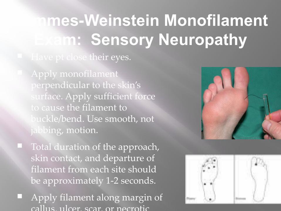
Semmes-Weinstein Monofilament Exam: Sensory Neuropathy
Have pt close their eyes. Apply monofilament
perpendicular to the skin’s surface. Apply sufficient force to cause the filament to buckle/bend. Use smooth, not jabbing, motion.
Total duration of the approach, skin contact, and departure of filament from each site should be approximately 1-2 seconds.
Apply filament along margin of callus, ulcer, scar, or necrotic tissue. Do no apply filament over these lesions.

Semmes-Weinstein Monofilament Exam
Passing score 7/10 or greater Failed monofilament testing indicated loss of
protective sensation

Malformed Foot

Hammer & Claw toe

Charcot Foot

Ulcer Characteristics
Over a pressure point Usually painless. If have pain, not usually a good sign
if also has loss of protective sensation. May probe to bone Periwound with callous

Management
Treatment of infection Assess for arterial insufficiency Offload the wound Debridement Referral to podiatry May need urgent surgical referral Diabetic? Glycemic control Daily foot and shoe inspection

Requires Surgical Referral
Pearls from Case 2
Maggot infestation Copious irrigation (wear protective gear) Consider Dakins or other antimicrobial to decrease
bioburden Dispose in biohazard bag.
Will likely need multiple referrals to other disciplines for management
Assessment of arterial perfusion Will need follow up with podiatrist
specializing in neuropathic ulcers as outpatient Proper off loading and foot wear.

Questions????
Thank you