wound physiology cf
TRANSCRIPT

Clare Fenwick Griffith Univeristy Gold Coast 2010
Wound Physiology and AssessmentDr. Clare Fenwick
2010

Clare Fenwick Griffith Univeristy Gold Coast 2010
Aspects of wound care?
What causes wounds (pathophysiology)?Can I eliminate the cause?
How do wounds heal (healing physiology)? What am I looking for (clinical assessment)? How will I treat the wound (product
knowledge)? What skills do I need (clinical knowledge)? Did it work (evaluation)? If not, why not

Clare Fenwick Griffith Univeristy Gold Coast 2010
What causes/exacerbates wounds?
Wounds are generally classified as acute or chronic
Trauma/Surgery Underlying conditions Medications Lifestyle risks
Clare Fenwick Griffith Univeristy Gold Coast 2010
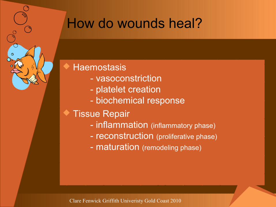
How do wounds heal?
Haemostasis- vasoconstriction- platelet creation- biochemical response
Tissue Repair- inflammation (inflammatory phase)
- reconstruction (proliferative phase)
- maturation (remodeling phase)

Clare Fenwick Griffith Univeristy Gold Coast 2010
Haemostasis
Vasoconstriction response: Arteries, arterioles and capillaries spasm to cease bleeding
Platelet response: Damaged endothelium exposes collagen fibres, platelets adhere resulting in a plug
Biochemical response: Clotting factors released causing clot formation, retraction and breakdown

Clare Fenwick Griffith Univeristy Gold Coast 2010
Tissue Repair - inflammation
Inflammation stage (0-5 days)
Capillaries spasm, a clot forms and the wound becomes ischemicHistamine is released causing vasodilation in surrounding tissueOedema and pressure initiates the pain pathwayDefence cells migrate protecting from bacteria and clearing debris
HEAT
OEDEMA
ERYTHEMA
PAIN

Clare Fenwick Griffith Univeristy Gold Coast 2010
Tissue Repair - reconstruction
Reconstruction stage (2-24 days): a time of cleaning and healing
Defence cells continue to clean up bacteria and clear debrisGranulation or angiogenesis appears (bumpy red new tissue) Contraction occursEpithelialisation commences

Clare Fenwick Griffith Univeristy Gold Coast 2010
Tissue Repair - maturation
Maturation stage (24days – 1 year)
Remodelling of the wound occurs.
Previously laid collagen fibres are broken down and new stronger collagen fibres are laid down
Rosy pink scar is still remodelling; scar similar to the surrounding skin has finished this stage
A scar has only 80% of strength and elasticity
Contracture
Remodelling
(Myers, 2004)

Clare Fenwick Griffith Univeristy Gold Coast 2010
Stage I – Nonblanchable erythema of intact skin
Stage II – Partial thickness, involving loss of the epidermis and/or dermis
Stage III – Full thickness, involving loss of the epidermis, dermis and subcutaneous
Stage IV – Full thickness, involving loss of the epidermis, dermis, subcutaneous and exposing muscle bone or supporting structures
Wound staging
STAGE ISTAGE IISTAGE IIISTAGE IV
Clare Fenwick Griffith Univeristy Gold Coast 2010
Are you totally lost yet?

Clare Fenwick Griffith Univeristy Gold Coast 2010
What am I looking for?
General client assessment
Assessment of the wound
Assessment of environmentaland local factors

Clare Fenwick Griffith Univeristy Gold Coast 2010
General client assessment
Overall health Co-morbidities Mobility status Nutritional status Sensory functioning status Psychosocial status Pain Current medication

Clare Fenwick Griffith Univeristy Gold Coast 2010
Assessment of the wound
Wound aetiology Wound location Wound dimensions Wound bed Wound drainage Wound temperature Wound infection Peri-wound

Clare Fenwick Griffith Univeristy Gold Coast 2010
Wound aetiology

Clare Fenwick Griffith Univeristy Gold Coast 2010
Wound location
PRESSURE ULCERS

Clare Fenwick Griffith Univeristy Gold Coast 2010
Wound dimensions
4.2cm length3.6cm width
Wound bed
12 o’clock 3cm9 o’clock 4cm6 o’clock 4.5cm3 o’clock 3.2cm
16cm circumference
TUNNELLING WOUND

Clare Fenwick Griffith Univeristy Gold Coast 2010
Wound Bed: Five tissue colours
Pearly pink colour – Epithelial tissue
Beefy red colour – Granulating tissue
Stringy yellow colour – Sloughy tissue
Hard black colour – Necrotic tissue
Pus green colour – Infected tissue
EPITHELIASING TISSUE
GRANULATING TISSUE
SLOUGHY TISSUENECROTIC TISSUE
INFECTED TISSUE
Clare Fenwick Griffith Univeristy Gold Coast 2010
Wound drainage
Exudate is assessed by;
Type Amount Colour Consistency Odour

Clare Fenwick Griffith Univeristy Gold Coast 2010
Wound Temperature
Wounds heal best at constant temperatures of 37 - 38 º C
Wounds left to cool down take about 3- 4 hours to restore to healing temperature (Shultz 2002)
Keep it warm, keep it moist

Clare Fenwick Griffith Univeristy Gold Coast 2010
Peri-wound
Attached or unattached Indistinct or well defined Macerated or dry Thickened and calloused Irregular or round Red, white or blue

Clare Fenwick Griffith Univeristy Gold Coast 2010
Nothing is how it seems

Clare Fenwick Griffith Univeristy Gold Coast 2010
Assessment of environmentaland local factors
INTRINSIC (inherent to the person)
EXTRINSIC (control from outside person)
Age Pressure, friction, shearing
Underlying disease processes Temperature
Nutritional status Infection
Gender Hydration
Psychological state Foreign bodies

Clare Fenwick Griffith Univeristy Gold Coast 2010
Modes of Healing
Primary Intention Secondary Intention Tertiary Intention Skin Graft Skin Flap
Primary Intention
Secondary Intention(Surgeries and procedures 2003)
Skin Flaps
Clare Fenwick Griffith Univeristy Gold Coast 2010
QUESTIONS???

Clare Fenwick Griffith Univeristy Gold Coast 2010
References
Carville, K. (2001). Wound Care Manual. Western Australia: Silver Chain Foundation.
Coleman, K. (2004). Wonderful World of Wounds. In C. Fenwick (Ed.). Gold Coast.
Hand-Surgical Approaches, Skin Flaps. (2003). Retrieved 17 February, 2004, from http://www.orthoteers.co.uk/Nrujp~ij33lm/Images9/flaps3.jpg
Edwards, S. L. (1998). High Temperature. Professional Nurse, 13(8), 521-526. Fierer, J., & Goldberg, C. (2002). Gangrene of the hand. Retrieved 2004, 2 February, from http://medicine.ucsd.edu/Clinicalimg/extremities-diabetic-foot-infection.html
Helmerd, D. (2003). Brown Recluse Spider Bite. In C. Fenwick (Ed.). Gold Coast.

Clare Fenwick Griffith Univeristy Gold Coast 2010
References
Morgan, S. (1990). A comparison of three methods of managing fever in the neurologic patient. Journal of Neuroscience Nursing, 22(1), 19-24
Myers, B. A. (2004). Wound Management Principles and Practice. New Jersey: Prentice Hall.
Shultz, M. (2002). Wound Care. Retrieved 17 February, 2004, from http://www.medpharm.co.za/sapj/2002/july/wound.html
SpinalNet. (2004). Skin and Wound Healing. Retrieved February 4, 2004, from http://www.spinalnet.co.uk/EEndCom/GBCON/Homepage.nsf/0/98BEE04F593CFD2D00256C46004B1D74?OpenDocument
Surgeries & Procedures, Skin Grafts. (2003). Retrieved 17 February, 2004, from http://health.allrefer.com/health/skin-graft-skin-graft.html
Wound Assessment. (2000). Retrieved 13 February, 2004, from http://www.medpharm.co.za/nursing/2000/sec2000/wound.html