wounds in older adults - brown university · wounds in older adults lisa j. gould, md, phd, ... •...
TRANSCRIPT

Wounds in Older Adults Lisa J. Gould, MD, PhD, FACS
Medical Director Wound Recovery and Hyperbaric Medical Center, Kent Hospital
USF Affiliate Professor Department of Molecular Pharmacology and Physiology
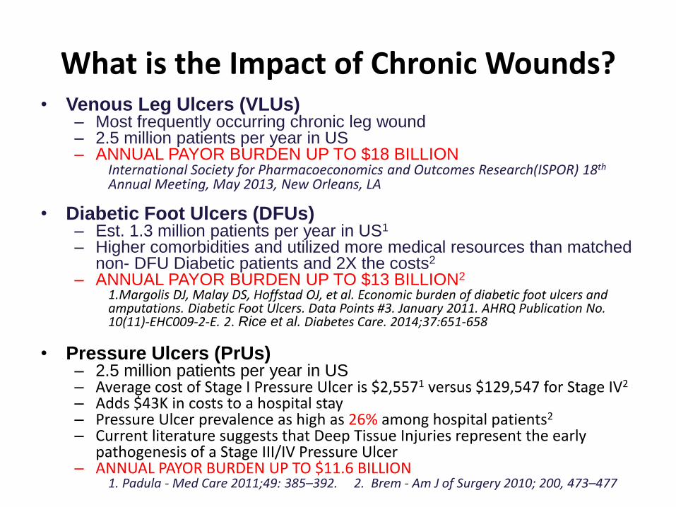
What is the Impact of Chronic Wounds? • Venous Leg Ulcers (VLUs)
– Most frequently occurring chronic leg wound – 2.5 million patients per year in US – ANNUAL PAYOR BURDEN UP TO $18 BILLION
International Society for Pharmacoeconomics and Outcomes Research(ISPOR) 18th Annual Meeting, May 2013, New Orleans, LA
• Diabetic Foot Ulcers (DFUs) – Est. 1.3 million patients per year in US1 – Higher comorbidities and utilized more medical resources than matched
non- DFU Diabetic patients and 2X the costs2 – ANNUAL PAYOR BURDEN UP TO $13 BILLION2
1.Margolis DJ, Malay DS, Hoffstad OJ, et al. Economic burden of diabetic foot ulcers and amputations. Diabetic Foot Ulcers. Data Points #3. January 2011. AHRQ Publication No. 10(11)-EHC009-2-E. 2. Rice et al. Diabetes Care. 2014;37:651-658
• Pressure Ulcers (PrUs)
– 2.5 million patients per year in US – Average cost of Stage I Pressure Ulcer is $2,5571 versus $129,547 for Stage IV2 – Adds $43K in costs to a hospital stay
– Pressure Ulcer prevalence as high as 26% among hospital patients2 – Current literature suggests that Deep Tissue Injuries represent the early
pathogenesis of a Stage III/IV Pressure Ulcer – ANNUAL PAYOR BURDEN UP TO $11.6 BILLION
1. Padula - Med Care 2011;49: 385–392. 2. Brem - Am J of Surgery 2010; 200, 473–477
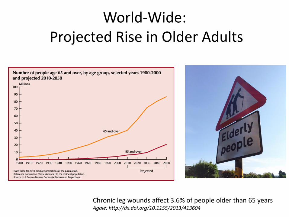
World-Wide: Projected Rise in Older Adults
Chronic leg wounds affect 3.6% of people older than 65 years Agale: http://dx.doi.org/10.1155/2013/413604

Conclusion?
• You will see chronic wounds in your practice
• You will see acute wounds in your practice
– Need to know what is reasonable to treat
– Need to know when to refer
– Need to know prevention strategies
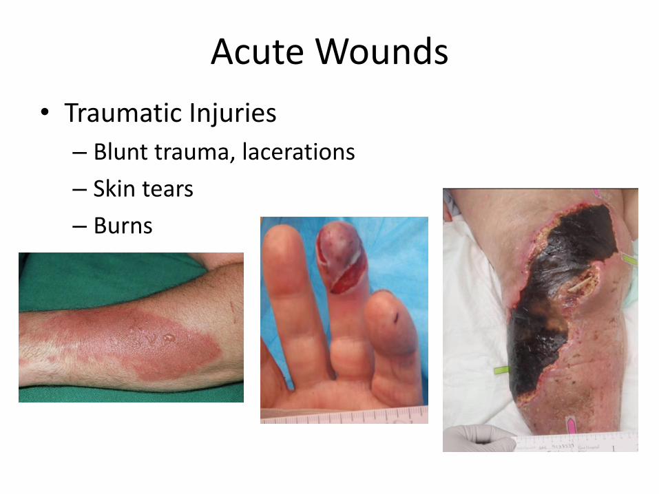
Acute Wounds
• Traumatic Injuries
– Blunt trauma, lacerations
– Skin tears
– Burns
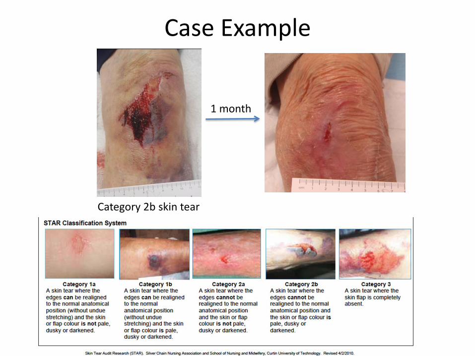
Case Example
Category 2b skin tear
1 month

Acute Wound Prevention
• Safe environment – Lighting – Furniture – Footwear – Avoid friction/shear when repositioning
• Good skin care – Avoid drying soaps – Use emollients – Hydration and good nutrition, essential fatty acid supplements – Manage incontinence
• Avoid adhesives • Protect vulnerable areas and fragile skin
– Tubular bandages – Long sleeved clothing – Skin protection devices

Keast et al WRR 2004;12:S1-S17

Identify and Treat the cause – Global assessment of patient
• Comprehensive H&P – Systemic illness known to impact wound healing
» Optimize respiratory, cardiac status, control hypertension » Diabetes – optimize glycemic control » Autoimmune/connective tissue disorder
– Nutritional assessment » Optimize glycemic control » Supplement calories/protein » Micronutrients, vitamin C
– Evaluation of systemic medications » Reduce medication known to impede wound healing if possible
– Tissue perfusion » Vascular status » Edema » Smoking » History of radiation
• Local Factors – Unrelieved pressure at site of wound – Foreign body – Infection – Malignancy – Incontinence – Topical agents

Lower Extremity Ulcers
• Arterial
• Venous
• Diabetic
• Mixed Etiology
• Other
Venous
Arterial
Diabetic
Other
Differential Diagnosis
• Venous
• Arterial
• Neuropathic
• Pressure
• Lymphatic
• Vasculitis
• Traumatic
• Autoimmune disease
• Sickle cell disease
• Pyoderma gangrenosum
• Hematologic disease
• Neoplastic
• Infectious

Differentiating features of leg ulcers
Site Skin Ulcer Other
Venous Lower 3rd of
leg; medial
malleolus
Edema
Hemosiderin
deposition
‘Weepy’
Irregular
Painful
Varicosities
‘Bottle leg’
Normal ABI
Arterial Distal
extremities
(toes)
Thin, atrophic
Shiny
Hair loss
No bleeding
Regular
Painful
Diminished
pulses
Low ABI
Diabetic
(neuropathic)
Pressure sites Callous Round, deep
Painless
Lack of
sensation
ABI>1.3
Adapted from Creager et al, ed, Vascular Medicine
Lower extremity Ulcers
All patients with lower extremity ulcers
should be evaluated for arterial disease
– Claudication
– Rest pain
– Dry, cool skin
– Distal Atrophy and Alopecia
– Dependent Rubor, Elevation Pallor
– Absent pedal (and proximal) pulses
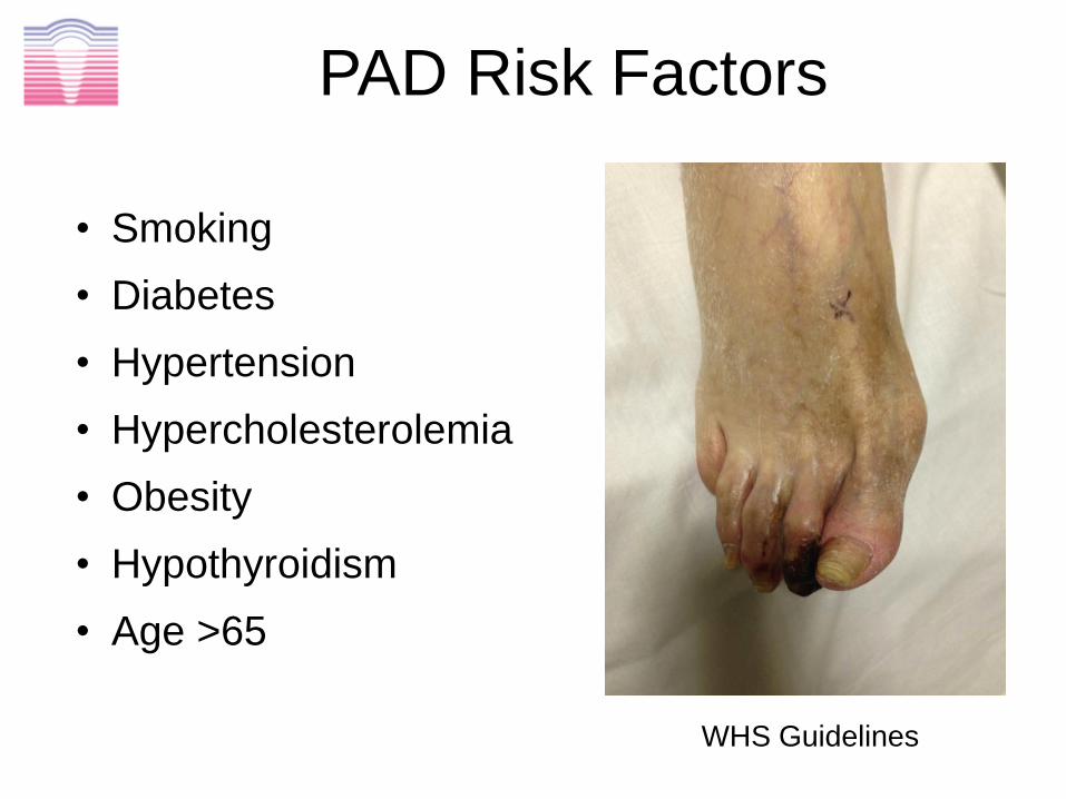
PAD Risk Factors
• Smoking
• Diabetes
• Hypertension
• Hypercholesterolemia
• Obesity
• Hypothyroidism
• Age >65
WHS Guidelines

Arterial Insufficiency Ulcers – Clinical Characteristics
•Deep punched out appearance
• Scant granulation
• Located between toes, on toe tips, outer ankle, or
where there is trauma and/or friction from walking
• Dry gangrene vs. wet gangrene
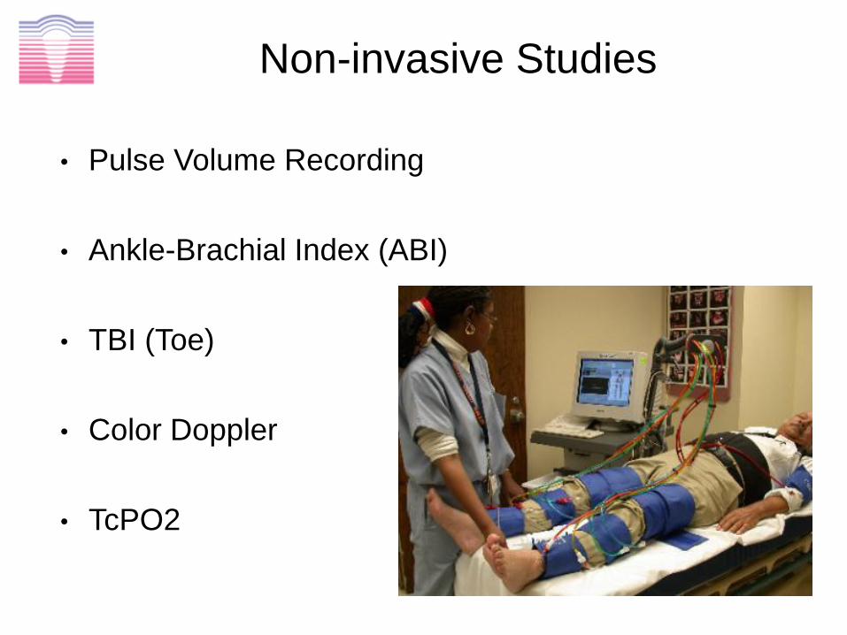
Non-invasive Studies
• Pulse Volume Recording
• Ankle-Brachial Index (ABI)
• TBI (Toe)
• Color Doppler
• TcPO2
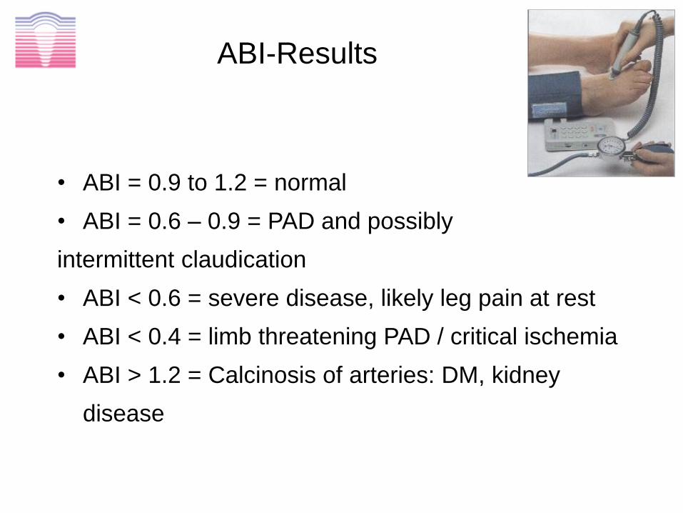
• ABI = 0.9 to 1.2 = normal
• ABI = 0.6 – 0.9 = PAD and possibly
intermittent claudication
• ABI < 0.6 = severe disease, likely leg pain at rest
• ABI < 0.4 = limb threatening PAD / critical ischemia
• ABI > 1.2 = Calcinosis of arteries: DM, kidney
disease
ABI-Results
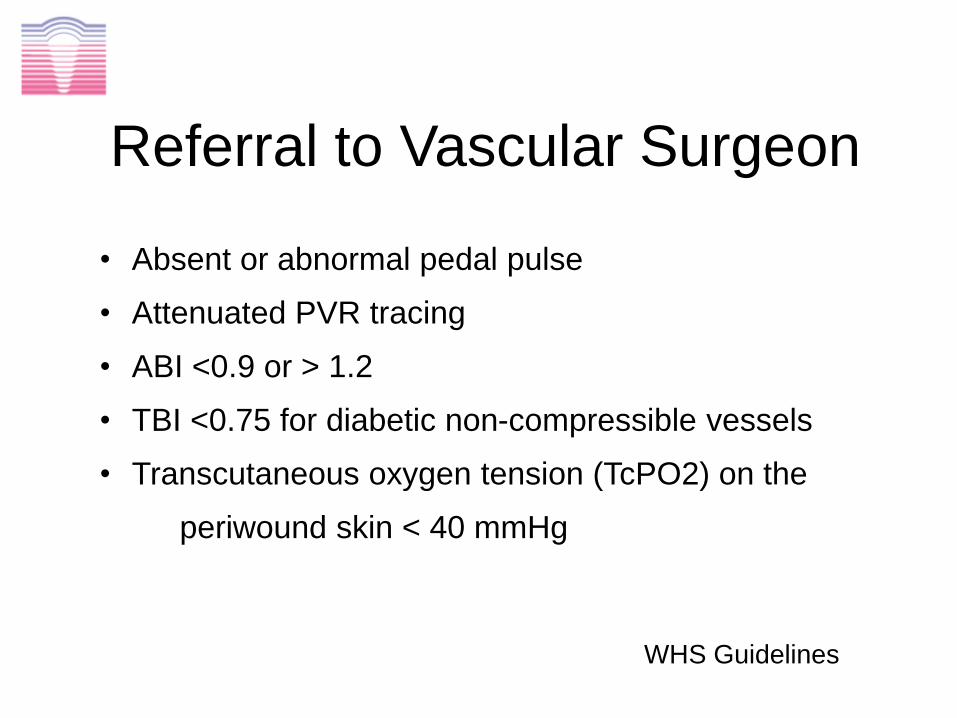
Referral to Vascular Surgeon
• Absent or abnormal pedal pulse
• Attenuated PVR tracing
• ABI <0.9 or > 1.2
• TBI <0.75 for diabetic non-compressible vessels
• Transcutaneous oxygen tension (TcPO2) on the
periwound skin < 40 mmHg
WHS Guidelines

Venous Leg Ulcers
• Dependent edema
• Varicose veins
• Reddish-brown pigmentation
• Eczematous changes
• Atrophie blanche
• Lipodermatosclerosis
• Ulceration
• At or above the ankle
• irregular, shallow
• fibrinous material at base
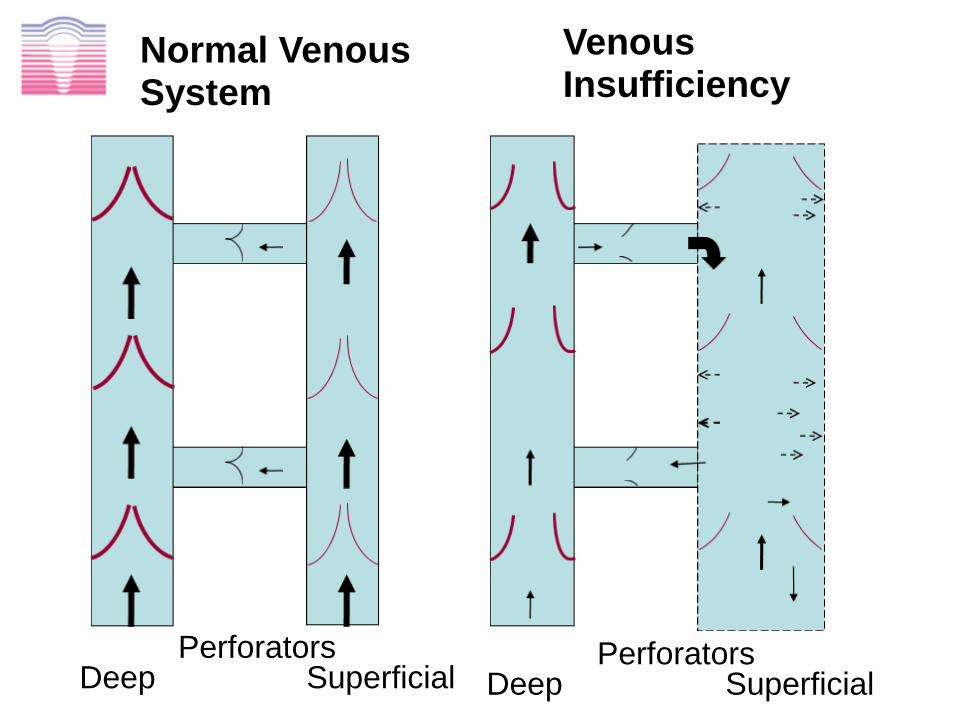
Deep Perforators
Superficial
Normal Venous System
Venous Insufficiency
Deep Perforators
Superficial
Risk Factors
• Deep vein thrombosis
• Family history of venous disease
• Varicose veins / phlebitis
• Previous trauma or surgery
• Prolonged immobilization
• Obesity
WHS Guidelines
Diagnosis
• Clinical History and Physical Examination
• Ulcer location
• Signs/symptoms of venous insufficiency
Swelling
Skin breakdown
Pigmentation
Eczema
• Diagnostic Procedures
• Duplex ultrasound/ Valsalva maneuver (not
ultrasound to r/o DVT)
• Velocity and direction of flow
• Rule out or consider arterial component
Mixed arterial/venous disease is common WHS Guidelines
Treatment of Venous Ulcers
• Elevation
• Compression therapy • Infection control – Local and Regional
• Debridement • Topical antimicrobials • Antibiotics for invasive infection
• Dressing • Moisture retentive vs. Exudate control • Protection of peri-wound skin • Pain control
• Adequate nutrition • Protein • Sodium and Sugar • Micronutrients
• Venous ulcers that show no sign of healing after six weeks of treatment should be biopsied
Edema: Excessive Fluid in Interstitium
• Inflammatory response to injury
• Histamine release
• Vasodilation
• Increased vascular permeability
• Limb dependency, venous hypertension
• Congestive heart failure
• Lymphedema

Edema Control
• Elevation
• Compression therapy
• Compression garments
• Pneumatic compression
• Layered wrap systems
• Classes of compression
• Class 1: 15 – 20 mmHg
• Class 2: 21 – 30 mmHg
• Class 3: 31 – 40 mmHg
• Class 4: 41 – 50 mmHg
• ANY compression is better than NO compression
Contraindications to
Compression Bandages
• Absolute - critical limb ischemia
• Relative
• ABI < 0.8
• CHF - be aware of rapid fluid shifts
• Unreliable attendance
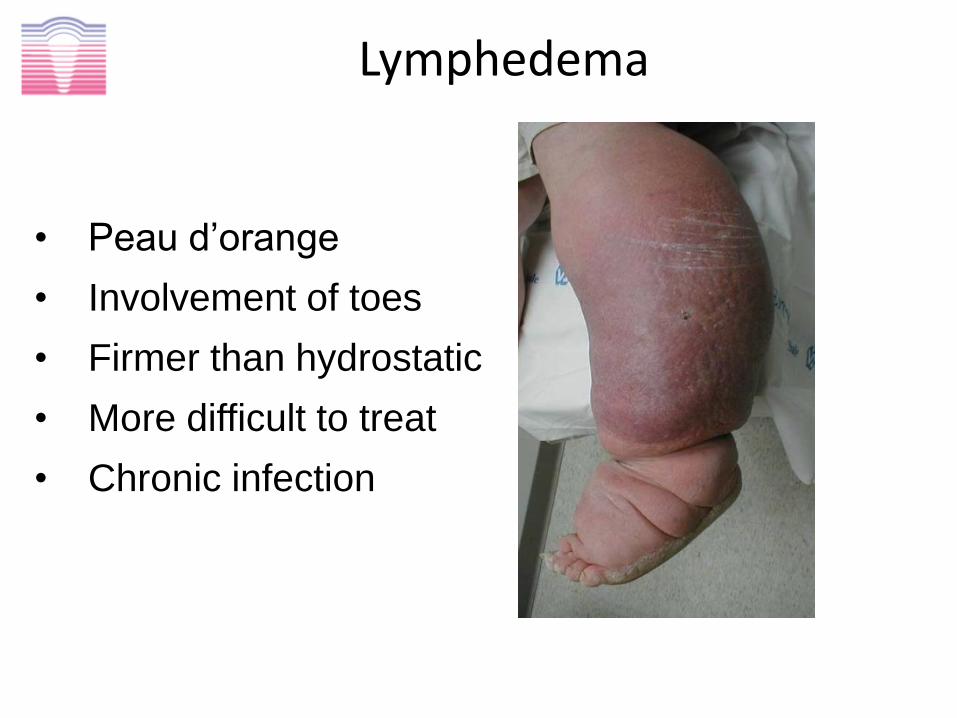
Lymphedema
• Peau d’orange
• Involvement of toes
• Firmer than hydrostatic
• More difficult to treat
• Chronic infection

BLEE vs Cellulitis
Unilateral, acute onset
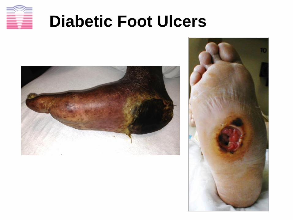
Diabetic Foot Ulcers

Diabetic Foot Ulcer Pathophysiology
Motor neuropathy
Altered Wound Healing
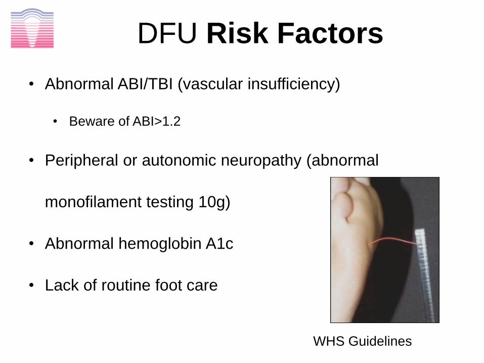
DFU Risk Factors
• Abnormal ABI/TBI (vascular insufficiency)
• Beware of ABI>1.2
• Peripheral or autonomic neuropathy (abnormal
monofilament testing 10g)
• Abnormal hemoglobin A1c
• Lack of routine foot care
WHS Guidelines

Three Major Categories of Diabetic Foot Wounds
Ischemic
Chronic Insensate
Deep Space Infection

Ulcer Characteristics
• Location
• Plantar midfoot
• Metatarsal pads
• Heel
• Site of repetitive trauma
• Wound Appearance
• Well defined margins, variable depth, granulation
frequently present, macerated perimeter
• Surrounding Skin
• Erythema (cellulitis), induration, callus
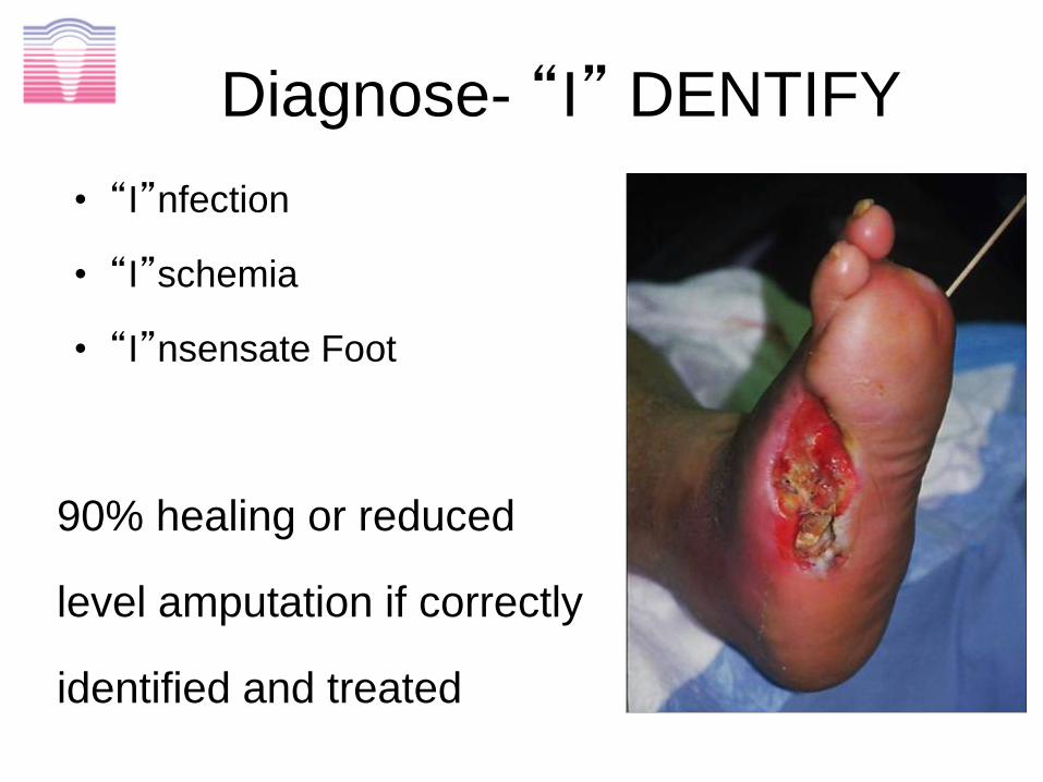
Diagnose- “I” DENTIFY
• “I”nfection
• “I”schemia
• “I”nsensate Foot
90% healing or reduced
level amputation if correctly
identified and treated

Treatment for Neuropathic DFU: Off-loading
Reverse the detrimental effects of neuropathy and deformity to decrease morbidity
Acceptable methods of offloading: • crutches • wheelchairs • custom shoes or inserts • diabetic boots • forefoot and heel relief shoes • C.R.O.W. • Removable cast walkers (RCW) • total contact cast (TCC/iTCC)
WHS Guidelines

Wound Assessment Determines Local Wound Care
– Categories
• Surgical/non-surgical
• Depth
• Specific type – Venous insufficiency
– Diabetic
– Arterial
– Pressure
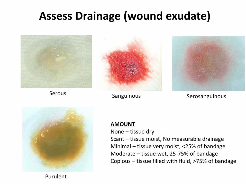
Assess Drainage (wound exudate)
Serous Sanguinous Serosanguinous
Purulent
AMOUNT None – tissue dry Scant – tissue moist, No measurable drainage Minimal – tissue very moist, <25% of bandage Moderate – tissue wet, 25-75% of bandage Copious – tissue filled with fluid, >75% of bandage

Tissue Description: Eschar
Eschar: Dry, leathery, black
Eschar: Soft, yellow, non-viable skin

Tissue Description: Necrotic Tissue
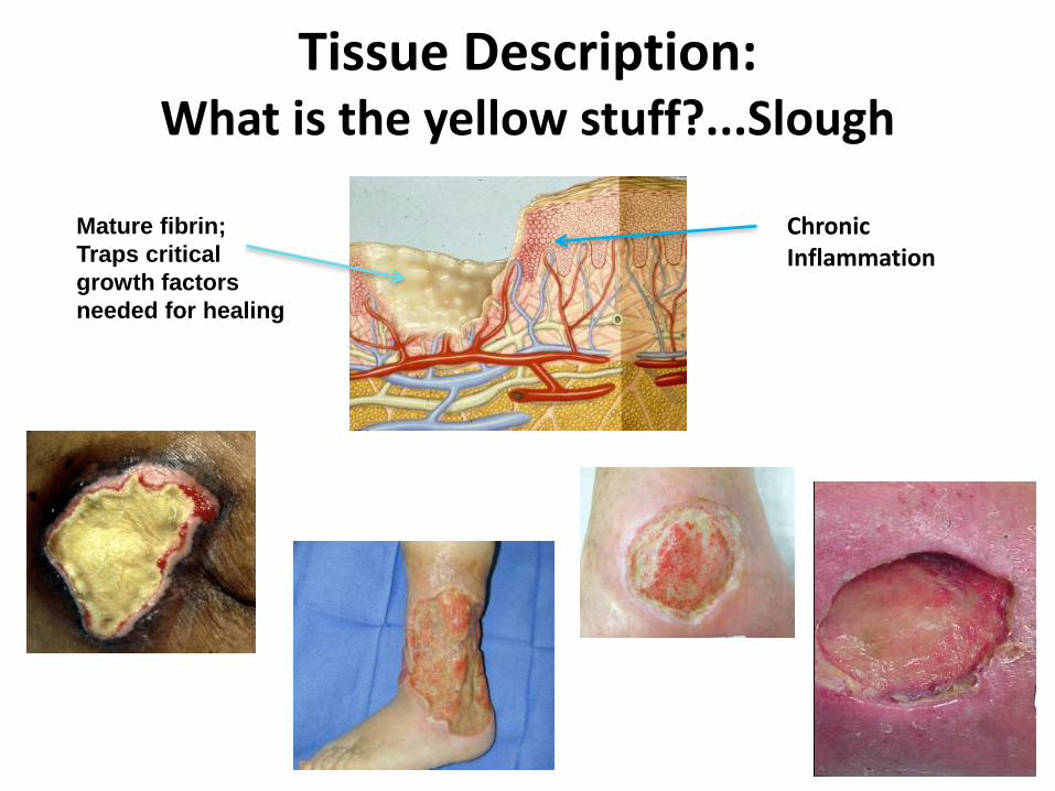
Tissue Description: What is the yellow stuff?...Slough
Chronic Inflammation
Mature fibrin;
Traps critical
growth factors
needed for healing

Measure Undermining

Measure Tunneling

Tissue Description Muscle

Tissue Description Tendon/fascia

Tissue Description Bone

Edge
• Hyperkeratotic
• Epibole
• Pocketing
• Maceration
• Advancing epithelium
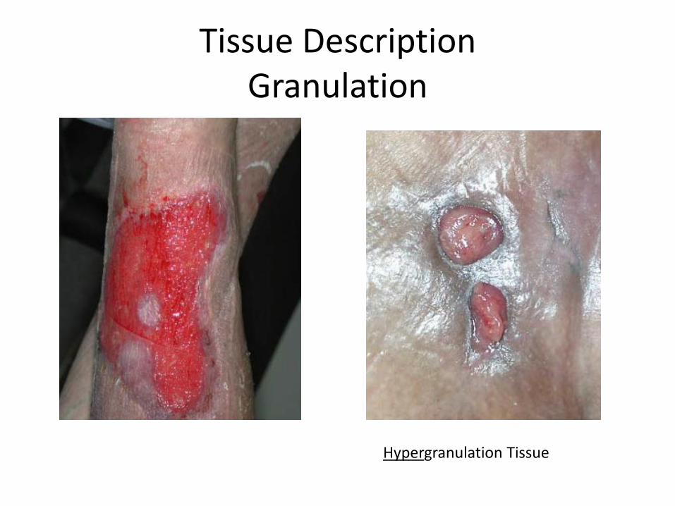
Tissue Description Granulation
Hypergranulation Tissue
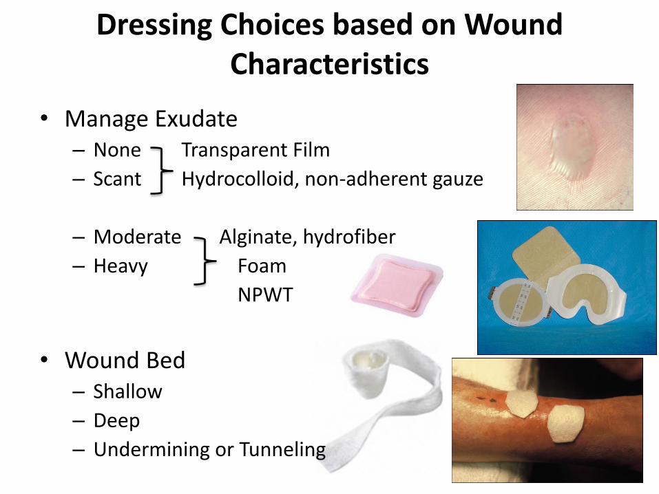
Dressing Choices based on Wound Characteristics
• Manage Exudate – None Transparent Film
– Scant Hydrocolloid, non-adherent gauze
– Moderate Alginate, hydrofiber
– Heavy Foam
NPWT
• Wound Bed – Shallow
– Deep
– Undermining or Tunneling

Undermining or Tunneling
Lightly pack with absorbent filler or gel impregnated gauze

Topical Agents That Destroy Tissue
• Betadine
• Hydrogen Peroxide
• Dakins’ Solution
• Kerosene
• Leaving open to air
Take Home Messages
• Patient is a ‘whole’ not a ‘hole’ – Primary disease control – Prevention – Nutritional support – Social Support and Stress management – Identify and Treat Infection – Edema Control – Simple dressings to maintain moist environment
• Develop Plan of Care – Patient capabilities – Need for home health – Ability to comply – Family – Cost
• Timely referral