wp3: theory for 360 degree fmt progress during year 1 giannis zacharakis institute for electronic...
TRANSCRIPT
WP3: Theory for 360 degree FMTProgress during Year 1
Giannis ZacharakisInstitute for Electronic Structure and Laser (IESL)
Foundation for Research and Technology – Hellas (FORTH)
FMT-XCT First Annual MeetingApril 24 2009

Whole animal imaging
Whole Animal
In Vivo Optical Imaging GroupIESL – FORTH
Cellular and Sub-Cellular

Ballistic Propagation (OPT)
Ballistic Propagation Beer´s Law ~ exp (-a*z)
FMT
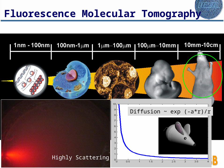
Highly Scattering
Fluorescence Molecular Tomography
Diffusion ~ exp (-a*r)/r

2nd generation FMT system
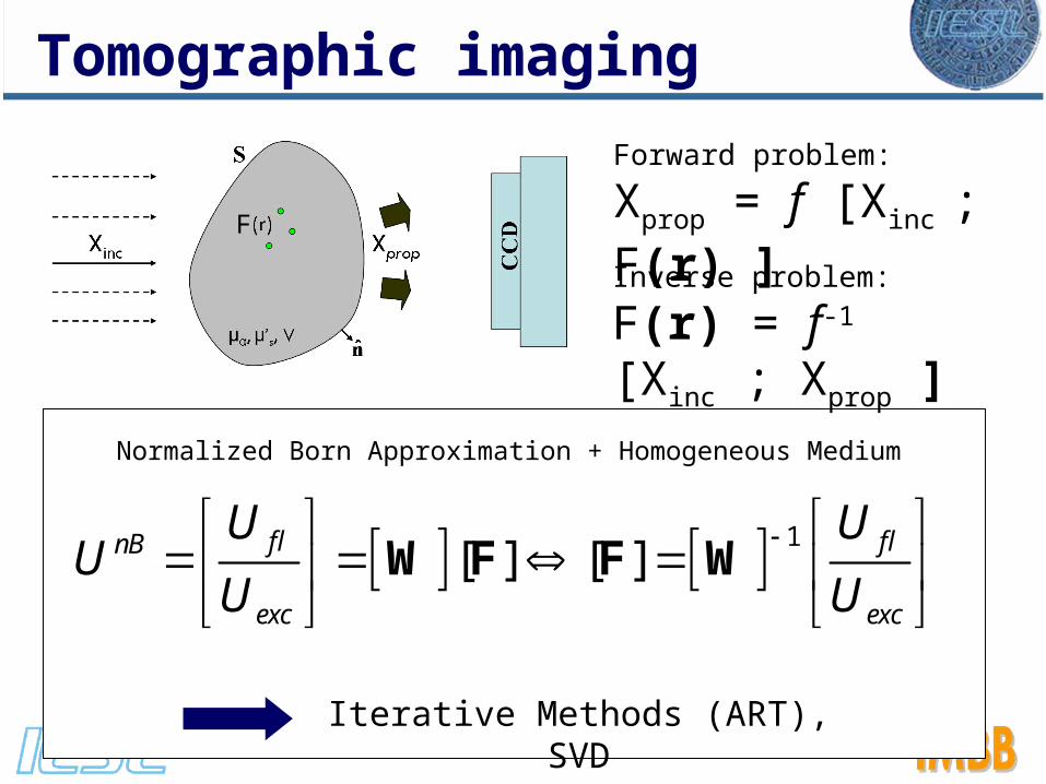
Inverse problem:
F(r) = f-1 [Xinc ; Xprop ]
Forward problem:
Xprop = f [Xinc ; F(r) ]
Tomographic imaging
1[ ] [ ]
W F F Wfl flnB
exc exc
U UU
U U
Iterative Methods (ART), SVD
Normalized Born Approximation + Homogeneous Medium
F

transgenicage ~ 8 weeks
non-transgenic age ~ 8 weeks
Fluorescence
NBornExcitation
Normalized measurements
hCd2-GFP mouse
GFP Control
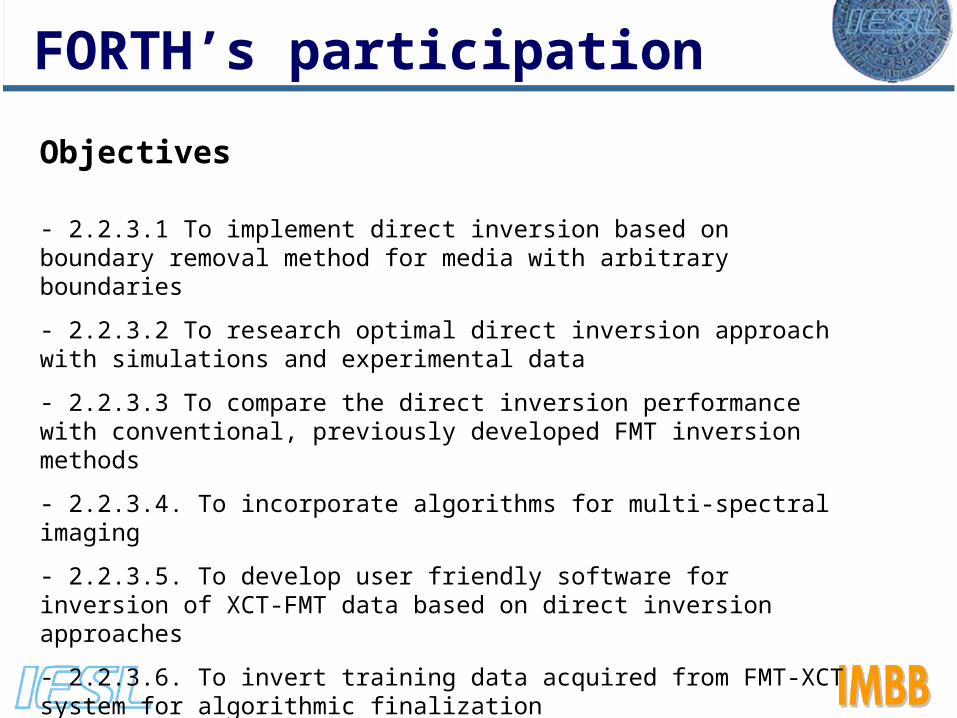
FORTH’s participation
Objectives
- 2.2.3.1 To implement direct inversion based on boundary removal method for media with arbitrary boundaries
- 2.2.3.2 To research optimal direct inversion approach with simulations and experimental data
- 2.2.3.3 To compare the direct inversion performance with conventional, previously developed FMT inversion methods
- 2.2.3.4. To incorporate algorithms for multi-spectral imaging
- 2.2.3.5. To develop user friendly software for inversion of XCT-FMT data based on direct inversion approaches
- 2.2.3.6. To invert training data acquired from FMT-XCT system for algorithmic finalization

Objectives
2.2.3.1 To implement direct inversion based on boundary removal method for media with arbitrary boundaries
- 2.2.3.2 To research optimal direct inversion approach with simulations and experimental data
- 2.2.3.3 To compare the direct inversion performance with conventional, previously developed FMT inversion methods
Theory for 360 FMT

Boundary removal
Ripoll and Ntziachristos, PRL 2006.

'1( , ) ( ', ' ) ',
2 'o o
A
gU z U z z dA z z
z
r r
R R
Assuming the interface locally plane at (R,z0), we can propagate the measurement U to anywhere in (diffuse) space:
(first Raleigh-Sommerfield integral formula)
(R,z0)
(R,z)
(infinite Diffuse Medium - virtual)
Original Diffuse Volume
Boundary removal
- Use of infinite Green’s functions
- Apply direct inversion
1. Backpropagation2. Complete Fourier Approach
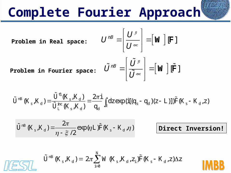
Complete Fourier Approach
flnB s d
s d s d s dinc zL s d d
U (K ,K ) 2 iU (K ,K ) dz exp(i[(q q )(z L)])F(K K ,z)
U (K ,K ) q
[ ]
W Ffl
exc
nB UU
U
[ ]
W F
fl
exc
nB UU
U
Direct Inversion!
Problem in Real space:
Problem in Fourier space:
nBs d s d
2U (K ,K ) exp( L)F(K K , )
/ 2
NnB
s d s d i s di 0
U (K ,K ) 2 W(K ,K ,z )F(K K ,z) z

LIMITATIONS:
1) It can only be used with large numbers of sources Grids of 64x64 sources to obtain relatively good data and are still prone to great Fourier artifacts. This is a great problem since all our FMT setups work with numbers of sources in the 100 range. Having more sources in unpractical, since the experiment times will increase unnecessarily.2) Fluorophore concentration in Fourier space. This means that once inverted the data has to be Inverse-Fourier transformed in 3D. This yields significant Fourier artifacts (seen as 'waves' surrounding the main central points of data) that worsen as the number of sources or detectors gets smaller. This means that in practice even larger numbers of sources need to be used.
Complete Fourier Approach
• After developing and implementing the main features of the direct inversion method, we have decided it is not the method to pursue for our FMT setups.
• Clearly we need a method that can solve fast the inversion needed but can work with small numbers of sources, in the order of <100 sources.
• We therefore opted to change this deliverable, DIRECT INVERSION method to a deliverable called 'ULTRA FAST INVERSION for FMT' that will be delivered on the next reporting period.
• In the meantime, the partners have access to an inversion method which is significantly faster that the currently existing ones, based on:
Boundary removal and ART inversion on the infinite homogeneous data.
Conclusion
Objective 2.2.3.4. To incorporate algorithms for multi-spectral imaging
Multi-color imaging
- A multispectral algorithm has been developed and tested for simultaneous detection of multiple fluorophores and absorbers.
- It will be incorporated in the FMT software in the next reporting period according to the time schedule.
1 1 1
2 2 2
n n n
G R M G1
G R M2 R
n MG R M
s s ... s CI
s s ... sI C
I Cs s ... s
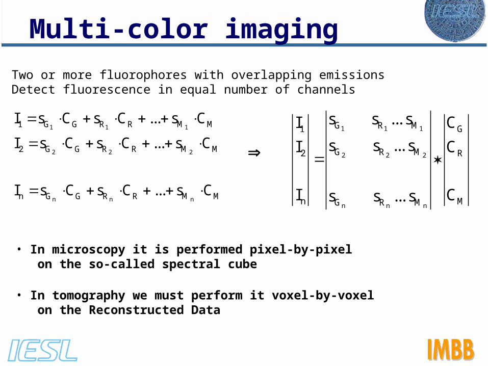
• Two or more fluorophores with overlapping emissions• Detect fluorescence in equal number of channels
1 1 1
2 2 2
n n n
1 G G R R M M
2 G G R R M M
n G G R R M M
I s C s C ... s C
I s C s C ... s C
I s C s C ... s C
• In microscopy it is performed pixel-by-pixel on the so-called spectral cube
• In tomography we must perform it voxel-by-voxel on the Reconstructed Data
Multi-color imaging

Multi-color imaging
nB flfl
exc
UU W fluo(r)
U
mua excabs
inc
UU W abs(r)
U
log / mua abs dz
Hb Hb HbO HbOmua C C
Tomographic calculations
Fluorescence reconstruction Absorption reconstruction
1 1
2 2
G R G1
2 G R R
s s Cfluo
fluo s s C
In the case of two fluorophores
Hb1 HbO1 Hb1
Hb2 HbO2 HbO2
Cmua
Cmua
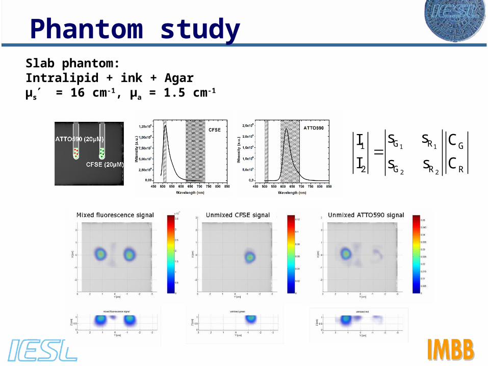
Slab phantom:Intralipid + ink + Agarμs = 16 cm-1, μa = 1.5 cm-1
1 1
2 2
G R G1
2 G R R
s s CI
I s s C
Phantom study
Phantom study
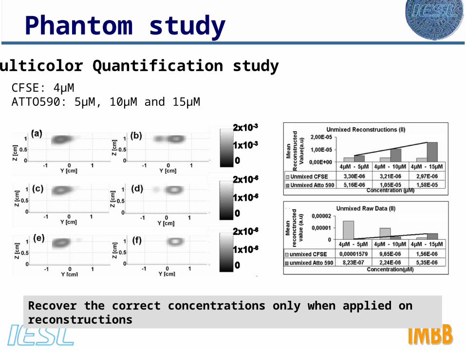
Multicolor Quantification studyCFSE: 4μMATTO590: 5μM, 10μM and 15μM
Recover the correct concentrations only when applied on reconstructions
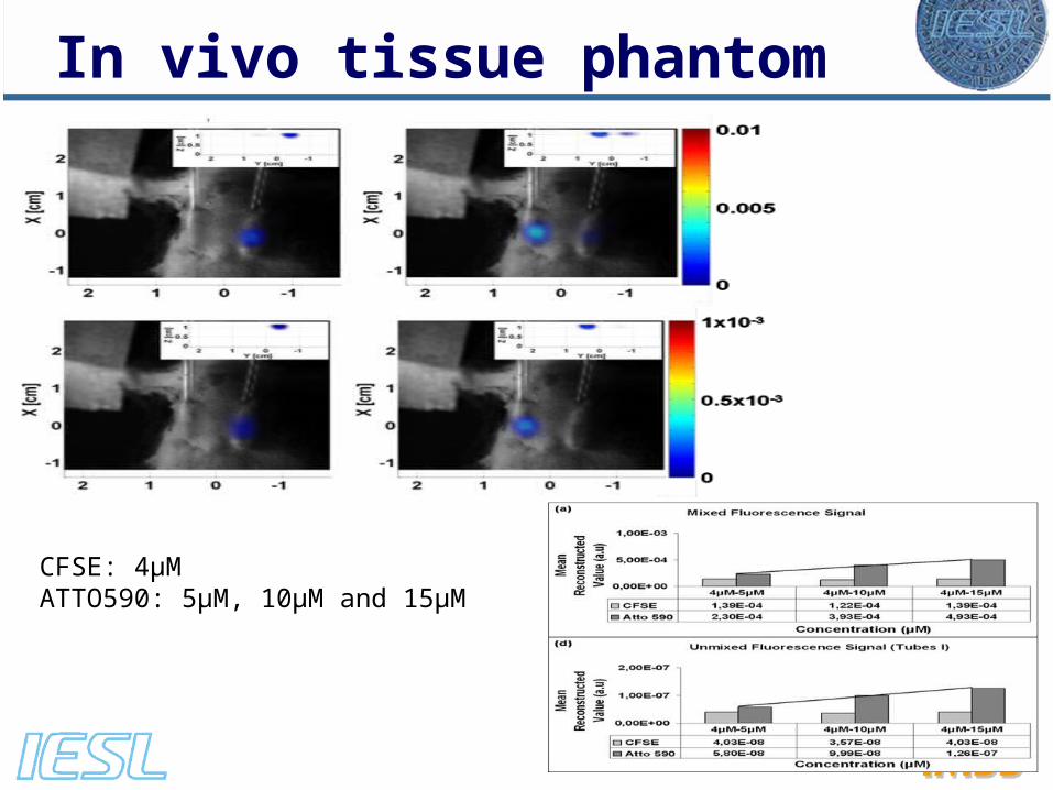
In vivo tissue phantom
CFSE: 4μMATTO590: 5μM, 10μM and 15μM
0log
Ic d
I
Hb HbOBV C C
100
HbO
HbT
COxySat
C
Hb1 HbO1 Hb1
Hb2 HbO2 HbO2
Cmua
Cmua
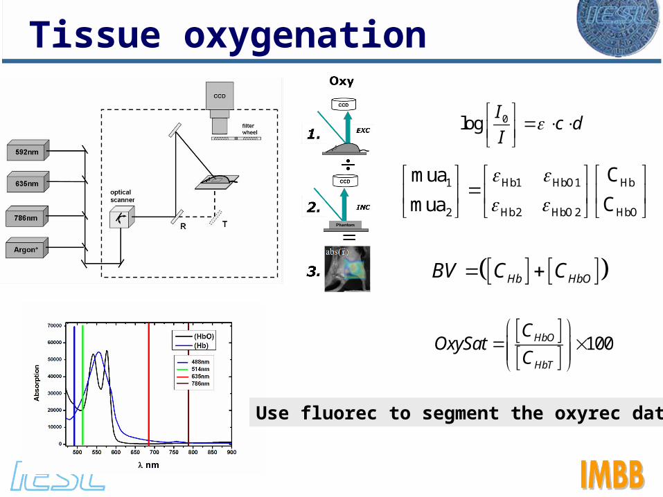
Tissue oxygenation
Use fluorec to segment the oxyrec data

• Image concurrently in 3D - Fluorescence activity - Oxygenation in hypoxic regions - Neo-vascular factors related to tumor proliferation - Measure dynamic parameters (BV and OxySat) - Identify cancer stage and phenotype (benign or malignant)
Tissue oxygenation

Objective 2.2.3.5. To develop user friendly software for inversion of XCT-FMT data based on direct inversion approaches
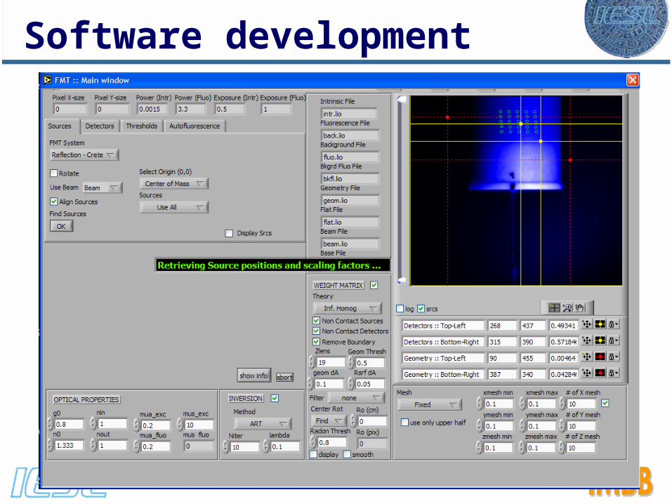
Software development
• Custom developed software based on Labview suitable for: 1. Data acquisition2. Data analysis (OPT and FMT)
• Automated user-friendly software for data analysisUses open source application for visualizatione.g. ImageJ, Amide, Osirix

Software development
Software development

Mini-FMT
Automated software for FMT acquisition and analysisThe main goal of the FMT software is to provide a user-friendly interface to take the measurement data and later perform the 3D reconstruction providing an image that can be analyzed with Open Source applications. e.g. ImageJ, Amide, Osirix
There is a complete manual available to the partners with detailed information and guidance for all the functions and parameters.
12 3
4 5
6
7 8 9
10 11 12
13
One button reconstruction
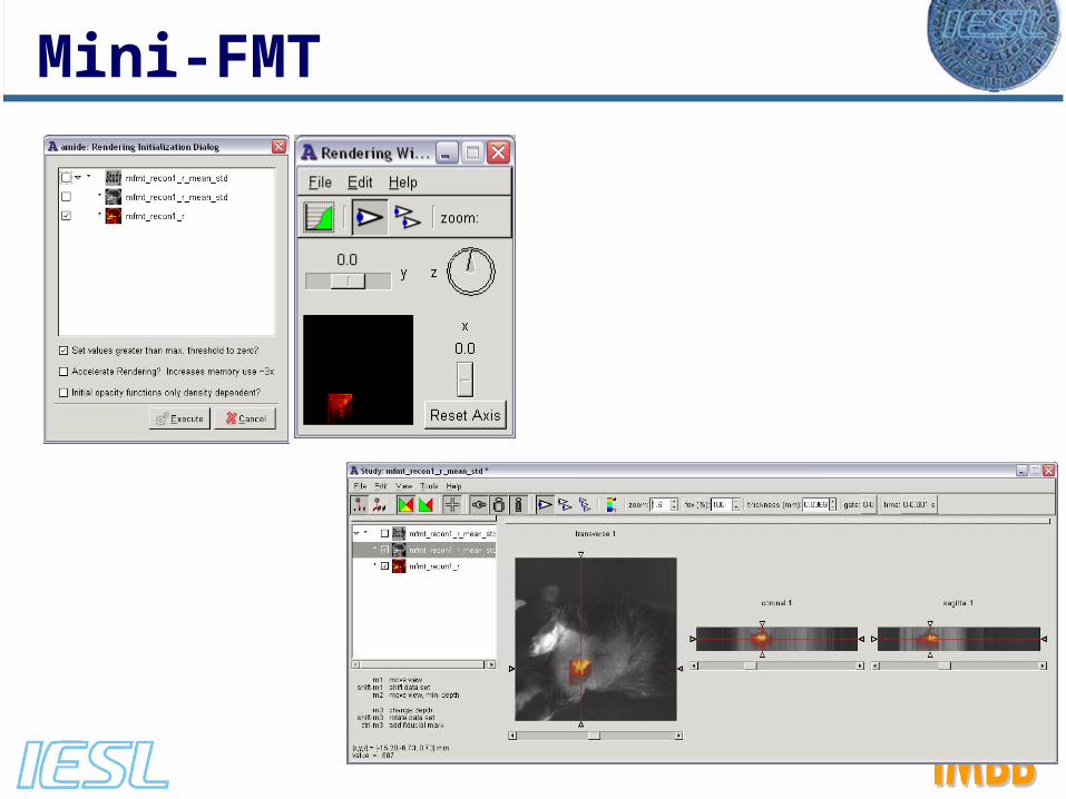
Mini-FMT

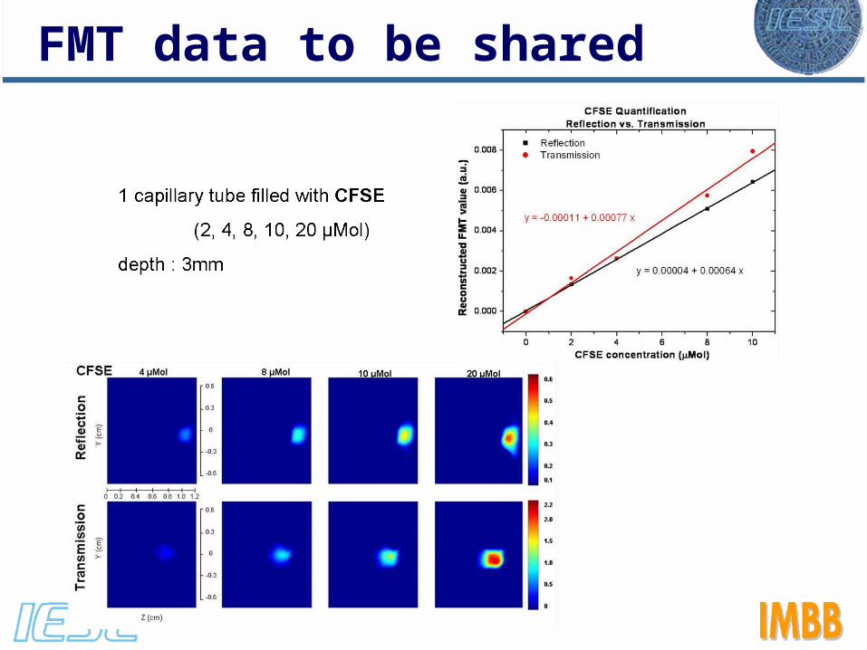
FMT data to be shared
Objective 2.2.3.6. To invert training data acquired from FMT-XCT system for algorithmic finalization
A large number of experimental measurements have been acquired that are available to all partners for optimization and finalization of algorithms. These measurements involve phantoms as well as in vivo experiments:
• Quantification• Resolution• Multispectral• In vivo studies (lymph nodes, tumor progression, oxygenation)

FMT data to be shared
FMT data to be shared

FMT data to be shared

FMT – TOAST comparison
Exchange of data with Partner 4UCLReconstructions by: Athanasios Zacharopoulos

FORTH – LIME dataExperiments performed with FORTH-FMT for comparison with LIME-FMT
Visit of Anikitos Garofalakis to FORTH
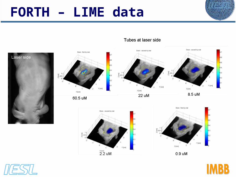
FORTH – LIME data
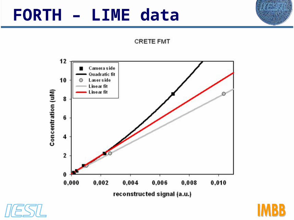
FORTH – LIME data
Second Year
• Ultra fast inversion for FMT (new Deliverable)
• Test and compare the performance
• Optimize and finalize Mini-FMT (feedback from Partners)
• Incorporate the multispectral algorithm (absorption & fluorescence)
• Increase the exchange of data! (essential for joint progress)
Vasilis Ntziachristos
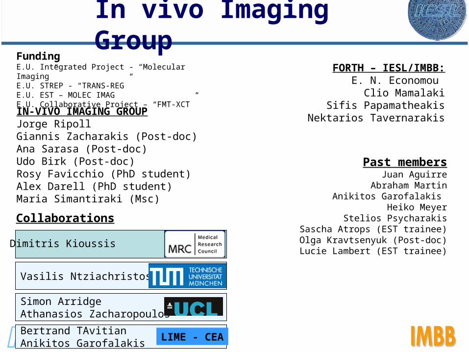
FundingE.U. Integrated Project - “Molecular Imaging” E.U. STREP - “TRANS-REG” E.U. EST – MOLEC IMAGE.U. Collaborative Project – “FMT-XCT”
Dimitris Kioussis
IN-VIVO IMAGING GROUPJorge RipollGiannis Zacharakis (Post-doc)Ana Sarasa (Post-doc)Udo Birk (Post-doc)Rosy Favicchio (PhD student)Alex Darell (PhD student)Maria Simantiraki (Msc)
FORTH – IESL/IMBB:E. N. Economou
Clio MamalakiSifis Papamatheakis
Nektarios Tavernarakis
Past membersJuan Aguirre
Abraham MartinAnikitos Garofalakis
Heiko MeyerStelios Psycharakis
Sascha Atrops (EST trainee)Olga Kravtsenyuk (Post-doc)Lucie Lambert (EST trainee)
In vivo Imaging Group
Simon ArridgeAthanasios Zacharopoulos
Bertrand TAvitianAnikitos Garofalakis
LIME - CEA
Collaborations

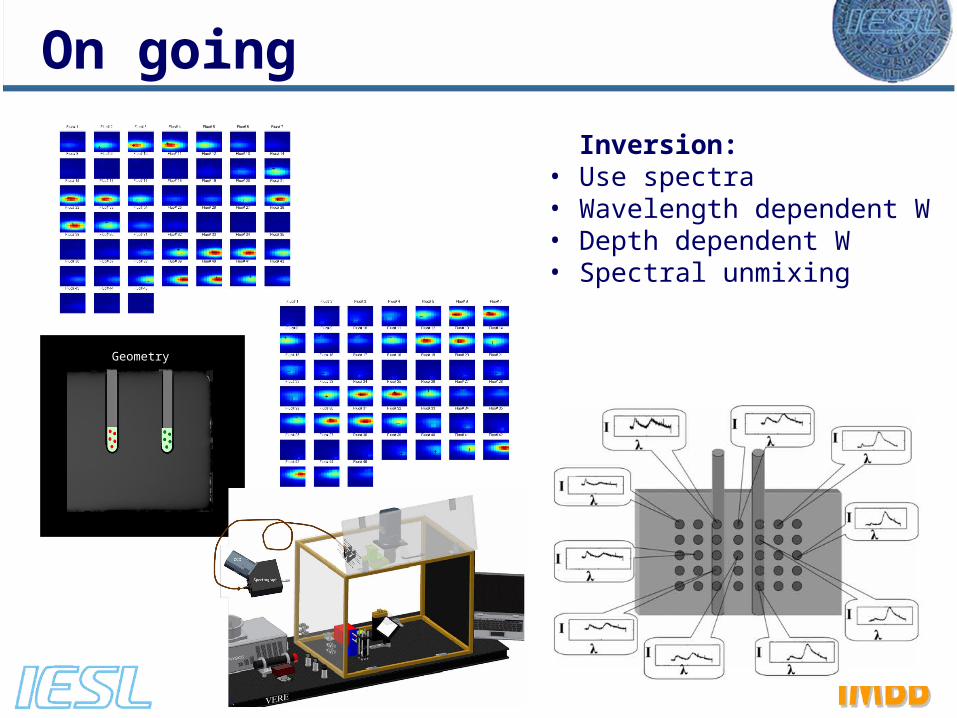
Geometry
Inversion: • Use spectra• Wavelength dependent W• Depth dependent W• Spectral unmixing
On going
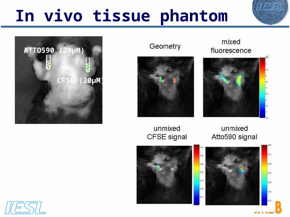
CFSE (20μM)
ATTO590 (20μM)
In vivo tissue phantom

Same number of GFP & DsRed T-cells injectedOnly DsRed cells recognize an antigen peptideRepetitive imaging of cervical lymph nodes
Following T-cell numbers