xpang_paper_a12
TRANSCRIPT

CORMIER ET AL. VOL. 7 ’ NO. 9 ’ 7562–7572 ’ 2013
www.acsnano.org
7562
August 26, 2013
C 2013 American Chemical Society
Molecular Structure of RADA16‑IDesigner Self-Assembling PeptideNanofibersAshley R. Cormier,†,‡ Xiaodong Pang,§ Maxwell I. Zimmerman,†,‡ Huan-Xiang Zhou,§ and
Anant K. Paravastu†,‡,*
†Department of Chemical and Biomedical Engineering, FAMU-FSU College of Engineering, 2525 Pottsdamer Street, Tallahassee, Florida 32310-6046, United States,‡National High Magnetic Field Laboratory, 1800 East Paul Dirac Drive, Tallahassee, Florida 32310, United States, and §Department of Physics and Institute ofMolecular Biophysics, Florida State University, Tallahassee, Florida 32306, United States
RADA16-I (COCH3-RADARADARADARA-DA-CONH2) forms nanofiber matricesin water with adaptive properties that
make them attractive for biomedical appli-cations. The formation of nanofiber net-works in physiological conditions allowsfor use as an extracellular matrix capableof providing mechanical support for tissueregeneration1,2 or three-dimensional cellculture.3,4 High effective porosities allowdiffusion of nutrients or drug molecules.5
The ability to add functionalmotifs further ex-pands the utility for tissue engineering.6�8
Dynamic reassembly following nanofiberfragmentation confers a “self-healing” prop-erty and may allow RADA16-I nanofibernetworks to adapt to stimuli such as celldivision ormigration.9 Injection of RADA16-Inanofibers has been shown to stop bleedingin surgery and repair severed neurons torestoreaxon function.10,11 Thepurely syntheticnature of RADA16-I avoids uncontrollable
* Address correspondence [email protected].
Received for review March 29, 2013and accepted August 26, 2013.
Published online10.1021/nn401562f
ABSTRACT
The designer self-assembling peptide RADA16-I forms nanofiber matrices which have shown great promise for regenerative medicine and three-
dimensional cell culture. The RADA16-I amino acid sequence has a β-strand-promoting alternating hydrophobic/charged motif, but arrangement of
β-strands into the nanofiber structure has not been previously determined. Here we present a structural model of RADA16-I nanofibers, based on solid-
state NMR measurements on samples with different schemes for 13C isotopic labeling. NMR peak positions and line widths indicate an ordered structure
composed of β-strands. The NMR data show that the nanofibers are composed of two stacked β-sheets stabilized by a hydrophobic core formed by alanine
side chains, consistent with previous proposals. However, the previously proposed antiparallel β-sheet structure is ruled out by measured 13C�13C dipolar
couplings. Instead, neighboring β-strands within β-sheets are parallel, with a registry shift that allows cross-strand staggering of oppositely charged
arginine and aspartate side chains. The resulting structural model is compared to nanofiber dimensions observed via images taken by transmission electron
microscopy and atomic force microscopy. Multiple NMR peaks for each alanine side chain were observed and could be attributed to multiple configurations
of side chain packing within a single scheme for intermolecular packing.
KEYWORDS: self-assembling peptide . peptide nanofiber design . solid-state NMR spectroscopy . structure determination .molecular modeling
ARTIC
LE

CORMIER ET AL. VOL. 7 ’ NO. 9 ’ 7562–7572 ’ 2013
www.acsnano.org
7563
composition issues with natural products such asMatrigel.12 RADA16-I has the potential for harmlessdegradation in vivo,13,14 eliciting little immune re-sponse when compared to other materials used insurgery.10
The RADA16-I sequence was discovered throughcombinatorial analysis of short amino acid sequenceswith alternating charged and hydrophobic residues.15
This patternwasoriginally observed in a segmentof yeastprotein15�18 and tends to promote β-strand formation.Self-assembly occurs through formation of hydrogenbonds between backbones of peptide molecules inextended conformations.19 Alternation of hydrophobicand hydrophilic residues also promotes formation ofhydrophobic and hydrophilic faces for β-sheets, resultingin a hydrophobic nanofiber core and a hydrophilic nano-fiber surface when two β-sheets stack into a basic fibrilunit.20 These features are consistent with the generalknowledge that sequence patterning can affect second-ary structure as well as the cores and solvent exposedregions of proteins. Sequence patterning, however, is notsufficient to fully determine the three-dimensional struc-ture; many sequences with similar patterning resultedin different structures and self-assembly behaviors.15,21
Furthermore, naturally occurring proteins that fold intoβ-strand domains or self-assemble into β-sheet fibrilsrarely exhibit such simple patterning.22,23 It has beenproposed that RADA16-I forms antiparallel β-sheets,which would allow interstrand staggering of positivelyand negatively charged side chains.9
We have employed solid-state nuclear magnetic reso-nance (NMR) spectroscopy to characterize the structureof RADA16-I nanofibers. Measurements on samples withdifferent schemes for 13C isotopic labeling allowed usto determine a structural model that describes howRADA16-I molecules are arranged within nanofibers. Peakpositions within NMR spectra indicate that RADA16-Inanofibers consist ofmolecules inβ-strand conformations,consistent with previous circular dichroism9 and Ramanscattering24 experiments. Distance-dependent 13C�13Cdipolar couplings in selectively labeled samples, however,are not consistent with the antiparallel β-sheet model.Instead, the results point to a structure consisting ofparallel β-sheets, in which staggering of oppositelycharged side chains is achievedby a registry shift betweenβ-strands in each β-sheet. We also compare nanofiberdimensions predicted by this model with those observedin images taken by transmission electron microscopy(TEM) and atomic force microscopy (AFM). The presentstructural characterization of RADA16-I nanofibers con-tributes to understanding self-assembling peptidedesignand provides a basis for improving the use of designerpeptide nanofibers in biomedical applications.25�27
RESULTS AND DISCUSSION
Imaging of Nanofiber Dimensions. RADA16-I nanofibermorphologies observed by TEM and AFM are shown in
Figure 1 and Figure S1 in Supporting Information,respectively. Nanofiber widths observed via TEM rangebetween 3 and 8 nm. Many nanofiber widths areless than previously reported values near 10 nmmeasured by TEM.28,29 However, RADA16-I nanofiberwidths have been observed by TEM to increase withtime in solution,29 suggesting lateral association ofmultiple subunits. AFM imaging, which directly reportsheight values of nanofibers deposited on mica sur-faces, exhibits heights of mostly 1.5 nm. While theobserved nanofiber heights are less than expected fora double layer of β-sheets with extended side chains,9
the observation that heights can vary in distinct stepsalong the length of single nanofibers or betweennanofibers has been used to infer the existence ofmultiple β-sheet layers.9,24 In our AFM images, smallsegments within nanofibers have heights of 0.75 nm(Figure S1).
RADA16-I NMR Peak Positions Are Consistent with OrderedLinearβ-Strands. Solid-state NMR spectra fromRADA16-Inanofiber samples are shown in Figure 2. Isotopiclabeling of RADA16-I nanofibers with uniform 13C(and 15N) on R9, A10, and D11 residues (Sample A,see Table 1) allowed assignments of NMR peaks tospecific labeled sites and assignment of β-strand sec-ondary structure. Sample A was analyzed by two-dimensional (2D) finite pulse radio frequency drivenrecoupling (fpRFDR), which yields off-diagonal peaks(crosspeaks) corresponding to directly bonded 13Cnuclei (Figure 2a).30 As indicated by colored horizontaland vertical lines in Figure 2a, crosspeak patternswere used for spectral assignments following 13C�13Cbonding patterns within each uniformly labeled sidechain. Detailed analysis of crosspeak positions and linewidths was performed via nonlinear fitting of Gaussianfunctions using Mathematica (Table S1). Secondarychemical shifts (peak positions relative to those ofcorresponding sites in random-coil model peptides)of carbonyl (CO), R-carbon (CR), and β-carbon (Cβ)sites are consistent with β-strand secondary structure(Table 2 and Table S1).31 This interpretation is based on
Figure 1. Negatively stained TEM image of RADA16-I nano-fibers. Nanofiber widths range from 3 to 8 nm.
ARTIC
LE

CORMIER ET AL. VOL. 7 ’ NO. 9 ’ 7562–7572 ’ 2013
www.acsnano.org
7564
negative secondary shifts for R9, A10, and D11 ofgreater than 1.3 ppm for CO and CR and positivesecondary shifts of greater than 1.9 ppm for Cβ. Linewidths on the order of 1 ppm (full width at half-maximum) are similar to previous line widths ob-served for amyloid fibrils, indicating similar levels ofstructural order.32 We also note that, like amyloidfibrils, RADA16-I nanofibers exhibit hydration-dependentline narrowing without any loss of signal associatedwith hydration-dependent molecular motion (seeFigure S2).
We observed 3 distinct 13C peaks with line widthsless than 1 ppm for each alanine Cβ site. This observa-tion indicates that distinct ordered structures coexist inthe samples, and is clearly seen in Figure 2 for A10 Cβlabeled in Sample A (between 20 and 24 ppm). We alsoobserved 3 peaks with similar positions and linewidthsin samples selectively labeled at A4 Cβ, A8 Cβ, and A14Cβ (Samples C, D, and E, respectively; see Table 1 andFigure S3). Three peaks at similar chemical shifts fromalanine Cβ sites were also observed in the naturalabundance 1H�13C cross-polarization magic anglespinning (CPMAS) spectrum from Sample B (Table 1and Figure 2b). It should be noted that alanine Cβ sitesare the only methyl carbon atoms in the peptide; other13C sites in RADA16-I are not expected to show signalsat these chemical shifts.33 In addition to the 3 promi-nent alanine Cβ peaks, there is a weak broad shoulderin the methyl signal below 20 ppm. This broad signal isobserved on the diagonal of the 2D-fpRFDR spectrumfrom Sample A (Figure 2a) and in the CPMAS spectrafrom Samples A, B, C, D, and E (see Figure S3), suggest-ing a minor population with disordered conformationsthroughout the amino acid sequence.We estimate thatthis minor signal represents less than 5% of the totalRADA16-I population. Although this peak position isnot inconsistent with β-strand secondary structure,further characterization of structure is precluded bythe low signal intensity.
Comparison of CPMASNMR spectra from Samples Aand B indicates that a consistent β-strand secondarystructure extends throughout the RADA16-I amino acidsequence (Figure 2b). As Sample B is not isotopicallylabeled, its natural abundance CPMAS NMR spectrumis due to equal contributions from 1% 13C atoms dis-tributed evenly throughout all C sites in the sample.Weperformed a nonlinear fit of the CPMAS spectrum ofSample B, with initial conditions based on the positionsand line widths of spectral components determinedprecisely from the 2D-fpRFDR spectrum of SampleA (Figure S4 and Table S2). The natural abundancepeak positions and line widths for arginine, alanine,and aspartate, compared to those of correspondinglabeled sites within Sample A, indicate that spectral
Figure 2. (a) 2D-fpRFDR 13C NMR spectrum of Sample Awith chemical shift assignment paths for each of the labeledamino acids. (b) CPMAS NMR spectra of Sample A (purple)and Sample B (blue). Vertical dotted lines are drawn toguide comparison of peak positions.
TABLE 1. RADA16-I Sample Designations Indicating
Different Schemes of Isotopic Labeling for Nanofibers
Prepared with the Same Protocol
sample
designation
uniformly 15N,13C-labeled
residues
selectively 13C-labeled
sites
A R9, A10, D11BC A4 CβD A8 Cβ, A14 COE A8 CO, A14 CβF A4 CO, A6 CO
TABLE 2. Peak Positions (δ) and line widths (full width
at half maximum; lw), in ppm, for Sample Aa
CO CR Cβ
RC δ lw RC δ lw RC δ lw
R9 174.6 171.4 0.9 54.3 53.0 1.3 29.2 32.2 1.3A10 176.1 173.0 1.3 50.8 49.2 1.4 17.4 20.8 0.7
48.1 1.0 22.0 0.948.6 1.4 23.4 0.8
D11 174.6 170.8 1.1 52.5 50.7 1.5 39.4 41.3 2.1
a Peak positions for random coil (RC) peptides are also tabulated. Estimated errors inboth δ and lw are (0.1 ppm.
ARTIC
LE
CORMIER ET AL. VOL. 7 ’ NO. 9 ’ 7562–7572 ’ 2013
www.acsnano.org
7565
components observed in Sample A are sufficient to fitthe spectra in Sample B and allow full assignment ofthe natural abundance signals (Figure S5). The obser-vation that chemical shifts for 13C nuclei within argi-nine, alanine, and aspartate residues do not varysignificantly across the primary structure (Tables S2and S3) indicates that secondary structure is also uni-form throughout themolecule. Further confirmation ofuniform secondary structure is seen in Table S3, whichindicates little deviation in peak positions betweennatural abundance signals (Table S2) and signalsfrom selectively labeled Samples C, D, and E (also seeFigure S3). When the natural abundance peak pos-itions were input into the TALOS software,34 we ob-tained estimates for φ and ψ backbone torsion angles(Table S4) that are consistent with β-strand secondarystructure.
Validation of β-strand secondary structure is shownin the circular dichroism spectrum in Figure S6 (see alsoSupplementary Methods). As observed previously,9
this spectrum exhibits characteristic maxima and mini-ma at 198 and 217 nm, respectively.
13C�13C Dipolar Recoupling NMR Data Constrain Intermole-cular Packing and Rule Out In-Register Parallel β-Sheets. Solid-state NMRmeasurements of 13C�13C dipolar couplingswere performed using the PITHIRDS-CT pulse se-quence in order to probe proximities between selec-tively 13C-labeled sites and test models for β-strandorganization into β-sheets.35,36 Decays in PITHIRDS-CTdata are sensitive to interactions between proximate13C nuclei in the sample because of the strong dis-tance dependence of the dipolar interaction: couplingstrength scales with r�3, where r is the internucleardistance. When arrangements of 13C atoms are char-acterized by multiple distances, PITHIRDS-CT decaysare most sensitive to the shortest 13C�13C distance,which we define here as the “nuclear spacing.” Itshould be emphasized that shapes of PITHIRDS-CTcurves are also significantly affected by the three-dimensional arrangement of 13C-labeled sites sepa-rated by distances close to the nuclear spacing.35
Samples prepared for PITHIRDS-CT measurementswere labeled with 13C at selected alanine Cβ and CO
sites (Samples C, D, E, and F, Table 1). The largechemical shift difference between methyl andCO NMR signals allows a single sample to be iso-topically labeled at a Cβ and a CO site withoutaffecting PITHIRDS-CT measurements on each site,as long as the Cβ and CO sites are separated by morethan 1 nm.
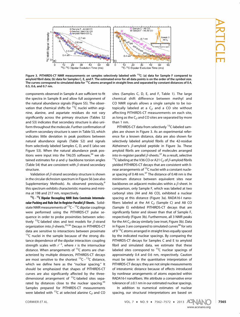
PITHIRDS-CT data from selectively 13C-labeled sam-ples are shown in Figure 3. As an experimental refer-ence for a known distance, data are also shown forselectively labeled amyloid fibrils of the 42-residueAlzheimer's β-amyloid peptide in Figure 3a. Theseamyloid fibrils are composed of molecules arrangedinto in-register parallel β-sheets.37 As a result, selective13C labeling at the V36 CO or A21 Cβ of β-amyloid fibrilsyielded PITHIRDS-CT decays that are consistent with li-near arrangements of 13C nuclei with a constant nucle-ar spacing of 0.48 nm.37 The distance of 0.48 nm is theminimum distance between equivalent sites nearbackbones on adjacent molecules within a β-sheet. Incomparison, only Sample F, which was labeled at twocarbonyl sites (A4 and A6 CO), exhibited a nuclearspacing at this distance (Figure 3a). RADA16-I nano-fibers labeled at the A4 Cβ (Sample C) and A8 CO(Sample E) exhibited PITHIRDS-CT decays that aresignificantly faster and slower than that of Sample F,respectively (Figure 3b). Furthermore, all 3 NMR peaksfor the A4 Cβ decay similarly (see inset, Figure 3b). Datain Figure 3 are compared to simulated curves38 for setsof 9 13C atoms arranged in straight lines equally spacedby the indicated nuclear spacings. By comparing thePITHIRDS-CT decays for Samples C and E to amyloidfibril and simulated data, we estimate that theselabeled sites correspond to 13C nuclear spacings ofapproximately 0.4 and 0.6 nm, respectively. Cautionmust be taken in the quantitative interpretation ofPITHIRDS-CT decays: they are not simplemeasurementsof interatomic distance because of effects introducedby nonlinear arrangements of atoms expected withinRADA16-I nanofibers. We attribute a conservative errortolerance of(0.1 nm in our estimated nuclear spacings.
In addition to numerical estimates of nuclearspacing, our structural interpretations are based on
Figure 3. PITHIRDS-CT NMR measurements on samples selectively labeled with 13C: (a) data for Sample F compared toamyloid fibril data; (b) data for Samples C, E, and F. The estimated error for all data points is on the order of the symbol size.The curves correspond to simulated data for 13C atoms arranged in straight lines and separated by constant distances of 0.4,0.5, 0.6, and 0.7 nm.
ARTIC
LE
CORMIER ET AL. VOL. 7 ’ NO. 9 ’ 7562–7572 ’ 2013
www.acsnano.org
7566
analysis of relative PITHIRDS-CT decay strengths forsamples labeled at different sites. It is immediatelyapparent that the PITHIRDS-CT decay for Sample E ismeasurably slower than that observed for Sample F,indicating that RADA16-I nanofibers are not composedof in-register parallel β-sheets. Furthermore, the fasterdecays for Sample C compared to the amyloid fibrildecays are not possible within isolated β-sheetsand will be discussed in terms of stacking of multipleβ-sheets.
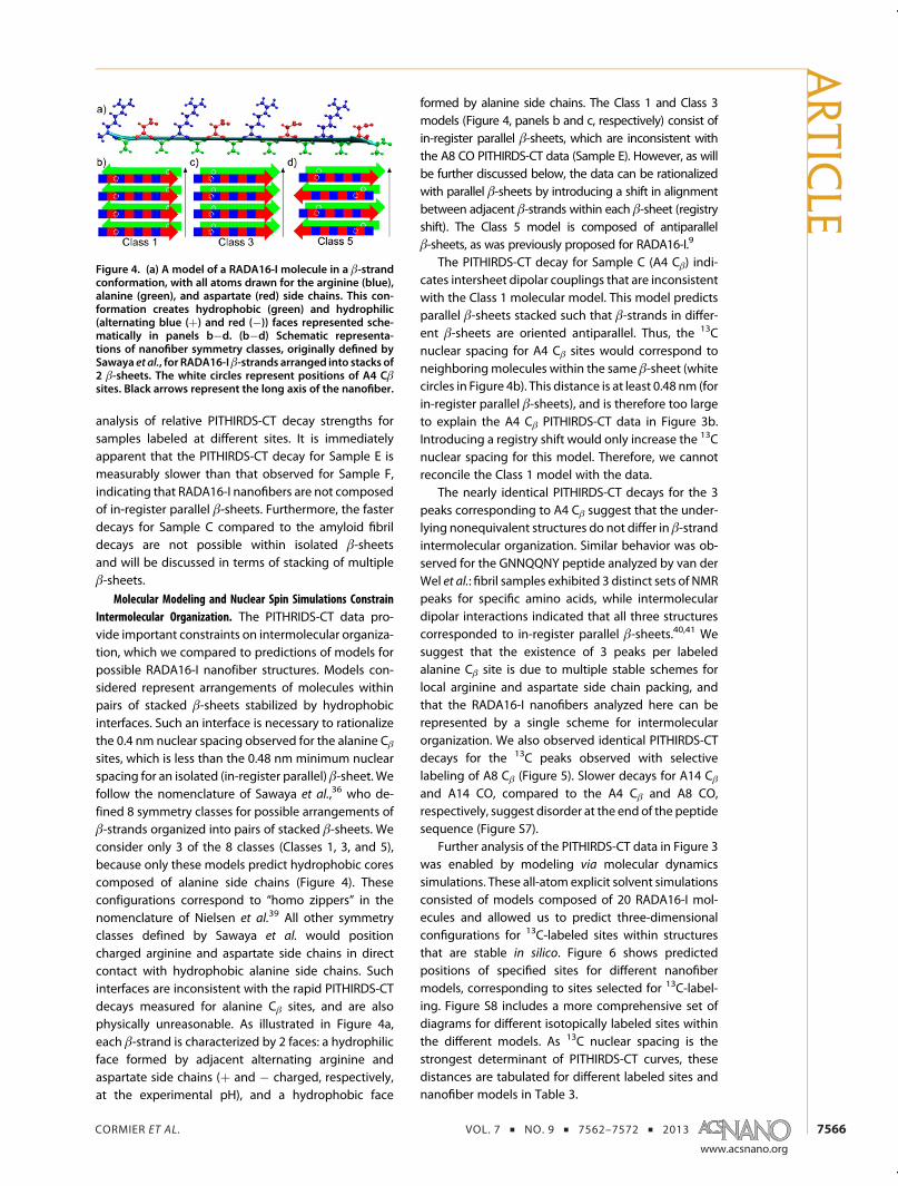
Molecular Modeling and Nuclear Spin Simulations ConstrainIntermolecular Organization. The PITHRIDS-CT data pro-vide important constraints on intermolecular organiza-tion, which we compared to predictions of models forpossible RADA16-I nanofiber structures. Models con-sidered represent arrangements of molecules withinpairs of stacked β-sheets stabilized by hydrophobicinterfaces. Such an interface is necessary to rationalizethe 0.4 nm nuclear spacing observed for the alanine Cβsites, which is less than the 0.48 nm minimum nuclearspacing for an isolated (in-register parallel) β-sheet. Wefollow the nomenclature of Sawaya et al.,36 who de-fined 8 symmetry classes for possible arrangements ofβ-strands organized into pairs of stacked β-sheets. Weconsider only 3 of the 8 classes (Classes 1, 3, and 5),because only these models predict hydrophobic corescomposed of alanine side chains (Figure 4). Theseconfigurations correspond to “homo zippers” in thenomenclature of Nielsen et al.39 All other symmetryclasses defined by Sawaya et al. would positioncharged arginine and aspartate side chains in directcontact with hydrophobic alanine side chains. Suchinterfaces are inconsistent with the rapid PITHIRDS-CTdecays measured for alanine Cβ sites, and are alsophysically unreasonable. As illustrated in Figure 4a,each β-strand is characterized by 2 faces: a hydrophilicface formed by adjacent alternating arginine andaspartate side chains (þ and � charged, respectively,at the experimental pH), and a hydrophobic face
formed by alanine side chains. The Class 1 and Class 3models (Figure 4, panels b and c, respectively) consist ofin-register parallel β-sheets, which are inconsistent withthe A8 CO PITHIRDS-CT data (Sample E). However, as willbe further discussed below, the data can be rationalizedwith parallel β-sheets by introducing a shift in alignmentbetween adjacent β-strands within each β-sheet (registryshift). The Class 5 model is composed of antiparallelβ-sheets, as was previously proposed for RADA16-I.9
The PITHIRDS-CT decay for Sample C (A4 Cβ) indi-cates intersheet dipolar couplings that are inconsistentwith the Class 1 molecular model. This model predictsparallel β-sheets stacked such that β-strands in differ-ent β-sheets are oriented antiparallel. Thus, the 13Cnuclear spacing for A4 Cβ sites would correspond toneighboringmolecules within the same β-sheet (whitecircles in Figure 4b). This distance is at least 0.48 nm (forin-register parallel β-sheets), and is therefore too largeto explain the A4 Cβ PITHIRDS-CT data in Figure 3b.Introducing a registry shift would only increase the 13Cnuclear spacing for this model. Therefore, we cannotreconcile the Class 1 model with the data.
The nearly identical PITHIRDS-CT decays for the 3peaks corresponding to A4 Cβ suggest that the under-lying nonequivalent structures do not differ in β-strandintermolecular organization. Similar behavior was ob-served for the GNNQQNY peptide analyzed by van derWel et al.: fibril samples exhibited 3 distinct sets of NMRpeaks for specific amino acids, while intermoleculardipolar interactions indicated that all three structurescorresponded to in-register parallel β-sheets.40,41 Wesuggest that the existence of 3 peaks per labeledalanine Cβ site is due to multiple stable schemes forlocal arginine and aspartate side chain packing, andthat the RADA16-I nanofibers analyzed here can berepresented by a single scheme for intermolecularorganization. We also observed identical PITHIRDS-CTdecays for the 13C peaks observed with selectivelabeling of A8 Cβ (Figure 5). Slower decays for A14 Cβand A14 CO, compared to the A4 Cβ and A8 CO,respectively, suggest disorder at the end of the peptidesequence (Figure S7).
Further analysis of the PITHIRDS-CT data in Figure 3was enabled by modeling via molecular dynamicssimulations. These all-atom explicit solvent simulationsconsisted of models composed of 20 RADA16-I mol-ecules and allowed us to predict three-dimensionalconfigurations for 13C-labeled sites within structuresthat are stable in silico. Figure 6 shows predictedpositions of specified sites for different nanofibermodels, corresponding to sites selected for 13C-label-ing. Figure S8 includes a more comprehensive set ofdiagrams for different isotopically labeled sites withinthe different models. As 13C nuclear spacing is thestrongest determinant of PITHIRDS-CT curves, thesedistances are tabulated for different labeled sites andnanofiber models in Table 3.
Figure 4. (a) A model of a RADA16-I molecule in a β-strandconformation, with all atoms drawn for the arginine (blue),alanine (green), and aspartate (red) side chains. This con-formation creates hydrophobic (green) and hydrophilic(alternating blue (þ) and red (�)) faces represented sche-matically in panels b�d. (b�d) Schematic representa-tions of nanofiber symmetry classes, originally defined bySawaya et al., for RADA16-Iβ-strands arranged into stacks of2 β-sheets. The white circles represent positions of A4 Cβsites. Black arrows represent the long axis of the nanofiber.
ARTIC
LE
CORMIER ET AL. VOL. 7 ’ NO. 9 ’ 7562–7572 ’ 2013
www.acsnano.org
7567
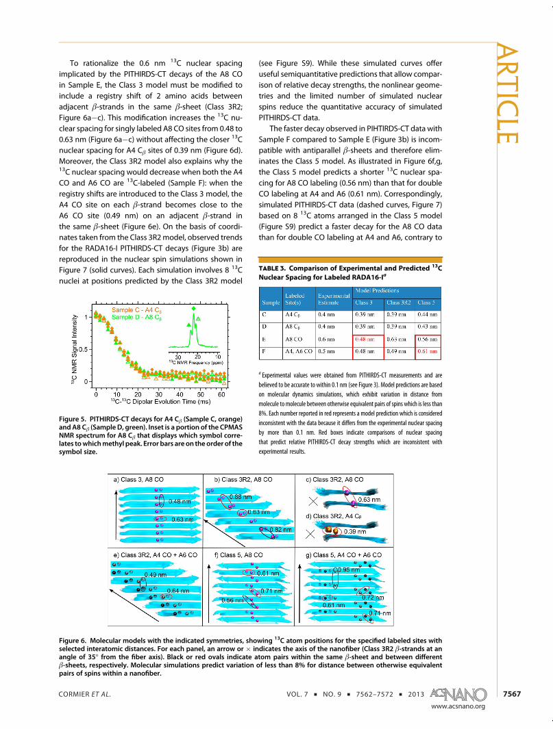
To rationalize the 0.6 nm 13C nuclear spacingimplicated by the PITHIRDS-CT decays of the A8 COin Sample E, the Class 3 model must be modified toinclude a registry shift of 2 amino acids betweenadjacent β-strands in the same β-sheet (Class 3R2;Figure 6a�c). This modification increases the 13C nu-clear spacing for singly labeled A8 CO sites from 0.48 to0.63 nm (Figure 6a�c) without affecting the closer 13Cnuclear spacing for A4 Cβ sites of 0.39 nm (Figure 6d).Moreover, the Class 3R2 model also explains why the13C nuclear spacing would decrease when both the A4CO and A6 CO are 13C-labeled (Sample F): when theregistry shifts are introduced to the Class 3 model, theA4 CO site on each β-strand becomes close to theA6 CO site (0.49 nm) on an adjacent β-strand inthe same β-sheet (Figure 6e). On the basis of coordi-nates taken from the Class 3R2model, observed trendsfor the RADA16-I PITHIRDS-CT decays (Figure 3b) arereproduced in the nuclear spin simulations shown inFigure 7 (solid curves). Each simulation involves 8 13Cnuclei at positions predicted by the Class 3R2 model
(see Figure S9). While these simulated curves offeruseful semiquantitative predictions that allow compar-ison of relative decay strengths, the nonlinear geome-tries and the limited number of simulated nuclearspins reduce the quantitative accuracy of simulatedPITHIRDS-CT data.
The faster decay observed in PIHTIRDS-CT data withSample F compared to Sample E (Figure 3b) is incom-patible with antiparallel β-sheets and therefore elim-inates the Class 5 model. As illustrated in Figure 6f,g,the Class 5 model predicts a shorter 13C nuclear spa-cing for A8 CO labeling (0.56 nm) than that for doubleCO labeling at A4 and A6 (0.61 nm). Correspondingly,simulated PITHIRDS-CT data (dashed curves, Figure 7)based on 8 13C atoms arranged in the Class 5 model(Figure S9) predict a faster decay for the A8 CO datathan for double CO labeling at A4 and A6, contrary to
Figure 6. Molecular models with the indicated symmetries, showing 13C atom positions for the specified labeled sites withselected interatomic distances. For each panel, an arrow or � indicates the axis of the nanofiber (Class 3R2 β-strands at anangle of 35� from the fiber axis). Black or red ovals indicate atom pairs within the same β-sheet and between differentβ-sheets, respectively. Molecular simulations predict variation of less than 8% for distance between otherwise equivalentpairs of spins within a nanofiber.
Figure 5. PITHIRDS-CT decays for A4 Cβ (Sample C, orange)and A8 Cβ (Sample D, green). Inset is a portion of the CPMASNMR spectrum for A8 Cβ that displays which symbol corre-lates towhichmethyl peak. Error bars are on the order of thesymbol size.
TABLE 3. Comparison of Experimental and Predicted 13C
Nuclear Spacing for Labeled RADA16-Ia
a Experimental values were obtained from PITHIRDS-CT measurements and arebelieved to be accurate to within 0.1 nm (see Figure 3). Model predictions are basedon molecular dynamics simulations, which exhibit variation in distance frommolecule to molecule between otherwise equivalent pairs of spins which is less than8%. Each number reported in red represents a model prediction which is consideredinconsistent with the data because it differs from the experimental nuclear spacingby more than 0.1 nm. Red boxes indicate comparisons of nuclear spacingthat predict relative PITHIRDS-CT decay strengths which are inconsistent withexperimental results.
ARTIC
LE

CORMIER ET AL. VOL. 7 ’ NO. 9 ’ 7562–7572 ’ 2013
www.acsnano.org
7568
experimental results. A distance as short as 0.5 nmamong the labeled A4 and A6 CO sites, as requiredby the PITHIRDS-CT data, is possible if the antiparallelβ-sheets were to have a registry shift of at least 6residues. Such a large registry shift would leaveresidues 12�16 of every strand without hydrogenbonding partners, resulting in a very unstable struc-ture. Therefore, only the Class 3R2 model remainsconsistent with all the PITHIRDS-CT data for COlabeling.
Further evidence in favor of the Class 3R2molecularmodel is obtained by comparing PITHIRDS-CT data forA4 Cβ (Sample C) to data for A8 Cβ (Sample D). Figure 5shows that the observed decays did not differ for thesetwo samples. This behavior is consistent with parallelβ-sheets, which predict no change in relative positionsof labeled sites for 13C labeling at different alanine Cβsites (see Figure S9), as well as simulated PITHIRDS-CTcurves in Figure 7 (solid curves). In contrast, the Class 5
model, with antiparallel β-sheets, predicts distinctthree-dimensional arrangements of 13C atoms for A4Cβ labeling compared to A8 Cβ labeling (Figure S9).Since A8 is in the middle of the β-strand, the Class 5model predicts spin interactions between an A8 Cβ siteand equivalent sites on neighboring molecules withinthe same β-sheet and in the adjacent β-sheet. Incontrast, A4 is close to the beginning of the peptidesequence, and an A4 Cβ site will not experience dipolarcouplings with equivalent sites in neighboring mol-ecules within an antiparallel β-sheet (distance >1 nm).Distinct three-dimensional configurations of 13C atomsspaced near the nuclear spacing are predicted tomeasurably affect shapes of PITHIRDS-CT decays (seedashed curves in Figure 7).35
We also probed the organization of RADA16-Iβ-strands within nanofiber β-sheets using Fouriertransform infrared spectroscopy. It has been suggestedthat antiparallel β-sheets exhibit a prominent peakat ∼1630 cm�1 and a weaker resolved signal at∼1695 cm�1, whereas a parallel β-sheet structure issuggested by the absence of the latter peak.42�44 Thespectrum shown in Figure S10 (see also SupplementaryMethods) is consistent with a parallel β-sheet structurefor RADA16-I nanofibers. The absence of the featureat 1695 cm�1 is also seen clearly in the attenuatedtotal reflectance Fourier transform infrared spectrumreported by Arosio et al.28
Figure 8 shows our structural model of the RADA16-I nanofiber, based on the solid-state NMR constraints.Table S5 summarizes our experimental constraints onRADA16-I nanofiber structure in comparison with dif-ferent model predictions. Only the Class 3R2 model isconsistent with the data presented here. Because ofthe registry shift, the parallel β-strands are predicted tobe at an angle of 35� from the fiber axis. This model isharmonious with the solid-state NMR chemical shifts,which indicate that each RADA16-I molecule forms alinear β-strand, as well as the PITHIRDS-CT 13C�13Cdipolar coupling data, which report on the three-dimensional organization of selectively isotopicallylabeled sites near peptide backbones. Conformationsof arginine and aspartate side chains are derived purelyfrom computer modeling and are not constrained byexperimental data. The nanofiber model predicts a
Figure 7. Simulated PITHIRDS-CT decay curves, each corre-sponding to 8 13C spins in positions predicted by theindicated labeling schemes within the Class 3R2 (solidcurves) and Class 5 (dashed curves) models, respectively(see Figure S10): (a) comparison of predicted decays forcarbonyl labeled Samples E (magenta) and F (black); (b)comparison of predicted decays for Samples C (orange) andD (green), respectively.
Figure 8. Molecular model for the RADA16-I nanofiber (Class 3R2), with peptide backbones rendered as ribbons, and atomsshown for alanine (green), arginine (blue), and aspartate (red) residues: (a) Three-dimensional representation of the RADA16-Inanofiber; (b) orthographic projection of the hydrophilic surface of a β-sheet, highlighting the predicted configuration ofarginine and aspartate side chains; (c) orthographic projection of the nanofiber cross section.
ARTIC
LE

CORMIER ET AL. VOL. 7 ’ NO. 9 ’ 7562–7572 ’ 2013
www.acsnano.org
7569
nanofiber height of 2.5 nm and a width of 3 nm asdepicted in Figure 8c. We consider both values to beconsistent with the smallest widths in TEM images(Figure 1). Note that an extended RADA16-I β-strandwould have a length of 5 nm; this dimension is largerthan the nanofiber width because of the 35� anglebetween the molecular axis and the fiber axis. Ob-served widths of greater than 3 nm for some nanofi-bers (Figure 1) would correspond to multiple layers ofthe fibril unit depicted in Figure 8, stabilized by inter-faces between surfaces created by arginine and aspar-tate side chains. RADA16-I nanofiber heights observedby AFM (1.5 nm) are less than the height of twoβ-sheets predicted by the model (2.5 nm). However,this model does not account for possible side chainrearrangements upon deposition onmica surfaces anddrying. Step variations in nanofiber heights first re-ported by Yokoi et al. and confirmed in our AFM images(Figure S1) are consistent with the existence of stackedlayers of β-sheets.9
The structural model of RADA16-I nanofibers inFigure 8 illustrates the complex influences of hydro-phobic, Coulombic, and hydrogen-bonding inter-actions on secondary, tertiary, and quaternary levelsof structure.45 The Class 5 model with antiparallelβ-sheets was proposed previously because this ar-rangement avoids unfavorable close proximity oflike-charged arginine and of like-charged aspartateside chains.9 While previous assignment of β-strandsecondary structure for RADA16-I nanofibers is based onexperimental data (e.g., circular dichroism and Ramanspectroscopy), there have been no direct measurementsof intermolecular organization.9,24 Our PITHIRDS-CT NMRdata indicate that nanofibers are uniquely consistent withthe Class 3R2 model, which constitutes an alternativearrangement that also avoids close proximity betweenlike-charged residues. The repetitive nature of theRADA16-I sequence allows the registry shift to occurwithout compromising hydrophobic interactions be-tween alanine side chains. It is likely that the observedregistry shift was influenced by the size disparity andCoulombic interactions between arginine and aspartateside chains. The importance of side chain Coulombicinteractions is supported by previous observations ofpH-dependentβ-sheet registry shiftswithinamyloidfibrilsformed by other peptides.46,47
In both designer peptide self-assembly and amyloidformation, a fundamental question relates to how theamino acid sequence influences the fibril structure.Amyloid fibrils with similar structures to RADA16-Inanofibers are usually composed of parallel β-sheetsand can exhibit structural features influenced bycharged or polar side chain interactions.37,45 Hydro-phobic interactions, often considered to drive amyloidformation, are usuallymaximized for in-register parallelarrangements through maximal overlap betweenhydrophobic side chains. Recently, mutants of the
Alzheimer's β-amyloid peptide involving charged ami-no acids (D23N and ΔE22) have been reported tosignificantly affect arrangement of β-strands withinβ-sheets.48,49 It should be noted that stabilizing inter-actions between charged and polar side chains cannotbe fully understood without modeling the full three-dimensional configurations of atoms. Structures suchas glutamine and asparagine polar zippers influenceamyloid formation for peptides that self-assembleprimarily through polar side chain interactions.50�53
Furthermore, combinatorial analysis of peptideswith hydrophobic/hydrophilic patterning similar toRADA16-I yielded several alternative structures.15 Thestructural model for RADA16-I presented here maycontribute to our understanding of design rules forself-assembled peptide structures.
The existence of 3 distinct NMR peaks correspond-ing to any alanine Cβ site indicates the coexistence of3 distinct substates in the RADA16-I nanofiber samples.The complete and consistent decays of the PITHIRDS-CT signals (Figure 3b) further indicates that the 3 sub-states correspond to similar three-dimensional config-uration of 13C atoms. Thus, the substates are unlikely tocorrespond to differences at the secondary, tertiary, orquaternary structural level. As suggested in previousstudies on polymorphic amyloid fibrils, heterogeneityin tertiary structure would lead to incomplete decay ofPITHIRDS-CT signals.48 We suggest that the 3 Cβ sub-states could be explained by alternative configurationsof arginine and aspartate side chains which in turnaffect the chemical shift of alanine Cβ within thehydrophobic core. Our molecular dynamics simula-tions starting from different rotameric states of argi-nine and aspartate all led to configurations in which anarginine at position i forms bifurcated hydrogen bondswith the aspartate at position i þ 2 of the sameβ-strand. The two stacked β-sheets have registry shiftsin opposite directions, resulting in the side chainhydrogen bonds either nearly perpendicular or parallelto the fiber axis (Figure S11a,b). The side chain hydro-gen-bonding pattern is formed in two rotameric statesof arginine (Figure S11c,d): I, accounting for ∼2/3 ofarginine�aspartate pairs, has the arginine side chaintorsion angles χ1, χ2, and χ3 all around 180�; II, account-ing for the remaining∼1/3 of arginine�aspartate pairs,has the arginine χ2 torsion angle changed to around60�. Variations in arginine side chain conformation donot have any significant effects on Cβ�Cβ spacings ofalanine side chains packed within the intersheet inter-face (Figure 8c) and hence are not expected to affect CβPITHIRDS-CT decays, but could result in different en-vironments for the alanine side chains, leading todistinct Cβ chemical shifts. The combination of argininerotameric states between the two β-sheets potentiallygives rise to 4 types of environments for the alanineside chains, with probabilities of 4/9, 2/9, 2/9, and 1/9,respectively. It is tempting to assign the first three types
ARTIC
LE
CORMIER ET AL. VOL. 7 ’ NO. 9 ’ 7562–7572 ’ 2013
www.acsnano.org
7570
to the central Cβ peak and the two side peaks, respec-tively, although we have no experimental evidence forsuch an assignment. It is also possible that interactionsbetween nanofibers along the hydrophilic surfaces affectarginine and aspartate side chain configurations. Thisenvironment-based explanation, as opposed to onebased on more localized structural variations, has someexperimental support. Using2Ddipolar assisted rotationalresonance (DARR) measurements (see SupplementaryMethods and Figure S12), we did not observe inter-sidechain contacts between nonequivalent alanine Cβ sites,suggesting that the two contacting alanine Cβ methylsfrom the two opposite β-sheets should always adopt thesame substate. This finding is consistent with the inter-pretation that the two contacting alanine Cβ methylssense the same environment.
Structural knowledge of the RADA16-I nanofibercould provide a basis formodelingmolecular phenom-ena underlying technologically important properties.These properties include environment-sensitive self-assembly and dynamic reassembly. Environment-sen-sitive self-assembly refers to the effects of parameterssuch as pH and ionic strength on nanofiber morphol-ogy and formation kinetics.28,54 These effects can beharnessed to engineer peptides which self-assembleunder specific desired conditions or design processeswhich avoid undesirable structures.55�57 One couldpredict howpH and ionic strength affect the stability ofthe assembly and whether alternative structures canbe stabilized via changes in solvent conditions. Dy-namic reassembly refers to the ability of RADA16-Inanofibers to spontaneously reassemble followingmechanical damage, without the addition of newmonomeric peptide.9,29 Our structural model can be
used to design specific labels or probes to test hy-pothesized dynamic reassembly processes such as thesliding diffusion model proposed by Yokoi et al.,9
ultimately leading to a better understanding of theself-healing property of RADA16-I.
CONCLUSION
Solid-state NMR peak positions, line widths, and13C�13C dipolar couplings are consistent with anordered RADA16-I nanofiber structure in which linearβ-strands are organized into parallel β-sheets. A reg-istry shift of two amino acids between neighboringβ-strands avoids close proximity between like-chargedside chains. Specifically, analysis of peak positions,observed through CPMAS and fpRFDR NMR spectra(Figure 2), correlates with a β-strand secondary struc-ture. The observed NMR line widths are consistent withthose observed for amyloid fibrils, indicating a similardegree of structural order. Through use of selective 13Clabels on alanine residues, PITHIRDS-CT decays werecompared via nuclear spin simulations to candidatemodels. Each model consists of two stacked β-sheetswith either Class 3, 3R2, or 5 nanofiber symmetry. Thestacking of β-sheets is supported by PITHRIDS-CTdecays for 13C-labeling at A4 and A8 Cβ sites, whichcannot be explained by an isolated single β-sheet;these results provide direct evidence for a hydrophobicnanofiber core. Further analysis of 13C�13C nuclearspacing and relative PITHIRDS-CT decays for sampleslabeled at different sites indicated that data are incon-sistent with in-register parallel (Class 3) or antiparallel(Class 5) β-sheets. Themodel in Figure 8, with Class 3R2symmetry, is consistent with all of the experimentalconstraints.
METHODS
RADA16-I was obtained following standard FMOC synthesisand purification procedures; see Supporting Information fordetails. Isotopic labels (see Table 1) were incorporated usingcommercial 13C-labeled protected amino acids. RADA16-I wasallowed to self-assemble into nanofibers at a concentration of 5mg RADA16-I in 1 mL of 10 mM phosphate buffer (pH 4.85). ForTEM imaging, a small aliquot (10 μL) of RADA16-I nanofibersolution was taken after 24 h of self-assembly and diluted 100-fold with water. The details of TEM specimen preparation aredescribed previously.29
For solid-state NMR measurements, nanofibers were re-covered by centrifugation (245 000g for 25 min at 4 �C) after48 h of self-assembly. Solid-state NMR samples were pre-pared by freeze-drying centrifuge pellets, packing dry pow-ders into NMR rotors, and then hydrating with 1 mg ofwater/mg of peptide. Measurements were performed onan 11.75 T (500 MHz 1H NMR frequency) Bruker Avance IIIsystem with a 2.5 mm Bruker MAS probe. Experimentsincluded CPMAS,58 2D-fpRFDR,30 and PITHIRDS-CT,35 withspecific parameters detailed in Supporting Information.Reported NMR chemical shifts are relative to tetramethylsilane, as calibrated before every experiment using a microcrystalline glycine system that was selectively labeled at theCO site.
For computer modeling of the RADA16-I nanofiber, a β-strand with standard torsion angles was built with Ambertools1.5. The β-strand was replicated to make parallel or antiparallelβ-sheets, and the β-sheets were paired according to the speci-fied symmetry classes. The pairs of β-sheets were solvated withwater (explicit solvent) and the whole systems were energyminimized and refined by molecular dynamics simulations.More details are found in Supporting Information.Nuclear spin simulations of PITHIRDS-CT experiments were
performed using SPINEVOLUTION. The simulations includedeither 8 or 9 13C atoms, arranged linearly for 9-atom simulations(Figure 3) or in positions predicted by the molecular models forthe 8-atom simulations (see Figure S10). More details are foundin Supporting Information.
Conflict of Interest: The authors declare no competingfinancial interest.
Acknowledgment. This research was supported by fundsfrom the National Science Foundation Grant DMR-105521 to A.Paravastu and the National Institute of Health Grant GM88187to H. Zhou. A portion of this work was performed at the NationalHighMagnetic Field Laboratory (NHMFL), which is supported byNational Science Foundation Cooperative Agreement No. DMR-0654118, the State of Florida, and the U.S. Department ofEnergy. This work was also partially supported by an NHMFL
ARTIC
LE
CORMIER ET AL. VOL. 7 ’ NO. 9 ’ 7562–7572 ’ 2013
www.acsnano.org
7571
User Collaboration Research Grant to A. Paravastu. The data onamyloid fibrils of the Alzheimer's β-amyloid peptide (Figure 3a)were produced by D. Huang and W. M. Tay. We also gratefullyacknowledge E. A. Bienkiewicz and L. Longo for assistancecollecting the circular dichroism spectrum, R. G. Alamo forassistance collecting the Fourier transform infrared spectrum,D. Huang for assistance with AFM imaging, and P. C. A. Van derWel for assistance configuring SPINEVOLUTION nuclear spinsimulation scripts.
Supporting Information Available: Supplementary methods,additional references, AFM imaging of RADA16-I nanofibers,additional solid-state NMR analysis, additional modeling andNMR simulation data, summary of experimental and modelingconstraints. This material is available free of charge via theInternet at http://pubs.acs.org.
REFERENCES AND NOTES1. Holmes, T. C. Novel Peptide-Based Biomaterial Scaffolds
for Tissue Engineering. Trends. Biotechnol. 2002, 20, 16–21.2. Cigognini, D.; Satta, A.; Colleoni, B.; Silva, D.; Donega, M.;
Antonini, S.; Gelain, F. Evaluation of Early and Late Effectsinto the Acute Spinal Cord Injury of an Injectable Functio-nalized Self-Assembling Scaffold. PLoS One 2011, 6, 1–15.
3. Gelain, F.; Horii, A.; Zhang, S. G. Designer Self-AssemblingPeptide Scaffolds for 3-D Tissue Cell Cultures and Regen-erative Medicine. Macromol. Biosci. 2007, 7, 544–551.
4. Cunha, C.; Panseri, S.; Villa, O.; Silva, D.; Gelain, F. 3D Cultureof Adult Mouse Neural Stem Cells within FunctionalizedSelf-Assembling Peptide Scaffolds. Int. J. Nanomed. 2011,6, 943–955.
5. Koutsopoulos, S.; Unswortha, L. D.; Nagaia, Y.; Zhang, S. G.Controlled Release of Functional Proteins through De-signer Self-Assembling Peptide Nanofiber Hydrogel Scaf-fold. Proc. Natl. Acad. Sci. U.S.A. 2009, 106, 4623–4628.
6. Horii, A.; Wang, X. M.; Gelain, F.; Zhang, S. G. BiologicalDesigner Self-Assembling Peptide Nanofiber Scaffoldssignificantly Enhance Osteoblast Proliferation, Differentia-tion and 3-D Migration. PLoS One 2007, 2, 1–9.
7. Kumada, Y.; Zhang, S. G. Significant Type I and Type IIICollagen Production from Human Periodontal LigamentFibroblasts in 3D Peptide Scaffolds without Extra GrowthFactors. PLoS One 2010, 5, 1–7.
8. Liu, J. P.; Zhao, X. J. Design of Self-Assembling Peptides andtheir Biomedical Applications. Nanomedicine 2011, 6,1621–1643.
9. Yokoi, H.; Kinoshita, T.; Zhang, S. G. Dynamic Reassembly ofPeptide RADA16 Nanofiber Scaffold. Proc. Natl. Acad. Sci.U.S.A. 2005, 102, 8414–8419.
10. Song, H.; Zhang, L. L.; Zhao, X. J. Hemostatic Efficacy ofBiological Self-Assembling Peptide Nanofibers in a RatKidney Model. Macromol. Biosci. 2010, 10, 33–39.
11. Ellis-Behnke, R. G.; Liang, Y. X.; You, S. W.; Tay, D. K. C.;Zhang, S. G.; So, K. F.; Schneider, G. E. Nano Neuro Knitting:Peptide Nanofiber Scaffold for Brain Repair and AxonRegeneration with Functional Return of Vision. Proc. Natl.Acad. Sci. U.S.A. 2006, 103, 5054–5059.
12. Silva, G. A.; Parpura, V. In Nanotechnology for Biology andMedicine: At the Building Block Level; Springer: New York,NY, 2011; p 242.
13. Shastri, V. P. In Vivo Engineering of Tissues: BiologicalConsiderations, Challenges, Strategies, and Future Direc-tions. Adv. Mater. 2009, 21, 3246–3254.
14. Tang, C. K.; Shao, X. M.; Sun, B. B.; Huang, W. L.; Zhao, X. J.The Effect of Self-Assembling Peptide RADA16-I on theGrowth of Human Leukemia Cells in Vitro and in NudeMice. Int. J. Mol. Sci. 2009, 10, 2136–2145.
15. Zhang, S. G. Emerging Biological Materials through Mo-lecular Self-Assembly. Biotechnol. Adv. 2002, 20, 321–339.
16. Zhang, S. G.; Lockshin, C.; Herbert, A.; Winter, E.; Rich, A.Zuotin, a Putative Z-DNA Binding-Protein in Saccharo-myces cerevisiae. EMBO J. 1992, 11, 3787–3796.
17. Zhang, S. G.; Holmes, T.; Lockshin, C.; Rich, A. SpontaneousAssembly of a Self-Complementary Oligopeptide To Form
a Stable Macroscopic Membrane. Proc. Natl. Acad. Sci.U.S.A. 1993, 90, 3334–3338.
18. Holmes, T. C.; de Lacalle, S.; Su, X.; Liu, G. S.; Rich, A.; Zhang,S. G. Extensive Neurite Outgrowth and Active SynapseFormation on Self-Assembling Peptide Scaffolds. Proc.Natl. Acad. Sci. U.S.A. 2000, 97, 6728–6733.
19. Zhang, S. G.; Holmes, T. C.; Dipersio, C. M.; Hynes, R. O.; Su,X.; Rich, A. Self-Complementary Oligopeptide MatricesSupport Mammalian-Cell Attachment. Biomaterials 1995,16, 1385–1393.
20. Jonker, A. M.; Loewik, D. W. P. M.; van Hest, J. C. M. Peptide-and Protein-Based Hydrogels. Chem.Mater. 2012, 24, 759–773.
21. Altman, M.; Lee, P.; Rich, A.; Zhang, S. G. ConformationalBehavior of Ionic Self-Complementary Peptides. ProteinSci. 2000, 9, 1095–1105.
22. Marcotte, E. M.; Pellegrini, M.; Yeates, T. O.; Eisenberg, D. ACensus of Protein Repeats. J. Mol. Biol. 1999, 293, 151–160.
23. Chen, Y. W.; Ding, F.; Nie, H. F.; Serohijos, A. W.; Sharma, S.;Wilcox, K. C.; Yin, S. Y.; Dokholyan, N. V. Protein Folding:Then and Now. Arch. Biochem. Biophys. 2008, 469, 4–19.
24. Taraballi, F.; Campione, M.; Sassella, A.; Vescovi, A.; Paleari,A.; Hwang, W.; Gelain, F. Effect of Functionalization on theSelf-Assembling Propensity of Beta-Sheet Forming Pep-tides. Soft Matter 2009, 5, 660–668.
25. Hilborn, J. In Vivo Injectable Gels for Tissue Repair. WileyInterdiscip. Rev.: Nanomed. Nanobiotechnol. 2011, 3, 589–606.
26. Loo, Y.; Zhang, S.; Hauser, C. A. E. From Short Peptides toNanofibers toMacromolecular Assemblies in Biomedicine.Biotechnol. Adv. 2012, 30, 593–603.
27. Wu, E. C.; Zhang, S. G.; Hauser, C. A. E. Self-AssemblingPeptides as Cell-Interactive Scaffolds. Adv. Funct. Mater.2012, 22, 456–468.
28. Arosio, P.; Owczarz, M.; Wu, H.; Butte, A.; Morbidelli, M. End-to-End Self-Assembly of RADA 16-I Nanofibrils in AqueousSolutions. Biophys. J. 2012, 102, 1617–1626.
29. Cheng, L.; Englander, O.; Paravastu, A. K.; Oates, W. S.An Effective Continuum Approach for Modeling Non-Equilibrium Structural Evolution of Protein Nanofiber Net-works. J. Chem. Phys. 2011, 135, 055102-1–055102-15.
30. Ishii, Y. C-13-C-13 Dipolar Recoupling under very FastMagic Angle Spinning in Solid-State Nuclear MagneticResonance: Applications to Distance Measurements,Spectral Assignments, and High-Throughput Secondary-Structure Determination. J. Chem. Phys. 2001, 114, 8473–8483.
31. Wishart, D. S. Interpreting Protein Chemical Shift Data.Prog. Nucl. Magn. Reson. Spectrosc. 2011, 58, 62–87.
32. Paravastu, A. K.; Leapman, R. D.; Yau, W. M.; Tycko, R.Molecular Structural Basis for Polymorphism in Alzheimer'sBeta-Amyloid Fibrils. Proc. Natl. Acad. Sci. U.S.A. 2008, 105,18349–18354.
33. Ulrich, E. L.; Akutsu, H.; Doreleijers, J. F.; Harano, Y.;Ioannidis, Y. E.; Lin, J.; Livny, M.; Mading, S.; Maziuk, D.;Miller, Z.; et al. BioMagResBank.Nucleic Acids Res. 2008, 36,D402–D408.
34. Cornilescu, G.; Delaglio, F.; Bax, A. Protein Backbone AngleRestraints from Searching a Database for Chemical Shiftand Sequence Homology. J. Biomol. NMR 1999, 13, 289–302.
35. Tycko, R. Symmetry-Based Constant-Time HomonuclearDipolar Recoupling in Solid State NMR. J. Chem. Phys.2007, 126, 064506–064506.
36. Sawaya, M. R.; Sambashivan, S.; Nelson, R.; Ivanova, M. I.;Sievers, S. A.; Apostol, M. I.; Thompson, M. J.; Balbirnie, M.;Wiltzius, J. J. W.; McFarlane, H. T.; et al. Atomic Structures ofAmyloid Cross-Beta Spines Reveal Varied Steric Zippers.Nature 2007, 447, 453–457.
37. Balbach, J. J.; Petkova, A. T.; Oyler, N. A.; Antzutkin, O. N.;Gordon, D. J.; Meredith, S. C.; Tycko, R. SupramolecularStructure in Full-Length Alzheimer's Beta-Amyloid Fibrils:Evidence for a Parallel Beta-SheetOrganization fromSolid-State Nuclear Magnetic Resonance. Biophys. J. 2002, 83,1205–1216.
ARTIC
LE

CORMIER ET AL. VOL. 7 ’ NO. 9 ’ 7562–7572 ’ 2013
www.acsnano.org
7572
38. Veshtort, M.; Griffin, R. G. SPINEVOLUTION: A Powerful Toolfor the Simulation of Solid and Liquid State NMR Experi-ments. J. Magn. Reson. 2006, 178, 248–282.
39. Nielsen, J. T.; Bjerring, M.; Jeppesen, M. D.; Pedersen, R. O.;Pedersen, J. M.; Hein, K. L.; Vosegaard, T.; Skrydstrup, T.;Otzen, D. E.; Nielsen, N. C. Unique Identification of Supra-molecular Structures in Amyloid Fibrils by Solid-State NMRSpectroscopy. Angew. Chem., Int. Ed. 2009, 48, 2118–2121.
40. van der Wel, P. C. A.; Lewandowski, J. R.; Griffin, R. G. Solid-State NMR Study of Amyloid Nanocrystals and FibrilsFormed by the Peptide GNNQQNY from Yeast PrionProtein Sup35p. J. Am. Chem. Soc. 2007, 129, 5117–5130.
41. van der Wel, P. C. A.; Lewandowski, J. R.; Griffin, R. G.Structural Characterization of GNNQQNY Amyloid Fibrilsby Magic Angle Spinning NMR. Biochemistry 2010, 49,9457–9469.
42. Bakota, E. L.; Sensoy, O.; Ozgur, B.; Sayar, M.; Hartgerink,J. D. Self-Assembling Multidomain Peptide Fibers withAromatic Cores. Biomacromolecules 2013, 14, 1370–1370�1378.
43. Cerf, E.; Sarroukh, R.; Tamamizu-Kato, S.; Breydo, L.; Derclaye,S.; Dufrene, Y. F.; Narayanaswami, V.; Goormaghtigh, E.;Ruysschaert, J. M.; Raussens, V. Antiparallel Beta-Sheet: ASignature Structure of the Oligomeric Amyloid Beta-Peptide. Biochem. J. 2009, 421, 415–423.
44. Mukherjee, S.; Chowdhury, P.; Gai, F. Effect of Dehydrationon the Aggregation Kinetics of Two Amyloid Peptides.J. Phys. Chem. B 2009, 113, 531–535.
45. Tycko, R. Molecular Structure of Amyloid Fibrils: Insightsfrom Solid-State NMR. Q. Rev. Biophys. 2006, 39, 1–55.
46. Verel, R.; Tomka, I. T.; Bertozzi, C.; Cadalbert, R.; Kammerer,R. A.; Steinmetz, M. O.; Meier, B. H. Polymorphism in anAmyloid-like Fibril-FormingModel Peptide. Angew. Chem.,Int. Ed. 2008, 47, 5842–5845.
47. Petkova, A. T.; Buntkowsky, G.; Dyda, F.; Leapman, R. D.;Yau, W. M.; Tycko, R. Solid State NMR Reveals a pH-Dependent Antiparallel Beta-Sheet Registry in FibrilsFormed by a Beta-Amyloid Peptide. J. Mol. Biol. 2004,335, 247–260.
48. Qiang, W.; Yau, W.; Tycko, R. Structural Evolution of IowaMutant Beta-Amyloid Fibrils from Polymorphic to Homo-geneous States Under Repeated Seeded Growth. J. Am.Chem. Soc. 2011, 133, 4018–4029.
49. Cloe, A. L.; Orgel, J.; Sachleben, J. R.; Tycko, R.; Meredith,S. C. The JapaneseMutant A Beta (Delta E22-A Beta(1�39))Forms Fibrils Instantaneously, with Low-Thioflavin T Fluo-rescence: Seeding ofWild-Type A Beta(1�40) into AtypicalFibrils by Delta E22-A Beta(1�39). Biochemistry 2011, 50,2026–2039.
50. Goehler, H.; Droge, A.; Lurz, R.; Schnoegl, S.; Chernoff, Y. O.;Wanker, E. E. Pathogenic Polyglutamine Tracts Are PotentInducers of Spontaneous Sup35 and Rnq1 Amyloidogen-esis. PLoS One 2010, 5, 1–13.
51. Chan, J. C. C.; Oyler, N. A.; Yau, W.M.; Tycko, R. Parallel Beta-Sheets and Polar Zippers in Amyloid Fibrils Formed byResidues 10�39 of the Yeast Prion Protein Ure2p. Bio-chemistry 2005, 44, 10669–10680.
52. Perutz, M. F.; Johnson, T.; Suzuki, M.; Finch, J. T. GlutamineRepeats as Polar Zippers�Their Possible Role in InheritedNeurodegenerative Diseases. Proc. Natl. Acad. Sci. U.S.A.1994, 91, 5355–5358.
53. Perutz, M. F.; Staden, R.; Moens, L.; Debaere, I. PolarZippers. Curr. Biol. 1993, 3, 249–253.
54. Ye, Z. Y.; Zhang, H. Y.; Luo, H. L.; Wang, S. K.; Zhou, Q. H.; Du,X. P.; Tang, C. K.; Chen, L. Y.; Liu, J. P.; Shi, Y. K.; et al.Temperature and pH Effects on Biophysical and Morpho-logical Properties of Self-Assembling Peptide RADA16�1.J. Pept. Sci. 2008, 14, 152–162.
55. Cormier, A. R.; Ruiz-Orta, C.; Alamo, R. G.; Paravastu, A. K.Solid State Self-Assembly Mechanism of RADA16-I De-signer Peptide. Biomacromolecules 2012, 13, 1794–1804.
56. Haines-Butterick, L.; Rajagopal, K.; Branco, M.; Salick, D.;Rughani, R.; Pilarz,M.; Lamm,M. S.; Pochan, D. J.; Schneider,J. P. Controlling Hydrogelation Kinetics by PeptideDesign for Three-Dimensional Encapsulation and
Injectable Delivery of Cells. Proc. Natl. Acad. Sci. U.S.A.2007, 104, 7791–7796.
57. Schneider, J. P.; Pochan, D. J.; Ozbas, B.; Rajagopal, K.;Pakstis, L.; Kretsinger, J. Responsive Hydrogels from theIntramolecular Folding and Self-Assembly of a DesignedPeptide. J. Am. Chem. Soc. 2002, 124, 15030–15037.
58. Schaefer, J.; Stejskal, E. C-13 Nuclear Magnetic-Resonanceof Polymers Spinning at Magic Angle. J. Am. Chem. Soc.1976, 98, 1031–1032.
ARTIC
LE