yakrit - the clinical application
TRANSCRIPT
Yakrit - the clinical application
Prof. S.N. Ojha , principal and Director, Hon Annasaheb Dange Ayurveda
Medical college, Ashta, Sangli, Maharashtra
Dr. Anant Prabhakar Samant , Associate professor department of Ayurveda
Samhita , J J Magdum Ayurveda college , Jaisingpur, Kolhapur.
Yakrit is considered as one of the koshtanga in human body.(Ca.Sa. 7/10) It is
Matruja avayava. (Ca. Sa.3/6) Yakrit is the moolasthana of raktavaha srotas
which maintains the quality as well as quantity of rakta.(Ca. Chi.4/10) Yakrit
and Pleeha are the mulasthana of Raktavahasrotas and Acharya Sushruta has
mentioned that both the organs have their origin in Garbhavasta from Rakta
dhatu. Thus by ASHRAYA ASHRAYI sambandha whenever the raktadhatu
gets vitiated it will diseased the Yakrit and vice versa is also true that whenever
Yakrit gets vitiated it will vitiate the Rakta dhatu.
In Samhita there is no specific importance given to Yakrit as avayava whereas
the classic have given importance to raktavaha srotas and rakta dhatu. In
modern era it is the need of the hour to understand the Hepatic and Biliary
disorders in Modern parlance.
Charak in Sutrasthan 24th chapter has mentioned the general causes of Rakta
dusti and described the symptoms. All the hetu mentioned may be classified as
some vitiating the dhatvagni, some vitiating the srotas ashrita vata and some
acting on the srotas. Those acting on the srotas will be considered here.
Pradustha, bahu, tikshna, ushna Madhya and wrongly prepared and
administered madya leads to rakta and yakrit dushti. Madhya when taken while
following rules and regulation acts like Amrita otherwise it acts like the Visha.
(Ca. Ci. 24/28) Madhya by its ten qualities which are opposite to Oja, help in
vitiating raktadi dhatu and Oja. The ushna tikshna and vyavayi guna contributes
in traversing various protective barriers within the body.
Alcohol (ethanol) is readily absorbed from the stomach, but most is absorbed
from the small intestine. Alcohol cannot be stored. A small amount is degraded
in transit through the gastric mucosa, but most is catabolized in the liver,
primarily by alcohol dehydrogenase (ADH) but also by cytochrome P-450 2E1
(CYP2E1) and the microsomal enzyme oxidation system (MEOS).
Metabolism via the ADH pathway involves the following:
ADH, a cytoplasmic enzyme, oxidizes alcohol into acetaldehyde. Genetic
polymorphisms in ADH account for some individual differences in blood
alcohol levels after the same alcohol intake but not in susceptibility to
alcoholic liver disease.
Acetaldehyde dehydrogenase (ALDH), a mitochondrial enzyme, then
oxidizes acetaldehyde into acetate. Chronic alcohol consumption enhances
acetate formation. Asians, who have lower levels of ALDH, are more
susceptible to toxic acetaldehyde effects (eg, flushing).

These oxidative reactions generate hydrogen, which converts
nicotinamide-adenine dinucleotide (NAD) to its reduced form (NADH),
increasing the redox potential (NADH/NAD) in the liver.
The increased redox potential inhibits fatty acid oxidation and
gluconeogenesis, promoting fat accumulation in the liver.
Chronic alcoholism induces the MEOS (mainly in endoplasmic reticulum),
increasing its activity. The main enzyme involved is CYP2E1. When induced,
the MEOS pathway can account for 20% of alcohol metabolism. This pathway
generates harmful reactive oxygen species, increasing oxidative stress and
formation of oxygen-free radicals.
Hepatic fat accumulation
Fat (triglycerides) accumulates throughout the hepatocytes for the following
reasons:
Export of fat from the liver is decreased because hepatic fatty acid
oxidation and lipoprotein production decrease.
Input of fat is increased because the decrease in hepatic fat export
increases peripheral lipolysis and triglyceride synthesis, resulting
in hyperlipidemia.
Hepatic fat accumulation may predispose to subsequent oxidative damage.
Laghu, Ushna, Tikshna, sukshma, Vyavayi, Ashu are the guna which bring
about easy absorption and the Oxidative stress.
Alcohol changes gut permeability, increasing absorption of endotoxins released
by bacteria in the gut. In response to the endotoxins (which the impaired liver
can no longer detoxify), liver macrophages (Kupffer cells) release free radicals,
increasing oxidative damage.
Oxidative stress is increased by
Liver hypermetabolism, caused by alcohol consumption
Free radical–induced lipid peroxidative damage
Reduction in protective antioxidants (eg, glutathione, vitamins A and E),
caused by alcohol-related undernutrition
Binding of alcohol oxidation products, such as acetaldehyde, to liver cell
proteins, forming neoantigens and resulting in inflammation
Accumulation of neutrophils and other white blood cells (WBCs), which
are attracted by lipid peroxidative damage and neoantigens
Inflammatory cytokines secreted by WBCs
A vicious circle of worsening inflammation occurs: Cell necrosis and apoptosis
result in hepatocyte loss, and subsequent attempts at regeneration result
in fibrosis. Stellate (Ito) cells, which line blood channels (sinusoids) in the liver,
proliferate and transform into myofibroblasts, producing an excess of type I
collagen and extracellular matrix. As a result, the sinusoids narrow, limiting
blood flow. Fibrosis narrows the terminal hepatic venules, compromising
hepatic perfusion and thus contributing to portal hypertension. Extensive
fibrosis is associated with an attempt at regeneration, resulting in liver nodules.
This process culminates in cirrhosis.
Sura, Sauvira, Sukta, are the different types of madya which contribute to
Yakrit vikar when taken in excess or in improper manner.
Madya has been the cause for Raktapitta, Shoth, Udara, Madatyaya. These are
all the diseases where Rakta is among the main dusya and involvement of
Yakrit is observed.
Lavana Rasa Atyadhik Sevan:
Lavan again contains Ushna, tikshna, sukshma guna as mentioned in Madya.
पितं्त कोियपि,रकं्त वधधयपि, िर्धयपि, मूर्च्ध यपि िाियपि, दारयपि,कुष्णापि मांसापन, प्रगालयपि
कुष्ठापन,पवरं् वधधयपि, शोफान् स्फोटयपि. Excess intake of lavana is among the cause for
rakta dusti.
Too much sodium led to a number of changes in the liver – such as misshapen
cells, higher rates of cell death and lower rates of cell division – all of which
can lead to liver fibrosis.
Liver fibrosis occurs when there is excessive accumulation of “extracellular
matrix” proteins like collagen that support the cells that do the work of the liver
– such as breaking down old and damaged cells and metabolizing fats for
energy.

The researchers suggest the mechanism through which too much salt may cause
liver damage and fibrosis in both adults and developing embryos is through
oxidative stress.
Oxidative stress is where the balance between the production of reactive oxygen
species (free radicals) and antioxidants is upset in favor of the former. Such an
imbalance can increase inflammatory cells and promote the death of liver cells,
leading to progressive fibrosis.
https://www.medicalnewstoday.com/articles/307028#Too-much-salt-led-to-changes-in-liver-cells-
linked-to-fibrosis
High-salt diet exacerbated nonalcoholic steatohepatitis (NASH) in high-fat diet-
fed LOX-1 transgenic /apoE knockout mice and that this effect was associated
with the stimulation of oxidative and inflammatory processes; this is the first
study to suggest the important role of excessive salt intake in the development
of NASH. Importantly, the survival rate was reduced in NASH patients, who
frequently died from liver-related causes in addition to cardiovascular events.
Because NASH is not a benign disease, it is considered to be an important
health-threatening component of Metabolic Syndrome. Excessive salt intake, an
oxidative stress-inducer, is an accelerator of the onset and progression of High
Fat Diet (HFD)-induced NASH due to ROS overproduction. Salt loading on
HFD may accelerate induction of NASH. High Salt (HS)/HFD induced
significant fatty degeneration around the hepatic central veins associated with
fibrotic changes. The deterioration of fatty metabolism, inflammation, and
pericellular fiborosis observed in HS/HFD-fed mice met the criteria of NASH in
human. https://www.ncbi.nlm.nih.gov/pmc/articles/PMC4337194/

Katu rasa helps in शोपििसङ्घािं पभनपत्त in normal quantity but when taken in excess
it is the cause for bleeding tendency. The ushna, tikshna, laghu guna are causes
of rakta sanghata bhinnatti.
Research of the effects of hot red pepper on rabbit's hepatic tissue shows that
when Rabbits were orally ingested 2 g/Kg hot red pepper every day for 10 days.
Hot red pepper induced a significant increase in temperature of rabbits after oral
ingestion of each dose. Hepatic tissue damage was recorded through
examination of the stained paraffin embedded sections. Inflammatory cellular
infiltration and hepatocytic vacuolation were marked in rabbits ingested with the
hot red pepper. Histochemical studies reveal a decrease in both of carbohydrates
and protein contents in the liver. Serum alanine aminotransferase (ALT),
aspartate aminotransferase (AST), cholesterol, triglycerides and glucose were
decreased in rabbit due to oral ingestion of hot red pepper. These scientific
evidences show that hot red pepper possesses some chemical and
pharmacological properties which are capable of inducing liver damage.
Therefore, based on the findings of this study, the excessive consumption of red
pepper is capable of inducing liver damage and so should be avoided.
https://www.researchgate.net/publication/228489328_Effects_of_extensive_consumption_of_hot_
red_pepper_fruit_on_liver_of_rabbit
Amla rasa कफं पवलाियपि, पित्तमपभवधधयपि, रकं्त दूर्यपि,मांसं पवदहपि. Pitta vridhi and
rakta dusthi is cause for yakrit dushti.
Virudha anna is cause for jalodara, pandu like disease which are related to
yakrit.

कुलत्थ, मार्, पनष्पाव, पिलिैलपनरे्विै, पिण्डालु, मूलकादी, हररिानां are some food items
which are among the factors which are cause for raktapitta, vatarakta as
discussed above are the cause for yakrit dushti.
अपशिस्यापिसङ्क्षोभाद्यानयानापिचेपििैैः, अपिव्यवाय, भाराध्व, वमन, व्यापधकशधनै are hetu for
pleehodar and this may also be cause for yakrit vikruti.
पवदाह्युष्णपिभोजनाि्, पवरुद्धाजीिध, सङ्लिि, पवर्मासात्म्यभोजनाि् व्यािन्न, बहुमद्यत्वा,
दे्वगसन्धारिार्च्र माि् पजह्मव्यायाम शयनादपिभाराध्व मैथुनाि् are common cause in
raktadushti and yakrit antahvidradi. Therefore such hetu will cause dusti of
yakrit. Similarly hetu in raktapitta, vatarakta, udara are common
Visha like madya has qualities opposite to Oja. Especially the Ushna, tikshna,
vyavayi and vikashi guna which helps the poison to cross all the protective
barriers within the body. Visha also has deteriorating effect on yakrit.
Hepatotoxicity implies chemical-driven liver damage. Drug-induced liver
injury is a cause of acute and chronic liver disease. The liver plays a central role
in transforming and clearing chemicals and is susceptible to the toxicity from
these agents. Certain medicinal agents, when taken in overdoses and sometimes
even when introduced within therapeutic ranges, may injure the organ. Other
chemical agents, such as those used in laboratories and industries, natural
chemicals (e.g., microcystins) can also induce hepatotoxicity. Chemicals that
cause liver injury are called hepatotoxins. Drug-induced liver injury is
responsible for 5% of all hospital admissions and 50% of all acute liver failures.
Due to its unique metabolism and close relationship with the gastrointestinal
tract, the liver is susceptible to injury from drugs and other substances. 75% of
blood coming to the liver arrives directly from gastrointestinal organs and the
spleen via portal veins that bring drugs and xenobiotics in near-undiluted form.
Several mechanisms are responsible for either inducing hepatic injury or
worsening the damage process. Many chemicals damage mitochondria, an
intracellular organelle that produces energy. Its dysfunction releases excessive
amount of oxidants that, in turn, injure hepatic cells. Activation of some
enzymes in the cytochrome P-450 system such as CYP2E1 also lead to
oxidative stress. Injury to hepatocyte and bile duct cells lead to accumulation
of bile acid inside the liver. This promotes further liver damage. Non-
parenchymal cells such as Kupffer cells, fat storing stellate cells,
and leukocytes (i.e. neutrophil and monocyte) also have a role in the
mechanism. https://en.wikipedia.org/wiki/Hepatotoxicity
Hetu other than those explained in Vidhi Shonitiya Adhyaya are discussed
below. Various diseasess are cause for yakrit vikar.
Jwar has its effect on Yakrit through the involvement of rakta. रक्तधात्वाश्रयैः प्रायो
दोर्ैः सििकं ज्वरम्. Vishamjwara having ashraya in raktadhatu will affect the rakta
dhatu and inturn yakrit. As understood by reverse pharmacology Pippalyadi
ghrita useful in jwara has its use in visham jwar and halimaka as its phalashruti.

The liver contains approximately one-third of the reticuloendothelial system
mass in humans. As the recipient of both the portal and systemic circulation, the
liver plays an important role in host defense against invasive microorganisms.
The impact of microbial pathogens on the liver can vary greatly, presenting with
a wide variety of manifestations from asymptomatic elevations in
aminotransaminases, acute liver failure, hepatic fibrosis, and cirrhosis.
Viruses Bacteria and
Mycobacteria
Parasites Fungi
Epstein Barr Virus
(EBV]
Salmonella
enterica serotype
typhi
Schistosoma
species
(Schistosomiasis)
Candida
species
Cytomegalovirus
(CMV)
Mycobacterium
tuberculosis
Plasmodium
species
(malaria)
Histoplasma
capsulatum
Herpes Simplex Virus
(HSV) and other
Herpes
Viruses
Brucella species
Yellow Fever Virus Coxiella bunerii
(Q
fever)

Viruses Bacteria and
Mycobacteria
Parasites Fungi
Dengue Virus Leptospira and
other Spirochetes
Typhoid Fever: Salmonella enterica serotype typhi is the causative agent.
Hepatic involvement with Salmonella occurs via both hematogenous seeding of
the liver during bacteremic periods and from infection of cells of the
reticuloendothelial system. Hepatic manifestations of typhoid fever include
incidental findings of hepatomegaly and abnormal liver function tests which
occur in 50% of cases. A severe form of disease with jaundice can occur in 0.4–
26% of cases. The elevations in serum transaminases are usually 3–5 times the
upper limit of normal with AST usually being higher than the ALT.
Malaria is caused by one of four species of the protozoan parasite, Plasmodium:
Approximately 60 percent of patients with Plasmodium falciparum or vivax
may have hepatomegaly and/or splenomegaly.
Brucellosis
Brucellosis is a systemic febrile illness caused by zoonotic infection with
Brucella species, which are small, intracellular gram negative diplococci. B.
melitensis infection found hepatomegaly as the most common physical finding,
occurring in 38% of cases.
Similar zoonotic infection caused by Coxiella burnetii, an intracellular gram-
negative coccobacillus formerly classified as a rickettsiae leads to hepatitis.
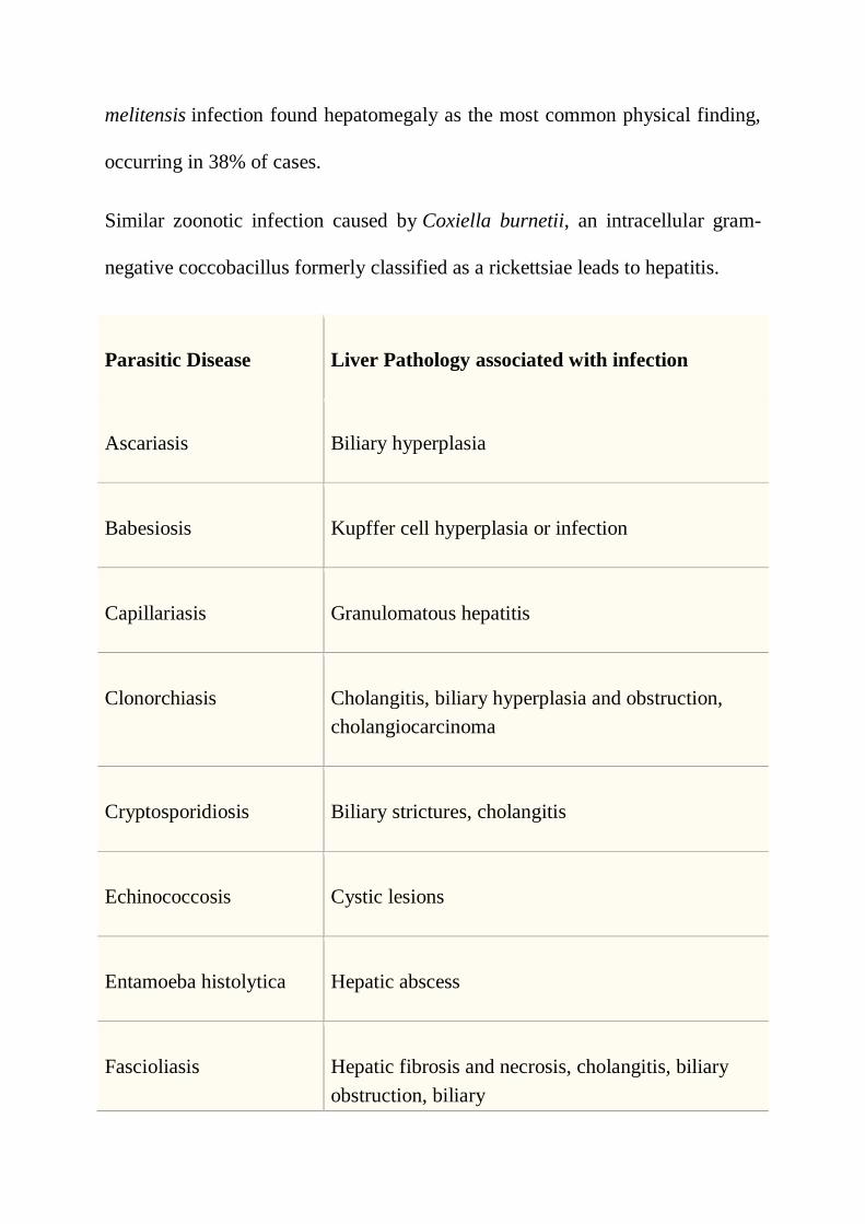
Parasitic Disease Liver Pathology associated with infection
Ascariasis Biliary hyperplasia
Babesiosis Kupffer cell hyperplasia or infection
Capillariasis Granulomatous hepatitis
Clonorchiasis Cholangitis, biliary hyperplasia and obstruction,
cholangiocarcinoma
Cryptosporidiosis Biliary strictures, cholangitis
Echinococcosis Cystic lesions
Entamoeba histolytica Hepatic abscess
Fascioliasis Hepatic fibrosis and necrosis, cholangitis, biliary
obstruction, biliary
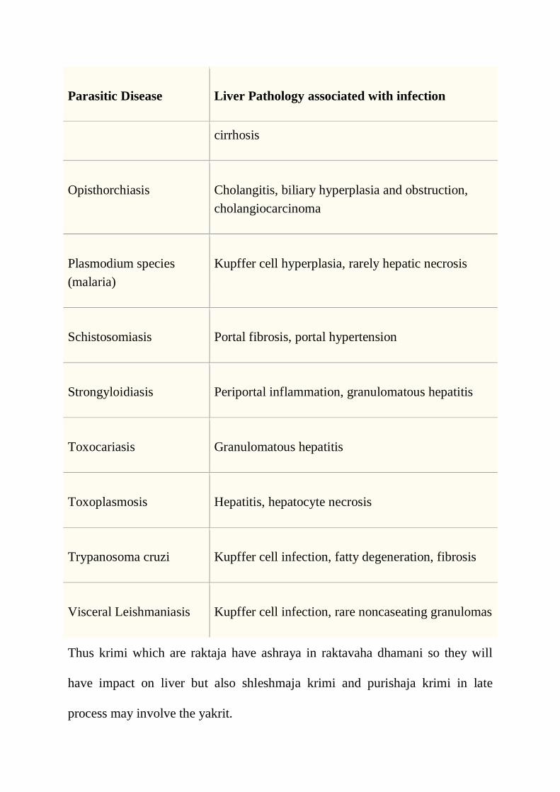
Parasitic Disease Liver Pathology associated with infection
cirrhosis
Opisthorchiasis Cholangitis, biliary hyperplasia and obstruction,
cholangiocarcinoma
Plasmodium species
(malaria)
Kupffer cell hyperplasia, rarely hepatic necrosis
Schistosomiasis Portal fibrosis, portal hypertension
Strongyloidiasis Periportal inflammation, granulomatous hepatitis
Toxocariasis Granulomatous hepatitis
Toxoplasmosis Hepatitis, hepatocyte necrosis
Trypanosoma cruzi Kupffer cell infection, fatty degeneration, fibrosis
Visceral Leishmaniasis Kupffer cell infection, rare noncaseating granulomas
Thus krimi which are raktaja have ashraya in raktavaha dhamani so they will
have impact on liver but also shleshmaja krimi and purishaja krimi in late
process may involve the yakrit.
As discussed above hetu explained in Raktapitta affects the liver function
leading to liver involvement. Coagulopathy affects the liver function and
damage liver function affects the coagulopathy. Grathita rakta if it takes place in
portal vein it affects yakrit.
Portal vein thrombosis (PVT’) is a vascular disease of the liver that occurs when
a blood clot occurs in the hepatic portal vein, which can lead to increased
pressure in the portal vein system and reduced blood supply to the liver.
An equivalent clot in the vasculature that exits the liver carrying deoxygenated
blood to the right atrium via the inferior vena cava, is known as hepatic vein
thrombosis or Budd-Chiari syndrome.
Thrombophilia (including inherited conditions such as factor V
Leiden deficiency, protein C or S deficiency, or antiphospholipid antibody
syndrome) is another common cause. Nearly one-third of patients have a
myeloproliferative disorder (e.g. polycythemia vera or primary thrombocytosis),
most commonly due to a Janus kinase 2 (JAK2) gene mutation.[1]
Oral
contraceptive use or pregnancy are other non-inherited tendencies for
thrombosis.
A long-standing hindrance in flow as in chronic PVT, also known as portal
cavernoma, can cause an increase in the hepatic venous pressure gradient (portal
hypertension) and increased blood flow through subsidiary veins.[ This may
lead to ascites or bleeding from varices
An infected thrombus may become septic, known as pylephlebitis; if blood
cultures are positive for growth at this time, the most common organism
is Bacteroides.
PVT is also a known complication of surgical removal of the spleen. During the
last several years, myeloproliferative neoplasms (MPNs) have emerged as a
leading systemic cause of splanchnic vein thromboses (includes PVT).
Hetus mentioned in gulma also contribute to liver diseases. कट्वम्लिीक्ष्िोष्ण पवदापह
रूक्ष क्रोधापिमद्याकध हुिाशसेवा| आमापभघािो रुपधरं च दुिं िैत्तस्य गुल्मस्य पनपमत्तमुक्तम्||१२||
Above are the causes for Pittaj gulma leading to noninfective origin hepatitis as
discussed before but diseases like pancreatitis leads to fatty liver.
Even though Liver and Pancreas organs are quite different, they exhibit a
number of general structural and functional similarities. Furthermore, the
diseases mediated by alcohol abuse in these organs exhibit some striking
similarities. The diseases in both organs are characterized by parenchymal cell
damage, activation of stellate cells, aberrant wound healing, and fibrosis.
Because of the similarities between the liver and the pancreas, and the alcohol-
associated diseases of these organs, we may be able to apply much of the

knowledge that we have gained regarding the effects of alcohol on the liver to
the pancreas.
Cyst formation, pseudocyst formation, Calcification process, tumours (benign
or malignant) have their pathophysiology as like gulma. If they get formed in
yakrit it will lead to hepatic diseases and if the same occur in pancreas then as
complication yakrit involvement is observed.
Stenosis of the bile duct resulting in persistent jaundice (more than a few
weeks) is uncommon and usually secondary to pancreatic fibrosis.
POLYCYSTIC LIVER DISEASE 2 : PCLD2 is an autosomal dominant disease
characterized by the presence of multiple liver cysts resulting from structural
changes in the biliary tree during development. Abnormal biliary structures may
be present early in life, but they usually remain asymptomatic until cyst growth
initiates during adulthood. In advanced stages, patients may develop massively
enlarged livers, which cause a spectrum of clinical features and complications.
Madhumeha is cause for madhumehi pidaka. Madhumeha when not controlled
leads to accumulation of kleda in the space between mansala Pradesh leading to
Pidaka. One aamong the same is vidradi and Acharya has classified that Yakrit
is one of the sthan of vidradi. Thus abscess in Liver may be cause madhumeha.
Dyslipidemia due to diabetes is well known so also Non Alcoholic Fatty Liver
Disease (NAFLD) is common in Metabolic Syndrome. Common factors
underlying the pathogenesis of NAFLD and DM may be insulin resistance,
oxidative stress, and inappropriate secretion of inflammatory cytokines by
steatotic and inflamed liver.
Skin diseases have their own impact on liver function.
Mycobacterium tuberculosis: There are a variety of clinical manifestations of
hepatic tuberculosis prompting some investigators to further classify the various
forms as miliary, granulomatous, and localized hepatic tuberoculosis.
Autoimmune disorders have their interrelationship directly or indirectly with
functioning of liver. Autoimmune hepatitis is a disease in which the
body’s own immune system attacks the liver and causes it to become
inflamed. The disease is chronic, meaning it lasts many years. If
untreated, it can lead to cirrhosis and liver failure.
There are two forms of this disease. Type 1, or classic, autoimmune
hepatitis is the more common form. This is the form that mostly
affects young women and is often associated with other
autoimmune diseases. Type 2 autoimmune hepatitis is less common
and generally affects girls between the ages of 2 and 14.

Pathology https://www.msdmanuals.com/professional/hepatic-and-
biliary-disorders/alcoholic-liver-disease/alcoholic-liver-disease#:
Marmopaghata explained as Hetu in Shoth is again a cause for Yakrit
vikar. Congestive heart failure causes blood to back up from the
heart into the inferior vena cava. Such congestion increases pressure
in the inferior vena cava and other veins that carry blood to it,
including the hepatic veins (which drain blood from the liver). If this
pressure is high enough, the liver becomes engorged (congested)
with blood and malfunctions.
Similarly, Hepatic involvement in polycystic kidney disease is
common and can cause significant mass effect, compressing
structures such as the great veins (leading to impaired venous
drainage) and abdominal viscera (causing anorexia and weight loss,
referred to as “lethal exhaustion” in severe cases).
Pleehodar hetu are the same for yakritodar hetu. Similarly
spllenomegally can further complicate as hepatomegally. शोपििं वा
रसापदभ्यो पववृदं्ध िं पववधधयेि् is pathogenesis explained for yakritodar. Chakrpani
commentary is significant which explains as शोणितं वा रसाणदभ्य इणत अत्राणदशब्दः
प्रकारवाची; तेन रसस्य कारिस्य वृद्ध्या कार्यस्य रक्तस्य वृद्धिस्तथा मांसाणदभ्योऽणि

रक्तवृद्धिर्यवणत; तेनाहारणवहारेभ्यो रक्तवृद्धिरुक्ता र्वणत| Metabolic disease such as
Niemann-Pick disease,
Gaucher's disease wherein Hepatosplenomegally
observed as cholesterol, proteins get deposited in Liver and spleen leading to
hepatospleenomegally.
Pandu is a disease of Rakta poshak rasa sara bhag thus any impact on rakta
dhatu will surely lead to Yakrit vikar. Hemolytic Anaemia is the best example.
Secondly, Pernicious anaemia (PA) is the end stage of atrophic gastritis which results in the loss of
parietal cells in the fundus and body of the stomach. Loss of parietal cells is associated with the
failure of intrinsic factor production and results in vitamin B12 deficiency and megaloblastic
anaemia. Extramedullary hematopoiesis is the cause for hepatomegally.
https://www.ncbi.nlm.nih.gov/pmc/articles/PMC4316289/
Aplastic anemia occurs when bone marrow doesn’t make enough red
and white blood cells, and platelets. Having fewer red blood cells
causes hemoglobin to drop. Hepatomegally is observed.
In Kamala the dusya is rakta and mansa and the dosha is pitta. Haem and
globin are portions of blood. Globin a protein is degraded into amino acids and
plays no role in Jaundice whereas two reactions take place with haem
molecules. The first oxidation reaction is catalysed by the microsomal enzyme
haem oxygenase and result in bilverdin (harita varna), iron and carbon
monoxide. The next step is the reduction of bilverdin to yellow colour called
bilirubin. It may be noted that 20 percent comes from other haem sources

including ineffective erythropoiesis and the breakdown of other haem
containing proteins such as muscle myoglobin and cytochromes. This explains
why Caraka has used the word mansa dagdva. Myoglobin is iron and oxygen
binding protein found in muscle and is related to haemoglobin. Myoglobin is
only found in blood stream after muscle injury and is an abnormal finding and
can be diagnostically relevant when found in blood. Myoglobin is primary
oxygen carrying pigment of muscle tissue. Thus alpha rakta leads to reduced
mansa poshan causing damage to mansa dhatu as jeevan karma of rakta dhatu is
hampered.
It can be understood from abpve discussion that not only Yakrit alone is cause
for Kamala. The Kamala may be cause by Intrahepatic or by extrahepatic cause
but rakta involvement is essential whereas rakta as a whole needs to be
considered in Kamala in a broad sense but this does not reduce the influence of
Yakrit in kamala.
Shwas is also cause for Yakrit vikar.
Cor pulmonale is a state of cardiopulmonary dysfunction that may result from
several different aetiologies and pathophysiologic mechanisms:
Pulmonary vasoconstriction (secondary to alveolar hypoxia or blood acidosis).
Anatomic reduction of the pulmonary vascular bed (emphysema, pulmonary
emboli, etc.)
Increased blood viscosity (polycythaemia, sickle-cell disease, etc.)
Increased pulmonary blood flow.

The most frequent cause of cor pulmonale is chronic obstructive pulmonary
disease (COPD) due to chronic bronchitis or emphysema . Cor pulmonale later
by back pressure leads to hepatomegally.
Following are the Pulmonary disease which are cause for Cor Pulmonale
complicating into Yakrit vikar.
Lung disease
Chronic obstructive pulmonary disease
Cystic fibrosis
Interstitial lung diseases
Disorders of the pulmonary circulation
Pulmonary thromboembolism
Primary pulmonary hypertension
Tumour emboli
Sickle cell anaemia
Schistosomiasis
Pulmonary veno-occlusive disease
Disorders of ventilatory control
Primary central hypoventilation
Sleep apnoea syndromes
Lastly, Cor Pulmonale is also observed in Neuromuscular diseases such as

Amyotrophic lateral sclerosis, Myasthenia gravis, Poliomyelitis, Guillain-Barre
syndrome, Spinal cord lesions, Bilateral diaphragmatic paralysis, Thoracic cage
deformities, Kyphoscoliosis. This list is important because these diseases are
classified in Vatavikar and they can lead to yakrit vikar in late complication.
Conclusion:
Ayurveda has not given much importance to anatomy while explaining the
Medicinal Disease. They have given importance to pathophysiology hence we
do not find reference of organs being mentioned at gross level. Yakrit is such
organ which plays essential role in body functioning and pathology. There are
various pathology explained in Samhita which can affect the function of Yakrit.
The above discussion is in reference to know how various pathology can affect
the Yakrit function and lead to Hepato Biliary Disease.