yoshiyuki sowa et al- direct observation of steps in rotation of the bacterial flagellar motor
TRANSCRIPT
8/3/2019 Yoshiyuki Sowa et al- Direct observation of steps in rotation of the bacterial flagellar motor
http://slidepdf.com/reader/full/yoshiyuki-sowa-et-al-direct-observation-of-steps-in-rotation-of-the-bacterial 1/4
Direct observation of steps in rotation of thebacterial flagellar motorYoshiyuki Sowa1*, Alexander D. Rowe2*, Mark C. Leake2, Toshiharu Yakushi3, Michio Homma3,
Akihiko Ishijima1,4 & Richard M. Berry2
The bacterial flagellar motor is a rotary molecular machine thatrotates the helicalfilaments that propel many species of swimming bacteria1,2. The rotor isa set ofringsup to45 nmin diameterin thecytoplasmic membrane3; the stator contains about ten torque-generating units anchored to the cell wall at the perimeter of therotor4,5. The free-energy source for the motor is an inward-
directed electrochemical gradient of ions across the cytoplasmicmembrane, the protonmotive force or sodium-motive force forH1-driven and Na1-driven motors, respectively. Here we demon-strate a stepping motion of a Na1-driven chimaeric flagellarmotor in Escherichia coli
6 at low sodium-motive force and withcontrolled expression of a small number of torque-generating units. We observe 26 steps per revolution, which is consistent withthe periodicity of the ring of FliG protein, the proposed site of torque generation on the rotor7,8. Backwards steps despite theabsence of the flagellar switching protein CheY indicate a smallchange in free energy per step, similar to that of a single iontransit.
Direct observation of steps in ATP-driven molecular motors hasrevealed much about the fundamental mechanisms of these protein
machines
9–11
. Steps characteristic of the ATP-driven F1 part of ATPsynthase have been observed when the whole enzyme is driven by ionflow in the FO part12, but steps have never been seen in a purely ion-driven molecular machine. Steps have not previously been resolvedin the flagellar motor13 because of its high speed14,15, the presence of multiple stator units in a single motor4 and the small step sizepredicted—either from structural data on rotor periodicity or frommechanical data obtained by dividing the free energy of one ioncrossing the membrane by the torque that the motor generates. Wetook several measures to overcome these factors. We detectedrotation by back-focal-plane (BFP) interferometry of 500-nm diam-eter polystyrene beads attached to spontaneously sticky flagellarfilaments of E. coli, as described previously 16, or by high-speedvideo recording of 200-nm fluorescent beads attached in the sameway (Fig. 1b, c, inset). The structure and function of Hþ and Naþ
motors are similar, and functional chimaeras have been madecontaining different mixtures of Hþ and Naþ motor components17.We slowed motor rotation to the point at which steps could beresolved by decreasing the sodium-motive force (SMF) in E. coli cellscontaining Naþ-driven chimaeric motors (in which the Hþ-drivenstator proteins MotA and MotB are replaced by the Naþ-drivenPomA and the chimaeric fusion protein PotB, respectively 6; Fig. 1a).We decreased the SMF by a low external Naþ concentration inexperiments with BFP detection and by cumulative photodamagein fluorescence experiments. We controlled the number of statorunits by inducible expression of stator proteins in a strain with
wild-type stator proteins deleted. We also deleted cheY and pilA toavoid possible complications due to motor switching or sticking of beads to cell bodies, respectively. We obtained rotation speed by means of power spectra of the combined x and y signals16, and beadangles by modelling the raw data as the projection of an obliquecircular orbit18.
Fast Naþ-dependent rotation and reduced speed when Liþ
replaced Naþ (data not shown) indicate that the chimaera behavesas a typical Naþ motor15,19. For step experiments, we grew cells withlow inducer levels to produce fully energized motors with a smallinitial number of stator units (typically three or four; Supplementary Information). Reduction of the SMF, either by lowering the Naþ
concentration in BFP experiments (Supplementary Information) orby photodamage in fluorescence experiments, led to a decrease inboth the number of stator units and the speed per unit. Figure 1bshows the speed of a 500-nm bead in a typical BFP experiment. TheNaþ concentration was reduced from 5 mM to 0.1 mM for theinterval between the black arrows, during which the motor rotatedat about 1 Hz. This cell rotated with four stator units when firstobserved in 5 mM Naþ, but this number was decreased to one by a
previous reduction of [Na
þ
] (data not shown). Discrete speedincrements identical to those occurring after the induced expressionof stator proteins16 were typical during recovery after the transient
LETTERS
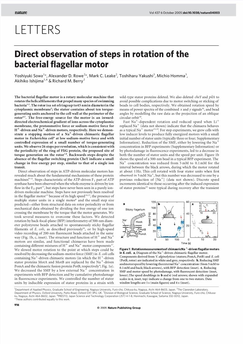
Figure 1 | Rotationmeasurementsof chimaericNa1
-driven flagellar motors
in E. coli . a, Diagram of the Naþ-driven chimaeric flagellar motor.Components derived from V. alginolyticus (stators; PomA, PotB) and E. coli(PotB, rotor) are indicated in white and grey, respectively. b, Reducing SMFandmotorspeed by lowering theexternal Naþ concentration (from 5 mM to0.1 mM and back; black arrows), with BFP detection (inset). c, Reducing SMF and motor speed by photodamage, with fluorescent detection (inset,lower).The speed doublings in b and c (red arrows; shown with expandedscales in c, inset, top) indicate a change from one to two stators. Data
window lengths are 1 s (main figures) and 4 s (inset).
1Department of Applied Physics, Graduate School of Engineering, Nagoya University, Furo-cho, Chikusa-ku, Nagoya, Aichi 464-8603, Japan. 2The Clarendon Laboratory,
Department of Physics, Oxford University, Parks Road, Oxford OX1 3PU, UK. 3Division of Biological Science, Graduate School of Science, Nagoya University, Furo-cho, Chikusa-
ku, Nagoya, Aichi 464-8602, Japan. 4PRESTO, Japan Science and Technology Corporation (JST) 4-1-8, Honmachi, Kawagoe, Saitama 332-0012, Japan.
*These authors contributed equally to this work.
Vol 437|6 October 2005|doi:10.1038/nature04003
916
© 2005 Nature Publishing Group

8/3/2019 Yoshiyuki Sowa et al- Direct observation of steps in rotation of the bacterial flagellar motor
http://slidepdf.com/reader/full/yoshiyuki-sowa-et-al-direct-observation-of-steps-in-rotation-of-the-bacterial 2/4
removal of Naþ(Fig. 1b), indicating reversible inactivation of stator units on disruption of the SMF. Similar phenomena havebeen reported for Hþ-driven motors of E. coli20 and Rhodobacter sphaeroides21. Figure1c shows the speed of a 200-nm fluorescent beadin a typical fluorescence experiment. We attribute periods of smoothspeed reduction to photodamage-induced reduction of the mem-brane voltage22, and discrete speed changes (inset) to the consequentreversible inactivation of stator units. Speed doublings such as those
in Fig. 1b, c were interpreted as a change from one to two stators.However, faster speed fluctuations were common and we were notalways able to assign a definite stator number.
Figure 2a shows a sequence of images of a rotating 200-nmfluorescent bead with calculated bead centres superimposed(Methods). Figure 2b shows stepping rotation of 500-nm plain and200-nm fluorescent beads attached to chimaeric flagellar motors.The diameter of bead orbits (insets) is consistent with attachment toa short flagellar filament stub. Steps were detected at speeds below 7and 40 Hz in BFP and fluorescence experiments, respectively, andwere not restricted to episodes with an estimated single stator unit.Figure 3a shows expanded sections of the traces in Fig. 2b (moreexamples are given in Supplementary Information), with the outputof a step-finding algorithm (Methods) superimposed. Backwardssteps were observed in both BFP and fluorescence experiments, with
higher probability at lower speeds. As the strain lacks the switch-inducing protein CheY and never rotated backwards at high Naþ
concentration, backwards steps represent microscopic reversibility
rather than motor switching. Similar steps have been seen, althoughless frequently, in ATP-driven molecular motors10,11. Figure 3b showsseveral revolutions of a 200-nm bead, the histogram of all dwellangles during those revolutions and the power spectrum of thathistogram. The peak at 26 per revolution in the spectrum corre-sponds to steps of 13.88 and indicates that successive revolutionsshow the same stopping angles.
We combined and analysed step data from different cells and both
experimental techniques. Figure 4a shows the step-size histogram forall steps found. Assuming that larger steps are in fact unresolvedmultiple steps, we fit the distribution as a sum of gaussian distri-butions with means equal to integer multiples of step size, allowingdifferent sizes and variances for forward and backward fits. The fittedstep sizes are 13.78 (26 per revolution) and 210.38 (35 per revolu-tion) for forward and backward steps, respectively. The differencebetween step sizes may be due to reorientation of the rotation axis onreversal, or it may be an artefact of measurement and analysis.Figure 4b shows the sum of histogram power spectra, similar tothose of Fig. 3b, for all step data. Histograms of the levels betweensteps found by the step algorithm were also made; the sum of theirpower spectra is shown in the inset to Fig. 4b. The most striking peak is at 26 per revolution, with others at 11, 16 and 23 per revolution.After subdivision of the data, neither step size nor periodicity
depended on the individual cell, experimental method, angle,estimated number of stator units or average speed of rotation.
Stepping motion in ATP-driven molecular motors reflects both thediscrete molecular nature of the fuel and the periodicity of the ‘track’along which the motor runs9–12. Our observation of 26 steps per
Figure 2 | Stepping rotation. a, Selected frames at 21-ms intervals during one rotation of a 200-nm fluorescent bead. Red and yellow dots mark thecalculated bead centre in the current and previous images respectively. Eachpixel is 80nm square. b, Stepping rotation of flagellar motorswith a range of average speeds. Bead positions are shown in the insets (scales innanometres); bead angles are plotted against time in the main figure. Grey lines indicate (360/26)8 in both main and inset figures. Blue and black tracesare from BFP and fluorescence experiments, respectively. Backwards andforwards steps were observed at all speeds.
Figure 3 | Analysis of step size and periodicity. a, Selected sections of thetraces in Fig. 2b, with the output of a step-finding algorithm superimposed(red). Blue and black traces are from BFP and fluorescence experiments,respectively, as in Fig. 2b. The dominant step size is about 14 8. b, Plot of angle againsttime duringthreerevolutionsof a 200-nmbead, a histogramof dwell angles for the same revolutions and the power spectrum of thathistogram. The peak at 26 per revolution corresponds to a step size of 13.8 8,and shows that themotorstops at thesame angles on successiverevolutions.
NATURE|Vol 437|6 October 2005 LETTERS
917
© 2005 Nature Publishing Group

8/3/2019 Yoshiyuki Sowa et al- Direct observation of steps in rotation of the bacterial flagellar motor
http://slidepdf.com/reader/full/yoshiyuki-sowa-et-al-direct-observation-of-steps-in-rotation-of-the-bacterial 3/4
revolution is consistent with the periodicity of the ring of FliGprotein, the track on the rotor where torque is generated7,8. The10-fold or 11-fold periodicity matches the filament and hook 23; otherperiodicities observed may be due to interactions between othercomponents of the flagellum with as-yet undetermined symmetry.Some cells in BFP experiments rotated at about 1 Hz even in 5 mMNaþ. Steps were seen in these unusually slow episodes (data notshown), similar to or possibly slightly smaller than 1/26 of a
revolution. Locking of one stator in onestate of its mechanochemicalcycle while other stators continued normally could producesteps with theperiodicityof thelocked state. Unusually slow episodesare excluded from our data analysis, but we cannot entirely rule out apossible contribution of a locking effect. Interval length distributionsfor data with a narrow range of average speed were sometimes, butnot always, single-exponential. The apparent independence of stepsize on stator number, non-exponential interval length distributionsand the observation of occasional steps smaller than 1/26 of arevolution may require the revision of existing models of the flagellarmotor as a set of independent poisson-stepping stators16,24.
Our experiments cannot determine whether single steps corre-spond to single ion transits. Regardless of the nature of the steppingprocess, the ratio of forwards to backwards step probabilities isexp(2DG/kT ), where DG is the free energy available to drive one
step and kT is the average thermal energy. In our experiments DGrangedfrom0 to3kT , dependingon rotation speed.This is equivalentto the free energy of one ion transit through a SMF of up to 275 mV.Measuring the SMF in E. coli under these conditions would revealwhether the observed steps could be driven by single-ion transits.However, previous data indicate that single ions in fully energizedwild-type motors cannot drive steps as large as 148. If about tentorque-generating units pass 1,200 Hþ ions per revolution25,then one ion in one unit should step about 3 8, assuming tightly coupled independent units16. Energy conservation sets an upperbound to the angle coupled to one ion transit, equal to (free energy per ion)/(maximum torque per unit). A wild-type E. coli cell with a
protonmotive force of about 150 mV (6kT per ion)26 drives a 1-mmbead with an estimated 280 pN nm per unit16, giving an upper boundof about 58. It is possible that stoichiometry changes at low SMF orstator number or that smaller substeps will be resolved in future.Alternatively, a mechanical step may be coupled to several iontransits, requiring the accumulation either of ions or of mechanicalstrain between steps. The latter mechanism is believed to occur inATP-synthase, coupling three or four ion-driven steps in FO to a
single ATP-synthesizing step in F1. By combining high-resolutionmeasurements of flagellar rotation with manipulation of the SMF, thechimaeric flagellar motor described here will enable a detailedcomparison to be made between the mechanism of the bacterialflagellar motor and those of its ATP-driven relatives.
METHODS E. coli chimaera. E. coli strain YS34 (DcheY , fliC::Tn10 , D pilA, DmotAmotB)was derived from strain RP4979 (ref. 27) (DcheY ) and transformed withplasmids pYS11 ( fliC sticky filaments28, ampicillin resistance, pBR322 deriva-tive) and pYS13 ( pomApotB (ref. 6), isopropyl bDthiogalactoside inducible,chloramphenicol resistance, pMMB206derivative). Deletions of pilA and motAB
were made as described in ref. 29; fliC::Tn10 was transduced from HCB1271(ref. 16). Cells were grown for 5 h at 30 8C from frozen stocks, with shaking, intryptone broth (TB) containing antibiotics to preserve plasmids. Isopropyl
bD
thiogalactoside (1–10 mM) was added for low-level expression of statorproteins.Rotation measurement. All experiments were performed at 23 8C. Speeds wereobtained from power spectra of combined ( x , y ) data16, using data windows of length 1 or 4 s beginning at intervals of 0.1 s. Motor angles were obtained by fitting an ellipse to bead trajectories under the assumption that trajectoriesrepresent the projection of a circular orbit onto the focal plane of themicroscope18.
For BFP experiments, polystyrene beads (diameter 535nm; Polysciences)were attached to truncated flagella of immobilized cells, and bead position ( x , y )was measured by BFP laser interferometry (sample rate 4 kHz) as described16.NaCl (or LiCl) was added to the Naþ-free motility medium in variousconcentrations; the total concentration of added salt was adjusted to 85 mMwith KCl. Custom-made flowchambers alloweda completeexchange of mediumin about 5 s.
For fluorescence experiments, fluorescent polystyrene beads (diameter
227 nm, ‘yellow/green’; Molecular Probes) were attached as above. A mercury-arc lamp provided epifluorescence excitation (475-nm bandpass) at an intensity of about 1 W cm22. Images (16 £ 16 pixels, each 80 nm square in the sampleplane; 505nm long-pass) were sampled at a frame rate of 2.4 kHz with a cooled,back-thinned, electron-multiplying charge-coupled device camera (iXonDV860-BI, Andor Technology). Bead position was determined with a precisionof about 5 nm by a two-dimensional gaussian fit to the bead image.Step resolution. The relaxation time of a 500-nm bead attached to an elastichook rotating about an axis 150 nm from its diameter is about 1.1 ms (ref. 30).For a 200-nm bead the time is about 0.15ms. These times are lower limitsassuming no contribution from the flagellar stub16, and they set upper limits of about 900 and 6,500 to the number of steps that can be detected per second inBFP and fluorescence experiments respectively. Our actual limits were lower,namely about 200 s21 and about 1,000 s21, respectively. The latter limit wasattained by analysis of dwell-time histograms.Step analysis. A computer algorithm divided angle versus time records intostraight-line portions corresponding to episodes of constant average speed. Asecond algorithm based on the method of J. W. Kerssemakers (personalcommunication) detected steps as follows. First, an entire episode was dividedinto two intervals at the point giving the best least-squares fit to a single-stepfunction. Second, an excess of steps ( N max per revolution on average) wasassigned by repeatedly dividing, as in the first step, the interval containing thelargest range of angles in the previous iteration. Third, a ‘quality factor’ Q wasdefined for each assigned step as Q2
¼ ( x 1 2 x 2)2/[(var.1/n1) þ (var.2/n2)],where x i, var.i and n i are the mean angle, the variance and the number of points in an interval, respectively, and i ¼ 1 and i ¼ 2 indicate intervalsimmediately before and after a step. The lowest-quality step was removed andadjacent intervals were concatenated, but only if the quality was below athreshold Qmin. Last, the previous step was repeated until no steps remainedwith Q , Qmin. The accuracy and sensitivity of the algorithm were tested and asuitable value of Qmin for each episode was chosen by applying the algorithm tosimulated data with N o ¼ 10, 20, 30, 40 and 80 Poisson-distributed steps perrevolution and brownian noise and average speed similar to real data.
Figure 4 | Summary of step analysis. a, A histogram of step sizes found inall episodes of approximately constant speed (1,400 revolutions, 9 differentcells, 28,611 steps). A multiple-gaussian fit (red line) gives step sizes of 13.78(j ¼ 5.168) and 210.98 (j ¼ 3.98) for forward and backward steps,respectively. b, The sum of histogram power spectra (see Fig. 3b) for thesame data set. Inset, summed spectra of histograms of mean levels betweenfound steps. The forward step size is in close agreement with the peak in thesummed spectra, confirming that there are 26 steps per revolution.
LETTERS NATURE|Vol 437|6 October 2005
918
© 2005 Nature Publishing Group

8/3/2019 Yoshiyuki Sowa et al- Direct observation of steps in rotation of the bacterial flagellar motor
http://slidepdf.com/reader/full/yoshiyuki-sowa-et-al-direct-observation-of-steps-in-rotation-of-the-bacterial 4/4
Received 1 April; accepted 11 July 2005.
1. Berry, R. M. & Armitage, J. P. The bacterial flagella motor. Adv. Microb. Physiol.
41, 291–-337 (1999).
2. Berg, H. C. The rotary motor of bacterial flagella. Annu. Rev. Biochem. 72, 19–-54
(2003).
3. Thomas, D. R., Morgan, D. G. & DeRosier, D. J. Rotational symmetry of the
C ring and a mechanism for the flagellar rotary motor. Proc. Natl Acad. Sci. USA
96, 10134–-10139 (1999).
4. Blair, D. F. & Berg, H. C. Restoration of torque in defective flagellar motors.
Science 242, 1678–-1681 (1988).5. Berry, R. M., Turner, L. & Berg, H. C. Mechanical limits of bacterial flagellar
motors probed by electrorotation. Biophys. J. 69, 280–-286 (1995).
6. Asai, Y., Yakushi, T., Kawagishi, I. & Homma, M. Ion-coupling determinants of
Naþ-driven and Hþ-driven flagellar motors. J. Mol. Biol. 327, 453–-463 (2003).
7. Suzuki, H., Yonekura, K. & Namba, K. Structure of the rotor of the bacterial
flagellar motor revealed by electron cryomicroscopy and single-particle image
analysis. J. Mol. Biol. 337, 105–-113 (2004).
8. Lloyd, S. A. & Blair, D. F. Charged residues of the rotor protein FliG essential for
torque generation in the flagellar motor of Escherichia coli. J. Mol. Biol. 266,
733–-744 (1997).
9. Mehta, A. D. et al. Myosin-V is a processive actin-based motor. Nature 400,
590–-593 (1999).
10. Schnitzer, M. J. & Block, S. M. Kinesin hydrolyses one ATP per 8-nm step.Nature 388, 386–-390 (1997).
11. Yasuda, R., Noji, H., Kinosita, K. Jr & Yoshida, M. F1-ATPase is a highly efficient
molecular motor that rotates with discrete 120 degree steps. Cell 93, 1117 –-1124
(1998).
12. Diez, M.et al.
Proton-powered subunit rotation in single membrane-boundFOF1-ATP synthase. Nature Struct. Mol. Biol. 11, 135–-141 (2004).
13. Berg, H. C. in Cell Motility Vol. A (eds Goldman, R., Pollard, T. & Rosenbaum, J.)
47–-56 (Cold Spring Harbor Press, New York, 1976).
14. Berg, H. C. & Turner, L. Torque generated by the flagellar motor of Escherichia
coli. Biophys. J. 65, 2201–-2216 (1993).
15. Sowa, Y., Hotta, H., Homma, M. & Ishijima, A. Torque-speed relationship of the
Naþ-driven flagellar motor of Vibrio alginolyticus. J. Mol. Biol. 327, 1043–-1051
(2003).
16. Ryu, W. S., Berry, R. M. & Berg, H. C. Torque-generating units of the flagellar
motor of Escherichia coli have a high duty ratio. Nature 403, 444–-447 (2000).
17. Yorimitsu, T. & Homma, M. Naþ-driven flagellar motor of Vibrio. Biochim.
Biophys. Acta 1505, 82–-93 (2001).
18. Yasuda, R., Noji, H., Yoshida, M., Kinosita, K. Jr & Itoh, H. Resolution of distinct
rotational substeps by submillisecond kinetic analysis of F1-ATPase. Nature
410, 898–-904 (2001).
19. Liu, J. Z., Dapice, M. & Khan, S. Ion selectivity of the Vibrio alginolyticus flagellar
motor. J. Bacteriol. 172, 5236–-5244 (1990).
20. Fung, D. C. & Berg, H. C. Powering the flagellar motor of Escherichia coli with an
external voltage source. Nature 375, 809–-812 (1995).
21. Armitage, J. P. & Evans, M. C. Control of the protonmotive force in
Rhodopseudomonas sphaeroides in the light and dark and its effect on the
initiation of flagellar rotation. Biochim. Biophys. Acta 806, 42–-55 (1985).
22. Neuman, K. C., Chadd, E. H., Liou, G. F., Bergman, K. & Block, S. M.
Characterization of photodamage to Escherichia coli in optical traps. Biophys. J.
77, 2856–-2863 (1999).
23. Samatey, F. A. et al. Structure of the bacterial flagellar hook and implication
for the molecular universal joint mechanism. Nature 431, 1062–-1068
(2004).
24. Samuel, A. D. & Berg, H. C. Torque-generating units of the bacterial flagellar
motor step independently. Biophys. J. 71, 918–-923 (1996).25. Meister, M., Lowe, G. & Berg, H. C. The proton flux through the bacterial
flagellar motor. Cell 49, 643–-650 (1987).
26. Gabel, C. V. & Berg, H. C. The speed of the flagellar rotary motor of Escherichia
coli varies linearly with protonmotive force. Proc. Natl Acad. Sci. USA 100,
8748–-8751 (2003).
27. Scharf, B. E., Fahrner, K. A., Turner, L. & Berg, H. C. Control of direction of
flagellar rotation in bacterial chemotaxis. Proc. Natl Acad. Sci. USA 95, 201–-206
(1998).
28. Kuwajima, G. Construction of a minimum-size functional flagellin of Escherichia
coli. J. Bacteriol. 170, 3305–-3309 (1988).
29. Datsenko, K. A. & Wanner, B. L. One-step inactivation of chromosomal genes
in Escherichia coli K-12 using PCR products. Proc. Natl Acad. Sci. USA 97,
6640–-6645 (2000).
30. Block, S. M., Blair, D. F. & Berg, H. C. Compliance of bacterial flagella measured
with optical tweezers. Nature 338, 514–-518 (1989).
Supplementary Information is linked to the online version of the paper atwww.nature.com/nature.
Acknowledgements We thank H. Berg and K. Fahrner for the gift of strain
HCB1271. The research of R.B., M.L. and A.R. was supported by combined UK
research councils through an Interdisciplinary Research Collaboration in
Bionanotechnology, that of A.I., M.H. and T.Y. by Grants-in-Aid from the
Ministry of Education, Science, Sports, Culture and Technology of Japan, that of
M.H. and T.Y. by Soft Nano-Machine Project of JST, and that of Y.S. by JSPS
Research Fellowships for Young Scientists.
Author Contributions BFP experiments were performed by Y.S. and A.R.,
fluorescence experiments by A.R. and M.L., experimental design was by R.B.,
A.I., A.R. and Y.S., data analysis by R.B., Y.S. and A.R., and strain construction by
Y.S., T.Y. and M.H. Y.S. and A.R. contributed equally to this work.
Author Information Reprints and permissions information is available at
npg.nature.com/reprintsandpermissions. The authors declare no competing
financial interests. Correspondence and requests for materials should be
addressed to R.M.B. ([email protected]).
NATURE|Vol 437|6 October 2005 LETTERS
919
© 2005 Nature Publishing Group