zbtb17 (miz1) is important for the cardiac stress response
TRANSCRIPT
1
Title page
ZBTB17 (MIZ1) is important for the cardiac stress response and a novel
candidate gene for cardiomyopathy and heart failure
Buyandelger et al., ZBTB17 mutations cause cardiomyopathy
Authors
Byambajav Buyandelger, PhD1,2
; Roddy Walsh, PhD1; Sawa Kostin, PhD
3; Catherine Mansfield,
PhD1; Onjee Choi, PhD
1; Angharad M. Roberts, PhD
1; James S. Ware, PhD
1; Francesco
Mazzarotto, PhD1; Francesco Pesce, PhD
1; Rachel Buchan, PhD
1; Rivka L. Isaacson, PhD
4;
Josee Vouffo, PhD2; Sylvia Gunkel, PhD
2; Gudrun Knöll
1,2; Sara J. McSweeney, PhD
1; Heming
Wei, PhD20,21
; Andreas Perrot, MD5,6
; Conny Pfeiffer2; Mohammad Reza Toliat, MD
7; Kristina
Ilieva4; Ewelina Krysztofinska
4; Marina M. López-Olañeta
9; Jesús M. Gómez-Salinero
9;
Albrecht Schmidt2; Keat-Eng Ng, PhD
1; Niels Teucher, MD
2; Ju Chen, PhD
10; Martin
Teichmann, PhD11
; Martin Eilers, PhD12
; Wilhelm Haverkamp, MD6; Vera Regitz-Zagrosek,
PhD13
; Thomas Braun, PhD3; Dudley J. Pennell, MD
14; Ian Gould, PhD
15; Paul J.R. Barton,
PhD14
; Enrique Lara-Pezzi, PhD1,9
; Sebastian Schafer22,23
; Norbert Hubner22,23
; Leanne A.
Felkin18
; Declan P. O’Regan16
; Enrico Petretto, PhD16
; Thomas Brand, PhD18
; Hendrik Milting,
PhD17
; Peter Nürnberg, PhD7,8,19
; Michael D. Schneider, PhD1; Sanjay Prasad, MD
14; Stuart
Cook*, PhD,14,20,21
; and Ralph Knöll*, MD. PhD1,2
Conflict of interest
The authors have declared that no conflict of interest exists.

2
Affiliations
1National Heart & Lung Institute, Faculty of Medicine, Imperial College London, Hammersmith
Campus, Imperial Centre for Translational and Experimental Medicine, London W12 0NN, UK.
2Heart Centre, Georg-August University of Göttingen, Robert-Koch-Str. 40, 37075 Göttingen,
Germany.
3Max-Planck-Institute for Heart and Lung Research, 61231 Bad Nauheim, Germany.
4Division of Molecular Biosciences, Department of Life Sciences, Faculty of Natural Sciences,
Imperial College London, London, UK.
5Charité-Unversitätsmedizin Berlin, Experimental and Clinical Research Center (ECRC), a joint
cooperation between the Charité Medical Faculty and the Max-Delbrück Center for Molecular
Medicine, 13125 Berlin, Germany.
6Charité-Unversitätsmedizin Berlin, Dept. of Cardiology at Campus Virchow-Klinikum, 13353
Berlin, Germany.
7Cologne Center for Genomics, University of Cologne, Cologne, Germany.
8Cologne Excellence Cluster on Cellular Stress Responses in Aging-Associated Diseases
(CECAD), University of Cologne, Cologne, Germany.
9Cardiovascular Development and Repair Department, Centro Nacional de Investigaciones
Cardiovasculares (CNIC), Melchor Fernández Almagro 3, 28029 Madrid, Spain.
10Department of Medicine, University of California at San Diego (UCSD), 9500 Gilman Drive,
San Diego, CA, USA.
11Institut Européen de Chimie et Biologie, INSERM U869, Bordeaux, France.
3
12Theodor Boveri Institute, Biocenter, University of Würzburg, Am Hubland, 97074 Würzburg,
Germany.
13Berlin Institute of Gender in Medicine, Charité-Unversitätsmedizin Berlin, Germany.
14NIHR Cardiovascular Biomedical Research Unit at Royal Brompton & Harefield NHS
Foundation Trust and Imperial College London, London SW3 6NP, UK
15Department of Chemistry and Institute of Chemical Biology, Imperial College London, South
Kensington, SW7 2AZ, UK.
16Medical Research Council (MRC) Clinical Sciences Centre, Faculty of Medicine, Imperial
College London, Hammersmith Hospital, Du Cane Road, London W12 0NN, UK.
17Ruhr-Universität Bochum, Herz & Diabeteszentrum NRW, Erich & Hanna Klessmann-Institut,
Bad Oeynhausen, Germany.
18Heart Science Centre, National Heart and Lung Institute, Imperial College London, Harefield
Middlesex UB9 6JH, UK.
19Center for Molecular Medicine Cologne (CMMC), University of Cologne, Cologne, Germany.
20Duke-NUS Graduate Medical School, 8 College Road, 169857 Singapore.
21National Heart Centre, Singapore, 17 Third Hospital Avenue, 168752 Singapore.
22Max-Delbruck-Center for Molecular Medicine (MDC), 13125 Berlin, Germany.
23DZHK (German Centre for Cardiovascular Research), Partner Site Berlin, Germany.

4
*Correspondence to:
Professor Ralph Knöll, MD, PhD
Chief Scientist
AstraZeneca R&D Mölndal
R&D | Innovative Medicines & Early Development | Cardiovascular & Metabolic Diseases iMed
Pepparedsleden 1
SE-431 83 Mölndal, Sweden
Phone: (+46) (0) 706 165801
Email: [email protected]
ICMC (Integrated Cardio Metabolic Centre)
Karolinska Institutet
Karolinska University Hospital in Huddinge (M54)
SE 141 86 Stockholm, Sweden
Email: [email protected]
The total word count: 6973
[130] Animal models of human disease
[16] Myocardial cardiomyopathy disease
Abstract

5
Background- Mutations in sarcomeric and cytoskeletal proteins are a major cause of hereditary
cardiomyopathies, but our knowledge remains incomplete as to how the genetic defects execute
their effects.
Methods and Results- We used cysteine and glycine-rich protein 3 (CSRP3), a known
cardiomyopathy gene, in a yeast two-hybrid screen and identified zinc finger and BTB domain
containing protein 17 (ZBTB17) as a novel interacting partner. ZBTB17 is a transcription factor
that contains the peak association signal (rs10927875) at the replicated 1p36 cardiomyopathy
locus. ZBTB17 expression protected cardiac myocytes from apoptosis in vitro and in a mouse
model with cardiac myocyte-specific deletion of Zbtb17, which develops cardiomyopathy and
fibrosis after biomechanical stress. ZBTB17 also regulated cardiac myocyte hypertrophy in vitro
and in vivo in a calcineurin-dependent manner.
Conclusions- We revealed new functions for ZBTB17 in the heart, a transcription factor which
may play a role as a novel cardiomyopathy gene.
Key words- Heart Failure; Cardiomyopathy; Genetics
6
Introduction
Mutations in over 50 genes, mostly encoding sarcomeric and Z disc proteins, cause hypertrophic
cardiomyopathy (HCM) and dilated cardiomyopathy (DCM) that lead to heart failure.1 Genetic
variation in a Z disc gene can cause either HCM or DCM perhaps due to pleiotropic effects on
survival and/or hypertrophic pathways.2 The muscle LIM protein (MLP, CSRP3) is a Z disc
protein involved in cardiac mechanosensation that is important for myocyte-specific survival
pathways through interactions with other Z disc proteins.3-5
CSRP3 mutations have been found in
both HCM and DCM patients3, 4, 6
and we used protein-protein interaction studies to identify new
CSRP3-interacting partners that may be important for cardiac myocyte survival and/or
cardiomyopathy.
7
Methods
All primers and antibodies used are listed in Supplemental Tables. Tissue culture experiments
were performed using primary neonatal and adult rat cardiac myocytes and animal experiments
were performed using conditional knockout (cKO)-Zbtb17, overexpressing transgenic (TG)-
ZBTB17 and TG-ZBTB17/knockout (KO)- protein phosphatase 3, catalytic subunit, beta
isozyme (calcineurin Aβ, Ppp3cb) animals. Yeast two-hybrid, Western blot, quantitative Real-
time PCR (qRT-PCR) and other assays were performed as described elsewhere.3-5, 7
Statistical
evaluations were performed by Fisher exact, ANOVA or Student’s t-test. (For more details
please see the online supplemental information.)

8
Results
MLP interacts with ZBTB17
Yeast two-hybrid screens using CSRP3 as bait 3 classified -actinin and telethonin as CSRP3-
interacting proteins that are important determinants of cardiac function5, 8
and also identified
ZBTB17 (Figure 1A). ZBTB17 is a member of the poxvirus and zinc-finger (POZ or BTB)
domain/zinc-finger transcription factor family with no previously described role in the heart.9
We confirmed the CSRP3-ZBTB17 interaction by co-immunoprecipitation in vivo (Figure 1B),
cross-linking of recombinant proteins (Figure 1C) and immunohistochemistry studies (Figure 1D
and Supplemental Figure 1). These data identify ZBTB17 as a novel CSRP3 interacting protein.
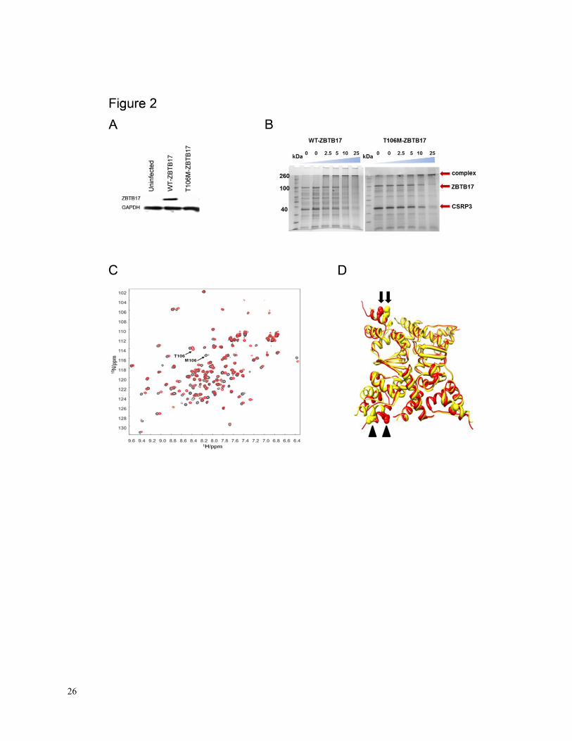
Analysis of a human ZBTB17 variant (T106M)
ZBTB17 is encoded on human chromosome 1 at the replicated 1p36 cardiomyopathy locus.10-12
The peak DCM association at this locus (rs10927875, P = 1.3x10⁻⁷
)12
is located in intron 2 of
ZBTB17, with further evidence of association of this SNP with DCM in Chinese population.12, 13
Considering rare polymorphisms (allele frequency <0.1%; population prevalence of
cardiomyopathies ~1:5001), we identified a p.T106M variant in the Exon variant server (ESP).
The p.T106M variant could not be stably expressed in cardiac myocytes (Figure 2A), bound
poorly to CSRP3 (Figure 2B) and caused structural abnormalities to the POZ domain (Figure 2C,
D), and may represent a rare haplo-insufficient polymorphism. Together, these data point to
ZBTB17 as a possible disease gene at the 1p36 cardiomyopathy locus and suggest a role for a
9
transcription factor in heart failure, as recently shown for left ventricular non compaction
cardiomyopathy.14
Analysis of ZBTB17 function in vitro
To study function in vitro we overexpressed ZBTB17 in cardiac myocytes and found that it
induced hypertrophy (Figure 3A, B and Supplemental Figure 2A). CSRP3 and other CSRP3-
interacting proteins have important roles in cardiac myocyte survival, and this was also observed
for ZBTB17 (Figure 3C and Supplemental Figure 2B). Calcineurin activity is of central
importance for pro-hypertrophic signaling and myocyte survival15, 16
and in both the hypertrophy
and the cell death experiments inhibition of calcineurin attenuated the effects of ZBTB17,
suggesting an interaction (Figure 3A, C). Hence, ZBTB17 regulates cardiac hypertrophy and cell
survival, a recognized dual-property of genes that are of central importance for cardiac myocyte
biology.15-18
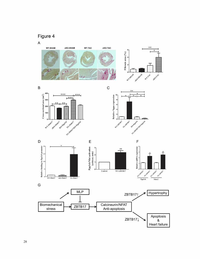
Analysis of ZBTB17 function in vivo
Mice globally deleted for Zbtb17 die due to a gastrulation defect.19
We therefore crossed floxed
Zbtb17 mice20
with the MLC2v Cre deleter line21
to test Zbtb17 gene function in cardiac
myocyte-specific conditional knockout mice (cKO) (Supplemental Figure 3A-D). cKO mice
were viable and, in the absence of a spontaneous functional cardiac phenotype, we employed the
established model of transverse aortic constriction (TAC)5 to unveil potential gene effects. After
10
TAC the left ventricles of cKO mice became dilated and exhibited impaired contractile
performance, which are the cardinal features of DCM (Table 1). As compared to controls, failing
cKO hearts had higher apoptotic events (8.5-fold, P < 0.001), increased activated caspase 3 (6.3-
fold, P < 0.001) and marked replacement fibrosis (Figure 4A, Supplemental Figure 3E and Table
3). These data show that Zbtb17 modulates biomechanical stress-induced apoptosis and
interstitial fibrosis in vivo, which are important in the pathobiology of both HCM and DCM.
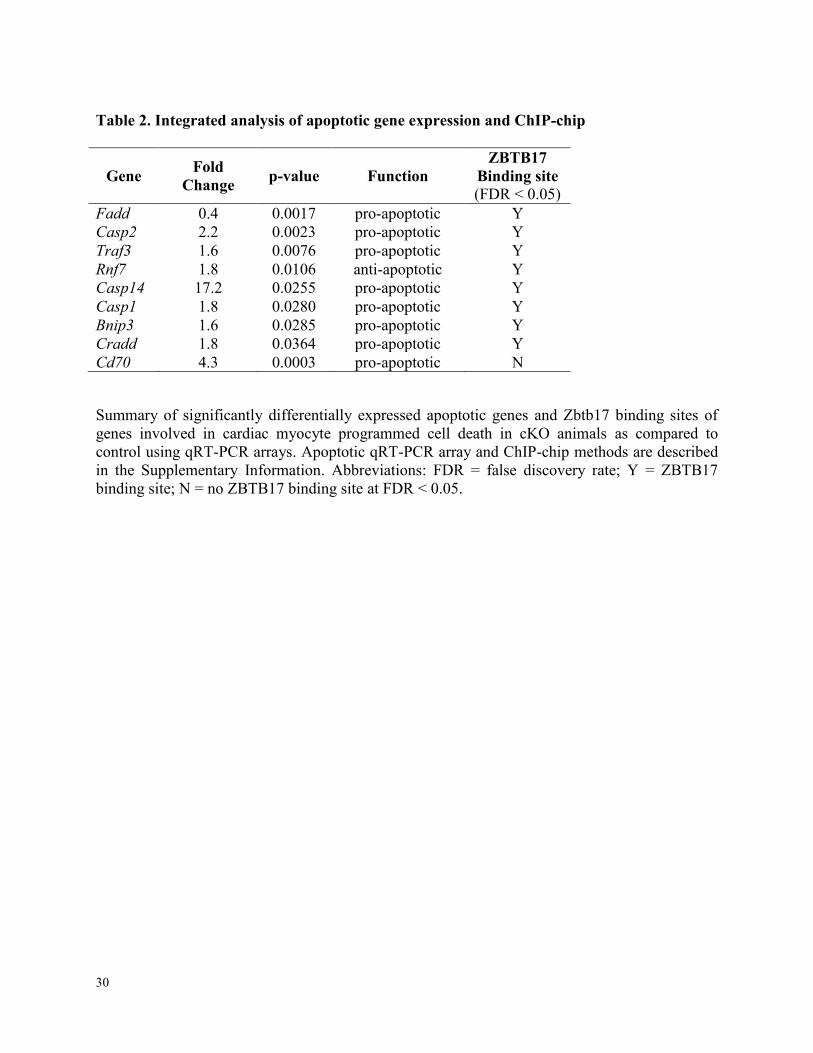
To begin to understand the molecular mechanisms by which ZBTB17 regulates cardiac myocyte
survival, based on the functions of ZBTB17 in other tissues19
, we applied apoptosis gene-focused
PCR array analysis combined with genome wide chromatin immunoprecipitation-chip (ChIP-
chip). In cKO hearts subjected to TAC, pro-apoptotic gene expression was increased with
evidence of direct regulation of a subset of these genes in ChIP-chip studies (Table 2). Hence
ZBTB17 regulates a transcriptional program that protects cells against apoptosis.
In addition, ZBTB17 overexpression drives hypertrophy in vitro (Figure 3). To examine further
the effects of ZBTB17 on hypertrophy we overexpressed ZBTB17 in mice using a cardiac
myocyte specific promoter22
(Supplemental Figure 4). Transgenic mice exhibited spontaneous
cardiac hypertrophy with enlarged cardiac myocytes and increased Nppa expression (Table 3;
Figure 4B, C). In vitro experiments suggested a link between ZBTB17 and calcineurin and ChIP-
chip data showed binding of Zbtb17 to the calcineurin Aβ (Ppp3cb) promoter that we confirmed
by ChIP-qRT-PCR and reporter assays (Figure 4D, E). There was increased expression of
Ppp3cb and Nfatc2 in TG-ZBTB17 hearts and decreased expression in cKO (Figure 4F and
Supplemental Figure 5A). We crossed ZBTB17 transgenic mice into a Ppp3cb deficient

11
background that prevented cardiac myocyte hypertrophy confirming the interaction of ZBTB17
with the calcineurin pathway (Figure 4B and Supplemental Table 1).
12
Discussion
CSRP3 mutations cause heart failure in various animal models4, 23
and have been identified in
various patients affected by DCM3, 24
or HCM25-27
. Although many cardiomyopathy and heart
failure models have been developed, the molecular mechanisms which link sarcomeric and Z
disc proteins to these phenotypes remain not well understood. Here we show that CSRP3
interacts with ZBTB17 and provide the first detailed analysis of this transcription factor in the
cardiovascular system. In particular we demonstrate that ZBTB17 causes cardiac myocyte
hypertrophy and is essential for cell survival. The extent of hypertrophy in cardiac myocytes is
comparable to the effects seen with trophic signals including mitogenic serum in these post-
mitotic cells.28, 29
So far, ZBTB17 is the only known transcription factor to interact with CSRP3
and to be expressed in cardiac myocytes (for a brief review:30
). The effects of ZBTB17 on
hypertrophy and survival are not restricted to the in vitro situation, but can also be observed in
genetically altered animals under in vivo conditions. Interestingly, ZBTB17 overexpressing
transgenic animals did not develop heart failure due to massive increase in apoptosis as
frequently seen in other genetically altered mouse models (for a review:31
).32-34
The combination of ChIP-chip, whole gene expression- and apoptosis array data clearly point to
an important role of ZBTB17 in orchestrating a cardio-protective gene expression program. This
is indicated by the fact that pro-apoptotic genes were consistently upregulated, while anti-
apoptotic genes were down regulated in cKO hearts following biomechanical stress (Table 2).
We also show that ZBTB17 targets various calcineurin/NFAT genes and activates this pathway.
This notion is supported by our whole gene expression analysis, ChIP-chip assays, analysis of
the calcineurin/NFAT pathway activation, promoter assays and by the significant loss of
hypertrophy when TG-ZBTB17 animals are crossed into the calcineurin Aβ deficient background
13
(Figure 3, 4D and E). These data are also supported by our finding of enhanced calcineurin Aβ
and Nfatc2 mRNAs in hearts of ZBTB17 transgenic animals and decreased calcineurin Aβ and
Nfatc2 mRNAs in hearts of Zbtb17 cKO animals after TAC (Figure 4F and Supplemental Figure
5A).
CSRP3 translocates into the nucleus upon TAC35
and is essential for the adaptation of cardiac
myocytes to biomechanical stress.36
CSRP3, like telethonin, is primarily expressed in muscle
tissues. By interacting with ZBTB17, CSRP3 plays a role in the initiation of myocyte specific
survival pathways or mechanoptosis (mechanosensitive types of cell death5). This notion is
supported by the fact that Csrp3 deficient hearts exhibit increased rates of apoptosis37
hence the
interaction of CSRP3 with ZBTB17 can also be described as a form of Z disc transcriptional
coupling.38
Calcineurin/NFAT signaling is an important mediator of cardiac hypertrophy39, 40
and this
pathway is also known to exert cardio-protective effects by avoiding apoptosis during ischemia
reperfusion15
, which emphasizes the effects of ZBTB17 on cell survival.
CSRP3 directly interacts with calcineurin and is required for its activation41
, thus, by interacting
with ZBTB17, CSRP3 introduces an additional level of control which ensures a medium level of
calcineurin/NFAT activation (Figure 4G). For a long time c-myc is known to be induced during
TAC42
, but its downstream effects in cardiac myocytes remained elusive. However, c-myc is
known to inhibit ZBTB1743
and therefore may have adverse effects on survival and hypertrophy
in cardiac myocytes. Interestingly, CSRP3 interacts with the aminoterminal POZ domain which
is predicted to interfere with ZBTB17 tetramerization.
An additional layer of complexity is added by the fact that ZBTB17 can be phosphorylated at
position S428 by the Akt kinase, which is another important mediator of hypertrophy.44
This
14
post-translational modification enables the interaction of ZBTB17 with 14-3-3 ŋ and leads to its
inactivation, thus avoiding prolonged activation of ZBTB17, which can also have adverse
effects.45
In the Exome Variant Server (EVS) database there are 10 T106M variants annotated out of at
least 13006 alleles of European-American and Afro-American origin combined. The EVS does
not provide data on individual phenotypes, but the frequency for this variant is certainly very low
and a disease causing or at least modifying role cannot be excluded. Nevertheless, a two-hit
hypothesis whereby a structural perturbation combines with a defect in a ZBTB17 mediated cell
survival pathway could also lead to heart failure.
As a result we expect this variant to impair calcineurin dependent hypertrophy and being unable
to efficiently protect cardiac myocytes from apoptosis. However, this variant may also have
specific effects on remodeling and hypertrophy46
by specifically affecting protein degradation
pathways, for example via the ubiquitin proteasome system (UPS)47
, and aside from affecting its
interaction with CSRP3 (Figure 2B) ,may specifically interfere with other protein / protein
interactions.
ZBTB17 is a transcription factor that contains the peak association signal (rs10927875) at the
replicated 1p36 cardiomyopathy locus. Although other genes in the genomic region of ZBTB17,
such as HSPB7 and CLCNKA, have been suggested as possible candidate genes by GWAS10, 48
,
ZBTB17 might well be another novel DCM-associated gene. However, except for this report, the
effects of ZBTB17 on the cardiovascular system have never been analyzed before. It is
interesting to note that HSPB7 which encodes heat shock protein 27, a factor involved in the
immediate stress response, CLCNKA, which encodes a Ka renal chloride channel involved in the
15
regulation of hypo-osmotic cell stretch, as well as ZBTB17 are all involved in cellular primary
stress response and survival signaling.
At least 50 single genes have been identified as linked to familial DCM, some of which encode
proteins of the sarcomere49
, costamere, Z disc3, 4
and nuclear membrane50
while others function
as phosphatases and transcriptional activators (EYA4).1, 51
Unfortunately not much is known
about how these effects are linked to changes in gene transcription. An elegant recent study
found pro-fibrotic gene expression profiles in hypertrophic cardiomyopathy mouse models and
identified Transforming growth factor beta 1 (TGFβ1) as the initiating event, but the underlying
transcriptional events remained elusive.52
However, ZBTB17 may well play a role in these
genetic circuits. Defective Z disc mediated survival signaling may also contribute to the DCM
phenotype observed in patients carrying truncating Titin (TTN) mutations.49
In summary, we identify ZBTB17 as a candidate for a new cardiomyopathy gene, which may also
be important for heart failure syndromes in general, and suggest that its primary function is to
protect cardiac myocytes from apoptosis5 through modulation of both hypertrophic and cell death
pathways (Figure 4G).
Acknowledgments
Dr J. Molkentin is kindly acknowledged for providing the calcineurin Aβ Promotor-Luciferase
plasmid and calcineurin Aβ (Ppp3cb) deficient animals. Dr S.C. Wright is thanked for providing
the ZBTB17-POZ plasmid.
16
Sources of Funding
R. Knöll is supported by Leducq, DFG Kn 448/9-1, /10-1, /10-2, Fritz Thyssen Stiftung, FP7-
PEOPLE-2011-IRSES Proposal No 291834 – Acronym: SarcoSi, and British Heart Foundation
(BHF) grants PG/11/34/28793, RG 11/20/29266 and SI/11/2/28875. R. Isaacson is supported by
an MRC New Investigator Research Grant. A. Perrot is funded by ECRC. M. Schneider is the
British Heart Foundation Simon Marks Chair in Regenerative Cardiology and supported by BHF
grants RE/08/002 and SI/11/2/28875. S. Cook, P Barton, J Ware and A Roberts are supported by
grants from the MRC UK, the NMRC Singapore, the British Heart Foundation, the Wellcome
Trust and the Leducq Foundation, Heart Research UK, The Academy of Medical Sciences and
Arthritis Research UK. This project was also supported by the NIHR Cardiovascular Biomedical
Research Unit of Royal Brompton and Harefield NHS Foundation Trust and Imperial College
London. P. Barton, S. Cook and E Lara-Pezzi are supported by EU FP7 (CardioNeT-ITN-
289600). M. Schneider is the British Heart Foundation Simon Marks Chair in Regenerative
Cardiology and supported by BHF grants RE/08/002 and SI/11/2/28875.
Disclosures
None
17
References
1. McNally EM, Golbus JR, Puckelwartz MJ. Genetic mutations and mechanisms in dilated cardiomyopathy.
J Clin Invest. 2013;123:19-26
2. Bos JM, Ackerman MJ. Z-disc genes in hypertrophic cardiomyopathy: Stretching the cardiomyopathies? J
Am Coll Cardiol. 2010;55:1136-1138
3. Knöll R, Hoshijima M, Hoffman HM, Person V, Lorenzen-Schmidt I, Bang ML, Hayashi T, Shiga N,
Yasukawa H, Schaper W, McKenna W, Yokoyama M, Schork NJ, Omens JH, McCulloch AD, Kimura A,
Gregorio CC, Poller W, Schaper J, Schultheiss HP, Chien KR. The cardiac mechanical stretch sensor
machinery involves a z disc complex that is defective in a subset of human dilated cardiomyopathy. Cell.
2002;111:943-955.
4. Knöll R, Kostin S, Klede S, Savvatis K, Klinge L, Stehle I, Gunkel S, Kotter S, Babicz K, Sohns M, Miocic
S, Didie M, Knoll G, Zimmermann WH, Thelen P, Bickeboller H, Maier LS, Schaper W, Schaper J, Kraft
T, Tschope C, Linke WA, Chien KR. A common mlp (muscle lim protein) variant is associated with
cardiomyopathy. Circ Res. 2010;106:695-704
5. Knöll R, Linke WA, Zou P, Miocic S, Kostin S, Buyandelger B, Ku CH, Neef S, Bug M, Schafer K, Knoll
G, Felkin LE, Wessels J, Toischer K, Hagn F, Kessler H, Didie M, Quentin T, Maier LS, Teucher N,
Unsold B, Schmidt A, Birks EJ, Gunkel S, Lang P, Granzier H, Zimmermann WH, Field LJ, Faulkner G,
Dobbelstein M, Barton PJ, Sattler M, Wilmanns M, Chien KR. Telethonin deficiency is associated with
maladaptation to biomechanical stress in the mammalian heart. Circ Res. 2011;109:758-769
6. Geier C, Perrot A, Ozcelik C, Binner P, Counsell D, Hoffmann K, Pilz B, Martiniak Y, Gehmlich K, van
der Ven PF, Furst DO, Vornwald A, von Hodenberg E, Nurnberg P, Scheffold T, Dietz R, Osterziel KJ.
Mutations in the human muscle lim protein gene in families with hypertrophic cardiomyopathy.
Circulation. 2003;107:1390-1395
7. Knöll R, Postel R, Wang J, Kratzner R, Hennecke G, Vacaru AM, Vakeel P, Schubert C, Murthy K, Rana
BK, Kube D, Knoll G, Schafer K, Hayashi T, Holm T, Kimura A, Schork N, Toliat MR, Nurnberg P,
Schultheiss HP, Schaper W, Schaper J, Bos E, Den Hertog J, van Eeden FJ, Peters PJ, Hasenfuss G, Chien
KR, Bakkers J. Laminin-alpha4 and integrin-linked kinase mutations cause human cardiomyopathy via
simultaneous defects in cardiomyocytes and endothelial cells. Circulation. 2007;116:515-525
8. Louis HA, Pino JD, Schmeichel KL, Pomies P, Beckerle MC. Comparison of three members of the
cysteine-rich protein family reveals functional conservation and divergent patterns of gene expression. J
Biol Chem. 1997;272:27484-27491.
9. Stead MA, Trinh CH, Garnett JA, Carr SB, Baron AJ, Edwards TA, Wright SC. A beta-sheet interaction
interface directs the tetramerisation of the miz-1 poz domain. J Mol Biol. 2007;373:820-826
10. Matkovich SJ, Van Booven DJ, Hindes A, Kang MY, Druley TE, Vallania FL, Mitra RD, Reilly MP,
Cappola TP, Dorn GW, 2nd. Cardiac signaling genes exhibit unexpected sequence diversity in sporadic
cardiomyopathy, revealing hspb7 polymorphisms associated with disease. J Clin Invest. 2010;120:280-289
11. Stark K, Esslinger UB, Reinhard W, Petrov G, Winkler T, Komajda M, Isnard R, Charron P, Villard E,
Cambien F, Tiret L, Aumont MC, Dubourg O, Trochu JN, Fauchier L, Degroote P, Richter A, Maisch B,
Wichter T, Zollbrecht C, Grassl M, Schunkert H, Linsel-Nitschke P, Erdmann J, Baumert J, Illig T, Klopp
N, Wichmann HE, Meisinger C, Koenig W, Lichtner P, Meitinger T, Schillert A, Konig IR, Hetzer R, Heid
IM, Regitz-Zagrosek V, Hengstenberg C. Genetic association study identifies hspb7 as a risk gene for
idiopathic dilated cardiomyopathy. PLoS Genet. 2010;6:e1001167
12. Villard E, Perret C, Gary F, Proust C, Dilanian G, Hengstenberg C, Ruppert V, Arbustini E, Wichter T,
Germain M, Dubourg O, Tavazzi L, Aumont MC, Degroote P, Fauchier L, Trochu JN, Gibelin P, Aupetit
JF, Stark K, Erdmann J, Hetzer R, Roberts AM, Barton PJ, Regitz-Zagrosek V, Aslam U, Duboscq-Bidot
L, Meyborg M, Maisch B, Madeira H, Waldenstrom A, Galve E, Cleland JG, Dorent R, Roizes G, Zeller T,
Blankenberg S, Goodall AH, Cook S, Tregouet DA, Tiret L, Isnard R, Komajda M, Charron P, Cambien F.
A genome-wide association study identifies two loci associated with heart failure due to dilated
cardiomyopathy. Eur Heart J. 2011;32:1065-1076
13. Li X, Luo R, Mo X, Jiang R, Kong H, Hua W, Wu X. Polymorphism of zbtb17 gene is associated with
idiopathic dilated cardiomyopathy: A case control study in a han chinese population. European journal of
medical research. 2013;18:10
14. Arndt AK, Schafer S, Drenckhahn JD, Sabeh MK, Plovie ER, Caliebe A, Klopocki E, Musso G, Werdich
AA, Kalwa H, Heinig M, Padera RF, Wassilew K, Bluhm J, Harnack C, Martitz J, Barton PJ, Greutmann
18
M, Berger F, Hubner N, Siebert R, Kramer HH, Cook SA, Macrae CA, Klaassen S. Fine mapping of the
1p36 deletion syndrome identifies mutation of prdm16 as a cause of cardiomyopathy. Am J Hum Genet.
2013;93:67-77
15. Bueno OF, Lips DJ, Kaiser RA, Wilkins BJ, Dai YS, Glascock BJ, Klevitsky R, Hewett TE, Kimball TR,
Aronow BJ, Doevendans PA, Molkentin JD. Calcineurin abeta gene targeting predisposes the myocardium
to acute ischemia-induced apoptosis and dysfunction. Circ Res. 2004;94:91-99
16. Molkentin JD, Lu JR, Antos CL, Markham B, Richardson J, Robbins J, Grant SR, Olson EN. A
calcineurin-dependent transcriptional pathway for cardiac hypertrophy. Cell. 1998;93:215-228
17. Cook SA, Matsui T, Li L, Rosenzweig A. Transcriptional effects of chronic akt activation in the heart. J
Biol Chem. 2002;277:22528-22533
18. Ruppert C, Deiss K, Herrmann S, Vidal M, Oezkur M, Gorski A, Weidemann F, Lohse MJ, Lorenz K.
Interference with erk(thr188) phosphorylation impairs pathological but not physiological cardiac
hypertrophy. Proc Natl Acad Sci U S A. 2013;110:7440-7445
19. Adhikary S, Peukert K, Karsunky H, Beuger V, Lutz W, Elsasser HP, Moroy T, Eilers M. Miz1 is required
for early embryonic development during gastrulation. Mol Cell Biol. 2003;23:7648-7657
20. Gebhardt A, Kosan C, Herkert B, Moroy T, Lutz W, Eilers M, Elsasser HP. Miz1 is required for hair
follicle structure and hair morphogenesis. J Cell Sci. 2007;120:2586-2593
21. Hirota H, Chen J, Betz UA, Rajewsky K, Gu Y, Ross J, Jr., Muller W, Chien KR. Loss of a gp130 cardiac
muscle cell survival pathway is a critical event in the onset of heart failure during biomechanical stress.
Cell. 1999;97:189-198.
22. Bhuiyan MS, Pattison JS, Osinska H, James J, Gulick J, McLendon PM, Hill JA, Sadoshima J, Robbins J.
Enhanced autophagy ameliorates cardiac proteinopathy. J Clin Invest. 2013;123:5284-5297
23. Arber S, Hunter JJ, Ross J, Jr., Hongo M, Sansig G, Borg J, Perriard JC, Chien KR, Caroni P. Mlp-
deficient mice exhibit a disruption of cardiac cytoarchitectural organization, dilated cardiomyopathy, and
heart failure. Cell. 1997;88:393-403
24. Mohapatra B, Jimenez S, Lin JH, Bowles KR, Coveler KJ, Marx JG, Chrisco MA, Murphy RT, Lurie PR,
Schwartz RJ, Elliott PM, Vatta M, McKenna W, Towbin JA, Bowles NE. Mutations in the muscle lim
protein and alpha-actinin-2 genes in dilated cardiomyopathy and endocardial fibroelastosis. Molecular
genetics and metabolism. 2003;80:207-215
25. Bos JM, Poley RN, Ny M, Tester DJ, Xu X, Vatta M, Towbin JA, Gersh BJ, Ommen SR, Ackerman MJ.
Genotype-phenotype relationships involving hypertrophic cardiomyopathy-associated mutations in titin,
muscle lim protein, and telethonin. Molecular genetics and metabolism. 2006;88:78-85
26. Geier C, Gehmlich K, Ehler E, Hassfeld S, Perrot A, Hayess K, Cardim N, Wenzel K, Erdmann B,
Krackhardt F, Posch MG, Osterziel KJ, Bublak A, Nagele H, Scheffold T, Dietz R, Chien KR, Spuler S,
Furst DO, Nurnberg P, Ozcelik C. Beyond the sarcomere: Csrp3 mutations cause hypertrophic
cardiomyopathy. Hum Mol Genet. 2008;17:2753-2765
27. Newman B, Cescon D, Woo A, Rakowski H, Erikkson MJ, Sole M, Wigle ED, Siminovitch KA. W4r
variant in csrp3 encoding muscle lim protein in a patient with hypertrophic cardiomyopathy. Molecular
genetics and metabolism. 2005;84:374-375
28. Sadoshima J, Aoki H, Izumo S. Angiotensin ii and serum differentially regulate expression of cyclins,
activity of cyclin-dependent kinases, and phosphorylation of retinoblastoma gene product in neonatal
cardiac myocytes. Circ Res. 1997;80:228-241
29. Ueno H, Perryman MB, Roberts R, Schneider MD. Differentiation of cardiac myocytes after mitogen
withdrawal exhibits three sequential states of the ventricular growth response. J Cell Biol. 1988;107:1911-
1918
30. Buyandelger B, Ng KE, Miocic S, Piotrowska I, Gunkel S, Ku CH, Knoll R. Mlp (muscle lim protein) as a
stress sensor in the heart. Pflugers Arch. 2011
31. Dorn GW, 2nd. Mitochondrial pruning by nix and bnip3: An essential function for cardiac-expressed death
factors. J Cardiovasc Transl Res. 2010;3:374-383
32. Adams JW, Sakata Y, Davis MG, Sah VP, Wang Y, Liggett SB, Chien KR, Brown JH, Dorn GW, 2nd.
Enhanced galphaq signaling: A common pathway mediates cardiac hypertrophy and apoptotic heart failure.
Proc Natl Acad Sci U S A. 1998;95:10140-10145
33. Diwan A, Krenz M, Syed FM, Wansapura J, Ren X, Koesters AG, Li H, Kirshenbaum LA, Hahn HS,
Robbins J, Jones WK, Dorn GW. Inhibition of ischemic cardiomyocyte apoptosis through targeted ablation
of bnip3 restrains postinfarction remodeling in mice. J Clin Invest. 2007;117:2825-2833

19
34. Yussman MG, Toyokawa T, Odley A, Lynch RA, Wu G, Colbert MC, Aronow BJ, Lorenz JN, Dorn GW,
2nd. Mitochondrial death protein nix is induced in cardiac hypertrophy and triggers apoptotic
cardiomyopathy. Nat Med. 2002;8:725-730
35. Boateng SY, Belin RJ, Geenen DL, Margulies KB, Martin JL, Hoshijima M, de Tombe PP, Russell B.
Cardiac dysfunction and heart failure are associated with abnormalities in the subcellular distribution and
amounts of oligomeric muscle lim protein. Am J Physiol Heart Circ Physiol. 2007;292:H259-269
36. Boateng SY, Senyo SE, Qi L, Goldspink PH, Russell B. Myocyte remodeling in response to hypertrophic
stimuli requires nucleocytoplasmic shuttling of muscle lim protein. J Mol Cell Cardiol. 2009;47:426-435
37. Heineke J, Wollert KC, Osinska H, Sargent MA, York AJ, Robbins J, Molkentin JD. Calcineurin protects
the heart in a murine model of dilated cardiomyopathy. J Mol Cell Cardiol. 2010;48:1080-1087
38. Knöll R, Buyandelger B, Lab M. The sarcomeric z-disc and z-discopathies. J Biomed Biotechnol.
2011;2011:569628
39. Dorn GW, 2nd, Force T. Protein kinase cascades in the regulation of cardiac hypertrophy. J Clin Invest.
2005;115:527-537
40. Heineke J, Molkentin JD. Regulation of cardiac hypertrophy by intracellular signalling pathways. Nat Rev
Mol Cell Biol. 2006;7:589-600
41. Heineke J, Ruetten H, Willenbockel C, Gross SC, Naguib M, Schaefer A, Kempf T, Hilfiker-Kleiner D,
Caroni P, Kraft T, Kaiser RA, Molkentin JD, Drexler H, Wollert KC. Attenuation of cardiac remodeling
after myocardial infarction by muscle lim protein-calcineurin signaling at the sarcomeric z-disc. Proc Natl
Acad Sci U S A. 2005;102:1655-1660
42. Izumo S, Nadal-Ginard B, Mahdavi V. Protooncogene induction and reprogramming of cardiac gene
expression produced by pressure overload. Proc Natl Acad Sci U S A. 1988;85:339-343
43. Wanzel M, Herold S, Eilers M. Transcriptional repression by myc. Trends Cell Biol. 2003;13:146-150
44. Shiojima I, Sato K, Izumiya Y, Schiekofer S, Ito M, Liao R, Colucci WS, Walsh K. Disruption of
coordinated cardiac hypertrophy and angiogenesis contributes to the transition to heart failure. J Clin
Invest. 2005;115:2108-2118
45. Wanzel M, Kleine-Kohlbrecher D, Herold S, Hock A, Berns K, Park J, Hemmings B, Eilers M. Akt and
14-3-3eta regulate miz1 to control cell-cycle arrest after DNA damage. Nat Cell Biol. 2005;7:30-41
46. Knöll R, Iaccarino G, Tarone G, Hilfiker-Kleiner D, Bauersachs J, Leite-Moreira AF, Sugden PH,
Balligand JL. Towards a re-definition of 'cardiac hypertrophy' through a rational characterization of left
ventricular phenotypes: A position paper of the working group 'myocardial function' of the esc. Eur J Heart
Fail. 2011;13:811-819
47. Schlossarek S, Carrier L. The ubiquitin-proteasome system in cardiomyopathies. Curr Opin Cardiol.
2011;26:190-195
48. Cappola TP, Matkovich SJ, Wang W, van Booven D, Li M, Wang X, Qu L, Sweitzer NK, Fang JC, Reilly
MP, Hakonarson H, Nerbonne JM, Dorn GW, 2nd. Loss-of-function DNA sequence variant in the clcnka
chloride channel implicates the cardio-renal axis in interindividual heart failure risk variation. Proc Natl
Acad Sci U S A. 2011;108:2456-2461
49. Herman DS, Lam L, Taylor MR, Wang L, Teekakirikul P, Christodoulou D, Conner L, DePalma SR,
McDonough B, Sparks E, Teodorescu DL, Cirino AL, Banner NR, Pennell DJ, Graw S, Merlo M, Di
Lenarda A, Sinagra G, Bos JM, Ackerman MJ, Mitchell RN, Murry CE, Lakdawala NK, Ho CY, Barton
PJ, Cook SA, Mestroni L, Seidman JG, Seidman CE. Truncations of titin causing dilated cardiomyopathy.
N Engl J Med. 2012;366:619-628
50. Fatkin D, MacRae C, Sasaki T, Wolff MR, Porcu M, Frenneaux M, Atherton J, Vidaillet HJ, Jr., Spudich S,
De Girolami U, Seidman JG, Seidman C, Muntoni F, Muehle G, Johnson W, McDonough B. Missense
mutations in the rod domain of the lamin a/c gene as causes of dilated cardiomyopathy and conduction-
system disease. N Engl J Med. 1999;341:1715-1724
51. Schonberger J, Wang L, Shin JT, Kim SD, Depreux FF, Zhu H, Zon L, Pizard A, Kim JB, Macrae CA,
Mungall AJ, Seidman JG, Seidman CE. Mutation in the transcriptional coactivator eya4 causes dilated
cardiomyopathy and sensorineural hearing loss. Nat Genet. 2005;37:418-422
52. Teekakirikul P, Eminaga S, Toka O, Alcalai R, Wang L, Wakimoto H, Nayor M, Konno T, Gorham JM,
Wolf CM, Kim JB, Schmitt JP, Molkentin JD, Norris RA, Tager AM, Hoffman SR, Markwald RR,
Seidman CE, Seidman JG. Cardiac fibrosis in mice with hypertrophic cardiomyopathy is mediated by non-
myocyte proliferation and requires tgf-beta. J Clin Invest. 2010;120:3520-3529

20
Figure legends
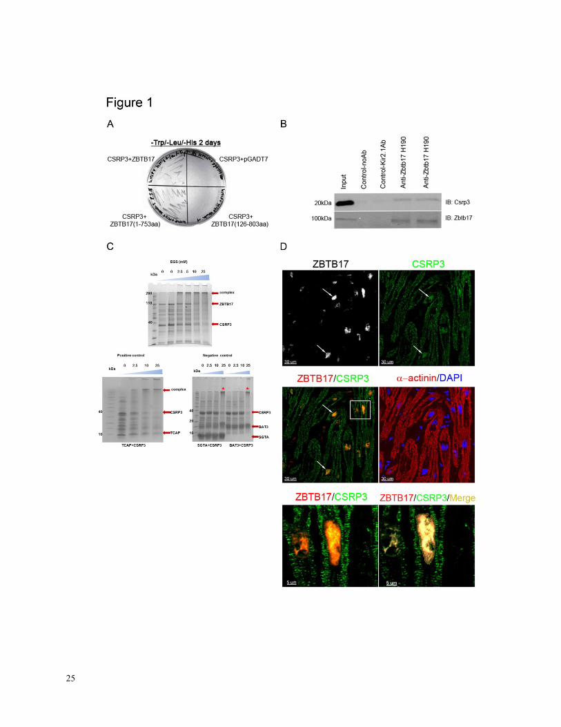
Figure 1. A, Analysis of the interaction between CSRP3 and ZBTB17 by yeast two-hybrid
assays. No interaction between proteins when the ZBTB17-POZ domain is deleted (lower right
quadrant). A total of 106 clones were screened and 650 positive clones were identified
3, one of
which encoded the 5’ sequence of Zbtb17. B, Immunoprecipitation of ZBTB17 from heart
lysates results in the co-precipitation of CSRP3. Input indicates mouse heart extract, a Csrp3
antibody (Santa Cruz Biotechnology; sc-30274) has been used for immunoprecipitation and a
well characterized Zbtb17 (Santa Cruz H-190; sc 22837) antibody has been used to detect the
protein. No antibody or an unrelated anti-Kir2.1 antibody served as negative controls. C, Cross-
linking experiments of recombinant proteins confirm binding between ZBTB17 and CSRP3. Top
panel: With increasing concentration of cross-linking reagent the individual components
decrease while a large complex emerges. The complex contains multiple copies of each protein
reflecting the oligomeric nature of the individual components. Bottom left: Positive control
showing cross-linking between CSRP3 and Telethonin (TCAP), a known interaction. Bottom
right: Negative controls - we attempted to cross-link CSRP3 separately with two different
proteins that have no documented association but are known to cross-link well to their binding
proteins. These were small glutamine-rich tetratricopeptide repeat-containing protein alpha
(SGTA) and HLA–B associated transcript 3 (BAT3). As expected, increasing cross-linking
reagents showed no binding between CSRP3 and the negative control proteins but only the usual
CSRP3 oligomers indicated with stars. D, Co-localization of ZBTB17 and CSRP3 in the nuclei
of human cardiac myocytes. Arrows indicate typical cardiac myocyte nuclei that are positive for
both ZBTB17 and CSRP3. Lower left panel is the three-dimensional image of the boxed region
(left middle panel). Lower right panel is a three-dimensional co-localized color intensity image

21
of ZBTB17 with CSRP3: mean percentage of nuclear ZBTB17 co-localized with nuclear CSRP3
= 79.4% ± 5.5; mean Pearson’s coefficient of co-localized volumes = 0.88 (n = 4, ~125 nuclei
per heart).
22
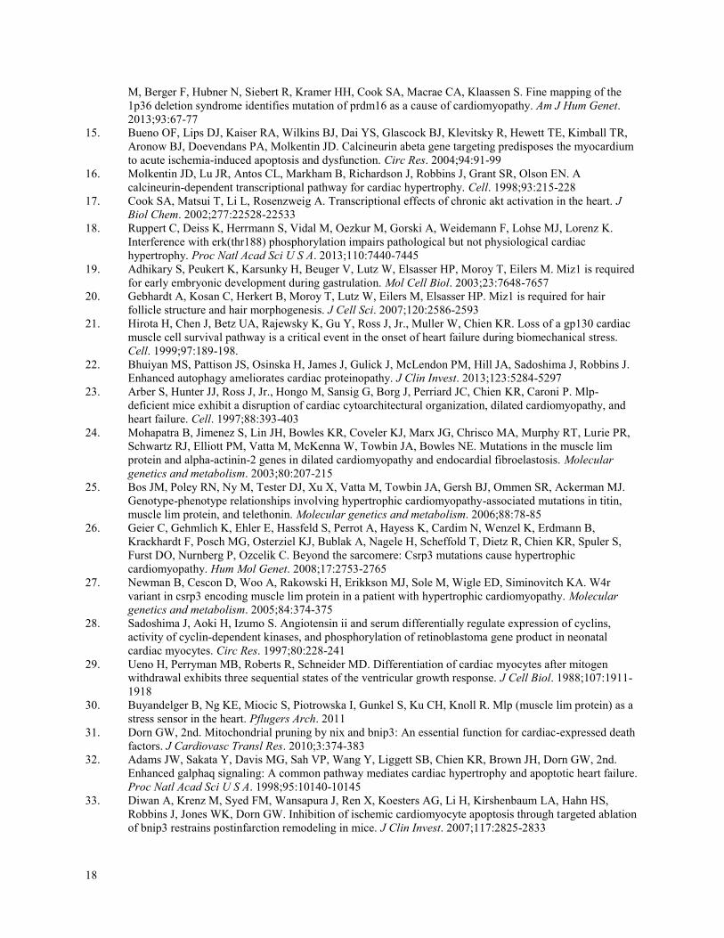
Figure 2. A, Analysis of WT- and T106M-ZBTB17 proteins via adenoviral overexpression in
NRCMs. While WT-ZBTB17 protein is readily detectable, ZBTB17:p.T106M was not and is
most likely unstable in eukaryotic cells. B, Interaction of T106M-ZBTB17 with CSRP3 as
studied via cross-link interaction. CSRP3 interacts with WT-ZBTB17 (left), but interacts less
well with T106M-ZBTB17 (right). C, NMR 1H-
15N HSQC spectrum of WT (red) and T106M
(black) ZBTB17-POZ domain with pertinent residues assigned based on predicted chemical
shift. The mutation causes significant structural disruption to the POZ domain. D, The available
ZBTB17–POZ structure was used to model the T106M mutation. 50 nanoseconds of simulation
of the WT and the T106M structures which have been run as the full quadrameric structures as in
the PDB 2Q81. Red indicates the native and yellow the mutant ZBTB17, spheres represent the T
and M residues. It can clearly be seen that the structures of the alpha-6, where the mutation is
located, have changed. Also the simulations reveal that there is a much higher level of structural
mobility for the mutant case. The mutation causes significant structural disruption to the POZ
domain, a finding consistent with our NMR data, arrowheads indicate position 106, arrows
indicate a position far away).

23
Figure 3. A, Analysis of ZBTB17 effects on hypertrophy as determined by impedance
measurements. Bar graphs represent cell index values after 24 hours (n = 10, experiments done
in duplicate). Dotted lines indicate cell indices in calcineurin inhibition experiments (CsA:
0.2µM; n = 4; *P<0.05, **P<0.01). B, Representative images of rat cardiac myocytes with and
without ZBTB17 adenoviral gene transfer. C, Effects of ZBTB17 overexpression on doxorubicin
(DOX, 1µM)-induced cell death analysis (n = 7). Dotted lines indicate data from
inhibition experiments (CsA: 0.2µM; n = 4; *P<0.05)
24
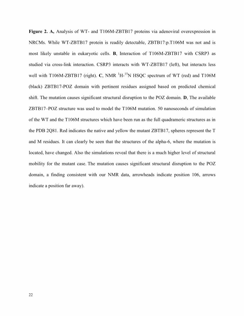
Figure 4. A, Representative micrographs of fibrosis staining of mouse hearts following sham or
TAC procedures and quantification of fibrosis (4 fields; 2 independent animals per group).B,
Analysis of cardiac myocyte cross sectional area under spontaneous conditions in WT, cKO-
Zbtb17, calcineurin Aβ-/-
(KO-Ppp3cb), TG-ZBTB17, TG-ZBTB17/KO-Ppp3cb (~150 cardiac
myocytes per animal; n = 5 per group). C, Upregulation of the pro-hypertrophic biomarker Nppa
in TG-ZBTB17 hearts (n = 3 per group). D, ChIP qRT-PCR analysis of Zbtb17 binding to
the Ppp3cb promoter in vivo. E, Luciferase reporter assay of ZBTB17 activation of the Ppp3cb
promoter via transient transfection. F, Increased Ppp3cb and Nfatc2 mRNA expression levels
in TG-ZBTB17 (n = 3 per group; *P<0.05, **P<0.01, ***P<0.001; error bars represent mean ±
SD).
25
26
27
28

29
Table 1. Echocardiography of cKO-Zbtb17 animals
Genotype/ Intervention
BW g
LVID;d mm
LVID;s mm
FS %
h/r
HW/BW
mg/g
LV Vol;s
LV Vol;d
EF %
Control.SHAM
31.90 3.77 2.06 45.61 0.54 4.26 14.40 61.39 45.62
Control.TAC 31.01 3.44* 1.65* 52.01* 0.63
* 5.03* 7.86* 49.04* 52.04*
cKO.SHAM 32.04 3.71 2.05 45.71 0.56 4.07 15.30 59.79 45.73
cKO.TAC 32.55 3.74†† 2.00 † 46.46† 0.57 4.82 13.22
† 59.69†
† 46.46
†
Functional data obtained via echocardiography 4 weeks after sham operations (SHAM) or 4
weeks after transverse aortic constriction (TAC) in Zbtb17 cKO and control animals. After TAC
cKO hearts enlarge (LVID;d and LVID;s) and decrease in function (%FS) (Control = flox/flox,
Cre-; cKO = flox/flox, Cre
+; *P < 0.05, **P < 0.01 for Control.SHAM vs Control.TAC; †P <
0.05, ††P < 0.01 for Control.TAC vs cKO.TAC; data = mean).
30
Table 2. Integrated analysis of apoptotic gene expression and ChIP-chip
Gene Fold
Change p-value Function
ZBTB17
Binding site
(FDR < 0.05)
Fadd 0.4 0.0017 pro-apoptotic Y
Casp2 2.2 0.0023 pro-apoptotic Y
Traf3 1.6 0.0076 pro-apoptotic Y
Rnf7 1.8 0.0106 anti-apoptotic Y
Casp14 17.2 0.0255 pro-apoptotic Y
Casp1 1.8 0.0280 pro-apoptotic Y
Bnip3 1.6 0.0285 pro-apoptotic Y
Cradd 1.8 0.0364 pro-apoptotic Y
Cd70 4.3 0.0003 pro-apoptotic N
Summary of significantly differentially expressed apoptotic genes and Zbtb17 binding sites of
genes involved in cardiac myocyte programmed cell death in cKO animals as compared to
control using qRT-PCR arrays. Apoptotic qRT-PCR array and ChIP-chip methods are described
in the Supplementary Information. Abbreviations: FDR = false discovery rate; Y = ZBTB17
binding site; N = no ZBTB17 binding site at FDR < 0.05.
31
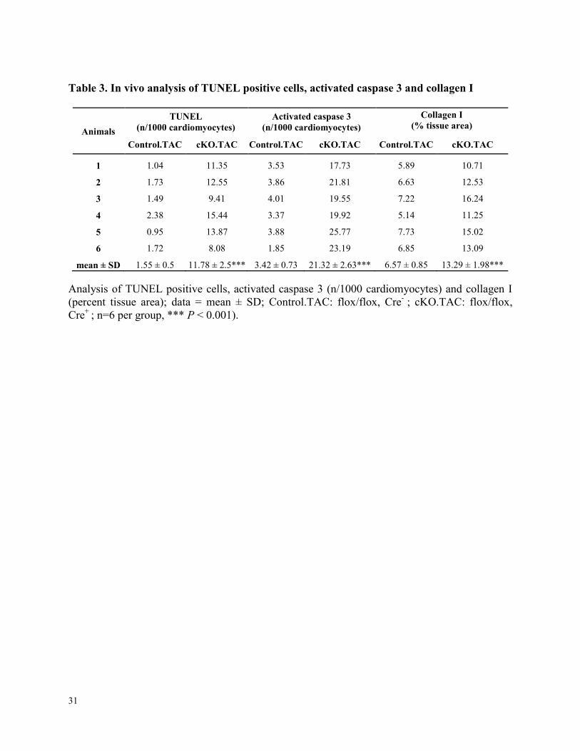
Table 3. In vivo analysis of TUNEL positive cells, activated caspase 3 and collagen I
Animals
TUNEL (n/1000 cardiomyocytes)
Activated caspase 3 (n/1000 cardiomyocytes)
Collagen I (% tissue area)
Control.TAC cKO.TAC Control.TAC cKO.TAC Control.TAC cKO.TAC
1 1.04 11.35 3.53 17.73 5.89 10.71
2 1.73 12.55 3.86 21.81 6.63 12.53
3 1.49 9.41 4.01 19.55 7.22 16.24
4 2.38 15.44 3.37 19.92 5.14 11.25
5 0.95 13.87 3.88 25.77 7.73 15.02
6 1.72 8.08 1.85 23.19 6.85 13.09
mean ± SD 1.55 ± 0.5 11.78 ± 2.5*** 3.42 ± 0.73 21.32 ± 2.63*** 6.57 ± 0.85 13.29 ± 1.98***
Analysis of TUNEL positive cells, activated caspase 3 (n/1000 cardiomyocytes) and collagen I
(percent tissue area); data = mean ± SD; Control.TAC: flox/flox, Cre- ; cKO.TAC: flox/flox,
Cre+
; n=6 per group, *** P < 0.001).