zek1424
DESCRIPTION
gggTRANSCRIPT

Colony Organization in the Green Alga Botryococcus braunii (Race B)Is Specified by a Complex Extracellular Matrix
Taylor L. Weiss,a Robyn Roth,b Carrie Goodson,c Stanislav Vitha,d Ian Black,e Parastoo Azadi,e Jannette Rusch,c
Andreas Holzenburg,a,d,f Timothy P. Devarenne,a and Ursula Goodenoughc
Department of Biochemistry and Biophysics, Texas A&M University, College Station, Texas, USAa; Department of Cell Biology, Washington University School of Medicine,St. Louis, Missouri, USAb; Department of Biology, Washington University, St. Louis, Missouri, USAc; Microscopy and Imaging Center, Texas A&M University, College Station,Texas, USAd; Complex Carbohydrate Research Center, University of Georgia, Athens, Georgia, USAe; and Department of Biology, Texas A&M University, College Station,Texas, USAf
Botryococcus braunii is a colonial green alga whose cells associate via a complex extracellular matrix (ECM) and produce prodi-gious amounts of liquid hydrocarbons that can be readily converted into conventional combustion engine fuels. We used quick-freeze deep-etch electron microscopy and biochemical/histochemical analysis to elucidate many new features of B. braunii cell/colony organization and composition. Intracellular lipid bodies associate with the chloroplast and endoplasmic reticulum (ER)but show no evidence of being secreted. The ER displays striking fenestrations and forms a continuous subcortical system in di-rect contact with the cell membrane. The ECM has three distinct components. (i) Each cell is surrounded by a fibrous �-1, 4-and/or �-1, 3-glucan-containing cell wall. (ii) The intracolonial ECM space is filled with a cross-linked hydrocarbon networkpermeated with liquid hydrocarbons. (iii) Colonies are enclosed in a retaining wall festooned with a fibrillar sheath dominatedby arabinose-galactose polysaccharides, which sequesters ECM liquid hydrocarbons. Each cell apex associates with the retainingwall and contributes to its synthesis. Retaining-wall domains also form “drapes” between cells, with some folding in on them-selves and penetrating the hydrocarbon interior of a mother colony, partitioning it into daughter colonies. We propose that re-taining-wall components are synthesized in the apical Golgi apparatus, delivered to apical ER fenestrations, and assembled onthe surfaces of apical cell walls, where a proteinaceous granular layer apparently participates in fibril morphogenesis. We furtherpropose that hydrocarbons are produced by the nonapical ER, directly delivered to the contiguous cell membrane, and passacross the nonapical cell wall into the hydrocarbon-based ECM.
The trebouxiophyte green fresh- to brackish-water alga Botryo-coccus braunii has a unique colonial organization: individual
cells of the colony are embedded in an extracellular matrix (ECM)composed of polymerized and liquid hydrocarbons (LH) (29, 35,46–48). Although they may serve additional functions, these hy-drocarbons notably allow B. braunii colonies to float, presumablyto increase exposure to light for photosynthesis at the surfaces ofponds or lakes (4). The presence of B. braunii oils and ECM fossilsin petroleum, coal deposits, and oil shale suggests that their hy-drocarbon products once contributed to these reserves (1, 2, 8, 10,16, 17, 34, 36, 52, 62, 63, 69, 70) and that these hydrocarbons couldbe used as a source of renewable energy. In fact, B. braunii hydro-carbons can be readily converted into petroleum equivalent,drop-in transportation fuels by conventional petroleum-process-ing techniques (22, 28).
The three races of B. braunii are classified on the basis of thechemical nature of the liquid hydrocarbons they produce. The Arace primarily produces alkadienes and alkatrienes derived fromfatty acids (38, 49, 65, 67); the B race (the focus of this study)produces two triterpenoids, tetramethylsqualene as a minor com-ponent, and botryococcenes ranging from C30 to C37 as the majorcomponents (41–43); and the L race produces a tetraterpenoidknown as lycopadiene (37, 39). The B race of B. braunii has gar-nered the most research attention due to its ability to accumulatebotryococcenes up to 86% of its dry weight (9).
The solid, polymerized-hydrocarbon portion of the ECM of allthree races has been found to be mainly composed of long-chainpolyacetal hydrocarbons that are covalently, and possibly evennoncovalently (mechanically), cross-linked with hydrocarbons
specific for each race (47, 48). For example, in the A race, theindividual polyacetals appear to be only covalently cross-linked toeach other (46). However, the B race further covalently cross-linkstetramethylsqualene diols to the polyacetals (48), while the L racefurther covalently cross-links lycopadiene diols to the polyacetals(6). The B and L races additionally link the tetramethylsqualeneand lycopadiene diols to large (C31 and C33) macrocyclic alde-hydes, which may then be mechanically linked to the linear poly-acetals (47). These cross-linked hydrocarbons are proposed toproduce a network for deposition of liquid hydrocarbons (47).
An important goal of B. braunii research is to develop a com-prehensive understanding of how and where the hydrocarbons areproduced and retained within the colony. Past research has uti-lized radioactive precursors to show that in the B race, C30 botryo-coccene is produced inside cells and higher-molecular-weightbotryococcenes, C31 to C34, are produced by methylation of C30
botryococcene with S-adenosylmethionine as the methyl donor(43, 74). These studies also indicate that the methylation reactionscan take place both inside cells and in the ECM and that intracel-
Received 6 July 2012 Accepted 28 August 2012
Published ahead of print 31 August 2012
Address correspondence to Timothy P. Devarenne, [email protected], or UrsulaGoodenough, [email protected].
Supplemental material for this article may be found at http://ec.asm.org/.
Copyright © 2012, American Society for Microbiology. All Rights Reserved.
doi:10.1128/EC.00184-12
1424 ec.asm.org Eukaryotic Cell p. 1424–1440 December 2012 Volume 11 Number 12

lular botryococcenes can be transported to the ECM (43), but themechanism(s) by which this occurs remains unknown. Most cur-rent research on B. braunii hydrocarbons is focused on identifyingthe genes responsible for botryococcene biosynthesis, and genesinvolved in the production of C30, C31, and C32 botryococceneshave been identified (54, 55). The enzyme activity associated withC30 botryococcene production, and presumably these enzymes,has been shown to be membrane localized (58), likely to the en-doplasmic reticulum (ER).
Light microscopy with the fluorescent lipophilic dye Nile red,coupled with confocal Raman microspectroscopy, has docu-mented the presence of botryococcenes in the ECM and in multi-ple cytoplasmic lipid bodies (LBs) (71), supporting the hypothesisthat the lipid-body botryococcenes are secreted out of the cells toform the liquid hydrocarbon portion of the ECM (45, 47). How-ever, direct evidence for this hypothesis has not been obtained.
At the electron microscopy level, several studies have probedthe ultrastructure of B. braunii cells and colonies, documentingthe presence of a wall around each cell (5, 11, 31, 56, 73) and anouter wall surrounding each colony (73). Fibrils of polysaccharidehave also been seen emanating from the apexes of individual cells(5, 31, 73). These studies were performed on several differentstrains of B. braunii, and several were carried out before the clas-sification of B. braunii into its three races; hence, it is difficult toextend the findings to any specific race. Additionally, the imagesare compromised by the fact that B. braunii, like many other algaewith highly impermeable cell walls (CW), is difficult to fix, dehy-drate, and embed in plastic (5); moreover, such procedures havethe capacity to modify the native organization of extracellularproducts.
We have therefore analyzed the ultrastructure of the B. brauniiB race using the quick-freeze deep-etch (QFDE) electron micros-copy technique, which employs no fixation or dehydration (20).Living colonies were snap-frozen at liquid helium temperatures(approximately �269°C), fractured at liquid nitrogen tempera-tures (approximately �196°C), and etched and replicated usingplatinum/carbon rotary shadowing. In parallel studies, B. brauniicells and colonies were subjected to histochemical analysis, whilepreparations of released outer-wall fragments were subjected tomass spectrometry analysis.
Our findings, many of which are diagrammed in Fig. 1, includethe following. (i) An outer retaining wall (RW) serves to sequesterthe liquid ECM within the colony by forming a drape betweencells. A fibrillar sheath extends from the outer face of the retainingwall into the growth medium. A purified preparation of discardedretaining walls/sheath fibrils is �97% carbohydrate, with 81% ofthe mass being arabinose (42%) and galactose (39%), the princi-pal sugars of many matrix elements in algae and land plants (30).(ii) The retaining wall also makes intimate contact with the apicalcell wall of each cell. A prominent Golgi body and a unique systemof fenestrated cortical ER appear to participate in the productionand secretion of the retaining wall and its sheath fibrils in thisapical domain. Notably, an amorphous secretion product(“toothpaste”) is converted into fibrils in conjunction with a pro-teinaceous granular layer. (iii) The drape also occasionally foldson itself and penetrates the colony’s hydrocarbon interior to sub-divide a mother colony into daughter colonies, a specialization weterm the zipper. (iv) The cell wall, but not the retaining wall, stainsintensely with the anionic dye Congo red, indicating that the twowalls are composed of distinctive materials and that the cell wall
contains �-1,4- and/or �-1,3-glucans. (v) The cytoplasmic inte-rior contains inclusions, corresponding to those visualized withNile red and confocal Raman microspectroscopy, that are visuallyindistinguishable from triacylglycerol (TAG)-containing lipidbodies in other algae. No evidence of their secretion was encoun-tered. (vi) In the nonapical portions of the cell, a fenestrated ER isin intimate contact with the cell membrane. We propose thatbotyrococcenes produced by this ER are directly delivered to thecell membrane, after which they are exuded across the cell wall andinto the ECM. (vii) The hydrocarbon ECM fills the intersticesbetween cells. The botryococcene liquid hydrocarbon phase is de-formable to fracture and largely extracted by n-hexane, while thepolymerized phase is fibrous and not extracted by n-hexane. Wepropose that the polymerized polyacetals function to stabilize thecolony and to facilitate gas exchange in the colony interior.
MATERIALS AND METHODSStrains and culture conditions. B. braunii strain Berkeley (Showa) race B(57) was grown in modified Chu 13 medium (19) using 13-W compactfluorescent 65 K lighting at a distance of 7.62 cm, which produced a lightintensity of 280 �mol photons/m2/s. Lighting was on a cycle of 12-h
FIG 1 Model of a B. braunii cell within a colony based on the studies presentedhere. (A) Color DIC microscopy image of a partial B. braunii colony. Theboxed area is depicted in panel B. Bar, 10 �m. (B) Model of the B. braunii celland its surrounding extracellular matrices. Portions of the hydrocarbon ECMaround the cell edge and the upper left quadrant are drawn with the liquidhydrocarbons removed to show the underlying structure of these regions. Forsimplification, not all cellular organelles are shown. Bar, 2 �m.
B. braunii ECM Structure
December 2012 Volume 11 Number 12 ec.asm.org 1425

light/12-h dark at 22.5°C. The cultures were continuously aerated withfilter-sterilized enriched air containing 2.5% CO2. Fifty milliliters of cul-ture was used to inoculate 750 ml of subsequent subcultures every 4 weeks.
Quick-freeze deep-etch electron microscopy. Floating colonies werepipetted from the surfaces of fresh cultures, placed on a cushioning ma-terial, and dropped onto a liquid He-cooled copper block; the frozenmaterial was transferred to liquid nitrogen and fractured, etched at �80°Cfor 2 min, and Pt/C rotary replicated as described previously (20). Thereplicas were examined with a JEOL electron microscope, model JEM1400, equipped with an AMTV601 digital camera. The images are photo-graphic negatives; hence, protuberant elements of the fractured/etchedsurface are most heavily coated with platinum and appear white.
Histochemical staining and microscopy. For visualization of the cellwall using Congo red (Sigma), 100 �l of B. braunii colonies in mediumwas treated with 1 �l of a 1-mg/ml stock solution of Congo red dissolvedin water. For visualization of botryococcenes and other lipids using Nilered (Sigma), 400 �l of B. braunii colonies in medium were treated with 1�l of a stock solution of Nile red dissolved in acetone (0.15 mg/ml) so thatthe final concentrations of Nile red and acetone were 0.375 �g/ml and0.25%, respectively. Both Congo red- and Nile red-stained samples werekept in the dark and incubated at room temperature for 15 min. Bothsamples were then diluted with 1 ml distilled water (dH2O) and centri-fuged at 10,000 � g for 30 s, and excess solution was removed by pipettingunder the floating layer of algal colonies. This rinsing process was quicklyrepeated three times, and the final stained algal samples were stored in aminimal volume prior to immediate microscopy.
The fibril sheath system was visualized using a modified periodic acid-Schiff reagent (mPAS) using propidium iodide (PI) (Sigma) as the Schiffreagent (53). B. braunii colonies (270 �l) in medium were treated with 30�l of a stock solution of 10% periodic acid in the dark at room tempera-ture for at least 30 min. The samples were diluted with 1 ml dH2O andcentrifuged at 10,000 � g for 30 s, and excess solution was removed bypipetting under the floating layer of algal colonies. This rinsing processwas repeated three times. The samples were then resuspended in 30 �l of10 mM PBS buffer, pH 8.0, and 1 �l of a 1-mg/ml PI stock solution wasadded. The samples were kept in the dark and incubated at room temper-ature for 20 min before an additional rinse with dH2O (three times) andimmediate microscopy visualization. Costaining of B. braunii colonieswith both Nile red and an mPAS reaction followed the same procedure asan mPAS reaction alone, except that 1 �l of Nile red stock solution wasadded to the sample during the PI incubation.
Microscopy imaging of B. braunii colonies was performed at the TexasA&M University Microscopy and Imaging Center. Fluorescence micros-
copy visualization was performed using an Olympus FV1000 (OlympusAmerica Inc., Center Valley, PA) laser scanning confocal microscopeequipped with an UPLSAPO 100�/1.4 oil immersion objective. Excita-tion and emission wavelengths were set for Congo red (excitation, 543nm; emission, 555 to 630 nm), mPAS with PI (excitation, 543 nm; emis-sion, 555 to 620 nm), Nile red (excitation, 488 nm; emission, 540 to 590nm), and Nile Red plus mPAS with PI double staining (excitation, 488nm; emission, 500 to 550 and 620 to 640 nm, sequential acquisition).Chlorophyll autofluorescence (excitation, 405 nm; emission, �650 nm)was recorded in parallel with the dyes listed above, using line-sequentialacquisition.
Shell preparation. Five liters of a 4-week-old B. braunii culture wasleft undisturbed for 12 h, allowing a majority of colonies to float andresidual material to settle. This residual material was then harvested byvacuum suction with care to minimize disruption of the floating algallayer. The harvested material was vacuum filtered through a 35-�mnylon cloth (Aquatic Eco-Systems, Inc., Apopka, FL) to remove resid-ual large algal colonies. The filtrate was centrifuged at 17,000 � g for 30min to pellet the shells, bacteria, and any remaining algal colonies insolution. The supernatant was carefully disposed of by vacuum suc-tion, and the soft layer of shells was gently resuspended and transferredinto fresh centrifugation tubes, leaving behind a hard pellet of bacteriaand algal colonies. The resuspended shells were diluted with dH2O,centrifuged at 48,000 � g for 15 min, and again harvested. This processof centrifugation, shell isolation, and dH2O rinsing was repeated atleast 4 times until there was no obvious pellet of bacteria after centrif-ugation. The shell fraction was then freeze-dried and analyzed by massspectrometry as described below.
Carbohydrate mass spectrometry analysis. For glycosyl composi-tion, the shell preparation was analyzed by the per-O-trimethylsilyl(TMS) and alditol acetate derivatization methods. For the TMS method,methyl glycosides were prepared from the dry sample by methanolysis in1 M HCl in methanol at 80°C for 17 h, followed by re-N-acetylation withpyridine and acetic anhydride in methanol (for detection of aminosugars). The sample was then per-O-trimethylsilylated by treatment withTri-Sil (Pierce) at 80°C for 0.5 h. The TMS derivatives were run on aShimadzu QP2010 gas chromatograph-mass spectrometer (GC-MS) inchemical ionization mode. For the alditol acetate method, the sample wasdepolymerized, reduced, and acetylated; the resultant alditol acetates(AAs) were analyzed by GC-MS for determination of monosaccharideidentity. Specifically, 200 �g of lyophilized shells was hydrolyzed using 2M trifluoroacetic acid for 2 h in a sealed tube at 121°C, reduced withsodium tetradeuteroborate (NaBD4), and acetylated using acetic anhy-
FIG 2 Survey views of B. braunii colony organization. (A) Two cells showing nucleus (N), chloroplast (cpst), chloroplast envelope (ce), and apical Golgi body(G). The intercellular space is filled with an h-ECM containing balloons (b). The retaining wall and cell wall cover the apex of the cell at left, and the retaining wallextends between cells to form a drape that holds in the h-ECM. The asterisks indicate the cell apex-drape junctions. Sheath fibrils extend from the retaining wall.Bar, 500 nm. (B) Cell with apical Golgi body (g), retaining wall carrying white tubular elements, cell wall, and mitochondria (mito). The drape retaining wall holdsin the h-ECM with balloons (b), with an asterisk at the cell-drape junction. The full extent of the fibrillar sheath is depicted (bottom right). Bar, 500 nm.
Weiss et al.
1426 ec.asm.org Eukaryotic Cell

dride/trifluoroacetic acid. The AAs were analyzed on an Agilent 7890AGC interfaced to a 5975 mass selective detector (MSD) operating in theelectron impact ionization mode. Separation of the AAs by GC was per-formed on a 30-m Supelco 2330 bonded-phase fused-silica capillarycolumn.
For glycosyl linkage analysis, the samples were permethylated, depo-lymerized, reduced, and acetylated, and the resultant partially methylatedalditol acetates (PMAAs) were analyzed by GC-MS as previously de-scribed (79). Initially, �1 mg of the sample was suspended in 200 �l ofdimethyl sulfoxide, and the sample was left to stir for 3 days. The samplewas then permethylated by treatment with sodium hydroxide and methyliodide in dry dimethyl sulfoxide (DMSO) (12). Following sampleworkup, the permethylated material was hydrolyzed using 2 M trifluoro-acetic acid for 2 h in a sealed tube at 121°C, reduced with NaBD4, andacetylated using acetic anhydride/trifluoroacetic acid. The resultingPMAAs were analyzed on an Agilent 7890A GC/5975 MSD as describedabove. Separation was performed on a 30-m Supelco 2330 bonded-phasefused-silica capillary column.
Protein determination and SDS-PAGE of lyophilized shells. Forprotein determination, 1.6 mg of shell lyophilate was suspended in 200 �lwater and assayed with the Bio-Rad Bradford Protein Assay reagent using
bovine serum albumin (BSA) as a standard. For SDS-PAGE, 22.5 mg ofsample was brought up in 100 �l extraction buffer (1 mM NaCl, 10 mMTris, pH 7, protease inhibitor cocktail), 100 �l 2� Laemmli buffer wasadded, and the sample was heated at 90°C for 5 min and then applied to a4 to 15% polyacrylamide gradient gel (Bio-Rad). The gels were stainedwith Coomassie blue.
Shells on coverslips. Coverslip chips (3 by 3 mm) were immersed in 1mg/ml low-molecular-weight (LMW) polylysine (1,000 to 4,000 MW;Sigma P0879) in 100 mM KCl for 1 h and rinsed in water. Two chips wereplaced on the bottom of a 1.5-ml microcentrifuge tube containing a pol-ished resin insert that provided a flat base, and 50 �l of a freshwater-washed shell pellet was layered over 400 �l water and spun in a clinicalcentrifuge (7,300 rpm) for 6 min. The chips were transferred to water,immediately quick frozen, and stored in liquid nitrogen until they weremounted in a Balzers freeze-etch device, where they were immediatelywarmed to �80°C, freeze-dried for 15 min in vacuo, and replicated withplatinum/carbon.
n-Hexane extraction. In a modified version of a previously describedprocess (15), colonies were applied to a 35-�m mesh nylon filter sand-wiched between 2 trimmed 1-ml pipette tips. Compressed air was passedthrough the filter for 1 min, the filter was removed, and colonies were
FIG 3 Cell apexes. (A) Apical Golgi body, fenestrated ER, and cell membrane (cm) underlie the cell wall, which fractures into 2 layers (labeled 1 and 2) at right,and retaining wall. The retaining wall carries white tubules (t), a layer of granules (g), and the fibrillar sheath. cpst, chloroplast. Bar, 100 nm. (B) Cell membrane,double-layered (labeled 1 and 2) fibrous cell wall, and retaining wall carrying tubules (t), granules (g), and fibrils. The asterisks mark continuities between tubulesand fibrils. Bar, 100 nm. (C) Cross-fractured fibrous double-layered (labeled 1 and 2) cell wall overlain by en face view of the retaining wall, which fractures into2 layers (labeled 3 and 4), showing tubules (t) and granules (g). The asterisks mark continuities between tubules and fibrils. Bar, 100 nm. (D) Retaining wall enface with tubules (t), granules (g), and fibrils. Bar, 100 nm.
B. braunii ECM Structure
December 2012 Volume 11 Number 12 ec.asm.org 1427

spread uniformly over its surface and air dried for 10 min. The driedcolonies were removed from the filter and placed in a capped glass tubecontaining 12 ml n-hexane (Sigma H9379) and a stir bar, and stirred for 1h, during which the n-hexane turned yellow. The n-hexane was removedby aspiration, and the cells were dispersed in 5 ml phosphate-bufferedsaline (PBS) and washed 2 times before freezing.
RESULTSOverview of colony organization. The organization of a B. brau-nii cell and the surrounding ECM of the colony are diagrammed inFig. 1. Each colony unit, containing 50 to 100 ovoid cells organizedinto smaller clusters and embedded in a hydrocarbon extracellularmatrix (h-ECM), is encased in a retaining wall. The outer surfaceof the retaining wall carries an extensive system of anastomosingfibrils that extends 2 to 3 �m into the growth medium and formsa fibrillar sheath around the entire colony; this presumably corre-sponds to the “tenuous mucilage layer surrounding the colony”described in 1936 (7) and the fibrillar sheath described later (73).The inner surface of the retaining wall has two configurations: (i)when overlying the apical portion of a cell, it makes direct contactwith the CW; (ii) when spanning the region between cells, it makesdirect contact with the h-ECM and forms a “drape” between thecells.
Figure 2 illustrates these general features in QFDE replicas (ad-ditional survey images are found in File S1 in the supplementalmaterial). The asterisks indicate junctions between cell wall-asso-ciated and drape domains of the retaining wall. Also evident is thehighly heterogeneous nature of the h-ECM, as detailed below.
The retaining wall-cell wall-ER system at the cell apex. Figure3A illustrates key features of the cell apex in cross-fracture (addi-tional images of the cell apex are found in File S2 in the supple-mental material). The thin RW is in direct contact with the thickerfibrous CW, which often fractures into two sublayers (Fig. 3A,labeled 1 and 2; see File S3 in the supplemental material), beneathwhich is the cell membrane (cm) and then a fenestrated ER cister-num. A large Golgi body also occupies the apical cytoplasm (Fig. 1and 2; see File S3 in the supplemental material). The outer aspectof the retaining wall carries the fibrillar sheath system plus twoadditional differentiations: protuberant tubular elements (t), re-sembling toothpaste, and a layer of granules (g).
Figure 3B to D shows these differentiations in en face fractures.The retaining wall, which fractures into two sublayers (Fig. 3C,labeled 3 and 4; see Fig. S2-4 in File S2 and Fig. S3-2 in File S3 in thesupplemental material), carries abundant tubular elements that“dive into” the granular material; fibrils emerge from the otherside of the granular layer. Asterisks in Fig. 3B and C denote tubulesthat display direct continuity with branching fibrils. It is therefore
FIG 4 (A and B) Apical fenestrated ER and the overlying cell membrane (cm),cell wall, and retaining wall with its tubules (t), granular layer (g), and fibrils.ce, chloroplast envelope. (C) The asterisk marks a secretion pore in the cellmembrane. Bars, 250 nm (A), 200 nm (B), and 100 nm (C).
FIG 5 Drape domain of the retaining wall with associated granules (g) andfibrils on its external face and LH on its internal face. Bar, 100 nm.
Weiss et al.
1428 ec.asm.org Eukaryotic Cell

proposed that a tubule-to-fibril transformation occurs at the re-taining-wall surface in association with the granules.
Figure 4A to C shows en face fractures of the fenestrated ER thatdirectly underlies the apical cell membrane. In Fig. 4A, the fractureplane has dropped below the cell membrane to reveal a segment ofthe ER; in Fig. 4B, a continuous expanse is exposed. The ER fenes-trae are irregular in size; whether this is native or due to differen-tial fracture/etching is not known. At higher magnification(Fig. 4C), the asterisk marks a candidate secretion pore in the cellmembrane (additional secretion pore candidates are found in FileS4 in the supplemental material). As detailed in Discussion below,these pores are interpreted as arising when polysaccharide mate-rial, delivered to the fenestrae via Golgi body-derived vesicles,passes through the cell membrane and then traverses the cell andretaining walls, emerging as the tubules on the external face of theretaining wall.
The retaining-wall system in intercellular drapes. At thejunctions marked by asterisks in Fig. 2, the retaining wall loses itsassociation with the apical cell wall and drapes across the h-ECM,eventually making contact with the apical cell walls of adjacentcells. Figure 5 shows such a drape at high magnification (addi-tional images of drapes and h-ECM are found in File S5 in thesupplemental material). The granular layer and the fibrils remainassociated with the drape, but the tubular elements are absent,indicating that the tubules are a unique feature of the retaining-wall– cell wall relationship.
The h-ECM in Fig. 5 displays two phases: delicate strands as-sociate with the inner aspect of the retaining wall, and amorphousmaterial fills the rest of the region. As detailed below, the amor-phous material represents the LH phase of the h-ECM. Thestrands are possibly polymerized hydrocarbons but are morelikely liquid hydrocarbons sheared during the fracturing process.
Retaining-wall “zipper.” Figure 6A shows two cells and theirintervening drape. At the asterisk, the drape dives into the h-ECM,forming a configuration that we designate a retaining-wall zipper.Figure 6B enlarges the region of zipper initiation. The drape, nolonger bearing fibrils, folds in on itself to form a trilaminar unit
consisting of a retaining wall, a shared granule domain, and asecond retaining wall. Candidate en face views of the zipper wallare shown in File S6 in the supplemental material. As detailed inDiscussion below, we propose that zippers function to segregate acolony into daughter colonies, with the granule domain possiblyserving as the plane of daughter-colony separation.
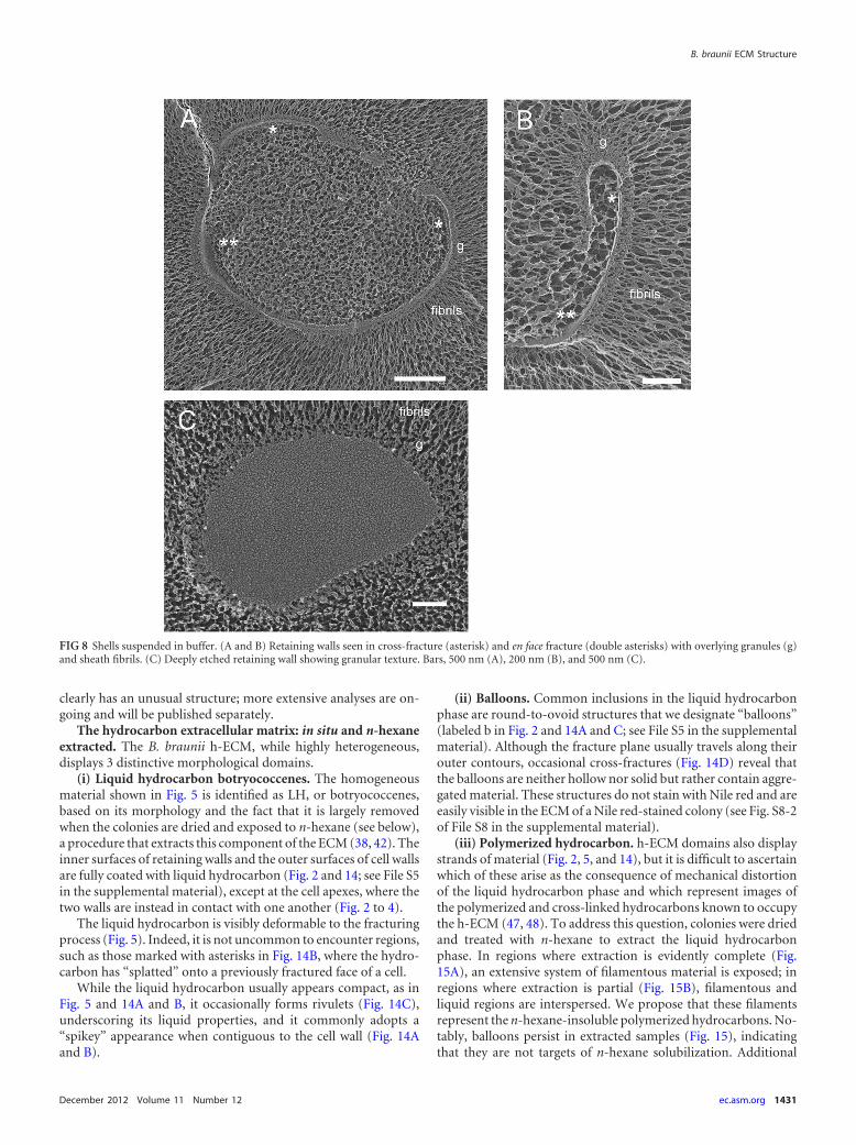
“Shells,” excised segments of the retaining wall. As B. brauniicultures grow, uniform cup-shaped structures, �3 by 5 �m insize, accumulate in the medium (Fig. 7A). These “shells” form afluffy pellet when the medium is centrifuged (Fig. 7B), and highlypurified shell preparations can be obtained with filtering andwashing (Fig. 7C). QFDE shows the shells to be segments of theretaining wall and its fibril sheath (Fig. 7D, 8, and 9; additionalimages are shown in File S7 in the supplemental material). Re-markably, although the culture is not axenic (Fig. 7B), the shellsresist degradation for many weeks.
Figure 8 shows representative images of shells snap-frozen inaqueous suspension. The retaining-wall fibrils radiate outward,with the layer of granules at their base (Fig. 8A and B), and theinner face of the retaining wall faces the curved interior, mirroringthe topology adopted at the cell apexes. Cross-fractures of theretaining wall (Fig. 8A and B, single asterisks) reveal it to be verynarrow (�15 nm); en face fractures (Fig. 8A and B, double aster-isks) show a smooth surface that, when deeply etched (Fig. 8C),has a roughened texture with no evidence of a fibrous component(see Fig. S7-1 to -3 of File S7 in the supplemental material foradditional images); in contrast, the cell wall is thicker (�50 nm)and visibly fibrous (Fig. 3A to C and 4A; see Fig. S3-1 and -2 of FileS3 in the supplemental material). Shells lack the tubular tooth-paste-like elements; this could indicate that they derive fromdrape domains, which lack these elements (Fig. 5), or that theyderive from apical domains that have lost the tubules during orfollowing the excision process.
Figure 9A and B shows shells adsorbed to a polylysine-coatedglass coverslip, revealing the smooth face of the retaining wall andthe full dimensions of the fibrillar sheath.
The granular material interfacing the tubules and fibrils of the
FIG 6 Retaining-wall zipper. (A) Apical domains of two cells embedded in LH and interconnected by a drape. At the asterisk, the drape forms a zipperthat extends into the colony interior. RW, retaining wall carrying a few white tubules; cpst, chloroplast; ce, chloroplast envelope. Bar, 500 nm. (B) Highermagnification of zipper involution (asterisk). Fibrils are absent, and granules (g) coalesce as a central domain of the zipper flanked by two RW domains.Bar, 250 nm.
B. braunii ECM Structure
December 2012 Volume 11 Number 12 ec.asm.org 1429

retaining wall (Fig. 3 to 5) persists in the shell preparation (Fig. 8Aand B). Since the granules resemble proteins, the presence of pro-tein in the shell lyophilate was assayed, and it was estimated torepresent 1.25% of the total mass (Table 1). SDS-PAGE of the shelllyophilate documented the presence of a single prominent poly-peptide (Fig. 10) with an apparent molecular mass of 150 kDa. Wepropose that this polypeptide forms the granules.
Histochemical and chemical analysis of cell and retainingwalls and the fibril sheath system. Histochemical studies pro-vided additional information about the ECM components of B.braunii. Nile red stained the intracellular LBs and the intercellularhydrocarbon matrix (Fig. 11A to D), as expected, and Congo red,which stains �-1,4- and �-1,3-glucans, including cellulose (13, 32,76), showed strong and specific staining of the cell perimeters (Fig.11E to I), indicating that the cell wall is composed of a cellulose-like polysaccharide. However, neither reagent stained the retain-ing wall/sheath (Fig. 11), nor is it visible with color differentialinterference contrast (DIC) (Fig. 12A), although a haze aroundthe colony edge is visible with black-and-white DIC (Fig. 12B).
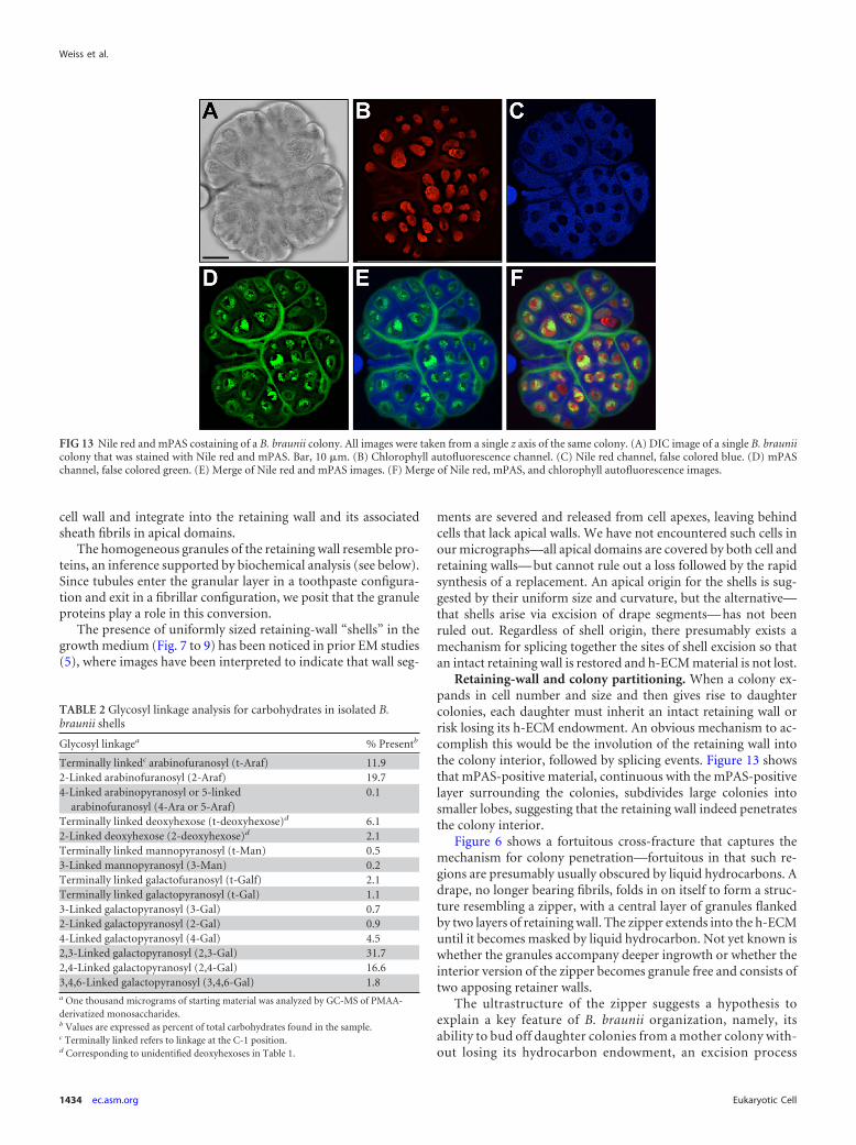
Since several previous studies indicated the presence of poly-saccharide associated with the colony exterior (5, 31, 73), we nextused a more general polysaccharide stain, the periodic acid-Schiffreagent (PAS) stain, in which carbohydrate vicinal diols are oxi-dized to aldehydes with periodic acid and the aldehydes are thendetected with the Schiff reagent (23). We employed an mPAS re-action that utilizes PI as the Schiff reagent (53), and this procedurewas capable of effectively staining polysaccharides in the chloro-plast (presumably starch), in the cell wall, and in the retaining-wall–fibrillar-sheath system (Fig. 12C to F; see Fig. S8-1 of File S8in the supplemental material), suggesting that the last is composedof carbohydrates. Costaining with mPAS and Nile red (Fig. 13A toF) revealed the extensive Nile-red-positive h-ECM in which eachof the cells was embedded and the mPAS-positive system thatsurrounds the entire colony and penetrates to define the lobes offuture daughter colonies. As detailed in Discussion below, we pro-pose that retaining-wall zippers correspond to the internal mPAS-positive system.
We next subjected the shell preparation, which includes re-taining wall/fibrillar sheath and granules (Fig. 8 to 9), to TMSand alditol acetate derivatization followed by GC-MS. Theanalysis (Table 1) showed that the shells are 97.9% carbohy-drate, with the major glycosyl residues being arabinose (46.7mol%) and galactose (36.3 mol%). Mannose and glucose weretrace components. Two unidentified deoxyhexose peaks con-stituting 9.3 mol% (Table 1) were detected in the sample usingthe TMS derivatization method, followed by chemical-ioniza-tion GC-MS detection. The sample also contained minoramounts of the uncommon glycosyl residues 3-methyl-arabi-nose and 6-methyl-galactose. Interestingly, the TMS de-rivatization did not detect any uronic acids or N-acetyl glycosylresidues (not shown), indicating that the polysaccharides in theshells do not possess additional chemical modifications com-monly associated with structural carbohydrates.
We also analyzed the glycosyl linkages of the polysaccharides inthe shell material. As would be expected from the compositionanalysis, the majority of the linkages involve galactose and arabi-nose (Table 2). The arabinoses and deoxyhexoses are mainlylinked at just one position: the 1 carbon (terminally linked) or the2 carbon (Table 2). The galactoses mainly have two linkages persingle unit, 2,3-linked and 2,4-linked; a small amount is triplelinked at the 3, 4, and 6 carbons or singly linked at the 4 carbon(Table 2). All other identified linkages were saccharides with onlyone linkage (Table 2), suggesting that the polysaccharide chainshave a backbone of galactose that terminates with, or has sidechains of, only one saccharide, mainly arabinose. Since there arenone of the more common 1,4 or 1,6 linkages, this polysaccharide
FIG 7 Isolation and imaging of B. braunii “shells.” (A) Portion of a B. brauniicolony, induced by pressure on the coverslip to exude h-ECM globules (notethat this does not occur spontaneously), with shells in the surrounding me-dium. Bar, 10 �m. (B) Centrifugation of nylon cloth-filtered B. braunii culturemedium pellets the shells, along with bacteria and dead algal cells. Shell puri-fication is achieved by repeated harvesting and centrifugation. (C) View offinal shell preparation by phase microscopy. Bar, 10 �m. (D) View of freeze-dried shells by QFDE showing their cupped shape and fibrillar endowment.Bar, 10 �m.
Weiss et al.
1430 ec.asm.org Eukaryotic Cell
clearly has an unusual structure; more extensive analyses are on-going and will be published separately.
The hydrocarbon extracellular matrix: in situ and n-hexaneextracted. The B. braunii h-ECM, while highly heterogeneous,displays 3 distinctive morphological domains.
(i) Liquid hydrocarbon botryococcenes. The homogeneousmaterial shown in Fig. 5 is identified as LH, or botryococcenes,based on its morphology and the fact that it is largely removedwhen the colonies are dried and exposed to n-hexane (see below),a procedure that extracts this component of the ECM (38, 42). Theinner surfaces of retaining walls and the outer surfaces of cell wallsare fully coated with liquid hydrocarbon (Fig. 2 and 14; see File S5in the supplemental material), except at the cell apexes, where thetwo walls are instead in contact with one another (Fig. 2 to 4).
The liquid hydrocarbon is visibly deformable to the fracturingprocess (Fig. 5). Indeed, it is not uncommon to encounter regions,such as those marked with asterisks in Fig. 14B, where the hydro-carbon has “splatted” onto a previously fractured face of a cell.
While the liquid hydrocarbon usually appears compact, as inFig. 5 and 14A and B, it occasionally forms rivulets (Fig. 14C),underscoring its liquid properties, and it commonly adopts a“spikey” appearance when contiguous to the cell wall (Fig. 14Aand B).
(ii) Balloons. Common inclusions in the liquid hydrocarbonphase are round-to-ovoid structures that we designate “balloons”(labeled b in Fig. 2 and 14A and C; see File S5 in the supplementalmaterial). Although the fracture plane usually travels along theirouter contours, occasional cross-fractures (Fig. 14D) reveal thatthe balloons are neither hollow nor solid but rather contain aggre-gated material. These structures do not stain with Nile red and areeasily visible in the ECM of a Nile red-stained colony (see Fig. S8-2of File S8 in the supplemental material).
(iii) Polymerized hydrocarbon. h-ECM domains also displaystrands of material (Fig. 2, 5, and 14), but it is difficult to ascertainwhich of these arise as the consequence of mechanical distortionof the liquid hydrocarbon phase and which represent images ofthe polymerized and cross-linked hydrocarbons known to occupythe h-ECM (47, 48). To address this question, colonies were driedand treated with n-hexane to extract the liquid hydrocarbonphase. In regions where extraction is evidently complete (Fig.15A), an extensive system of filamentous material is exposed; inregions where extraction is partial (Fig. 15B), filamentous andliquid regions are interspersed. We propose that these filamentsrepresent the n-hexane-insoluble polymerized hydrocarbons. No-tably, balloons persist in extracted samples (Fig. 15), indicatingthat they are not targets of n-hexane solubilization. Additional
FIG 8 Shells suspended in buffer. (A and B) Retaining walls seen in cross-fracture (asterisk) and en face fracture (double asterisks) with overlying granules (g)and sheath fibrils. (C) Deeply etched retaining wall showing granular texture. Bars, 500 nm (A), 200 nm (B), and 500 nm (C).
B. braunii ECM Structure
December 2012 Volume 11 Number 12 ec.asm.org 1431

images of n-hexane extraction are included in File S7 in the sup-plemental material.
Features of the nonapical cell. (i) Lipid bodies. Figure 16Ashows a cross-fracture through the nonapical portion of a cell andits contiguous h-ECM. Prominent in the cytoplasm is an organ-elle, designated a lipid body (L), that presumably corresponds tothe Nile red-positive inclusions in Fig. 11C and D and 13 (see FileS3 in the supplemental material). By Raman spectroscopy, theseinclusions have been shown to contain botryococcenes (71), al-though this assay does not rule out the presence of additionalcomponents. In Fig. 16B and C, a B. braunii lipid body is shown tobe similar in appearance to lipid bodies from a second trebouxio-phyte, Auxenochlorella protothecoides, that produces triacylglyc-erol (TAG) (51), but not liquid hydrocarbons.
The B. braunii lipid bodies are invariably located in the cellinterior, associated with both the ER/nuclear envelope and thechloroplast envelope, as is also the case for the chlorophyte Chla-mydomonas reinhardtii (18). No images indicate that their con-
tents are directly secreted into the extracellular matrix. Taken to-gether, these observations suggest that the B. braunii lipid bodiesmay serve to store liquid botryococcenes, and possibly other prod-ucts, for future mobilization.
FIG 9 Shells adsorbed to glass and freeze-dried. (A) Two shells with associatedfibrils. (B) Shell showing retaining wall and fibrils. Bars, 500 nm.
TABLE 1 Glycosyl composition of isolated B. braunii shells
Glycosyl residue Mass (�g)a Mol%a,b
Arabinose (Ara) 83.5 46.73-Methyl-arabinose 7.3 3.7Rhamnose (Rha) NDc
Fucose (Fuc) NDXylose (Xyl) NDGlucuronic acid (GlcA) NDGalacturonic acid (GalA) NDMannose (Man) 0.5 0.2Galactose (Gal) 77.8 36.36-Methyl-galactose 7.6 3.3Glucose (Glc) 0.9 0.4N-Acetyl galactosamine (GalNAc) NDN-Acetyl glucosamine (GlcNAc) NDN-Acetyl mannosamine (ManNAc) NDDeoxyhexosesd 18.3 9.3Total carbohydrate 195.8Total proteine 1.25a Two hundred micrograms of starting material was analyzed by GC-MS of alditolacetate-derivatized monosaccharides.b Values are expressed as mole percent of total carbohydrate.c ND, not detected by either the alditol acetate method shown or the TMSderivatization method (not shown).d Two deoxyhexoses were detected by GC-MS analysis of the TMS method but did notmatch available standards and thus were not positively identified.e Total protein was determined by Bradford assay and is reported as a percentage of thetotal weight of the sample.
FIG 10 SDS-PAGE of shell preparation showing a single band comigratingwith the 150-kDa marker.
Weiss et al.
1432 ec.asm.org Eukaryotic Cell

(ii) Fenestrated ER. The nonapical ER associates with internallipid bodies and Golgi bodies (Fig. 16; see File S3 in the supple-mental material). In addition, the system of cortical fenestratedER, prominent beneath the apical cell wall (Fig. 3 and 4), wherebotryococcenes are not secreted, continues beneath the nonapicalcell wall (Fig. 17), where botryococcenes are secreted. Images of
secretion pores in the nonapical cell membrane are presented inFile S4 in the supplemental material. As detailed in Discussionbelow, we propose that Golgi body-derived polysaccharide cellwall components pass through the fenestrae and pores in bothapical and nonapical domains, while in nonapical regions, botryo-coccenes are also elaborated, in conjunction with the nonapicalER membrane, and are directly transferred into the cell mem-brane, after which they cross the cell wall and enter the h-ECM.
DISCUSSION
The combined application of TEM, histochemistry, and biochem-ical analysis utilized in this study provided a number of novelinsights into the organization and composition of B. braunii race Bcolonies. Our findings pertaining to the ECM and its polysaccha-ride and hydrocarbon components are discussed below.
Retaining-wall morphology and functions. The retainingwall represents a unique and fascinating feature of B. braunii co-lonial organization.
The narrow nonfibrous retaining wall, which occasionally frac-tures into two sublayers, adopts 3 configurations. (i) When asso-ciated with a cell apex, it contacts the apical cell wall with its innerface. Its outer face is studded with “toothpaste tubules,” a layer ofgranular material, and an extensive sheath of fibrils that are con-tinuous with the tubules (Fig. 1 to 4) and presumably serve tointerface between the colony and its aqueous and biological envi-ronment. (ii) When serving as an intercellular drape to sequesterthe hydrocarbon ECM, its outer face harbors the granules andfibrils but is devoid of tubules (Fig. 1, 2, and 5). We interpret thispattern as indicating that the tubules represent fibrillar materialbeing secreted from the cell and, hence, absent when cell contact islost. (iii) When serving as a “zipper” wall, the drape harbors thegranules, at least initially, but is devoid of fibrils (Fig. 6).
Several candidate secretion pores have been found in the apicalcell membrane (Fig. 4C; see File S3 in the supplemental material).Since images of secretion pores are rarely captured by electronmicroscopy, except in cases of massive exocytosis (21), these poresare apparently relatively long-lived. As developed below, we pro-pose that polysaccharides pass through such pores and then eitherintegrate into the cell wall in nonapical domains or else cross the
FIG 11 Nile red and Congo red staining of B. braunii colonies. All images were taken from a single z axis of the same colony. (A to D) Close-up images of the edgeof a Nile red-stained colony. (A) DIC image of a Nile red-stained colony. Bar, 10 �m. (B) Chlorophyll autofluorescence channel. (C) Nile red channel, falsecolored green. (D) Merge of DIC, Nile red, and chlorophyll autofluorescence images. (E to I) Congo red-stained B. braunii colony. (E) DIC image of a singlecolony stained with Congo red. Bar, 10 �m. (F) Chlorophyll autofluorescence channel. (G) Congo red channel, false-colored green. (H) Merge of chlorophyllautofluorescence and Congo red images. (I) Merge of chlorophyll autofluorescence, Congo red, and DIC images. Note that two cells that appear to possess cellwalls by DIC imaging are not stained by Congo red, suggesting a difference in cell wall chemical makeup or organization. The reason for this is unclear, but as itwas a common observation it merits future investigation.
FIG 12 DIC imaging and mPAS staining of B. braunii colonies. All imageswere taken from a single z axis of the same colony. (A) Color DIC microscopyimage of a colony with enlarged image of the colony edge. Bar, 10 �m. (B)Black-and-white DIC microscopy image of a colony with enlarged image of thecolony edge showing presence of a colony sheath. Bar, 10 �m. (C to F)Close-up images of the edge of an mPAS-stained colony. (C) DIC image of anmPAS-stained colony. Bar, 2 �m. (D) Chlorophyll autofluorescence channel.(E) mPAS channel, false colored green. (F) Merge of DIC, mPAS, and chloro-phyll autofluorescence images.
B. braunii ECM Structure
December 2012 Volume 11 Number 12 ec.asm.org 1433
cell wall and integrate into the retaining wall and its associatedsheath fibrils in apical domains.
The homogeneous granules of the retaining wall resemble pro-teins, an inference supported by biochemical analysis (see below).Since tubules enter the granular layer in a toothpaste configura-tion and exit in a fibrillar configuration, we posit that the granuleproteins play a role in this conversion.
The presence of uniformly sized retaining-wall “shells” in thegrowth medium (Fig. 7 to 9) has been noticed in prior EM studies(5), where images have been interpreted to indicate that wall seg-
ments are severed and released from cell apexes, leaving behindcells that lack apical walls. We have not encountered such cells inour micrographs—all apical domains are covered by both cell andretaining walls— but cannot rule out a loss followed by the rapidsynthesis of a replacement. An apical origin for the shells is sug-gested by their uniform size and curvature, but the alternative—that shells arise via excision of drape segments— has not beenruled out. Regardless of shell origin, there presumably exists amechanism for splicing together the sites of shell excision so thatan intact retaining wall is restored and h-ECM material is not lost.
Retaining-wall and colony partitioning. When a colony ex-pands in cell number and size and then gives rise to daughtercolonies, each daughter must inherit an intact retaining wall orrisk losing its h-ECM endowment. An obvious mechanism to ac-complish this would be the involution of the retaining wall intothe colony interior, followed by splicing events. Figure 13 showsthat mPAS-positive material, continuous with the mPAS-positivelayer surrounding the colonies, subdivides large colonies intosmaller lobes, suggesting that the retaining wall indeed penetratesthe colony interior.
Figure 6 shows a fortuitous cross-fracture that captures themechanism for colony penetration—fortuitous in that such re-gions are presumably usually obscured by liquid hydrocarbons. Adrape, no longer bearing fibrils, folds in on itself to form a struc-ture resembling a zipper, with a central layer of granules flankedby two layers of retaining wall. The zipper extends into the h-ECMuntil it becomes masked by liquid hydrocarbon. Not yet known iswhether the granules accompany deeper ingrowth or whether theinterior version of the zipper becomes granule free and consists oftwo apposing retainer walls.
The ultrastructure of the zipper suggests a hypothesis toexplain a key feature of B. braunii organization, namely, itsability to bud off daughter colonies from a mother colony with-out losing its hydrocarbon endowment, an excision process
FIG 13 Nile red and mPAS costaining of a B. braunii colony. All images were taken from a single z axis of the same colony. (A) DIC image of a single B. brauniicolony that was stained with Nile red and mPAS. Bar, 10 �m. (B) Chlorophyll autofluorescence channel. (C) Nile red channel, false colored blue. (D) mPASchannel, false colored green. (E) Merge of Nile red and mPAS images. (F) Merge of Nile red, mPAS, and chlorophyll autofluorescence images.
TABLE 2 Glycosyl linkage analysis for carbohydrates in isolated B.braunii shells
Glycosyl linkagea % Presentb
Terminally linkedc arabinofuranosyl (t-Araf) 11.92-Linked arabinofuranosyl (2-Araf) 19.74-Linked arabinopyranosyl or 5-linked
arabinofuranosyl (4-Ara or 5-Araf)0.1
Terminally linked deoxyhexose (t-deoxyhexose)d 6.12-Linked deoxyhexose (2-deoxyhexose)d 2.1Terminally linked mannopyranosyl (t-Man) 0.53-Linked mannopyranosyl (3-Man) 0.2Terminally linked galactofuranosyl (t-Galf) 2.1Terminally linked galactopyranosyl (t-Gal) 1.13-Linked galactopyranosyl (3-Gal) 0.72-Linked galactopyranosyl (2-Gal) 0.94-Linked galactopyranosyl (4-Gal) 4.52,3-Linked galactopyranosyl (2,3-Gal) 31.72,4-Linked galactopyranosyl (2,4-Gal) 16.63,4,6-Linked galactopyranosyl (3,4,6-Gal) 1.8a One thousand micrograms of starting material was analyzed by GC-MS of PMAA-derivatized monosaccharides.b Values are expressed as percent of total carbohydrates found in the sample.c Terminally linked refers to linkage at the C-1 position.d Corresponding to unidentified deoxyhexoses in Table 1.
Weiss et al.
1434 ec.asm.org Eukaryotic Cell

that is readily triggered by applying pressure to a coverslipoverlying a colony. The central granular domain, or perhaps adomain of two apposing retainer walls if the granules fail topenetrate deeply, is posited to serve as a focus of slippage orshear, dissociating in response to natural or applied stimuli,thereby allowing the zipper to peel apart into half-zippers, eacha single retaining wall, that segregate with daughter lobes. Suchdissociation may be facilitated by cells that adopt an apicalorientation with respect to the zipper and initiate fibril forma-tion via “toothpaste” secretion. The fibrils would then serve topush the zipper, and hence the daughter colonies, apart.
Many questions are raised by this proposal. By what mecha-nism does the drape become fibril free at the drape-zipper junc-tion? How are the timing and position of zipper ingrowth regu-lated and coordinated with cell division? Does a single zipper growfrom one surface to the opposite side, or do two or more ingrow-ing zippers meet and fuse? What is the natural stimulus for initi-ating zipper dissociation? Do the granules, if present in the inte-rior, play an active or passive role, and what is the relationship, ifany, between the zipper mechanism and the accumulation ofshells in the growth medium?
The fibrillar sheath. A B. braunii colony presents itself to itsecosystem as a dense ball of intertwined polysaccharide fibrils. Anobvious function of this sheath, as with all algal ECMs, is to me-
diate an appropriate interface with the growth milieu; a secondmay be to aid in flotability; a third may be to protect from preda-tors/pathogens.
It may also mediate symbiosis. While there are reports ofaxenic cultures of B. braunii (3, 23–26, 40, 60, 61, 64, 66, 68, 72,78), maintaining the cultures as axenic over time has been diffi-cult, raising the possibility of symbiotic relationships. The surfaceof the sheath frequently carries one or more associated bacteria(see Fig. S1-3 of File S1 in the supplemental material), whereasbacteria are never observed within the sheath interior. Possibly,symbiotic exchanges between B. braunii and fibril-recognizingbacteria occur at the sheath boundaries.
Retaining-wall chemistry. Oligosaccharides rich in arabinoseand galactose are added to Ser, Thr, and Hyp residues of hydroxy-proline-rich glycoproteins (HRGPs) in chlorophyte green algae(30); genomic evidence for HRGPs has also been obtained forprasinophyte (77), trebouxiophyte, and charophyte (J.-H. Lee,unpublished data) green algae. In contrast, free arabinose-galac-tose polysaccharides have to date been reported only in land plants(30) and mycobacteria (75).
Assays of isolated retaining walls/fibrillar sheaths (shells),which are 97.9% carbohydrate (Table 1), reveal that 80% of thesugars are unusual polymers of arabinose and galactose; third inabundance (�10%) are two unidentified deoxyhexoses. It is not
FIG 14 LH ECM. (A) LH relationship to internal cell wall and retaining-wall drape; b, balloon; cm, cell membrane; cpst, chloroplast. Bar, 100 nm. (B)Fracture-induced “splat” (asterisks) of LH over previously fractured cell; ce, chloroplast envelope; cpst, chloroplast. Bar, 100 nm. (C) LH in a rivulet configu-ration; b, balloon. Bar, 250 nm. (D) Cross-fractured balloon (b). Bar, 100 nm.
B. braunii ECM Structure
December 2012 Volume 11 Number 12 ec.asm.org 1435

known how these components are distributed between the fibrilsand the retaining wall, but one hypothesis, concordant with theirrelative morphological abundance, is that arabinose and galactoseform the fibrils and the deoxyhexoses, which are important ECMcomponents in bacteria (33), form the retaining wall.
Shells also contain 1.25% protein. A single polypeptide migrat-ing as a 150-kDa species is detected by SDS-PAGE of shell samples(Fig. 10), where some of its mass may be sugar if the protein isglycosylated. We propose that this polypeptide corresponds to thegranules that appear to mediate the transformation of the tooth-paste product into a fibrillar configuration and that persist indrapes, copurify with shells, and accompany retaining walls intheir initial zipper configuration.
Cell wall morphology and chemistry. The retaining wall isnarrow (�15 nm), with its etched inner face resembling concrete(Fig. 8C; see Fig. S7-1 to -3 of File S7 in the supplemental mate-rial). In contrast, the cell wall is much thicker (�50 nm), fracturesinto 2 layers, and is formed of fine filaments (Fig. 3A to C and 4A;see Fig. S3-1 and -2 of File S3 in the supplemental material). Thecell wall has been proposed to be composed of polysaccharide(31), and its intense staining with Congo red (Fig. 11E to I) indi-cates it likely contains a �-1,4-glucan component, possibly but notnecessarily cellulose, since Congo red can bind other �-1,4-and/or �-1,3-glucan polymers in addition to cellulose.
The secretory system. A B. braunii cell secretes three classes ofECM materials: (i) the polysaccharides of the cell wall and theretaining wall/fibrillar sheath, (ii) the presumed proteinaceousgranules associated with the retaining wall, and (iii) the hydrocar-bons of the h-ECM.
As noted in the introduction, botryococcenes are believed to besynthesized in association with the cytoplasmic face of the ER, andthe enzyme activity associated with botryococcene biosynthesis ismembrane associated (58). The bulk of the nonapical B. brauniiER takes the form of a cisternum contiguous to the cell membrane
that apparently extends around the entire nonapical cell cortex(Fig. 17; see File S3 in the supplemental material), a configurationthat is highly suited to secreting hydrocarbons to the cell exterior.We therefore propose that the hydrocarbons “melt into” the ERand then the plasma membrane, cross the cell wall, and are depos-ited into the h-ECM. The continuous “spikey” layer of liquid hy-drocarbon directly contiguous to the cell wall (Fig. 14, 16, and 17)may represent newly secreted product.
The ECM polysaccharides, in contrast, are presumably synthe-sized by glycosyltransferases in the Golgi body, as previously sug-gested (58); packaged in vesicles; and delivered to the cell mem-brane for secretion. Were it the case that the cortical ER created adouble-membrane barrier between the cytoplasm and the cellmembrane, such a secretion mechanism would not be possible.Instead, the cortical ER is perforated by abundant fenestrae thatexpose the cell membrane to the cytoplasm (Fig. 3, 4, and 17). Wepropose that the fenestrae allow vesicle-mediated secretion. In thismodel, Golgi vesicles would dock at the fenestrae and deliver theircontents to the exposed cell membrane, in the process creating therelatively long-lived secretion pores noted earlier. Golgi vesiclescontaining cell wall and retainer wall/sheath polysaccharides andgranule proteins would, in this model, all utilize the fenestralports. The presence of a highly developed Golgi body in the apicalregion and a more modest Golgi endowment in the cell interiorsuggests a more specific model: the apical Golgi body may provi-sion the polysaccharides for the enormous project of producingthe extensive retaining-wall–sheath– drape–zipper system, whilethe interior Golgi body delivers polysaccharide to the nonapicalER fenestrae for the more modest project of building the cell wall.
To our knowledge, the only other example of fenestrated ER isthe annulate lamellar system, most commonly found in oocytesand in embryonic and neoplastic cells (27). Annulate lamellaeoccur in stacked arrays in association with a central nucleus, andtheir fenestrae are considered to be modified versions of nuclear
FIG 15 n-Hexane-extracted h-ECM. (A) Fully extracted region with polymerized hydrocarbons (PH) and balloons (b). Bar, 100 nm. (B) Partially extractedregion with PH, persistent LH, and balloons (b). Bar, 250 nm.
Weiss et al.
1436 ec.asm.org Eukaryotic Cell

pores. The cortical ER of B. braunii is clearly distinctive in both itsnonstacked arrangement and cellular location, and the fenestraeare distinctive in morphology from B. braunii nuclear pores (seeFile S3 in the supplemental material). That said, structural ele-ments are presumably necessary to create the fenestrae between
apposed ER membrane sheets, and possibly a subset of nuclear-pore components has been recruited to this end.
The cortical ER does not engage in h-ECM secretion at theapical ends of the cells. One explanation might be that B. brauniipossesses two physically independent ER systems: a nonapical ER
FIG 16 Lipid bodies. (A) Survey of nonapical cell interior and associated h-ECM. LB associates with the ER and chloroplast envelope (ce). cpst, chloroplast; b,balloon; asterisk, cross-fractured balloon. Bar, 250 nm. (B) Lipid body associated with ER/nuclear envelope around nucleus (N), ER element, and chloroplastenvelope (ce). cpst, chloroplast. Bar, 250 nm. (C) Lipid body of A. protothecoides associated with nuclear envelope. N, nucleus. Bar, 200 nm.
FIG 17 (A and B) Interior fenestrated ER in en face fracture. ce, chloroplast envelope. Bars, 250 nm.
B. braunii ECM Structure
December 2012 Volume 11 Number 12 ec.asm.org 1437

endowed with h-ECM enzymes and an apical ER lacking theseenzymes. However, the ER is believed to be a continuous system inall eukaryotic cells (14). The alternative, therefore, is that the B.braunii ER is a continuous system, with specific functions dictatedby cellular location, which is consonant with many studies dem-onstrating that the ER can form locally differentiated domains(14). In C. reinhardtii, for example, one face of the ER can be seento give rise to Golgi body-targeted vesicles, while the opposite facemediates lipid body formation (see Fig. 8C in reference 18).
ER profiles, often fenestrated, are also encountered in the cellinterior, associated both with the cis face of the Golgi body (see FileS3 in the supplemental material) and with lipid bodies. The lipidbodies are Nile red positive (Fig. 11 and 13) (71), and their EMmorphology (Fig. 16A and B), including their enclosure in a mem-brane monolayer, is indistinguishable from that of the TAG-filledlipid bodies in other algae (Fig. 16C), where their close associationwith both chloroplast envelope and ER membranes is similar tothe arrangement in C. reinhardtii (18). Raman spectroscopy indi-cates the presence of botryococcenes in these lipid bodies (71),and although the TAG levels in the B race have not been analyzed(44, 50), they are predicted to be low (�14%) compared to otheralgae (37).
We have not encountered images of lipid bodies in the cellperiphery that appear poised for secretion into the h-ECM. Againmaking the assumption that the ER is a continuous system, onepossibility is that some of the hydrocarbons synthesized in associ-ation with the cortical ER are routed, via lipid body-associated ER,to the lipid bodies for storage and future release.
The hydrocarbon extracellular matrix. Liquid botryococ-cenes fill the colony interior and coat the inner surfaces of drapewalls and the outer surfaces of nonapical cell walls. Given that thematerial is deformable by the fracturing process (Fig. 14B), it isnot possible to ascertain whether features of its organization, e.g.,the delicate strands associated with the wall surfaces (Fig. 5 and14B), are native or induced by sample preparation.
To distinguish between polymerized and liquid hydrocarbons,colonies were dried and treated with n-hexane, which selectivelyextracts the liquid phase (38). Abundant fibrillar material is re-vealed (Fig. 15), some in the form of narrow fibers and some asaggregates, where aggregation is possibly induced by the harshdrying and extraction procedures employed. Collectively, the im-ages suggest that the polymerized fibers fill the ECM as a mesh-work that provides mechanical stability to the colony, as has beenpreviously proposed from the chemical analysis of these polymers(47). The meshwork may also create lacunae in the liquid-hydro-carbon phase to facilitate gas exchange in the colony interior.
The h-ECM also harbors round-to-oval inclusions that we des-ignate balloons; similar structures are also evident in publishedthin-section studies (73), where they are interpreted as extracellu-lar lipid bodies. The fact that the balloons persist after the liquidhydrocarbon is largely extracted by n-hexane indicates that theyare stable entities and not simply morphological configurations ofthe liquid phase; the fact that they do not stain with Nile red (seeFig. S8-2 of File S8 in the supplemental material) indicates thatthey are not lipid bodies. When cross-fractured, they are found tocontain aggregates of amorphous material (Fig. 14D and 16A).
We offer a proposal for the origin of balloons in the context ofa peculiar feature of B. braunii colony organization. Should anycell in a colony die, as has been reported in late-stage cultures (11)and observed at earlier stages in our studies (T. L. Weiss and T. P.
Devarenne, unpublished data), then the dead cell would by defi-nition be trapped inside the colony by the retaining wall. PossiblyB. braunii has evolved a senescence/apoptosis mechanism fordealing with this eventuality, with useful end products provision-ing the living cells in the colony and nondegradable end productstaking the form of balloons and the additional unidentified debristhat litters the h-ECM. Evidence in support of this hypothesis maycome from future genomic/transcriptomic identification of senes-cence/apoptosis-related genes in the B. braunii genome/expres-sion profile.
Implications for biofuel applications. Our images indicatethat B. braunii produces and secretes its high-value hydrocarbonproducts via a uniquely organized fenestrated ER system and thatthese products are sequestered within the colony via the special-ized retaining wall. Ongoing efforts to identify the genes respon-sible for the biosynthesis of these products and to express them infast-growing organisms may well be frustrated if there exist addi-tional ER-localized and/or cell membrane-localized features (e.g.,secretion mechanisms) that are required for execution of thepathway.
An alternative approach might be to characterize the structureof the retaining wall and identify inhibitors that prevent its forma-tion and/or mechanical mechanisms to disrupt it, releasing singlecells. Selection on these cells, under various light/growth condi-tions and with or without mutagenesis, would then be applied forsuch traits as unicellular viability, enhanced growth rates, and bot-ryococcene secretion. Such single cells, if viable, could also besubjected to targeted genetic manipulation, a challenging projectusing an obligate colonial organism like B. braunii, where a sexualstage has not been identified. The understanding of B. brauniicolonial organization provided in this report is expected to lendguidance to such approaches.
Evolutionary perspective. A colonial lifestyle has been ad-opted at numerous junctures in evolutionary history (59). B. brau-nii depends on its colonial organization to retain its hydrocarbonproducts for buoyancy, and hence access to light, and we docu-ment that a novel system, the retaining wall/sheath, mediates thistrait. Biosynthesis of the retaining wall/sheath is a shared endeavorof all the cells in a colony and is presumably synchronized withrates of cell and hydrocarbon proliferation. The ingrowth of theretaining wall to generate daughter colonies is presumably alsounder some sort of coordination, as is the regular excision of re-taining-wall segments (shells) during growth. Granted thatunique solutions to the challenge of organizing multicellularityhave repeatedly arisen, the bauplan evolved by the Botryococcuslineage is novel, elegant, and, judging by its ancient history anddiversity, highly robust.
ACKNOWLEDGMENTS
This work was supported by contract DE-EE0003046 awarded to theNational Alliance for Advanced Biofuels and Bioproducts (NAABB) fromthe U.S. Department of Energy. Carbohydrate MS analysis at the CCRC issupported in part by the Department of Energy-funded (DE-FG02-93ER-20097) Center for Plant and Microbial Complex Carbohydrates. TheOlympus FV1000 confocal microscope acquisition at the Texas A&MUniversity Microscopy and Imaging Center was supported by the Officeof the Vice President for Research at Texas A&M University.
We thank Shayani Pieris and Richard Sayre for Auxenochlorella pro-tothecoides samples and Michael Devarenne, Swamp-Side Studio, for pro-duction of the B. braunii colony model (Fig. 1).
Weiss et al.
1438 ec.asm.org Eukaryotic Cell

ADDENDUM IN PROOF
After acceptance of this manuscript, we learned of an additionalstudy on B. braunii ultrastructure (T. Noguchi and F. Kakami, J.Plant Res. 112:175-186, 1999) that documents the presence of afenestrated ER beneath the cell membrane and associated with theGolgi apparatus. This study was done on the A race of B. braunii,indicating that this type of ER may be conserved among the threeraces of B. braunii.
REFERENCES1. Adam P, Schaeffer P, Albrecht P. 2006. C40 monoaromatic lycopane
derivatives as indicators of the contribution of the alga Botryococcus brau-nii race L to the organic matter of Messel oil shale (Eocene, Germany).Org. Geochem. 37:584 –596.
2. Audino M, Grice K, Alexander R, Kagi RI. 2002. Macrocyclic alkanes incrude oils from the algaenan of Botryococcus braunii. Org. Geochem. 33:979 –984.
3. Baba M, Ioki M, Nakajima N, Shiraiwa Y, Watanabe MM. 2012.Transcriptome analysis of an oil-rich race A strain of Botryococcus braunii(BOT-88-2) by de novo assembly of pyrosequencing cDNA reads. Biore-sour. Technol. 109:282–286.
4. Banerjee A, Sharma R, Chisti Y, Banerjee UC. 2002. Botryococcus brau-nii: a renewable source of hydrocarbons and other chemicals. Crit. Rev.Biotechnol. 22:245–279.
5. Berkaloff C, et al. 1984. Variability of cell-wall structure and hydrocarbontype in different strains of Botryococcus braunii. J. Phycol. 20:377–389.
6. Bertheas O, Metzger P, Largeau C. 1999. A high molecular weight com-plex lipid, aliphatic polyaldehyde tetraterpenediol polyacetal from Botryo-coccus braunii (L race). Phytochemistry 50:85– 86.
7. Blackburn KB. 1936. A reinvestigation of the alga Botryococcus brauniiKützing. Trans. R. Soc. Edinburgh 58:841– 854.
8. Brassell SC, Eglinton G, Mo FJ. 1986. Biological marker compounds asindicator of the depositional history of the Maoming oil shale. Org.Geochem. 10:927–941.
9. Brown AC, Knights BA, Conway E. 1969. Hydrocarbon content and itsrelationship to physiological state in the green alga Botryococcus braunii.Phytochemistry 8:543–547.
10. Cane RF. 1977. Coorongite, balkashite and related substances: an anno-tated bibliography. Trans. R. Soc. S. Aust. 101:153–154.
11. Casadevall E, et al. 1985. Studies on batch and continuous cultures ofBotryococcus braunii: hydrocarbon production in relation to physiologi-cal state, cell ultrastructure, and phosphate nutrition. Biotechnol. Bioeng.27:286 –295.
12. Ciucanu I, Kerek F. 1984. A simple and rapid method for the permethyl-ation of carbohydrates. Carbohydr. Res. 131:209 –217.
13. Darken MA. 1962. Absorption and transport of fluorescent brighteners bymicroorganisms. Appl. Microbiol. 10:387–393.
14. English AR, Zurek N, Voeltz GK. 2009. Peripheral ER structure andfunction. Curr. Opin. Cell Biol. 21:596 – 602.
15. Frenz J, Largeau C, Casadevall E, Kollerup F, Daugulis AJ. 1989.Hydrocarbon recovery and biocompatibility of solvents for extractionfrom cultures of Botryococcus braunii. Biotechnol. Bioeng. 34:755–762.
16. Gelpi E, Oro J, Schneider HJ, Bennett EO. 1968. Olefins of high molec-ular weight in two microscopic algae. Science 161:700 –702.
17. Glikson M, Lindsay K, Saxby J. 1989. Botryococcus—a planktonic greenalga, the source of petroleum through the ages: transmission electron mi-croscopical studies of oil shales and petroleum source rocks. Org.Geochem. 14:595– 608.
18. Goodson C, Roth R, Wang ZT, Goodenough U. 2011. Structural corre-lates of cytoplasmic and chloroplast lipid body synthesis in Chlamydomo-nas reinhardtii and stimulation of lipid body production with acetateboost. Eukaryot. Cell 10:1592–1606.
19. Grung M, Metzger P, Liaaen-Jensen S. 1989. Primary and secondarycarotenoids in two races of the green alga Botryococcus braunii. Biochem.Syst. Ecol. 17:263–269.
20. Heuser JE. 2011. The origins and evolution of freeze-etch electron mi-croscopy. J. Electron. Microsc. (Tokyo) 60(Suppl. 1):S3–S29.
21. Heuser JE, Reese TS. 1981. Structural changes after transmitter release atthe frog neuromuscular junction. J. Cell Biol. 88:564 –580.
22. Hillen LW, Pollard G, Wake LV, White N. 1982. Hydrocracking of the
oils of Botryococcus braunii to transport fuels. Biotechnol. Bioeng. 24:193–205.
23. Ioki M, et al. 2012. Modes of hydrocarbon oil biosynthesis revealed bycomparative gene expression analysis for race A and race B strains ofBotryococcus braunii. Bioresour. Technol. 109:271–276.
24. Ioki M, Baba M, Nakajima N, Shiraiwa Y, Watanabe MM. 2012.Transcriptome analysis of an oil-rich race B strain of Botryococcus braunii(BOT-22) by de novo assembly of pyrosequencing cDNA reads. Biore-sour. Technol. 109:292–296.
25. Ioki M, Baba M, Nakajima N, Shiraiwa Y, Watanabe MM. 2012.Transcriptome analysis of an oil-rich race B strain of Botryococcus braunii(BOT-70) by de novo assembly of 5=-end sequences of full-length cDNAclones. Bioresour. Technol. 109:277–281.
26. Ioki M, Ohkoshi M, Nakajima N, Nakahira-Yanaka Y, Watanabe MM.2012. Isolation of herbicide-resistant mutants of Botryococcus braunii.Bioresour. Technol. 109:300 –303.
27. Kessel RG. 1992. Annulate lamellae: a last frontier in cellular organelles.Int. Rev. Cytol. 133:43–120.
28. Kitazato H, Asaoka S, Iwamoto H. 1989. Catalytic cracking of hydrocar-bons from microalgae. Sekiyu Gakkaishi 32:28 –34.
29. Knights BA, Brown AC, Conway E, Middleditch BS. 1970. Hydrocar-bons from the green form of the freshwater alga Botryococcus braunii.Phytochemistry 9:1317–1324.
30. Lamport DT, Kieliszewski MJ, Chen Y, Cannon MC. 2011. Role of theextensin superfamily in primary cell wall architecture. Plant Physiol. 156:11–19.
31. Largeau C, Casadevall E, Berkaloff C, Dhamelincourt P. 1980. Sites ofaccumulation and composition of hydrocarbons in Botryococcus braunii.Phytochemistry 19:1043–1051.
32. Maeda H, Ishida N. 1967. Specificity of binding of hexopyranosyl poly-saccharides with fluorescent brightener. J. Biochem. 62:276 –278.
33. Maki, M, Renkonen R. 2004. Biosynthesis of 6-deoxyhexose glycans inbacteria. Glycobiology 14:1R–15R.
34. Mastalerz M, Hower JC. 1996. Elemental composition and molecularstructure of Botryococcus alginite in Westphalian cannel coals from Ken-tucky. Org. Geochem. 24:301–308.
35. Maxwell JR, Douglas AG, Eglinton G, McCormick A. 1968. Botryococ-cenes— hydrocarbons of novel structure from the alga Botryococcus brau-nii Kutzing. Phytochemistry 7:2157–2171.
36. McKirdy DM, Cox RE, Volkman JK, Howell VJ. 1986. Botryococcanesin a new class of Australian non-marine crude oils. Nature 320:57–59.
37. Metzger P, Allard B, Casadevall E, Berkaloff C, Coute A. 1990. Structureand chemistry of a new chemical race of Botryococcus braunii. J. Phycol.26:258 –266.
38. Metzger P, Berkaloff C, Casadevall E, Coute A. 1985. Alkadiene-producing and botryococcene-producing races of wild strains of Botryo-coccus braunii. Phytochemistry 24:2305–2312.
39. Metzger P, Casadevall E. 1987. Lycopadiene, a tetraterpenoid hydrocar-bon from new strains of the green alga Botryococcus braunii. TetrahedronLett. 28:3931–3934.
40. Metzger P, Casadevall E. 1992. Ether lipids from Botryococcus braunii andtheir biosynthesis. Phytochemistry 31:2341–2349.
41. Metzger P, Casadevall E, CoutÉ A. 1988. Botryococcene distribution instrains of the green alga Botryococcus braunii. Phytochemistry 27:1383–1388.
42. Metzger P, Casadevall E, Pouet MJ, Pouet Y. 1985. Structures of somebotryococcenes: branched hydrocarbons from the B-race of the green algaBotryococcus braunii. Phytochemistry 24:2995–3002.
43. Metzger P, David M, Casadevall E. 1987. Biosynthesis of triterpenoidhydrocarbons in the B-race of the green alga Botryococcus braunii. Sites ofproduction and nature of the methylating agent. Phytochemistry 26:129 –134.
44. Metzger P, Largeau C. 1999. Chemicals of Botryococcus braunii, p 205–260. In Cohen Z (ed), Chemicals from microalgae. Taylor & Francis, Lon-don, United Kingdom.
45. Metzger P, Largeau C. 2005. Botryococcus braunii: a rich source for hy-drocarbons and related ether lipids. Appl. Microbiol. Biotechnol. 66:486 –496.
46. Metzger P, Pouet Y, Bischoff R, Casadevall E. 1993. An aliphatic poly-aldehyde from Botryococcus braunii (A race). Phytochemistry 32:875– 883.
47. Metzger P, Rager MN, Fosse C. 2008. Braunicetals: acetals from conden-sation of macrocyclic aldehydes and terpene diols in Botryococcus braunii.Phytochemistry 69:2380 –2386.
B. braunii ECM Structure
December 2012 Volume 11 Number 12 ec.asm.org 1439

48. Metzger P, Rager MN, Largeau C. 2007. Polyacetals based on polymethyl-squalene diols, precursors of algaenan in Botryococcus braunii race B. Org.Geochem. 38:566 –581.
49. Metzger P, Templier J, Largeau C, Casadevall E. 1986. An n-alkatrieneand some n-alkadienes from the A race of the green alga Botryococcusbraunii. Phytochemistry 25:1869 –1872.
50. Metzger P, Villarreal-Rosales E, Casadevall E, CoutÉ A. 1989. Hydro-carbons, aldehydes and triacylglycerols in some strains of the A race of thegreen alga Botryococcus braunii. Phytochemistry 28:2349 –2353.
51. Miao X, Wu Q. 2006. Biodiesel production from heterotrophic micro-algal oil. Bioresour. Technol. 97:841– 846.
52. Moldowan JM, Seifert WK. 1980. First discovery of botryococcane inpetroleum. J. Chem. Soc. Chem. Commun. 19:912–914.
53. Moreno N, Bougourd S, Haseloff J, Feijó JA. 2006. Imaging plant cells,p 769 –787. In Pawley JB (ed), Handbook of biological confocal micros-copy, 3rd ed. Springer Science and Business Media, New York, NY.
54. Niehaus TD, et al. 2012. Functional identification of triterpene methyl-transferases from Botryococcus braunii race B. J. Biol. Chem. 287:8163–8173.
55. Niehaus TD, et al. 2011. Identification of unique mechanisms for triter-pene biosynthesis in Botryococcus braunii. Proc. Natl. Acad. Sci. U. S. A.108:12260 –12265.
56. Noguchi T, Watanabe H. 1999. Brefeldin A effects on the trans-Golginetwork and Golgi bodies in Botryococcus braunii are not uniform duringthe cell cycle. Protoplasma 209:193–206.
57. Nonomura AM. 1988. Botryococcus braunii var. showa (Chlorophyceae)from Berkeley, California, United States of America. Jpn. J. Phycol. 36:285–291.
58. Okada S, Devarenne TP, Murakami M, Abe H, Chappell J. 2004.Characterization of botryococcene synthase enzyme activity, a squalenesynthase-like activity from the green microalga Botryococcus braunii, raceB. Arch. Biochem. Biophys. 422:110 –118.
59. Ratcliff WC, Denison RF, Borrello M, Travisano M. 2012. Experimentalevolution of multicellularity. Proc. Natl. Acad. Sci. U. S. A. 109:1595–1600.
60. Rivas MO, Vargas P, Riquelme CE. 2010. Interactions of Botryococcusbraunii cultures with bacterial biofilms. Microb. Ecol. 60:628 – 635.
61. Sato Y, Ito Y, Okada S, Murakami M, Abe H. 2003. Biosynthesis of thetriterpenoids, botryococcenes and tetramethylsqualene in the B race ofBotryococcus braunii via the non-mevalonate pathway. Tetrahedron Lett.44:7035–7037.
62. Stasiuk LD. 1999. Confocal laser scanning fluorescence microscopy ofBotryococcus alginite from boghead oil shale, Boltysk, Ukraine: selectivepreservation of various micro-algal components. Org. Geochem. 30:1021–1026.
63. Summons RE, Metzger P, Largeau C, Murray AP, Hope JM. 2002.Polymethylsqualanes from Botryococcus braunii in lacustrine sedimentsand crude oils. Org. Geochem. 33:99 –109.
64. Tanoi T, Kawachi M, Watanabe MM. 2011. Effects of carbon source ongrowth and morphology of Botryococcus braunii. J. Appl. Phycol. 23:25–33.
65. Templier J, Largeau C, Casadevall E. 1984. Hydrocarbon formation inthe green-alga Botryococcus braunii. 4. Mechanism of non-isoprenoid hy-drocarbon biosynthesis in Botryococcus braunii. Phytochemistry 23:1017–1028.
66. Templier J, Largeau C, Casadevall E. 1987. Hydrocarbon formation inthe green-alga Botryococcus braunii. 5. Effect of various inhibitors on bio-synthesis of non-isoprenoid hydrocarbons in Botryococcus braunii. Phyto-chemistry 26:377–383.
67. Templier J, Largeau C, Casadevall E. 1991. Biosynthesis of normalalkatrienes in Botryococcus braunii. Phytochemistry 30:2209 –2215.
68. Templier J, Largeau C, Casadevall E. 1993. Variations in external andinternal lipids associated with inhibition of the resistant biopolymer fromthe A race of Botryococcus braunii. Phytochemistry 33:1079 –1086.
69. Testa M, Gerbaudo S, Andri E. 2001. Botryococcus colonies in Miocenesediments in the western Woodlark Basin, southwest pacific (ODP Leg180). Proc. Ocean Drill. Prog. Sci. Results 180:1– 6.
70. Traverse A. 1955. Occurrence of the oil-forming alga Botryococcus inlignites and other Tertiary sediments. Micropaleontology 1:343–348.
71. Weiss TL, et al. 2010. Raman spectroscopy analysis of botryococcenehydrocarbons from the green microalga Botryococcus braunii. J. Biol.Chem. 285:32458 –32466.
72. Weiss TL, et al. 2010. Phylogenetic placement, genome size, and GC-content of the liquid hydrocarbon producing green microalga Botryococ-cus braunii var. Berkeley (Showa) (Chlorophyta). J. Phycol. 46:534 –540.
73. Wolf FR, Cox ER. 1981. Ultrastructure of active and resting colonies ofBotryococcus braunii (Chlorophyceae). J. Phycol. 17:395– 405.
74. Wolf FR, Nemethy EK, Blanding JH, Bassham JA. 1985. Biosynthesis ofunusual acyclic isoprenoids in the alga Botryococcus braunii. Phytochem-istry 24:733–737.
75. Wolucka BA. 2008. Biosynthesis of D-arabinose in mycobacteria: a novelbacterial pathway with implications for antimycobacterial therapy. FEBSJ. 275:2691–2711.
76. Wood PJ. 1980. Specificity in the interaction of direct dyes with polysac-charides. Carbohydr. Res. 85:271–287.
77. Worden AZ, et al. 2009. Green evolution and dynamic adaptations re-vealed by genomes of the marine picoeukaryotes Micromonas. Science324:268 –272.
78. Yonezawa N, Matsuura H, Shiho M, Kaya K, Watanabe MM. 2012.Effects of soybean curd wastewater on the growth and hydrocarbon pro-duction of Botryococcus braunii strain BOT-22. Bioresour. Technol. 109:304 –307.
79. York WS, Darvill AG, McNeil M, Stevenson TT, Albersheim P. 1986.Isolation and characterization of plant-cell walls and cell-wall compo-nents. Methods Enzymol. 118:3– 40.
Weiss et al.
1440 ec.asm.org Eukaryotic Cell