zhang, wenke and allen, stephanie and roberts, clive j...
TRANSCRIPT

Zhang, Wenke and Allen, Stephanie and Roberts, Clive J. and Soultanas, Panos (2006) The Bacillus subtilis primosomal protein DnaD untwists supercoiled DNA. Journal of Bacteriology, 188 (15). pp. 5487-5493. ISSN 0021-9193
Access from the University of Nottingham repository: http://eprints.nottingham.ac.uk/1109/1/Zhang_et_al.pdf
Copyright and reuse:
The Nottingham ePrints service makes this work by researchers of the University of Nottingham available open access under the following conditions.
This article is made available under the University of Nottingham End User licence and may be reused according to the conditions of the licence. For more details see: http://eprints.nottingham.ac.uk/end_user_agreement.pdf
A note on versions:
The version presented here may differ from the published version or from the version of record. If you wish to cite this item you are advised to consult the publisher’s version. Please see the repository url above for details on accessing the published version and note that access may require a subscription.
For more information, please contact [email protected]
The Bacillus subtilis primosomal protein DnaD untwists supercoiled DNA
Wenke Zhang, Stephanie Allen1, Clive J. Roberts1 & Panos Soultanas*
Centre for Biomolecular Sciences
School of Chemistry
and 1Laboratory of Biophysics and Surface Analysis
School of Pharmacy
University of Nottingham
University Park
Nottingham NG7 2RD
UK
SHORT TITLE: DnaD-mediated DNA-remodelling
* Corresponding author
Tel. 44-(0)115-9513525
E-mail: [email protected]
1
ABSTRACT
The essential Bacillus subtilis DnaD and DnaB proteins have been implicated in the
initiation of DNA replication. Recently DNA remodelling activities associated with both
proteins were discovered that could provide a link between global or local nucleoid
remodelling and initiation of replication. DnaD forms scaffolds and opens up supercoiled
plasmids without nicking to form open circular complexes while DnaB acts as a lateral
compaction protein. Here we show that DnaD-mediated opening of supercoiled plasmids
is accompanied by significant untwisting of DNA. The net result is the conversion of
writhe (Wr) into negative twist (Tw), thus maintaining the linking number (Lk) constant.
These changes in supercoiling will reduce the considerable energy required to open up
closed circular plectonemic DNA and may be significant in the priming of DNA
replication. By comparison, DnaB does not affect significantly the supercoiling of
plasmids. Binding of the DnaD C-terminal domain (Cd) to DNA is not sufficient to
convert Wr into negative Tw, implying that the formation of scaffolds is essential for
duplex untwisting. Overall our data suggest that the topological effects of the two
proteins on supercoiled DNA are different; DnaD opens up, untwists and converts
plectonemic DNA to a more paranemic form whereas DnaB does not affect supercoiling
significantly and condenses DNA only via its lateral compaction activity. The
significance of these findings in the initiation of DNA replication is discussed.
2

INTRODUCTION
DNA replication is the most fundamental function in all biology. It is divided in three
main stages known as initiation, elongation and termination. Initiation involves
remodelling of a replication origin (oriC) through the action of the main initiator protein
DnaA and primosomal multi-protein cascades that ultimately load two replicative ring
helicases, one on each strand of the DNA duplex. The helicases in turn recruit the DNA
primases thus signalling the switch from initiation to elongation and the two replication
forks migrate in opposite directions, one on the leading and one on the lagging strand (11,
21). Progression to the elongation stage does not guarantee completion of DNA
replication as replication forks could be challenged and arrested anywhere along the
DNA. In Escherichia coli, reconstitution of an active replication fork at arrested sites is
mediated by PriA and/or PriC primosomal pathways that include the PriA, PriB, PriC and
DnaT proteins (19, 25) while priming at the oriC is mediated by DnaA. Homologues of
both DnaA and PriA are found in gram positive bacteria but another primosomal cascade
involving the DnaD, DnaB and DnaI proteins is found only in some low G+C content
gram positive bacteria, including Bacillus subtilis. While DnaI is believed to be the gram
positive functional homologue of the Escherichia coli helicase-loader DnaC (not to be
confused with the Bacillus subtilis DnaC helicase), both DnaD and DnaB have no
homologues in gram negative bacteria. They are essential for viability and required for
both DnaA and PriA-mediated initiation of DNA replication (3, 27). The molecular
events that underpin the priming mechanism are unclear at present but data so far indicate
that DnaD interacts with DnaA, PriA and DnaB (16, 20) while DnaI interacts with the
3

DnaC helicase (15, 26, 32, 36). DnaD is believed to act early in the cascade setting the
stage for helicase recruitment.
We have recently discovered that DnaD and DnaB have global DNA-remodelling
activities. DnaD forms scaffolds and converts supercoiled DNA to an open circular form
whereas DnaB compacts laterally supercoiled and linear DNA (35, 40). The biological
significance of these activities is not clear at present but we have proposed a functional
model with DnaD and DnaB being the link between global or local nucleoid remodelling
and the initiation of DNA replication (35, 40). Atomic force microscopy (AFM) revealed
that DnaD converts all the writhe of supercoiled plasmids into twist, and suggested that
this DNA remodelling might be accompanied by significant untwisting of the duplex
(40). Untwisting of the DNA may compensate for the considerable force required to open
up supercoiled plasmids without nicking. Once opened up the plasmid is held firmly in
the perimeter of a circular protein scaffold made up of DnaD molecules.
The actual details of the molecular mechanism of the DnaD-induced topological effects
are not known. In an effort to understand this mechanism we examined the effects of
DnaD-binding to DNA on the activity of Escherichia coli topoisomerase I (topo I). Topo
I relaxes negatively supercoiled DNA and changes in topology can be probed by effects
on its activity. We reveal that DnaD untwists the DNA in a concentration dependent
manner. Untwisting requires the formation of scaffolds by intact DnaD, since a C-
terminal domain (Cd) that binds to DNA and exhibits a DNA-dependent oligomerisation
activity (5) is unable to significantly untwist supercoiled plasmids. By comparison, DnaB
4

does not affect the topology of DNA and its effect on DNA condensation is simply the
result of lateral compaction. Our combined data suggest that the two proteins have
different DNA remodelling effects. The significance of our findings and the putative
roles of these proteins in DNA replication are discussed.
MATERIALS AND METODS
Protein purifications
DnaD and DnaB proteins and the Cd domain of DnaD were purified as described before
(5, 35, 40).
Topo I relaxation assays
pBluescript plasmid was purified from XL1Bule Escherichia coli cells using a plasmid
midi-prep kit (Sigma). The same batch of pBluescript was used for all of the Topo I
relaxation assays shown in this manuscript. pBluescript (18 nM) was incubated in a total
volume of 25 µl with varying concentrations of protein(s) at 37 °C for 20 minutes to
allow complex formation. Subsequently, 3 µl of 10×NEB buffer 4 (50 mM potassium
acetate, 20 mM Tris-acetate, 10 mM magnesium acetate, 1 mM DTT), 1 µl BSA (30
mg/ml), and 1 µl of Topo I (New England Biolabs; 4u/µl) were added and the reaction
mixture (total volume 30 µl) was incubated at 37 °C for 40 minutes. All proteins in the
reaction mixture were removed by digestion with excess of PK at 37 °C for 20 minutes.
5 µl of 6×DNA gel loading buffer was added and the mixture loaded onto 1% w/v
agarose gel casted in TAE buffer. The gel was run at 40 V in TAE buffer, in a cold room
5

for 7~8 hours, stained in 1 µg/ml ethidium bromide for 40 minutes, and photographed
with a digital camera.
2D agarose gel elctrophoresis
Topo I relaxation assays were set up, as described above. The reaction mixtures were
subjected first to normal 1D agarose gel electrophoresis in 1.2 % w/v agarose gels, casted
in TAE buffer, at 4 °C (cold room). The samples were loaded into a well on the top left
corner of the gels, run at 40 V for 7 hours in one dimension and then soaked in TAE
buffer supplemented with 4.5 µg/ml chloroquine for two hours. The gels were then turned
clockwise by 90 ° and run in the second dimension at 20 V for 12 hours in TAE buffer,
containing 4.5 µg/ml chloroquine. The gel was stained in 10 µg/ml ethidium bromide for
35 minutes, destained in deionised water for two hours and photographed with a digital
camera.
RESULTS
DnaD untwists supercoiled plasmids in a concentration-dependent manner
Binding of DnaD to supercoiled pBR322 plasmid results in the formation of circular
protein scaffolds with the plasmids held firmly around the periphery of these scaffolds
(40). The contour lengths of the bound plasmids were found to be consistently longer
than unbound molecules. The actual contour lengths were directly proportional to the
sizes of the scaffolds, with larger scaffolds holding plasmid molecules with longer
contour lengths compared to smaller scaffolds (40). The net result was the conversion of
all the Wr into Tw. However, in the absence of nicking activity there will be no net
6

change in ∆Lk, implying that the elimination of Wr must be compensated by negative
twisting i.e. untwisting of the double helix. From statistical measurements of 134
pBR322 molecules bound to DnaD it has been established that this untwisting results in
an average increase of the double helical turn from 10.6 to 16.1 bp (40). There is however
no concrete biochemical data to reinforce these AFM observations. In an effort to provide
biochemical evidence for these observations we examined the effects of DnaD binding to
the activity of E. coli topo I. We argued that if DnaD binding to plasmids causes
compensatory untwisting, treatment with topo I will relax this compensatory untwisting
with a corresponding positive linking number change and subsequent removal of the
bound DnaD will yield more relaxed supercoiled DNA. Because purifications of low
copy number pBR322 plasmid (4,361 bp) yielded small amounts, to produce higher
amounts of plasmid required for our experiments we used the high copy number
pBluescript SK(-) plasmid (2,961 bp) instead.
We discovered that incubation of pBluescript with increasing concentrations of DnaD,
subsequent treatment with topo I and removal of all proteins by digestion with proteinase
K (PK) resulted in a stimulation of relaxation up to 16 µM DnaD, followed by apparent
inhibition of relaxation at 32 µM of DnaD (Fig.1A). By comparison free pBluescript was
relaxed less efficiently, as can be clearly seen from the appearance of distinct
topoisomers in the absence of DnaD compared to fully relaxed plasmid in the presence of
DnaD (0.1-10 µM) under identical conditions (Fig. 1B; left panel). In fact, in the presence
of 10 µM DnaD four to five times less topo I was sufficient to relax the bound plasmid
relative to the amount required to relax unbound plasmid (Fig. 1B; right panel). The
7
simplest interpretation of these data is that DnaD binding and remodelling of the DNA
causes increasing compensatory untwisting as the DnaD concentration is increased.
Treatment with topo I relaxes this compensatory untwisting with corresponding positive
linking number changes progressively increasing with increased untwisting. Subsequent
removal of the bound DnaD yields supercoiled DNA with progressively higher positive
linking number changes, resulting in the appearance of more relaxed negatively
supercoiled DNA. However, at high concentrations of DnaD inhibition of the topo I
activity is observed because the excess bound DnaD molecules sterically prevent access
to the DNA. These DnaD-induced superhelical changes were unequivocally confirmed by
2D gel electrophoresis (Fig. 1C).
Supercoiled pBluescript topoisomers were separated in the first dimension by simple 1D
agarose gel electrophoresis. The most supercoiled molecules have the fastest mobility
while fully relaxed molecules run slowest through the gel matrix. The gel was then
soaked in 4.5 µg/ml chloroquine turned 90o and topoisomers were separated in the second
dimension in TAE buffer supplemented with 4.5 µg/ml chloroquine (Fig. 1C).
Chloroquine relaxes negatively supercoiled DNA and can introduce positive supercoiling
depending on its concentration (38). Plasmid molecules that were least negatively
supercoiled will now be the most positively supercoiled and travel further toward the
anode. We confirmed that incubation with increasing concentrations of DnaD (0.5, 1 and
16 µM) and subsequent treatment with topo I does indeed increase the amount of
supercoiled plasmids with smaller negative linking number, compared to the supercoils
with higher negative linking number in the absence of DnaD (Fig. 1C). These data verify
8

that topo I relaxes the compensatory untwisting induced by DnaD, resulting in an overall
positive change in the Lk. At high concentrations (32 µM) DnaD inhibits the topo I
relaxation activity, as manifested by the presence of more supercoils with higher negative
linking number in 2D gels (Fig. 1C).
DnaB does not cause significant changes in plasmid supercoiling
Previous observations with AFM revealed that tetrameric DnaB binds to, and laterally
compacts, supercoiled plasmids forming bead-like structures with the DNA wrapped
around the protein (40). An important question to answer is whether this condensing
activity is accompanied by significant changes in the superhelical properties of the DNA,
analogous to that observed for DnaD. We examined the effects of DnaB binding on the
activity of topo I. DnaB up to 4 µM only slightly enhanced topo I relaxation (Fig. 2A lane
4 and Fig. 2B) that can be attributed to minor non-specific effects by DnaB binding rather
than specific significant untwisting, as was observed for DnaD. As the concentration was
raised to above 8 µM inhibition of the topo I activity was observed (Fig. 2A lanes 5-7),
because of excess bound DnaB preventing access to the DNA. We verified these data
with 2D gel electrophoresis (Fig. 2B).
Taken together the above data show that DnaD-dependent opening of supercoiled
plasmids is accompanied by significant concentration-dependent untwisting of the
duplex, whereas DnaB-mediated compaction does not change the superhelical properties
of the DNA and is simply the result of lateral compaction.
9

The DnaD scaffold is essential for untwisting the duplex
DnaD consists of two domains with distinct activities (5). Its N-terminal domain (Nd) has
a DNA-independent oligomerisation activity while its C-terminal domain (Cd) binds to
DNA and exhibits a separate DNA-dependent oligomerisation activity (5). An important
mechanistic detail to reveal is whether DNA-binding per sec is sufficient to untwist the
duplex. To answer this question we investigated the effect of Cd binding to pBluescript
on the activity of topo I (Fig. 3A). Increasing concentrations of Cd 0.5-1 µM did not
affect the activity of topo I (Fig. 3A, compare lanes 3, 4 and 5) but at 8 µM a slight
stimulation of topo I was apparent (Fig. 3A, compare lanes 3 and 6). This was better than
the non-specific effect observed for DnaB (Fig. 2A lane 4 and Fig. 2B) but much less
compared with the strong effect seen with the full length DnaD protein (Fig. 1),
indicating that simple Cd binding to pBluescript causes only minor untwisting of the
duplex. At 18 and 64 µM Cd there was apparent inhibition of topo I (Fig. 3A; lanes 7 and
8). Like before with excess DnaD and DnaB, this inhibition was simply a steric effect.
These observations were verified by 2D gel electrophoresis (Fig. 3B). The combined data
indicate that the DNA-binding and DNA-induced oligomerisation activities of Cd were
not sufficient to untwist the DNA significantly. Therefore, it is not simply the DNA-
binding event that untwists the duplex. The formation of the scaffold is also necessary
and thus duplex untwisting is the result of the the sum of both DNA-binding and scaffold
formation. Since the size of the scaffolds (40) and the extent of untwisting are both
concentration dependent the implication is that as the scaffold increases in size at higher
DnaD concentrations so does the untwisting of the duplex (see Discussion).
10

DnaB does not inhibit the DnaD-mediated untwisting activity
AFM images of DnaD and DnaB mixtures with supercoiled pBR322 revealed unique
bipolar nucleoprotein complexes with the two proteins located at diametrically opposite
ends of the plasmid (40). At higher molar ratios of DnaD relative to DnaB one end of the
plasmid was partially opened up whereas at higher molar ratios of DnaB relative to DnaD
the plasmid adopted a highly compacted rod-like conformation (40). These data indicated
that the two proteins bind to DNA simultaneously and antagonize each other for the
overall effects on supercoiled DNA. An important question to answer is whether the
DnaB antagonistic effect against DnaD is accompanied by a concomitant inhibition of the
DnaD duplex untwisting activity. To answer this question, we examined the effects of
DnaD and DnaB mixtures on the topo I activity. We investigated the effect of 4 µM
DnaB on the twisting activity of DnaD (Fig. 4B). At this concentration, DnaB does not
inhibit topo I; (see Fig. 2). Increasing the concentration of DnaD (0.5-1 µM) in the
presence of 4 µM DnaB resulted in a slight enhancement topo I relaxation, compared to
the presence of equivalent concentrations of DnaD alone (Fig. 4B, lanes 3-6). At 8 µM
DnaD plus 4 µM of DnaB non-specific steric inhibition of topo I was apparent (Fig. 4B,
compare lanes 7 and 8). At 8 µM of DnaB non-specific steric inhibition of topo I was
apparent throughout the range of 0.5-33 µM DnaD concentrations (Fig. 4A). The
combined data suggest that DnaB does not inhibit the untwisting activity of DnaD and if
anything it appears to slightly enhance it.
AFM revealed that both proteins bind simultaneously to the same plasmid resulting in
characteristic bipolar structures with the two proteins bound at diametrically opposite
11

ends of a rod-like supercoiled plasmid (40). Under the conditions of our experiment the
two proteins should be bound simultaneously to the plasmid and partial opening should
occur (40). This is also supported by the observation that as the concentration of DnaD is
increased in the presence of DnaB, eventually topo I is sterically inhibited suggesting that
DnaD binds to DNA simultaneously with DnaB. These data suggest that DnaB binding
does not inhibit untwisting by DnaD.
DISCUSSION
DnaD is a replication initiation protein in Bacillus subtilis but its relative abundance in
the cell, estimated at 3,000-5,000 molecules per cell (4), implies that it may also be
involved in other additional functions. The discovery of its global DNA remodelling
activity offered a possible link between replication and nucleoid remodelling (35, 40) but
the actual mechanism of this remodelling is unclear. This remodelling activity is the sum
of three separate activities residing on two distinct domains; an Nd domain with a DNA-
independent oligomerisation activity and a Cd domain with DNA-binding and DNA-
dependent oligomerisation activities (5). These separate activities must be coupled to
each other on the same polypeptide to remodel DNA. This remodelling when applied to a
supercoiled plasmid is also accompanied by an overall increase in the contour length of
the plasmid DNA (40). Such an increase can only be explained by a concomitant
untwisting of the helix and a statistical analysis of several DnaD-pBR322 complexes by
AFM suggested an average untwisting of the duplex from 10.6 to 16.1 bp per turn (40).
Our data offer direct biochemical evidence that DnaD binding to a supercoiled plasmid
causes significant duplex untwisting. Binding of DnaD to DNA via its Cd and the DNA-
12
induced oligomerisation event that accompanies this binding (5) are not sufficient to
remodel the DNA, implying scaffold formation by Nd linked on the same polypeptide is
essential (5). DnaD binds efficiently to a 19-mer oligonucleotide (5) and assuming 19
bases as the binding size, compared to the size of pBluescript (2,961 bp), this represents
approximately 155 binding sites per plasmid for DnaD. Therefore, at 28 and 56 molar
excess of DnaD it is unlikely that the whole of the plasmid will be open up by DnaD and
only at 448 molar excess it is likely that the whole of the plasmid will have been forced to
open up. In fact untwisting increased progressively as the concentration of DnaD was
raised and reached its maximum level at 8 µM (Fig. 1A), representing 1:448 molar excess
of DnaD over the plasmid. We hypothesize that initially at low concentrations DnaD
binds to few sites along the plasmid via its Cd domain and forms localized scaffolds that
untwist the duplex only partially. As the concentration of DnaD increases the sizes of the
local scaffolds also increase until they join to form a large circular scaffold holding the
open plasmid in the periphery. We envisage that as the scaffold grows the duplex
untwisting also increases progressively until the maximum possible open/paranemic
plasmid conformation is achieved (Fig. 5). We estimate this to be the case at around
1:440 molar excess of DnaD over pBluescript. Untwisting of the duplex will compensate
against the considerable force required to eliminate all the Wr, open up supercoiled DNA
without nicking, and increase its contour length in a paranemic conformation (Fig. 5).
Local DnaD-mediated superhelical changes at the oriC
The untwisting activity of DnaD may also be an essential function for oriC remodelling
to facilitate the initiation of DNA replication. Conversion of the duplex from plectonemic
13

to paranemic by DnaD binding and untwisting within the oriC, may be an essential
feature for the initiation of DNA replication in Bacillus subtilis. With an effective zero
Lk the paranemic helix within oriC could be easily separated, thus exposing the
necessary single strands for loading of the replicative helicase. DnaD may facilitate
DnaA-binding and melting of the oriC. There are 15 DnaA boxes in the Bacillus subtilis
oriC and DnaA has differing activities for these sites (23). It binds to strong sites before
weaker sites in vivo and the affinity of the binding sites plays a role in the staged
assembly of the unwound oriC (18, 23). Local DnaD remodelling of the oriC may be
targeted by the direct interaction with DnaA (16) and may enhance DnaA binding
particularly to the weaker sites thus stimulating the formation of the unwound orisome.
Indeed, the Escherichia coli DNA remodelling protein HU interacts with oriC and
enhances the ability of DnaA to unwind the origin in vitro (14). Changes in supercoiling
within oriC may also affect the binding affinity and/or specificity of other primosomal
proteins. Indeed such changes in other cases like the hix site can switch the local DNA
structure from an inefficient conformation for Hin interaction to an efficient one (2). A
paranemic duplex may also facilitate the helicase loading in replication restart sites away
from oriC and targeting of DnaD to restart sites may be mediated by a direct interaction
with PriA (20).
DnaD is a potential modulator of global superhelical density
The precise role of DnaD in vivo may not be confined to the initiation of DNA
replication. For example, could its DNA remodelling activity with the significant
untwisting of the DNA and stimulation of topo I activity be part of the essential
14

homeostatic control of global superhelical density in vivo? Bacterial nucleoids are
negatively supercoiled closed circular compacted structures separated into distinct
topological domains (6, 12, 27). Modulation of the superhelical density has been shown
to be an important factor in bacterial physiology affecting global transcriptional
regulation, replication, recombination and response to environmental challenges (8, 34,
37, 39), and nucleoid architectural changes are apparent during growth (1, 17). Excess
negative supercoiling is growth inhibitory (10) and in Escherichia coli, topA null mutants
exhibit a growth defect due to increased negative supercoiling and frequently acquire
compensatory mutations that inactivate DNA gyrase or over-produce topo IV resulting in
a reduction of negative supercoiling (7, 9, 28, 29). It is attractive to speculate that in
addition to other functions, DnaD may also act as a global regulator of superhelical
density in Bacillus subtilis. Indeed its relative abundance in the cell (4) suggests functions
additional to initiation of DNA replication. Over-production of DnaD in vivo may be an
alternative mechanism for stimulating topo I activity and thus relieving excess negative
supercoiling in Bacillus subtilis. A putative functional cooperation between DnaD and
topo I does not imply a direct interaction between the two proteins (although this has not
been excluded), as topo I activity can be modulated indirectly by DNA binding proteins
in the absence of a direct interaction (31).
The role of DnaB
The precise function(s) of DnaB in the initiation of DNA replication is rather ambiguous.
It has been suggested to act in conjunction with DnaI, effectively forming a pair of
helicase loaders that load DnaC onto DNA (36) or alternatively as a membrane
15

attachment protein to regulate initiation of DNA replication by regulating the recruitment
of DnaD from the cytoplasm to the membrane (13, 30, 33). The latter suggestion is
consistent with the localization of GFP-DnaD and GFP-DnaB fusions proximal to the cell
membrane (22) suggesting that the two proteins are likely to interact at some stage during
primosomal assembly. A mutation in dnaB (DnaBS371P) suppresses the temperature
sensitive phenotype caused by the dnaD23ts mutation and while DnaB does not interact
with DnaD, the resulting DnaBS371P mutant interacts directly with DnaD (30). These
data indicate that there is a genetic link between the two proteins and that DnaB can
adopt a conformation (induced by the S371P mutation) that interacts directly with DnaD.
Indeed, a DNA-dependent interaction between the two proteins has been detected (20)
but it has not been established whether this direct interaction alters the mode of DnaD
binding to DNA. Both proteins can bind simultaneously to a supercoiled plasmid
resulting in opposing remodelling effects (40) and in this paper we have established that
although DnaB opposes DnaD-mediated remodelling it does not inhibit the untwisting
activity of DnaD. On the contrary it slightly enhances untwisting.
ACKNOWLEDGEMENTS
This work was supported by a BBSRC grant to P.S., C.J.R. and S.A. (grant reference;
BB/C500579/1).
16

FIGURES
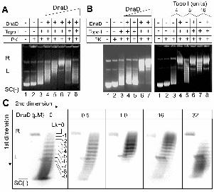
Figure 1
DnaD stimulates the topo I activity.
A. The effect of increasing concentrations of DnaD on the activity of topo I.
pBluescript (18 nM) was incubated with increasing concentrations of DnaD (0.5, 1, 8, 16
and 32 µM; lanes 4-8, respectively) and then treated sequentially with topo I (4 units) and
PK prior to electrophoresis. Controls are shown in lanes 1-3, as indicated. Relaxed, linear
and negatively supercoiled plasmid is indicated by R, L and SC(-), respectively.
Increased relaxation of the SC(-) plasmid is observed up to 8 µM DnaD (lanes 4-6),
manifested by the increasing appearance of R. At higher concentrations of DnaD, topo I
is inhibited as manifested by the appearance of supercoils with higher electrophoretic
mobility in lanes 7 and 8.
B. The left panel shows that the initial stimulation of the topo I relaxation activity is
evident at 1 and 10 µM DnaD (lanes 6 and 7, respectively) whereas at 0.1 and 0.01 µM
DnaD there was no observable effect (lanes 4 and 5, respectively). Controls are shown in
lanes 1-3, as indicated. The right panel compares the relaxation of pBluescript by
increasing concentrations of topo I (4, 8 and 16 units) in the presence and absence of 10
µM DnaD, as indicated. 16 units of topo I was required to relax pBluescript with
approximately the same efficiency as 4 units in the presence of 10 µM DnaD (compare
lanes 4 and 7). Controls are shown in lanes 1-3, as indicated.
C. 2D gel electrophoresis verifying that DnaD untwists duplex DNA.
The gels from left to right show 2D gels of the products from the reactions in lanes 3, 4,
5, 7 and 8 of panel A, respectively. The positions of the R, L and SC(-) plasmids are
17

indicated. Topoisomers with progressively increasing negative Lk are apparent from top
to bottom. Only topoisomers with Lk values from 0 to -9 are indicated for clarity.
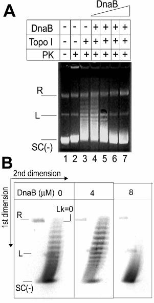
Figure 2
DnaB does not alter the supehelical properties of DNA.
A. The effect of increasing concentrations of DnaB on the activity of topo I.
pBluescript (18 nM) was incubated with increasing concentrations of DnaB (4, 8, 16 and
26.7 µM; lanes 4-7, respectively) and then treated with topo I (4 units). The reaction was
terminated and the mixture was treated with PK prior to electrophoresis. Controls are
shown in lanes 1-3, as indicated. Relaxed, linear and negatively supercoiled plasmid is
indicated by R, L and SC(-), respectively. Marginal non-specific stimulation is barely
visible at 4 µM DnaB (lane 4) and inhibition is observed at 8, 16 and 26.7 µM DnaB
(lanes 5, 6 and 7). Overall there is no significant effect on the topo I activity.
B. 2D gel electrophoresis verifying that DnaB does not untwist duplex DNA.
2D gels showing reactions in the absence (left) and presence of 4 and 8 µM DnaB
(middle and right, respectively), equivalent to the products of the reactions in lanes 3, 4
and 5 from panel A. The positions of the R, L and SC(-) plasmids are indicated together
with the position of the Lk=0 topoisomer. Topoisomers with progressively increasing
negative Lk are apparent from top to bottom.
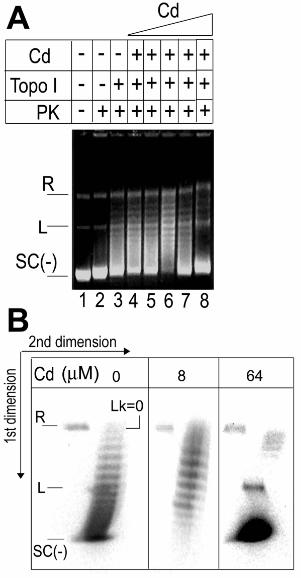
Figure 3
The Cd of DnaD cannot untwist duplex DNA.
A. The effect of increasing concentrations of Cd on the activity of topo I.
18

pBluescript (18 nM) was incubated with increasing concentrations of Cd (0.5, 1, 8, 16
and 64 µM; lanes 4-8, respectively) and then treated sequentially with topo I (4 units) and
PK prior to electrophoresis. Controls are shown in lanes 1-3, as indicated. No effect on
topo I activity was observed at 0.5 and 1 µM Cd (compare lanes 3, 4 and 5) but a slight
stimulation of relaxation was observed at 16 µM Cd (compare lanes 3 and 6). At higher
Cd concentrations (16 and 64 µM) inhibition of relaxation was observed.
B. 2D gel electrophoresis verifying that Cd inhibits the topo I at high concentration.
2D gels showing reactions in the absence (left) and presence of 8 and 64 µM Cd (middle
and right), equivalent to the products of the reactions in lanes 3, 6 and 8 from panel A.
The positions of the R, L and SC(-) plasmids are indicated together with the position of
the Lk=0 topoisomer. Topoisomers with progressively increasing negative Lk are
apparent from top to bottom.
Figure 4
DnaB inhibits untwisting by DnaD.
A. The effect of DnaB (8 µM) on the twisting activity of increasing amounts of DnaD
(0.5-33 µM), as indicated. Appropriate mixtures of proteins were incubated with
pBluescript (18 nM), sequentially treated with topo I and PK before electrophoresis. The
positions of fully relaxed, linear and supercoiled plasmids are indicated by R, L and SC,
respectively. Increasing amounts of DnaD and treatment with topo I resulted in gradual
positive increases of Lk by duplex untwisting, manifested as a gradual relaxation of the
negatively supercoiled plasmid (lanes 4, 6 and 8) followed by steric inhibition of topo I
activity at high concentrations of DnaD (lanes 10 and 12). Under identical conditions but
19

in the presence of 8 µM DnaB, the untwisting activity of DnaD was totally inhibited, as
manifested by the presence of mainly supercoiled plasmid throughout (lanes 5, 7, 9, 11
and 13). Controls are shown in lanes 1-3, as indicated.
B. The same experiment as the one shown in panel A was carried out with 4 µM DnaB at
0.5, 1 and 9.7 µM DnaD, as indicated. Increasing the concentrations of DnaD (0.5-1 µM)
in the presence of 4 µM DnaB resulted in a slight enhancement of topo I relaxation the
presence of equivalent concentrations of DnaD alone (lanes 3-6).
Figure 5
A schematic diagram illustrating conversion of DNA from plectonemic to paranemic by
DnaD-concentration dependent untwisting. DNA remodeling by DnaD is the sum of
separate DNA-independent and DNA-dependent oligomerisation activities residing on its
Nd and Cd domains, respectively, plus a DNA-binding activity on the Cd domain (15).
Based upon a DNA-remodelling model proposed before (15), we suggest that initial
binding of DnaD to DNA via its Cd domain causes only minor untwisting of the duplex
but as the scaffold forms at higher concentrations, via oligomerisation interactions
mediated by the Nd domain, gradual untwisting of the duplex becomes significant and the
duplex is eventually converted to a paranemic form. Only three helical turns are shown
for simplicity and although untwisting is depicted to open up individual helical turns, it is
likely that it will be distributed throughout all the helical turns in the circular plasmid
molecule as the DnaD scaffold grows.
20

REFERENCES
1. Altuvia, S., Almiron, M., Huisman, G., Kolter, R. & Storz, G. (1994). The dps
promoter is activated by OxyR during growth and by IHF and sigma S in stationary
phase. Mol. Microbiol., 13: 265-272.
2. Bae, S.H., Yun, S.H., Sun, D., Lim, H.M. & Choi, B.S. (2006). Structural and dynamic
basis of a supercoiling-responsive DNA element. Nucleic Acids Res., 34: 254-261.
3. Bruand, C., Farache, M., McGovern, S., Ehrlich, S.D. & Polard, P. (2001). DnaB,
DnaD and DnaI proteins are components of the Bacillus subtilis replication restart
primosome. Mol. Microbiol., 42, 245-255.
4. Bruand, C., Velten, M., McGovern, S., Marsin, S., Serena, C., Ehrlich, S.D., & Polard,
P. (2005). Functional interplay between the B. subtilis DnaD and DnaB proteins essential
for initiation and re-initiation of DNA replication. Mol. Microbiol., 55: 1138-1150.
5. Carneiro, M.J.V.M., Zhang, W., Ioannou, C., Scott, D.J., Allen, S., Roberts, C.J. &
Soultanas, P. (2006). The DNA-remodelling activity of DnaD is the sum of
oligomerisation and DNA-binding activities on separate domains. Mol. Microbiol., In
Press.
6. Dame, R.T. (2005). The role of nucleoid-associated proteins in the organization and
compaction of bacterial chromatin. Mol. Microbiol., 56: 8585-870.
7. DiNardo, S., Voelkel, K.A., Sternglanz, R., Reynolds, A.E. & Wright, A. (1982). E.
coli DNA topoisomerase I mutants have compensatory mutations in DNA gyrase genes.
Cell, 31: 43-51.
8. Dorman, C.J. (1996). Flexible response: DNA supercoiling, transcription and bacterial
adaptation to environmental stress. Trends Microbiol., 4: 214-216.
21

9. Drlica, K. (1992). Control of bacterial DNA supercoiling. Mol. Microbiol., 6: 425-433.
10. Drolet, M. (2005). Growth inhibition mediated by excess negative supercoiling: the
interplay between transcription elongation, R-loop formation and DNA topology. Mol.
Microbiol., 59: 723-730.
11. Fang, L., Davey, M.J. & O’Donnell, M. (1999). Replisome assembly at oriC, the
replication origin of E. coli, reveals an explanation for initiation sites outside an origin.
Mol. Cell, 4, 542-553.
12. Higgins, N.P. (1999). Organization of the prokaryotic genome. Ed. Charlebois, R.L.,
ASM, Washington DC.
13. Hoshino, T., McKenzie, T., Schmidt, S., Tanaka, T. & Sueoka, N. (1987). Nucleotide
sequence of B. subtilis dnaB: a gene essential for DNA replication initiation and
membrane attachment. Proc. Natl. Acad. Sci. USA, 84: 653-657.
14. Hwang, D.S. & Kornberg, A. (1992). Opening of the replication origin of E. coli by
DnaA protein with protein Hu or IHF. J. Biol. Chem. 267: 23083-23086.
15. Imai, Y., Ogasawara, N., Ishigo-Oka, D., Kadoya, R., Daito, T. & Moriya, S. (2000).
Subcellular localization of Dna-initiation proteins of Bacillus subtilis: evidence that
chromosome replication begins at either edge of the nucleoids. Mol. Microbiol., 36,
1037-1048.
16. Ishigo-Oka, D., Ogasawara, N. & Moriya, S. (2001). DnaD protein of Bacillus
subtilis interacts with DnaA, the initiator protein of replication. J. Bacteriol., 183, 2148-
2150.
22

17. Kim, J., Yoshimura, S.H., Himuze, K., Ohniwa, R.L., Ishihama, A. & Takeyasu, K.
(2004). Fundamental structural units of the E. coli nucleoid revealed by atomic force
microscopy. Nucleic Acids Res., 32: 1982-1992.
18. Leonard, A.C. & Grimwade, J.E. (2005). Building a bacterial orisome: emergence of
new regulatory features for replication origin unwinding. Mol. Microbiol., 55: 978-985.
19. Marians, K.J. (2000). PriA-directed replication fork restart in Escherichia coli.
Trends Biochem. Sci., 25, 185-189.
20. Marsin, S., McGovern, S., Ehrlich, S.D., Bruand, C. & Polard, P. (2001). Early steps
of Bacillus subtilis primosome assembly. J. Biol. Chem., 276, 45818-45825.
21. Marszalek, J. & Kaguni, J.M. (1994). DnaA protein directs the binding of DnaB
protein in initiation of DNA replication in Escherichia coli. J. Biol. Chem., 269, 4883-
4890.
22. Meile, J.C., Wu, L.J., Ehrlich, S.D., Errington, J. & Noirot, P. (2006). Systematic
localization of proteins fused to the green fluorescent protein in Bacillus subtilis:
Identification of new proteins at the DNA replication factory. Proteomics, 6, In Press
(published on line).
23. Messer, W. (2002). The bacterial replication initiator DnaA. DnaA and oriC, the
bacterial mode to initiate DNA replication. FEMS Microbiol. Rev., 26: 355-374.
24. Moriya, S., Imai, Y., Hassan, A.K. & Ogasawara, N. (1999). Regulation of initiation
of Bacillus subtilis chromosome replication. Plasmid, 41, 17-29.
25. Ng, J.Y. & Marians, K.J. (1996). The ordered assembly of the phiX174-type
primosome. I. Isolation and identification of intermediate protein-DNA complexes. J.
Biol. Chem., 271, 15642-15648.
23

26. Noirot-Gross, M.F., Dervyn, E., Wu, L.J., Mervelet, P., Errington, J., Ehrlich, S.D. &
Noirot, P. (2002). An expanded view of bacterial DNA replication. Proc. Natl. Acad. Sci.
USA, 99, 8342-8347.
27. Postow, L., Hardy, C.D., Arsuaga, J. & Cozzarelli, N.R. (2004). Topological domain
structure of the E. coli chromosome. Genes Dev., 18: 1766-1779.
28. Pruss, G.J., Manes, S.H. & Drlica, K. (1982). E. coli DNA topoisomerase I mutants:
increased supercoiling is corrected by mutations near gyrase genes. Cell, 31: 35-42.
29. Raji, A., Zabel, D.J., Laufer, C.S. & Depew, R.E. (1985). Genetic analysis of
mutations that compensate for loss of E. coli DNA topoisomerase I. J. Bacteriol., 162:
1173-1179.
30. Rokop, M.E., Auchtung, J.M. & Grossman, A.D. (2004). Control of DNA replication
initiation by recruitment of an essential initiation protein to the membrane of B. subtilis.
Mol. Microbiol., 52: 1757-1767.
31. Sikder, D., Unniraman, S., Bhaduri, T. & Nagaraja, V. (2001). Functional cooperation
between topoisomerase I and single strand DNA-binding protein.
32. Soultanas, P. (2002). A functional interaction between the putative primosomal
protein DnaI and the main replicative DNA helicase DnaB in Bacillus. Nucleic Acids
Res., 30, 966-974.
33. Sueoka, N. (1998). Cell membrane and chromosome replication in B. subtilis. Progr.
Nucleic Acid Res., 59: 34-53.
34. Travers, A. & Muskhelishvili, G. (2005). DNA supercoiling – a global transcriptional
regulator for enterobacterial growth? Nat. Rev. Microbiol., 3: 157-169.
24

35. Turner, I.J., Scott, D.J., Allen, S., Roberts, C.J. & Soultanas, P. (2004). The Bacillus
subtilis DnaD protein: A putative link between DNA remodelling and initiation of DNA
replication. FEBS Letters, 577: 460-464.
36. Velten, M., McGovern, S., Marsin, S., Ehrlich, S.D., Noirot, P. & Polard, P. (2003).
A two-protein strategy for the functional loading of a cellular replicative DNA helicase.
Mol. Cell, 11, 1009-1020.
37. Wang, J.V. & Syvanen, M. (1992). DNA twist as a transcriptional sensor for
environmental changes. Mol. Microbiol., 6: 1861-1866.
38. Waring, M. (1970). Validation of the supercoils in closed circular DNA by binding of
antibiotics and drugs: Evidence for molecular models involving intercalation. J. Mol.
Biol., 54: 247-279.
39. Weinstein-Fischer, D., Elgrably-Weiss, M. & Altuvia, S. (2000). E. coli response to
hydrogen peroxide: a role for DNA supercoiling topoisomerase I and Fis. Mol.
Microbiol., 35: 14-13-1420.
40. Zhang, W., Carneiro, M.J.V.M., Turner, I.J., Allen, S., Roberts, C.J. & Soultanas, P.
(2005). The B. subtilis DnaD and DnaB are DNA remodelling proteins with different
activities. J. Mol. Biol. 351: 66-75.
25