zoology universitymicrofilms, inc., annarbor, michigan
TRANSCRIPT
This dissertation has been 64-2657microfilmed exactly as received
THOMSON, Donald Arthur, 1932-A HISTOLDGICAL STUDY AND BIOASSAY OFTHE TOXIC STRESS SECRETION OF THE BOXFISH, OSTRACION LENTIGINOSUS.
University of Hawaii, Ph.D., 1963Zoology
University Microfilms, Inc., Ann Arbor, Michigan

Copyright by
Donald Arthur Thomson
1964

A HISTOLOGICAL STUDY AND BI0~SSAY OF
THE TOXIC STlIESS SECRETION OF
THE BOXFISH, OSTRACION LENTIGINOSUS
A THES IS SUBMITTED TO THE GRADUATE SCHOOL OF THE
UNIVERSITY OF HAWAII IN PARTIAL FULFILLMENT
OF THE REQUIREMENTS FOR THE DEGREE OF
DOCTOR OF PHILOSOPHY
IN ZOOLOGY
JUNE 196)
By
Donald Arthur Thomson
Thesis Committee:
Professor Pieter Bo van Weel, ChairmanProfessor Albert H. BannerProfessor William Ao GoslineProfessor Sidney Co HsiaoAssociate Professor Judson Lo Ihrig
PLEASE NOTE: Tables and Fi~~res are not originalcopy. Some of these pages tend to"curl". Filmed in the best possibleway.
UNIVERSITY MICROFILMS, INC.

" . ,- "" .' -' ~,' . ,. " .
-.•1' •. • 0 ••••• '.. - • ,M,
• J • ..'
FRONTISPIECE
The adult boxfish, Ostracion lentigino8us
male: topfemale: bottom

PREFACE
A fire which leveled one of the buildings of theHawaii Marine Laboratory on Coconut Island, December 30,1961,destroyed all the original notes and data pertainingto this thesis collected since 1959. Therefore, some ofthe information in Chapters 5 through 13 is based uponprogress reports submitted at the end of each schoolsemester to Dro Piater B. van Weel, Chairman of theThesis Committeeo The important experiments in thesecha,pters were repeated and the entire hi stologicalinvestigation was conducted after the fire.
The University of Hawaii, through the intercession ofthe Dean of the Graduate School, Dr. Robert WQ Fiatt,awarded me a one_year grant to compensate for lossessuffered in the fire and to enable me to complete thisthesiso For this I am very gratefulo I would also liketo thank Drs. Albert H. Banner and Philip Helfrich ofthe Hawaii Marine Laboratory for their interest in thisinvestigation and their attempts to pronure funds for thi~
study. I wish to give a special acknowledgement toDr. van Weel for his foresight in requiring detailedprogress reports, a measure which prevented more seriouslosses of ~ta in the fi~e, and fo£bis guidanoe and soundjudgment during the course of this research. I amindebted to many students and faculty members, especiallyCharles Bo Alender, William Eger, David Boylan, andRalph T. Hinegardner for stimulating discussions whichled to many new ideas on how to approach this problem.
I wish to thank Mro John Marr, Director of theHonolulu Biological Laboratory, Bureau of Commercial Fisheries,and Mr. Vernon Eo Brock, former Director, for theirpermission to use the docksite facilities at Kewalo Basin.Thanks also to ~{ro Dick Holoway and his assistants of thesame laboratory for providing tuna blood and saline solution.
I am greatly indebted to my wife, Jenean, for thegra9hs, fine illustrations and the typing of the ma~uscript9
arduous tasks which I would not have accomplished WIthouther helpo

TABLE OF CONTENTS
LIST OF TABLES e 0 0 e 0 0 0 0 0 0 0 0 0 0 0 0 0 e 0 0 x
LIST OF FIGURES 0 0 0 0 0 0 0 0 0 0 0 0 0 e 0 0 0 0 0 xi.
.ABSTRACT 0 0 0 0 0 0 0 0 0 0 0 0 0 0 0 0 0 0 0 0 0 0 0 xi v
CHAPTER 1
INTRODUCTION 0 0 0 0 0 0 0 0 0 0 0 0 0 0 0 0 .. 0 0 0 0 1
CHAPTER 2
SYNOPSIS OF THE FAMILY OSTRACIONTIDAE: SYSTEMATICAND ECOLOGICAL CONSIDERATIONS 0 0 0 0 0 0 0 e 0 0 0 7
ECOLOGY OF TRUNKFISH 0 e e 0 0 0 0 0 e 0 e 0 e 0 0 e 8PROTECTIVE ADAPTATIONS OF PLECTOGNATHS IN GENERAL 0 09
CHAPTER 3
THE HAVIA I IAN TRU1~1CF ISHES 0 0 • 0 • 0 0 e 0 0 • • 0 • 0 11
CHAPTER l.J
COLLECTION AND PREPARATIVE TECHNIQUES 0 • 0 e 0 0 • 0 15
COLLECTION OF TRUNKFISH e 0 e 0 0 • 0 • 0 0 " e
COLLECTION OF BOXFISH MUCOUS SECRETIONS 0 e e e
PRELIMINARY ISOLATION OF OSTRACIN e 0 eo. 0 0
• 0
o 0
o e
151822
CHAPTER 5
DEVELOPnffiNT OF A BIOASSAY FOR OSTRACIN e e e .. . . o 0 23
TOXICITY OF OSTRACIN TO AQUATIC ORGANISMS 0 0 e 0 0 24COELENTERATA 0 .. 0 0 • 0 • • eo" e 0 0 .. • 0 0 25PLATYHELMINTHES • 0 • 0 0 0 0 0 0 0 0 0 e .. .. 0 0 28MOLLUSCA 0 0 0 0 0 0 0 0 0 0 0 0 0 e 0 e 0 e 0 0 29ECHINODERMATA 0 0 e e eo. 0 0 0 0 0 0 0 0 0 0 0 29CRUSTACEA 0 0 • 0 0 0 0 0 eo.. 0 0 0 • 0 e e 0 0 31PISCES 0 0 0 • 0 e e 0 0 0 0 0 • 0 .. .. 0 .. 0 0 0 31
Effect of Body Size on Death Time • 0 • 0 0 0 32C~itical Exposure Period of Fishto Ostracin • 0 0 0 0 • 0 0 • 0 0 eo. 0 • 0 33
Response of Different Species to Ostracin .... 34Effect of Salinity on Toxic Actionof Ostracin 0 0 0 0 0 • .. .. • • 0 e e 0 0 0 0 35
TOXICITY OF OSTRACIN TO T'E PEARLFISH •
000TOXICITY OF OSTRACIN TO BOXFISH
iv
• 000
o 0 0 "
01)0
I) 0 41
42
TABLE OF CONTENTS (continued)
CHAPTER 5 (continued)
TOXICITY OF OSTRACIN TO mUTE MICE .. 0 .. .. .. .. .. .. 0 43Symtoms 0 .. .. 0 .. .. 0 0 0 .. .. '0 .. " .. 0 .. 0 " 0 .. ~Lethal dose of ostraci~ .. " 0 0 0 0 .. .. 0 .. .. 0 0 44Effect of injection media .. 0 " .. 0 0 0 0 .. 0 .. .. 459ontrol injections " .. .. .. 0 .. .. .. 0 0 .. 0 0 0 0 45
CHOICE OF ASSAY FISH ." • .. .. .. .. .. .. 0 .. .. .. .. .. 0 0
DOSE-RESPONSE DATA .. .. .. 0 .. .. .. .. .. .. .. .. .. • 0 0 •
Crude ostracin .. .. .. • .. .. .. .. .. • • .. .. .. .. .. ..Semi~pure ostracin .. 0 • .. .. .. 0 0 .. • " .. " .. ..
CHAPTER 6
ESTABLISHlffiNT OF A STANDARD BIOASSAY FOR OSTRACIN .. .. 0
THE STANDARD DOSE • .. 0 0 .. .. .. .. • .. • .. .. .. .. .. .. .. 50Advantages and Disadva.ntages of Fish Bioassay .... 53SJ-'1lIools and Defini tions .... 0 0 0 0 .. 0 .. 0 .. 0 .. 54
CHAPTER 7
FLUCTUATIONS IN OSTRACIN OCCURRENCE IN BOXFISHSECRETIONS; ECOLOGICAL CONSIDERATIONS 55
MATERIALS AND METHODS .. .. .. .. .. .. .. .. 0 .. 0 .. .. .. 0 .. 55
GENERAL TOXICITY .. .. 0 .. 0 0 .. .. .. .. .. .. .. 0 • .. .. 0 56
SIZE AND TOXICITY .. .. .. .. .. .. .. .. " .. 0 .. .. .. .. 0 ..
GOO 6 e 0 0 0 • eSEASONAL EFFECTS ON TOXICITY .. ..
EFFECT OF LOCALITY ON TOXICITY .. .. • Q • 0 • 0 0 • 0
57
58
59
o • 0 0 0 59
62
o .. .. 59
o e " 59
.. 0 0
.. .. .
.. .. ..
.. 0 •o ~ 0
o 000
o 0 0 0 0 0 0 • G 8 0 0DISCUSSION
EFFECT OF AGE AND SEX ON TOXICITY .. .. .. 0
EFFECT OF METHOD OF CAPTURE ON TOXICITY 0
TOXICITY OF BOXFISH I~ CAPTIVITY 0
CHAPTER 8
GENERAL PHYSICAL AND CHEMICAL PROPERTIES OF OSTRACIN " 0 64
STABILITY OF AQUEOUS OSTRACIN SOLUTIONS .. 0 0 0 0 0 .. 64
HEAT STABILITY OF OSTRACIN 000 0 • ~ • 0 0 0 0 • 0 66
v

o 0 0 0 0 0 0 75
767680
EFFECT OF OSTRACIN ON RED BLOOD CELLS 0 • 0 0 • • 0 •
Qualitative Test for Hemolysis and Agglutination 0
Quantitative Hemolytic Test for Ostracin •• 0 0 0
TABLE OF CONTENTS (continued)
CHAPTER 8 (continued)
ACID-ALKALINE STABILITY OF OSTRACIN • 0 0 0 0 0 0 0 0 67
POSSIBLE BACTERIAL DECOMPOSITION OF OSTRACIN 0 0 0 0 0 68
SOLUBILITY OF OSTRACIN 0 0 0 0 0 0 0 0 • 0 0 0 0 0 0 0 73
DIALYSIS OF OSTRACIN 0 0 0 0 • • 0 0 0 0 • • 0 • 0 0 0 74
FOAMING OF OSTRACIN SOLUTIONS 0 • 0 0 0 0 0 0 0 0 0 0 74
CHAPTER 9
SPECIAL 'CHARACTERISTICS OF OSTRACIN 0 0 0 0
Results • 0 • • • 0 • 0 • • • • 0 0 0 • 0 0 0 • 0
.0.0. ~ 0 • 0 • • 0 0 • 0 • 0 0 0 • • 0DISCUSSION
PRECIPITATION OF OSTRACIN BY CHOLESTEROL 0 • • 0 0 0 • 82Materials and Methods 0 •• 0 0 •• 0 • 0 • 0 0 0 82
83
84
CHAPTER 10
SUlThaRY OF EVIDENCE FOR THE STEROID SAPONINNATURE OF OSTRACIN 0 0 0 • • 0 0 • • • 0 • 0 0 0 • 0 • 86
e 0 0 • 0 0 0 0
.. . .
CHAPTER 11
PURIFICATION TECHNIQUES FOR OSTRACIN •
PRECIPITATION WITH CHOLESTEROL 0 .. 0 .00 o 0
o 90
o 90
CHRO~aTOGRAP}IT • 0 • • • .. • 0 0 0 0 0 0 • • • 0 • 0 0 90Foam Chromatography • 0 0 0 0 0 0 0 0 0 .. 0 • 0" 90Paper Chromatography • 0 • .. 0 0 0 0 0 0 0 0 0 0 0 90
EXTRACTION WITH ORGANIC SOLVENTS 0 0 0 0 0 0 0 0 0 0 0 91Acetone • 0 0 0 0 • • • 0 0 • • 0 0 0 0 0 • 0 0" 91Chloroform 0 • 0 • 0 0 0 0 • 0 • 0 .. 0 0 0 0 • 0 0 92
CENTRIFUGATION AND DIALYSIS 0
PREPAIATION OF OSTRACIGENIN 0
000
000
000
000
000
o .. 0
000
000
92
94
CHAPTER 12
TOXIC SECRETIONS OF OTHER PLECTOGNATHS Q 0 e 0 000 95
vi
TABLE OF CONTENTS (continued)
CHAPTER 12 (continued)
o 000 0 0 0 0 0 0 0 0 0 0 0• 0 .00 0
TRUNKFISH ..
PUFFERS ..
.. .. .. 0 o 0 .00 .. 0 .. .. .. .. .. o 0 0 0 95
97
000 • • 000 0
o Q 0 0 Q 0 000
o 0 • 0 0 • 000
CHAPTER I)
SITES OF OSTRACIN SECRETIONS IN THE BOXFISH ..
ASSAY OF ORAL AND SKIN SEClillTIONS ..1mterials and Methods ..........Results .. .. 0 .. .. .. .. .. .. .. .. ..
000 • 99
100100100
EXTRACTION OF OSTRACIN FROM BOXFISH TISSUES .. .. .. .." 102Materials and Methods " " .... " " .. .. 102Results " " .. .. .. .. .. 105
DISCUSSION
CHAPTER 14
o 000 0 0 Q 0 0 0 0 0 0 0 0 0 0 000 0 106
HISTOLOGY OF THE PRESUMED POISONOUS GLANDS OF THE BOXFISH:MATERIALS AND 1ffiTHODS " " " " .108
COLLECTION AND FIXATION OF TISSUES • .. • " .. o 0 0 • • 108
000 ~ 0 0 0
DEHYDRATING, CLEARING, mmEDDING, SECTIONINGAND MOUNTING • • • .. " • .. " • • .. " .. • ..
STAINS AND STAINING .. .. .. .. .. .. ..
.. " .. .. .... 0 •
109
110
CHAPTER 15
HISTOLOGY OF BOXFISH SKIN .. 0 .. 0 0 o 0 0 0 0 e e 00. 112
o 0 0 0 0 0 0 e 0 0GENERAL MORPHOLOGY OF THE SKIN ..
STRUCTURE OF BOXFISH EPIDERMIS 0 .. .. o 0 e 0 o 000
II)
115
122o 000
FomaTION OF POISON SKIN GLANDS " " 0 " " .. " 0 .. .. 0 122
Trill DISTRIBUTION OF ~mcous AND CLUB CELLS INTHE EPIDERMIS " " .. .. 0 " .. .. .. 0 " .. .. 0 ..
STAINING AFFINITIES OF ~fUCOUS AND CLUB CELLS .. .. .." 126Harris Hematoxylin and Triosin (or Eosin) .... "" 126Mallory's Triple Stain " 0 • 0 .. 127Gomorits Aldehyde Fuchsin and TrichromeCounterstain " .. .. • .. .. .. • • • • • .. 0 0 .. .... 129
Periodic Acid LeucoflJchsin Schiff! s Method(PAS) .. .. .. 0 0 0 .. .. .. .. 0 " .. 0 0 .. .. .. 0 .. 0 " 1)1
vii
TABLE OF CONTENTS (continued)
CHAPTER 15 (continued)
Toluidine Blue 0 0 ~ 0 .. 131Dresbach's Mucicarmine Metbod 0 131Sudan Black B .. .. .. .. .. .. 0 .. .. .. .. .. 0 .. .. .. 0.. 133
DISCUSSION
CHAPTER 16
o 0 0 0 0 0 0 ~ 0 0 e 0 000 • • e 000 133
HISTOLOGY OF THE LABIAL GLANDS .. .. .. .. .. e .. .. .. 0 .. .. 0 135
GENERAL MORPHOLOGY ..... o 0 0 0 0 0 00. 006 o .. 0 .. 135
137139
139o 000DISTRIBUTION OF THE LABIAL GLANDS .. .. 0 .. 0 0
HISTOLOGICAL STRUCTURE OF THE LABIAL VILLI .. .. .. .. .. .. 135Harris Hematoxylin and Triosin (or Eosin) ...... 0 137Mallory' 8 Triple Stain .. "'0 .. '.. .. .. ,,'.. .. 0 0 0 0 137Gomor1 t s Aldebyde Fuchsin and Trichrome .... 0 .... 137
CounterstainPeriodic Acid Leucofuchsin Schiff!s Method(PAS) • • • eo. 0 0 0 e 0 0 • • • 0 • 0 • 0 0 •
Toluidine Blue ........ 0 0 0 .. 0 0 0 0 0 0 0 00 0
STRUCTURE OF THE LIP SKIN AND PROBABLE ORIGINOF THE LABIAL G~NDS .. .. 0 .. ~ .. .. 0 .. .. .. .. • $ CD 0 "4".l .l
DISCUSSION .. .. .0. • 0 0 • 0 0 0 0 • • • e e 0 0 e 144
CHAPTER 17
HISTOLOGICAL AND HISTOCHEMICAL EVIDENCEOF OSTRACIN SECRETION .. .. .. .. .. .. 0 .. o .. .. 000 CD 0 148
HISTOLOGICAL CHANGES IN THE LABIAL AND SKIN GLANDSOF CAPTIVE BOXFISH .. .. .. .. 0 .. .. .. .. .. .. .. .. .. .. .... 148
Epidermis .. .. .. .. .. .. .. .. .. .. .. .. .. .. .. .. .. .. .. 0 149Labial Glands .. .. 0 .. .. .. .. 0 .. .. .. .. .. .. .. .. .... 151
GLANDULAR HISTOLOGY OF STRESSED VERSUSNON-STRESSED. BOXFISH 0 0 0 153
Epidermi s ". .. 0 0 • .. .. .. .. .. .. .. 0 0 " .. .. 0 0.. 156Labial Glands .. .. ·0 .. .. 0'" .. .. .. .. .. .. 0 .. .. 156
SPOT TESTS OF OSTRACIN .. .. .. .. .. .. .. 0 " .. .. .. .. 158Materials and Methods .. .. .. .. .. 0 .. .. .. .. 0 .. 158Resul ts .. 0 0 .. .. .. ..'.. .. 0 .. 0 ... ' 0 ......... 0 0 159
viii

TABLE OF CONTENTS (continued)
ClaPTER 17 (continued)
Mallory's Triple Sta,in " 159Gomorils Aldehyde Fuchsin-Trichrome Stain 161PAS Method 0 0 0 0· 161PAS + Toluidine Blue .. 0 0 0 .. .. .. .. • • 0 .. 0 161Mallory'1 s Triple Stain .. .. .. .. • .. .. .. • .. 0 0 162Gomorils Aldehyde Fuchsin Trichrome Stain.. .. 162Toluidine Blue .. 0 • .. .. .. .. .. 0 .. .. .. .. .. .. .. .. .. 162
DISCUSSION
CHAPTER 18
.. .. .. o 0 e 0 a 0 0 0 0 G • 0 Q 0 GOO 006 163
COMPARATIVE HISTOLOGY OF THE COr&ISH AND OTHERPLECTOGNATHS 165
EPIDERMIS OF THE CmVFISH .. .. .. .. .. .. • .. .. .. .. 165Staining Properties of Mucous and Club Cellsof Cowfish 0166
LABIAL GLANDS OF THE COI~ISH .. .. .. .. .. .. .. .. .. .. .. .. .. 166
EPIDERMIS OF OTHER PLECTOGNATHS .......... 0 0 .. .. .... 170
DISCUSSION 0 0 171
o G • 0 • 0 0 0 0 • 0 0
CHAPTER 19
DISCUSSION AND CONCLUSIONS .. • ..
OSTRACIN, THE STEROID SAPONIN OF THE BOXFISH ..
SIGNIFICANCE OF THE SKIN AND LABIAL GLANDS TOOSTRACIN SECRETION .. .. .. .. .. .. .. .. .. .. .. .. ..
CHAPTER 20
o • e 0
eo. e 178
SUlillfARY .. .. o e 0 0 0 0 • • o .. e 000 Q 000 • • 0 • 0 184
LITERATUP~ CITED .. .. .. .. .. .. .. .. .. 0 .. .. .. 0 0 .. .. .. .... 189
ix
TABLE 10
TABLE 110
TABLE 1110
LIST OF TABLES
Toxicity of Ostracin to Some Aquatic Animals oo 26
Ostraciu Secretion by Captive Boxfishas Estimated by the Standard Bioassay 0 0 0 0 61
The Stability of Crude Ostracin SolutionsStored Frozen and at Room Temperatures 0 0 0 65
TABLE IV. The Effect of pH on the Stabilityof Ostracin 0 0 • • 0 • • • • • 0 o 0 o • • 69
TABLE V. Comparison of the Detoxification Rateof Semi_Pure and Crude OstracinaDistilled Water Solutions • 0 • 0 0 0 o • 0 70
TABLE Vlo Detoxification of Crude and PureOstracin in Sea Water Solutions • e 0 0 0 0 72
TABLE VlIo Ostracin Hemolysis. and Agglutination ofthe Erythrocytes of Some Vertebrates 0 • 0 0 78
TABLE VIII. Comparison of the General Propertiesof Ostracin with Digitonin andSaponin (Merck) 0 0 • • • 0 • 0 0 • 0 o • 0 88
TABLE IX.
TABLE X.
TABLE XI.
TABLE XII.
Ichthyotoxicity of the Oral Secretionsof Boxfish 0 0 • • 0 • 0 • 0 • • • 0 0 • 0 0 101
Ichthyotoxicity of Boxfish SkinSecretions • 0 0 0 0 .. • • • 0 0 • 0 • 0 0 0 103
Staining Reactions of Secretory Productsin. Boxfish Epidermal Gland. CellsAfter Bouin1s Fixation 0 • 0 0 0 0 0 0 0 0 0 146
Relative Numbers of Mucous and Club Cellsin the Caudal Epidermis of Boxfish Kept inCaptivity and Freshly Captured 0 0 0 • 0 O. 152
TABLE XIII. Comparison of Numbers of Skin GlandCells in Boxfish Before and After Stress o 0 157
TABLE XIVo Comparison of the General Propertiesof Ostracin with Other "Saponins" fromMarine Organisms 0 ••• 0 •• 0 0 0 0 • 0 0 176
x
LIST OF FIGURES
FRONTISPIECE.. The adult boxfish, Ostracion lentiginosuso
FIGURE 10 Juvenile boxfish 0 o 0 0 0 o 0 ~ a 0 0 0 o 19
FIGURE 2 0
FIGURE 30
Foamy mucous secretions of a distressedboxfish out of water 0 " .. 0 .... 0 ...... 0 19
Collection of the entire stress secretionsof female boxfish ....... 0 0 0 0 .. 0 0 0 20
FIGURE 40
FIGURE 50
Effect of crude ostracin on sailfinmollies acclimated to differentsalinities .. 0 0 .. 0 0 .. 0 ........ 0
Effect of crude ostracin on survivaltime of aholeholes acclimated todifferent salinities. 0 0 .......
.. 0
.. 0
37
39
FIGURE 60 Dose-response curve of crude ostracinon sailfin mollies in sea water .... 0 .. .. 51
FIGURE 70 Dose-response curve of semi-pureostracin on sailfin mollies insea water .. • .. .. .. • 0 • .. • 0 .. • • • • 51
FIGURE 80 Erythrocytes of skipjack tuna(Katsuwonis pelamis) before additionof ostracin 0 0 • 0 .. 0 • 0 0 • 0 .. 0 o 0 79
FIGURE 90
FIGURE 100
FIGURE 110
FIGURE 12 0
FIGURE 130
FIGURE 140
Same erythrocytes seconds after additionof ostracin • 0 .. 0 .. 0 0 0 .. 0 0 0 .. .... 79
Hemolysis of skipjack tunaerythrocytes by ostracin measured witha spectrocolorimeter .. 0 .. 0 0 .. .. 0 0 0 .. 81
Preliminary purifica,tion procedurefor ostraci n .. 0 .. ..'.. .. .. .. • 0 0 0 .. 0 0 93
Surface view of mid-later~l carapa~e
of boxfisb .. 0 ........ 0 0 .. 0 ...... 0 .. 0 114
Surface view of lateral carapacebehind pectoral fin showing mucoidcoating .. 0 .. .. 0 .. .. .. .. .. .. 0 .. .. .. .... 114
Mucous cell in the process ofextrusion .......... 0 .. 0 ...... 0 0 0 .... 117
xi
FIGURE 150
FIGURE 160
FIGURE 17 ..
FIGURE 180
LIST OF FIGURES (continued)
Stage I club cell ........ 0 .. 0 ........ 0 117
Stage II club cell 0 0 .. 0 0 .. 0 ...... 0 0 119
Stage III club cell showing formationof the apical cap .. .. 0 ... 0 0 0 .. .. .. 0 119
Stage IV club cell showing the homogenou~
colloidal secretion and flattenednuclei .. 0 • • .. • .. 0 .. 0 0 0 .. 0 .. .. 0 121
Early stage V club cell not yet in theprocess of involution .. 0 • .. • • 0 • .... 121
o • 0 0
FIGURE 19 ..
FIGURE 2O ..
FIGURE 210
FIGURE 22 0
FIGURE 230
FIGUHE 240
FIGURE 250
Drawing of a caudal skin section ofQo lentigingsus .. .. 0 0 .. .. • • .. •
Club cell glands in the caudal skinof O. lentiginosus • • .. .. .. • .. • •
Club cell gland showing pseud.oduct ..
Harris hem::ttoxylin-triosin sectionof caudal skin of Qo )entiginoBus ..
hmlloryis triple stain section ofth~ ~audal skin • .. .. .. • • • • 0 •
Mallory.s triple stain caudal skinsection showing mid-epidermisextrusion of a stage III club cell
.. .. ..
• •
o • ..
• 0 0
.. . ..
123
124
125
0128
128
130
FIGURE 260
FIGURE 27 ..
FIGURE 280
FIGURE 290
Malloryts triple stain caudal skin sectionshowing extrusion by stages IV and Vclub cells •• 0 0 0 0 0 .. 130
Caudal skin section stained with G<>mori1saldehyde fuchsin-trichrome stain • .. .. 0 .. 132
Caudal skin section stained withPAS reagent • ,- 0 • 0 • .. .. .. .. .. .. .. 0 .. 0 132
Drawing of sagittal section of the lipsof a boxfish showing the position ofthe labial villi glands .. 0 .. 0 .. .. .. .... 136
FIGURE 300 Labial glands stained with Mallory.striple stain ..... 0 .......... 0 .... 0 ..
xii
138
FIGURE 310
FIGURE 32 0
FIGUHE 330
FIGURE 340
FIGUHE 350
FIGURE 36.
FIGURES37 and 38.
LIST OF FIGURES (continued)
Labial glands stained with'GomoriBsaldehyde fuchsin_trichrome stain showingthe positive aldehyde fuchsin granules ... 138
Labial villi stained with the PAS reagent. 138
Sagittal section of the lips showinglabial pocket glands. and their pseudo-ducts .. • .. .. • .. .. .. • .. .. .. .. 0 .. .. .. 0 140
Cross section of the lips showing thelabial pocket glands .. ., 0 .. 0 ... 0 .. 0 140
Drawing of a cross section of the mouththrough the dorsal teeth showing thearrangement of labial villi aroundthe teeth • 0 .. 0 .. .. • • • • .. .. .. .. 0 142
Lip skin showing mucous and club cellsamong the thick layer of epidermal cellso 0143
Labial epidermal folds showing the transitionfrom skin gland cells to labial cells •••143
FIGURE 39" Skin of a starved boxfish(32 days in captivity) .... 0 •• o .0" 150
FIGURE 40 0 Labial pocket glands of a starved.boxfish (48 days in captivity) ... o .. ..
FIGURE 41.
FIGURE 42 0
FIGUHE 43.
FIGUilli 440
FIGURE 45 ...
FIGUllli 460
FIGURE 470
Labial pocket glands of the same fishstained with Gomori l s aldehyde fuchsin_trichrome .. • • • .. .. • • .. .. • .. .. 0 .... 154
~fucoid coating peeled from the skinof a boxfish killed in Bouin l s fluid ...... 160
Caudal skin of the cowfish L. fornasin~o 0 167
Cowfish skin stained with Mallory'striple stain .. 0 .. .. .. 0 • • .. .. • • .. .. .. 168
Cowfish skin stained with GomoriBsaldehyde fuchsin_trichrome stain .. .. .. .. .. 168
Cowfish skin stained with the PAS reagent. 169
Cowfish skin stained orthochromaticallywith Toluidine blue 0 ......... 0 ...... 0 169
xiii

A.BSTRACT
An icbthyotoxin was isolated and partially purified
from the foamy mucous secretions collected from the skin
and mouth of distressed boxfish p Ostracion lentiginosuso
The thermostable, non.dialyzable toxin foamed profusely in
aqueous solutions and was highly toxic to fisb and many
other aquatic organisms at concentrations as low as 1 0 0 ppm o
In addition, this toxin caused agglutination and hemolysis
of vertebrate erythrocytes and formed a water... insoluble,
non.toxic complex with cholesterolo Thus, the toxin secreted
by the boxfisb was tentatively identified as a steroid
saponin and the name "ostracin li was proposed, marking the
first known occurrence of saponins in vertebrateso
A histological investigation of the integument and
mouth of boxfish revealed three types of gland cells:
mucous cells in the superficial layers of the epidermis;
so-called "club cells li in tbe intermediate and basal
layers, characterized by mid.epidermal extrusion of their
secretory products; and "labial cells" lining the labial
villi and forming compound glands in the buccal cavityo
Developmental, or restitutional stages of the club
cells were described and some staining prope~ties of their
homogenous and granular secretions were determined o Clusters
of these cells formed primitive multicellular epidermal
glands characteristic of those found in venomous fishes,
and it was proposed that these club cell glands were the
xiv

source of ostracin secretion in the skin o
Ostracin could not be extracted from the skin of dead
boxfish p although large amounts (100 mg of semi~pure toxin)
could be readily collected in the mucous secretions of
living p but distressed boxfish o Furthermore p ostracin
was toxic to the boxfish itself and caused agglutination
and hemolysis of boxfish erythrocytes in vitro, suggesting
that ostracin was activated during p or just prior to,
extrusion from the gland cello
The ichthyotoxicity of the foamy oral mucus of distressed
boxfish was attributed to secretions of the labial glands
in the buccal cavity although these oral exudations did
not contribute significantly to the total toxicity of the
stress secretions. The chemical identity of the oral and
skin toxins was not determined.
A comparative histological study of the cowfish p
Lactoria fornasini, a closely related species that also
secreted a hemolytic ichthyotoxin with its mucus, corroborated
the results obtained with Qo lentiginosuso
xv
INTRODUCTION
Poisonous fish have been considered as belonging
to one of two categories: (1) fish that are only
poisonous when eaten and (2) fish that have venomous
spineB or stingso The first category m~y be divided
into t~o groups: fish witb poisons believed to be of
exogenous origin, i0609 the ciguatera toxinp which is
of regional occu~reoce io the tFopicsp may be found
in many widely divergent species of bony fish and is
thought to be of environmental origin; and fish with
poisons believed to be endogenous, ioeo, the puffer.fishes
which naturally contain toxins in their flesh, viscera
and skin that are believed to be metabolically pro-
duced by the fish themselveso The functional signifi.
oance of sucb toxins is unknown.
The seoond category includes all sOacalled venomous
fishes suoh as the stonefish (SYR!Dceia), weeverfish
jT~achinus), OF lionfiGh (Pt~roi~) that have venom sacs
at the base of their spines or venomous skin glands
assooiated with spineso Such poisonous spines are
clearly evolutionary speciali2ations and are assumed
to have defensive fUDctionso
A possible third category of poisonous fishes,
those that seorete substanoes into the water that
are poisonous to other fish, has long been neglectedo
2
Recently Clark and Gobar (1953) reported that a
Red Sea boxfish (Trunkfish family: Ostraciontidae)
seemed to secrete a poison into the ~ter killing
other fiSheso The authors write:
mIt is, however. common experience at theBiological station of Al Ghardaqa that it is~engerous to put boxfishes into small aguariumtanks containing other fishes, as this oftenleads to the death of msny of tha fish inhabaIt&ot8 of those aquaria. The boxfishes seemto pass some poisonous secretion into the water,but the exact nature of the problem stillawaits invGatigationo=
MOEe zecently Brock (1955) reported that the Hawaiian
boxfish, Ostracion lentiginosus appeared to produce a
substance poisonous to fishes. Brock noted that only
boxfish under stress seemed to produoe this poison &s
boxfish acclimatized to aqnarium conditions did not
poison the aquarium fish inhabitants. Only newly
introduoed, highly excited boxfish secreted this
poisonous substance.
Tropical saltwater fieh hobbyists have probably
become a~ra of the poisaDoua cheracter of boxfiBhes
through exp~rienceo Straughmn (1959) noted that the
Atlantic spotted trunkfish (probably Laotophry~
bicaudalis, au1hor does not give species) apparently
gave off a highly poisonous Bubstance when disturbed
and cautioned aquariets on the use of thig fish in a
merine aquariumo
Poisonous secretions by distreBsed fish apparently

:3
are not confined to the trnnkfisheso 11hitley (1957)
reports a similar phenomenon in a fFOgfish, a distantly
related species (family Batrachoididae; frogfish =toadfish):
=Sir Edward Hallstrom has informed me that alooal frogfish is exhibited at Taronga ParkAquarium, Sydney, where they have also had speci ....mens of a more ornamental New Guinea species;but these fishes squirt out a liquid or slimewhich fouls the water and kills other fish.Some speeies have a pore in the pectoral axilor iarmpit i , the function of which is unknown,thongh it resembles the poisonous gland ofcatfisheso t2
Some pufferQfishes, fish closely related to trunk....
fishes, apparently prodnce toxic stress secretions.
During preliminary investigations of boxfish secretions,
it ~9 ecoident!y disoo~e~ed th~t the mucous secretions
of a pUffer.fisb, Arothron hispiduB (family Tetraodontidae)
were highly toxio when injected into fish and miceo
However, unlike boxfish secretions, puffer seoretions
were not poisonous to fish immersed in seB water oon~
taining these secretions.
FUrther investigation showed that two other puffers o
Diodon hystxix; (family Diodontidae) and Canthigaster
rivulatu8 (family Canthigasteridae) also produoed
mucous secretions that had a similar toxic effect as
those of !~ hispiduso It should be emphasized that these
puffers contain a potent endotoxin in their flesh 9
viscere, and Skin and the relationship between this
endotoxin Bnd the toxic mucous secretions requires
clarifioatioDo
The faot that fish under strese prodnoe substanc~~
that are not produced under normal oonditions has
long beeD recognized, although such secretions are
not neoessarily toxico Von Frisch (1941) showad that
in minnows an a!arrn remotion WB8 induced by substances
produced by distressed minnows or en extract of minnow
~inso Fn~the~orep Skinner at alo (1962) and Verheijen
(1962) reported species-specific alarm substances from
the top smelt (Atherinops affinis) and the freshwater
creek chub (SemotiluB atromaculatus)e More recently
Tester (1963) found that hungry sharks could detect a
scent released by alarmed but uninjured prey fish and
postulated that distressed fish produced a substance
attractant to sharks o
The trnnkfishes, because of their toxic stress
secretions, provide extreme examples of the production
of stress substances by highly axcitad figh, end this
toxicity has possibly caused these fish to be included
in general literature on poisonous fishsso lor ex~
ample, Bandall (1958), in a review of ciguater~
poisoning, mentions that Ostracton is poisonous and
also gives a reference (Brown 1945) to a toxic
Atlantic tFumkfish (believed to be LactopbrY§ bic!udalis)
5't7hicb states:
°0 0 0 the poison is localized in small pooketsof jelly on each side within the carapace justbehind the gill opening. Market vendors inNassau eut this out before selling the fish:symptoms are unsteadiness in ga.it similar todrunkeness (Clarke 1920) and the effects maybe serious. o (page 36).
In another instance Halstead and Bunker (1954)
Doted that muscle and visoeral extraots of ~~r_a_c_io~n_
meleagris (synonym of 00 lentiginoB~s) and 0 0 cubicus
from Johnston Isla~d, a cigoatera toxic area, were
toxic to mice, however it was not clea.r whether the
toxic effects were attributed to a ciguatera poison
or a speciel trunkfish toxin.
Unfortunately the literatnre on poisonous trunkfish
has been conducted on trnnkfish toxinso
The purpose of this thesis is to investigate the
phenomenon of toxic stress seoretioDs by the boxfish,
Ostraci2D lentigiuosu~ Bloch 8ud Schneider~ the most
common trunkfish in Hewa.iian waters and the species
that Brock (1955) had reported as produoing a poisonous
aubstanceo The aims of this investigation are: (1) to
determine the effect of the boxfish toxin on various
living systems in order to develop a bioassay for the
toxin, (2) t~ gain information on the probable origin
of the toxin, i.e., exogenous or enaogeuouso (3) to
isolate and partially purify the toxin contained in
the stress secretions in orner to (4) oetermine some
of thA toxinas physical properties and learn its
general chemical nature, (5) to conduct a thorough
descriptive histological stud~r to determine the site
of secretion of the toxin and the probable gland cells,
(6) to compare the toxic stress secretions of the cow
fish, Lactoria fornasini Bianconi, to those of the
boxfish with respect to the general nature of the
secretions and the histology of the secretory tissues,
to obtain a broader under~tanding of toxic stress
secretions by trunkfish.
6
CHAJPTER 2
SYNOPSIS OF THE FAMILY OSTRACIONTIDAE:SYSTEMATIC AND ECOLOGICAL CONSIDERATIONS
Fishes of the family Ostraciontidaes commonly called
tFunkfishesg ~re readily identified by their rigid dermal
carapace consisting of firmly united polygonel bony
plateso Conspicuous flsshy lips, slit-like gill openings D
flexible pecto%al, dorsal and snal fins, and & muscular
caudal peduncle with a rudder~like caudal fin complete
the identification for the moet cBsual observero
The systematio position of the trunkfishes is in the
order Tetraodontiformes (Plectognathi) which includes
7
filefishes, triggerfishes and the ooean sunfisheso
Superfioially, these fishes do not resemble one another
exoept that all bave neither normal pelvic fins nor normal
scales and all have evolved an assortment of curious
protective devioeso The basis for classifying the plec~
tognaths is the Bi~ilarity of osteological characteristios
__whiCh indicates ths.t this g!'onp \'mS derived from an
acanthuroid-like fish@
The systematics of the family Ostraciontidae 1s
somewhat confusedo FrassreErunner (1935) lists 12 genera
under two subfamilies, using the 5 longitudinal ridges
of the carapace as key characteristics in separating the

8
g~nerao In a later paper the same author (1941) ssparates
the subfamilies into two distinct families: Aracanidae
and Ostreciontidee p while Gosline and B~ock (1960) prefer
to include all trunkfishes under a single familYD
Ostraciontidaeo At present there seams to be nead for s
revision of the farnilyQ
ECOLOGY OF TBUNKFISH
The trunkfishes, like most of the plectognaths D are
coral reef fishes9 inhabitin8 the shallow inshore waters
of tropical seaso They are circumtropical in distribution
and their pelagic larvae are frequently collected in surface
plankton tows many miles from shoreo The adults, with a few
exceptions, are olosely associated with a substrateo
The feeding habits of trunkfish are poorly knOWDe
Hiatt and Strasburg (1960) in a study of the ecology of
Marshall Island fishes examined the gut contents of 6
specimens of Ostracion cubicus in Eniwetok and stated
that trunkfish are completely omnivorousp taking what
food itecs they oen gato However, the authors recognized
the trunkfish8 s essentially herbivorous nature when they
placed them in the category of grazers. They further
considered all the Marshallese pleetognaths, which include
triggerfish e filefish, trunkfish, and two families of
puffers, as facultative omnivores, ioe09 fish capable of
taking full meals of animal or plant matter, depending on
which appears to be most &vailablsQ
9
PROTECTIVE ADAPTATIONS OF PLECTOGNllTHS IN GENERAL
The most striking charaoteristics of the plectognaths
and most pertin~nt to this study are the diverse protective
adaptations that have evolved in this groupo The best
known of these is the ability of the puffers p also known
as balloon fiah g blowfish etco p to inflate with either eir
or water p thus effectively increasing their size and
discouraging predatorso The well known toxic quality of
their flesh may also serve as a determent to predatioDo
The filefishes and triggerfishes heve developed a rigid
dorsal spine which locks in aD upright position when these
fish are threatened p while the trunkfishes have a tough
bony oarapace which provides a durable protective armoro
Besides these aevice~,
been recently found to secrete atoxio mucusnp the
~ lelucidation of the trunkfish secretion being the theme
of the present investigation.
The value of such peculiar defensive adaptations to
the plectognaths is better appreoiated when one considers
their poor swimming abilities, making them easy prey for
fa.st-swimming fishaso /':.1 though juvenila plectognaths are
commonly found in the stomaohs of yellowfin tuna (Reintjes
and King, 1953), this is not disconoerting when we oonsider
the relative sizes of prey to predator, whereupon a proG
teotive device of almost any sort would be funotionally
uselesso In their normal habitat on the reef where large,

10
fast-moving predators like the tuna are replaced by
smaller, more deliberate stalkers like the barracuda,
carangids and moray eels, the plectognaths have the
advantages of both an access to shelter and their unusual
protective devioeso
Although it is indeed difficult to ascertain the
survival value of an adaptation, it has long been recognized
(for example sse Dobzhansky, 1951) that if a modification
~enefited an organism in only ODe instance out of a thousand
it would have sufficient survival value to the species as
a whole to be genetically selected in subsequent generationso

11
CHAPTER 3
THE HAWAIIAN TRU1iKFISHES
The Hawaiian tEunkfish fauna is represented by 3
genera and 5 species according to Gosline and B~ock (1960)0
Only 2 speoies are common to inshore waters: Ostracion
lentiginoBus and Lactoria fornasini o Lactoria diaphanus
is more frequently encountered in offshore wate~8 and is
essentially a pelagic speciss o Ostracion solorensis is
quite rare, being known only from a single Johnston Island
specimen and Aracana aculeate 10 known from Hawaiian waters
from two specimens dredged at a depth of 500 feet e Another
species, Rhynchostracion(?) was collected over deep water
off Pokai Bay, Wainae, Oahu during the course of this inves_
t1gationo
The most common species of trunkfish around the
island of Oahu, and perhaps throughout the Hawaiian Islands,
is the boxfish, Ostracion lentiginosus mlooh and Schneider,
whioh is the species most thoroughly studied in the present
investigationo There is soma disagreement concerning the
nomenclature of this specieso Briggs (1962) states that
~o lentiginoBus is a synonym of ~o meleagris Shawp basing
hie decision on the work of Rofen (1958)0 Rofen (1958)
states that he follows both FrasereBrunner (1935) and
Schultz (MS)o Since ~e lentiginosus Bloch and Schneider
(1801) appears to have priority over ~o meleagrie Shaw (1804)p
12
the former name util be used in this invGstigatioDo
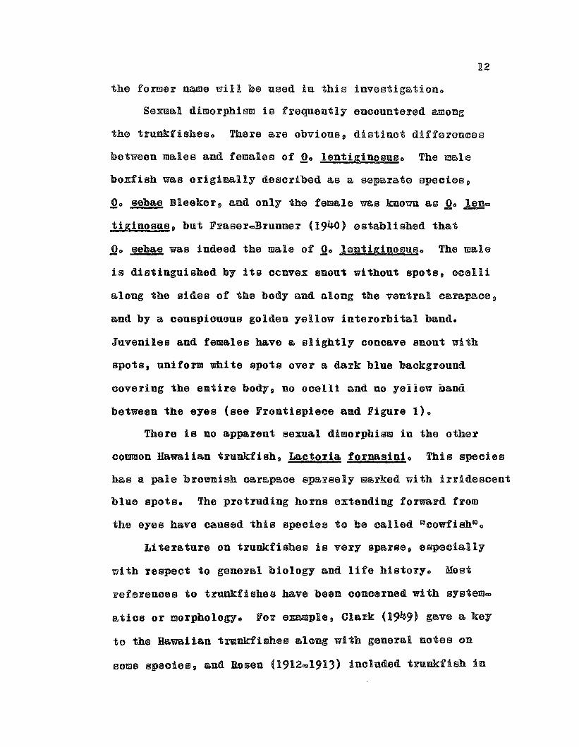
Sexual dimorphism ie frequently encountered among
the trunkfisheso There are obvions p distinct differeuc6a
between males and females of Qo lentiginosuso The male
boxfish was originally described as a separate epeciss p
00 sebae Bleekerg and only the female was known as 00 len=- --tiginosus, but Fr&sercBrunner (1940) established that
~o sebae was indeed the male of Qo lentiginosuBo The m~le
is distinguished by its ocnvex snout without spots, ooelli
along the sides of the body and along the ventral oarapece p
and by a conspiouous golden yellow interorbital band.
Juveniles and females bave a slightly concave snout with
spots, uniform white spots over a dark blue baokground
covering the entire body, no ocelli and no yellOW nann
between the eyes (see Frontispie~e and Figure 1)0
There is DO apparent sexual dimorphism in the other
common Hawaiian trunkfish, Laotoria fornasini o This species
has a pale brownish c&rapece sparsely marked with irridescent
blue spots. The protrUding horns extending forward from
the eyes have caused this species to be called scowfish~o
Literature on trunkfishes is very sparse p especially
with respect to general biology &nd life history. Most
references to trunkfishss have bean conoerned with system=
atlcs or morphologye For examplep Clark (1949) gave a key
to th0 Hawaiian trmnkfishss along with general notes on
soms speciasp and Rosen (1912@191J) included t~nkfi8b in
13
his studies on the plectognaths p a se~ies of four papsrs
dealing with the blood vascular system p the eir seop the
integument p end the body mU8clese Al~Hu88aini (1947)
briefly described the feeding habits and morphology of
the alimentary tract of a Red Sea trunkfish and Willem
(1941) described the function of the respiratory
apparatus of 00 cornutuso The major taxonomio works have
been done by Fraser.Brunner (1935, 1941)0
References to the possible production of a poisonous
substance by trunkfish have already been cited but these
were merely brief notes commenting on ~he apparent poison-
ing of other fishes by boxfish in aquariao
An often quoted observation on trunkfish locomotion
by Goode (Evermann, 1902) is typical of the kind of
information available:
-The locomotion of the trunkfishes is verypeculiaro The propelling foroe is exerted by thedorsal and anal fins, whioh bave a half-rotary,sculling motion, resembling 1hat of a sorew propeller;the caudal fin aots as a rudder, save when it isneeded for unusually rapid swimming, when 1t is usedas in other fishes; the chief function of the broadpectorals seems to be that of forming & current ofwater through the gills, thus aiding respiration,which would otherwise be difficult on aooount of thenarrowness and inflexibility of the braohial aperturesoWhen taken from the water one of these fishes willlive for two or three hours, all the time solemnlyfanning its g111s, and when restored to its nativeelement seems Done the worse for its 8xpgrience,except that on aooount of the air absorbed, it cannotat once sink to the bottome= (po 261)
The last sentence of the above qnotation was certainly
notwue for Hawaiian trunkfishaso Boxfish and cowfish
became moribund if left out of water longer than ten
minutes and the fanning of the pectoFel fine appeared
to be related to the secretion of toxin Father than an
emergency Fespiratory aid as the above quotation seems
to ~nggesto

Jl5
CHAPTER 4-
OOLLECTION AriD PREPARATIVE TECHNIQUES
COLLECTION OF TRUNKFISH
Nearly all boxfish used in this study ware oaptured
in :K.&oooha Bay, the Ala. Moamn. reef and Ke~la Be-atop Oa.huo
The majG~ collactlng gft~ ~6 Kewalo Besin p B harbor fOE
small fishing vssseise The actmal collecting occurred
on the aec~rd oids of the basin opposite the Fish and
Wildlife Service Laboratory along the boat piers and along
the stoDe wall parallel to the 8ea~rd channelo Here the
boxfish could be captured by traps or dipnetso Along the
Ala MOana reef adjacent to Kewalo Basin, boxfish and cowfish
were cap1nred only with dipnetso The largest boxfish
were collected by fish traps in Kaneohe Bayo
Trapping with commercial fish traps constructed of
iron frames and l.inoh ohioken wire proved moderately
suocessful in oapturing adult boxfish and large cowfiBh,
although difficultias ~ara G0t ~h~n traps were set in
the shallow water preferred by these trunkfishes as
traps were inevitably stolen or tampered with by curious
fishermen or Skindivers. Traps set in water sufficiently
deep to prevent the trap from being sean from the surface
usually caught few boxfish and were difficult to locate
and haulQ ibe traps set in the shallow waters opposite

16
the government biological laboratories in Keualo Basi&
were relatively safe from intruders during the working
week bnt during weekends had to be lifted and stoFed on
lando
The problems of trapfiShing made it necessary to
capture the m~jority of boxfish by dipnetting o There was
a definite advantage in capturing boxfish this waYo
Trap-captured fish usually had bean confined in the trap
for several hours or days before being removedo As a
mle, under anch conditions of prolonged streBS the highly
excited boxfish lost much of their toxic secretions,
especially if the fish had injured themselves by swimming
against the wire walls of the trapo Boxfish oaught with
a dipnet were always in oetter. oOfiuitiou Qud ~h6 6t~egg
of captnre could be minimized by a skillful pursuero
For these reasons it is important to discuss methods of
stalking and netting boxfisho
Boxfish ~ere often ~ncountered around coral mounds
in small aggregations of from 2 to 5 fish o When approaohed
by a skin diver, the boxfiSh would often watch the diver
cautiously, allow him to swim within a few feGt~ than
dart into a hole or under a ledgs o Unlike many reef
fiShes! the boxfieh would not remain hidden for more than
a few seoonds and would frequently depart via a back
axito I~ was more difficult to net boxfish among oor&l
mounds in water deeper than 5 feat, therefore it was
17
often necessary to manuever the boxfish toward more shallou
water, and if possible, away from shelter o If the diver
succeeded in doing tbis, he could chase the boxfish toward
shore where it could be trapped and scooped up easily with
the aid oH two dipnetso Often the boxfish would inadvertenta
ly abeach~ iiself in ita frantic efforts to e8c~pe~ Althongh
catching boxfish in this manner ~s quite laboriou8, and
weather and tidal conditions had to be favorable, the method
was fairly efficient o
Juveniles (under 100 ~ tot~l length) ~0rc easily
captured by the method described but adults were usually
more wary and less easily tricked. The large females ~ere
especially difficult to catch. The males were less cautious,
even appearing curious, and were sometimes as easily
captured as the juveniles.
Cowfish, ~. fornasini, were not as common as the boxfish.
They were much slower and more awkward swimmers than boxfish
and were very easily captured with dipnets, even in deeper
watero Cowfish ware coamonly 3ssn on the reef flats at
night with the aid of gas lanterns and could be easily
scooped up with dipnets e Boxfish, on the other bands were
rarely seen OD the reef at night at whieb time tbey
apparently seek shelter like most diurnal reef fisheso
18
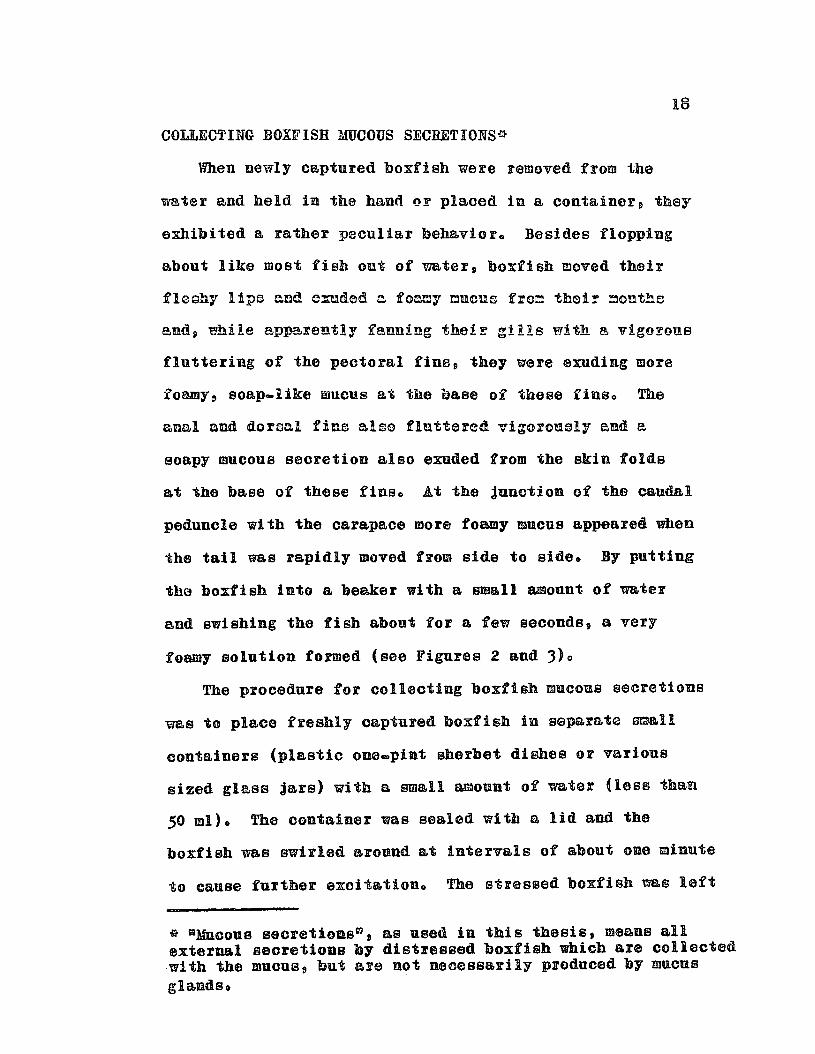
COLLECTING BOXFISn MUCOUS SECRETIONS*
When newly captured boxfish were removed from the
v~ter and held in the hand OF plaoed in a container, they
exhibited a rather peouliar behavioro Besides flopping
about like most fish out of water, boxfish moved their
fleahy lipe end o:uded c foamy cuellO froo their couths
end» ~ile apparently fenning theiF gills with B vigorous
fluttering of the pectoral fins, they were exuding more
foamy & soap-like mucus at the base of theBe finBo The
annl ond dorsal fins elso fluttered vigorou~ly end e
soapy mucous seoretion also exuded from the skin folds
at the base of these finsa At the junction of the caudal
peduncle with the oarapace more foamy mucus appeared when
the tail was rapidly moved from side to 81a60 By putting
tha boxfish into a beaker with a small amonnt of water
and swishing the fish abont for a few seconds, a very
foamy solution formed (see Figures 2 and 3)0
The procedure for colleoting boxfish mucous secretions
was to place freshly captured boxfish in separate ~&11
containers (plastio one.pint sherbet diShes or various
sized glass jars) with a small amount of water (les8 than
50 mil. The container was sealed with a lid and the
boxfish was swirled around at intervals of about one minute
to cause further exoit&tioDo The stressed boxfish ~s left
~ ~MUcouB secretioGss , as used in this thesis, means allexternal secretions by distressed boxfish which are collectedwith the mucus, but are not necessarily produced by mucusglandso

Figure I.to the
Juvenile boxfish.adult female.
Note the resemblance
Figure 2. The foamy mucous secretions of adistressed boxfish out of water.
l"......
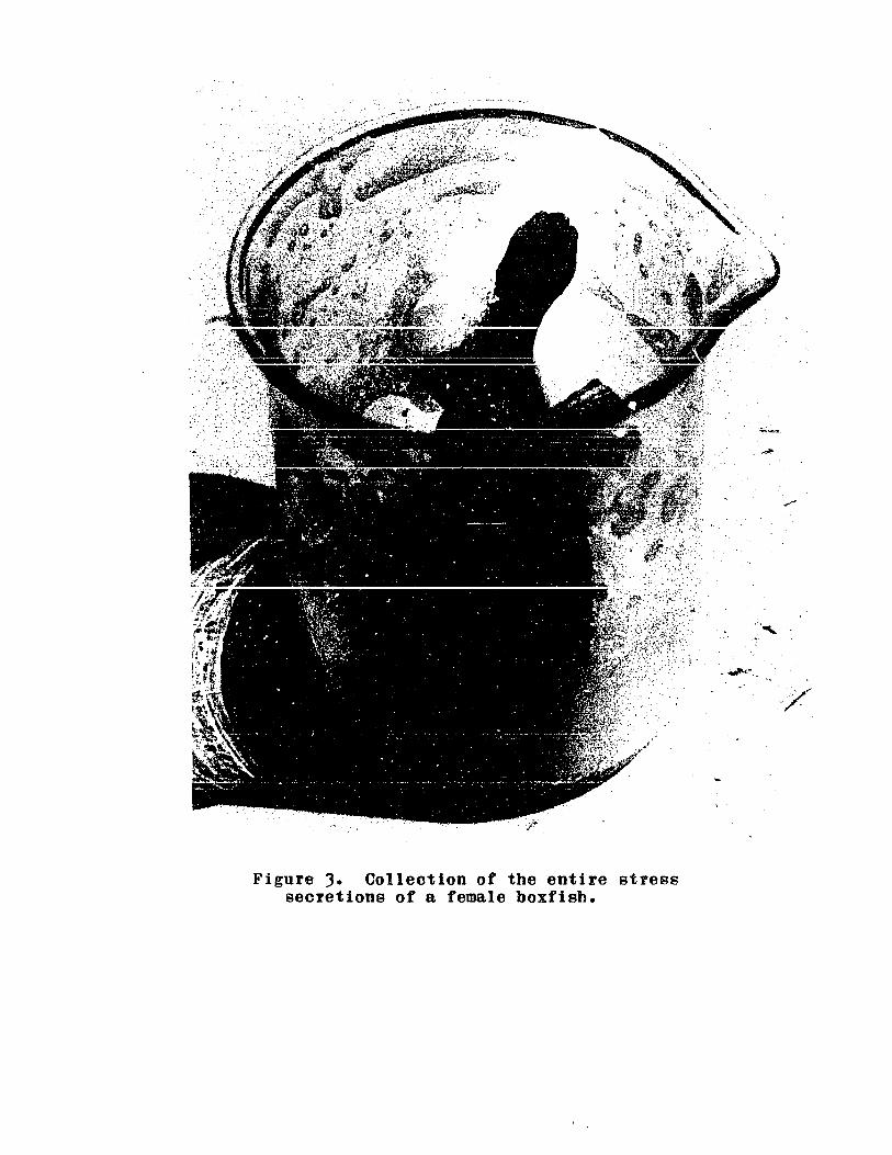
Figure 3. Collection of the entire stresssecretions of a female boxfish.
·Y··/~

21
in the container for 5 Q 10 minutes or until death,
although most of the mucous secretions appeared to be
given off within the first few minuteBo When removed
from the container, the boxfish was rinsed with distilled
or Bea water to oollect the foamy mucus adhering to the
sUFfBce of its skine It appeared that a more concentrated
mucus was obtained when an adequate volume of rinse water
was used, since water bathing the skin seemed to facilitate
mucous 8ec~atiGno (Too greet dilutions of the mucus
~ere avoided since it was later found that the boxfish toxin
was labile in greatly diluted aqueous solutionso)
The resultant diluted mucous secretions were a fommy,
colloidal Boluti~n which, when dissolved in sea water,
was poisonous to marine fishes at dilutions as great as
1125,000e The ichtbyotoxic suhstance(s) contained in the
mucous secretion of 90 lenti«lnosuB will be heretofore
referred to as aostracin", a tentative name derived from
the genus Ostracione The justification for uming this
name will be stQtod in a leter chBptero~
* Through convention a compound is usually not named untilan empirical formula is determined, however the distinctiveproperties of Dostracinn (which will be elucidated later)appear to justify the tentative naming of tbis toxin(s)without· this informationo
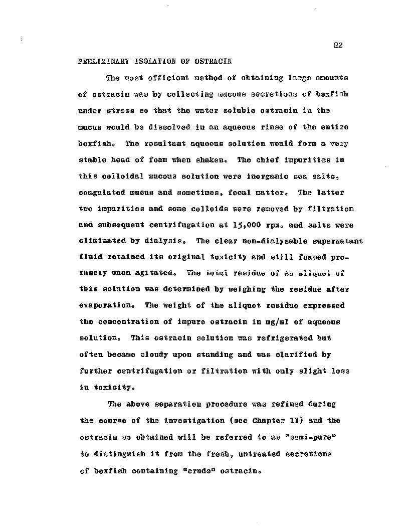
PRELIMINARY ISOLATION OF OSTRACIN
The most officiant method of obtaining large amounts
of oatracin was by collecting mucous sGCretioDs of b@xfish
under stress 80 that the water soluble ostracin in the
mucus would b~ dissolved in an aqueous rinse of the sniiFs
boxfisho The resultant aqueous solution would form a very
stable head of foam when ehakeno The chief impurities in
this colloidal mucou~ solution were inorganic sea salts,
ooagulated mucus and sometimes, fecal mattero The latter
two impurities and some colloids ware removed by filtration
and subsequent centrifugation at 15.000 rpmo and salts were
eli~iu&ted by dialysiso The clear non-dialyzable supernatant
fluid retained its original toxicity and still foamed proo
fuseiy when agitateuo ~ne ~otai ~esiuu~ ot au aliquvt vf
this solution was determined by weighing the residue after
evaporationo The weight of the aliquot residue expressed
the concentration of impure ostracin in mg/ml of aqueous
solutioDo This Gstracin solution was refrigerated but
often became cloudy upon standing and was olarified by
further centrifugation or filtration with only slight loss
in tox10ityo
The above separation procedure was refined during
the course of the investigation (see Chapter 11) and the
ostracin so obtained will be referred to as ~semi-pure~
to distinguish it from the fresh, untreated secretions
of boxfish containing ~crudeR ostraciuo
23
CHAPTER 5
DEVELOPMENT OF A BIOASSAY FOR OSTRACIN
The development of a bioassay sensitive enough to
detect minute amounts of biologically active substances
is en iapo~tant step toward isolating and studying U~
known toxiuso Toxins~ b~tng highly patent substances, oan
be readily detected by their action on living systemso
The response of a living aY8t~c to e toxin must then be
converted to B quantitative expression which will accurately
assay the toxicity of an unknown sampleQ Toxioity is
defined as the total amount of toxic material in a sample
while the potency of a toxin is the capaoity of a given
amount of pure toxin to elioit a response. A good bioassay
should provide an acourate determination of the former and
a reliable estimate of the latter.
Ostracin. the toxio, water soluble fraction of boxfish
muoous secretions. has already been shown to be poisonous
to fish and in this sensa rGsam_l~holcthurin. the sea
cucumber toxin (Nigrelli &nd Jakoweka, 1960) and some red
tide toxins (Abbott and Ballantine, 1957; Shilo and Rosenberg,
1960; Gates and Wilaon 9 1960)0 Both holothnrin aad the red
tide toxins have aleo been reported to exhibit a variety of
effects on diverse organisms from protozoans to mammalso
Because of the ichthyotoxic parallelism of oetracln witb
holothurin and the red tide toxins. it was deoided to
24
test o8tr~cin on various ~quatic organismso This approacb
would not only facilitate the development of an adequate
bioassay for ostracin but uould provide comparative infor~
mation on the mode of action of o8t~acin ~ith respect to
other marine biotoxinso
TOXICITY OF OSTBACIN TO AQUATIC ORGANISMS
Various species of aquatic animals were placed in
sea water containing crude oetracin in concentrations whioh
were highly toxic to marine fish, ioeo, caused de~th of
such fish within 10 minutes. The animals used were those
that were readily available and the list of species tested
represents a more or less fortuitous selection of phylao
Since the various animals tested feil into three
general groups, the reaction groups proposed by Abbott
and Ballantine (1957) for the §l!nodinium toxin were used
and are as follows:
(A) E2! affecteuo Animals of this group ~howed normal
behavior during immersion in ostracin solutions and survived
when returned to fresh sea watero Polycbaetes, colonial
&scidians, some mollusks and most crustaceans belonged to
this groupo
(B) Slowly affeoted& In this category were inoluded
animals which showed various symptoms of poisoning while
immersed in oetraoin-sea water for several minutes to
hours but usually recovered when removed snd returned to
25fresh sea uatero Anemones and sea urchins are exampleso
(c) Rapidly killedo This lest category included all
the animals which were killed within minutes after exposure
to ostracino Fish and cephalopods fell into this groupo
A summary of reactions of various species is found
in Table 1 0 The results summarized in this table compare
favorably with those obtained by Abbott and Ballantine (1957)
using toxic cultures of Gymnodinium veneficum, a red tide
dinoflagellsts o These authors concluded that the rate of
penetration of the Gymnodinium toxin into the test organisms
was the critical factor in delineating between groups B and Co
This explanation appears feasible in regard to 08tra010
since many interesting parallels with the Gymnodinium toxin
were observed.
A description of the symptoms of animals of groups
Band C is given belowe
COELENTERATA
Sea anemones often retracted their tentacles slightly
and wound the distal ends into tight spiralso The swimming
anemone Nectothelia lilae, however, relaxed its tentacles
Which subsequently became insensitive to probing. The polyps
of the colonial hydroid Pennaria tiarella also became
insensitive to probing whereas the oontrol animals would
immediately contract when touched with a dissecting needle~
All these ~oelenterates would recover if placed in freSh
sea water before they became moribund in the ostracin

TABLE I. TOXICITY OF OSTRACIN TOSOME AQUATIC ANnIALS
26
animal
COELENTERATA
Anemonia mutabilis (anemone)~ctothelia lilae (anemone)Pennaria tiarella (hydroid)
PLATYHELMINTHES
Dugesia spo (planaria)
ANNELIDA
Sabillid wormsUnidentified polychaetesParasitic marine leech
MOLLUSCA
Helcioniscus argentatu8 (Opihi)PolyPUS spo (octopus)
ECHINODERMATA
Echinometra mathaei (sea urchin)Echinometra oblonga (sea urchin)Echinothrix diadema (sea urchin)
ARTHROPODA: CRUSTACEA
Spirontocaris marmoratus (shrimp)Stenopus hispidus (banded shrimp)Penaeid shrimp (prawn)Pachygrapus spo (crab)Carpi Ius convexus (crab)Artemia salina (brine shrimp)Labidocera sp. (copepod)
TUNICATA
ascidia spo (sea squirt)
Reaction Group
BBB
B
A
AC
BBB
AABAAAB
A
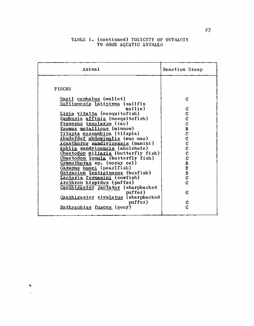
TABLE 10 (continued) TOXICITY OF OSTRACINTO SOME AQUATIC ANIMALS
27
Animal Reaction Group
PISCES
Mugil cephalus (mullet) CMollienesia latipinna. (sailfin
mollie) CLimia vitatta (mosqui tofish) CGambusia affinis (mosqui tofi sh) CPranesus insolarum (iao) CBsofins metallicus (minnow) BTilapia mossambic~ (tilapia) CAbudefduf abdominalis (mao mao) CAcanthurus sandiyicensis (manini) CKuhlia sandvicensis (aholehole) CChaetodon miliaris (butterfly fish) CChaetodon lunula (butterfly fi sh) CGymnothorax spo (moray eel) BCarapus horne! (peai'lfisl1) BOstraci.on lentiginosus (boxfish) BLactoria fornasini (cowfish) CArothron hispidus (puffer) CCanthtgaster jaetator (sharpbacked
puffer) CCanthi gaster rivulatus (sharpbacked
puffer) CBathygobius fusells (gooy) C

28
solutioDo The desensitization of the tentacles suggested
a neurotoxic effect on the nerve-net systerno
PUt.TYHELMINTHES
Planariaus exposed to toxic solutions ot ostracin in
fresh water disintegrated upon transfer to clean watero
Quaglia at alo (1957) reported similar results with
holothurin.treated planarianso These authors also reported
that planarians exposed to toxic holothurin solutions for
les8 than one minute survived indefinitelyp however when
these animals were dissected transversely and returned to
fresh water the anterior portions regenerated into complete
animals, whereas the majority of the posterior portions
di~inte~reted ~~ f8!!e~ to ~eg~ne~ate~ A similar experiment
was conducted in which 2 groups of 20 planaria (Dngeeia ~o)
were exposed to concentrations of semi-pure ostracin
of 80 a.nd 40 ppm (parts per million) for 15 .., 25 and 5 ... 15
minutes respectively. (These concentrations were highly
toxic to fisho) The planarians were cut transversely while
in the toxic solution and transferred to fresh water kept
at room temperature (24 G 260 C)o The anterior and posterior
portions regenerated at the same rate as the controlso
Eyespots were observed by the third day and the 40 ppm
group appeared normal and survived as long as the controlso
Although both portions regenerated equally well in the
80 ppm group, these animals were very inactive compared to

29
the control end 40 ppm groupse Many planarimns were curled
in a loop and were very nsticky~ to touch. While the
planarians in the control and 40 ppm groups were able to
glide smoothly over the gless bottoms of their petri dishes,
the 80 ppm group moved about by worm-like contractioDs.
At the end of e month there was e 15% mortality rate in
the 80 ppm group as compared to no mortalities in the 40 ppm
and control groupso No anti-metabolic activity of ostracin
was demonstrated in any group.
MOLLUSCA
An octOPUB (Polypus ~o) about one ponnd in weigbt
was placed in a plastic bucket containing crude boxfish
8ee~etic~so The e~!~B! c!~ng te~c!c~s!y tc the sides cf
the bucket and was pried loose with great difficulty.
After about 5 minutes exposure to the toxic secretions
the octopus was released in & large saltwater pond. The
moribund octopus was found dead the next Dorning in the
Bame spot where it had been releasedo
ECHINODERMATA
The tube feet of sea urchins contracted in ostr8cin
solutions and the animals became immobilized, while control
animals aotively moved about in their containerBo Injection
of ostraoin at the muscular base of the spine of the long
spined species Echinotrix diadema oaused riaidity of that
spine but did not affect adjoining spineso Control aaline

I .
30
injections had no effecto
Ruggieri and Nigrelli (1960) found that holothnrin A
was not only very toxic to sea urchin embryos but, in
sublethal concentrations p caused abnormalities such as
aniwalization and fragmentatioDo Because of the similarity
of ostracin to holothurin, a saries of experiments was
conduoted to determine the effect of low concentrations of
ostracin on the developing embryos of Tripneustes gratillao
Although the data of these experiments were destroyed in the
fire at the Coconut Island Marine Laboratory, December 30,
1961, a brief summary of results should be of some interest o
Fertilized eggs kept in sea water containing 1 to 10 ppm
of ostraoin showed marked inhibition of cleavagoo In one
experiment, eggs kept in soa water with 1 ppm ostracin
develo,ed only to the motile blastula stage while the
oontrols developed into normal plutei during the same period.
With a few exceptions, both in the oontrols and teat animals p
no abnormal larvae were observedo
Fertilizsd eggs of e see cuoumber, Holotburia
fuscorubra were immersed in equivalent ichthyotoxic solutions
of ostracin and holothurin (obtained from the cuvierian
organs of Actinoplga obesa). Ostracin was lethal at 1 ppm
while crude holothurin was non.toxic at this level but
effective at only slightly higher oonoentrations (5 ppm)o

31
CRUSTACEA
All crustaoeans tested showed great resistanoe to
ostracino As suggested before, this may be due to the
rate of penetration of astrecin through the gills or
other body membranceso An ioshore marine copepod p
L~bidece~B ~op nOFm~!ly B positively phototactic animal,
ioeop one that is attraoted toward light, lost its photoe
tactic response in sea water containing ostrecin and a 50%
mortality ensued after It hourso The control animals
responded no~ally ~ad sUFviv0d the duration of the ex=
perimento
PISCES
All fish immersed in ostracin-sea water were rapidly
killed, usually within 10 minuteso The symptoms shown by
all fish were identioal and were very similar to symptoms
reported by Abbott and Ballantine (1957) for the GymnodiniYm
toxin and by Nigrelli and Jakowska (1960) ror holothuriuo
In ostracin concentrations causing death within 10 minutes
(100 ppm), the initial reaction of fish when ostracin was
added to the water was of marked irritabilityo The fish
made violent attempts to swim away from the irritation and
in some cases jumped out of the containero Such frantic
swimming usually lasted 1 - 2 minutes, again depending
upon the concentration of ostracin used o Opercular
movements were erratic g spasmodio ~vomitingQ occarred g and
)2
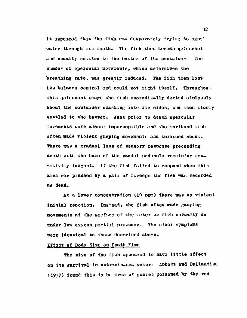
it &ppeared that the fish was desperately trying to expal
water through its moutho The fish then became quiescent
and usually settled to the bottom of the oontalnel'o The
number of opercular movements, which determines the
breathing rate, was greatly reduced. The fish thon lost
its balance control and could not right itsello Throughout
this quiesoent stage the fish sporadically darted aimlessly
about the oontainer orashing into ita sides, and than slowly
sottled to the bottom. Just prior to death operoular
movements were almost imperceptible and the moribund fish
often made violent gasping movements and thrashed about o
There was a gradual lOBS of sensory response preoeeding
death with the base of the caudal peduncle retaining sen
sitivity longest. It the fish failed to respond when this
area was pinohed by a pair of forceps the fish was recorded
8.S dead.
At a lower concentration (10 ppm) there was no violent
initial reactioDo IDstead, the fish often made gasping
movament~ at the surface of the water ~B fish normally do
under low oxygen partial pressure. The other symptoms
were identioal to those desoribed above.
Effect of Body Size on Death Time
The size of the fish appeared to have little effect
on its survival in ostracin.sea watero Abbott and Ballantine
(1957) found this to be true of gobles poisoned by the red

33
tide Gymnodinium toxin and explained this by the fact that
the toxin penetrates the gills and the active gill surface
18 roughly proportional to body size within a specieso
The Oritical Exposure Period of Fish to Ostracin
The duration of exposure of fish to ostraoin was oriticalo
Beyond a certain exposure time the effeot was irreveroible,
ioeo, ostracin did irreparable damage and the fish did not
recover if returned to fresh sea watero Apparently a lethal
dose penetrated the fish very quiokly or did morbid damage
to vital membranes like the gills o
An experiment was performed using juvenile tilapia p
Tilapia mosswwbica (15 - 20 mm total length), in which
groups of 4 fish were immersed in ostraoinosea water (about
5 ppm ostracin) for varying periods and returned to fresh
sea water for recovery. The results are summarized below:
Exposure time (min.)
151065J2It1
50% mortality (mino)
15151617222936
000(1 fish diedin 2 hours)
000(1 fish diedin 2t hours)
The tilapia exposed to ostracin for 30 seconds to 2
minutes showed no clear stress symptoms but became agitated
after 3 minutes and quiesoent after 5 minuteso

Complete mort&lity ensued in all Ce8GS except Bt exposures
of 30 seconds and 1 minute. These data suggested that
ostracin either penetrated very rapidly or interfered with
the surface of the gill membranes and that a lethal dose
had been aocumnlated before outward symptoms were recognizedo
nO~H9nso of DiffeFeat Speo!s8 to Ostraoin
The 8nsce~tibility of various SpeCiG8 of fish to
ostraoin appeared to vary acoording to the aotivity level
of diffe~ent species. This can beet bo illuatrat0d by
relative 8urvivBl times of different species immersed in
equal concentrations of ostraoin (about 5 ppm) in Bea water:
(6 juvenile fiSh used per test; complete mortality ocourred
in all tests o )
Speoies
Abudefduf abdominalisAcanthurus sanvicensisKuhlia 8&nviceusisMagil oepbaluBMollienisia latipinnaBathygobius fuscus
Common name
mao maomanini&holeholemulletsailCin mollygoby
SUrvival time of50% of fiSh (min.)
600805
10.0120515 0 0)0.0
The first two species on the lIst are very active stenohaline
reef fishss 9 the next three are moderately active euryhaline
forms and the last, the goby, is a bottom dwelling, relatively
inactive, tidepool specieso Susceptibility to ostracin may
be correlated with the frequency of op~rcular (respiratory)
movements 0 The goby, typical of most bottom living fiShes,
has a much lower respiratory rate than aotive swimmers suoh

35
as the mao mao and rn~nini and therefore, might be expected
to show a higher resistanoe to the toxino
The Effect of Salinity on Toxio Aotion of Ostraoin
The affect of salinity on the toxio action of astreoin
had a much more significant and dramatic effect on survival
rate than physiologioal variablsso Juvenile tilapia acolimated
to sea water had a mean survival time of 20 minutes in
ostracin-sea water, but the euryhaline tilapia acclimated
to fresh water survived 70 minutes in the same conoentration
of ostracin in fresh watero The freshwater cyprinid Esomu8
metalious was even more resistant to ostractn in fresh water
than freshwater acclimated tilapia at the same conoentratioDo
The mean survival time of these cyprinids was J hours and
50 minutes. Such anomalies led to the investigation of the
toxic action of crude ostraoin on fish immersed in salinities
ranging from fresh water to sea water.
Juvenile sa11fin mollies (12 • 15 mm total length)
were acclimated to water of various salinities ranging from
f%0ah to aG& water for 25 hOnlee Groupe af 6 fiab ~ere
placed in beakers containing 100 ml of the desired sea water
dilutioDo No aeration was used since it had been determined
that small moltles can survive without auxilIary aeration
for several hours to days under such conditiouso A lethal
unit of freshly collected boxfiBh secretions was added to
eaoh beakeroThis experiment was repeated J times and the
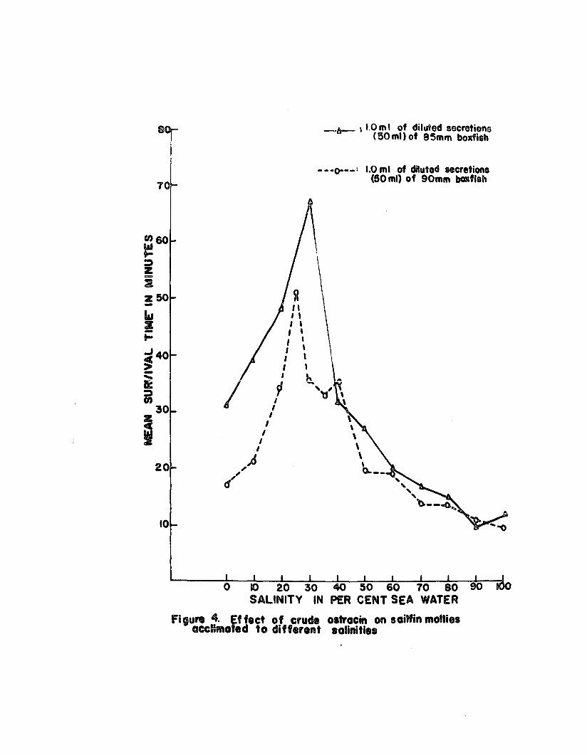
results of the laBt 2 tFials are depicted in Figure 40

The data showed that there was a gradual inorease in
survival time of sailfin mollies as their ssa water medium
was diluted and at salinities ranging from 25 to 30% sea
water there was & Sharp inorease in survival t!meo There
was an equally sharp reduction in survival time in salinities
belo~ 25% sea ~1e~ and in f~Gah ~t0r the sBrv!ve! ties
wag only ~bout t~iee Be long 88 it ~88 in 88~ WBte~e The
experiment was repeated with Tilapia m08sambioa and similar
results were obtainedo
The pH of the various dilutioBa ~angcd frem {079 in
20% saa water to 8004 in fresh water. The pH of the sea
water used was {085. Although it seemed unlikely that the
pH itself was exerting some effect on inhibiting the action
of ostraoio p an experiment was performed in whioh the pH
of sea water was adjusted by adding acid or baseo Groups
of 4 tilapia (12 a 28 mm total length) were plaoed in
beakers containing 400 ml of Bea water of a pH rangingftom
50 80 to 80800 An equal amount of crude ostracin was
introduced into each beaker and the mean su~vival time
was reoorded:
Mean survival time (mino)
24 0 025 0 024.02)00210022 0 0
70
-fJt-- \ 1.0 mI of diMed secretions{50 mil of 85mm bodiah
- - -0--_: 1.0 rnl of diluted secretione(50 ml) of 90mm bGKflsh
20
10
,II,,,,
/ lI,
II
I
,~,($'
,,,,,,b..',,'
t\\\\\
~--'""""'b.--~...
ostracin on saiffin molliessalinitie.

38
It ~s clear that pH had little or no effect on survival
times of tilepie immersed in ostracinesea watero
Since sailfin mollies and tilapia are essentially
freshwater fishes w.bich have secondarily invaded braokish
and salt water, it might be argued that these fish would be
more resistant to ostraoin while immersed in salinities
ears closely corresponding to tbeir natural environmento
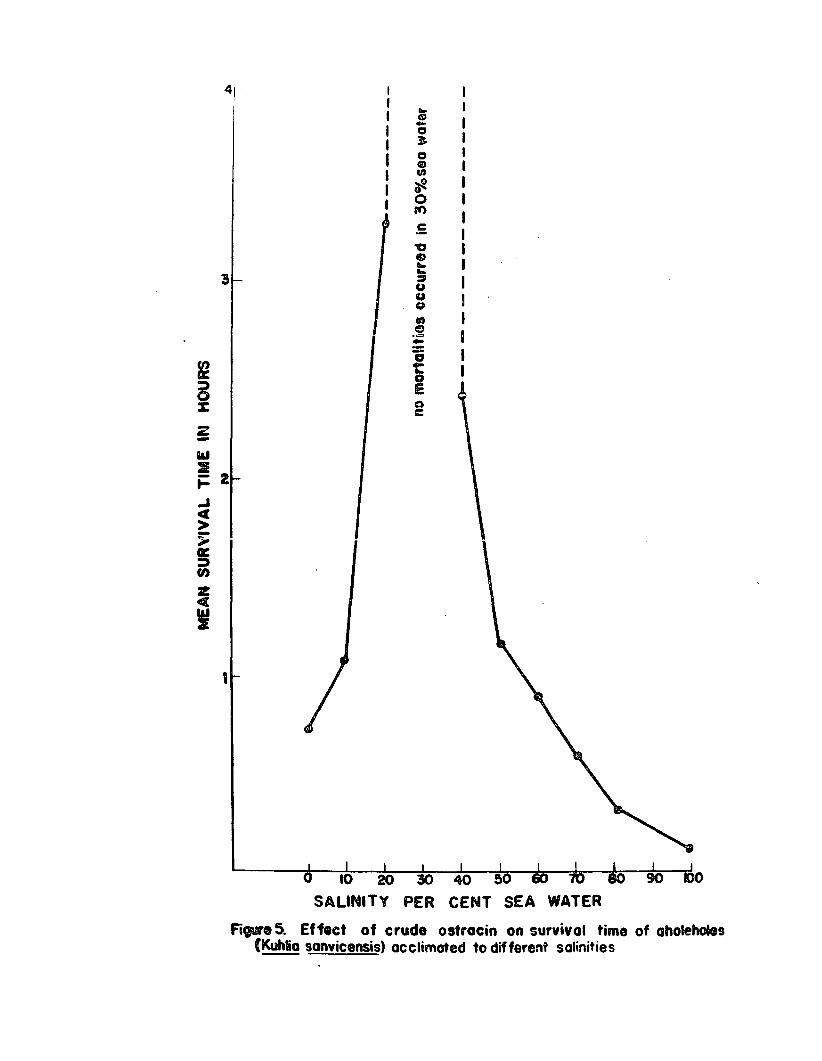
Therefore, a more typioslly marine species, the aholehole
(Kuhlie 8Bnv!oeusis), was tested in a similar mannere
This euryhaline speoies lives and spawns in sea water but
the young are frequently enoountered in braokish and
freshwater streamso Juvenile· aholeholes (25 - 40 mm total
length) were acolimated for 24 hours to different salinities
and 2 fish each were placed in beakers containing 100 ml
of water of the desired s&linit1o A lethal unit of orude
oatracin was put in each beakero No aeration was usedo
The results are shown in Figure 50
The mean Burvival time of the aholahole at low salinities
was much longer than th~t ef the 8Biltin molly Bnd tilapiao
Although this species was shown to be more susceptible to
poisoning by ostracin in sea water than were sailfin molliss 9
the aholehole was found to be considerably mors resistant
to ostracin in dilute sea water than the molly was, espeeially
in 20 to 40~ sea watero
There are at least two possible explanations to this
phenomenon. rna first concerns the osmotic equilibrium of
,
IIIIIIIII
Figure 5. Effect of crude ostracin on survival time of aholeholes(Kuhlio sonvicensis) acclimated to different salinities
40
fish in changing salinitieso I~ appeared that the rete of
penetration of ostracin into the tissues of fish immersed
in ostracin solutions was dependent upon the salinity of
the mediumo Osmotio regulation then was probably an im
portant faotoro Aocording to Black (1957) the freezing
point depression of the blood of freshwater teleosts is
about 00 0 570 C and in marine tele08t8 is about 00 0 78° C~
whioh 1s lower than that of the sea water (0108 to c202° 0)0
Consequently, marine tele08ts must actively take in water
to replace water lost osmotical1Ye The external membranes
(gills, gut, skin) then favor water intake and are able
to absorb WB.ter against an osmotic gr&dient o One can then
expeot a corresponding change in permeability and membrane
potentials as marine fish are gradually acclimated to fresh
watero The conoentration of the blood of teleosts oorresponds
to 25 to 40% sea water, ioeo, at this ooncentration the
blood is isotonic with the external medium and osmotic
work (exchange of water, salts and ~aate products) should
be einimale This coincides with the range of salinities
in which ostracin was least effective and suggests that
isotonicity is an important factor in the resistance of
fish to ostracino
The seoond possible ~xpla.nation is that advanced by
Yariv and Hestrin (1958) as reported by Shilo and Rosenberger
(1960). The former workers, studying the ichthyotoxius
f~om the phytoflagellata, Przmuesium parvum, have suggested

41
that a speoial property of the Pe parvum ichthyotoxin
was its requirement of cofactors for activity and that
the toxicity of Po parvum solutions aypeared to be a
function of the concentration of both the iohthyotoxin end
of inorganic s81t80 In the absence of inorganic salt8 p
this ichthyotoxin does not kill fish o
The Eo parvua ichthyotoxin has enough properties in
common with ostracin to consider this cofactor theory as
another possible explanatioD p except that ostracin, unlike
the P. parvum toxiD is @OFe toxic in fresh water than in
diluted sea watero
The important practical consideration here is that s
ohange in salinity of the external medium will have a
significant effect on toxicity of ostracin to euryhaline
fishes and this fact should be taken into &ccount in the
development of a bioassay using fish immersed in water
containing unknown amounts of ostracino
TOXICITY OF OSTRACIN TO THE PEARLFISB
Pearl fishes, inquilines in the cloaca and respiratory
cavities of certain sea cucumbers and starfishes» have been
shown to be susceptiblep but highly resistant to the Bea
cucumber poison D holothnrin (Nigrelli, 1952) and Carapus
bermudensis bas been used for evaluating the toxicity of
holothurin from the cuvieriSborgans of Actinoprga agassizi
(Aronson and Mosherp 1951)0

42
Pearlfish (Carapus homei) were exposed to icbthyotoxic
solutions of ostracin and crude holotnurin (from skin of
Holothuria ~) and their survival time recordedo Althongh
the holothurin solution was more ichthyotoxic to sailfin
mollies than the ostracin solution used (sailfin mol lies
died within 10 minutes in holothurin-sea water and in 15
minutes in ostracin=@ea water), pearlfish lived for more
than 6 hOUfS in ostracin-sea watero
The pearlfish!s resistanoe to both these toxins, and
particularly to holothurin suggests some protective
mechanism not possessed by other marine species (with ex
ceptions in Gymnothorax and 08tracion)0
TOXICITY OF OSTRACIN TO THE BOXFISH
Experience with collecting and transporting boxfiBh
or cowfish showed that freqnent mortalities occurred despite
seemingly adequate aerationo This was especially true
during transport of freshly captured trnnkfish from which
stress secretions had not been previously collectedo
Boxfish often became moribund during collection of thei%
secretions if they were kept in the concentrated oetracin
solution longer than 5 minuteso Cowfish appeared to be
exceptionally sensitive to both their own and boxfish
secretioDso Thus, it appeared these trunkfisb were being
poisoned by their own toxinse
Experi~ent8 were performed to test this hypothesiso
Intramuscular and intraperitoneal injeotions of semi_pure
ostracin in sea water media at doses of 30 0 to 7.0 mg/boxfish
(80 to 117 mID long) did not oause death and immersion of
boxfish in 15 ppm ostracin-seawater had no affeot o
However. when the crude mucous secretions of freshly captur
ed boxfish were injected into the body cavity via the
caudal-oarapace juncture, the boxfish became moribund almost
immediately, showing typical symptoms of ostracin poisoningo
About 1.0 ml of concentrated mucous seoretions
representing approximately one-quarter of the total stress
secretions (equivalent to 20 - 30 mg of semi-pure ostracin)
was needed to kill a 80 - 110 mm boxfish in 5 to 20 minuteso
Lesser amounts were ineffectiveo
Thus, it is apparent that boxfish are susceptible to
ostracin but are considerably more resistant to this toxin
than other fisheso
TOXICITY OF OSTRACIN TO WHITE MICE
White mice have been widely used as assay animals for
various biologically active compounds including plant and
animal toxinso In studies on marine biotoxins white mice
have been used as a standard assay for paralytic shellfish
poisons. puffer toxins and ciguatera-type toxins and it
has been customary to express the potency of any toxin by
its LDSO in white miC8e (LD50 is the minimum lethal dose
that will kill just 50% of the assay Bnimalso) Because of
the widespread nae of white mice as indicators of toxin
potency, it was necessary to determine the effect and
potency of ostracin injected into miceo
§ymtoms
Albino White mice (closed colony Carworth Farms
Webste~ strain) ranging from 109 to JOg were injected
intraperitoneally with crude and semicpure ostrecino The
characteristic symtoms of mice injected with ostrac1n are
the following: about 2 - 3 cinutes after injection the
mouse appeared very sleepy and began to breathe deeply;
this was soon followed by ataxia, the loss of its grasping
and righting reflexes; breathing became more labored until
the mouse appeared to be literally gasping for breath;
DdeathR followed quiokly, usually without convulsions.
~utopsy revealed that the heart was etil1 beating, the
lungs were collapsed and, judging from the appearance of
the peritoneum, there had been no vascular damage. If
a sublethal dose was given, similar symptoms would often
occur but recovery was usually complete within a few hOU~8c
Leth~~ Doee of Ostracin
The lethal doss of oetraoin in mice was quite high.
At least 1 cc of a highly toxic 10 ml rinse was needed to
kill a 20=gram mouse injected intraperitoneallyc A dose
of about 0.20 mgl g mouse of semiQpure ostraoin appeared
to be the minimum lethal doeeo This amounted to abont

4 mg per 20-gram mouse, a fairly large amount which certainly
prohibited the use of mice as standard bioassay animalso
Effect of Injection Media
Since it was found that ostracin was less toxic to
fish in fresh and 25 - 40% sea water than in 100% sea water,
ostracin dissolved in distilled water and 25% sea water
was injected intraperitoneally into mice to determine if
similar reductions in toxicity occurred. Suoh injections
caused no intoxication symptoms While sea water.ostracin
injections of the same concentration were rapidly lethal.
The cause of these anomalies is unknown, but may be ex.
plained by the same mechanism(s) suggested for ichthyo
toxicity (see ppo 39 • 41)0
Control Injections
Control injections of physiological saline, distilled
water and sea water had no effect on mice. To determine
if there was a toxic reaction to fish mucus in general,
the mucous secretions of 4 species of reef fish were
collected and assayed in the same manner as were boxfish
secretions. The species used were: the manini, Acanthurus
sanvicensis, a butterfly fish, Chaetodon miliaris, a
filefish, Pervagor spilosoma and the puffer, Arothron
hispiduso The mucous secretions of the first 3 species
were non-toxic to mice but the secretions of the puffer,
Aronthron hispidus, were more toxic to mice than boxfish

46
secretionso Arothron hispidus is known to contain a
highly toxic substance, tetraodotoxin, in its flesh and
viscerao The toxic mucous secretions of this species are
further discussed in Chapter 120
47
CHAPTER 6
ESTABLISHMENT OF ASTANDARD BIOASSAY FOR OSTRACIN
To detect ostracin in the mucous secretions of
individual boxfish and to measure the potencies of various
o8t~QoiB prepaFatious, it uas necessaFy to uevelop a
stQndQ~d bioassay ~i~h ~ould not only be sensitive Qnd
reliable, but simple to perforIDo A bioassay using juvenile
fiGh met these specificationso
CHOICE OF ASSAY FISH
The choice of a suitable bioassay fish depended upon
its availability, hardiness, size and sensitivity to
ostracioe A number of reef fishes were screened and,
although all were very sensitive to the toxin, they were
found lacking in other qualifications such 8S availability
throughout the year and general hardinesso The most
suitable test species was a brackish water cyprinodont,
the sailfin molly, Mollienesia latipinna which had been
introduced into Bome of the streams and·brackish water
ponds on Oahu for mosquito control, and are therefore
known, along with a few other cyprinodont fishes as
~mosquito fisb D by local resldents o
The sailfin molly was very abundant in the brackiSh
ponds and canals in Ala Momna park in Honolulu and thousands
of these fisD could be collected by seining the canal in
a single afternooDo The seasonal fluctuation in their
numbers was only slight and these fish could be collected
throughout the yearo This was not true of most reef fishes
whose numbersD espeoially of the young g fluctuated widely
during a single yearo
The sB!!fin molly met ~!! the reqnirements of a good
B8say speciss e Besides being readily Qvailable, this
enryhaline fish was quite hardy and could survive as well
in fresh water as in sea watero Sin~e all Qolli~3 ~r~ liveQ
bearers, their young are precocious Bnd very hardy in com=
parison to the fry of oviparous fish e The newly born molly
was about 8 - 10 mm in total length and conld be reared
easily in an aquariumo Because of the small size and
availability of newborn mollies. it was decided to use
only these and juvenile mollies for the bioassay. The
UBe of Buch a small fish for a bioassay had obvious
advantages 0 A small volume of water and oonsequently, a
small amount of toxin could be used for testingo
Since it ~8 not always possible to colleot newborn
mollies in the canal, as they are quiokly eaten by predators,
pregnant mol lies were oollected and kept in 35 e 45 gallon
aquaria partitioned by hardware clotho The newly born
mollies would swim through the wire soreen after being
obased by the adults and take Shelter at the safe end of
the aquarium~ They could be oollected with a dipnet and

transferred to another aquariumo Hundreds of young
mollies would be produced in a tank cont~i~iDg about
J dozen pregnant femaleso The spent females were re~
moved from the breeding tank and released or disposed oi D
as they preyed upon the young, oompeted with the others
for food or died and fouled the watero Either brackish
or see water was ueed in these rearing tanks &s it did
not appear to affect the survival rate of young or adnlts o
Another assay fish which was used for many experiments
was the African mouth-breeding cichlid. Tilapia moseambicao
The young tilapis (10 a 12 mm total length) could be
collected in one of the brackish ponds in Ala Mosua park
during the Bummer monthso This 8pe~ies was also found in
fresh and brackish waters throughout the Hawaiian Islands
and was as hardy and as adequate an 8ssay species as the
sailfin molly. However» the supply of these fish was too
erratic to satisfy its use as a standard assay specieso
DOSE RESPONSE DATA
Crude Ostracin
The mucous secretions of a ~uvenile boxfish (about 90 mID
total length) were collected in a 50 ml distilled water
rinse and assayed with sailfin mollies in 100 ml of sea
watero The experiment wa,s conducted at room temperatures
(25 a 260 C) and 0.02 to 10 0 0 ml of the 50 rot boxfish rinse
were added to beakers of 100 ml of sea ~ter. each oontain=
iog 4 assay fisho Complete mortalities occurred in all
50
concentrations tested bnt only the 50% mortality time was
recorded and plottedo The resulting curve obtained by
plotting dose against survival time is Shown in Figure 6 0
The assay curve obtained was very similar to the
curves obtained by Abbott and Ballantine (1957) for
GYmnodiniUB toxin and by Hashimoto and YasUIDoto (1960)
fo~ 8ta~fish saponino An abrupt change in the conrse of
the curve was oharacteristic of all these curveso
Semi.pure OstF&cin
An aqueous stock solution of 1 mg/ml semi.pure 08tracln
was added to sea water to prepare dilutions of ostracin of
1:25eOOO (40 ppm) to 1920 9 000,OOO (0.05 ppm)o Four sailfin
mollies were used per beaker containing 50 ml of sea water
and the mean survival time was recorded (obtained by averag
ing individual deaths)o
Complete mortality occurred in dilutions as low as
1.5 ppm (1:67,000) and a 50~ mortality was noted at 0.1 ppm
(1:10,000 9 000)0 At 40 ppm the mean survival time was 800
minutes and at 105 ppm it ~s 2 houra and 4) minuteso
The dose response curve resembles the curve obtained
with crude ostracin (see Figure 7)0
THE STA.NDARD DOSE
To quantitatively determine the concentration of ostracin
secreted by stressed boxfish, a standard procedure of
collecting and assaying whole crude secretions was adoptedo

o
o
o
o
o.aDOSE: ... 0f1 GO ... RaNI! OF OJ?!~~
Al7JI'Cl 6. DoH-respcllfll cane (14'1'aol:r:::,..:.r" .... In Mel water
1201~ .
~IO~~'Z '
~ 90r~ I
J Tsi0: .
;J GOr~ :er45t-::> 'Ul
z3()!4UJ
2 I~
°t·...J11+H+H+H.-_---;!;;-__4:-__-:h-__~~2-7-0C-_±_--_},,d~!A'+.§~ m Jo Js ~'o 3!l io
DOSE. ppr,e OF OSTRACIN
FIgure 1. DOte-reGPOnlll curv. 0' eetN-.... 0IIr_ ClII IaIfin 11'IO_1" wa IIrCItw(4f1s!1/60IIII ....,)

52
A ganersl description of the collection of boxfish secretions
has already been giveno In standardizing the procednrep
the boxflsh ware kept under stress for five minutes and
the volume of the rinse uater was recordedo From 10 to
50 ml of distilled or sea water was used depending upon
the size of the boxfi8h o
The dilute secretions were then assayed by pipetting
a measured amount into 100 ml of sea water oontaining from
2 to 6 essay fisho A standard dose (1150 of rinse) was
arbitrarily chosen that corresponded to the inflection
point on the dOBe.response curveo Hence, leO ml of a
50 ml rinse, 0 0 5 ml of a 25 ml rinse and 0.2 ml of a 10 ml
r!~ee ~e~e de9!g~Bted Be stanrla?d d08e8~ The mean survival
time (determined by averaging individual deaths) or the
time of 50% mortality, was recorded in minutes. Thus, if
the mean survival time of 6 mollies at the standard dose
was 8 minutes, the toxicity of the secretions was expressed
as: TSD = 800 (TSD: toxicity of the standard dosB)o This
notation will be used for oonvenienoe throughout the thesis
and unless otherwise stated will denote the mean survival
time. in minutes! of sailfin motlles immersed in 100 ml of
sea water to whioh one standard dose of crude diluted boxfish
mucous seoretions bas been added. If les8 than 100 ml of
sea water was used for the &SS&10 a proportional reduction
would be m&de in the standard doseD

53
Advantages and Disadvantages of Fish Bioaasay
The advantages for using suoh a bioassay instead of
the oonventioal mouse assay waFe many: (1) it was rapid
and easy to use; it could be readily carried ont in the
field with fairly crude equipmento No animal colonies p
syringes or extraotion solvents were required o (2) Sen
sitivity was high; less than 005% of the total ostracln
secreted by a single boxfish was needed for a responS6o
(3) Variability of essey fiSh SBS minimal; at the standard
dose the 50% mortality differed from the 100% mortality
by usually only one or two minuteso At smaller doses
this difference increased sliightly (5 G 10 minutes)o
(4) Size of assay fish had little effect on survival rate;
within the range used (10 - 30 mID total length) the size
of the molly had little effeot on mean survival timeo
(5) Comparisons of toxicity were possible; the amount of
ostracin secreted by individual boxfish could be estimated
and the relative toxicity of crude and semi.pure preparations
of ostF&cin could be accuFately deta~inado
The disadvantages of this standard bioassay were:
(1) the standard dose was a little too large; a smaller
dose would have been more precise, however this would have
extended the survival time and introduced variables such
as possible deoomposition of ostraoin; (2) it was assumed
that the imposed stress oonditions oaused nearly maximal
ostracin seoretion although all boxfish may not have

responded alike to the imposed stress situationo
Symbols and Definitions
The following symbols will be used for convenience
throughout the thesis:
SD = the standard dose; 100 wI of a 50 ml ~inse
or its equivalent added to 100 ml of sea
uater or equivalent containing 2 or more
saiifin mollies or other specified sp8cieso
TSD = toxicity a.t the sta.ndard dose expressed in
the mean or 5°% survival rate of the a.ssay
fish in minntese
T(l:ou) = the toxicity of a semi-pure 08traoin solution
in mean survival time, in minutes, of assay
fish; the concentration of ostracin is given
within the parenthesis o
The standardbioassay = the fish immersion bio&ssayo

55
CHAPTER 7
FLUCTUATIONS IN OSTRACIN OCCURRENCE IN BOXFISHSECRETIONS: ECOLOGICAL CONSIDERATIONS
Many poisonous marine organisms show great fluotuations
in toxioityo The eiguatera-typs toxins found in oertain
coral reef fishes are known to be of ~egioua1 OCCUFFeoco
and are believed to have their oFigin in ~ food chain
(Banner et a10, 1960)0 The oocnrrenoe of Tetraodotoxin
in the flesh and visoera of several species of pUffer, or
balloon fishesg undergoes wide fluotua~ionBg and ons
investigator (Tani, 1945) has suggested a seasonal
oorrelation, with these fish being most toxic during the
o~eedieg 8eB80n~ The occurrenoe of red tides depends upon
an unknown oomplex of hydrographic factors and oonsequently
the occurrence of paralytic shellfish poisons bas been
sporadic and unpredictable (Sohantz, 1960).
Therefore, it seemed reasonable to determine whether
great fluctuations also oocur in the toxicity of boxfish
seoretions and to determine if ostreoin is an exogenous
or endogenous taxine
MATERIALS ~ METHODS
The muoous secretions of about 150 boxfish were
individually oollected and bioassayed during a period from
July 1959 to June 19610 The standard fish bioassay for
ostraoin was applied to the secretions of about 110 boxfisho

o
The totel length of each boxfish was reoorded and the sex
was determined, when possible9 by secondary sexual characterso
Toxioity values (TSD) were recorded for eeoh fish and used
as an estimate of relative toxicityo Comparisons of TSD
values were made with respect to sizG p SGX 9 time of year
captured and diet of boxfishe
GENERAL TOXICITY
Of the muoous seoretions of about 110 boxfish assayed
for toxicity by the standard bio&ssay~ 104 boxfish secretions
were toxic while only 6 ~eFa non-toxico However, the
~nonQtoxio~ secretions were actually found to be weakly
toxic when the standard dose was increasedo Thusp the
frequency oocurrence of ostracin as estimated by the fish
immersion &ssay was virtually 100%0
The relative toxicity of individual boxfish secretions
was analyzed by establishing arbitrary toxioity categories.
The toxicity values (TSD) of 73 boxfish as estimated by the
standard bioassay are summarized in the following table:
noo ofToxicity category TSD value boxfish
Highly toxic less than 10 30 4101
Moderately toxic 10 GIl 25 28 38 0 4
Weakly toxic mol'S than 25 9 12 0 3
I13Non""toxic~ c=== 6 80 2

57Although this semple of 73 boxfish represents ~n aS8orto
ment of variou8asized fish caught under different conditions
at different times of the year, almost 92~ of the 10tal had
secretions sufficiently toxic to be readily detected by the
standard bioessayo Since the boxfish secretes astreoin only
under stressp the actnal variations in toxicity may even be
smaller than shown here because it is almost impossible 10
precisely control the stress involved in the capture of these
fisho Some ostrecin is probably lost during capture and
handlinge
SIZE AND TOXICITY
The smallest boxfish tested for toxicity (39 mID t01al
length) by the standard assay had a TSD of 8 0 5 which puts
it in the highly toxic categoryo The largest boxfish (178
rom tete! length assayed for ostraoin had a TSD of 30 0 0
Higher toxicity values are expected in larger fish, however
it is evident that the standard dose does not give a true
picture of ostracin secretion in the larger boxfisho The
significant point here io that even very young boxfish secrete
a highly toxio ~uousm and there appears to be no def0ciency
in oBtracin aecretion in juvenile boxfisho The average TSD
values of 3 size groups arranged arbitrarily are given below:
NOQ of boxfish Total length in nml Mean TSD Ran.ge
21 39 to 79 1400 toO to 34 0 024 80 to 100 1208 toO to 28.512 101 to 178 H.05 30 0 to 26 0 0
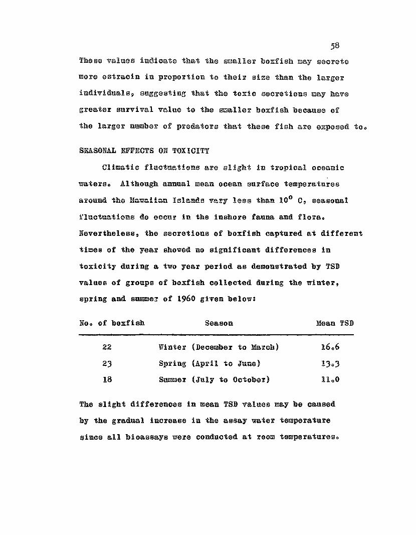
58
Thess values indioate that the smaller boxfish may secrete
more ostracin in proportion to their size than the larger
individualsp suggesting that the toxic secretions may have
greater survival value to the amaller boxfish beoause of
the larger numbex of predators that these fish &re exposed too
SEASONAL EFFECTS ON TOXICITY
Climatic fluctaations ere slight in tropical oceanic
waterso Although annual mean ocean surface temperatures
around the Hawaiicn Islends very less than 100 C, seasonal
fluctuations do oocnr in the inshore fauna and florae
Nevertheless, the seoretions of boxfish captured at different
times of the year showed no significant differences in
. toxicity during a two year period as demonstrated by TSD
values of groups of boxfish collected during the winter,
spring and Bumme~ of 1960 given below:
Noo of boxfish Season Mean TSD
22 Winter (December to March) 1606
23 Spring (ApFil to JUDS) 1303
18 Summer (July to October) 11 0 0
The slight differenoes in meen TSD v&lueB may be caused
by the gradual inorease in the assay water temperature
since all bioassays were conduoted at room temperatureso

59
EFFECT OF LOCALITY ON TOXICITY
Most boxfi~h investigated were captured in Kowala
Basin= Ala Moana ~eef area in Honolnluo The few boxfish
captured at Diamond Head beach park g Kenaohe BaYI) hnu1uu
and Nanakuli p Oahu were as toxio as KawaloeAla Moana fisho
EFFECT OF AGE AND SEX ON TOXICITY
Juvenile boxfish p &S well as mature cales and females,
secrete ostracin under stres80 No obvious differenoes in
secretory toxicity was found among male and female fish
although juvenile boxfish appeared to be almost as toxic
a.s OOultso
EFFECT OF METHOD OF CAPTURE ON TOXICITY
While the seoretions of netmcaptured boxfish were
usually highly toxic, those of boxfish caught in fish traps
were often less sOo The average TSD values for trapoo&ngbt
fish were about 17 0 0 to 18 0 0 while for net_captured fish
they were about 11 0 0 to 1200 0
Since it is known that o8trscin is only secreted
under stress, the prolonged stress conditions caused by
confinement in a fiah trap might cause a premature loss
of oBtracino
TOXICITY OF DOXFISH IN CAPTIVITY
Boxfish collected in Kewalo Basin during February
July 1960 were routinely assayed for toxicity and held

60
in captivity fOF varying periods of timeo These oaptive
boxfish were then periodically assayed for ostreoin
secretions to deteFmine whether a 1080 in toxicity occurFed
during confinement o
One group of fish was kept in B lOO=gallon aquaFium
and led frozen brine shrimp and a oommercial tront food
(Purina fry fead)o However, boxfish p espeoially the larger
oneB~ were often reluctant to feed in captivityo ThuSD
only 4 boxfish were observed feeding regularly (feeding
group) while 7 boxfish would not eat the prepared diet
(non-feeding group)o
Another group of boxfish was kept in a wire live cage
in Kewalo Basin and was not fed (starved group)o
The TSD values of secretions collected from these 3
groups are presented in Table 110 These data show a great
variability in ostracin secretion of captive boxfish,
but firm conclusions cannot be made because too many
variables were uncontrolled and too few boxfish would
feed in ceptivityo
Neverthelesso it was clear that the capacity of boxfish
of all 3 groups to secrete ostracin under stress was
not greatly impaired by confinement of 2~3 waekso This
period was extended much longer in feeding fish o In one
instance a moderately high level of ostracin was found in
a feeding boxfish after almost 3 months in an aquarium
while the ostr&cin secretion of the other 3 feading fiSh
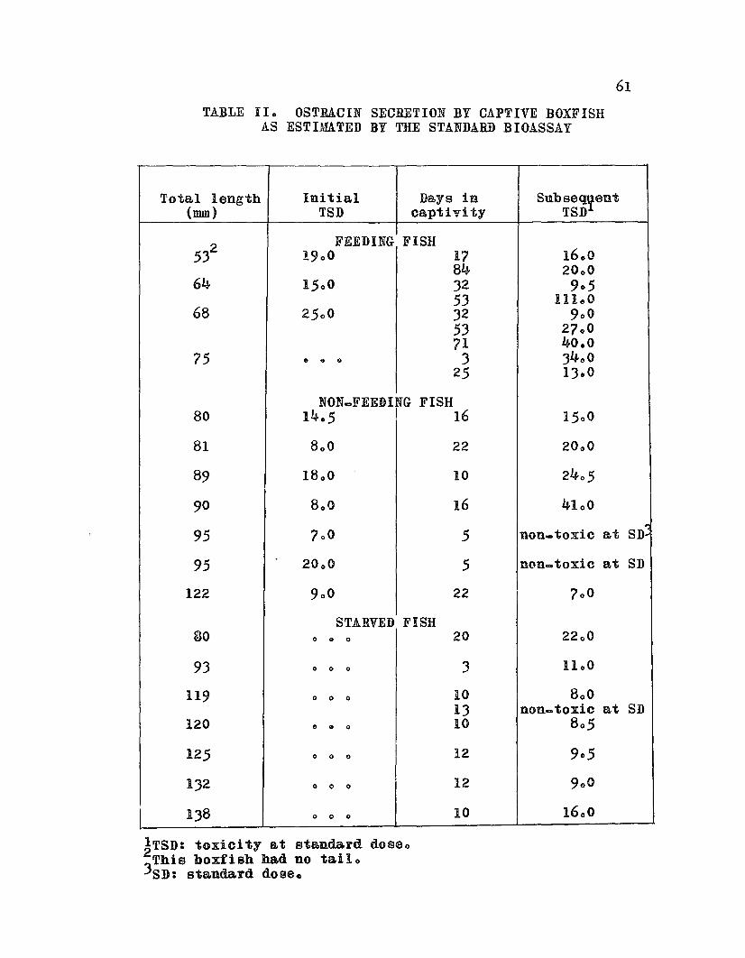
TABLE 110 OSTRACIN SECRETION BY CAPTIVE BOXFISHAS ESTIMATED BY THE STANDARD BIOASSAY
61
Total length Initia.l Days in Subseqyent(nun) TSD ca.ptivity TSD
532 FEEDING FISH
1900 17 160084 2000
64 1500 32 90553 111,,0
68 2500 32 90053 270071 40.0
75 e .. 61 3 340025 13eO
NONeFEEDING FISH80 1405 16 1500
81 800 22 2000
89 18 00 10 2405
90 8 00 16 4100":
95 700 5 non-toxic at SD
95 20 0 0 5 non... toxic at SD
122 900 22 700
STARVED FISH80 o 0 0 20 2200
93 o 0 0 3 1100
119 o 0 0 10 80013 non-toxic at SD
120 o 0 0 10 805
125 o 0 0 12 905
132 o 0 0 12 9 0 0
138 o 0 0 10 1600
~TSD: toxicity a.t standard dosao
3This boxfish had no tail o
SD: standard doseo
62
first sharply increased in captivity, then gradually
became reducedo
The non_feeding and starved boxfish became emaciated
(marked by a "caving inn of the sides of the carapace)
and rarely lived for more than a month in captivityo
Unfortunately these (starved) fish were not assayed after
J weeks because a tsunami destroyed the live cage in which
they were being kept and all fish were losto Nevertheless,
some of these boxfish contained a moderately high amount
of ostracin in their mucous secretions when assayed 10 to
20 days after captureo
DISCUSSION
The in~e8tigation of variabilities in the occurrence
of ostracin in boxfish mucous secretions was prompted by
the great fluctuations of other marine biotoxins of both
exogenous and endogenous origin. The slight fluctuations
found in ostracin secretion of freshly captured fish
appeared to be only those normally expected due to experi_
mental error and did not suggest environmental influenceso
The consistency of ostracin occurrence in secretions
of various-sized boxfish captured in different places at
different times of the year, and the capacity of aquarium
boxfish 9 maintained on an unnatural diet, to secrete
ostracin under stress indicate that ostracin is metabolic
ally produced by the boxfish independent of specific

precursors of environmental oFigino Thus» ostF8ciID tao
in all probability~ an endogenous toxino

64
CHAPTER 8
GENERAL PHYSICAL AND CHEMICALPROPERTIES OF OSTRACIN
The refinement of a preparative isolation procedure
for ostraoin depended npon the physical and chemical
oharaoteristics of the toxiuo Knowledge of suoh properties
as solubility and stability of ostracin were of primary
importance in deciding upon an appropriate separation
systemo Both crude and semiDpure ostracin solutions ~ere
used in determining the general nature of the toxino
STABILITY OF AQUEOUS OSTRACIN SOLUTIONS
The stability of crnde ostracin solutions, either when
frozen or at room temperatures p was very unpredictableo
Initially, the crude secretions were collected and stored
frozen in sea watert however a great reduction in toxicity
was often noted after frozen samples were thawed and
bioassayedo Toxicity data on crude sea water ostracin
solutions stored frozen and at room temperatures have been
presented in Tabla 1110 The only significance of these
data was its great variabilityo
Some breakdown of active compounds was expected at
Foom temperatures but the significant 1088 in toxicity
that ocourred in Borne frozen samples was surprisingo This
unexplained instability of crude ostracin in sea water

TABLE III. THE STABILITY OF CRUDE OSTRACINSOLUTIONS STORED FROZEN AND AT ROOM TEMPERATURES
Volume of Initia.l Duration Finalsolution (mI) TSD of storage TSD
FROZEN IN SEA WATER
10 700 62 days 605
10 900 2 days 1205
10 805 85 days 1)00705 (5xSD)
40 16 0 5 41 days 5200 (5xSD)
50 7 0 0 1 day 1000
50 900 1 day 4)00
50 6 0 0 51 days 3400
50 .., " '>.{:. deys 9~O(e V j-
STORED IN SEA WATER ATROOM TEMPERATURES
50 7 0 0 1 day 1000
50 900 1 day non... toxic
5° 700 4 days non-toxic6300 (3xSD)
50 6 00 3 days 5200 (5xSD)
100 800 It hours 400
500 50 0 3 days 1100

66
led to future collection and storage of boxfish secretions
in distilled wateFo Toxicity was better retained in frozen
distilled water solutions, although in one notable instanoe
when the combined mucous secretions of about 25 trapocaugbt
boxfish were frozen in distilled water, they were only very
weakly toxic when thawed and assayed 1 month latero
Thus, the unpredictable reduction in the activity of
crude ostracin solutions prohibited prolonged storage of
crude secretions end necessitated eBrly treatment of samplese
HEAT STABILITY OF OSTRACIN
Ostracin was found to be a very thermostable toxino
Boiling crnde ostracin solutions had no effect on toxicityo
Sawpies of
heated'for a total of 3 hours in a hot water bath (91 a 94°c)
and aliquots were assayed for toxicity at intervals of 15,
40, 105 and 180 minuteso*
Ostracin solution
distilled water
sea water
Initial TSD
1100
Final TSD(after 3 hrso)
The reduction in the toxicity of the sea water e ostracin
solution occurred during the first 15 minutes and no further
~ TSDis recorded at intervals destr9yed by fire; only finalTSD value available~

67
change was noted~ again emphasizing the greater
instability of ostracin in sea water (see preceding table)o
It was possible to boil an aqueous ostrRcin solution
to dryness without appreciable 108s in toxicityo In faot n
boiling an ostracin solution for a few minutes appeared
to have a stabilizing effect on toxicity as such solutions
could be stored for prolonged periods at room temperature
or under refrigeration without serious 1088 in toxicityo
ACID-ALKALINE STABILITY OF OSTRACIN
Ostracin was relatively stable over a pH range from
200 to 11 0 6, however with the addition of excess strong
base (KOH) ostracin was completely detoxifiedo
To determine the effect of pH on ostracin, a 001%
ostracin solution in 009% NaCI was prepared and 1 ml of
this solution was pipetted into several vials to which was
added 1 ml of an appropriate phosphate buffer to obtain a
pH range of about 4 0 0 to 900. Acetic acid and 100% KOH
were added to oiller vials to extend the pH range beyond
40 0 and 9.00 The pH was recorded with a Beckman Zeromatic
pH meter and the vials were refrigerated for 36 hourso
The pH was then recorded again and an aliquot from each viel
was assayed for toxicity by the standard bioassayo
A second experiment was performed using distilled
water ostracin solutions made alkaline by addition of 100%
KOH and refrigerated for 45 hourso The data of both experiments
68
are presented in Table IVo
The data of these experiments indicate that the effect
of acidity ot alkalinity was not a significant factor in
the detoxification of ostracin sempleso
POSSIBLE BACTERIAL DECOMPOSITION OF OSTRACIN
The instability of erude ostracin solutions and the
stability of heated ostrecin solutions Buggested a
possibility of bacterial detoxificationo Experiments were
performed to ascertain the possible role of bacteria in
detoxificationo Samples of semi=pure ostracin and cTUde
ostracin in distilled water were treated as follows:
control (no treatment); heated to boiling and &sceptically
stoppe~edi heated to boiling and alluwed tu cuol hefuE6
stoppering (not &sceptic); addition of an antibiotic
(streptomycin sulfate); filtration through a bacterial
filter (pore size 0 0 45 u)o All solutions were kept in
test tubes at room temperatures and tested after en interval
of 51 days and Bome after 72 dayso The results are shown
in Table Vo
Th0 only significant loases in toxicity occnrred in
the crude ostracin solutions except when these solutions
were heatedo Apparently the antibiotic and bacterial filter
were less effective than heating, indicating that perhaps
conditions were not really aeceptico
To determine whether crude boxfish secretions contained
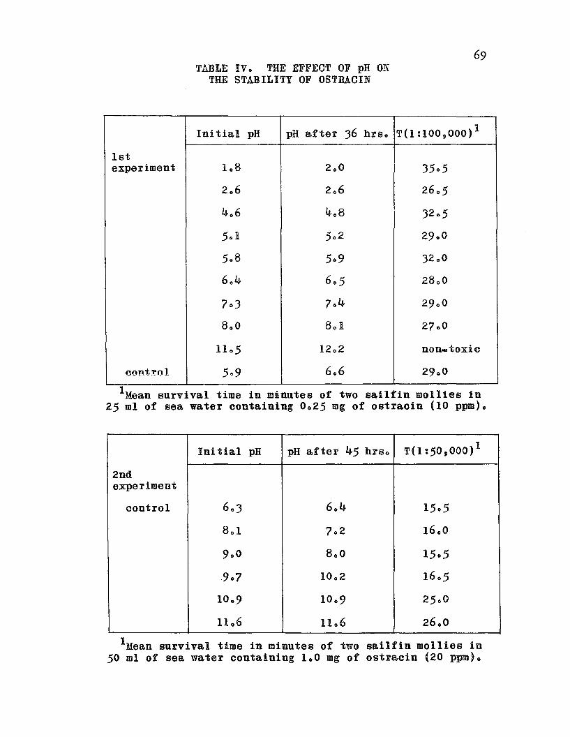
69TilLE IV 0 THE EFFECT OF pH ON
THE STABILITY OF OSTRACIN
Initial pH pH after 36 hrso T(l : 100 9 000 )1
1stexperiment 108 2 00 3505
206 206 2605
406 408 3205
501 502 29 00
508 509 3200
60 4 605 28 00
703 704 2900
800 801 2700
1105 12 02 non-toxic
cont!'ol 5,,9 6.6 2900
IMean survival time in m~nutes of two sailfin mollies in25 ml of sea water containing 0 0 25 mg of ostracin (10 ppm).
Initial pH pH after 45 hrso TO :50,000) 1
2ndexperiment
control 603 6 .. 4 1505
8 01 702 16 00
900 8 0 0 1505
.907 10 0 2 1605
1009 1009 2500
11 06 1106 26.0
IMean survival time in minutes of two sailfin moilies in50 mi of sea water containing 1.0 mg of ostracin (20 ppm)o
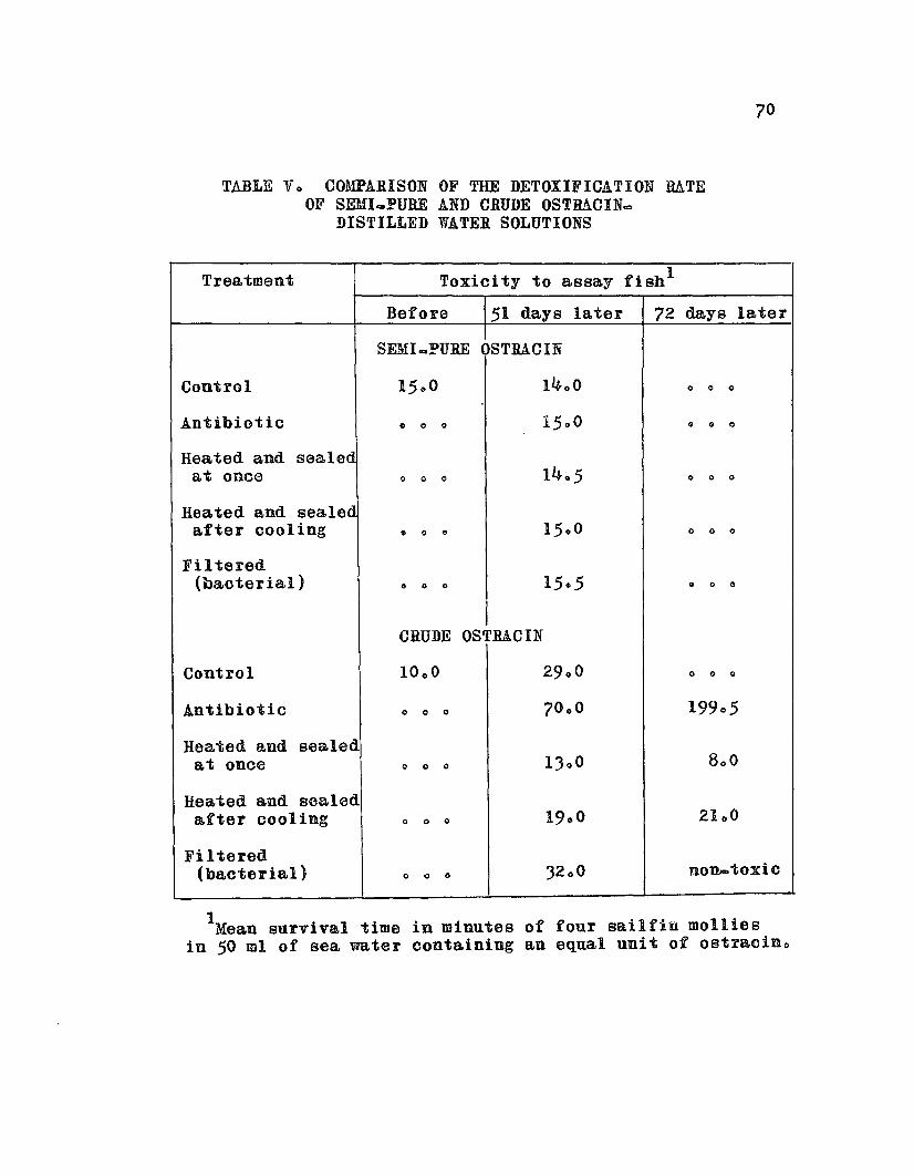
70
TABLE Vo COMPARISON OF THE DETOXIFICATION RATEOF SEMIQPURE AND CRUDE OSTRACIN=
DISTILLED WATER SOLUTIONS
Treatment Toxicity to assay fish l
Before 51 days later 72 days later
Control
Antibiotic
Heated and sealedat once
Heated and sealedafter cooling
Filtered(bacterial)
Control
Antibiotic
SEMI.PURE OSTRACIN
1400
o " 0
00"
• " e
" 0 "
CRUDE OSTRACIN
o " 0
o " "
" <II "
" 0 "
o " "
000
o " 0
19905
Heated and sealedat once
Heated and sealeda.fter cooling
Filtered(ba.cterial)
" 0 0
o 0 0
o 0 "
1300
3200
2100
non...toxic
1Mean survival time in minutes of four sailfin moillesin 50 ml of sea water containing an equal unit of ostracin"

71
a detoxifying agent (such as an enzyme) or if sea water
itself could cause detoxification, another experiment was
performed using crude and semi-pure ostracin in sea water
solutions kept at room temperatureso The results are
shown in Table VIo
Since heated sea water_ostracin solutions retained
toxicity and unheated ostracin=sea water solutions lost
their toxicity to a considerable extent, it appeared that
sea water in itself was not responsible for detoxification
although the possibility still exists that a detoxifying
agent (enzyme) occurs in the crude secretionso
Possible detoxification by oxidation could be another
explanationo Since heating would drive off most of the
dissolved oxygen from an ostracin solution, its stability
could be explained by lack of oxygeno Preliminary experi
ments showed that bubbling pure oxygen or air through an
ostracin-sea water solution for I hour caused considerable
loss in toxicity. On the other hand, bubbling oA~gen
through a heated ostracin-sea water solution for 1 hour
caused no appreciable loss in toxicity, indicating that
oxygen per ~ does not cause detoxificationo
In conclusion, it, appears that bacterial action is
at least partly responsible for losses in toxicity of
ostracin sampleso Abbott and Ballantine (1957) noted that
the toxicity of Gymnodinium cultures decreases when the
bacterial population1becomes high, although Shilo and
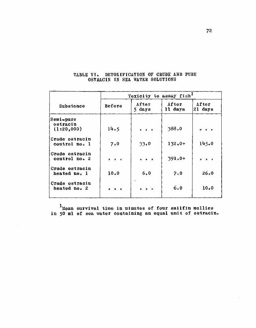
72
TABLE VIo DETOXIFICATION OF CRUDE AND PUREOSTRACIN IN SEA WATER SOLUTIONS
Toxicity to a.ssay fish1
Substance Before After After After5 days 11 days 21 days
Semi-pureostracin(1:20,000) 1405 0 0 0 38800 ? 0 •
Crude ostracincontrol noo 1 700 33.0 132 .0+ 14500
Crude ostracincontrol noo 2 0 0 0 0 • 0 39100+ 0 0 .,
Crude ostracinhea.ted noo 1 10.0 600 700 2600
Crude ostraoinheated noo 2 0 e 0 0 0 0 600 1000
IMean survivel time in minutes of four sailfin molliesin 50 ml of sea water oontaining an equal unit of ostracin.

73
Rosenberger (1960) reported that the fo parvum toxins
undergo rapid inactivation under field conditions, and
even in bacteria=free laboratory cultureso The 108s in
toxicity of frozen ostracin samples suggest that other
faotors, such as detoxifying enzymes p are operating since
rapid !nBctivBtion oocurred only in cruds, unheated sscretionso
SOLUBILITY OF OSTRACIN*
Solubilities were determined by heating an ostracin
solution to dryness, weighing the residue and mixing it
in the desired solvent at room temperatureo The solution
was then centrifuged and the supernatant fluid was filteredo
The filtrate was dried and the residue diseolved into water
and assayed for toxicityo
Ostracin was very soluble in water and was less soluble
in other polar 891venta sucb as metbanol, ethanol and
aoetone. The solubility of ostracin was greater in absolute
methanol and 50 to 95% ethanol than in absolute ethanol
and ostracin was only partly soluble in acetoneo Ostracin
was insoluble in the non_polar solvents diethyl ether and
benzene, but was very soluble in ohloroform. Liquid=
liquid extraction of ostracin from distilled water by
chloroform was BuccesBfu19 although after vigorous shaking
of these liquids in a separatory flask and standing over=
night p the water layer was significantly more toxic than
the chloroform layer (T 1:50,000 = 10 0 0 of the water
~ Since ostracin is a relatively large molecule it formscolloidal dispersions rather than true 801ut10D80

layer and 1805 of the chloroform layero) None of the
specified solvents caused detoxifioetioDo
DIALYSIS OF OSTBACIN
Ostracin, as mentioned previously, was non~dialyzable,
indicating that it is a rather large molecale in colloidal
solution. Ostraoin shares this property with many other
marine ichthyotoxins, inoluding the sea cncumber toxin,
holothurin A, the red tide toxins of gymnodinium veneficum
and Prymnesinm parvum and the starfish poisonso
FOAMING OF OSTRACIN SOLUTIONS
Stable foams are very characteristic of ostracin
solutions and there appeared to be a direct correlation of
toxicity with degree of foaminesso The most toxic solutions
foamed readily and profusely when agitated and the general
toxicity of an ostracin solution conld be estimated by
its capacity to prodnce a stable, rich foam when shakeno
75CHAPTER 9
SPECIAL CHARACTERISTICS OF OSTRACIN
The superficial resemblance of ostracin to holothurin Ap
the steroid saponin from the sea cucumber Aotinopyga agassiz!
(Nigrelli Bnd Jako~ska, 1960), th~ GtQ~fiGh saponin
(Hashimoto Bnd Yasllmoto i 1960) end the red tide to~ins from
GYmnodinium venefioum (Abbott and Ballantines 1957) and
Prymnesium pervum (Shilo end Rosenberger o 1960) suggested
that osiracin might be a saponin-like compound o
Saponins* in general heve diagnostio properties which
distinguish them from other toxins. They are poisonous to
fish and many other coldeblooded animals; they are surfaceD
active agents whioh produce a soapy lather in aqueous
solutions; and they are hemolytio, i.eo, they lyse red
blood cellso Furthermore, steroid saponins, such as
holothurin and digitonin, form stable, non-toxic complexes
with cholesterolo
Since ostracin bad been shown to be icbthyotoxic and
to form soap-like fOBms D its effect on erythrocytes and
its reaction with cholesterol was etudiedo
~ The general term Dsaponinn refers to all the plantglycosides which possess these properties (eogo. digitonin)QUnfortunately, this term also refers to a specificcompound with an emperical formula of C12H52e170 To avoidconfusion p this saponin will be referred to as saponin (Merck)o
EFFECT OF OSTRACIN ON RED BLOOD CELLS
Whole blood was oollected from the hearts of several
speoies of bony fishes and stored in a sodium citrate
solution (8 g sodium citrate, 7025 g sodium chloride and
2005 g dextrose per liter distilled water) specially
prepared by the serological laboratory of the Honolulu
Biological Laboratory (UoS. Bureau of Commercial Fisheries)
for tuna bloodo The red blood cells (RaC) were washed
several times with oitrate-saline and stored under refrigere
atioDo Human, mouse, rabbit and toad erythrocytes wore
prepared and stored in a similar mannero
Qualitative Test for Hemolyeis and Agglutination
Semi-pure ostracin dissolved in citrate-saline was_0-~
!e!!gth
75 mm)o The wet end of this capillary tube was immersed
in a washed BBC suspension to draw the erythrocytes into
the tube. The dry end was sealed with modeling clay and
the oapillary tube was set vertically in a racke After
the RBC passed through the ostr&cin solution and settled
at the bottom of the tube, a reading was taken by comparing
the appearance of the RBC suspension ~ith a saline oontralo
If hemolysis oecurred~ hemoglobin from the lysed RBC would
color the suspension a uniform redo If agglutination
occurred, the RBC would clump as they settled in the tubeo
Ostracin, at a dilution of 1:1,000 caused complete
hemolysis of all RBC suspensions testedo At this CODa
centration, stFong agglutination of RBC preceded hemolysis

77
in all fish and rabbit RBC, whereas no agglntination was
observed in human or mouse ROC suspensionso Control solutions
of crude holothurin (aqueous extract of cuvierian tubules
of ActinopYga obesa) and saponin (Merck) caused complete
hemolysis but no agglutination of all RBC usedo
Agglutination of skipjack tuna blood occurred at
ostracin dilutions as high as 1:20,000 whereas hemolysis
was observed (in the capillary tubes) in dilutions as
great as 1:100,000 0 The list of species used and their
RBC reactions is given in Table VIlo
The so.called agglutination reaction which occurred
when 08tr&c1n was added to fish or rabbit erythrocytes
superficially resembled an antibody-antigen agglutination
by denaturing the protein impurities in the 08tracin sample,
it must be attributed to ostracin itself o Microscopical
examination showed that a characteristic apparent reduction
in size of the agglutinated and hemolyzed erythrocytes
occurred within seoonds after introduotion of ostraoin
(see Figures 8 and 9)0
Hemolysis and agglutination of boxfish erythrooytes
by ostracin occurred in vitro and both reactions appeared
as strong as those using other RBCo Undoubtedly some
mechanism has evolved in the boxfish to proteot the fish
from its own toxin although complete protection from the
active toxin has not been achieved judging by its harmful
TABLE VIlo OSTRACIN HEMOLYSIS ANDAGGLUTINATION OF THE ERYTHROCYTES
OF S01m VERTEBRATES
78
SpeciesHemolysis
Aggluti_nation
Conger marginatus (Conger eel) + +
Gymnothorax spo (Moray eel) + +
KatBuwonus pelamis (Skipjack tuna) + +
Thunnus ala1unga (albacore) + +
Neothunnus macropterus (Yel10wfin tuna) + +
Parathunnus sibi (Bigeye tuna) + +
CoryPhaena hippurus (dolphin) + +
Abudefduf abdominalis (mao mao) + +
Acanthurus sanvicensis (manini) + +
Scaru8 spo (parrot fish) + +
Ostracion lentiginosus (boxfish) + +
.!!!!.f2 marinus (toad) + 0 0 0
Mouse + -Rabbit + +
Man + -
+: reaction occurred
_: no reaction
ostracin concentration: 1:1,000
Figure 8. Erythrocytes of skipjack tuna(Katsuwomis pelamls) before additionof ostracin. x440.
Figure 9. The same erythrocytes seconds afteraddl tlon of ost·racin showing agglutinationand hemolysis. Only the cell nuclei of theerythrocyte ghosts are visible. x440.
80
effect on erythrocytes and general toxicity to the boxfish
(see pages 42 c 43)0
The toxic action of ostracin on erythrocytes is probably
not a direct cause of its lethal effect to assay fish since
examination of the blood of fish killed by ostracin failed
to show any evidence of hemolysis or agglutination.
9uantitative Hemolztic Test for Ostracin
To determine the relative potency of ostracin as a
hemolysin, dilutions of semi-pure ostracin of 0 0 1 to 20 ppm
were prepared to which O~l mi of a tuna ROC suspension was
added. The test tubes were shaken and allowed to stand at
room temperature for I - 2 hours. • They were then centri.
fnged for 5 minutes and the supernatant fluid was tranSq
farred to clean test tubes to determine the percent
transmittance at wave lengths of 510, 525 and 540 ~, using
a Beckman Spectrocolorimetero The transmittance of the
RHC.saline control was set at 100%. The results of one
experiment are graphed in Figure 10c
Some hemolysis of tuna blood oocurred at very low
ostracin concentrations (0 0 1 ppm). This roughly corresponds
to the approximate LD50 of assay fish obtained with semi.pure
ostracin (see Figure 7). The sensitivity of RBC to lysis
by ostracin made a quantitative hemolytic bioassay possibleo
However, since such a bioassay required frequent time
consuming standardization, it was deferred in favor of the
simpler fish bioassayo

0N
l!•I0
~..U
"tu.=..-11..;•
:% IA. cL -0Z CI
c.- iz-Co) 1Jcar: •~ @lm -0 i~ ti ..I •!i Jb.Ifnl&- I0
&D AZ 'I2!« '5at: •... 1-z
~ )0 ::I:
gClt>.=0i&:i -~-~- --i- -r~-~--T--2- 0
J.NV.1.YNH3clnS 1f'Y!»"\".1'".~ .1N3:> Y3d
82
PRECIPITATION OF OSTRACIN BY CHOLESTEROL
At pres08t, all saponins and s&pogenins (the hydrolytic
prodnot of saponins) are divided into two large classes:
the steroids and the triterpenoidso Although both types
of saponins are hemolytic, only some steroid saponius
form water-insoluble binary complexes with cholesterol
(Ransom, 1901; Sokolskaya, 1951; Wall et alo, 1952)0
Holothurin A, the steroid saponin of the sea cuoumber,
forms a complex with cholesterol and this property has been
useful in the purifioation of this toxin (Chanley et al0 9
1959)& Abbott and Ballantine (1957) found that 20 mg of
cholesterol added to 100 ml of sea water containing a
highly toxio U]mnodinium culture gave complete protection
to the assay fisho Although the authoTs did not give
reasons for this, B.C. Abbott (personal communication)
suspected that the Gymnodinium toxin was a saponin.
Thus, the saponin-like aotivity and properties of
ostracin led to investigation of the reaction of ostraoin
w~th cholssterolo
Materials and Methods
First Experiment
Cholesterol was dissolved in hot 95% e~hanolo Test
tubes oontaining 4 mg of semi=pure ostracin in 95% ethanol
were prepared and 20 9 50 and 100 mg cholesterol, rsspectivelY9
were added to 3 test tnbeso Ethanol controls of both
cholesterol and ostracin were also prepared and all solutions

83
were kept at room tamperatureo After 2 days, eliquots of
each test tube were dried and the residues were assayed
for toxicity by the fish standard assay and for hemolytic
activity by the capillary tube methodo
Seoond E!perimeni
The first experiment was repeated using 5 mg of a
different ostracin preparation together with 10, 25 and
50 mg of cholesterol in 95% ethanolo The test tubes
were refrigerated for 5 days and then assayed for toxicity
as described in Experiment 10
Third Experiment
About 20 mg of a semi_pure ostracin preparation
(chloroform extract) was dissolved in absolute methanol
containing 20 mg of cholesterol and refrigerated for 2 dayso
The residue was assayed for iohthyotoxic and hemolytic
activitYo
Results
Both the precipitate and the supernatant fluid of all
ethanol cholesterolcostracin solutions were nOD=toxic to
assay fish, whereas the ethanol.ostracin control lost no
toxicityo Besides losing its ichthyotoxic activitY9
cholesterol-treated ostracin was non_hemolytic and no~
agglutinative when t05ted with fish RBCo The methanol
ostracin-cbolesterol solution retained some ichthyotoxioity
and hemolytic-agglutinative activity, but heating an aqueous
solution of the methanol cholesterol-ostracin residue for
84
a few minutes caused considerable reduction in total activitYo
SOIDe of the activity of the ostracinmcbolesterol residue
was restored after extraction with cold ethero Dissolving
the ostracinocholesterol complex in hot pyridine followed
by extraction with cold ether of the residue after the
pyridine bad been removed by evapo~ation oaused greator
restoration of activitYo ReQoval of the ether~goluble
cholesterol left an etherQinsoluble~ water=801uble icilthyo~
toxic precipitateo No toxic precipitate was formed when
ether was added to a pyridine solution of the ostracin=
cholesterol complexo
Discussion
The relationship between hemolysis of red blood cells
by saponins and precipitation of steroid saponins by
cholesterol becomes clearer when we consider how a lytic
substance &cts o Danielli (1958) states that a lytic substance,
such 8S saponin, penetrates into the cholesterol film
surrounding the RBO to form & stable complex with the
cholesterol. As saponin penetrates, it disperses the
protein monolayer causing the leakage of hemoglobin, or
hemolysiso
Ruyssen (1957-) has shown that saponins, such as digitonin,
have no solubilizing action on cholesterol as do other
hemolytic agent8~ such as butanol, detergents, etcQ' and
that an equimolar digitonin-cholesterol complex remained
combined on the ghosts of the erythrocytes hemolyzed by
digitonino Thu89 combining saponin with cholesterol prior

85
to addition to a RBC suspension prevents hemolysiso
Daniell! (1958) compared the capacity of surface active
or lytic agents to penetrate cholesterol mono layers and
found that, in general p increase in number of =CH2= groups
in the molecule increased penetration and that the effect=
iveness of polar groups of suoh agents gave a series:
-Iffi3+ >-804":;7 .80j > ...000· > ""H(<fH3); 0 Danielli pointed out
that whereas substances that penetrate and disperse protein
monolayers are oytolic; substenoes which are adsorbed on
protein monolayers, but do not disperse them are agglutin
ative but not oytolioo It was shown that ostracin, unlike
holothurin and saponin (Merck), is both agglutinative and
hemolytic~ This suggests that initially ostracin is
adsorbed on the protein layer of RBC causing agglutination,
but as it eventually penetrates the cholesterol layer it
causes dispersion of the protein monolayer and consequent
hemolysiso
The parallelisms between ichthyotoxicity, hemolytic."
agglutinative activity and precipitation by cholesterol
strongly suggest that ostraoin p the active substanoe in
boxfish mucous seoretions p is a steroid saponioo

8 6CHAPTER 10
SUMMARY OF EVIDENCE FOR THE STEROID SAPONINNATURE OF OSTRACIN
As mentioned before, saponins have been defined as
plant glycosides that have the distinctive property of
forming a soapy lather in water (Fieser and Fieser, 1959)0
These glycosides* are extremely effective as surface active
agents because of their ability to lower surface tensioDo
This property is associated with their ability to alter the
permeability of cell membranes and to hemolyze erythrooyteso
ThUS, saponins exert a general toxicity on most tissues,
however oertain saponins possess potent physiological
surface tension effectso Such saponins are sometimes'
referred to as Qsapotoxins A , and holothurin A, the steroid
saponin of the sea cucumber, is a prime exampleo
The principal products of the hydrolytic cleavage of
saponins are sapogenins (aglycone portion) and sugars
(glycosidic portion)o The aglycone struoture of a steroid
saponin consists of a reduoed cyclopentenophenanthrene
ring with a spiroketoacetal side ohain with methyl groups
~ Certain steroid glyoosides Which possess similar propertiesas saponins but exert a characteristio physiological effecton the heart are clas~ified as cardiac glycosides, e.g••digitoxin, one of the cardiac.active glycosldes isolatedfrom the leaves of the purple foxglove, Digitalis purpureao

87
attached to the 010 and 013 atoms and with a hydroxyl
(OR) group attaohed to the C3 atoIDo The glycosidic portion
(sugars) attaohes to the 03 hydroxyl group through a
glyoosidio linkage, or bondo The water solubility and
pharmaoological activity of saponius are due to this
glyoosidic portioDo The sapogenins are usually biologically
inactive because of their low solubility in water, but are
often more soluble than saponins in other solvents such &s
ether, benzene, etce (Sokolskaya, 1951)0
The properties of ostracin, as elucidated in the
preceding chapters, clearly indicate that this toxin is a
saponin. Furthermore, the precipitation and detoxification
of ostracin by cholesterol suggest that ostracin is a
steroid saponinQ Table VIII presents a comparison of the
properties of ostracin with two commonly known saponins,
digitonin and saponin (Merck)o From these data it appears
that ostracin is remarkably similar to both saponin (Merck)
and digitonin in general properties with the exception of
solubilities, wherein ostr~cin seems to be much more
soluble in a wider range of solvents than these saponioso
Ostracin, however, is considerably more toxic to fish
than saponin (Merck)o A conoentration of pnre saponin
(Merck) of 1:5,000 (200 ppm) was required to kill sailfin
mollies within 30 minutes whereas a semi.pure ostr3cio
concentration of only 1:250,000 (4 ppm) was lethal to
these assay fish in the same timeo Evidently ostracin,
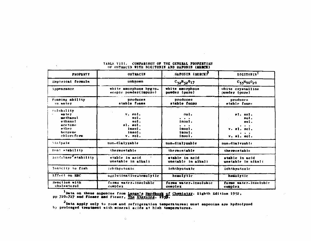
HULt, \'111. COMPAlllson or THE GEREIu.L PIiO.EITU;Sor USTKACIN WITH DIGITTORIR ARB HAPORIR (KlICK)
PlionaTY OSTII1Cn SA.ONIR CMEICK1 DIGITON I"1
t;l!Ipirical fOlllula unk!'own C)2HS2011 C~~H90029
·\PPf'arance white ••orphoull hYI:lO_ "hUe ..orphoVl8 ubit. crystalline~copic powder(i.puro) powder (pure) DOwde r (pu re )
-I ~'oaming abllHy produces produceo prOdUCf'Ri
\lI .•ater 8table foa.1I etable tOMS Il tab 1f' tOllllll'
~,,111bJlit1
- w. tE' r v. p.ol. 001. (III. Bol.lWfIthanol 1101. . . . 1'101 •('thano! 801. inool. Bol.aCt-tone sl. 1101. . . . . . ."tht>f i1ll801. iOBOI. T. • 1. 1!!01 •
I benzent' ineol. i8BOI. . . .eh lore· t() I'IIl v. eol. inllol. v. el. I!!ol.
'i 1/1 l:t ~ t " nOfl-dla1)'zab I. non.diahaDbte non.rlial vzah I f
IIpot " ••biUty thenloetablt- thenlOstable the 1'\11011 tab It·
Ae 1il-~!aHe~ R tab iIi ty Iltable in acid .tabl .. in acid etablt' in acidunstable in alkali unatabla· in alkal i unetablt' in alka!1
l"~icjty .0 fieh ichtbyotosic ichth70 t oz 1c lchtb1otos i ("
[Crt'ct on HUe ug~lutinatlve.hc.ol,tic hll.c> 11 ti c heaol,tlc
llt'BC t 100 .1 th for.~ •• tt'r-in8oluble fOIll. water.ineolublc tonas •• tt' r.i niJ() I ub If·cholesterol cQOIples cOIlIplel: cOlltplt's
1Data 0111 the.. onpoDio~ trom ~lIIle'. ~~J&!Cheal.t". Etchth ~dltlon 1952,
pp 720-727 aDd raeaeR aad Fie.er. --! Ster~~".2
Data .ppl, 01111, to r008 and refricer.tion te.perature•• moet ..poolae ar. h,drol,Bedb> prololllled t ...~t with .ln8ral acidl!! at hlah te-peraturee.
i
89
perhaps because of its solubility properties, is bio=
logioally more aotive than saponin (Merck) and should be
olassified as & UsapotoxlnU along with holothurin and the
starfish saponin (see Chapter 19)0
The resemblanoe of ostracin to steroid saponins is
suffici6at 6videnoe to tentatively ne~e the iohthyotoxic
hemolysin in bo~fi8h sec~etions °ostxacinMo Following
the nomenclature rules for saponios, the sapogenin should
then be named ~o8traoigenin~e This does not dismiaG the
possibility of other toxins OccuFFing in boxfish mucous
secretions, but until different toxic fractions can be
demonstrated, ostraoin will be Rssumed to be a single
toxinG
90
CHAPTER 11
PURIFICATION TECHNIQUES FOR OSTBACIN
Although the objectives of this thesis did not include
complete purification of ostraclu, a sufficiently pure
prepara.tion was needed to determine the chemi.cal cha.racter"",
Istic8 of the toxin o Many purification techniques were
attempted, including methods used by investigators in
isolating other sapotoxinse A summary of these techniques
and results is discussedo
PRECIPITATION WITH CHOLESTEROL
Chanley et alo (1959) successfully purified holothurin A
by precipitating it with cholesterol, reactivating the toxin
with pyridine, and precipitating the glycoside from pyridine
by ethero This procedure was attempted with ostracin,
however, this substance could not be readily precipitated
from a pyridine solution by ether and the method was
a.bandoned.
CHROMATOGRAPHY
Foam ChromatographY
Foam chromatography was tried because aqueous solutions
of ostracin, like all saponins, produced very stable foamso
Preliminary trials using compressed air bubbling through
crude ostracin solutions were partly successful but a
reduction in toxicity occurred in both the foam and the

91
foamateQ
Paper Chromatography
Paper chromatography has been successfully used to
separate saponins (Dutta, 1955)0 Although not a preparative
method, paper chromatography can be useful in determining
the numbeF of components in e ~ixtureo Ascendin~ paper
chromatograms were prepared with the following solvent
systems: n-butanol, glacial acetic acid and water (4:1:5);
ethyl acetate, glacial acetic acid and wateF (3:1:3); and
pyridine ethyl aoetate and wateF (2:1:2)0 The pBpeFs were
developed with ninhydrin, a silver nitrate-ammonium hydroxide
solution and a spray reagent developed by Lemieux and Bauer
(1954) for use in the detection of saponins e
The best separation occurred in the pyridine~ethyl
acetate.water (2:1:2) solvent system but ostraoin was
eluted from an unstained portion of the chromatogramo
The ninhydrin.stained band was non.toxic, and the Lemieux
and Bauer spray reagent stained only tbe non=toxic solvent
fronto Unless a spray reagent is found that will color
the toxic band of the chromatogram, paper chromatography
of ostracin will have only limited valueo
EXTRACTION WITH ORGANIC SOLVENTS
Acetone
Acetone soxhlet extraction* for 24 hours of the crude
* Done by David Boylan, Chemistry Department, Universityof Hawaiio

92
residue obtained after boiling an aqueous ostracin solution
to dryness was BUcc6ssfuio The oily yellowish acetone
residue dissolved in water killed assay fish within 45
minutes at a concentration of 1:500,000 0
Chloroform
The residue of a boiled aqueous ostraoin solution
was extracted with chloroform at room temperatureo The
ostracin in the residue was readily dissolved in chloroform
and the chloroform residue had a toxicity comparable to
that of the acetone residu6e
CENTRIFUGATION AND DIALYSIS
Initially, all semi.pure ostracin solutions were
prep9,red by centrifuging them at 15~OOO rpm~ discarding
the non.toxic precipitate, and dialyzing the snpernantant
fluid against running tap water or distilled watero The
toxic residue from dialysis was stored in distilled watero
It was later discovered that heating the aqueous ostraoin
solution stabilized the activity ofostracin and that
storage in chloroform was preferable to water since some
detoxification occurred, either by bacterial decomposition
or hydrolysis, in aqueous solutioDso
Thus, the scheme recommended for preliminary purie
fication of ostracin is presented in Figure 110 The
semi-pure ostraoin obtained in step IV WRS9 in most
instances, the preparation nsed for all toxicity axperi
mentse The addition of the chloroform extraction was a
recent refinement o
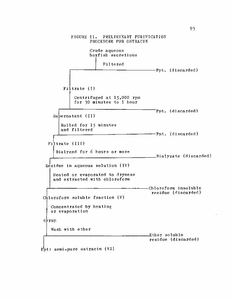
93
FIGURE II. PRELIMINARY PURIFICATIONPROCEDURE FOR OSTRACIN
Crune aqueousboxfish secretions
Filteredr----~-----------Ppt.(discarded)
Fil trate (I)
Centrifuged at 15,000 rpmfor 30 minutes to 1 hour
Ppt. (niscarned)Superna tant (II)
Boiled for 15 minutesand fi I tered
....-L- Ppt. (fl i sca rded )
F i I t ra t e (II I )
Dialy~erl for 6 hours or more~----------------------------------------nialyzate(discarned)
Residue in aqueous solution (IV)
Heated or evaporated to nrynessand extracted with chloroform
r-'-------------------------------------- Ch I 0 ro f 0 rrn in so I ubI eresidue (rliscarned)
Chloroform soluble fraction (V)
Concentrated by heatingor evaporation
Syrup
Wash with ether,-L-------------------------------Ethe r so 1ub 1e
residue (discarded)
Ppt: semi-pure ostracin (VI)

PREPARATION OF OSTRACIGENIN
An aqueous ostracin solution (fraction IV), acidified
by adding concentrated HCI (1 ml concentrated HCI to 5 ml
of 6 mg/m ostracin solution), was heated in boiling water
for 1 houro After 15 minutes a precipitate began to form
and after 1 hour an oily syrup collected at the surface
and sides of the glass containero This syrup was washed
several times with water and after drying in an oven was
completely dissolved in benzene. The toxicity of the
acidified supernatant was considerably reduced, indicating
that acid-hydrolysis occurred and that the syrupy fraction
was ostracigenin.
95
CHAPTER 12
TOXIC SECRETIONS OF OTHER PLECTOGNATHS
Reports of ichthyotoxins produced by trunkfishes
(Clark and Gohar, 1953; Brock, 1955; Straughan, 1959)
are suggestive that ostracin or ostracin-like toxins might
be found throughout the trunkfish familyo There are many
examples of similar toxins ocourring in closely related
organismso To cite a few marine examples, holothnrina
like toxins have been reported in many genera of sea
cucumbers (Yamanouchi, 1955; Nigrelli and Jakowska, 1960)
and there appear to be similarities in red tide toxins from
Gymnodinium venificum (Abbott and Ballantine, 1957) and
Gonyaulax monilata (Gates and Wilson, 1960). The pulfera
fishes, whioh include se~eral families~ay also contain
similar or identical toxinso The important unanswered
question is whether a single toxin persists throughout a
family or group of related families or whether analogous
but distinctly different toxins occur in different specieso
The answer to this question could have important significance
in understanding the phylogenetic relationships between
specieso
TRUNKFISH
Besides the boxfish, another common Hawaiian 1runkfish
is the cowfisb 9 Lactoria lornasini& This species was often
96
encountered in shallow uaters over sand flats while collect~
ing boxfisho It is a rather inconspicuous, slow swimming
trunkfish and was easily captured by dipnettingo
The mucous secretions of a few cowfish were collected
and assayed for toxicity, and of 19 cowfish (48 to 102 mID
total length)9 toxic eecFetions ~ere detectod in only
11 fish o The cowfish mucous secretions foamed B8 well 8S
those of the boxflsh, but the cowfish toxin appeared to
be muoh more labile than ostracino This toxin instability
probably accounted for the 8 nODQtoxic fish, as merely
filtering or centrifuging the cowfish toxin seemed to
cause considerable detoxificationo Cowfish secretions
also caused hemolysis and agglutination of fish ROC
indicating that the active principle may also be a
saponin-like toxino
The secretions of two other species of trunkfish were
collected and assayed for ichthyotoxicity and hemolytic
activity. One juvenile Lactoria diaphanus was captured
in a mid~~ter trawl by the Charles H. Gilbert, research
vessel of the U.So Bureau of Commercial Fisheries and
kept in a shipboard aquariumo The secretions were collected
from the weakened trunkfieh immediately, &s it was taken
from the trawl, and again a few days IBtero* The samples
were frozen and tested on shoreo Only the second sample
~ Collected by Robert Ao Morris
97
was iohthyotoxic, Bnd hemolyzed and agglutinated fish
erythrocyteso
Two specimens of a rare trunkfisb, tentatively
identified as Rhynoostracion ~o, were collected off
Pokai Bay, Oahu and kept in an aquariumo* The secretions
of both fish were collected and assayed and found to be
icbtbyotoxic, agglutinative and hemolytico
Thus, all fOUl Hawaiian trunkflshes examined contained
an ichthyotoxic hemolysin in their mucous secretions. The
similarities and differences of these toxins to ostracin
were not determined.
PUFFERS
!t ~~8 mentioned previously that the mucous secretions
of the puffer, !o bispidus, were highly toxic to mice. The
puffer mucus was collected by placing a puffer in a beaker
and rinsing the fish with distilled water. This aqueous
mucous solution, injected intraperiotoneally into white
mice and fish (Acanthurus sanvicensis), caused death within
a few minutes, depending upon the doseo
The puffer mucous t.oxin, unlike ostracin, was non
hemolytic and non-ichthyotoxic, i.60, it was non-toxic to
fish when dissolved in their aquarium water. However,
application of this puffer toxin in Ringeris solution
to an isolated soiatic nerve of the toad blocked the action
* Collected by Donald Eo Morris and R~bert A0 Morriso
~
potentials of the nerve in less than 1 minute, indicating
that a potent neurotoxin was presento
Two other species, the spiny puffer, DiodoD hxstrix
and a sharpbacked puffer, Canthigaster rivulatu8 also
produced mucous secretions that had a toxic effect in
fish siwilaF to that of !o bispi~us mucuSo
Th~ endog~nons toxin of the puffeFs, tetFuodotoxin
(or tetrodotoxin). has been reported to occur in the
puffers examined and has probably confused the pictureo
The relationship between the endotoxin and the toxic
mucous secretions has been investigated by WoRo Eger (1963)0
99
CHAPTER 13
SITES OF OSTRACIN SECRETIONS IN THE BOXFISH
Foamy oral exudations ~ere observed when freshly
captured boxfish were taken ont of watero Furthermore,
~80apyn mncous secretions appeared at the base of the
pectoral, anal and dorsal fins and at the oaudal peduncleo
Suoh mucous secretions were oharacteristio of all Hawaiian
trunkfish examined Bud although Aloaming of the mouth" has
been reported by Gordon (1937) in Atlantic trunkfish, he
attaohed no significance to ito
The definite oorrelation between foaminess of ostrecin
solutions and toxicity suggested a relationship between the
foamy mucus and ostracin secretion. During the earliest
stages of this investigation, both the oral foam and skin
mucus were found to be ichthyotoxic, thus it became
important to search for poisonous glauds in the mouth and
integument of the boxfishe The approach to this problem
consisted of three parts: (1) to collect secretions from
various portions of living boxfish and assay them for
ichthyotoxio and hemolytio aotivitY9 (2) to extract various
tissues with different solvents and assay the extractsa and
(3) to conduct a thorough histological study of the
probable glandular tissueo
100
ASSAY OF ORAL AND SKIN SECRETIONS
Materials ~ Methods
The oral foam WRS separately collected from 12 boxfish
with an eyedropper while a cloth was wrapped around the
fish l s body just posterior to its eyes to prevent skin
a~cr0tionm fFom conterninBtine the sample. However, it was
difficult to prevent skin secretions from being mixed with
the oral exudations since the oral foam wonld cling to the
skin of the lips and head of the boxfiaho If the eyedropper
was ins6Fted into the boxfishls mouth the oral secretion
was inhibitedo In addition, the foamy mucus produced at
the pectoral fin base could possibly be sucked into the
branchial cavity through the opercular opening and eventually
be exuded through the moutho
The skin secretions were collected by wrapping the
entire head of the boxfish with cloth and rinsing the skin
with water, or pipetting off foamy mucus directly with an
eyedroppero Samples were assayed for hemolytio and/or
ichthyotoxic activitYo
Results
The oral 80cretions of only 6 fish were demonstrated
to be ichthyotoxic and in one instance a sample was non
toxic to fish but caused hemolysis of tuna erythrocyteso
Ichthyotoxicity data on the oral secretions of 10 fish are
presented in Table IXo
The skin secretions, collected by rinsing the boxfish

TABLE IX. ICHTHYOTOXICITY OF THEOR~L SECRETIONS OF BOXFISH
101
Total Volume of Volume Volume Toxicitylength of diluted assayed of assay (mean survivalboxfish secretions (ml) sea water in min. of 2
(mm) (ml) sailfin mollies)
94 506 100 100 600O
105 1100 200 100 5°00
120 6 0 8 2.0 I 50 8600
113 202 202 50 non-toxic
95 6 0 4 604 50 non-toxic
95 6 03 603 50 2700
117 2 I, 2 .. 4 ~() 36,,00..... ./-
691 1.6 0 0 1 15 non-toxic
75 101 001 15 non_toxic
80 2 01 0.05 15 59.00.10 15 4700
1The samples from these fish were hemolytic; theothers were not tested ..

102
with water, were considerably more toxic than oral secretions
and in the one case tested with BRC, caused strong agglutin=
etlon of tuna erythrocytes prior to hemolysis o Ichthyotoxicity
data on skin secretions are presented in Tabla Xo The
failure of the oral secretions tested to cause agglutination
was probably due to ostracin concentration since agglutinQ
ation occurs only at strongly ichthyotoxic concentrations
(I : 20 t 000 ) 0
The date indicate that the major secretion of ostracin
occurs in the skin, and assuming that the oral exudations
were not contaminated by skin secretions, ostracin is also
produced in the oral cavitYe The nonotoxicity of the 2
skin samples may be due to the dose used in the &ssay,
whereas the non.toxicity of the 4 oral secretions could
mean that the skin secretioDs were actually responsible
for the 6 toxic reactions obtained with oral foarnQ
EXTRACTION OF OSTRACIN FROM BOXFISH TISSUES
Materials ~ Methods
Various extraction techniques were tried and the
extracts were assayed for ichthyotoRic and hemolytic activitYo
In all oases the tiElsues were washed thoroughly in distilled
water to remove snperfioial secretions prior to extractioDo
The extraotion methods and the tissues used are listed
eeparatelyo
(1) A 110 mm long female boxfish was killed immediately

TABLE X. ICHTHYOTOXICITY OFBOXFISH SKIN SECRETIONS
103
Total Volume of Volume Volume Toxicitylength of diluted assayed of assay (mean survivalboxfish secretions (ml) see water in min. of 2
(mm) (ml) (ml) sailfin mollies
94 1200 2 00 100 1005
105 1500 300 100 2005
ooo? 700 lc,4 100 18 0 0
120 12 00 1.0 100 non-toxic
120 6.5 2.0 50 6 00
721 ooo?
80 01P0? 0.°5 15 185,,0
0.10 15 80.0
116 10 0 0 0.50 15 non_toxio
IThe skin secretions of this fish were stronglyhemolytic and agglutinative on tuna erythrocytes; theothers were not tested on blood o

104
after capture by immersion in a strong solution of MS 222
(tricaine methanesulfonate) and the following tissues
were dissected, extracted with sea water and centrifuged:
skin, muscle, esophogus, stomach, intestine, liver, kidneys,
ovaries, urinary bladder, and some 8ubcarapace cheek rojellyt3 o
The supernatant fluid was assayed for ichthyotoxic1ty by
the standard assayo
(2) The tissues of a large male boxfish which had been
frozen were homogenized in a blender and e~racted with
hot ethanol (95%~o The tissues extracted included the
gall bladder, viscera, liver p anal and pectoral fin bases,
gills, caudal skin, dorsal musculature, air bladder, lips,
oral cavity epithelium, tongue and the subcarapace cheek
"jelly"o The ethanolic extract was assayed for ichthyo~
toxicity by the standard assayo
(3) The lips and the tail of a 90 mm long juvenile boxfish
were severed at capture and frozen in sea watero After
thawing they were extracted with boiling distilled water
and the extract assayed for ichthyotoxicityo
(4) The lips of a 210 mID long male were severed at capture
and both the lips and boxfish were frozeno After thawing,
the lip skin was dissected from the jaws and both portions
were extracted with hot watero The caudal skin and epidermal
scrapings from the carapace were also extracted with hot
watero All extracts were tested for ichthyotoxio and
hemolytic activitYo

105
(5) The lips of a 175 mm long female were severed, frozen
and extracted with hot water and the extract assayed as in
(4) 0
(6) The caudal skin~ carapace, base of fins, lips and trunk
muscles of a 175 mm long female boxfish were extracted with
hot water followed by hot ethanol (70%) and the ethanol
extract was dissolved in a citrate saline solution and
a.ssayed for hemolytic activitYo
(7) The caudal and fin base skin of a 170 mm long male
boxfish were extracted with acidified (HCI) hot water and
the aqueous extract assayed as in (4)0
(8) The lips of 4 large boxfish were severed, frozen and
extracted with acidified (HCI) hot water and the aqueous
extract was assayed as in (4)0
(9) The lips, lateral and ventral carapace and caudal
skin of a 110 mm freshly captured female boxfish killed by
immersion in 35% isopropyl alcohol were cut up, extracted
with sea water and assayed for ichthyotoxicityo
(10) The lips of 4 freshly captured boxfish were severed,
extracted with citrate saline and assayed for hemolytic
activitYo
Results
All the extracts, with some exceptions in (3), (4), and
(5) were non-toxic and non.hemolytico Of the exceptions,
extracts of the lips in (3) and (5) and the jaws in (4) were
we~klY ichthyotoxico The jaws and lips in (4) and (5) were
106
strongly hemolytic but non-agglutinativ6o None of the skin
extracts had any ~ctivityo
The results obtained in (6) canno~ be oonsidered valid
since this fish had apparently been kept in captivity for
a long time judging by its emaciated appearance and small
livero
DISCUSSION
The contradictory nature of the seoretion and extraction
results presents an intriguing enigmao A high yield of
ostracin can be obtained from the stress secretions of living
boxfish but praotioally no ostracin could be extracted from
the suspected glandular tissues of dead boxfisho There are
at least three possible explanations to this paradox: (1) the
extraction techniques were inadequate or were causing
toxification; (2) the tissues extracted were not producers
of ostracin; or (3) ostracin is activated by some unknown
mechanism just prior to or during the actual secretion processo
The first explanation appears unlikely because the
extraction techniques employed work well for other saponius
and most water soluble, non-proteinacious toxinso DetoxifiQ
cation would only be possible in (7) and (8) where acidified
water was used.
The second explanation must be carefully considered,
but since all the major organs, with the exception of the
nervous and skeletal systems» were extracted and assayed,
it seems improbable that poison glands would be overlookedo
107
The foamy ostracin sscFetions appeared to form on all
epidermal areas of living, distressed boxfish and highly
toxic samples could be colleoted easily from the surface
of the skino
The third explanation appears at present to have the
most merito Since ostracin seems to be an active cytotoxin
with high ichthyotoxic potency, it Is possible that a
detoxification mechanism exists in boxfish tissue wherein
ostracin is stored in an inactive stateo During secretion,
ostracin might be activated by an enzyme or some other means
as it is extrudedo The boxfish, although highly resistant
to ostracin, can be poisoned by this toxin (see Chapter 5)
and hemolysis of boxfish erythrocytes by ostracin occurred
in vitro (see Chapter 9)0 Thus, an accumulation of active
toxin in the glandular tissues could bave an adverse effect
on the boxfish if ostracin secretory oells were damaged
and ostracin leaked into the blood stream.
The toxic extracts of the lips and jaws and the
non-toxic skin extracts present another paradox since the
oral secretions were only mildly toxic in comparison to
skin secretions. However, these results could not be
repeated and are probably artifacts caused by secretions
clinging to the surface of these tissueso
Further discussion of these problems follows the
histologioal study of the suspected glandular tissueo

108
CHAPTER 14
HISTOLOGY OF THE PRESUMED POISONOUS GLANDSOF THE BOXFISH: MATERIALS AND METHODS
The evidence discussed in the preceding chapter,
although contradictory and enigmatic, suggested that
poisonous glands occurred in the oral cavity and skin of
boxfishe A histological investigation was designed to
test this hypothesis and to describe the structure of the
glands and the nature of their secretionso
COLLECTION AND FIXATION OF TISSUES
Allboxfish used for histological study were oollected
in Kewalo Basin and on the Ala Moana reef, Oahu 0 The fish
were either killed immediately upon capture by immersing
them in a fixative or by pithing t or they were left in a
container with their toxic secretions until they diede
The purpose was to obtain glandular tissue before and after
the secretion processo Actually, it was almost impossible
to be certain that secretion had not occurred prior to
fixation since capturing and killing the boxfish were
undoubtedly "distressing" and the degree of stress secretions
remained an incalcuable variableo
Selected tissues were dissected from the freshly
killed boxfish and immediately immersed in an appropriate
fixativeo Fixatives used were: Bouin1 s fluid, 10% formalin
in sea water» 10% formalin in 70% ethanol» a modified Gilson1 s

109
fluid (formaldehyde used instead of merouric chlo7ide: 10 ml
95% ethanol p 004 mi glacial acetic acid~ 108 rol nitric acid D
5 ml formaldehyde and 88 mi distilled water, and Champyns
fluido Bouinus fluid was far superior to the other fixatives
in preserving cellular structure and secretory products, and
consequently became the standard fixative in this stndyo
Tissues selected for microscopical examination were:
the integument, which included skin from the caudal peduncleD
the folds at the junction of the carapace and caudal trunk,
the base of the pectoral, anal and dorsal fins, the dorsal,
lateral and ventral carapace and the lips; the oral and
bucoal cavity, which included the jaws, the tongue, the
palate to and including the esophagus and gills; the sub.
carapace cheek njellyn and the opercular flapo
All Bouin.fixed tissues were washed and stored in 70%
ethanol and certain tissues, e.go p the lips which contained
the jaws and teeth, the carapace and the palate were deQ
oalcified in J changes of a 5% nitrio acid solution for
a week to 10 daysG
DEHYDRATING, CLEARING, EMBEDDING,SECTIONING AND MOUNTING
Tissues were dehydrated with absolute ethanol and
cleared with either cedarwood oil, methylbenzoate celloidin
O~ toluene, with toluene giving the best resultso Tissues
were embedded in a medium grade paraffin (melting point
56 = 58° C) after infiltration for 3 to 5 hourso

110
The paraffin tissue blocks uere sectioned at thicknesses
of 4, 6, 8 and 10 po Serial s6ctions (cross, saggital OF
tangential) were prepared of all tissueso Most skin
s6ctions were cut at 6 PD whereas other tissues (lip, mouth
and carapaoe) were out at 8 to 10 po
The paraffin ribbons weFe afixed to microscopioal
slides with an albumen fixative diluted uith distilled
water (2 drops albumen to 20 ml water)o
STAINS AND STAINING
(1) Harris hernatoxylino This nuclear stain was used on
all sections to determine general histological structure o
Hematoxylin sections were counterstained with eosin or
(2) Heidenhaints iron hematoxylino Allents modification
of this stain was used with and without counterstains
(eosin or trioain)o Finer cyt~logical detail was possible
with this stain than with Harris hematoxylino
(3) Mallory!s triple staino This oonneotive tissue stain
was useful in differentiating the integumentary sections
and glandular seoretioDso Brilliant staining occurred
after Bouinls fixationo
(4) Gornori's aldehyde fuohsino This stain was developed
by Gomori (1950a) for elastic tis8ueso He states that the
staining properties of this dye are almost unique in that
it has been found to stain speoial types of seoretory
III
cells such as mast cellsg Beta cells of pancreas and
certain basophile of the anterior pituitary, and uill also
stain certain types of mucin and elastic fibers of all
tis8ueso This dye was used with a trichrome stain originally
developed by Gomori (1950b) and later modified by various
workers as follows: seotions first mordanted in a 4%phosphotungstic acid ~Q 1% phosphomolybdic acid solution for
10 minutes and stained for 1 hour in an aqueous trichrome
stain containing 004% fast green, 1% orange Gs 005% chromotrope
2R and 0 0 28% acetio acido
(5) Periodic acid leucefuchsin Schiff method (PAS).
The PAS procedure recommended by Conn et alo (1960) was
followed, although the nuclear and counterstains were not
used. This stain was employed to characterize epithelial
mucins and other oytoplasmic inclusionso
(6) Toluidine bineo This is a metaohromatic basis dye
whioh stains basophilic substances, including many acidic
mnains, and was used to characterize epithelial rnncinso
Both aqueous and ethanolic solutions were used following
the procedures listed in Pearse (1960, pgo 834)9
(7) Dresbach's mucicarmine methodo This method was used
to demonstrate epithelial mncinso
(8) Sudan Black Do This stain was used after fixation in
Champyts fluid to demonstrate lipids in the cytoplasm of
various glandular cells in paraffin sections (sea Pearse,
1960, pgo 850)0
112
CHAPTER 15
HISTOLOGY OF BOXFISH SKIN
Teleost skin typically consists of an epidermis of
s!~atifiad epithelium oontaining mucous cellsp sensory
:oe118 and ordinary squamous or cuboidal epidermal cells,
and a dermis oontaining scales of various types in different
specieB~ There are numerous variations of this basic
pattern and a few ourious specializatioDso
In some families of teleosts, serous gland cells and
specialized secretory cells of a dubious function occur in
the epidermiso Rabl (1931) has listed a few families of
bony fishes, widely separated phylogenetically, that
contain in their epidermis special secretory cellse called
nclub cel1s" because of their charaoteristio shape, which
are thought to be modified serous gland cellse Club oells
are characterized by the dischargin~ of their seoretion
while in the middle layers of the epidermiso The resulting
degenerate cell is often pushed onto the surface of the
epidermis and discarded (Andrew, 1959)0
There is an extensive literature on the distribution
and structure of these cells in the bony fishes (see Babl,
1931) and many speculations on their fUDctiono Because
these club cells are also associated with the poisonous
glands of venomous fishes such as the Scorpaenidae (soorpion
fishes), Trachinidae (weever fishes), Synancejidae (stonefishes)D
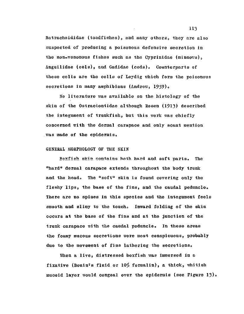
113
Batrachoididae (toadfishes)p Bud many othersp they are also
suspected of produoing a poisonous defensive secretion in
the nonwvenomous fishes such as the Cyprinidae (rninnous)p
Anguilidae (eels)~ and Gadidae (cods)o Counterparts of
these cells are the cells of Leydig which form the poisonous
secretions in many amphibians (Andreu, 1959)0
No literature was available on the histology of the
skin of the Ostraciontidae although Rosen (1913) described
the integument of trunkfish, bnt this work was chiefly
concerned with the dermal carapace and only scant mention
was made of the epidermis o
GENERAL MORPHOLOGY OF THE SKIN
Bo~f!sh skin contains both ha~d and 80ft parts. The
nhard~ dermal carapace extends thronghout the body trunk
and the heado The "soft a skin is found covering only the
fleshy lips, the base of the fins, and the caudal peduncleo
There are no spines in this species and the integument feels
smooth and slimy to the toucho Inward folding of the skin
occurs at the base of the fins and at the jnnction of the
trunk carapace with the caudal peduncleo In these areas
the foamy mucous secretions were most conspicuous, probably
due to the movement of fins lathering the seoretionso
When a live, distressed boxfish was immersed in a
fixative (Bouin's fluid or 10% formalin), a thick~ whitish
muooid layer would congeal over the epidermis (see FiguF6 13)0
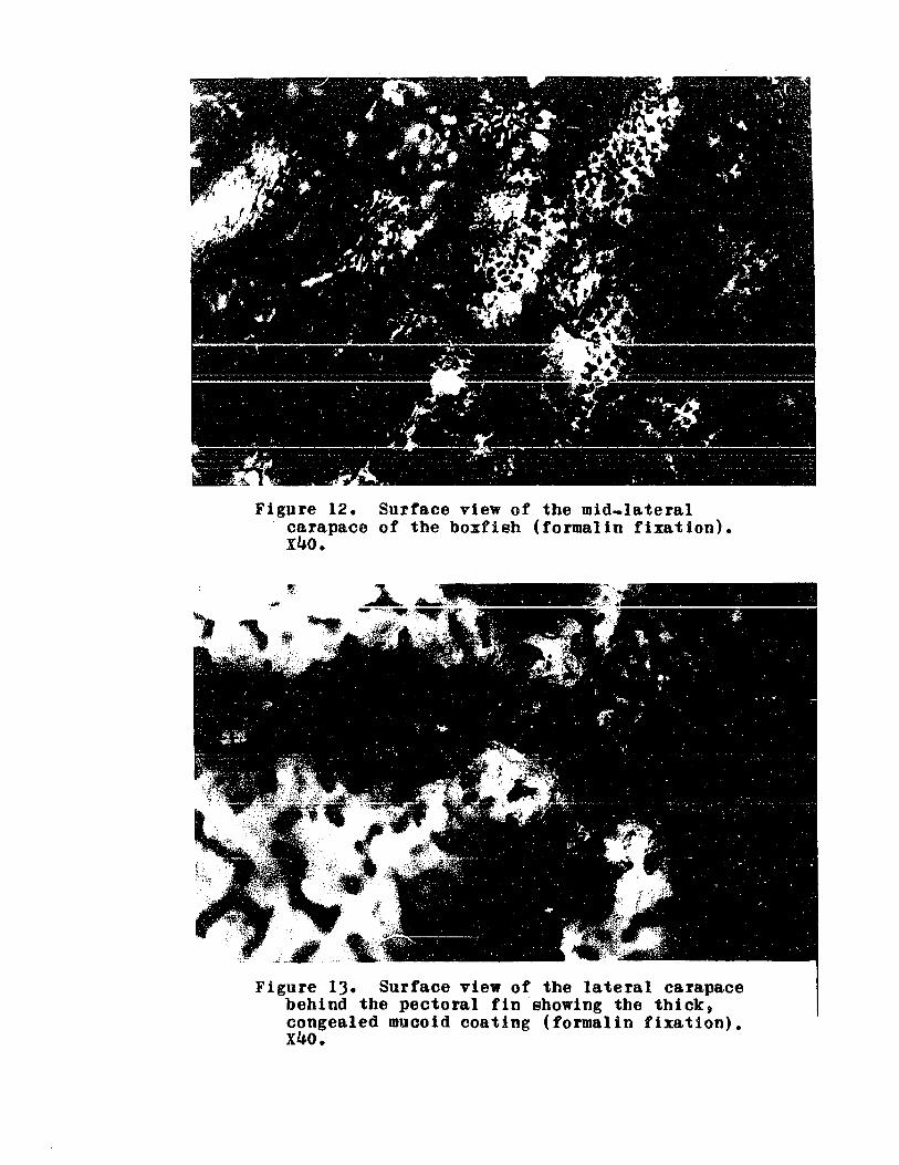
Figure 12. Surface view of the mid-lateralcarapace of the boxfish (formalin fixation).x40.
Figure 13. Surface view of the lateral carapacebehind the pectoral fin showing the thick,congealed mucoid coating (formalin fixation).x40.

Jl15
This layer uas thickest on the carapace behind the pectoral
fin, although this mucoid coating was of sufficient thick=
ness on other portions of skin 80 that it could be peeled
off intact as one peels o~ sunburned skino
The epidermis on the lips, parts of the carapace and
caudal peduncle is partitioned by dermal septa into reticu~
la-ted shallow npockets C3 o The surface of these C3pockets U
can be seen in Figures 12 and IJo In other sections of
the epidermis, such as around the fin bases and parts of
the carapace, the °pockets Q are indistinot, but dermal
ridges, grooves and protuberances cause irregularities in
the epidermis and effectively increase its surfacee
No axillary glands or other accessory skin glands
were found in a maoroscopic examination. Nor could any
su'bcarapace pockets of 03jellyll be found behind the gill
opening as was noted by Brown in an Atlantic trunkfish
(see Chapter 1, pgo 4)0
STRUCTURE OF BOXFISH EPIDERMIS
The epidermis consists of stratified squamous
epithelium and is sharply delineated from the dermis by a
border of melanophoreso At least J distinct cell types
can be recognized in the epidermal layer:
(1) Epidermal cel180 These are the unspecial~zed supporting
cells which may be considered as forming the Dm&trix~ of
the epidermis. They are distinguished by their largeg
usually sphericalg nuclei which measure about 4 p in diametero

116
The polyhedral cell outlines are irregular and the cyto~
plasmic portion of the cell is quite small in comparison
to the large uucleau80 In the basa.l layer of the epidermis
the nuclei are often fusiform and the cells are columnarQ
shaped, whereas near the surface and in the intermediate
layers polyhedral cells with spherical nuclei pi'evailo On
the surface of the epideI'iilis oceuI'S ex. single layer of
squamous cells with serrated edges, forming the external
boundary of the stratified epitheliuIDo
(2) Mucous cellso These cells, about 12 ~ 16 P in diameter,
are only found at or near the surface of the epitheliumo
Their nuclei are spindle-shaped and peripheral and the
intact cells were always full of IDUCUSo On some sections
"ruptured" cells could be seen discharging their mucous
content (see Figure 14)0 Evidently the cell membrane
collapses and the mucons contents are eliminated during the
process of secretion, ioe09 the secretion appears to be of
the merocrine typeo
(3) Club cellso These large (25 to 50 ~), variable_shaped
~serousn cells were the most conspicuous cells in the
epidermis o They are classified as nclub calls n because they
were found secreting their products while in the middle
layers of the epidermis, and they resemble the so_called
"club cells~ of other teleost8 in structure and in the
appearance of °secretory productsUo Their secretion also
appears to be of the merocrine typeo

Figure 14. A mucous cell in the process ofextrusion. Heidenhain's iron hematoxylin.XIOOO.
Figure 15. A stage I club cell. Note the largespherical nuoleus with prominent nuoleolusand fine granular cytoplasm. Heidenhain'siron hematoxylin. XIOOO.

118
These club cells may be seen in various stages of
development 0 The description of these proposed stages 9
based upon studies of Bouin-fixed, Heidenhainis iron
hematoxylin stained sections cut at 6 p is as follows:
(a) In the early developmental stage (1) a large
(7 to 8 p) spherical nucleus may be seen in the center
of the cello The cytoplasm consists of very fine
granular material which stains a uniform grayo Such
cells were usually found in the basal layers of the
epidermis (see Figure 15).
(b) In about the same position in the epidermis
are cells in the second stage of development (II),
(see Figure 16)0 These cells are characterized by
coarse, deeply stained granules forming among the
fine granular cytoplasmo At this stage orientation
of the cell into apical and basal poles is obviouso
The sperical nucleus can now be seen at the basal
end, apparently having been forced there by the
accumulating secretio~ prodncts o
(c) In the third developmental stage (III) the
coarse granules accumulate at the apical pole and a
thick cap of homogenous, colloidal secretion forms
at the apex and tapers down along both sides (see
Figure 17)0 At this stage the cell can be seen
discharging the homogenous secretion while still in
the middle layers of the epidermiso The nucleus
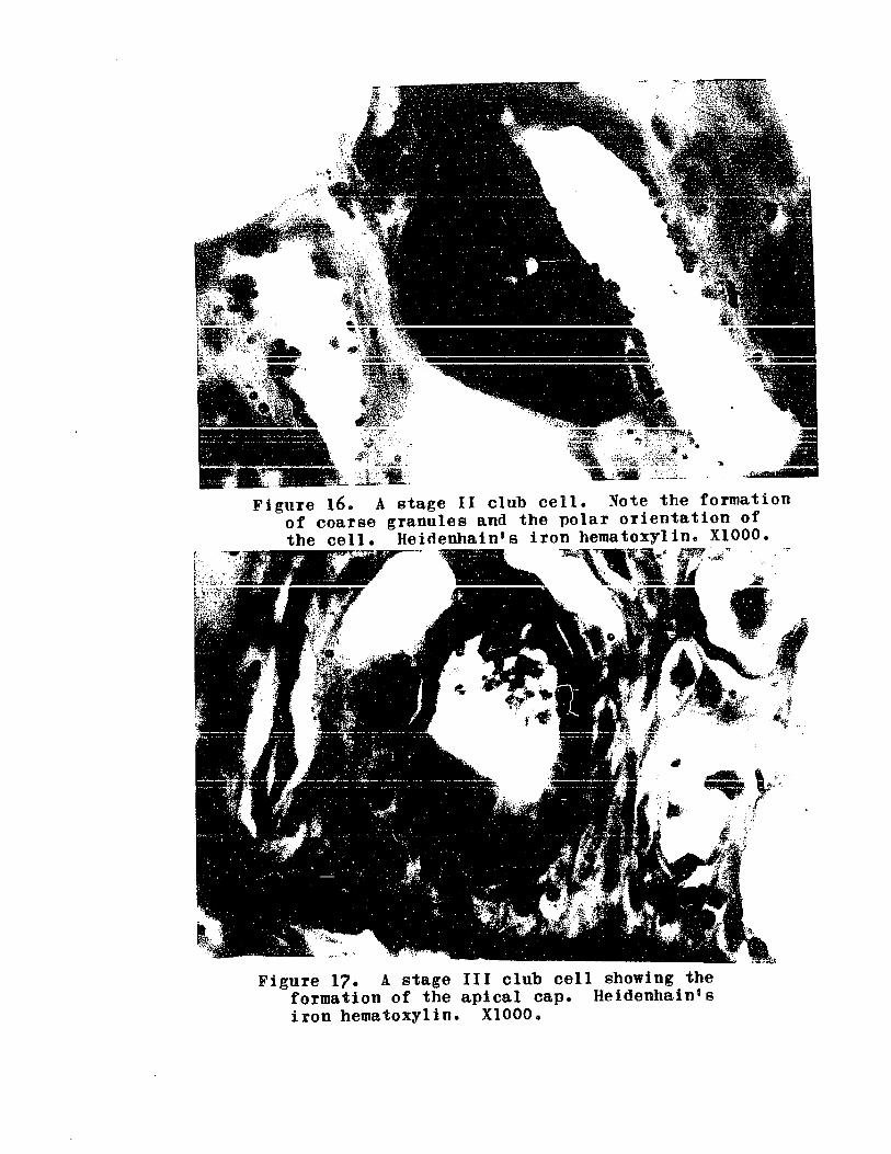
"; '. ..-~ .
Figure 160 A stage II clubof coarsethe cell.
--.. ,..
.. y'.,4: ·,c·
Figure 17. A stage III club cell showing theformation of the apical cap. Heidenhain'siron hematoxylin. XIOOO.

120
is somewhat flattened against the basal end of the
cell and is often difficult to recognizeo
(d) The fourth developmental stage (IV) is
characterized by a large globule of homogenous9
colloidal secretion o The coarse granules are absent
or are incorporated within the globuleo These cells
are usually found closer to the surface than those of
the preceding stages and the homogenous secretion
often may be seen being extruded as the cell wall
ruptureso The oblong, somewhat flattened nucleus is
found along the periphery of the cell (see Figure 18)0
(e) Cells in the fifth developmental sta,ge (V) are
found closest to the surface and are somewhat reduced
in sizeo They appear to be in a state of involutiono
The globula,r secretion is rednced in size and appears
more sphericalo The greatly flattened nucleus is
still evident (see Figure 19)0
'Size and position of club cells are not necessarily
indicative of developmental stages, as these criteria are
variableo The most important criterion is the type of cell
inclusions, or secretory products present o
Many of the club cells appear as empty vacuoles, aud
even the cells containing secretions have extensive open
spaceso The latter is probably a fixation artifact, as the
flattened peripheral nuclei of the club cells suggest that
the cells are ful10 The empty vacuoles indicate that either

Figure 18. A stage IV club cell showing thehomogenous. colloidal secretion and theflattened nuclei. Heidenhain's ironhematoxylin. XlOOO.
Figure 19. An early stage V club cell not yetin the process of involution. Heidenhain'siron hematoxylin. XI000.

122
secretion has already taken plaoe or the secretory material
was lost in seotioningo
A composite drawing of a crOBS section of boxfish
skin showing all the cell types and stages of club cell
development is presented in Figure 200
FORMATION OF POISON SKIN GLANDS
In the skin aFeas where shallow pockets OF grooves are
formed by dermal septa (see Figures 12 and 13), groups of
3 • 5 club cells were found in the depressions between the
septae The apices of the club cells converge toward the
center of the pocket and discharge their secretions into a
common groove or pseudo.ducto Figure 21 shows 2 club cell
nglands" and Figure 22 shows a pseudo_duct through which
the secretions paSSo
Multicellular skin glands 9 though rare in fishes9 do
occur as poison glands in certain elasmobranchs and teleosts
and as phosphorescent organs in some bathypelagic fish
(Van Ooaten, 1957)0 Muoous glands in fish skin are unicellular
and when mature, line the surface of the epidermis 9 whereas
in amphibians there occur true multicellular glands extending
into the dermiso The presence of primitive multicellular
glands in boxfish epidermis marks an advance over most
fishes, and since these glands consist of club cellsD
suggests a specialized functiono
THE DISTRIBUTION OF MUCOUS AND CLUB CELLS IN THE EPIDERMIS
Mucous and club cells were fonnd in all skin sections 9

rlgu~e 20. D~awiDg of a caudal skin sectionof !. len~111n08u8.
Epf ep!delllisbill b••8I1en" membraneDel dena1s1111 .elaDopbo~e
IDC I snOOU8 ceIInc: nuolei of club cell
1118: IIOC008 secretionape: epide~1 cellsI.V: stages or olub cellshe: homogenous seo~e~ion
of club oellgsa granula~ secretion
of club cell

~~;,:.
Figure 21. Club cell glands in the caudal skin of o. lentigJn08us.dermal septum separating the glands and the dark secretory productextruded by the gland on the right. Hl3idenhain's iron hematoxylin.
\,
Note thebeingx440.

Figure 22. A club cell gland clearly showingthe pseudo-duct. Harris hematoxylintriosin.. x400o
126
including those of the fin bases, lips and the carapacGo
The epidermis covering the tough dermal carapace was
surprisingly rich with both mucous and club cel180 As a
general rule, naked or sparsely scaled fish are more richly
provided with both numbers and kinds of mucous cells than
the well scaled teleosts~ end some investigators have postuQ
lated an inverse relationship between squemat!on and abundance
and type of mucous oells (Van Oosten, 1957)0 Their reasoning
is that the skin 2 a protective function is assumed by the
inoreased number of mucous oells in poorly scaled fisho
The presence of such a glandnlar epidermis over the boxfishgs
hard carapace contradicts this generalizationo
Club cell "glandeR were found only in sections of the
lateral carapace where epidermal pockets were observed
superficiallyo However, not enough sections were examined
to conclude that they are absent in the ventral and dorsal
carapace. These nglande" were aleo found in the lip skin
but were not encountered in ~0ctions of the dorsal, anal and
pectoral fin baseso
STAINING AFFINITIES OF MUCOUS AND CLUB CELLS
The descriptions of staining properties pertain to
skin sections fixed in Bouings fluid unless otherwise
specified.
Harris Hematoxylin and Triosin (or Eosin)
The colloidal nature of the club cell sec~etion
127
(stage III = V) was evident in these sectionso Fine granules
within the otherwise homogenous secretion in stages II and
III stained only weakly in Harris hematoxylin and were not
as conspiouous as they were in Haidenhain's iron hematoxylin
preparations of the same tissue sect10no The homogenous
secretion in club cel18~ stage III ~ V9 stained red witb
triosin or eosin Bnd pale lavender in sections without
these oounterstainso
The mucous cells ~ere stained a pale pink by triosin
or eosin and fine granular material could be distinguished.
Figure 2) shows a typioal Harris hematoxylinctriosin
section of the caudal skino
Mallory's Triple Stain
The club cell (III ~ V) secretions stained yellow
orange to a brilliant red and the sparse fine granules
were still evident within the globule. Some of the club
cell globules (IV) were bicolored, with the basal portion
staining blue, and secretions of stage V cells often stain
ing pale orange~ red or blu80 The color of the coarse
granules in stage II and III appeared orange to dark red or
purple, depending upon the thickness of the s8ction and
degree of destaining.
The mucous cells stained light blue and their granular
content was evident o
The nuclei of all cells stainod pale orange and were
difficult to seet thus limiting the utility of this stain

Figure 2)- A Harris hematoxylin-triosinsection of the caudal skin ofo. lentiginosus. x400.
Figure 240 Mallory's triple stain section ofthe caudal skino Note the blue stainingmucous cells and the red homogenoussecretions of the club cells. x400.

129
to color reactions of the various secretion productso
Clusters of coarse red granules were found for the
first time in the basal layers of the epidermis o These
granules were encountered in a few sections in both the
epidermis and among the dermal connective tissueo Some
resembled the nuclei of erythrocytes in size, structure and
staining properties, whereas others appear~d as disintegrated
nuclei. Possibly, hemolysis of these erythrocytes by ostracin
occurred during capture and handling prior to fixation o This
would corroborate the in vitro hemolysis of boxfish erythro
cytes by semi_pure ostracin.
Figure 24 depicts a typical caudal skin section with
these red granules, and Figures 25 and 26 show extrusion of
the secretory products of stages III and IV stained with
Malloryls triple stain.
Gomori's Aldehyde Fuchsin and Trichrome Counterstain
The coarse granules in club cell stages II and III
and the granules in the mucous cells were stained purple by
aldehyde fuchsino The homogenous secretions in III - V
were stained orange, red and green by the counterstain and
bicolored (green basal and redqorange apical portions)
globules were commODo Green-colored globules (IV and V)
were much more numerous than the corresponding blue from
Mallory stained sections although the colloidal secretion
in stage III always stained red or orange in both stainso
The coarse red granules (believed to be disintegrated
Figure 25. A Malloryis triple stain caudalskin section showing the mid_epidermisextrusion of a stage III club cell. x4000
Figure 26. A Mallory's triple stain caudalskin section showing extrusion by stagesIV and V club cellso x4oo.

1)1
erythrocytes observed in Mallory sections were stained
red_orange by the trichrome counterstaino The nuclei of
all cells were weakly stained by the counterstain and an
intercellular matrix interwoven among the epidermal cells
was stained light greeno
A caudal skin section stained with these dyes is
shown in ~igu~e 270
The Periodic Acid Leucofuchsin Schiff1s Method (PAS)
The PAS method was used to demonstrate glyco- and
mucoproteins after digestion uith amylase (from saliva)o
The only positive PAS substance in skin sections were the
granules in the mucous cells which were strongly stained a
deep red-violet coloro The globular secretions in club
cells (stage III - V) were stained pale pink, only slightly
darker than the epidermal cells, indicating a negative
reaction, or absence of glyco~ and/or mucoproteinso
A skin section stained by the PAS method is shown
in Figure 28 0
Toluidine Blue
The homogenous secretions in club cells III _ V were
orthochromatically stained a deep aqna blue by this dyec
All nuclei were stained the color of the dye whereas
the mucons cells were unstainedc
Dresbach's Mucicarmine Method
In the epidermis only the homogenous secretion in
the club cells was stained red by this commonly used mucous
Figure 27. A caudal skin section stainedwith Gomorils aldehyde fuchsin-trichromestain. Note the aldehyde fuchsinpositive granules in the mucous cellsand in a stage III club cell and the"hemolyzed erythrocytes" in the basallayerso x400o
Figure 28. A caudal skin section stainedwith the PAS reagent. Note the PAS+mucous cells. x4oo.
133
stain, but the connective tissue matrix in the dermis
appeared pinko This method was recommended for epi thelia.l
and connective tissue mucins in generalo
Sudan Black B
The homogenous club cell secretions were stained
weakly with this dye in sections fixed with Charnpy!s fluid
but were not, or only weakly, stained in Bouin-fixed materialo
This suggests that lipids were present in the secretory
materials bUE only in insignificant amounts o
Discussion
The staining affinities of the secretory materials
in the epidermis corroborate that the mucous and club cell s
B~e distinct cell types with different secretory material
and the developmental stages of the club cells, as described
from iron hematoxylin sections, are in proper sequenceo
There are apparent contradictions in the muous staining
properties of both glandular cellso The mucous cells stained
positively with Mallory!s triple (blue), PAS (red-violet),
and aldehyde fuchsin (purple), indicating their mucoid
nature. However, the club cell homogenous secretions
were stained by toluidine blue and mucicarmine which are
also mucous stainso
ThUS, it appears that both cells contain mucins but
of distinctly different typeso The PAS positive reaction
in the mucous cells shows that muco- and/or glycoproteins
1)4
are present and the negative PAS reaction in the club cells
demonstrates their absence.. By process of elimination,
the other common epithelial mucins are the mucopolysaccharides~
which are PAS negative substances and toluidine blue
<p'metachromaSia) positiveo However, at this point, the
exact nature of the mucoid component is irrelevant o The
important consideration is which cell, the mucous cell Or
the club cell, is responsible for ostracin secretion?

135
CHAPTER 16
HISTOLOGY OF THE LABIAL GLANDS
As previously mentioned (Chapter 13), the oral secretions
of boxfish contained an ichthyotoxic, hemolytic substance,
although the skin secretions appeared to be the major source
of ostracino Histological sections were prepared of the
entire lips, including the jaws and epithelial lining of
the buccal cavity to and including, the esophagus e The
more striking features of the histology of these regions
are presented in this chaptere
GENERAL MORPHOLOGY
The most conspicuous structures in the anterior portion
of the buccal cavity of boxfish are the prominent labial
villi attached to the ventral floor of the vestibular region
between the external lips and the teeth o A drawing of a
median sagittal section of the lips of a female boxfish
(Figure 29) clearly shows the position of these villi in
the buccal cavityo
HISTOLOGICAL STRUCTURE OF THE ~~BIAL VILLI
Cross and sagittal sections (8 ~ lOp) of the lips
revealed that the labial villi were extensions of dermal
connective tissue lined with a mucous membrane composed of
stratified cuboidal epitbeliumo
The epithelial cells were nearly uniform in size
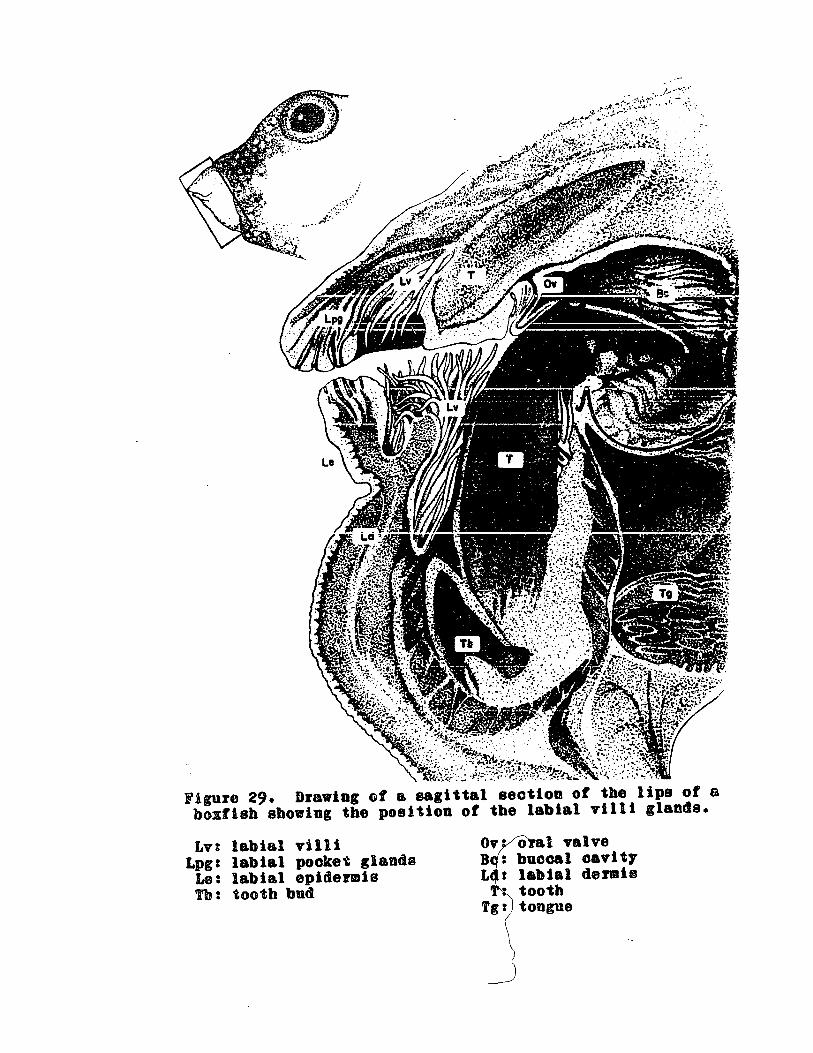
figure 29. Drawing of a sagittal section of the lips of abcxflsh sboving the position of tbe labial villi glands.
Lv:Lpg:
I.e:Tb:
labial villilabial pocket glandalabial epidermistooth bud
o a1 valvebuccal cavitylabial dermiotoothtongue

137
(about 12 p) and occurred in great clusters at the distal
ends of the villio The cells appeared empty in iron
hematoxylin sections p except for irregularly shaped spherical
nucleio Serrated edges were found on the peripheral callso
These ulabial n cells were further characterized by their
staining affinities with stains used on the skin sections
in Chapter 150
Harris HematoxYlin and Triosin (or Eosin)
Labial cells appeared the same as they did in iron
hematoxylin sectionso The spherical nuclei 9 agranular
cytoplasm and serrated edges were evident.
Malloryls Triple Stain
The cytoplasm appeared clear in this stain also.
The nuclei stained pale red and the dermal supporting
framework deep blue (see Figure 30)0
Gomori Aldehyde Fuohsin and Trichrome Connterstai~
Dramatic results were obtained by staining the
labial cells with aldehyde fuchsino Fine and coarse
purple granules were abundant in the cytoplasmo The
distribution and relative density of these granules were
more or less uniform from cell to cell and none of the
cells was empty (see Figure 31)0
Periodic Acid Leucofuchsin Shiffss Method (PAS)
PAS positive granules were demonstrated in the labial
cells, however fewer granules were shown in these s6ctions
than in aldehyde fuchsin stained sections (see Figure 32)0·

Figure 30. Labial glandsstained with Mallory's triplestain. Note the clear cytoplasm and red nuclei. x4000
Figure 31. Labial glands stained with Gomori 1 s aldehyde fuchsin-trichrome stain. The positive aldehyde fuchsin granulesare clearly visible. x4000
Figure 320 Labial villi stainedwith the PAS reagento Note thepositive PAS granules in thelabial cellso x4000

139
Toluidine Blue
This metachromatic dye did not stain the labial cel180
The staining reactions of the contents of the labial
cells were very similar to those of the mucous cells of
the skino The mucous cells however, stained more strongly
in these dyes than the labial cal180 This may be due to
the Fslative amount of granular material in the cells
rather than different types of reactants o
DISTRIBUTION OF THE LABI~L GLANDS
The labial villi were found to be only a portion of
an extensive elaboration of the mucous membrane of the
oral cavity into what will be referred to as "labial
sufficient to olassify the labial glands as compound
acinous glandso
In Ostracion, the labial glands consisted not only
of dorsal and ventral villi but "glandular pocketsn extend.
ing deeply into the dermis of the lips through infolding
of the lip epidermis, therefore effectively forming a
duct emptying outside the buccal cavity (see Figure 33)0
These Upockets U are found in both the dorsal and
ventral portions of the lip where they extend in an anterior
to posterior direction with the duct opening at the surfaoe
of the lip epidermiso Other such pockets occur deep into
the epithelial lining of the buccal cavity with ducts
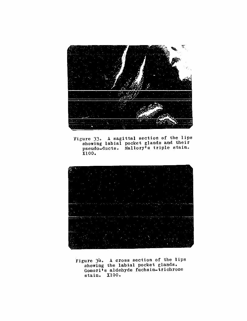
Figure 330 A sagittal section of the lipsshowing labial pocket glands and theirpseudo_ductso Mallory's triple stainoXIOO.
Figure 340 A cross section of the lipsshowing the labial pocket glands.Gomori's aldehyde fuchsin-trichromestaino XIOOo

141
opening into the oral cavity behind the teetho See Figure )4
for a cross section of these dermal pooketso
The labial villi are contained in large pockets formed
by conneotive tissue septa, both above the dorsal row of
teeth and below the ventral row, with each pocket opposite
eech tootho These will be referred to as the "dental
pocketsao Figure 35 is a drawing of a cross section of the
lips and buocal cavity cut at the level of the dorsal row
of teeth. The dermal pockets, and the villi within the
dental pockets are ShOWDo
The labial gland cells extend throughout the mucous
membrane of the bucoal cavity and are found lining the
folds and papillae of the palate but do not occur in the
esophaguso
STRUCTURE OF THE LIP SKIN AND PROBABLE ORIGINOF THE LABIAL GLANDS
The epidermis of the lips is considerably thicker and
more compact than that of the rest of the skino The
epidermal~ or supporting oells, predominate although mucous
and club cells are found in the upper layers, and in
Rglandular pockets~, similar to those found in caudal skin
sections, with distinct pseudo-ducts (see Figure )6)0
In the anterior portions of lip sections extensive
folding and invagination of the epidermis was evident o
Some "glandular pockets" extended deeply into the lips
and cross sections through these dermal pockets revealed

o
v
Figure 35. Drawing of a cross section of the mouththrough the dorsal teeth showing the arrangement ofImb!al villi around the teeth.
Lv: labial villiLpg: labial pocket glandsLsg: labial skin glands00: oral cavity
D: dorsalV: ventralT: teeth

Figure 360 Lip skin showingmucous and club cells amongthe thick layer of epidermalcells. Gomori!s aldehydefuchsin-trichrome. x4000
Figures 37 and 380 Labial epidermal foldsshowing the transition from the skin glandcells to labial cells. (Left: Gomori!saldehyde fuchsin trichrome stain. x400oRight: Mallory's triple stain. X250.)
mucous cells, club cells and labial callso A transition of
cellular types occurred, with the mucous and club cells
being found in the more superficial folds and labial cells
baing found exclusively in the deep pockets, but inter~
mediate regions contained all 3 types of cells (see Figures
37 and 38) which suggested that the labial glands are formed
by an invagination of the lip ep!dermis o
Although the staining properties of the labial cells
are similar to those of the skin mucous cellsp there are
enough differences to conclude that these cells are a
distinct type: (1) the labial cells possibly have a merocrine
secretion but no cells were observed in the process of
extrusion, (2) serrated edges were found on some labial
cells, but could not be demonstrated in mucous cells,
(3) the nucleus of mucous cells was always peripheral and
spindle_shaped like that of typical mucous oells, whereas
the labial cell nuclei were irregularly-shaped spheroids,
(4) mucous cells stained blue in Mallory's triple stain
but the labial calls remained clear, (5) mucous cells ~ere
strongly PAS positive but labial cells contained only a
few PAS positive granules, and (6) granules of both cells
stained purple with aldehyde fuchsin but staining was
much more intense in the muoous callso
DISCUSSION
Compound glands rarely ocour in the buccal cavity of
fishes, although Andrew (1959) notes multicellular glands
145
in tIle lungfish Protopterus annectans p Nishida (1954) in
adult salmon Onooorhynchns keta and Monod (1950) in the
characin Alestes Spe The function of such glands is unknown
and true salivary glands are thought to be absent in fisheso
The multicellular labial glands of the mucous membrane
of boxfish are prominent and extensive enough to justify
consideration of them as poisonous glandso An ichthyotoxin
hemolysin has been found in the oral secretions collected
from the region near the labial villi (see Chapter 13)0 On
the other hand, the skin secretions were considerably more
toxic than the oral ones and no active substanoes could be
extracted from the skin or lips of dead boxfiaho Furthermore,
the labial and skin glands are histologically distinct, and
the cytomorphology and histochemical propertie3 of the
secretory cells of the gland cells are differento See
Table XI for a summary of the staining reactions of the
secretory material within the gland cellso
Such evidence suggests that 2 or more distinct
secretions containing different toxins are occurring, but
this has not yet been demonstratedG Because of difficulties
met in extracting ostracin from the skin and the small
yield of toxin from lip extracts, it has not been possible
to characterize chemically the respective toxinso Nevertheless,
highly toxic skin secretions could be collected from disc
tressed boxfish from which the lips, containing the labial
glands, were completely severedo Assuming that ostracin
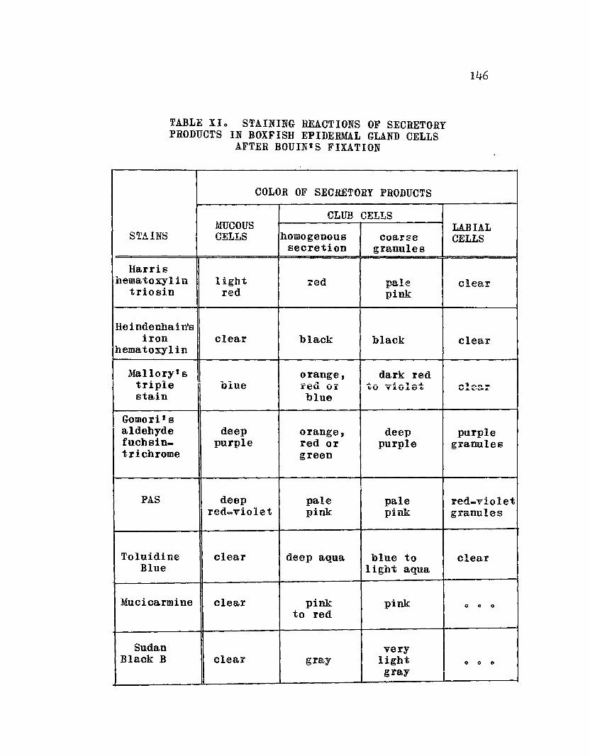
TABLE XIo STAINING REACTIONS OF SECRETORYPRODUCTS IN BOXFISH EPIDERMAL GLAND CELLS
AFTER BOUINtS FIXATION
146
COLOR OF SECRETORY PRODUCTS
CLUB CELLSMUCOUS LABIAL
STAINS CELLS homogenous coarse CELLSsecretion granules
Harrishematoxylin light red pale clear
triosin red pink
Heindenhain'siron clear black black clear
hematoxylin
Mallory's orange, dark redtriple biue red Or to violet ,,",l~r'lI'"
""" ........ \,JWJL
stain blue
Gomori'saldehyde deep orange, deep purplefucbsin_ purple red or purple granulestrichrome green
PAS deep pale pale red-violetred...violet pink pink granules
Toluidine clear deep aqua blue to clearBlue light aqua
Mucicarmine clear pink pink 0 0 0
to red
Sudan varyBlaok B clear gray light 0 0 0
gray
147
secretion has a protective function, this indicates that
the labial glands play a minor role in the total ostracin
secretion by distressed boxfisho The position of the
glandular labial villi and pockets in the bucoal cavity
suggests a possible feeding, rather than defensive,
functiono
148
CHAPTER 17
HISTOLOGICAL AND HISTOCHEMICALEVIDENCE OF OSTRACIN SECRETION
The experimental evidence of the site of ostracin
secretion and the description of the histological structure
of the skin and labial glands olearly suggest that ostracin
is being secreted by epideFmal glands. However, the anomolies
obtained in tissue extracts~ together with the establishment
of 3 distinct gland cells, make it difficult to ascertain
the precise source of ~tracino Additional studies attemptG
ing to resolve this problem are presented in this chaptero
HISTOLOGICAL CHANGES IN THE LABIAL ANDSKIN GLANDS OF CAPTIVE BOXFISH
In Chapter 7 it was shown that boxfish in captivity
would retain their ability to secrete ostracino However,
poorly fed or starving fish secreted very little ostracin
under stress. Thus, the glandular tissues of captive boxfish
were fixed, sectioned and stained to correlate the reduction
in toxicity with changes in histological structureo
One 81 mm long juvenile boxfish was kept in an aquarium
for 32 days and given a subminimal diet of Purina trout
food and frozen brine shrimp. On the thirtycsecond day the
stress se~retions of this fish were collected in 70 2 mi of
tap water until death of the boxfish and assayed for
ichthyotoxicity with 2 sailCin mollies in 100 mi of sea
water. The results were as follows:

Volo assayed ofthe 702 ml rinse
149
Mean survival time ofassay fish (mino)
non-toxic
non. toxic
102 0 0
Calculation of the total amount of ostraoin from the
dose_response curve (Figure 8) shows that less than 1 mg
of ostracin was present in the secretions of this fisho
This represents a oonsiderable reduotion in ostracin
yield from freshly captured boxfish o
The lips and portions of the caudal skin of this fish
were fixed in Bouinls and processed for histological ex=
aminationo Results of this examination are as follows:
The Epidermis
There was a marked change in the epidermis. Both
mucous and club cells were greatly reduoed in number and
many sections of the skin were completely devoid of these
cells, consisting only of undifferentiated epidermal cells
(see Figure 39)0 The mucous cells, though few in number,
were full of mucus, and the clUb oells were either empty
or contained coarse granules which were stained purple by
aldehyde fuchsin. Only a very few club cells contained a
homogenous secretiono
Another captive fish, a 115 mm long male, lived for
48 days in an aquarium. 'Vhen this boxfish became moribund
its tissues were fixed in Bouinls and processed for histological
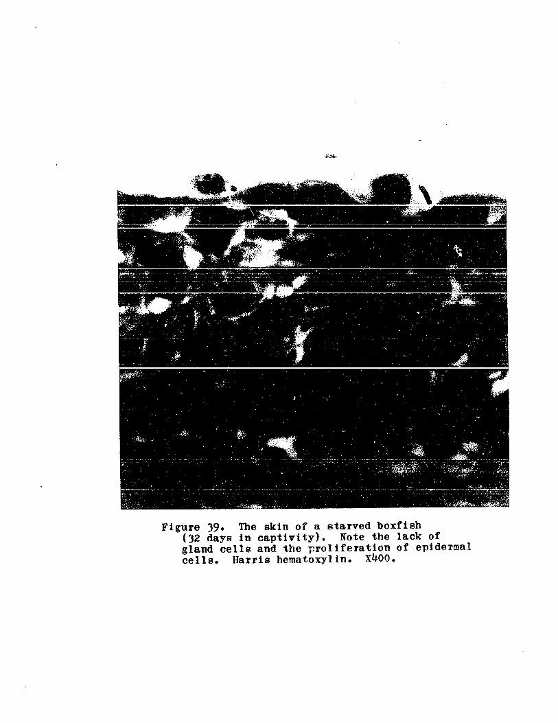
Figure 39. The skin of a starved boxfish(32 days in captivity). Note the lack ofgland cells and the ~roliferation of epidermalcells. Harris hematoxylin. x400.

151
examinationo The skin sections of this fish resembled
those of the captive juvenile fish in their scarcity of
mucous and club cellso This fish had been feeding better
than the first but was so emaciated and weak when it died
that no stress secretions were collectedo
The numbers of mucous and club cells in the caudal
epidermis of botb boxfish kept in captivity were counted
and compared with numbers of these cells in freshly captured
boxfisho The counts were averaged per linear mm from
several skin sections (6)1) and, although variations in
gland cell numbers can be expected to depend upon the section
chosen, general informaticTI on the relative abundance of
these cells can be obtained. The results are "tabulated
in Tab1e XI I 0
The data indicate that there is a greater proportionate
reduction in the number of club cells as compared to mucous
cells in boxfish held in captivityo The ratio of mucous
cells to club cells is even greater when a skin section
containing "glandular pocket~n is examined, however such a
section cannot be used for a direct comparison since the
captive boxfish skin sections did not contain "glandular
pockets". Nevertheless o these data could be interpreted
to suggest that the club cells are responsible for ostracin
secretiono
Labial Glands
A great reduction in the number of labial cells

TABLE XII.. RELATIVE NUMBERS OF MUCOUS ANDCLUB CELLS IN THE CAUDAL EPIDERMIS OF BOXFISH
KEPT IN CAPTIVITY AND FRESHLY CAPTURED
152
Mean no o of Mean noo of Apprmrocaudal skin mucous cells club cells ratio ofsection (6 p) per mm per mm mucous to
epidermis epidermis club cells
Boxfish in captivity(glandular ~pocketsM
absont) 1 507 10 .. 3 (1:2)
Freshly capturedboxfish(glandular "pockets"absent)2 1206 4500 (1:4)
(glandulat "pockets il
present») 909 6106 (1:6)
1Average of 6 sections (total 19 .. 7 mm) from 2 boxfish,
)2 and 48 days in captivityo
2Average of 5 sections (total 1606 mmt from 2 boxfish,fr~shly capturedo
3Average of 2 sections (total 806 mm) of 1 ·boxfishfreshly capturedo
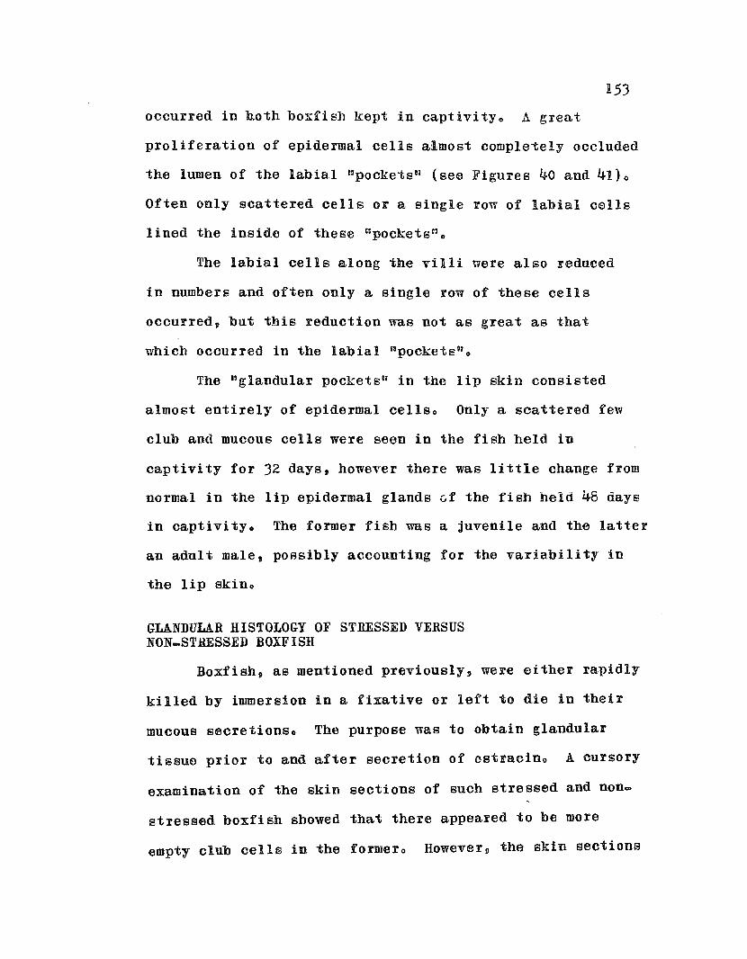
153
occurred in hoth boxfish kept in captivityo A great
proliferation of epidermal cells almost comple~ely occluded
the lumen of the labial ~pocketsll (see Figures 40 and 41)0
Often only scattered cells or a single row of labial cells
lined the inside of these «pockets n o
The labial cells along the villi were also reduced
in numbers and often only a single row of these cells
occurred, but this reduction was not as great as that
which occurred in the labial "pockets U o
The "glandular pockets!; in the lip skin consisted
almost entirely of epidermal cellso Only a scattered few
club and mucous cells were seen in the fish held in
captivity for 32 days, however there was little change from
normal in the lip epidermal glands vf the fisb held 48 days
in captivityo The former fish was a juvenile and the latter
an adult male, possibly accounting for the variability in
the lip skino
GLANDULAR HISTOLOGY OF STRESSED VERSUSNON.STHESSED BOXFISH
Boxfish, as mentioned previously, were either rapidly
killed by immersion in a fixative or left to die in their
mucous secretionso The purpose was to obtain glandular
tissue prior to and after secretion of ostracino A cursory
examination of the skin sections of such stressed and nOD=
stressed boxfish showed that there appeared to be more
empty club cells in the formero However, the skin sections

'Figure 40. Labial pocket glands of astarved boxfish (48 days in captivity).Mallory's triple stain. XIOO.
'Figure 41. Labial pocket glands of thesame fish stained with Gomorifsaldehyde fuchsin_trichromeo Note thefew labial cells among the proliferatingepidermal cells. x400.

155
used were not comparable and the degree of stress was not
controlledo
In an attempt to control the amount of stress that
boxfish were subjected to, 2 juvenile boxfish (62 and
66 mm long) were carefully oaptured with a dipnet on
Ala Moana reef and immediately transferred into a 20 gallon
bucket sealed wit~ a lido The 2 fish had been obser~ed
swimming together and both were easily captured within a
few minuteso They were handled as little as necessary and
were not unduly excitedo
Both fish were kept in a 35 gallon aquarium for 3 dayso
On the third day one of the boxfish (62 mm), chosen at random,
was quickly netted and rapidly killed by immersion in Bouin's
fluid. The other (66 mm) was placed in I liter of sea water
and kept there for 2 hours e Air was periodically bubbled
through the water and the sides of the container tapped to
disturb the boxfish and cause a stress situationo A. small
puffer (Canthigaster jactator) was placed in with the boxfish
as an assay for ostracino The puffer died after 28 minutes,
indicating that ostracin had been secreted by the distressed
boxfisho When the boxfish appeared weakened, it was killed
by immersion in Bouin~s fluido This fish had not been
handled during the induced stress experiment since direct
pressure on the epidermis might have caused excessive
secretion of the glandso
The glandular tissues (fixed in Bonin's) of both fish
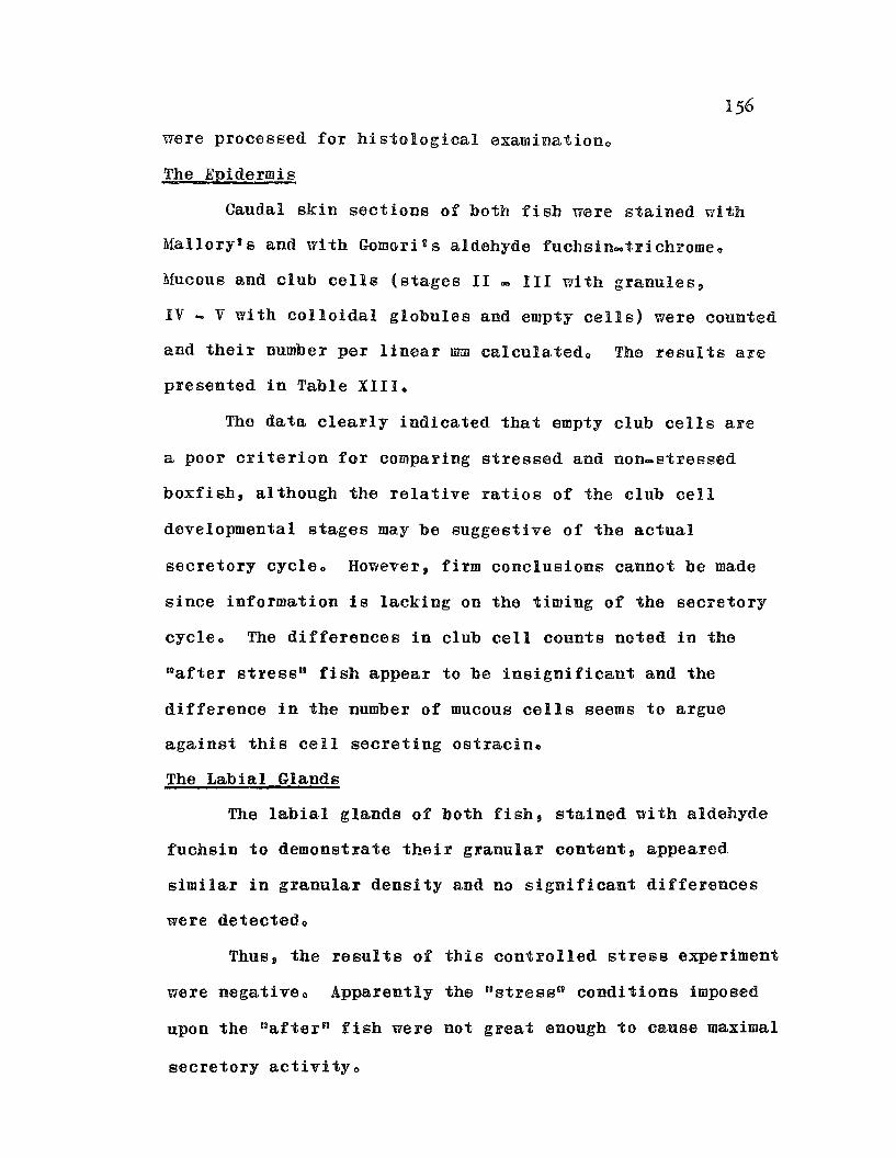
were processed for histological examinationo
The Epidermis
Caudal skin sections of both fish were stained with
Malloryts and with Gomori 2 s aldehyde fuchsin~trichromeo
Mucous and club cells (stages II = III with granules,
IV - V with colloidal globules and empty cells) were counted
and their number per linear rom calculatedo Tha results are
presented in Table XIIlo
The data clearly indicated that empty club cells are
a poor criterion for comparing stressed and non-stressed
boxfish, although the relative ratios of the club cell
developmental stages may be suggestive of the actual
secretory cycleo However, firm conclusions cannot be made
since information is lacking on the timing of the secretory
cycleo The differences in club cell counts noted in the
~after stress" fish appear to be insignificant and the
difference in the number of mucous cells seems to argue
against this cell secreting ostracino
The Labial Glands
The labial glands of both fishs stained with aldehyde
fuchsin to demonstrate their granular content, appeared
similar in granular density and no significant differences
were detectedo
Thus, the results of this controlled stress experiment
were negativeo Apparently the n13tress ti conditions imposed
upon the Uafter" fish were not great enough to cause maximal
secretory activitYo
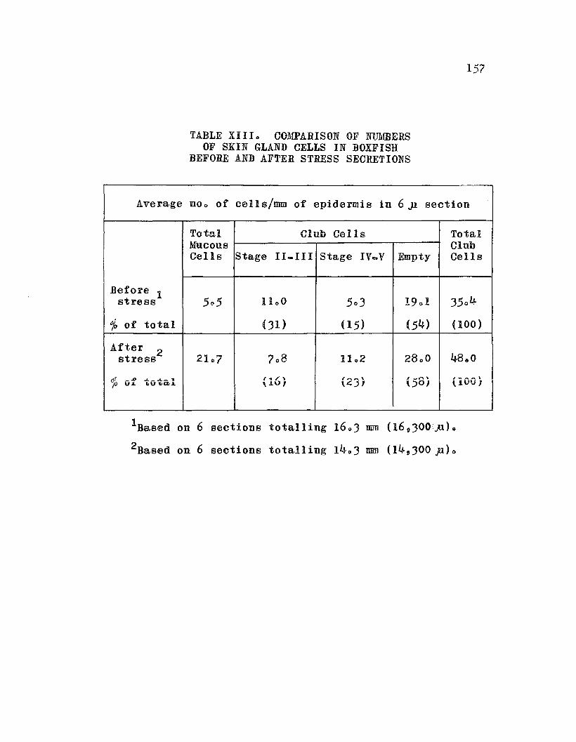
TABLE XlIIo COMPARISON OF NUMBERSOF SKIN GLAND CELLS IN BOXFISH
BEFORE AND AFTER STRESS SECRETIONS
157
Average no o of cells/mm of epidermi 8 in 6 Jl section
Total Club Cells TotalMucous ClubCells Stage II-III Stage Iv...V Empty Cells
Before 1505 1100 503 19 0 1 35 0 4stress
%of total (31) (15) (54) (l00)
After 2stress 21 07 708 11 02 2800 48&0
01. ,,00 .... _.A._ .. l .. , \ 1'--.\ , ..... n \.t _ ...... _ \
/0 JL ILIU IIQ,L \.L°I \~-' I \.)0J \lVV/
IBased on 6 sections totalling 1603 mm (16,300p)0
2Based on 6 sections totalling 14~3 mm (14,300 p)o

158
SPOT TESTS OF OSTRACIN
tJSpot U tests have been employed by several \1orkers
(see Pearse, 1960) to correlate the staining properties of
a purified substance with the staining of certain cell in~
elusions in order to develop a histochemical test for the
desired substance in the cello There are some objections
to the approacho One is that it has been shown that the
staining properties and histochemical characteristics of
'Dsecretion granules" in gland cells often bear no relation""
ship to the chemical and physiological properties of the
products secreted by the glandso "SerQus ll granules are a
good example of this as they often show no similarities
in staining and histochemical properties with the actual
secretory product of the· serous glands (Gabe and Arvy, 1961)0
Nevertheless, there are types of glands in which the
"secretion granules" retain their staining properties,
histochemical characteristics and their shape after extrusion o
Mucous glands of vertebrates and the ink sac gland of Sepia
are examples (Gabe and Arvy, 1961)0
Thus, "spot tests" were employed to find out whether
relationships between the staining properties of the assumed
glandular secretory products and crude and semi.pure
ostracin existo
Materials and Methods
Drops of crude and semi.pure ostracin were placed on
microscopical slides followed by a few drops of Bouinus
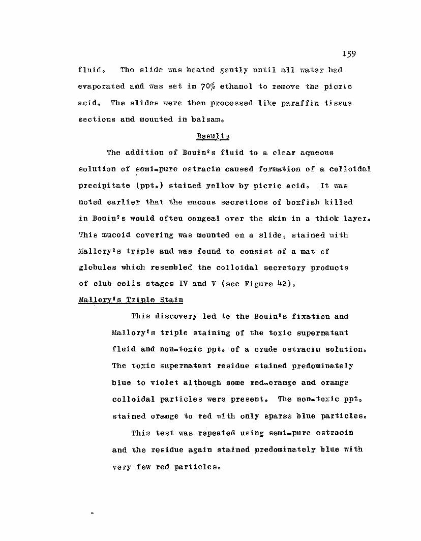
159
fluido The slide vms heated gently until all water had
evaporated and uas set in 70% ethanol to remove the picric
acid o The slides were then processed like paraffin tissue
sections and mounted in balsamo
Resut t8
The addition of Bouines fluid to a clear aqueous
solution of semi=pure ostracin caused formation of a colloidal
precipitate (ppt o ) stained yellow by picric acido It was
noted earlier that the mucous secretions of boxfish killed
in Bouinis would often congeal over the skin in a thick layero
This mucoid covering was mounted on a slides stained with
Mallorygs triple and was found to consist of a mat of
globules which resembled the colloidal secretory products
of club cells stages IV and V (see Figure 42)0
Mallory1s Triple Stain
This discovery led to the Bouin's fixation and
Malloryls triple staining of the toxic supernatant
fluid and non-toxic ppto of a crude ostracin solutioDo
The toxic supernatant residue stained predominately
blue to violet al~hough some red.orange and orange
colloidal particles were presento The non.toxic ppt o
stained orange to red with only sparse blue particleso
This test was repeated using scmiapure ostracin
and the residue aga.in stained predominately blue with
very few red particle so

· .~ . - '. '. ,. . "
,•.~~, .. " ~ ~:;;.: _ .1 .. • ....;t ••1 ~ .'~' '. -
Figure 42. The mucoid coating peeled fromthe skin of a boxfish killed in Bouin'sfluid. Note the homogenous secretionsof the club cells at the surface of thecoating. Mallory's triple stain. x400.

Jl.6Jl
GomoFi~s Aldehyde Fucbsin~Trichrome Stain
The supernatant residue of a crude ostl'acin
solution stained with these dyes was found to consist
of numerous purple granUles similar to those found
in the mucous celis and stage II and III club cellso
Green colored particles were also seeno The ppt o
contained vari~colored particles~ including purple
granules, but these aldebyde-fuchsin positive granules
were not as numerous as they were in the supernatant
residueo
Staining semi-pure ostracin residues gave
simi lar reaul ta.
PAS Method
PAS positive particles were found in the super=
natant residue of a crude ostracin solution whereas
such particles were absent in the precipitateo These
results were confirmed with semi-pure ostracino
PAS + Toluidine Blue
Toluidine blue, used as a counterstain after
PAS staining, completely masked the PAS positive
components in a supernatant residue preparationo
This is in contrast to what happened in tissue sections
since, after FAS treatment, the club cell secretions
would not stain with toluidine blueo
All of the above tests were performed on crude and
semi.pure ostracin solutions that were not completely

162
deproteinizedo When pureI' extracts of os1r~~in \7eli'e used,
such as after hot acetone or chloroform extraction, results
were somewhat differento
Malloryns Triple Stain
Acetone extracts of ostracin lIouid not .stain in
these dyes, but a branching crystalline structure
was formeda These crystals were not an artifact of
the stain since they were found on unstained, processed
slidcso
Gomori!s Aldehyde Fuchsin and Trichrome StaiD
ACbtone-ostracin residues contained very fine,
weakly stained purple granules and variQcoloredr,
colloidal particleso No crystals were fonnd e
Toluidine Blue
Toluidine h!ue stained acetone and chloroform
ostracin residues both ortho- and metachromatically
<P> but the aqua color observed in tissue sections
was not seeno In the chloroform residue preparation,
numerous blue, needle=like crystals were formedo
To determine what effect the fixative had on ostracin
in vitro, an aqueous, semi-pure ostracin solution was mixed
with 5 times its volume of Bouinns fluid, immediately
forming a precipitateo The whole mixture was dialyzed for
6 hours to remove the fixative and then assayed for activityQ
Although the solution was still weakly ichthyotoxic after
dialysis and foamed when agitated, it lost its toxicity when
· .\
163
it WBS heated gently. to dryness and dissolved in citrate
salineo It was non=hemolytic to tuna erythrocyteso The
initial weak toxic reaction after dialysis nas probably due
to residual fixative contaminents o ¥ihen picric acid was
used instead of Bouiong fluid and dialysis was carried out
overnight, ostracl0 was completely detoxifiedo
At first 9 the "spot testst~ of impure ost1'ac1n showed
notable parallels with the ~secl'etory gl'anules ,a in the
mucous and labial cells and presented good evidence
supporting these cells as the source of ostraclno However,
as purer samples of 08tracin were tested, the staining
properties of the test residue were considerably reduced,
indicating that the "spot tests" were actually demonstrating
difficult to control the amount of residue on the slide
because processing caused loss of incaluable amounts of the
original nspot n and it was impossible to be certain that
the ostracin fraction was not 108t o
DISCUSSION
The evidence so far presented on the cellular source
of ostracin in glandular tissue has been both inadequate
and contradictoryo Evidently there is a complexity of
f~ctors operating within the secretory cycle which cannot
be detected by simple meanso The club cell 1Bsecretory
products lO stain "Variably in triple stains suggesting that
chemical transformations are occurring and it is uncertain
if the coarse granules in stage III aye extruded uith the
colloidal apical product o The inactive extracts of skin
sections present a formidable, but highly intriguing,
pJl.'Oblern and the labial glands further confuse the picture 0
Therefore, on the basis of the histological structure
of the glands, the extraction and collection of their
secretions s the staining and histochemical properties of
their secretory products, the coxrelation of ostracin
secretion with numbers of gland cells in captive fish,
and parallelisms and differences of the staining properties
of ostracin with the "secretory partlcleso within the cells,
the following hypotheses are proposed:
(1) The club cells are responsible for ostracin
secretion in the skino
(2) The mucous cells, containing muco~ and/or
glycoproteins, are true mucous cells having no
other specialized functiono
{3) The labial cells contain an ostracin-like toxin
but have a different function than do the skin
club cellso
There are, of course, other possible hypotheses and the
above proposals must not be misconstrued as final conclusionso
They are only believed to be the most likely explanations
of a complex, unresolved problemo

CHAPTER 18
COMPARATIVE HISTOLOGY OF THE SKIN OF COWFISHAND OTHER PLECTOGNATHS
The cor~elation of a poisonous skin secretion with
the presence of club cells in the skin of boxfish led to
a preliminary histological Gtudy of the skin of the co'rlish,
Lactoria fornasini, another Hawai ian trunkfi sil l{nown to
secrete a saponin-like toxino The integument of a few
species of puffers and one fl1efish were examined for
compara.tive purposes since the puffers were also shown to
contain toxic components in their mucous secretionso
Both skin and lips of the cowfish and the skin only
of four species of puffer and one filefish were fixed in
Bouinvs fluid and processed in the manner described for
boxfish tissues (Chapter 14)0
EPIDERMIS OF THE COWFISH
The structure of the epiaermis of cowfish has much in
common with that of boxfisho The upper layers contain
numerous mucous cells (12 to 16 p) and a thin layer of
serrated squamous epitheliumo The middle and basal layers
contain the large (40 to 80 )1) glandular "club cel18a~
characteristic of boxfish epidermis o Many of these cells
were found in the process of ext~usion of secretions through
pseudo~duct8 while in the middle layers of the epidermis p
although no glandular rlpockets Pa bordered by dermal ridges

166
Vlere observed in the sections examined o Developmental
stages contain! ng granular and agl'anulal' ~lsecretol"Y
products~ were present in most sections o
The chief structural differences noted in cowfish
epidermis are: (1) the height of the epidermal layer is
greater than that of boxfi8h~ (2) a greater number of mucous
cells is found in the upper layers (usually there are 2 - J
layers of mucous cells in comparison to one in boxfish),
and ()} clear vacuoles are often found in the homogenous
material in the club cells of cowfish but are not commonly
seen in those of boxfisho
Figure 4) shows a typical cr08S section of the caudal
skin of a cowfisho
Staining Properties of the Mucous and Club Cellsof_.-Cowf! sh
Using the same stains and staining procedures employed
successfully on boxfish epidermis, an excellent correlation
in the staining properties of the mucous and club cells
of cowfish and boxfish was demonstrated o In every instance
the staining reactions listed in Table XI for boxfish
gland cells were identical for their counterparts in the
cowfish o See Figures 1~ to 47 for color photomicrographs
of skin sections prepared with 4 diagnostic staining
procedureso
THE LABIAL GLANDS OF COVmISH
Labial villi containing gland cells similar to those

Figure 4)0 Caudal skin of the cowfish L. fornasinio Its superficial resemblance to boxfish skin is apparent. Mucous andclub cells (sta~es III _ V) may be seen. Heidenhain 8 g ironhematoxylin-triosin. x4oo.

Figure 440 Cowfish skin stained withMalloryts triple stain. Note the similarities in staining properties of the glandcell secretions to those of boxfishox4000
Figure 450 Cowfish skin stained withGomori1s aldehyde fuchsin-trichromestain. The numerous mucouS cells distinguish cowfish skin from that of theboxfish (Figo 28). x4000

Figure 460 Cowfish 8kin stained withthe PAS reagento Compare this photowith that of the boxfisb (Fig. 28).x4oo.
Figure 47. Cowfish skin stainedorthochr~maticallywith toluidine blue.Identical results were obtained withboxfish skin. Note that the mucouScells are unstained. x400.

170
of boxfish were also found in this specieso Glandular
npockets n extending into the lips and villi and dental
nipocketsl' were 611 so present 0 No extensive study Vias made
of the labial gland system of this species» but superficially
the glands resembled those of Ostraciono
THE EPIDE~1IS OF OTHER PLECTOGNATHS
The skin sections of four species of puffe:r fish,
Canthigaster jaotato1'» Aro~hron hispidus, !o meleagris and
Diodon hystrix, and one file fish, Pervagor spilosoma were
fixed in Bouin's and processed as described for boxfish
tissueo In some cases decalcification of the skin in 5%nitric acid was necessary.
~ complete descEipt.ion of the epidermis of these
plectognaths will not be attemptedo Only the salient
features will be discussed.
Mucous cells9 slightly larger than those found in
trunkfish skin, were the most common gland cell in the
epidermis of all these specieso In the puffers a few small
c.ells with agranular inclusions were found in some sections
but the club cells, so characteristic of trunkfish skin,
could not be found o
However, Eger (1963) has reported gland cells with
homogenous and granular inclusions around the nprickle n
spines of Arothron hispidus and he has extracted a potent
Uexotoxin" from the mucous secretions and skin of this specieso

171
These gland cells became apparent when sections thl'('mgh
the spines of Ao hispidus were processeno Possibly the
apparent absence of these cells in the other puffers was
because sections through these dermal spines were not
obtainedo
The staining properties of both the mucous calls of
all species and the gland cells around the dermal spines
of Ao hispidus (see Eger, 1963) correlated perfectly with
the staining properties of the mucous and club cells of
the trunkfish. However, certain structural differences
were evident in puffer epidermis: (1) the mucous cellp
are somewhat larger and are goblet-shaped, (2) the dermal
spine gland cells resemble stage V boxfish club cells,
but IV ~e!'e not
(3) extrusion of secretions in the middle layer of the
epidermis, suppC'sedly characteristic of true nclub cells n ,
was not demonstrated.
The ephl.ermis of the filefish~ a species wi th non-
toxic skin aecretions, consisted essentially of unspecial-
ized epidermal cells with a few mucous ce11so
DISCUSSION
The occurrence in the trunkfishes and puffers of
glandular epidermal cells possessing identical staining
properties correlates with the toxic stress secretions
produced by these fisho Although the trunkfish toxins
appear to be chemically qUite distinct from the ~oxin found
172
in the mucous secretions of the puffers, the remarkable
similarity in the staining properties of their gland cells
suggest that these cells are homologous in structureo
In the venomous fishes large epidermal gland cells
containing both granular and agl'anular rasecFetory producto U
are found to be associated with spines (see for example
Halstead at eloi 1955a~ 1955b and 1956; Evans, 1924) and
are believed to secrete venomo Babl (1931) states that
such venomous glands consi st of club cells which :ue
responsible for the production and secretion of venom, and
list the families of teleo8~s in which club cells have
been found o Among these fami.lies are preswnably non_poisonous
fishes like the cypriairls, eels and codso Apparently, club
cells have various functions in different fishes but are
primarily of a protective natureo
It was pointed out previously that the "secretory
granules" of gland callp are not necessarily synonomous to
the active secr-atoxy prodnct of the gland, ioeo, sometimes
only the supporting substance (inactive portion) is identified
in histological preparationso Thus, assuming that both
trunkfish and puffer toxint;! are being secreted by homologous
epidermal gland cells, the similarities in the staining
properties of the respective cells suggest that the support
ing substances, and not the toxins, are being demonstrated
histologicallyo
173
Therefore, the correlation of club cells in the boxfish
and cowfish with toxic skin secretions is reinforced by
similar correlations in the venomous fishes, and by parallels
with an uhomologousO gland cell and toxic skin secretions
in the puffer, Ao hispiduso

CHAPTER 19
DISCUSSION AND CONCLUSIONS
The two most significant findings of this investigation
are the tentative characterization of the toxic component
of the stress secretions of Ostracion lentigino8us as a
steroid saponin (ost~acin), and the discovery of specialized
skin and labial glands believed to be associated with the
production and secretion of ostracin o Thus, this thesis
can be divided into two major portions: the nature of the
toxin of Ostracion and the histology of the poison glandso
The development of a bioassay to detect ostracin aided in
achieving the former and histological and histochemical
studies supported the latter. The major unresolved problems
are the mechanism of ostracin secretion and the precise
roles of the skin and labial glandso
OSTRACIN, THE STEROID SAPONIN OF THE BOXFISH
The physical, ohemical and biological properties of
ostracin clearly indicate that this toxin is a steroid
glycoside and may be classified as a saponin along with
other plant and animal glycosides having such properties.
The evidence supporting the tentative identification
of ostracin as a saponin was summarized in Table VIII,
including a comparison of its general properties with those
of the plant glycosides9 digitonin and saponin (Merck)Q
175
But it is more meaningful to compare ostracin with saponins
of animal origin, such as holothurin and the starfish
saponin, and with the unidentified but clearly saponi~
like toxins produoed by red tide dinoflagellateso Table XIV
lists the diagnostic characteristics of ostracin and of
these other marine nsaponins"o
It is at once apparent that oatracin has many properties
in common with these marine UsapotoxinsUo From the information
available, all foam profusely in water, are strongly
ichthyotoxic and hemolytic, non-dialyzable, thermostable
(one exception), unstable in alkali, insoluble in ether
and form non.toxic complexes with cholesterolo high toxicity
to a variety of living systems is charaoteristic of these
substances which are many times more poteu'~ tha.n the plant
saponinso
Aside from the similarities of ostracin to such
Usapotoxins ll , there are notable differenceso Ostracin
caused agglutination of erythrocytes prior to hemolysiso
Suoh a reaction has not been reported for other hemolysins,
nor could it be demonstrated for holothurin and saponin
(Merck), which indicates that ostracin has unique phY8io~
logical properties not possessed by other hemolysinso
The solubility of ostracin in organic solvents was
greater than the other G2 saponins Q and its high solubility
in chloroform was surprising since polar substances are
usually insoluble in lipid solvents o However, solubility
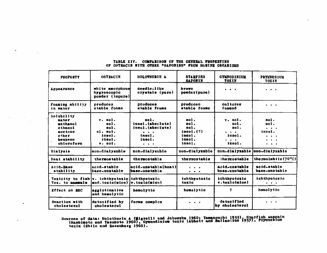
TABLE IIV. COKPAIISOI Gr THE GENlIAL ••O'£I'IISOF O~TJllCIR WITH O'I'REI ·SArolURS· PIlOM IIl\IIR OIGAIIBJIB
PSOPES'l' OSTBACIN HOLO'I'IIUIIJI A 8TAIlV181l1 G1'DODIRIUH PBYUNES IUlISAPOlIlR TOIIB TOlIN
Appearance "hi~e asorpboul needhl.like brown · · · · · ·h71r08copic cr7_tO" (pure) po_de r (pore )po.de r (illlpu re )
FOaJlllol abl I l,t)' produces producea pl'odticeo oultures · · ·in water stable fotUls stable fC>Bals stable lOUlIl fOUled
Solubilitywater v. 801. sol. eol. v. 001. Dol.lIIothaool 801. i0801.(ab8olnte) 801. lIOI. 1iI01.ethanol sol. insol.(absolute) 801l. 801. · · ·aoetone ul. 801. . . . i08011.(1) · · · ioool.e~her 10801. inaol. i08011. in801. · · ·beoztloe ln801. i0801. insoR. · · · · · ·chloroform v. 801. . . . in80ll. hll60l. · · ·-
D1alyeh nno.dialyzable non.dielyzoble noo.dial7sable oon.4ial,..able ooo.dialyzable
Heat stabil ity thtlNo.table theno&l1ablle themostable UaeftJo.,&.ble theroolabile(700C)
Aciel.Base acld.8tabl0 aoi4.un8tablle(heat) · · • a"ld_tllo.table aolet.stable8tabil itl' balle.uo.table ba.e-uo.tmblle • • • bOlle-tin.table balle.unetable
Toxicity to fieh .... lchthyotosio lohth70tozlo lohtllt,.otcxlo lolbtb7'ot01l:10 iohth70t 01l:1oTOI:. to .....1. 1II0d.toxlo(lIIloe) y.toxlo(.loo) t01l:10 Y. toxiohiiioe) · · ·Effect on RBC al.lutinaUye heool7110 helllclyUo l' hemol7tic
and helllolyUo
Heact ion .l th detoIIiUed by torae ooaapll'x · · · dl8toEltleet · · ·oholestel'ol ohoResterol by obol811t81'01
Souroes ot datal Holothurln A (.1,rell1 ~Dd ~mko.lIka 1960, tamaaouobl 1955), St&rfioh .aPODiD(H.8bll11oto and ' ••ua010 1960), CJDoodiolU8 toxin (Abbott aed Ball.otice 19'1), P~.lumto~in (Sbilo and aooeDbel'1 1960). , '
\177
may be a reflection of the impurity of ostracin since
impure substances often are more soluble than the purified
compounds.
The toxicity of ostracin to mice was much less than
that of the other "sapotoxinsU , whereas the ichthyotoxicity
of partially purified ostracin compared favorably with that
of purified holothurin Ao Ostracin also caused, at concen-
trations of less than 10 ppm, 100% hemolysis of a rabbit
erythrocyte suspension, whereas 100 ppm of holothurin A
was neceesary for complete hemolysis of rabbit erythrocytes
(Nigrelli and Jakowska, 1960)0
The antimetabolic effects shown by holothurin A in
planaria and sea urchin plutei were not demonstrated with
ostracin although a general toxio aotiQu nas e~ident
inhibition of development occurred in sea urchin embryos.
Therefore, ostracin can be classified with these
"sapotoxins n because its potency and mode of action appear
to compare favorably with the ecllinoderm and red tide toxins,
but the differences noted suggest that ostracin is distinct
chemically.
The saponins, like the alkaloids, have until recently,
only been isolated from plantso Within the past ten years
saponins have been found in the sea cucumbers and starfish
(Nigrelli, 1952.; Yamonouchi, 1955; Hashimoto and Yasumoto, 1960)0
The isolation of ostracin from the stress secretions of the
boxfish is the first time that steroid saponins have been
178
found in a vertebrateo The steroid genins found in the
venom of the parotid glands of toads are structurally
related to the sapogenins and the cardiac aglycones but
these toad poisons are not glycosideso The occurrence of
saponins in the Animal Kingdom, and especially in the
Vertebrata, could be of great significance in tracing the
paths of phylogeny by the biochemical evolution of the
steroids and their glycosidese
THE SIGNIFICANCE OF THE SKIN AND LABIAL GLANDSTO OSTRACIN SECRETION
The discovery of histologically distinct glands in the
integument and oral cavity of 00 lentiginosusp both of which
correlate with ostracin secretion, complicates the problem
of precisely identifying the actual source and site of
ostracin secretion. For simplicity's sake, ostracin has
been considered as one toxin and there is, as yet, no
concrete contrary evidenceo Nevertheless, two or more
saponins arecften found naturally occurring together in
the same specieso Two distinct saponins have been isolated
from the sea cucumber (holothurin A and B) and also from
the starfisho Similar examples may be found in the plant
saponins. In such cases, usually only one saponin is highly
potent thus masking the presence of the other components
in a crude extract bioassay. Therefore, it is reasonable
to suspect that "ostracinR may consist of different com~
ponents as does holothurino The difficulties met in obtaining
179
adequate sa~nJples of ostracin from the labial glands and the
failure to successfully prepare paper chromatograms showing
separation of different toxic components has militated
against this hypothesis, although this possibility has
definitely not been disproved and should be borne in mind
while considering which gland cells are responsible for
"ostracin" secretioDo
It has been established that two distinct gland cells,
the so.... called "mucous DJ and "club cellsI\' occur in the
integument, whereas only one type, the labial cells, form
the labial glands in the buccal cavityo Therefore, se7eral
theories as to the source of ostracin are possibleo In
the integument either one or both gland cells may secrete
ostracin, while the labial cells a~e the sGle 8U8POCtS
in the labial villi glands in tr~ oral cavity, although
the gland cells of the lip epidermis may contribute to the
oral secretionso
There appears to be little doubt that the skin str3ss
secretions are the major source of ostracin sinc€ severing
the lips from freshly captured boxfish did not seriously
affect the total yield of ostracin from such fisho
Histological and histcchemical studies of the skin showed
that both gland cells are actually "mucous secretingn cells~
One is a common type found in the epidermis of many fishes
and was simply called a "mucous" celIe Its mucins are
muco. and/or glycoproteinso The second type, the so_called

180
"club cells", appears to be a specialized secretory cell
that has been reported to occur in several ~enomous fishes
and in a few familia8 of non=poisonous teleosts o These
G1club cells n are believed to be modified serous cells
(Rabl p 1931) but a.ccording to Gabe and Arvey (1961), the
present classification of mucous and serous cells is an
oversimplification and its use is questionableo
The hmucins h of the club cells are not muco= or glyco
proteins but possibly mucopolysaccharideso Unfortunately
it is difficult to identify polysaccharides in tissues
since there are no specific tests for these substances
(Hale, 1957)0 The homogenous secretions of club cells are
orthochromatically stained by toluidine blue but stained
Led by WucicaLWine iudicating theix mucoiu natux~o Fu~iher.
more, their staining properties clearly indicate their
basophilic nature, whereas the mucous cell secretions are
acidophil iCe Although PAS negative, the coarse granules of
restitutional stages II and III of the club cells are stained
br Gomoriis aldehyde fuchsine Halmi and Davies (1953),
after comparing the staining reactions of the Shiff reagent
(PAS), aldehyde fuchsin and metachromasia by toluidine blue
on various tissues, concluded that most metachromatic and
orthochromatic PAS positive substances are also aldehyde
fuchsin positive but there are some exceptions with a few
PAS negative elements being aldehyde fuchsin positiveo
Thus p the histochemical techniques were largely unsuocessful

181
in elucidating the nature of the substance in the club
cells, but this was expected since the recent discovery of
steroid saponins in animal tissues precluded specific
histochemical tests for these substanceso
Fixation and staining of whole mucous secretions
showed that the club cell mucins were the major component
in the secretions and the numbers and relative sizes of
mucous and club cells in the epidermis corroborate this
observationo The non-toxic precipitate in a freshly collected
crude ostracin solution consisted largely of material from
the club cells and the spot tests of ostracin indicated
that the impurities or supporting substances were reacting
with the stains. Therefore, since the yield of ostracin
from a single fish~s secretions is so high (50 10 10C @g),
it seems more reasonable to suspect the larger and more
numerous club cells as being the source of ostracin rather
than the mucous cellso
The formation of club cell "glands" in certain areas
of the epidermis lends further support to the club cell
theory of ostracin secretion since it seems unlikely that
ordinary mucous cells would have formed so specialized a
structure. Similar wclub cell type" glands around the base
of the spines of venomous fishes have been clearly shown
to be specialized poison glands (HabI, 1931; Saunders, 1960;
Russell and Emery, 1960). Thus, it is logical to propose
t~at such is the case in Ostracion end possibly Lactoriao
182
The prominent labial glands in the mouth of trunkfish
present contradictory evidence, suggesting that the labial
gland cells and the small mucous oells in the skin are the
source of ostracino It appears that the labial glands
consist of modified skin mucous celIe judging by similarities
in staining properties and structureo Since there are
indications that the labial glands contain a hemolytic
ichthyotoxin, it is reasonable to suspect that the skin
mucous cells do alsoo However, if true, one would expect
these cells to be more numerous in the epidermis than they
are. Furthermore, the labial cells are distinct from the
mucous cells in that they were never observed in the process
of extrusion whereas the mucous cells were often seen
extruding their products o
Although the oral secretions were ichthyotoxic and
hemolytic, they probably accounted for less than 10% of
the total toxicity of the stress secretions, and since
the labial glands in the oral cavity are histologically
distinct from the skin glands, it is likely that their
secretory products are distinct alsoo
Comparative studies of other plectognaths add support
to the club cell theory of toxin secretioDo The epidermis
of the cowfish contains club cells that resemble those of
the boxfish in structure and staining properties, but
although the toxic secretio~~ of this species are similar
in action to ostTacin, they appear to be much less toxico
183
The mucous cells in the superficial layer of the epidermis
were more numerous in the cowfish p thereby providing a
negative correlation with toxicityo
"Homologous" gland cells are found in the epidermis
of puffers, which possess toxic skin secretions 9 and
although the chemical nature of their toxins is different
from ostreoin, the staining properties of the secretory
materials within the gland cells correlate perfectly with
those of the trunkfisho
The failure to extract ostracin from the suspected
poison glands in the boxfish was at first puzzling and
disturbingo However, it was shown later that ostracin is
highly toxic to the boxfish and causes both agglutination
and hemolysis of boxfish erythrocytes in vitroo Obviously,
the boxtish must have some control over its own potent
toxin and since ostracin is not a protein, immunological
protection is unlikely.. Therefore, either o.stracin is
stored in glands which ereot an ostracin-impermeable
barrier to the body fluids or an inactive precursor
accumulates in the glandular tissue and is activated by
an enzyme or other means during extrusion. The latter
explanation appears more likely in view of the inability
to extract ostracin from the poison glands o

184
CHAPTER 20
SUMMARY
The main objectives of this thesis were to characterize
chemically the ichthyotoxin secreted by distressed boxfish
Ostracio~ lentiginosus and to describe the histology of
their poison glands. The major results of these inves...
tigations are:
(1) An ichthyotoxic substance was found in the foamy
mucous secretions of distressed boxfish.
(2) This toxin was isolated and partially purified by
filtration, cent~ifugation, boiling and dialysis of the
mucous secretions and extraction of the water soluble
residue with acetone or chloroformo The toxic extract
so prepared was named "ostracinlto
(3) Ostracin was shown to be toxic to a variety of aquatic
organisms and, in general, it exhibited the same effect on
living systems as does holothurin (the sea cucumber toxin),
the starfish saponin and the red tide toxins of $ymnodinium
veneficum and pyrmnesium parvumo
(4) Ostracin was only moderately toxic to white mice.
About 4 mg of the purest preparation injected intraperitoneally
was needed to kill a 20 g mouse, whereas this same prepar
ation, diluted as great as 1:1,000,000 was lethal to fish o
(5) Since ostracin was toxic to most marine fishes at
concentrations as low as 1 ppm~ a fish immersion bioassay
185
was developed to detect ostracin in crude and semi_purified
preparationso
(6) Ostracin was detected in the stress secretions of
boxfish oollected, regardless of maturity, sex, time of
the year oaught or habitat, suggesting that ostracin is an
endogenous toxin.
(7) Ostracin is thermostable, non-dialyzable, stable in
acid and unstable in alkali at room temperatures, but is
readily hydrolyzed by heating in strong acido
(8) Ostracin is soluhle in water, methanol, ethanol,;
acetone and chloroform but is insoluble in diethyl ether
and benzen~0
(9) Profuse foaming in aqueous solutions was characteristic
of all ostracin preparationso
(10) Ostracin was shown to be hemolytic to vertebrate
erythrocytes and to form a water.insoluble, non-toxic
complex with cholesterol o Thus, ostracin was tentatively
identified as a steroid saponin.
(11) Besides its hemolytic activity, ostracin was found to
cause agglutination of erythrocytes of some species, a
property which distinguishes it from other saponins.
(12) The boxfish is poisoned by its own toxin, however it
is more resistant to ostracin than other fish. Both
hemolysis and agglutination of borfish erythrocytes by
ostracin occurred in vitroo
(13) The cowfish, Lactoria fornasini and two other species
186
of Hawaiian trunkfishes give off an ichthyotoxic, hemolytic
stress substance with their foamy mucous secretionso
(14) Several species of puffer_fishes also release a toxin
with their mucous secretionso However, the puffer toxin
is different from the trunkfish toxin and appears similar
in action to Tetraodotoxino
(15) Ostracin was readily detected both in the oral ex
udations and the skin mucous secretions of boxfish but could
not be extracted from the corresponding tissues of freshly
killed fish, suggesting that ostracin is activated only
during extrusion by the glandso
(16) A histological investigation of the skin (Bouin's
fixation) showed that 3 cell types occurred in the epidermis:
ordinary, unspecialized epidermal cells, mucous cells and
specialized gland cells called club cellso
(17) Clusters of club cells glands were found in dermal
pockets on some areas of the skin; especially in the
carapace epidermis behind the pectoral fino
(18) Suggested developmental stages of club cells were
described f:rom study of Bouin l s.fixed, Heidenhainf/s iron
hematoxylin sections.
(19) Some staining properties of the "secretory" material
within the mucous and club cells were determined with
Harris hematoxylinctriosin, Heidenhainis iron hematoxylin,
Malloryls triple stain, Gomori's aldehyde fuchsin.trichrome
stain, Toluidine blue, Periodic acid.Achiffls (PAS),

187
Dresbach's mucicarmine and Sudan Black Bo
(20) The fine granules in the mucous cells and the homogenous
secretions in the club cells have distinctive staining
properties. The mucous cells contain rouco-and/or glycoprotein
whereas the material in the club cells was not identified
by histochemical techniqueso
(21) Club cells were found extruding their products in the
middle layers of the epidermis, supposedly characteristic
of such cel180 Only club cell stages III a V appeared to
be capable of extrusion.
(22) Labial glands of a compound acinous type were found
in the oral cavity of boxfish and are believed to be.
associated with the toxic oral secretions although toxic
extracts of these glands could not be consistently obtained.
(23) The labial gland system consisted of dorsal and ventral
labial villi and dermal glandular pockets extending into
the lips and emptying both inside and outside the oral
cavity.
(24) Only one type of 8ecretory cell was recognized in the
labial glandso It resembled the mucous cells of the skin
in structure and staining properties but appeared to be a
distinct gland cell derived from the epidermis of the lipso
(25) Both the skin and. labial glands of starved boxfish
held in captivity were characterized by a great proliferation
of ~~idermal cells and reduction in numbers of gland cel18 0
This correlated with a significant decrease in the ostracin
188
secretion of such fish.
(26) nSpot tests 5D of ostracin and a controlled stress
experiment were inconclusive and did not aid in localizing
ostracin within the gland cellso
(27) Histological comparison of the skin of the cowfish
Lactoria fornasini with that of the boxfisI1 showed excellent
correlations with the structure and staining properties of
the mucous and club cells of this specieso Similar results
were obtained with the skin of some puffers, wher6as the
skin of a non.toxic filefish had no specialized gland
cells.
(28) It was proposed, based on the histological and
experimental evidence~ that the club cells in the epidermis
of boxfiah are the major source of ostracin secretion,
but the labial cells, and possibly the mucous cells of the
skin, may also be associated with the stress secretiono
189
LITERATURE CITED
Abbott, Bo Co and Do Ballantine. 19570 The toxin fromGymnodinium veneficum Ballantineo J. Maro BioloAsso Uo Ko 36: 169-1890
AI_Hussaini, Ao Ho 1947. The feeding habits and themorphology of the alimentary tract of some te1eostsliving in the neighborhood of the Marine BiologicalStation, Ghardaqa, Red Sea. Pub10 of the Maro BioloStaG Ghardaqao Noo 5: 1.61.
Andrew, W. 1959. Textbook of comparative histology.Oxford Univ. Press. New York. 652 po
Aronson, L. Ro and Co Mosher. 19510 Observations on thebehavior and ecology of the West Indian pearl fish.Anat. Record. .ill: (Abstr~ 105).
Banner, A. H., Po Jo Scheuer, S. Sasaki, Po Helfrich,C.Bo Alender o 1960 e Observations on Ciguatera_typetoxin in fisho Anno N. Yo Acad. Sci. 2Q (3): 770-7870
Black, U. S. 19570 Excretion and osmoregulation,po 163.206. In The Physiology of Fishes. Volo 10M. E. Brown, Ted.;. Academic Press Inco New Yorko447 po
Briggs, Jo Co 19610 The East Pacific barrier and thedistribution of marine shore fishes. Evol.12 (4)~
545-5540
Brock, V. E. 19550 Possible production of a substancepoisonous to other fish by the boxfish Ostracion1entiginosus Schneider. Copeiao 1 (195-196).
Brown, H. H. 1945. The fisheries of the windward andleeward islandso Development and Welfare in theWest Indieso Bullo No o 20 0 98 p.
Chanley, J.DollJl~Ledeen, J. Wax, Ro Fo Nigrelli, H. Sobotka.19590 Ho10thurin. I. The isolation, properties, andsugar components of holothurin Ao Jo Am. Chemo Soco.§l: 5180-5183·
Clark, Eo 1949. Notes on some Hawaiian plectoguathfishes, including a key to the specieso Amero MusoNovitates. (l937): 1...22.
Clark, Eo and H. A. Fo Gohar. 1953. The fishes of theRed Sea: order Plectognathio Publ. Mar. Biolo StaGAl Ghardaqao 8: 1...80.
190
LITERATURE CITED (continued)
Conn, H. Jo, M. A. Darrow s V. M. Emmelo 1960. Stainingprocedures Hsed by the Biological Stain Commission.Williams and Wilkins Co. Baltimore. 289 po
Danielli, Jo Fo 19580 Surface chemistry and cellmembranes, po 246-2650 In Surface phenomena in chemistryand biology. J. F. Danielli, K. Go A. Pankhurst,Ao C. Riddiford, (edo), Pergamon Presso N. Yo, London,Paris, Loa Angeleso
Dobzhansky, T. 1951.species. 2nd edo428 p.
Genetics and the origin of theColUmbia Univo Presso New Yorko
Dutta, N. L. 19550chromatography.
Separa.tion of saponins by paperNature. ill: 85.
Eger, W. 19630 An exotoxin produced by the puffer,Arothron BJspidus, with notes on the toxicity ofother plectognath fisheso Master's thesis. Univ o ofHawaii, Honolulu, Hawaii.
Evans, Ho M. 19240 The defen'sive spines of fishes, livingand fossil, and glandular structure in connectiontherewitho Observatious Oil the nature of fish venoms~
P~ilo Trans. R. Soc. London B. 212: 1-33.
Evermann, B. W. 1902. Investigations of the aquaticresources and fisheries of Porto Rico. U. So Fish.Corum. Bull. Goverment Printing Office. Washington.350 p.
Fieser, 10 Fo and M. A. Fieser. 1959.Reinhold Publishing Corp. New York.
Steroids.945 po
Fraser-Brunner, Ao 1935. A synopsis of the genera ofthe family Ostraciontidaeo Ann. Mag. Nato RistolQ.: 313-320.
_--::-~ ~_~. 1940"family Ostraciontidae.
Sexual dimorphism in theAnn. Mago Nat. Hist. ~: 390-392.
____~~~__~~-o 19410 The Aracanidae, a distinct familyof Ostraciontoid fishes with descriptions of two newspecieso Ann. Mag o Nato Rist. 8: 306-313.
00
Frisch, Ko von. 19410 Uber einen Schreckstoff der Fischhautund seine. Biologische Bedeutung. Z. Vergleiche Physio1.~: 46-1450

191
LITERATURE CITED (continued)
Gabe, Mo and Lo Arvey. 19610 Gland cellso (po 1~88) InThe cello V(2)0 J. Brachet and A. Eo Mirsky. AcademicPresso New York. Londono 597 po
Gates, J. Ao and W. B. Wilsono 1960. The toxicity ofGonyaulax monilata Howell to Mugil cephaluso Jouro Limoand Oceanogr. j (2): 171-174.
Gomo~ip Go' 1950ao Ald6hyde.fuchsin: a new stain forelastic tissueo Am. Jour. C1ino Path. 20: 665-666.
__~__~_. 1950bo A rapid one.step trichrome stain.Am. Jouro C1in. Path. 20: 661-6640
Gordon, Mo 1937. The fishes l art of se!fQdefenseoNato Histo N. Yo 40: 433-4410
Gosline, W. A. and V. E. Brock. 1960. Handbook of Hawaiianfishes. Univo of Hawaii Press. Honolulu. 372 po
Hale, A. J. 1957. The histochemistry of polysaccharides,p. 194.2630 !n International Review of Cytology (VI)oG. H. Bourne and J. F. Danielli, (edo) 556 po
Halmi, N. S. and Jo Davies. 1953. Comparison ofaldehyde fuchsin staining, metachromasia andperiodic acid. Schiff reactivity of various tissues.Jouro Histochem. and Cytochem. 1(6): 447-4590
Halstead, B. W. and N. Co Bunker. ~1954. A survey of thepoisonous fishes of the Phoenix Islands. Copeia. 1: 1.11.
Halstead, B. W., M. J. Chitwood, F. R. Modglin. 1955a.The venom apparatus of the California scorpion fish,Scorpaena guttata Girard. Transo Amero Micro Socoz!±: 145-1580
• 1955bo The anatomy of the venom apparatus----o-f--t-h-e--z-e-brafish, Pterois volitans (Linnaeus). Anat o Reco
1£&: 317-3330 ------- -
________~ • 19560 Stonefish stings and the venomapparatus of Synanceja horrida (Linnaeus). Trans. AmeroMicro Soc. 1j: 381_3970
Hashimoto, Yo and To Yasumoto o 1960. Confirmation ofsaponin as a toxic principle of starfisho Bull. JapoSoco Scio Fisho 26 (11): 1132-11380

192
LITERATURE CITED (continued)
Hiatt, R. W. and D. A. Strasburg o 1960. Ecologicalrelationships of the fish fauna on coral reefs of theMarshall Islands. Ecological Monographs. 1Q: 65-1270
Lemieux, R. U. and H. F. Bauer o.detect ion of carbohydrates.
1954. Spray reagent forAnal. Chem. £2: 920 •
Monod, T. 1950. Notes dVichtiologie Ouest-AfricaineoBull. Inst o Franc. Afrique Noire. 12: 1-71.
Nigrelli, R. F. 1952. The effect of holothurin on fishand mice with Sarcoma-180. Zoologica. JZ: 89-90.
Nigrelli, R. F. and S. Jakowska. 1960 0 Effects of holothurin, a steroid saponin from the Bahamian sea cucumber(A£!inopyga agassizi) on various biological systems.Ann. N. Y. Acad. Sci o 2Q (3)= 884-892.
Nishida, H. 1954. Occurrence of remarkable glands in theoral cavity epidermis of adult salmon, Oncorhynchus ~.Scio Repo Hokkaido Fisho Hatcheryo 2: 147-150.
Pearse, A. Go E. 1960. Histochemistry, theoretical andapplied. 2nd edo Little~ Brown and Co. Bostono 998 po
Quaglio, N., So F. Nolan, A.. M. Veltri, P. M. Murray,S. Jakowski, R. F. Nigrelli. 1957. Effects of holothurinon survival and regeneration of planarianso Anato Record o128: 604.6050
Rab1, Ho 1931. 10 Integument der Ananmier, po 271-345.In Handbuch der Vergleichenden .A.natomie der WirbeltiereoBOlk, Lo, E. Goppert, Eo Kal1ium, Wo Lubosch, (ed.)Urban and Schwarzenberg, Berlin-Wieno 752 p.
Randall, J. E. 1958. A review of ciguatera, tropical fishpoisoning, with a tentative explanation of its cause.Bull. Maro Sci. Gulf and Carib. 8 (3): 236.2670
Ransom, F. 19010 Saponin und sein GegengiftoWochschro £1: 194.1960
Deut o medo
Reintjes, J. W. and J. E. King. 1953. Food of yellowfintuna in the central Pacific. U. S. Fish and WildlifeServo Fish. Bullo 81. ~: 91-110 0
Rofen, R. R. 1958. The. marine fishes of Rennell Island,po 149-218. 1Y The Natural History of Rennell Islando(I). Danish Science Press, Ltd. Copenhagen.

193
LITERATURE CITED (continued)
Rosen, N. 19l2ao Studies on the plectognathso The blood....vascular system. Ark. Zool. Stockholm o Z (25): 1-240
________• 19l2b. Studies on the plectognaths o The air_sac with notes on other parts of the intestinesoArko Zoolo Stockholm. 2 (30): 23 po
__~ • 1913ao Studies on the plectognathso Theintegument. Ark. Zoolo Stockholm. 8 (10): 1-29.
______~. 1913b. Studies on the plectognathso The bodymuscles. Arko Zool. Stockholm 0 8 (18): 1_14.
Ruggieri, G. D. and Ro Fo Nigrelli o 19600 The effects ofholothurin, a steroid saponin from the sea cucumber, onthe development of the sea urchin. ZoologicRo ~ (1): 1_160
Russell, Fo Eo and Jo A. Emery. 1960. Venom of the weeversTracbinu8 draco and Trach}nusvipera. Anno No Y. Aca.d.Sci. 90 (3): 798....804.
Ruyssen, Ho 1957. Haemolysis by tensioactive substancesoPrQc.·of Second International Congress of Surface Activity.Iv:a 271...276.
Saunders, P. R. 1960. Pharmacological and chemical studiesof the venom of the stonefish (genus Synanceja) andother scorpionfishes. Anno No "Y. Acad. Sci • .2Q (3): 805-8190
Schultz, L. The fishes of the Marsnalls and Marianaso !!!o(Manuscript) 0
Schantz, E. Jo 1960 0
shellfish poisons.Biochemical studies on paralyticAnno No Y. Acado Sci. 2Q (3): 843-8550
Shilo, Mo and Ho F. Rosenberger. 1960. Studies on the toxicprinciples formed by the chrysomonad Prymnesium parvumCarter. Anu. No Y. Acado Scio .2Q (3): 866-8760
Skinner, W.' A., R. D. Mathews, R. M. Parkhurst. 19620Alarm reaction of t~e top smelt, Atheripops affinis (Ayres).Science. 138: 681-682.
Sokolskaya, Ao Mo 1951•. The saponin of the roots ofPatrinia intermedia Ro et Schult. Soviet PharmaceuticalResearcho l!!: 378-3800 Pharmacognosy. (Translated intoEnglish by Consultants Bureau Inco)

LITERATURE CITED (continued)
Straughan, R. P. L. 19590 The salt water aquarium inthe home. A. S. Barnes Inc. New York. 262 p.
Tani, I. 19450 Toxicological studies on Japanese puffers.Imperial Chem. Corp. Japan. £ (3): 1-103.
Tester, A. L.predation.
19630 The role of olfaction in sharkPacif o Scio 12(2): 145-1700
Van Oosten, J. 19570 The skin and scales, p. 163-2060In The Physiology of Fishes. (Volo 1)0 M. Eo Brown p (ado)ACademic Press, Inco New Yorko 447 po
Wall, M. Eo, Co R. Eddy, M. L. McClennon, M. E. Klumpp.19520 Detection and estimation of steroidal sapogeninsin plant tissue. Anal. Chern. ~: 1337-1341.