zurich open repository and year: 2011 filemouse model. moreover, both glycosaminoglycans reduced...
TRANSCRIPT
Zurich Open Repository andArchiveUniversity of ZurichMain LibraryStrickhofstrasse 39CH-8057 Zurichwww.zora.uzh.ch
Year: 2011
Ascidian dermatan sulfates attenuate metastasis, inflammation andthrombosis by inhibition of P-selectin
Kozlowski, E; Pavão, M S G; Borsig, L
Abstract: Background: Cancer-associated thrombosis and enduring inflammation are strongly associatedwith cancer progression and metastasis. Heparin is the mostly clinically used anticoagulant/antithrom-botic drug, and has recently been shown to exhibit antimetastatic and anti-inflammatory activities thatare linked to inhibition of P-selectin and/or L-selectin. P-selectin-mediated platelet–tumor cell and tu-mor cell–endothelium interactions facilitate the initial steps of metastasis. Objectives and Methods: Theaim of the present study was to determine the capacity of dermatan sulfates to inhibit P-selectin and totest their potential to affect thrombosis, inflammation and metastasis in respective experimental mousemodels. Results: Two dermatan sulfates isolated from the ascidians Styela plicata and Phallusia nigra,composed of the same disaccharide core structure (IdoA2-GalNAc)n, but sulfated at carbon 4 or 6 ofthe GalNAc, respectively, have opposed heparin cofactor II (HCII) activities and are potent inhibitors ofP-selectin. The ascidian dermatan sulfates effectively attenuated metastasis of both MC-38 colon carci-noma and B16-BL6 melanoma cells and the infiltration of inflammatory cells in a thioglycollate peritonitismouse model. Moreover, both glycosaminoglycans reduced thrombus size in an FeCl3-induced arterialthrombosis model, irrespective of their HCII activities. The analysis of arterial thrombi demonstratedmarkedly reduced platelet deposition after dermatan sulfate treatment, suggesting that the glycosamino-glycan inhibited P-selectin and thereby the binding of activated platelets during thrombus formation.Conclusions: Collectively, these findings provide evidence that specific inhibition of P-selectin representsa potential therapeutic target in thrombosis, inflammation and metastasis, and that ascidian dermatansulfates may serve as antiselectin agents.
DOI: https://doi.org/10.1111/j.1538-7836.2011.04401.x
Posted at the Zurich Open Repository and Archive, University of ZurichZORA URL: https://doi.org/10.5167/uzh-53579Journal ArticleAccepted Version
Originally published at:Kozlowski, E; Pavão, M S G; Borsig, L (2011). Ascidian dermatan sulfates attenuate metastasis, inflam-mation and thrombosis by inhibition of P-selectin. Journal of Thrombosis and Haemostasis, 9(9):1807-1815.DOI: https://doi.org/10.1111/j.1538-7836.2011.04401.x

1
5/23/2011 Manuscript
Ascidian dermatan sulfates attenuate metastasis, inflammation and
thrombosis by inhibition of P-selectin
Eliene Oliveira Kozlowski1,2, Mauro S.G. Pavao2, Lubor Borsig1
1Institute of Physiology, University of Zürich and Zürich Center for Integrative Human
Physiology, 8057 Zürich, Switzerland 2Laboratório Bioquímica e Biologia Celular de Glicoconjugados, Hospital Universitário
Clementino Fraga Filho and Instituto de Bioquímica Médica – Programa de Glicobiologia,
Universidade Federal do Rio de Janeiro, Rio de Janeiro, RJ 21941-590, Brazil.
Corresponding author: Lubor Borsig
Institute of Physiology, University of Zürich
Winterthurerstrasse 190
CH-8057 Zürich, Switzerland
Phone: +41 44 635-5134
Fax: +41 44 635-6814
Email: [email protected]
Running Title: Dermatan sulfates block thrombosis and metastasis
Key words: dermatan sulfate, metastasis, inflammation, thrombosis, P-selectin
2
Summary
Background: Cancer-associated thrombosis and enduring inflammation are strongly associated
with cancer progression and metastasis. Heparin, is the mostly clinically used
anticoagulant/antithrombotic drug, which has recently been shown to exhibit antimetastatic and
antiinflammatory activities that are linked to inhibition of P- and/or L-selectin. P-selectin-
mediated platelet-tumor cell and tumor-cell endothelium interactions facilitate the initial steps of
metastasis. Objectives and Methods: The aim of the present study was to determine the capacity
of dermatan sulfates to inhibit P-selectin and to test their potential to affect thrombosis,
inflammation and metastasis in respective experimental mouse models. Results: Two dermatan
sulfates isolated from the ascidians Styela plicata and Phallusia nigra, composed of the same
disaccharide core structure [IdoA2-GalNAc]n, but sulfated at carbon 4 or 6 of the GalNAc
residues, respectively, contain opposed HCII activities and are potent inhibitors of P-selectin.
The ascidian dermatan sulfates effectively attenuated metastasis of both MC-38 colon carcinoma
and B16-BL6 melanoma cells, and the infiltration of inflammatory cells in a thioglycollate
peritonitis mouse model. Moreover, both glycosaminoglycans, reduced thrombus size in a FeCl3-
induced arterial thrombosis model, irrespective of their HCII activities. The analysis of arterial
thrombi demonstrated a markedly reduced platelet deposition after dermatan sulfate treatment,
suggesting that the glycosaminoglycan inhibited P-selectin and thereby the binding of activated
platelets during thrombus formation. Conclusions: Collectively, these findings provide evidence
that specific inhibition of P-selectin represents a potential therapeutic target in thrombosis,
inflammation and metastasis, and ascidian dermatan sulfates may serve as anti-selectin agents.

3
Introduction
The relationship between hypercoagulability and cancer was first observed by Trousseau more
than a century ago [reviewed in 1]. Activation of blood coagulation in cancer patients is a rather
common complication that has been linked to cancer progression [2]. Tumor cells express tissue
factor and secrete cytokines that contribute to a prothrombotic microenvironment, which also
includes activation of platelets. The pathophysiology of thrombosis in cancer is complex, but
clinical and experimental evidence indicate that platelets represent the link between coagulation
and cancer progression [3-5]. Activated platelets interact with the activated endothelium and
contribute to recruitment of leukocytes to inflammatory sites [6]. Platelets are a rich source of
pro- and anti-angiogenic factors that upon activation may contribute to angiogenesis [7].
Furthermore, platelets support the integrity of angiogenic, inflamed [8] and tumor microvessels
[5]. Experimentally induced thrombocytopenia in tumor bearing animals leads to massive
hemorrhage at the tumor-stroma interface, indicating a central role of platelets in tumor vascular
homeostasis [5]. Although thromboembolism and inflammation are linked to cancer in a number
of different ways, accumulating experimental evidence indicates that P-selectin has an
integrating role in cancer progression [9-11].
P-selectin is a member of the selectin family of cell adhesion molecules that facilitates
interactions among platelets, leukocytes and endothelial cells [12]. The contribution of selectins
to physiological processes such as inflammation, reperfusion injury or hemostasis are well
described [12]. P-selectin is present in the storage granules of endothelial cells (Weibel-Palade
bodies) and platelets (α-granules), thus enabling rapid cell-surface expression upon activation
4
[12]. Most selectin ligands are based on the terminal tetrasaccharide structure sialyl Lewisx
(sLex) [12]. During hematogenous metastasis carcinoma cells carrying sLex-containing mucins
enter the circulation and become potential candidates for selectin-mediated interactions with
platelets, leukocytes and endothelium [11]. The absence of P- and/or L-selectin leads to
attenuation of experimental metastasis in different animal models, implicating selectins in cancer
progression [4, 13, 14]. Formation of platelet-tumor cell emboli is largely mediated by P-selectin
[4] and contribute to evasion of host responses and to colonization of distant organs [3, 15].
Accumulating evidence points to the critical role of P-selectin in cancer-associated thrombosis
[16]. Elevated levels of activated P-selectin-expressing platelets have been observed in advanced
stages of cancer [17]. Furthermore, the presence of tissue-factor–bearing microparticles
contributing to the prothrombic state has been observed in the circulation of gastric and
pancreatic cancer patients [18]. P-selectin signaling through its receptor on leukocytes, P-selectin
glycoprotein ligand 1 (PSGL-1), induces the generation of tissue factor-positive, highly pro-
coagulant microparticles [10, 19]. Consequently, P-selectin inhibition might have a beneficial
effect on survival of cancer patients, by attenuating both hematogenous metastasis and
thromboembolic complications.
Unfractionated heparin (UFH) and low molecular weight heparins (LMWHs) are commonly
used for prevention and treatment of cancer-associated thromboembolism [20-22]. Currently
emerging clinical evidence implicates heparin in prolonging survival of cancer patients [20, 21].
There is abundant experimental data indicating that heparins attenuate metastasis by affecting
angiogenesis, heparanase, P- and L-selectins or binding of cytokines [for review see 23, 24].
These observations are further supported by findings that non-anticoagulant heparin derivatives
also attenuate metastasis [25, 26]. Despite promising results, an effective and safe antithrombotic
5
treatment remains a very challenging clinical task in cancer patients, because of the high risk of
bleeding complications and thrombotic events in heparin-treated patients [20, 22].
Previously, we reported the presence of highly sulfated dermatan sulfates (DSs) in solitary
ascidians (sea squids) from the orders Phlebobranchia (Phallusia nigra) and Stolidobranchia
(Styela plicata) [27]. These polymers are composed of the same disaccharide backbone,
consisting of [→4IdoA(2S)β-1→3GalNAcβ-1→], but differ in the position of sulfation on the
GalNAc, which can be sulfated at carbon 4 or 6 [27]. The DS from the stolidobranchia S.plicata
is sulfated at carbon 2 of IdoAc and carbon 4 of GalNAc [27] and is a potent activator of heparin
cofactor II (HCII). In contrast, the DS from the phlebobranchia P.nigra is sulfated at carbon 2 of
IdoAc and at carbon 6 of GalNAc, and is a poor activator of HCII [27].
In the present work, we show that ascidian DSs, but not mammalian DS, attenuate
hematogeneous metastasis, inflammation-induced leukocytes recruitment and thrombosis.
Inhibition of P-selectin has been identified as the major biological activity of ascidian DS that
affects all three processes closely associated with cancer progression.

6
Materials and Methods
Cell lines and reagents - Human colon carcinoma cells LS180 (ATCC, Manassas, VA) were
grown in alpha-MEM media (Invitrogen, Carlsbad, CA) supplemented with 10% FCS
(Invitrogen). Mouse colon carcinoma cell line MC-38, stably expressing GFP MC-38GFP [13]
and mouse melanoma cell line B16-BL6 [25], were grown in DMEM with 4.5 g/l of glucose
supplemented with 10% FCS medium (Invitrogen). All reagents were from Sigma (St. Louis,
MO) unless otherwise stated. Unfractionated heparin – Liquemine (UFH) was obtained from
Roche Pharma, Switzerland.
Dermatan sulfates - The ascidian P.nigra was collected in Angra dos Reis, Rio de Janeiro,
Brazil; S.plicata was collected at Praia da Urca, Rio de Janeiro, Brazil. Dermatan sulfates (DSs),
were isolated from ascidian visceras by proteolytic digestion followed by anion exchange
chromatography as described previously [27]. Both ascidian DS contained 2-O sulfation on α-L-
iduronic acid. While S.plicata had additional sulfation in 4-O position of N-acetyl-β-D
glucosamine unit, P.nigra had sulfated 6-O position. Therefore S.plicata DS was designated as
2,4-DS while that from P.nigra as 2,6-DS. Mammalian DS was obtained from Sigma Company.
Oversulfated DS was prepared from a pig mucosal dermatan sulfate [28].
Activated partial thromboplastin time assay- aPTT clotting assays were carried out as
described previously [27]. Briefly, normal human plasma (90 μL) was incubated with 10 μL of
DS at several dilutions and 100 μL of cephalin. After three minutes at 37oC, 100 μL of 0.25 M
CaCl2 were added to the mixtures and the clotting time was recorded. The activity is expressed
as units/mg using a standard unfractionated heparin (200 IU/mg) curve.
7
Inhibition of tumor cell binding to immobilized selectins – The ability of DSs to inhibit the
adhesion of calcein AM labeled LS180 cells to immobilized P-selectin chimeras was examined
in a serial dilution of glycosaminoglycans as described previously [25].
Platelet-tumor cell aggregation in vivo – Lungs were prepared and analyzed at 30 min or 3 h
after intravenous injection of tumor cells as described previously [4]. Briefly, LS180 cells were
harvested with 2 mM EDTA in PBS; labeled with Calcein AM and injected intravenously in
mice with or without previous intravenous application of 100 µg of DSs or 1 mg of UFH (200
IU/mg) [4]. Lung sections were stained with rat anti-mouse CD41 antibody (Becton Dickinson,
Mountain View, CA), followed by the goat anti-rat-Alexa 568-conjugated antibody (Invitrogen)
and analyzed by immunofluorescence microscopy. Calcein-labeled cells present in 40 view fields
of six different lung sections were scored as “associated” or “not associated” with platelets.
Experimental metastasis model – Wild type (wt) or P-selectin-deficient mice (P-sel-/-) in
C57Bl/J6 background (The Jackson Laboratory) were intravenously injected either with 150’000
B16-BL6 cells or 300’000 MC-38GFP cells via the tail vein. Mice received either PBS or 100 µg
of mammalian DS, mammalian oversulfated DS, ascidian 2,4- or 2,6-DS intravenously injected
10 minutes prior to tumor cell injection. Mice injected with melanoma cells were terminated 15
days later and mice receiving carcinoma cells were terminated after 28 days. PBS-perfused lungs
were macroscopically evaluated for the presence of metastatic foci. GFP measurement in the
lungs homogenates from MC-38GFP cells-injected mice were performed as described previously
[4].
Thioglycollate-induced peritonitis – Mice were injected with 1 ml of 4% thioglycollate broth
into the peritoneum. Mice were intravenously injected with PBS, P-selectin function-blocking
8
antibody [29], 2,4-DS or 2,6-DS (4 mg/kg) five minutes after thioglycollate injection. After 4
hours, mice were terminated and peritoneal cells harvested by injection of 4 ml of PBS
containing 0.5% BSA and 1 mM EDTA. The total number of cells in peritoneal lavage was
counted with a hemocytometer. The differential count of polymorphonuclear leukocytes was
determined in cytospin preparations stained by Hematoxylin and Eosin.
FeCl3-induced arterial thrombosis – Wt or P-sel-/- C57Bl6 mice were intravenously injected
with 50 µL of 2,4-DS, 2,6-DS, P-selectin function blocking antibody (4 mg/kg) or PBS, 10
minutes before induction of thrombosis. Mice were placed in a supine position and the common
carotid artery (CCA) was exposed. Thrombosis was induced by placing a Whatman filter paper
saturated with 10% FeCl3 in 10% glycerol on CCA. After three minutes, the FeCl3-paper was
removed and CCA was rinsed with PBS. Blood flow was monitored with an ultrasonic flow
probe (Transonic System, USA) until complete vessel occlusion occurred [30]. For histological
analysis, mice were terminated 20 minutes after removing FeCl3-paper and CCA was carefully
dissected and frozen in OCT compound (Tissue-Tek, Sakura). Tissue sections were stained with
hematoxylin and eosin. Platelets were detected with rat anti-mouse CD41 antibody, followed by
Alexa 568-conjugated goat anti-rat antibody and analyzed by fluorescence microscopy. DAPI
was used for visualization of cell nuclei. At least five images acquired by Zeiss AM200
microscope were analyzed by Imaris® software per group (Bitplane AG, Zürich,
Switzerland).
9
Results
Dermatan sulfates inhibit tumor cells binding to P-selectin
To study the structural requirements of ascidian and mammalian dermatan sulfates (DSs) for P-
selectin recognition, we tested the capacity of DSs to inhibit binding of LS180 cells to P-selectin.
(Figure 1A). Ascidian DSs (2,4-DS or 2,6-DS) as well as a chemically oversulfated mammalian
DS (OSDS) inhibited tumor cell binding to P-selectin at comparable levels, with an IC50 of 13.5
µg/ml (ascidian 2,4-DS) and 12.2 µg/ml (ascidian 2,6-DS) and 12.6 µg/ml (OSDS). All
disulfated DSs were superior to UFH (24.5 µg/ml). In contrast, single sulfated mammalian 4-DS
from porcine skin did not inhibit P-selectin binding to tumor cells even at higher concentrations.
These observations indicate that the sulfation degree of dermatan sulfate is critical for P-selectin
recognition (Table 1). While ascidian DSs contain high levels of 2,4- or 2,6-disulfated
disaccharides, 66% and 78%, respectively [27], mammalian DS contains mostly 4-monosulfated
disaccharide units (80%). Disaccharide analysis of OSDS from porcine skin revealed a distinct
sulfation pattern composed of mostly 4,6- disulfated disaccharides (35.5%) and containing small
amount of 2,6-disulfated, 6-monosulfated and trisulfated disaccharides, 18.3%, 20.4% and 23.4%
respectively. OSDS or ascidian DSs showed similar IC50 values in the P-selectin binding assays,
suggesting that degree of sulfation, rather than the position of sulfate groups, is critical for P-
selectin interaction. In contrast, the anticoagulant activity of ascidian DSs has been shown to
depend on the position of sulfation on the N-acetylgalactosamine [27].
10
L-selectin also binds GAGs such as heparin and heparan sulfate [31]. Although, L-selectin
binding to 2,4 and 2,6-DSs was less efficient than of P-selectin (2-fold lower), it was comparable
to the binding of unfractionated heparin (Suppl. Figure 1).
P-selectin-mediated binding of platelets to selectin ligands on tumor cells significantly
contributes to tumor cell emboli formation [4]. To test ascidian DSs as inhibitors of P-selectin-
mediated interactions in vivo, mice were injected with calcein-labeled LS180 cells 10 minutes
after application of DSs (100 µg/mouse), PBS, or UFH (1 mg/mouse). The extent of platelet-
tumor cell emboli in the lung microvasculature was evaluated 30 min and 3 hours later (Figure
1B). Initial seeding of tumor cells in lungs was comparable among all groups after 30 min but the
number of viable tumor cells detected after 3 h significantly decreased in mice injected either
with DSs or heparin. These results confirmed that ascidian DSs are potent inhibitors of P-
selectin-mediated interactions also in vivo, while the inhibitory effect of DSs lasted for at least 3
hours.
Ascidian DSs attenuate experimental metastasis
Inhibition of P-selectin or the absence of P-selectin has been previously shown to attenuate
metastasis [4, 14]. To determine the ability of ascidian DSs to attenuate metastasis, wt mice were
intravenously injected with 100 µg of each ascidian DS followed by injection of MC-38GFP
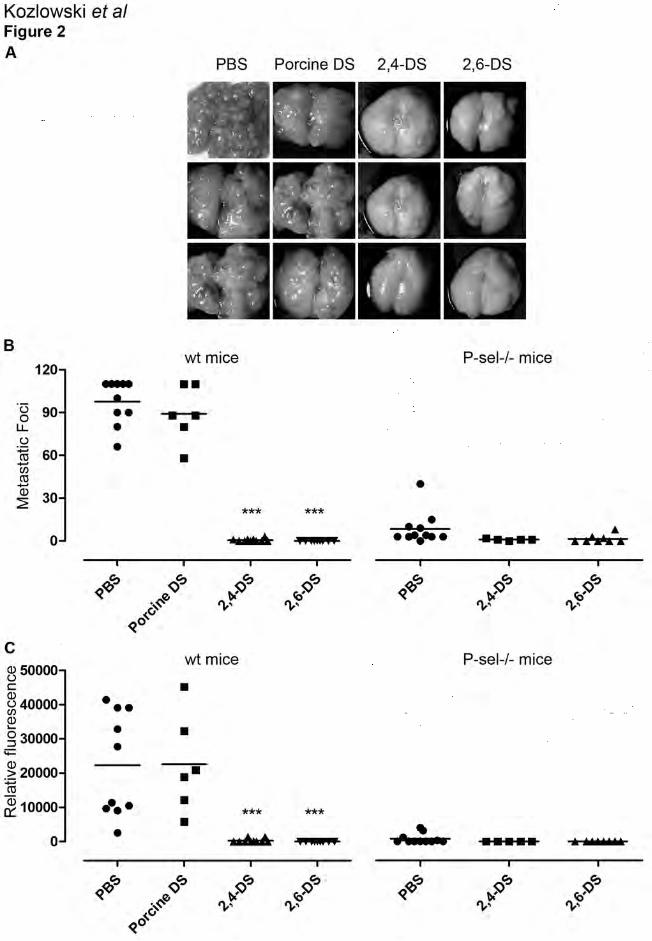
cells. After twenty eight days, mice were terminated and lungs evaluated for metastasis (Figure
2). Both ascidian DSs (2,4-DS or 2,6-DS) markedly attenuated metastasis, when compared to
PBS injected control mice with lungs displaced by metastasis (Figure 2A). Lungs of ascidian
DSs-treated mice showed only a few metastatic foci, 0-7 foci per lung (Figure 2B). Injection of
11
mammalian DS did not affect metastasis, which is in agreement with the previous observation
that 4-DS had no effect on P-selectin inhibition (Figure 1A). To confirm that the antimetastatic
effect of DS is dependent on P-selectin, we intravenously injected ascidian DSs followed by
MC-38GFP in P-selectin-deficient mice (P-sel-/-) (Figure 2B). While P-selectin deficiency alone
markedly reduced metastasis [4, 13], small number of metastatic foci were clearly detectable (3-
25 foci per lung). However, 2,4- or 2,6-DS had only minimal additional effect on metastasis,
indicating that the anti-metastatic effect of ascidian DSs in wt mice depends on inhibition of P-
selectin-mediated interactions.
The anti-metastatic effect of ascidian DSs was also evaluated with B16-BL6 melanoma cells,
which were previously reported to express P-selectin ligands, albeit to a lesser extent than MC-
38 cells [25]. Both ascidians DSs attenuated metastasis of B16-BL6, although less efficiently
than by MC-38GFP cells (Figure 3).
Ascidian DSs inhibit recruitment of polymorphonuclear cells
There is accumulating evidence that inflammatory cells affect tumorigenesis and metastasis,
although the underlying mechanism is still under investigation [32, 33]. Since ascidian DSs are
potent P-selectin inhibitors, we tested their capacity to inhibit leukocyte recruitment in a
thioglycollate-induced peritonitis mouse model. Intraperitoneal injection of 4% thioglycollate
was followed by intravenous injection of 100 µg of 2,4- or 2,6-DS or P-selectin function-
blocking antibody (P-sel Ab), respectively. Analysis of peritoneal lavage leukocytes showed a
three-fold increase in the number of polymorphonuclear cells (PMN) in thioglycollate treated
mice when compared to controls (Figure 4A). Both ascidian DSs significantly reduced leukocyte

12
recruitment, primarily PMNs (Figure 4B). Ascidian DSs reduced peritoneal recruitment of PMNs
to the similar extent as observed in mice treated with P-sel Ab. Thus, inhibition of P-selectin and
possibly L-selectin are primarily responsible for the anti-inflammatory effect of ascidian DSs.
Ascidian DSs inhibit arterial thrombosis
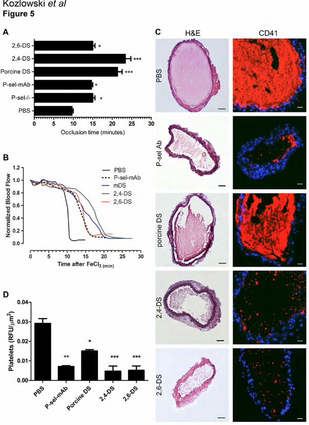
To test whether P-selectin inhibition by DSs affects thrombosis, we applied ascidian DSs in the
FeCl3-induced carotid artery lesion model. An increased time to occlusion has been observed for
both anticoagulant dermatans, 2,4-DS and mammalian DS respectively (Figure 5A). Although
2,6-DS has low HCII-mediated anticoagulant activity [27], it was able to trigger a small but
significant prolongation of the occlusion time. Furthermore, we observed similar prolongation of
the occlusion time both in P-sel-/- mice and wt mice treated with P-sel Ab (Figure 5A).
Representative normalized blood flow curves obtained from experiments with mice treated either
with 2,6-DS or P-sel Ab revealed a tendency to an increase of the initial plateau, followed by a
sharp drop in the blood flow (Figure 5B). Interestingly, the curve obtained in the experiments
with 2,4-DS-treated mice showed not only the same prolonged initial plateau, but also a milder
drop (green line). To test whether P-selectin inhibition by DSs affects thrombus formation, we
analyzed the carotid arteries 20 min after the induction of thrombosis. Histological analysis
revealed smaller thrombi in mice treated with 2,4- and 2,6 DS, but a marked difference in
thrombi composition was observed (Figure 5C). Whereas control mice and mammalian DS-
treated mice showed massive thrombi, mice treated with ascidian DSs or P-sel-Ab showed less
compact thrombi. Further analysis of platelet content revealed platelet-poor thrombi in mice
treated either by P-sel Ab or ascidian DSs, while platelet content in mammalian DS-treated mice
13
was only partially reduced (Figure 5C). These observations provide evidence that inhibition of P-
selectin by ascidian DS attenuates thrombi formation by inhibition of platelet deposition.
Discussion
Cancer patients are at high risk for venous thromboembolism (VTE) and P-selectin was recently
identified as an independent risk factor [16, 34]. The capacity of tumor cells to activate the
hemostatic system appears to be critical not only for thrombotic events but also favors tumor
progression, angiogenesis, and metastasis [2, 20]. P-selectin binding to PSGL-1 that is mainly
present on leukocytes triggers, together with other mediators, the release of procoagulant
microparticles from leukocytes and platelets [10]. The number of microparticles is elevated in
patients with deep venous thromboembolism [35] and also in experimental mouse model of
venous thrombosis [19, 36]. Here we provide evidence that inhibition of P-selectin by ascidian
DS affects thrombosis (Figure 5). The anticoagulant activity of DSs is given by the enhancement
of the catalytic activity of Heparin Cofactor II (HCII), a serine protease inhibitor (SERPIN)
present in the plasma, which inhibits thrombin directly and selectively and reduces thrombin
generation [37]. The presence of (IdoA2S-GalNAc,4S) units is essential to the anticoagulant
activity of DS polymers. Accordingly, 2,6-DS presents no appreciable anticoagulant activity
while 2,4-DS is highly anticoagulant (Table 1) [27]. This difference in the anticoagulant activity
of ascidian DSs was confirmed by aPTT ex vivo and a stasis/hypercoagulability-induced model
of venous thrombosis [38]. The 2,4-DS decreased the thrombus weight by 90% while the 2,6-DS
did not have any significant effect. Further analysis on an arterial endothelial photochemical

14
injury model indicated that 2,4-DS as well as mammalian DS (4-DS) significantly prolonged the
occlusion time in wild type mice. Interestingly, 2,6-DS treatment resulted in a visible tendency to
prolong the occlusion time, despite its low HCII activity [37]. Moreover both ascidian DSs and
not porcine DS, had a low but significant antithrombotic activity in HCII-/- mice. The aPTT
performed with plasma from HCII-/- mice did not show any variation after treatment with
mammalian or 2,6-DS and only a slight prolongation after 2,4-DS. Altogether, the antithrombotic
activity of ascidians DSs may involve HCII-dependent and HCII-independent mechanisms [37].
Although the HCII-independent antithrombotic effect of ascidian DSs remains to be fully
determined, the presented evidence (Figure 5) suggests that 2,6-DS inhibits P-selectin and
thereby platelet accumulation, which reduces thrombi formation. Previously, prolonged time to
occlusion was observed in P-selectin deficient mice using the FeCl3-induced arterial thrombosis
model [39]. Thrombi formed in the carotid artery of wt mice showed a higher content of
leukocytes in comparison to P-sel-/- mice. Inhibition of P-selectin by function-blocking antibody
decreased leukocyte accumulation and deposition of fibrin into growing thrombi in an
arteriovenous shunt model in baboons [40]. Although we did not analyze the leukocyte content
of thrombi, the inhibition of the P-selectin-dependent formation of large aggregates containing
platelets and leukocytes might be an important mechanism for the antithrombotic effect of
ascidian DSs.
Cancer patients are in a pro-thrombotic state and soluble P-selectin (sP-selectin) has been
suggested to be a relevant marker of cancer-related thrombosis that strongly correlates with
mortality [34]. The binding of sP-selectin to its main ligand expressed on the leukocyte surface
(PSGL-1) leads to leukocyte activation, resulting in a release of tissue factor-bearing
microparticles from leukocytes [41]. Such microparticles have been shown to accumulate in the

15
developing thrombi in a P-selectin- and PSGL-1-dependent manner [42]. Although we have
shown that inhibition of platelet P-selectin affects thrombi formation, the subsequent production
of microparticles by leukocytes, as well as their deposition in the forming thrombus may also be
hampered by treatment with ascidian DSs.
P-selectin expressed on activated endothelium and/or platelets, has been shown to be essential
for inflammatory leukocyte recruitment allowing cell tethering and rolling on activated
endothelium [43]. The results obtained from the thioglycollate-induced peritonitis model showed
that ascidian DS was as potent as the inhibition of P-selectin by function-blocking Ab or the
absence of P-selectin in inhibition of PMN recruitment (Figure 4). Since ascidian DSs also
efficiently bind to L-selectin (Suppl Figure 1), direct inhibition of leukocyte recruitment through
L-selectin can be expected. In addition to selectin inhibition, ascidian-derived
glycosaminoglycans (including DSs) have the potential to bind cytokines and growth factors and
thereby interfere in cytokine-mediated inflammatory responses or growth factor mediated tumor
progression [44]. Whether ascidian DSs contribute to a reduced leukocyte recruitment or
attenuation of metastasis by displacing cytokines and/or growth factors remains to be
determined.
There is accumulating evidence that P-selectin with its activity ranging from a cell-adhesion
molecule to a signaling mediator [45], might be a valuable pharmacological target in cancer [4,
46], inflammation [47] and thrombosis [19]. In comparison to clinically used heparins, ascidian
DS are rather abundant biological compounds. Further characterization of ascidian DS is
certainly necessary, but their potential to affect thrombosis, inflammation and metastasis by
specifically targeting P-selectin-mediated interactions makes them a potential agents for further
therapy development.
16
Acknowledgements
We thank Annamaria Naggi for providing us with oversulfated mammalian dermatan sulfate.
This work was supported by grants from Swiss National Foundation #31003A-133025 (to L.B.),
and Mizutani Foundation for Glycoscience, CNPq, FAF and FAPERJ (to M.S.G.P.). M.S.G.P. is
a research fellow from CNPq and FAPERJ. E.O.K. was supported by a Sanduiche Fellowship
from CNPq.
17
References
1. Varki A. Trousseau's syndrome: multiple definitions and multiple mechanisms. Blood
2007; 110: 1723-9.
2. Rickles FR. Mechanisms of cancer-induced thrombosis in cancer. Pathophysiol Haemost
Thromb 2006; 35: 103-10.
3. Karpatkin S, Pearlstein E, Ambrogio C, Coller BS. Role of adhesive proteins in platelet
tumor interaction in vitro and metastasis formation in vivo. J Clin Invest 1988; 81: 1012-9.
4. Borsig L, Wong R, Feramisco J, Nadeau DR, Varki NM, Varki A. Heparin and cancer
revisited: mechanistic connections involving platelets, P-selectin, carcinoma mucins, and tumor
metastasis. Proc Natl Acad Sci U S A 2001; 98: 3352-7.
5. Ho-Tin-Noe B, Goerge T, Cifuni SM, Duerschmied D, Wagner DD. Platelet granule
secretion continuously prevents intratumor hemorrhage. Cancer Res 2008; 68: 6851-8.
6. Frenette PS, Moyna C, Hartwell DW, Lowe JB, Hynes RO, Wagner DD. Platelet-
endothelial interactions in inflamed mesenteric venules. Blood 1998; 91: 1318-24.
7. Italiano JE, Jr., Richardson JL, Patel-Hett S, Battinelli E, Zaslavsky A, Short S, Ryeom S,
Folkman J, Klement GL. Angiogenesis is regulated by a novel mechanism: pro- and
antiangiogenic proteins are organized into separate platelet alpha granules and differentially
released. Blood 2008; 111: 1227-33.
8. Goerge T, Ho-Tin-Noe B, Carbo C, Benarafa C, Remold-O'Donnell E, Zhao BQ, Cifuni
SM, Wagner DD. Inflammation induces hemorrhage in thrombocytopenia. Blood 2008; 111:
4958-64.

18
9. Ludwig RJ, Schon MP, Boehncke WH. P-selectin: a common therapeutic target for
cardiovascular disorders, inflammation and tumour metastasis. Expert Opin Ther Targets 2007;
11: 1103-17.
10. Polgar J, Matuskova J, Wagner DD. The P-selectin, tissue factor, coagulation triad. J
Thromb Haemost 2005; 3: 1590-6.
11. Läubli H, Borsig L. Selectins promote tumor metastasis. Semin Cancer Biol 2010; 20:
169-77.
12. Kansas GS. Selectins and their ligands: current concepts and controversies. Blood 1996;
88: 3259-87.
13. Borsig L, Wong R, Hynes RO, Varki NM, Varki A. Synergistic effects of L- and P-
selectin in facilitating tumor metastasis can involve non-mucin ligands and implicate leukocytes
as enhancers of metastasis. Proc Natl Acad Sci U S A 2002; 99: 2193-8.
14. Ludwig RJ, Boehme B, Podda M, Henschler R, Jager E, Tandi C, Boehncke WH, Zollner
TM, Kaufmann R, Gille J. Endothelial P-selectin as a target of heparin action in experimental
melanoma lung metastasis. Cancer Res 2004; 64: 2743-50.
15. Nieswandt B, Hafner M, Echtenacher B, Mannel DN. Lysis of tumor cells by natural
killer cells in mice is impeded by platelets. Cancer Res 1999; 59: 1295-300.
16. Connolly GC, Khorana AA. Emerging risk stratification approaches to cancer-associated
thrombosis: risk factors, biomarkers and a risk score. Thromb Res 2010; 125 Suppl 2: S1-7.
17. Sierko E, Wojtukiewicz MZ. Platelets and angiogenesis in malignancy. Semin Thromb
Hemost 2004; 30: 95-108.
19
18. Thomas GM, Panicot-Dubois L, Lacroix R, Dignat-George F, Lombardo D, Dubois C.
Cancer cell-derived microparticles bearing P-selectin glycoprotein ligand 1 accelerate thrombus
formation in vivo. J Exp Med 2009; 206: 1913-27.
19. Myers DD, Jr., Rectenwald JE, Bedard PW, Kaila N, Shaw GD, Schaub RG, Farris DM,
Hawley AE, Wrobleski SK, Henke PK, Wakefield TW. Decreased venous thrombosis with an
oral inhibitor of P selectin. J Vasc Surg 2005; 42: 329-36.
20. Falanga A, Marchetti M. Heparin in tumor progression and metastatic dissemination.
Semin Thromb Hemost 2007; 33: 688-94.
21. Zacharski LR, Loynes JT. Low-molecular-weight heparin in oncology. Anticancer Res
2003; 23: 2789-93.
22. Kuderer NM, Ortel TL, Francis CW. Impact of venous thromboembolism and
anticoagulation on cancer and cancer survival. J Clin Oncol 2009; 27: 4902-11.
23. Mousa SA, Petersen LJ. Anti-cancer properties of low-molecular-weight heparin:
preclinical evidence. Thromb Haemost 2009; 102: 258-67.
24. Borsig L, Stevenson JL, Varki A. Heparin in Cancer: Role of Selectin Interactions. In:
Khorana AA, Francis CW, editors. Cancer-Associated Thrombosis New York: Informa
Healthcare; 2007. p. 97-113.
25. Hostettler N, Naggi A, Torri G, Casu B, Vlodavsky I, Borsig L. P-selectin- and
heparanase-dependent antimetastatic activity of non-anticoagulant heparins. FASEB J 2007; 21:
3562-72.
26. Kragh M, Binderup L, Vig Hjarnaa PJ, Bramm E, Johansen KB, Frimundt Petersen C.
Non-anti-coagulant heparin inhibits metastasis but not primary tumor growth. Oncol Rep 2005;
14: 99-104.
20
27. Pavao MS, Aiello KR, Werneck CC, Silva LC, Valente AP, Mulloy B, Colwell NS,
Tollefsen DM, Mourao PA. Highly sulfated dermatan sulfates from Ascidians. Structure versus
anticoagulant activity of these glycosaminoglycans. J Biol Chem 1998; 273: 27848-57.
28. Gigli M, Ghiselli G, Torri G, Naggi A, Rizzo V. A comparative study of low-density
lipoprotein interaction with glycosaminoglycans. Biochim Biophys Acta 1993; 1167: 211-7.
29. Läubli H, Spanaus KS, Borsig L. Selectin-mediated activation of endothelial cells
induces expression of CCL5 and promotes metastasis through recruitment of monocytes. Blood
2009; 114: 4583-91.
30. Kurz KD, Main BW, Sandusky GE. Rat model of arterial thrombosis induced by ferric
chloride. Thromb Res 1990; 60: 269-80.
31. Koenig A, Norgard-Sumnicht K, Linhardt R, Varki A. Differential interactions of heparin
and heparan sulfate glycosaminoglycans with the selectins. Implications for the use of
unfractionated and low molecular weight heparins as therapeutic agents. J Clin Invest 1998; 101:
877-89.
32. Mantovani A, Allavena P, Sica A, Balkwill F. Cancer-related inflammation. Nature 2008;
454: 436-44.
33. Grivennikov SI, Greten FR, Karin M. Immunity, inflammation, and cancer. Cell 2010;
140: 883-99.
34. Ay C, Simanek R, Vormittag R, Dunkler D, Alguel G, Koder S, Kornek G, Marosi C,
Wagner O, Zielinski C, Pabinger I. High plasma levels of soluble P-selectin are predictive of
venous thromboembolism in cancer patients: results from the Vienna Cancer and Thrombosis
Study (CATS). Blood 2008; 112: 2703-8.
21
35. Rectenwald JE, Myers DD, Jr., Hawley AE, Longo C, Henke PK, Guire KE, Schmaier
AH, Wakefield TW. D-dimer, P-selectin, and microparticles: novel markers to predict deep
venous thrombosis. A pilot study. Thromb Haemost 2005; 94: 1312-7.
36. Ramacciotti E, Hawley AE, Farris DM, Ballard NE, Wrobleski SK, Myers DD, Jr.,
Henke PK, Wakefield TW. Leukocyte- and platelet-derived microparticles correlate with
thrombus weight and tissue factor activity in an experimental mouse model of venous
thrombosis. Thromb Haemost 2009; 101: 748-54.
37. Vicente CP, He L, Pavao MS, Tollefsen DM. Antithrombotic activity of dermatan sulfate
in heparin cofactor II-deficient mice. Blood 2004; 104: 3965-70.
38. Vicente CP, Zancan P, Peixoto LL, Alves-Sa R, Araujo FS, Mourao PA, Pavao MS.
Unbalanced effects of dermatan sulfates with different sulfation patterns on coagulation,
thrombosis and bleeding. Thromb Haemost 2001; 86: 1215-20.
39. Yokoyama S, Ikeda H, Haramaki N, Yasukawa H, Murohara T, Imaizumi T. Platelet P-
selectin plays an important role in arterial thrombogenesis by forming large stable platelet-
leukocyte aggregates. J Am Coll Cardiol 2005; 45: 1280-6.
40. Palabrica T, Lobb R, Furie BC, Aronovitz M, Benjamin C, Hsu YM, Sajer SA, Furie B.
Leukocyte accumulation promoting fibrin deposition is mediated in vivo by P-selectin on
adherent platelets. Nature 1992; 359: 848-51.
41. Hrachovinova I, Cambien B, Hafezi-Moghadam A, Kappelmayer J, Camphausen RT,
Widom A, Xia L, Kazazian HH, Jr., Schaub RG, McEver RP, Wagner DD. Interaction of P-
selectin and PSGL-1 generates microparticles that correct hemostasis in a mouse model of
hemophilia A. Nat Med 2003; 9: 1020-5.

22
42. Falati S, Liu Q, Gross P, Merrill-Skoloff G, Chou J, Vandendries E, Celi A, Croce K,
Furie BC, Furie B. Accumulation of tissue factor into developing thrombi in vivo is dependent
upon microparticle P-selectin glycoprotein ligand 1 and platelet P-selectin. J Exp Med 2003; 197:
1585-98.
43. Ley K, Laudanna C, Cybulsky MI, Nourshargh S. Getting to the site of inflammation: the
leukocyte adhesion cascade updated. Nat Rev Immunol 2007; 7: 678-89.
44. Mulloy B. The specificity of interactions between proteins and sulfated polysaccharides.
Anais Da Academia Brasileira De Ciencias 2005; 77: 651-64.
45. Geng JG, Chen M, Chou KC. P-selectin cell adhesion molecule in inflammation,
thrombosis, cancer growth and metastasis. Curr Med Chem 2004; 11: 2153-60.
46. Ludwig RJ, Alban S, Bistrian R, Boehncke WH, Kaufmann R, Henschler R, Gille J. The
ability of different forms of heparins to suppress P-selectin function in vitro correlates to their
inhibitory capacity on bloodborne metastasis in vivo. Thromb Haemost 2006; 95: 535-40.
47. Wang L, Brown JR, Varki A, Esko JD. Heparin's anti-inflammatory effects require
glucosamine 6-O-sulfation and are mediated by blockade of L- and P-selectins. J Clin Invest
2002; 110: 127-36.
23
Figure Legends
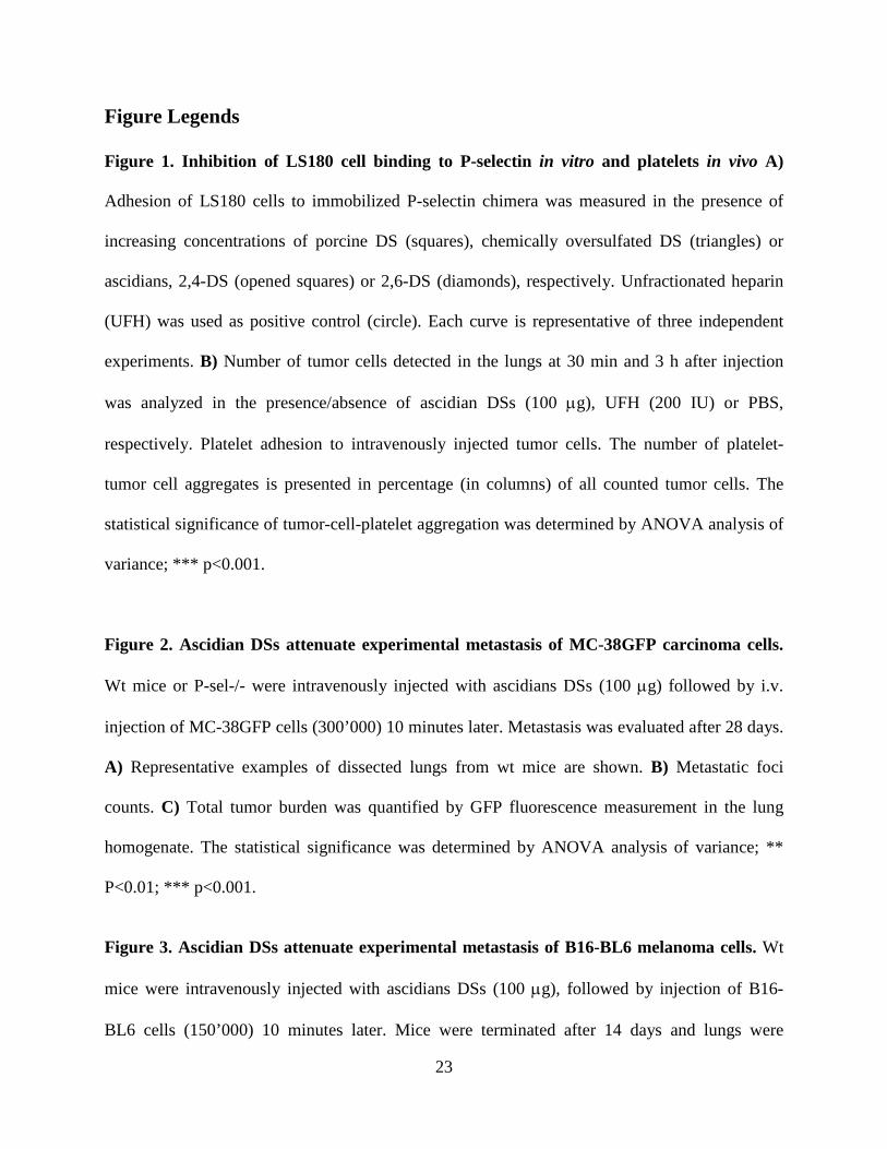
Figure 1. Inhibition of LS180 cell binding to P-selectin in vitro and platelets in vivo A)
Adhesion of LS180 cells to immobilized P-selectin chimera was measured in the presence of
increasing concentrations of porcine DS (squares), chemically oversulfated DS (triangles) or
ascidians, 2,4-DS (opened squares) or 2,6-DS (diamonds), respectively. Unfractionated heparin
(UFH) was used as positive control (circle). Each curve is representative of three independent
experiments. B) Number of tumor cells detected in the lungs at 30 min and 3 h after injection
was analyzed in the presence/absence of ascidian DSs (100 µg), UFH (200 IU) or PBS,
respectively. Platelet adhesion to intravenously injected tumor cells. The number of platelet-
tumor cell aggregates is presented in percentage (in columns) of all counted tumor cells. The
statistical significance of tumor-cell-platelet aggregation was determined by ANOVA analysis of
variance; *** p<0.001.
Figure 2. Ascidian DSs attenuate experimental metastasis of MC-38GFP carcinoma cells.
Wt mice or P-sel-/- were intravenously injected with ascidians DSs (100 µg) followed by i.v.
injection of MC-38GFP cells (300’000) 10 minutes later. Metastasis was evaluated after 28 days.
A) Representative examples of dissected lungs from wt mice are shown. B) Metastatic foci
counts. C) Total tumor burden was quantified by GFP fluorescence measurement in the lung
homogenate. The statistical significance was determined by ANOVA analysis of variance; **
P<0.01; *** p<0.001.
Figure 3. Ascidian DSs attenuate experimental metastasis of B16-BL6 melanoma cells. Wt
mice were intravenously injected with ascidians DSs (100 µg), followed by injection of B16-
BL6 cells (150’000) 10 minutes later. Mice were terminated after 14 days and lungs were
24
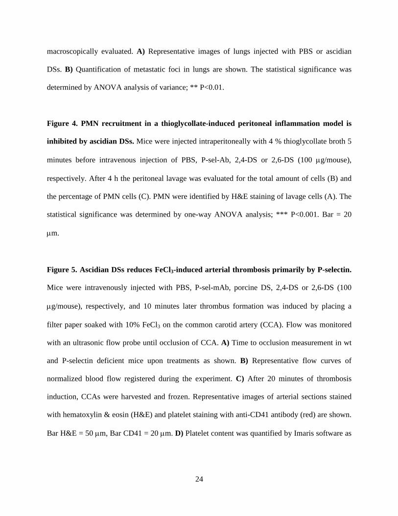
macroscopically evaluated. A) Representative images of lungs injected with PBS or ascidian
DSs. B) Quantification of metastatic foci in lungs are shown. The statistical significance was
determined by ANOVA analysis of variance; ** P<0.01.
Figure 4. PMN recruitment in a thioglycollate-induced peritoneal inflammation model is
inhibited by ascidian DSs. Mice were injected intraperitoneally with 4 % thioglycollate broth 5
minutes before intravenous injection of PBS, P-sel-Ab, 2,4-DS or 2,6-DS (100 µg/mouse),
respectively. After 4 h the peritoneal lavage was evaluated for the total amount of cells (B) and
the percentage of PMN cells (C). PMN were identified by H&E staining of lavage cells (A). The
statistical significance was determined by one-way ANOVA analysis; *** P<0.001. Bar = 20
µm.
Figure 5. Ascidian DSs reduces FeCl3-induced arterial thrombosis primarily by P-selectin.
Mice were intravenously injected with PBS, P-sel-mAb, porcine DS, 2,4-DS or 2,6-DS (100
µg/mouse), respectively, and 10 minutes later thrombus formation was induced by placing a
filter paper soaked with 10% FeCl3 on the common carotid artery (CCA). Flow was monitored
with an ultrasonic flow probe until occlusion of CCA. A) Time to occlusion measurement in wt
and P-selectin deficient mice upon treatments as shown. B) Representative flow curves of
normalized blood flow registered during the experiment. C) After 20 minutes of thrombosis
induction, CCAs were harvested and frozen. Representative images of arterial sections stained
with hematoxylin & eosin (H&E) and platelet staining with anti-CD41 antibody (red) are shown.
Bar H&E = 50 µm, Bar CD41 = 20 µm. D) Platelet content was quantified by Imaris software as

25
described in ‘Material and Methods’. The statistical significance was determined by one-way
ANOVA analysis (*P<0.05; ** P<0.01; *** p<0.001).

26
Kozlowski et al.
Table 1.
Disaccharide content, anticoagulant and P-selectin inhibitory activities of DSs
Dermatan sulfate Major disaccharide unit (%) aPTT (IU/mg)c
Inhibition of tumor cell adhesion to
P-selectin IC 50 (µg/ml)
Porcine DS α-∆HexUA-GalNAc(4S)a
(80 %) 2a -
2,4-DS
α-∆HexUA(2S)-GalNAc(4S)a
(66 %) 8a 13.51
2,6-DS
α-∆HexUA(2S)-GalNAc(6S)a
(75 %) 0.4a 12.19
OSDSd
α-∆HexUA-GalNAc(4S,6S)b
(36 %) 11.5 12.56 a Data from Pavão MS et al (27)
b Obtained by disaccharide analysis c The anticoagulant activity of heparin and its derivatives was determined in human plasma samples as described (27). d OSDS = oversulfated mammalian dermatan sulfate.