© copyright acebac, la trobe university 2017
TRANSCRIPT
© Copyright ACEBAC, La Trobe University 2017
2
Learning outcomes
Facilitator: Click on the play icon to view the film clip.
This module will describe:• what the bones, joints and muscles do• what happens when the bones, joints and muscles get older• what can damage the bones, joints and muscles• how to recognise changes to movement and mobility• how to report the changes.
Facilitator: Before going to the next slide, ask participants what the different partsof the skeleton are.
2

3
Movement and mobility
The following parts of the body are responsible for
movement and mobility:
▪ bones
▪ cartilage
▪ muscles
▪ connective tissues
▪ tendons
▪ ligaments.
3

4
The skeleton is the bony framework of the body
Bones are needed for:
▪ support, to help make the body
stand upright
▪ movement
▪ protection of organs, the brain and
spinal cord
▪ making red blood cells
▪ storage of calcium and other
nutrients.
Bones are living body parts and are stronger than steel.There are 206 bones in the human body.Every bone in the body – except for one – is connected to another bone. The one lonely bone is in the throat (the hyoid bone); this bone helps with speech.The skeleton provides the framework that supports the body and protects the organs.In partnership with the muscles the skeleton creates movement.Even inside the bones there is important activity; bones act as a storage place for vital nutrients and minerals such as calcium, and red blood cells are made there.
4

5
Joints
When two bones meet they form a joint.
Joint
Ligaments
The bones are connected to each other with ligaments.
The picture shows the knee joint.The bones are connected to each other with strong flexible bands called ligaments.Ligaments are able to stretch, which allows the joint to move.In turn, the joints allow the body to move.
5

6
The muscles
Muscles are needed for:
▪ movement
▪ stability
▪ control of body openings and passages
▪ keeping the body warm.
The human body has more than 650 muscles, which make up half of a person’s body weight. All muscles must work in pairs. One group of muscles relaxes and the other contracts. Muscles are needed for:• physical movement like walking, bending and sitting, and function of organs
inside the body (e.g. heart, eyes, intestines, lungs)• stability (muscles hold the bones securely together so that joints remain stable)• control of body openings and passages. These muscles are called sphincters and
behave like a valve that opens and closes. This type of muscle can be found in the bladder and digestive system
• keeping the body warm (muscles help the body maintain a normal temperature by releasing heat when the muscle contracts).
When you want to move, a signal is sent from the brain, along the spinal cord and through the nerves to the muscle fibres. This signal tells the muscles to contract. When the muscle contracts it pulls on the bone to cause movement.A tendon connects the muscle to bone.
6

7
Muscles
▪ Muscles are connected to the
bones of the skeleton by tendons.
▪ Only some muscles can be
controlled by choice.
In this picture the Achilles tendon attaches the calf muscles to the heel bone.
7

8
Age-related changes
▪ Joints become less flexible
▪ Bones become thin and brittle
▪ Muscles get
smaller and
weaker
As muscles age they get smaller, weigh less and lose their strength. Facilitator: Ask participants how much muscle they think we lose by the time we get to the age of 80.Answer: By the age of 80 we can lose between 23% and 50% of our muscle size.
As well as muscles getting smaller, the amount of body fat increases, particularly in women.The facial muscles show this change as the person ages.
As bones age they become thin and brittle, and their ability to heal slows down.Facilitator: Ask participants what they think bone ageing increases the risk of.Answer: The bone can break more easily (a fracture).
You may have noticed that the older person often shrinks a little and their spine starts to curve, changing the person’s height and posture and giving them a ‘hump’ shape in their upper back.
8
9
Movement and mobility problems
What common conditions can
change movement and
mobility in the older person?
Considering the number of bones, muscles and joints that make up the adult body, it is not surprising that many people experience problems as they age. Facilitator: Ask participants what they think are the most common conditions affecting movement and mobility which may cause discomfort and pain for the older person.
Go to the next slide to reveal the answer.
9
10
Movement and mobility problems continued
The most common conditions that change movement and
mobility in the older person are:
▪ arthritis
▪ osteoporosis
▪ fractures.
Expected answers:• arthritis• osteoporosis – a common change that occurs in the bones as they age. While this
condition does not in itself cause pain, it does increase the risk of falls and broken bones (fractures).
10

11
Know the resident: PCWs/PCAs should draw on their knowledge of the resident and how they usually mobilise.
Know the care plan: The care plan should alert PCWs/PCAs to the resident’s mobility needs and whether the resident is at risk of falling.
Know what changes the ability to move and mobilise: PCWs/PCAs should draw on their knowledge of ageing and the factors most likely to affect the resident’s mobility.
Know the signs of bone, muscle and joint problems: PCWs/PCAs should draw on their knowledge of conditions that may affect mobility.
Facilitator: Before moving to the next slide, ask participants to describe some of the opportunities they have during the day when they are with residents to recognise when something may have changed.
11

12
Every day, check…
▪ The resident’s movement
and mobility
▪ For pain
When assisting the
person with…
▪ Getting out of bed
▪ Washing, showering
or bathing
▪ Dressing
▪ Mobilising
Facilitator: Before going to the next slide ask participants what signs they would look out for in a resident who is likely to experience movement and mobility problems.
12

13
Recognising changed movement and mobility
▪ Groaning
▪ A grinding sound as
the joint moves
▪ Swelling of the joint
▪ Limited movement
▪ Colour change
▪ Non-verbal signs of pain
The joint
▪ Grinding as it moves
▪ Warm to the touch
SEEHEAR
FEEL
Hear• Verbalisations of pain such as moaning and groaning• A grinding sound when moving caused by bone-on-bone movement.See• Limited range of movement – stiffness, especially in the morning, or if sitting for
a long time• Changes in the shape of the joint (swelling and/or deformity)• Colour change, such as redness or the blue/purple colouring of bruising• Non-verbal expressions of pain (i.e. guarding or grimacing).Feel• The joint grinding as it moves• Warmth around the joint.
Facilitator: If an intermission is needed, the presentation could be paused hereand continued the next day.
13
14
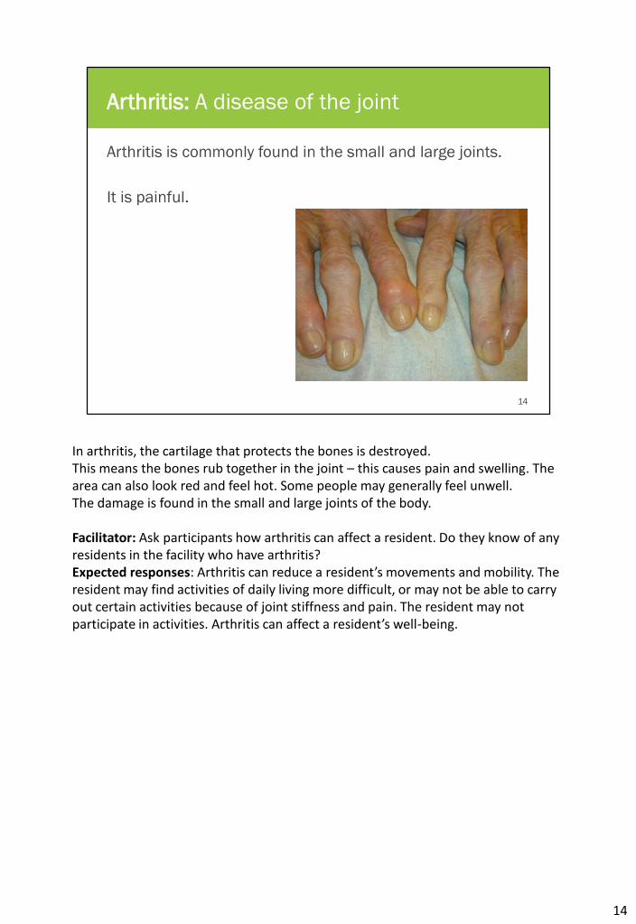
Arthritis: A disease of the joint
Arthritis is commonly found in the small and large joints.
It is painful.
In arthritis, the cartilage that protects the bones is destroyed.This means the bones rub together in the joint – this causes pain and swelling. The area can also look red and feel hot. Some people may generally feel unwell. The damage is found in the small and large joints of the body.
Facilitator: Ask participants how arthritis can affect a resident. Do they know of any residents in the facility who have arthritis?Expected responses: Arthritis can reduce a resident’s movements and mobility. The resident may find activities of daily living more difficult, or may not be able to carry out certain activities because of joint stiffness and pain. The resident may not participate in activities. Arthritis can affect a resident’s well-being.
14

15
Activity: Arthritis
In your workbook, mark the
parts of the body where you
think arthritis could be present.
Facilitator: Direct participants to complete the Arthritis activity in their workbooks.
Body parts that should be marked include the:• hands• spine• hips• knees• ankles• fingers.
15

16
Joint changes
Facilitator: Click on the play icon to view the film clip (NOTE: There is no sound or text in this film clip).
Explain what is happening in the film clip; that it is arthritis, which changes the ball and socket joint of the hip.The purple area is the cartilage, which protects the ends of the bone. The cartilage is surrounded by fluid to stop wear and tear. When the fluid dries up and the cartilage wears away, there is nothing to protect the bone. The bones rub together and become damaged. This causes pain and the loss of bone tissue and eventually the joint no longer functions properly. The diagram shows the major joints in the body that can be affected by arthritis.
16
17
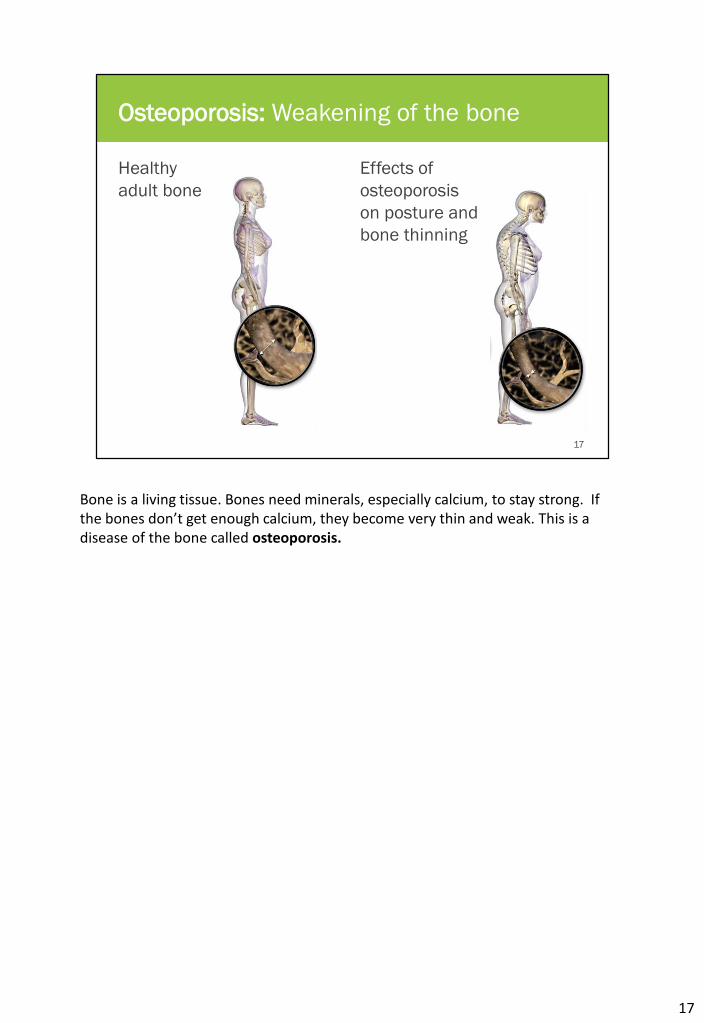
Osteoporosis: Weakening of the bone
Healthy
adult bone
Effects of
osteoporosis
on posture and
bone thinning
Bone is a living tissue. Bones need minerals, especially calcium, to stay strong. If the bones don’t get enough calcium, they become very thin and weak. This is a disease of the bone called osteoporosis.
17
18
Osteoporosis: A silent disease
▪ Men and women are affected
▪ Menopause increases the risk of osteoporosis
▪ There are no symptoms until the condition is advanced
▪ People with osteoporosis can fracture their bones very
easily, especially the:
▪ spine
▪ hip
▪ upper arm, forearm, wrist
▪ ribs.
Osteoporosis does not have any symptoms until it is very advanced.Men and women can have osteoporosis, but women are at greater risk because of menopause. Bone loss happens faster in menopause, with women losing up to 10% of bone mass. Other risk factors for menopause include lack of vitamin D, lack of exercise, too much nicotine (from smoking), and too much coffee and alcohol. The biggest risk to people with osteoporosis is having a fall, as a fall can cause fractures.
18

19
Falls
A fall is an event which
results in a person coming
to rest inadvertently on the
ground, or floor, or other
lower level. Slips, trips and
stumbles are the most
common type of fall.Why do falls
happen?
Facilitator: Ask participants why they think falls happen.Compare participants’ answers with those on the next slide.
19
20
The reason for falls
▪ Confusion and dementia
▪ Unsteadiness when walking
▪ Inactivity resulting in loss of strength
▪ Some medications that cause sleepiness or dizziness
▪ Low blood pressure
▪ Toileting difficulties
▪ Unsuitable footwear
▪ Poor eyesight
▪ Trip hazards
▪ Use of restraints
Facilitator: Ask participants which residents in the facility they think are at risk of a fall and why. What should they look for in a resident who has had a fall? Does the facility have a procedure which tells staff what they should do if a resident has had a fall? Can participants describe the procedure?
20
21
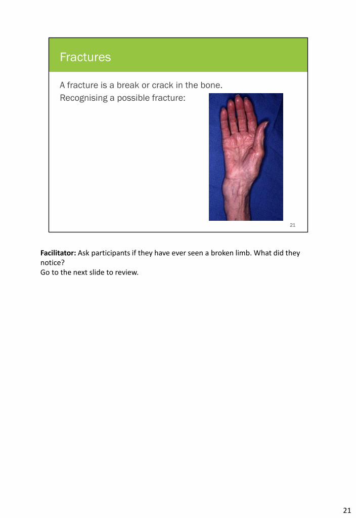
Fractures
A fracture is a break or crack in the bone.
Recognising a possible fracture:
Facilitator: Ask participants if they have ever seen a broken limb. What did they notice?Go to the next slide to review.
21
22
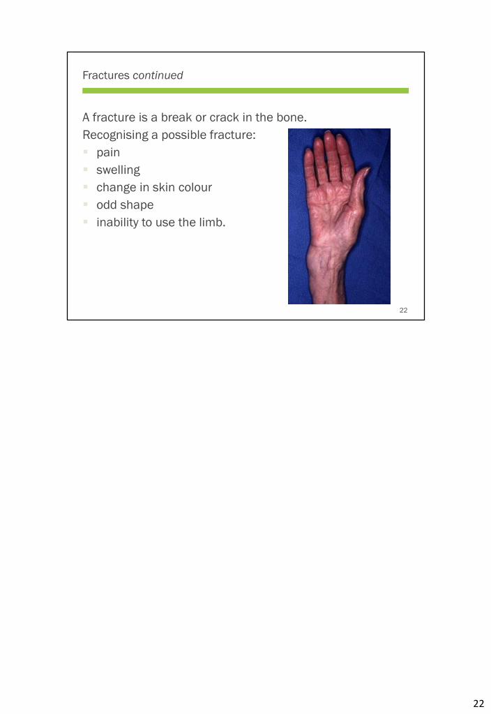
Fractures continued
A fracture is a break or crack in the bone.
Recognising a possible fracture:
▪ pain
▪ swelling
▪ change in skin colour
▪ odd shape
▪ inability to use the limb.
22

23
Immobility
Residents become immobile for a number of reasons:
▪ pain and joint changes
▪ poor eyesight, poor balance and dizziness
▪ some medicines can cause sleepiness
▪ the lack of opportunity to move about.
A general rule for keeping bones, muscles and joints
moving is “use it or lose it”.
23

24
Immobility continued
Immobility affects the whole body and:
▪ bones, muscles and joints age faster
▪ digestion slows down
▪ the heart doesn’t work as well
▪ the lungs don’t fill properly
▪ the kidneys and bladder become less effective.
Inactivity or immobility affects a person’s whole body.It speeds up the ageing of the bones, muscles and joints.The digestive system slows down, so older people feel less hungry, and they are more likely to get constipated.The heart does not work as well as it used to. The body then has trouble keeping the blood pressure steady, so the person feels dizzy and unsteady when getting out of a chair, or walking.The lungs don’t expand properly and breathing becomes slow and shallow. Older people are more likely to have chest infections and are less likely to recover from them.The kidneys and bladder don’t work so well, causing incontinence and urine infections.
24
25
Immobility and emotional wellbeing
Immobility can affect emotional wellbeing, causing:
▪ loss of confidence in walking safely
▪ a fear of falling
▪ anger and frustration at the loss of independence
▪ sleep disturbances
▪ disorientation and confusion
▪ isolation
▪ depression.
25
26
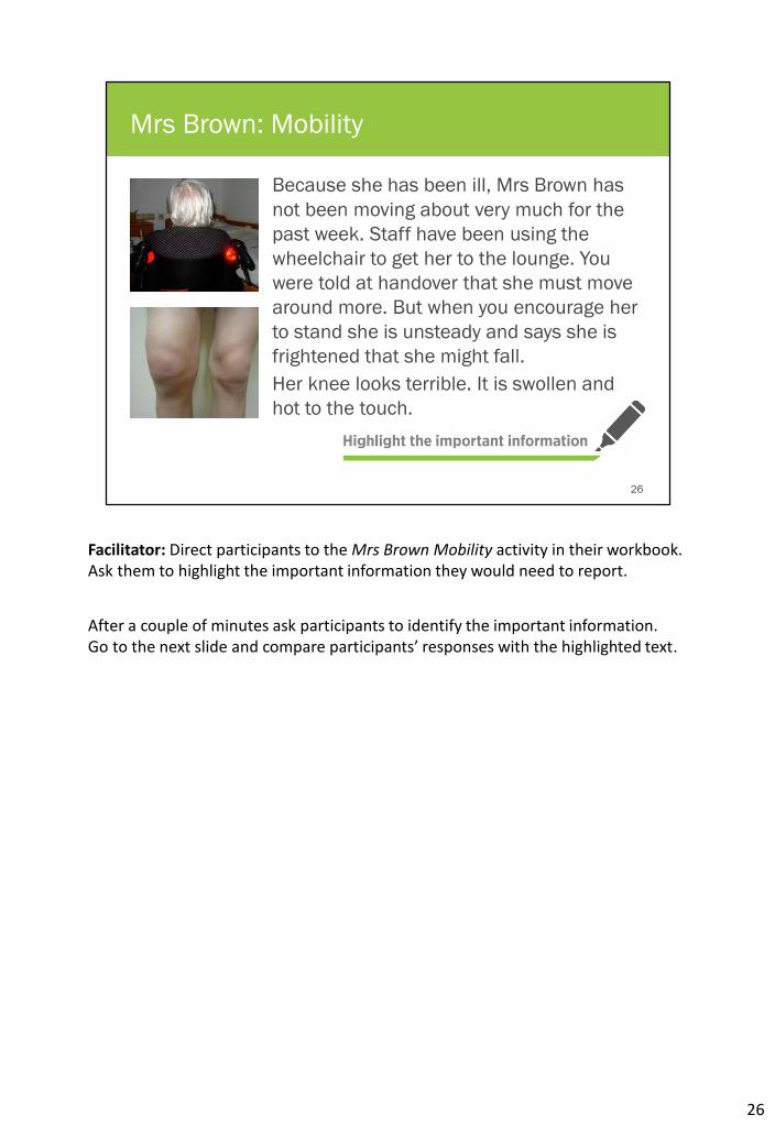
Because she has been ill, Mrs Brown has
not been moving about very much for the
past week. Staff have been using the
wheelchair to get her to the lounge. You
were told at handover that she must move
around more. But when you encourage her
to stand she is unsteady and says she is
frightened that she might fall.
Her knee looks terrible. It is swollen and
hot to the touch.
Mrs Brown: Mobility
Facilitator: Direct participants to the Mrs Brown Mobility activity in their workbook. Ask them to highlight the important information they would need to report.
After a couple of minutes ask participants to identify the important information.Go to the next slide and compare participants’ responses with the highlighted text.
26
27
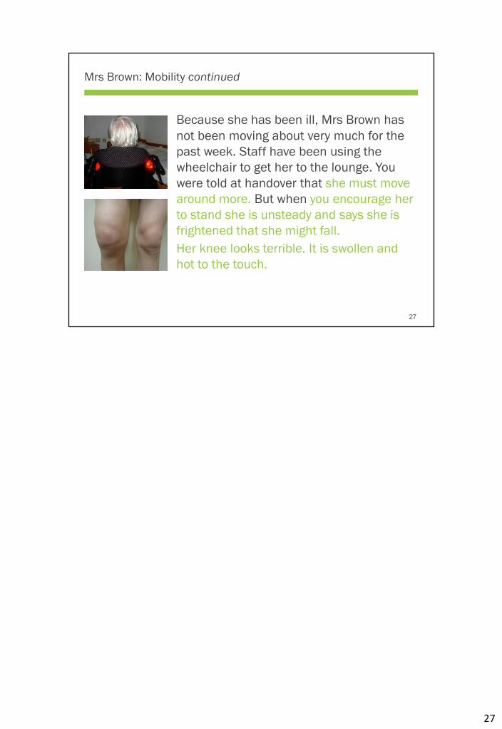
Mrs Brown: Mobility continued
Because she has been ill, Mrs Brown has
not been moving about very much for the
past week. Staff have been using the
wheelchair to get her to the lounge. You
were told at handover that she must move
around more. But when you encourage her
to stand she is unsteady and says she is
frightened that she might fall.
Her knee looks terrible. It is swollen and
hot to the touch.
27

28
Activity: Report the change
What did you see? What did you hear? What did you feel?
Make your report for the progress notes
Facilitator: Using the important information highlighted in the previous slide ask participants to complete the Report the change activity in their workbook.
Go to the next slide and compare participants’ responses with what is on the slide. Ask for volunteers to read out what they have written for the progress notes.
28

29
Activity: Report the change continued
What did you see? What did you hear? What did you feel?
▪ Mrs Brown is
unsteady on
standing
▪ The right knee is
swollen
▪ She is anxious that
she might fall
▪ Right knee is hot to
the touch
Make your report for the progress notes
This morning Mrs Brown was encouraged to move around more as per
handover instructions. She was unsteady on standing and said that she was
frightened of falling. Her right knee was swollen and hot to the touch. Staff
need to watch out for pain.
29

30
Summary
▪ Ageing reduces muscle and bone strength, and the flexibility of the joints.
▪ Arthritis and osteoporosis are common health conditions that affect movement and mobility.
▪ Arthritis can affect any of the major joints in the body and is painful.
▪ Osteoporosis makes the bones weaker and increases the risk of fracture on falling.
▪ Most falls can be prevented.
▪ Immobility affects the whole body. Residents should be encouraged and supported to be as active as possible, for as long as possible.
30
31
Acknowledgements
Slide 1
Assisted living nursing home elderly woman. By Wisconsinart. From Dreamstime
https://www.dreamstime.com/stock-photo-assisted-living-nursing-home-elderly-woman-
old-senior-sits-her-bed-medical-walker-aid-female-alone-needs-image65233609 Used
under RF-LL
Slide 2
Dancing skeletons. Used under license from Shutterstock
Perséphone - Retro Funky (SUNDANCE remix). By Sundance. From Soundcloud
https://soundcloud.com/sundancemusic/pers-phone-retro-funky Used under
CC-BY-3.0
Slide 4
Skeleton. From Pixabay https://pixabay.com/en/skeleton-human-skull-bone-bones-
146310/ Used under CC0 1.0
Slide 5
Anatomy of the knee, viewed from the side. By “Blausen gallery 2014”. From Wikiversity
Journal of Medicine. DOI:10.15347/wjm/2014.010. ISSN 20018762. Used under
CC-BY-3.0
Slide 6
Bodybuilder. Adapted from Pixabay https://pixabay.com/en/bodybuilder-muscles-man-
human-146791/ Used under CC0 1.0
Slide 7
Rupture du tendon d'Achille gauche. By Grook Da Oger. From Wikimedia Commons
https://commons.wikimedia.org/wiki/File:Rupture_tendon_achil%C3%A9en_l%C3%A9g
ende.jpg. Used under CC-BY-SA 3.0
Slide 8
Body building. From Pixabay https://pixabay.com/en/bodybuilding-man-body-male-
fitness-311351/ Used under CC0 1.0
Skeletons. By Clker. Adapted from Pixabay https://pixabay.com/en/skeletons-danse-
macabre-bones-dance-303877/ Used under CC0 1.0
Slide 11
Assisted living nursing home elderly woman. By Wisconsinart. From Dreamstime
https://www.dreamstime.com/stock-photo-assisted-living-nursing-home-elderly-woman-
old-senior-sits-her-bed-medical-walker-aid-female-alone-needs-image65233609 Used
under RF-LL licence
Slide 12
Abstract eye. By Daniela Spyropoulou. From Dreamstime
https://www.dreamstime.com/royalty-free-stock-photography-abstract-eye-
image139587 Used under RF-LL licence.
Slide 13
Female. From Pixabay https://pixabay.com/en/female-feminine-girl-lady-person-
161202/ Used under CC0 1.0
Slide 14
Heberden-Arthrose. By Drahreg01. From Wikimedia Commons
https://commons.wikimedia.org/wiki/File:Heberden-Arthrose.JPG Used under CC-BY-SA
3.0
Slide 15
Human body, upper part, front. By Häggström, M. From Wikimedia
https://commons.wikimedia.org/wiki/File:Human_body_silhouette.svg Used under
CC0 1.0
Slide 16
Rheumatoid arthritis of hip joint. Used under licence from Shutterstock
3D female skeleton anatomy. By Bernhard Ungerer. Adapted from Wikimedia
https://commons.wikimedia.org/wiki/File:3D_Female_Skeleton_Anatomy.png Used
under CC-BY-3.0
Slide 17
Osteoporosis. By Blausen.com staff. "Blausen gallery 2014". From
Wikiversity Journal of Medicine. DOI:10.15347/wjm/2014.010. ISSN 20018762.
https://commons.wikimedia.org/wiki/File:Blausen_0686_Osteoporosis_01.png Used
under CC-BY-3.0
Slides 21 & 22
Distal radius fracture. By Curtishand. From Wikipedia
https://commons.wikimedia.org/wiki/File:Distalradiusfracture.jpg Used under CC0 1.0
Slides 26 & 27
Knee effusion. By Heilman, J. From Wikimedia Commons
https://commons.wikimedia.org/wiki/File:K.neeffusion.JPG Used under CC-BY-SA 3.0
Woman. By Geralt. From Pixabay https://pixabay.com/en/woman-old-age-grey-hair-
grandma-593456/ Used under CC0 1.0
31