----------------------------------------------------------------------- · j. hallermann (1994)...
TRANSCRIPT

--------------------------------------------------------------------------------------------------------------------- Jakob Hallermann:
On the morphology of the ethmoidal region of iguanians (Squamata) – a comparative anatomical study*†
Bonner Zoologische Monographien no. 35 (1994), 133 pages
CONTENTS Page Introduction ..........................................................................................................3 Systematic overview ....................................................................................3 Historical review..........................................................................................3 Goals of the work.........................................................................................4 Acknowledgments....................................................................................................4 Materials and methods .............................................................................................4 Comparative and functional anatomy of the ethmoidal region................................5 Nomenclature...............................................................................................5 List of terms and their synonyms for the regio ethmoidalis of Squamata ...6 Nasal capsule ...............................................................................................6 Septum nasi......................................................................................6 Tectum nasi......................................................................................8 Paries nasi ......................................................................................11 Planum antorbitale .........................................................................13 Commissura sphenethmoidalis ......................................................15 Solum nasi......................................................................................15 Bulbus and nervus olfactorius, n. ethmoidalis ...........................................18 Nasal cavity................................................................................................20 Vestibulum nasi .............................................................................20 Cavum nasi proprium.....................................................................22 Choanal groove ..............................................................................25 Ductus nasolacrimalis ................................................................................26 Glandula nasalis lateralis ...........................................................................27 Jacobson’s organ........................................................................................27 Comparative analysis .............................................................................................28
* Original citation: Hallermann, J. 1994. Zur Morphologie der Ethmoidalregion der Iguania (Squamata) – eine vergleichend-anatomische Untersuchung. Bonner Zoologische Monographien, No. 35: 1-133. Translation © 2009 by Krister T. Smith. † PREFATORY REMARKS: In cases where Hallermann has used the Latin anatomical term, I have left it as is (except for the names of bones). In translating German anatomical terms into the English, I follow when possible Oelrich (1956). Note that there is a distinction between figures in the text (“Textfiguren”) and those that come at the end (“Abbildungen”); I use Text Fig. to translate the former, Fig. the latter. Footnotes are always those of Hallermann. Square brackets always indicate addenda to the text; the content of the brackets is italicized when it represents my own clarification and in quotation marks when it represents Hallermann’s original text (always provided subsequent to my translation). German terms are occasionally left in the text, always in italics, either where the corresponding term is unknown to me or where Hallermann specifically discusses the German one. There are many sentences that might be phrased more elegantly, but I feel the disadvantageous stretch of the diminishing-returns curve has been reached.
J. Hallermann (1994) Translation Page 2
General preliminary remarks on the ontogeny and phylogeny of the nasal capsule ....................................................................................28 Septum nasi................................................................................................30 Fenestra septi nasi ..........................................................................30 Processus praenasalis .....................................................................31 “Roofing cartilage” of Jacobson’s organ .......................................31 Tectum nasi................................................................................................32 Cupula nasi anterior .......................................................................32 External and internal nasal muscles...............................................33 Cartilago parietotectalis .................................................................35 Fenestra superior nasi ....................................................................35 Fenestra lateralis nasi.....................................................................36 Fissura lateralis nasi.......................................................................36 Paries nasi ..................................................................................................37 Concha nasalis ...............................................................................37 Processus maxillaris.......................................................................39 Planum antorbitale .....................................................................................40 Processus rostralis of the planum antorbitale.................................40 Commissura sphenethmoidalis ......................................................41 Solum nasi..................................................................................................42 Lamina transversalis anterior.........................................................42 Concha of Jacobson’s organ ..........................................................44 Cartilago paraseptalis.....................................................................44 Cartilago ectochoanalis..................................................................45 Vestibulum nasi .........................................................................................47 Cavum nasi proprium.................................................................................51 Choanal groove ..........................................................................................54 Ductus nasolacrimalis ................................................................................56 Glandula nasalis lateralis ...........................................................................58 Jacobson’s organ........................................................................................60 Dermal bones of the ethmoidal region.......................................................62 Hypothetical condition of the basic pattern of the regio ethmoidalis ........65 Characters outside of the regio ethmoidalis...............................................67 List of characters........................................................................................70 Phylogenetic hypothesis.............................................................................74 Summary [German] ...............................................................................................— Summary [English] ................................................................................................— Literature cited .......................................................................................................— Appendix ........................................................................................................75 Explanation of abbreviations .....................................................................— Figures [Captions] .....................................................................................75
J. Hallermann (1994) Translation Page 3
INTRODUCTION
Systematic overview The Class “Reptilia” comprises a class [“Sammelgruppe”] of hair- and featherless amniotes. The extinct representatives of the group ancestral to mammals (e.g., therapsids) as well as those of birds (archosaurs) and Recent crocodiles, turtles, and lepidosaurs belong to it. The external similarity of these reptiles is based in their common possession of primitive characters (= symplesiomorphies sensu Hennig, 1950). Lepidosauria includes the recent squamates (lizards and snakes) and their sister-taxon Sphenodon, a “living fossil” of the Order Sphenodontida, known since the Lower Triassic (Carroll, 1988). Lepidosauria and Archosauria (birds, crocodiles) are sister groups (Benton, 1985). Since the work of Camp (1923), the phylogenetic relationships of the individual larger groups within Squamata have for the most part been corroborated by further studies (Estes et al., 1988). The following phylogenetic diagram will explain this: [Cladogram] *paraphyletic group (from Estes et al., 1988) One of the most speciose subgroups of Squamata is Iguania, to which Agamidae1, Chamaeleonidae2, and “Iguanidae”3 belong. The monophyly of Iguania and its position as sister-group to the other squamates (Scleroglossa) are well founded (Etheridge and de Queiroz, 1988). While the chamaeleons are distinguished as a monophyletic group by numerous autapomorphic characters, no autapomorphic characters for agamas and iguanas could thus far be found (Etheridge and de Queiroz, 1988). Therefore, these taxa were arranged into new monophyletic families4 (Frost and Etheridge, 1989). The relationships of these nine subgroups of Iguania to one another, however, are unclear.
Historical review The regio ethmoidalis of Squamata had already received intensive scrutiny during the last century and the beginning of the present one (Solger, 1876; Born, 1879; v. Mihalkovics, 1899; Gaupp, 1900; Beecker, 1903; Fuchs, 1908). In these publications the anatomical foundations were lain and the terminology introduced that even today is used. While in these early works, for the most part, only a few middle European species were studied, comprehensive comparative studies have appeared, especially in the 1940s (Malan, 1946; Pratt, 1948; Stebbins, 1948). In these, as in more recent publications, comparative anatomy was of foremost interest. Here, mostly only parts of the regio ethmoidalis were emphasized. The nasal cavities and their mucus membranes were studied in a comparative-anatomical fashion by Gabe and Saint Girons (1976), the development of the nasal capsule by Słaby (1979a, 1979b, 1979c, 1981, 1982a, 1982b,
1 This taxon comprises Agaminae and Leiolepidinae (sensu Frost and Etheridge, 1989 = Agamidae + Uromastycidae sensu Moody, 1980); it is probably not monophyletic, for the chamaeleons, according to this and previous studies, are more closely related to a part of agamas. 2 This term is employed here in the traditional sense. It corresponds to Chamaeleoninae of Frost and Etheridge (1989). 3 This is the old term, which includes all iguanas. This family is probably not monophyletic. Iguanidae in the sense of Frost and Etheridge (1989) comprises only the large iguanas, which earlier were termed Iguaninae. 4 Chamaeleonidae, Corytophanidae, Crotaphytidae, Hoplocercidae, Iguanidae, Opluridae, Polychridae, Phrynosomatidae, Tropiduridae.

J. Hallermann (1994) Translation Page 4
1982c, 1984), and the relationships of the lacrimal-nasal duct to the choanal groove by Bellairs and Boyd (1950). Parsons (1970) and Bellairs and Kamal (1981) recapitulated previous results. Besides these there are several studies of individual species. The ethmoidal region, however, has only been studied in only relatively few representatives of the highly speciose and diverse group of iguanas (Sceloporus, Iguana: Malan, 1946; Anolis: Stimie, 1966; Ctenosaura: Oelrich, 1956). No author has yet concerned himself with the features of the basic pattern of the regio ethmoidalis of Squamata or its subgroups. In the present work, this part of the skull was selected for phylogenetic-systematic analysis because of its great variety of structure and its complexity.
Goals of the work In the first part of the present work, the ethmoidal region of numerous representatives of monophyletic subgroups of Iguania is described in a comparative-anatomical fashion for the first time. The arrangement here is not according to systematic units; rather, the different organization of individual structures in the various taxa are presented. In the second part the basic pattern of the regio ethmoidalis of squamates and its subgroups will be constructed. The analysis here is guided by the principles of strict phylogenetic systematics (sensu Hennig, 1950). As a first-order outgroup to Iguania, Scleroglossa is available. The second-order outgroup is Sphenodon. By using data on individual taxa that other authors had investigated, 29 genera of iguanians could be incorporated into the analysis. Several genera could not be taken into consideration for lack of embryonic or juvenile material (e.g., Morunasaurus, Leiosaurus, Enyaloides [sic], Leiolepis, Moloch). In a concluding phylogenetic hypothesis the phylogenetic relationships within Iguania are discussed in a recapitulatory fashion. Special weight was placed on viewing characters of the ethmoidal region in an evolutionary and functional context.
ACKNOWLEDGMENTS Herrn Prof. Dr. W. Maier … I thank for advising this dissertation. For review of the manuscript, encouragement, and fruitful discussions I thank … To Dipl. Biol. Stefan Eßwein and … I am thankful for the many tips and assistance. I would like sincerely to thank Herrn PD. Dr. W. Böhme, Bonn, for his interest in this work and his manifold support. The majority of agamas and iguanas studied here come from the Sammlung des Zoologischen Forschungsinstituts und Museums Alexander Koenig. Dipl. Biol. R. Wicker, … provided embryos and hatchlings from their own breederies. For the numerous valuable tips I very sincerely thank our institute’s illustrator Margret Roser, and also of the technical employees, Monika Meinert for support during the preparation of the microscopic serial sections. The work was supported financially by funds from the Sonderforschungsbereichs 230 of the Universities of Tübingen and Stuttgart.
MATERIALS AND METHODS Twenty-six different species from eight of the nine new families of Iguania, which were established as monophyletic by Frost and Etheridge (1989) (see above), constitute the foundation of the study. The taxa marked with * are not included in the description, for studies by other authors are already available or the material was not suitable for histologic work.

J. Hallermann (1994) Translation Page 5
[The list of materials is not translated here. The following correspondences for words neither trivial nor obvious may be of use to those interested: Aufhellungspräparat = cleared-and-stained specimen, Schnittserie = (series of) serial sections, Schlüpfling = hatchling, Schädel = skull.] The embryos used for serial sections were decapitated, decalcified with 6% nitric acid and EDTA (Ethylendinitrilotetraessigsäure [“Essigsäure” = acetic acid]), imbedded in paraffin, sliced transversely, and dyed with Azan after Heidenhain. Section thickness was 8 µm in Anolis marmoratus, 10 µm in the other species. Partial, 100:1-scale models of the nasal capsule of the younger stage of Crotaphytus and of Phymaturus were prepared by the Plattenrekonstruktions-methode. Models of the entire cranium of Anolis lineatopus (6.5 mm TL [total length: “KL” or “Körperlänge”]) and A. marmoratus (5.1 mm TL) were produced at 100:1- and 75:1-scale, respectively. The sections were projected with the help of a microscope-attached camera lucida onto 2-mm thick Styroporplatten and the cartilage and bone boundaries marked with different colors, then the outlines trimmed with a scalpel and glued together. The illustrations of the cartilaginous nasal capsule were prepared on the basis of the Plattenrekonstruktionen or from photographs of the cleared-and-stained specimens. Numerous adult specimens were studied using sections as well. The nasal capsules (Nk), chondrocrania (Cr), or nasal sacs (Ns) of the following species within Iguania have previously been worked on: …
COMPARATIVE AND FUNCTIONAL ANATOMY OF THE ETHMOIDAL REGION
Nomenclature The division of iguanas and agamas into several monophyletic units (families, subfamilies) has gained acceptance among experts in the last ten years. However, there is not yet a uniform nomenclature. In the present work the taxonomic arrangement of Iguania to the family level, as suggested by Moody (1980) and Frost and Etheridge (1989), is used. In his extensive phylogenetic analysis of agamas, Moody (1980) divided the old “Agamidae” into two families, Uromastycidae and Agamidae (= Agaminae sensu Frost and Etheridge, 1989). The latter he divided into five probably monophyletic subfamilies. The latest taxonomic arrangement of Iguania is that of Frost and Etheridge (1989). The subfamilies of “Iguanidae”, already described for some time, were characterized through autapomorphic characters by these authors and raised to eight monophyletic families. Chamaeleons and agamas, in contrast, were subsumed into the Family Chamaeleonidae. This new taxonomic arrangement has been met with well-founded criticism (Böhme, 1990; Lazell, 1992) and was not accepted by several authors (e.g., Norell and de Queiroz, 1991). The new taxonomic arrangement of Iguania in nine families, as carried out by Frost and Etheridge (1989), is accepted, but only in part, in my work. The new Family Chamaeleonidae (“Agamidae” + Chamaeleonidae) is not used, for this term does not at all discriminate chamaeleonids in the traditional sense. Moreover, Chamaeleonidae s.s. is the most well-founded subgroup of Iguania (Estes et al., 1988). The name Acrodonta, introduced by Cope (1864), is used instead. The monophyletic subgroups of Acrodonta are designated Agamidae, Chamaeleonidae and Uromastycidae, following the nomenclature of Moody (1980). For the

J. Hallermann (1994) Translation Page 6
polyphyletic genera “Agama” and “Amphibolurus”, the new synonyms as used by Joger (1991), Cogger (1992) and Leviton et al. (1992) are taken into consideration. Most technical terms for the squamate cranium were introduced by E. Gaupp (1900) and de Beer (1937). This nomenclature is also used in more recent works (e.g., Bellairs and Kamal, 1981). I essentially use this nomenclature. Where I deviate from it, this is noted.
List of terms and their synonyms for the regio ethmoidalis of Squamata The following list is certainly not complete; it was also usually not possible to discover the original author. Numerous terms were first used by Gaupp (1900). The bold-faced terms were newly introduced when the structure in question was important but not yet described. Several terms were Latinized, for thus far there have only been English or German names for them. In the first position are the most common terms, which are also used here; but they are placed in brackets if they have seldom been of use in the literature. …
Nasal capsule The nasal capsule of squamates can be arranged into different topographic regions. To begin with, the septum nasi divides the nasal capsule into its two halves. The septum nasi is connected dorsally to the tectum nasi, which is divided into the cupula nasi anterior and the cartilago parietotectalis (de Beer, 1937). The cartilago parietotectalis is connected laterally to the cartilago paranasalis. The solum nasi closes the cartilaginous nasal capsule ventrally. The solum nasi consists of two braces in all squamates. It can be constructed of three cartilaginous structures: the lamina transversalis anterior (this corresponds to the ventral part of the zona annularis of Gaupp (1900)), the cartilago paraseptalis, and the cartilago ectochoanalis. The planum antorbitale closes the nasal capsule caudally. Septum nasi The septum nasi of the embryo and hatchling of Crotaphytus collaris is a vertically oriented, cartilaginous plate that rises from rostral to caudal. Its lower edge is only weakly bent; thus, it slopes down slightly rostrally. Caudally, in the hind third of the septum nasi, there is a large, trapezoidal fenestra septi nasi, closed only by connective tissue (Fig. 3); because of it, the ventral cartilaginous part of the septum nasi is rodlike in this region (Figs. 36, 37). The fenestra septi nasi passes caudodorsally into the undivided foramen olfactorium (Fig. 37). The septum nasi is connected dorsally to the cartilago parietotectalis along its entire length. Rostrally, the septum nasi ends in a short processus praenasalis (Figs. 2, 3). The latter is surrounded ventrally by the two processus maxillares [lateral processes of Oelrich] of the premaxilla. Caudal to the processus praenasalis, at the ventral edge of the septum nasi, the lamina transversalis anterior splits off laterally (Figs. 2, 3). Midway up the septum nasi in the region of Jacobson’s organ a cartilaginous crest is developed, on which the septomaxilla abuts dorsally (Fig. 32). I call this crest the crista lateralis septi nasi [septomaxillary ridge of Oelrich]. The lower edge of the septum nasi of the specimen of Phymaturus palluma, in contrast to most other examined iguanians, has a much stronger, concave arch, and the processus praenasalis points ventrally. Comparisons with adults of the same species and with other taxa allow one to conclude that elongation takes place in this region, and therefore, it is an age-dependent phenomenon [“… lassen sich darauf schließen, daß eine Streckung in dieser Region stattfindet

J. Hallermann (1994) Translation Page 7
und es sich daher um ein altersbedingtes Phänomen handelt”] (cf. Calotes: Ramaswami, 1946). In all non-acrodont iguanians the processus praenasalis is short and directed rostrally (Figs. 1 to 6). It is lacking in all studied acrodontans (Figs. 16, 18). A large fenestra septi nasi, taking up about half of the total length of the septum nasi, can be found in Phymaturus, Liolaemus, Stenocercus, Leiocephalus, Tropidurus, Brachylophus, Chamaeleolis and Uromastyx. In addition to this fenestra, there are two smaller openings in Tropidurus. In Stenocercus and the younger embryo of Phymaturus, the fenestra septi nasi is separated from the foramen olfactorium by a thin rod of cartilage that extends from the cartilago parietotectalis to the planum antorbitale; the foramen olfactorium is therefore enclosed on one side by cartilage (Fig. 4). This cartilaginous connection is lacking in the 10.0-mm embryo of Phymaturus. One may assume that the for. olfactorium is undivided in older stages, like in the forms cited above. The fenestra septi nasi of Oplurus, Corytophanes, Basiliscus, Petrosaurus, Polychrus and Pogona, in contrast, is slitlike and lies in the ventral and hind parts of the septum nasi. It extends rostrally to about the level of the caudal end of the septomaxilla. All examined developmental stages of Anolis and Physignathus lack the fenestra septi nasi. In the region of the foramen olfactorium in all examined species, a dorsal incision in the septum nasi is developed. In this incision lie the ventral parts of the bulbi olfactorii. The membrane-closed fenestra septi nasi of iguanians could have arisen during the course of ontogeny in places of less mechanical load, once a sufficient stability through dermal bones is achieved. In early ontogenetic stages no fenestra septi nasi is developed. In view of the immobility of the septum nasi serves well to save material and is not part of a zone of flexibility within the nasal capsule, as the case is, for example, in birds (Weber, 1990; Hallermann, 1992). The dorsal incision in the region of the foramen olfactorium of several squamates, in contrast, could be related to the need for space for the bulbus olfactorius. The crista lateralis septi nasi in the region of Jacobson’s organ, present in Crotaphytus, can be found in all examined iguanians. This crest is rather broad in Polychrus and Chamaeleolis; in Phymaturus and Physignathus it is widened into a narrow plate that partly covers Jacobson’s organ (Figs. 17, 22, 38, 45, 49). Here, it probably represents the remnant of the Dachknorpel (“roofing cartilage” sensu Malan, 1946). The septomaxilla overlies this cartilaginous plate dorsally; caudally it narrows to a crest. In Phymaturus it extends dorsally and fuses with the ventral side of the cupula nasi anterior (Fig. 38). A nearly complete cartilaginous covering of Jacobson’s organ can be found in Sceloporus and Phrynosoma (cf. Malan, 1946). In Phrynosoma the septomaxilla follows only caudal to Jacobson’s organ, while in Sceloporus the roofing cartilage is perforated and covered by the septomaxilla (Malan, 1946). The septum nasi of Corytophanes cristatus is in front very flat and broad (Figs. 40, 41). Its lower edge is concave. This curvature of the palate is filled by the voluminous tongue. The septum nasi becomes higher and narrower caudally. Behind the processus praenasalis, the cupula nasi anterior as well as the lamina transversalis anterior depart laterally in the same level of section (Fig. 40). In the hind section of Jacobson’s organ the septum nasi possesses at its ventral edge a lateral crest that probably represents the cartilago paraseptalis fused to the septum. Caudal to Jacobson’s organ it runs separately from the septum nasi (Fig. 41). The septum nasi of Uromastyx, compared to other iguanians, evinces several peculiarities. As is also evident from the figures of Lemire et al. (1970), the fenestra septi nasi, which is located in the hind region of the nasal capsule, is concealed in lateral view by the connection of the cartilago paraseptalis to the middle part of the septum nasi. Thus, in cross-

J. Hallermann (1994) Translation Page 8
section the septum appears forked, whereby the ventral, rodlike part of the septum nasi lies under the fenestra septi nasi and between the two descending, cartilaginous plates (see Text Fig. 1). Text Fig. 1: Uromastyx acantinurus [sic]. Transverse section in the region of the concha nasalis. Cartilago paraseptalis fused to septum nasi. Redrawn after Lemire et al. (1970). Tectum nasi The roof of the nasal capsule of squamates is divided into a front section, the cupula nasi anterior, and a hind section, the cartilago parietotectalis (Bellairs and Kamal, 1981). A separate anlage of the two structures cannot be observed in the examined forms and stages. The cupula nasi anterior of Crotaphytus is a cartilaginous shell open laterally and ventrally, which surrounds the external naris and the vestibulum nasi (Figs. 1, 3). The ventral, outer edge of the cupula nasi, which ends in a process, is termed the processus alaris inferior (Fig. 2). The processus alaris superior an the dorsal, outer edge of the cupula nasi is only weakly developed (Fig. 1). Medially the cupula is connected to the dorsal edge of the septum nasi (Fig. 32). There is located the foramen apicale, through which the ramus ethmoidalis medialis leaves the nasal capsule (Fig. 1). A small break in the outer surface of the nasal capsule marks the boundary between cupula nasi and cartilago parietotectalis. On the inner side of this break is located a transversely oriented crest, on which posterior process of the septomaxilla ventrally abuts. I term this crest the crista septomaxillaris (Figs. 1, 34). The cartilago parietotectalis of Crotaphytus is a broad, horizontal cartilaginous plate without fenestration (Fig. 1). Its lateral edge is turned in ventrally. It underlies the vestibulum nasi and borders here on the cartilago paranasalis. It is, however, not fused with this, but rather separated from it by the fissura lateralis (Figs. 33, 34, 35). The fissura lateralis is filled with connective tissue in the younger stage (10.0 mm TL) and in the older stages (11.8 mm, 13.0 mm TL) closed by fusion of the cartilaginous elements. Caudolaterally, the cartilago parietotectalis surrounds the caudal section of the vestibulum nasi like a dome and on its hind edge passes ventrally into the cartilago paranasalis (Fig. 1). Caudally, on the border of the planum antorbitale, there are several foramina in the cartilago parietotectalis (Figs. 1, 37). It is reasonable to term them foramina olfactorii as well, for the fila olfactorii do not enter the nasal capsule through a unitary foramen; rather, the lateral portions of the nerve are separated by numerous cartilaginous laminae. Thus, several openings arise in the nasal capsule. The form of the cupula nasi anterior does not vary significantly within Iguania. In the following, several peculiarities will be presented that appear only in a few genera. The cupula nasi anterior of Anolis is still flat in early developmental stages (5.2 mm TL); it lacks the processus alares superior and inferior (Figs. 24, 25). The cupula first takes on a dome-shaped form in later development. On its dorsal and ventral edges, less prominent processus alares superior and interior arise. The cupula nasi anterior of Chamaeleolis ends rostrally in a broad, flat cartilaginous plate that lies dorsal to the premaxilla (Fig. 23). I term it the processus rostralis cupulae nasi. It is perforated in its rostral section and possess a short, medial process (Fig. 23). A function cannot be attributed to this developed process. A narrow, similarly located rostral process can also be found in a hatchling of Lyriocephalus. This process appears to be ossified on both sides of the body in this specimen. Only on the right side are further parts of the cupula nasi ossified. These so far unique findings of ossification of the nasal capsule cannot, however, be interpreted, for only a single specimen

J. Hallermann (1994) Translation Page 9
and no serial sections are available. According to structure and coloration in the cleared-and-stained specimen, it is not calcified cartilage Ramaswami (1946), in the figures of the nasal capsule of Calotes versicolor, illustrated a rostral process of the cupula nasi anterior that is located in the same position as that of Lyriocephalus. He terms it, however, the processus alaris inferior, and the “true” processus alaris inferior the processus alaris superior. In comparison to other iguanians, the cupulae nasi anteriores of Corytophanes, Phrynosoma, Phrynocephalus and Physignathus are large and in part laterorostrally divergent (Figs. 20, 31). Caudal to the external naris in Physignathus, on the lower edge of the cupula nasi, a round, membrane-closed foramen can be found through which no nerves or vessels extend (Fig. 27). A large part of the surface of the cupula nasi anterior in all examined agamas forms the origin of an extensive, external, smooth nasal muscle (m. nasalis externus). This muscle extends from the cupula nasi anterior, the processus alaris superior and inferior, and the connective tissue surrounding the external naris obliquely caudoventrally to the periosteum of the maxilla and the skin overlying it (Fig. 26). In Pogona this muscle is rather large and additionally makes use of lateral parts of the nasal as an area of insertion [“Ansatzfläche”]. Through contraction of the external nasal muscle, the skin and cartilage structures that surround the external naris are shifted caudally and ventrally, such that the diameter of the nasal opening is reduced. Radially arranged smooth muscle fibers (m. radialis internus) additionally runs from the inner side of the cupula and from the external skin that surrounds the naris to the outer epithelium of the vestibulum nasi (Figs. 53, 54). Contraction of these fibers causes widening of the naris. The processus alaris superior is a less prominently developed process in Phymaturus, Liolaemus, Stenocercus, Oplurus, Basiliscus, Chamaeleolis and Anolis (Figs. 1, 3, 4, 28). The process is more striking, in contrast, in Leiocephalus, Petrosaurus, Corytophanes, Physignathus and Pogona (Figs. 10, 20, 27, 30). The processus alaris superior is lacking in the examined stages of Tropidurus, Brachylophus and Basiliscus. The processus alaris inferior is long in Tropidurus, Leiocephalus, Petrosaurus, Chamaeleolis, Physignathus, Pogona and Uromastyx, while it is only weakly developed in Phymaturus, Stenocercus, Liolaemus, Oplurus and Anolis (Figs. 10, 19, 21, 22). Brachylophus and Corytophanes lack it (Figs. 15, 17). This may be age-dependent, at least in the embryo of Brachylophus (cf. Anolis). As in Crotaphytus, the very broad cartilago parietotectalis also surrounds the hind loop of the S-shaped vestibulum nasi in Phymaturus, Oplurus, Liolaemus, Stenocercus and Uromastyx (Figs. 1, 4 to 9). Caudolaterally the cartilago paranasalis is more extensively overlain by the cartilago parietotectalis in Stenocercus and Phymaturus than in the species described before (Figs. 4, 8). Therefore, the part of the cartilago paranasalis visible from above is only very narrow. The tectum nasi in the front region of Brachylophus and Leiocephalus is significantly narrower than that of the species cited above, for the vestibulum nasi is only weakly S-shaped. A partial underlying of the vestibulum nasi by the cartilago parietotectalis, however, still takes place behind the septomaxilla (Fig. 6). The extent to which the vestibulum nasi is underlain by the cartilago parietotectalis differs among the species (cf. Figs. 4 to 9). On the other hand the cartilago parietotectalis in those forms without a vestibulum nasi twisted into an S is relatively narrow; the vestibulum nasi does not become underlain (Figs. 10, 11, 21). Further differences principally regard the length of the tectum nasi. A rather narrow and long tectum is found in Petrosaurus, Tropidurus and in the examined representatives of

J. Hallermann (1994) Translation Page 10
Polychridae (Figs. 10, 21). However, these forms differ significantly from one other in the internal construction of the nose (see below). In contrast to this, the nasal capsules of Corytophanes, Physignathus and Pogona are strikingly short. A more quadratic form to the tectum nasi results from this, in comparison to other iguanians (Figs. 20, 31). One can also find the crista septomaxillaris (see above) on the inner side of the tectum nasi in Oplurus Phymaturus (Figs. 3, 4). In Uromastyx two conspicuous transverse crests are developed on the inner side of the tectum nasi. The front one is located behind the apertura nasalis externa; I term it the crista anterior tecti nasi (Figs. 7, 18). This crest is connected ventrally to the floor of the vestibulum nasi by connective tissue. Thus, there is a partition wall behind the front loop of the vestibulum nasi. The hind crest (crista posterior tecti nasi) lies further medially, directly behind the septomaxilla and in front of the mouth of the vestibulum into the cavum nasi (Fig. 7). This crest is connected laterally with the floor of the vestibulum nasi. It functionally replaces the posterior process of the septomaxilla in other squamates. This crest may be equated with the crista septomaxillaris. Because the homology is not assured, it is supplied with another name. The roof of the nasal capsule of Phymaturus, Leiocephalus, the younger embryo of Physignathus, Corytophanes and Basiliscus is pierced in its middle region by round fenestra superior nasi (oval in the last taxon) (Figs. 11, 20, 31). It is mostly completely covered by the nasal. In the second examined Physignathus embryo (12.0 mm TL, serial sections) and in all other examined iguanians, the fenestra superior nasi is lacking. The connections of the lateral edge of the cartilago parietotectalis to the lamina transversalis anterior (zona annularis) and to the cartilago paranasalis are developed to differing extents. Apposition of these cartilaginous elements, not fusion, usually takes place. Age-dependent differences must be taken into consideration here. The gap between cartilago parietotectalis and cartilago paranasalis is termed the fissura lateralis. This gap can be connected rostrally with the apertura nasalis externa. In such cases, then, no zona annularis is developed. The fissura lateralis is developed in Petrosaurus, Anolis, Chamaeleolis and Polychrus, while it is partially closed in Liolaemus, Oplurus, Tropidurus, Physignathus and Pogona. In its place, a deep groove marks the boundary between cartilago parietotectalis and paranasalis (Figs. 9, 10 ,27, 28). The closure of this gap is more extensive in the older Tropidurus specimens than in the younger ones. At the caudal end of the groove or the fissure, a round foramen is developed in the cartilago parietotectalis through which the duct of the glandula nasalis lateralis enters the nasal capsule (foramen ductus glandulae nasalis lateralis; Figs. 12, 21). This hole at the same time marks the caudal end of the vestibulum nasi. In Petrosaurus, Anolis, Polychrus and Chamaeleolis the fissura lateralis is widened caudally into a window in which the glandula nasalis lateralis lies (Fig. 10). This window lies in the hind part of the nasal capsule in Polychrus, for the vestibulum nasi is longer than that of Anolis and Chamaeleolis. In the latter two species it is located in about the middle of the nasal capsule. A descending cartilaginous brace of the cartilago parietotectalis separates the widened fissura lateralis from the large fenestra lateralis that lies caudal to it. In the region of the widened fissura lateralis in Petrosaurus, the lateral edge of the cartilago parietotectalis is turned in ventrally to form a horizontal tongue that is overlain by the caudolateral edge of the septomaxilla (Fig. 12). Rostral to this, the outer edge of the cartilago parietotectalis is somewhat thickened and abuts on the cartilago paranasalis. Caudal to the fissura

J. Hallermann (1994) Translation Page 11
lateralis in all iguanians there exists a smooth and uninterrupted [“homokontinuierlich”] connection of the cartilago parietotectalis to the cartilago paranasalis (e.g., Fig. 10). The fissura lateralis is closed again during ontogeny in Phymaturus, Leiocephalus, Stenocercus, Brachylophus and Uromastyx. It is not developed in Basiliscus and Corytophanes (Figs. 4 to 8, 11, 31). The foramen epiphinale, the egress from the nasal capsule of the ramus ethmoidalis lateralis, cannot be identified incontestably in all forms, for the course of the nerve can only be determined on the basis of serial sections (see Figs. 1, 5, 6, 9–11, 21, 31). On the lower edge of the nasal capsule in Pogona, Physignathus, Oplurus, Liolaemus and Tropidurus, like in Crotaphytus, there are several smaller foramina olfactorii next to a larger one in the cartilago parietotectalis (Figs. 1, 5, 9, 20, 21). In Corytophanes a small, separate foramen for the entry of the ramus ethmoidalis medialis into the nasal capsule can be found just rostral to the large, paired foramen olfactorium (Fig. 31). The foramen olfactorium of Corytophanes, Pogona, Anolis and Basiliscus is paired (Figs. 8, 9, 11, 44). The other examined iguanians possess an undivided foramen olfactorium (e.g., Figs. 10, 37). The two pairs of foramina olfactoria in Brachylophus are concealed in dorsal view by the dome-shaped dorsal X of the cartilago parietotectalis (Fig. 6). Paries nasi The paries nasi of squamates is, as a topographic term, synonymous with the cartilago paranasalis. The latter is connected rostrally to the lamina transversalis anterior and caudally to the planum antorbitale. These places of contact are recognizable in early ontogenetic stages by non-chondrified regions (Kamal and Abdeen, 1972 in Acanthodactylus; pers. obs. in Anolis). In later ontogeny these boundaries disappear, because the cartilaginous elements completely fuse with one another. The caudomedial connection to the cartilago parietotectalis is smooth and uninterrupted already in early ontogenetic stages (Skinner, 1973 in Mabuya; pers. obs. in Anolis). The position of the foramen epiphaniale, the fissura lateralis, and the fenestra lateralis constitutes a topographic boundary in older stages, insofar as these are present. The cartilago paranasalis of Crotaphytus rostrally underlies the vestibulum nasi as a broad, slightly arched cartilaginous plate (Figs. 34, 35). In the front area its medial edge is overlain by the lateral side of the septomaxilla. The cartilago paranasalis is connected to the lamina transversalis anterior at the level of the hind end of Jacobson’s organ (Fig. 2). The precise area of fusion cannot be determined. Behind the mouth of the vestibulum nasi into the cavum nasi, the cartilago paranasalis roofs the choanal tube (Fig. 36). This region is sunk into a depression, which is overlain by the glandula nasalis lateralis (Figs. 1, 36). Medial to that, a ventrally directed cartilaginous ridge can be found that runs caudally (Fig. 2). In position and mode of construction the depression of the nasal capsule and ridge correspond to a reduced concha nasalis (Fig. 3). This ridge delimits the boundary between the cavum nasi and the descending choanal tube that lies lateroventral to it (Fig. 36). The laterocaudal edge of the cartilago paranasalis passes caudally into the lateromedially flattened processus maxillaris posterior. Rostral to it, a laterally open, shallow groove can be found in which the ductus nasolacrimalis runs (Figs. 1, 36). The hind end of the processus maxillaris posterior does reach past that of the nasal capsule. The processus maxillaris anterior is very short (Fig. 3). The cartilago paranasalis is connected caudomedially to the planum antorbitale (Fig. 2).

J. Hallermann (1994) Translation Page 12
The cartilago paranasalis in Anolis arises at the caudolateral edge of the cartilago parietotectalis, grows out ventrally as a narrow band, and is connected to it [the cartilago parietotectalis] in a smooth and uninterrupted fashion in all examined developmental stages. In Anolis and Chamaeleolis the cartilago paranasalis covers the cavum nasi laterally and is rather narrow rostrally. It borders the fissura lateralis medially and the fenestra lateralis caudally (Fig. 23). The fenestra lateralis is already developed in pre-cartilaginous stages of Anolis and therefore apparently does not arise by resorption of cartilage (Figs. 24, 25). It is also present in the examined hatchling of Chamaeleolis but is missing in Polychrus and all other examined iguanians. Caudolateral to and underneath the fenestra lateralis, the cartilago paranasalis is trough-shaped. In this groove the ductus nasolacrimalis extends rostrally (Fig. 52). The shape and position of the cartilago paranasalis depends on the shape of the vestibulum nasi and the size of the cavum nasi. The features in those species in which the vestibulum nasi is S-shaped and no concha nasalis with cavum conchale is developed are very similar to those in Crotaphytus. In Phymaturus, Liolaemus, Stenocercus and Oplurus, then, the rostral part of the cartilago paranasalis visible from above is restricted to a narrow region lateral to the vestibulum nasi. It is connected medially to the ventral edge of the cartilago parietotectalis and partly underlies the vestibulum nasi (Figs. 4, 5, 8, 9, 19, 22). It cannot always unambiguously be determined in the examined species, whether the cartilago paranasalis or the cartilago parietotectalis underlies the vestibulum nasi, for these structures fuse with one another, and serial sections or different developmental stages are not everywhere available to establish the precise point of connection. In Phymaturus and Iguana (after Malan, 1946), the cartilago paranasalis, together with the dorsally located cartilago parietotectalis, partially constitute a double floor for the vestibulum nasi (Fig. 39). In contrast to forms that have an S-shaped vestibulum nasi, the rostral portion of the cartilago paranasalis in species with an extended vestibulum nasi (e.g., Petrosaurus, Tropidurus, Polychrus; Figs. 10, 21) is broader and more strongly arched. In Corytophanes and Basiliscus the nasal capsule is altogether rather short, and the dome-shaped cartilago paranasalis is less conspicuously separated from the cartilago parietotectalis by a groove or shallow depression (Figs. 11, 31). On the ventral edge of this depression there is a low crest that can be seen as a reduced concha nasalis. The glandula nasalis lateralis overlies it (Figs. 42, 43). Rostrally the outer edge of the cartilago paranasalis in Basiliscus is connected to the lamina transversalis anterior by a narrow cartilaginous brace that is separated laterally from the medially located part of the cartilago paranasalis by a fissure (Figs. 11, 13). Petrosaurus, Anolis and Chamaeleolis completely lack remnants of a concha nasalis. The cartilago paranasalis is altogether larger in forms in which a concha nasalis with cavum conchale is developed (Brachylophus, Leiocephalus, Physignathus, Pogona). The cartilago paranasalis in these species surrounds the lateral recess of the cavum nasi as a transversely oriented cartilaginous shell. Its front edge is folded in to deep concha that hangs freely into the cavum nasi (Figs. 6, 20). The cavum conchale is filled by the glandula nasalis lateralis (Fig. 55). The lateral edge of the cartilago paranasalis has a ventrally open groove in which the ductus nasolacrimalis runs. The floor of the aditus conchae in Brachylophus, Leiocephalus and Uromastyx is build of the caudal continuation of the very cartilaginous plate that underlies the vestibulum nasi (Figs. 6, 7). In Brachylophus and Leiocephalus this cartilaginous plate is perforated rostrolateral to the concha nasalis. Uromastyx possesses a very deep concha. Here, the floor of the aditus conchae is slightly dorsally arched on its lateral edge and, caudally, it rises dorsally very steeply (Fig. 7).
J. Hallermann (1994) Translation Page 13
Caudally, the cartilago paranasalis constitutes a narrow, transverse dome that passes ventrally and caudally into the planum antorbitale. On the mediocaudal edge of this dome, rostral to the departure of the commissura sphenethmoidalis, a small protrusion can be found (Fig. 7). This large glandula nasalis lateralis completely fills this deep depression (cf. Lemire et al., 1970; Text Fig. 1). The deep in-folding of the nasal capsule can be seen as the concha nasalis. Frost and Etheridge (1989) stated, however, that the concha nasalis is lacking in Uromastyx. However, this interpretation also stands in contrast to early descriptions (Gegenbaur, 1873; Lemire et al., 1970). Physignathus and Pogona have a narrower concha nasalis, but one that hangs more deeply into the cavum nasi (Fig. 20). Dorsolateral to the concha in Pogona and Physignathus, the cartilago paranasalis is hollowed out. This cavity is filled by the recessus lateralis (= extraconchalis) of the cavum nasi (Fig. 55). Beside the concha nasalis, which is reduced to a low ridge, there are additionally several crests and processes on the inner side of the cartilago paranasalis in several species worth mentioning. In Phymaturus, lateral and somewhat caudal to the reduced concha, a further, short, ventrally directed processes lies between the “Winkeltasche” [i.e., the lateral choanal fissure] (sensu Beecker, 1903) and the lateral recess of the choanal tube (Fig. 22). Stenocercus possesses a low crest on the lower edge of the cartilago paraseptalis that is perpendicular to a longitudinal crest on the medial edge of the cartilago paranasalis (reduced concha) and is connected laterally to the processus maxillaris. I call it the crista transversalis paranasalis (Fig. 8). In Petrosaurus a longitudinal crest (crista longitudinalis paranasalis), which medially borders a ventrally open groove on the outer edge of the cartilago paranasalis, depends from the ventral side of the nasal capsule (Figs. 10, 12). The ductus nasolacrimalis runs rostrally in this groove. All examined species, with the exception of Phrynocephalus, possess a processus maxillaris anterior; it for the most part rather short, however. In Leiocephalus, Oplurus and Brachylophus, in contrast, it is rather long (Figs. 6, 28, 29). The processus maxillaris posterior is short in Crotaphytus, Stenocercus, Liolaemus, Phymaturus, Anolis and Phrynocephalus; in Leiocephalus, Oplurus, Brachylophus, Basiliscus, Corytophanes, Physignathus, Pogona, Petrosaurus, Polychrus, Chamaeleolis and Uromastyx it is long and extends into the lateroventral part of the orbits (cf. Figs. 3–5, 8 with 6, 7, 10, 28). It borders ventrally and laterally on the maxilla, medially on the palatine. At the level of the hind edge of the nasal capsule in Phymaturus, in the caudal extension of the processus maxillaris anterior, an isolated piece of cartilage can be found that represents a part of the processus maxillaris posterior (Fig. 4). The processus maxillaris posterior could not be established in any examined stage of Tropidurus. This process presumably plays a role in the anchoring of the nasal capsule to the maxilla. Planum antorbitale The term “planum antorbitale”, introduced by Gaupp (1900), is used here as a synonym of the “lamina orbitonasalis” (de Beer, 1937). The planum antorbitale of Crotaphytus has the shape of a caudally flattened, triangular dome whose longest side lies ventrally. It closes off the nasal capsule caudally and surrounds the cavum antorbitale caudally and ventrally. Medially—ventral to the foramina olfactorii—the planum antorbitale borders on the septum nasi in a narrow region but is not connected to it (Fig. 2). Dorsally there is a connection to the cartilago parietotectalis. Its boundary is defined by the departure of the commissura sphenethmoidalis (Fig. 1).

J. Hallermann (1994) Translation Page 14
A ventral view shows the broad extension of the planum antorbitale in a mediolateral direction, by which it is connected to the cartilago paraseptalis medially (Fig. 2). In the caudal extension of the cartilago paraseptalis one finds a short process on the planum antorbitale (processus laminalis, Fig. 2 = “laminal process” sensu Ramaswami, 1946). Lateral to it, about in the middle of the planum antorbitale, a short, rostrally directed cartilaginous process can be found (Fig. 2). This process is introduced as the “processus rostralis plani antorbitalis” as a new term [“Dieser Fortsatz wird als ‘Processus rostralis plani antorbitalis’ als neue Bezeichnung eingeführt”]. The planum antorbitale borders laterally on the cartilago paranasalis and the processus maxillaris (Figs. 2, 3). The planum antorbitale of the examined species, with few exceptions, differs only slightly in its form. As a result of the relatively large cavum antorbitale, the planum antorbitale of Phymaturus, Brachylophus, Tropidurus, Phrynocephalus, Physignathus and Pogona has the shape of a conspicuous dome (Figs. 4, 6, 14, 16). In Phrynocephalus the planum antorbitale is strongly expanded ventrally by the entrance of the vestibulum nasi into the cavum antorbitale, whereby the foramen olfactorium is also displaced. Thus, several peripheral foramina olfactorii can be found on the ventrocaudal side of the nasal capsule. The planum antorbitale of Anolis, Chamaeleolis and Polychrus is a flat cartilaginous plate. This is related to the reduction of the cavum antorbitale in these forms. This reduction, in turn, is probably brought about by the diminution of the surface area of the olfactory epithelium (Saint Girons, 1975; Gabe and Saint Girons, 1976). In addition, the enlargement of the eyes and their spatial requirement has an influence on the caudal expansion of the planum antorbitale. Both appear to be correlated with the arboreal way of life of these species. Furthermore, the connection to the cartilago paranasalis is very short in Anolis and Chamaeleolis because of the large fenestra lateralis (Fig. 23). The ventral portion of the planum antorbitale is very narrow in Physignathus, Pogona and Uromastyx, in contrast to the non-acrodont iguanians (Figs. 16, 18). A medial connection of the planum antorbitale to the cartilago paraseptalis is present in numerous iguanians. In Oplurus and Polychrus, however, there is only a caudal remnant of the cartilago paraseptalis on the planum antorbitale (processus paraseptalis posterior, Fig. 19). Tropidurus, Anolis and Chamaeleolis also lack this process (Figs. 14, 23). One can find a processus rostralis on the planum antorbitale in all examined iguanas with the exception of Basiliscus. Within Acrodonta it is present in Pogona and Phrynocephalus. This process is especially well developed—triangular in ventral view and bordering the choana medially—in Brachylophus, Petrosaurus, Tropidurus, Leiocephalus, Stenocercus and Chamaeleolis (Figs. 12, 14, 23, 17). In these it separates the cavum nasi from the ventrally located descending choanal tube as a triangular plate and additionally borders the external choana; it is partly underlain by the palatine (Figs. 17, 23). The rostral connection of this process to the lamina transversalis anterior appears only with Phrynosomatidae. And namely, in Petrosaurus and adult Phrynosoma by tight connective tissue, while in Sceloporus and juvenile Phrynosoma [it] is chondrified (cf. Malan, 1946). This cartilaginous brace lies between the cartilago paraseptalis and the cartilago ectochoanalis and supports the medial choanal fold. This connection was termed by Malan (1946) the “lateral paraseptal cartilage”. The short caudal process on the planum antorbitale (processus laminalis, “laminal process” sensu Ramaswami, 1946) in the imaginary [“gedachten”] extension of the cartilago

J. Hallermann (1994) Translation Page 15
paraseptalis is, like in Crotaphytus, also developed in Liolaemus, Stenocercus, Oplurus and Basiliscus (Figs. 5, 8, 9, 11, 13). Commissura sphenethmoidalis The commissura sphenethmoidalis arose from the front orbital cartilage (Kamal, 1969). Because it has close relations with the ethmoidal region, however, it will be described here. In squamates it connects the roof of the nasal capsule to the septum interorbitale or the planum supraseptale as a thin brace (Bellairs and Kamal, 1981). The rather short commissura sphenethmoidalis of the younger stage of Crotaphytus (10.0 mm TL) is, in contrast [“dagegen”], not free but fused with the dorsal edge of the planum antorbitale (Figs. 1, 3). In an older specimen of Crotaphytus the commissura sphenethmoidalis extends in a bow-shaped path to the front, dorsal edge of the septum interorbitale. In all examined iguanians, with the exception of Anolis and Chamaeleolis, the commissura sphenethmoidalis is connected to the septum interorbitale (e.g., Figs. 7–11). In Phymaturus a lateral crest is developed on its dorsal edge, caudal to the place of connection to the septum interorbitale; the crest reaches as far caudally as the planum supraseptale. It probably represents the commissura sphenethmoidalis, which has been fused to the septum interorbitale. The commissura sphenethmoidalis in Brachylophus, in contrast to the species described above, departs nearly perpendicular from the nasal capsule and is connected to the dorsal edge of the septum interorbitale (Fig. 6). This course is related to the caudally more expanded nasal capsule. Text Fig. 2: Anolis lineatopus TL 6.5 mm, section 3-3-3. Transverse section in the region of the bulbus olfactorius. Caudal end of the commissura sphenethmoidalis as an insertion [“Ansatz”] of the oblique eye-muscles. Scale bar = 0.5 mm. In all examined developmental stages of Anolis, like in Chamaeleolis, only a short process on the hind edge of the nasal capsule could be observed, which has no contact with the septum interorbitale (Fig. 25). Only in Anolis do several fibers of the m. obliquus superior attach to this structure, which can be interpreted as a reduced commissura sphenethmoidalis (Text Fig. 2). Solum nasi In this work those structures that directly form the floor of the nasal capsule are counted as the solum nasi (Gaupp, 1900). I follow the nomenclature of Malan (1946) and term lamina transversalis anterior only the ventrally located structure adjacent to Jacobson’s organ; the further caudally and dorsally located cartilaginous plate that forms the floor of the vestibulum nasi, in contrast, is termed the cartilago paranasalis (see above). The solum nasi of Crotaphytus consists of a lamina transversalis anterior and a cartilago paraseptalis. The lamina transversalis anterior is connected rostrally to the ventral edge of the septum nasi. At this place of connection a short, caudally directed process is developed, which is lacking, however, in older stages (Fig. 2). The lamina transversalis anterior has the form of a narrow plate, which in front lies horizontally, further behind obliquely, and rises (Figs. 2, 3). On its medioventral edge a cartilaginous ridge is developed, which projects into Jacobson’s organ laterally (Fig. 32). The dorsal edge of the lamina transversalis anterior is broadened and serves as a surface for the septomaxilla to lie on (Fig. 33). Its caudal end is hollowed out in front of the
J. Hallermann (1994) Translation Page 16
connection to the cartilago paranasalis. This cavity is filled by a blind recess of the ductus nasolacrimalis (Fig. 33). The caudolateral, dorsal edge of the lamina transversalis anterior is connected with the cartilago paranasalis. At the level of the cavity a narrow cartilaginous rod establishes the connection of the lamina transversalis anterior to the cartilago paraseptalis (Fig. 2). Because of its position behind Jacobson’s organ this cartilaginous rod is designated the “commissura vomeronasalis posterior” and is introduced as a new term. This commissure apposes the septum of the septomaxilla dorsally (A, B in Fig. 3). A short, caudal process of the lamina transversalis anterior lies lateral to the rostral end of the choanal groove and probably represents the remnant of the cartilago ectochoanalis (Figs. 2, 3). The cartilago paraseptalis is connected rostrally with the lamina transversalis anterior and extends forward as an obliquely oriented cartilaginous lamella, caudally as a cartilaginous rod—approaching the septum nasi closely but separate from it (Figs. 2, 3, 32, 35). In the rostral region the cartilago paraseptalis borders medially on Jacobson’s organ and forms its side wall (Figs. 32, 33). Caudally it is connected to the planum antorbitale (Fig. 2). The cartilago paraseptalis is underlain along its entire length by the vomer and palatine. The lamina transversalis anterior in Anolis arises on the ventral edge of the septum nasi, rostral to the anlage of Jacobson’s organ (Fig. 25). It grows out laterocaudally in the course of ontogeny and in the hatchling borders Jacobson’s organ rostrally and laterally as a relatively narrow cartilaginous plate (Fig. 49). In Chamaeleolis the organ is additionally still bordered by a vertical, slightly arched cartilaginous plate on which the septomaxilla lies dorsally. This palte is connected caudally with the lamina transversalis anterior and the broad cartilago ectochoanalis (Fig. 23). The lamina transversalis anterior in Liolaemus, Stenocercus, Leiocephalus, Petrosaurus, Basiliscus and Tropidurus is rather broad rostrally; in the latter, triangular in ventral view (Figs. 12, 13, 14). In Physignathus and Pogona, at its connection with the septum nasi, it is greatly thickened, hollowed out, and underlies Jacobson’s organ in its rostral area. This small hole is filled by the rostral part of Jacobson’s organ. In Anolis one likewise finds a similar cavity, but the small Jacobson’s organ does not project into it (Fig. 48). In contrast to Physignathus, Jacobson’s organ in Pogona is covered by a medially directed process of the lamina transversalis anterior in the caudal section where the septomaxilla is lacking. This narrow cartilaginous plate is connected medially to the cartilago paraseptalis and is penetrated by the nervus vomeronasalis. The lamina transversalis anterior in Uromastyx constitutes a capsule for Jacobson’s organ laterally, rostrally, and caudally (Fig. 18; cf. Lemire et al., 1970). The caudal edge of the lamina transversalis anterior of Phymaturus and Physignathus, just like in Crotaphytus, is hollowed out and filled by a blind recess of the ductus nasolacrimalis. The features [“Verhältnisse”] look somewhat different in Corytophanes, for a septomaxilla is missing. The lamina transversalis anterior of Corytophanes is connected to the ventral edge of the septum nasi as well as the cupula nasi anterior immediately behind the processus praenasalis (Fig. 40). A small cavity thereby arises, which is filled by the rostral section of Jacobson’s organ. Jacobson’s organ is therefore covered by the lamina transversalis anterior. This reminds one strongly of the features in Bradypodion (Chamaeleonidae), where the septomaxilla is likewise lacking (Malan, 1946). The lamina transversalis anterior separates the medially lying Jacobson’s organ from the dorsolaterally lying vestibulum nasi (Fig. 40). The ventrally depending part of the lamina transversalis anterior is somewhat thickened and ends free (Fig. 15). This process lies ventral to the choanal tube and medial to the choanal groove; the lateral edge of the vomer abuts on it ventrally (Fig. 41).

J. Hallermann (1994) Translation Page 17
The lamina transversalis anterior borders Jacobson’s organ caudolaterally as a straight plate in Basiliscus and Petrosaurus and as one arched toward the front in Tropidurus (Figs. 12, 13, 14). In the caudal region of this plate in Basiliscus there is a short fissure in which the caudolateral part of the septomaxilla, drawn out into a point, lies (Fig. 13). Behind it the lamina transversalis anterior is connected to the cartilago paranasalis by a thin cartilaginous brace that lies dorsomedial to the maxillary tooth row. The arching [“Vorwölbung”] of the lamina transversalis anterior termed the concha of Jacobson’s organ (Gaupp, 1900) is in part developed in very different ways in the examined species. Most species possess a ridge-shaped concha that rises from laterorostral to caudodorsal (e.g., Polychrus, Basiliscus, Brachylophus, Petrosaurus, Phymaturus, Liolaemus, Leiocephalus, Stenocercus and Tropidurus Figs. 12–14, 17, 22). In Uromastyx the concha lies on the mediorostral edge of the lamina transversalis anterior (Fig. 18). Polychrus has only a very small concha, while it is missing in Anolis and Chamaeleolis. In Pogona, similar to in Crotaphytus, the concha protrudes into the organ laterally. The concha is cone-shaped, in contrast, in Oplurus, Corytophanes and Physignathus (Figs. 15, 16, 19). In the latter this small arching [“Vorwölbung”] lies on the laterocaudal edge of the lamina transversalis anterior (Fig. 16). A commissura vomeronasalis posterior is developed numerous examined iguanians (Phymaturus, Liolaemus, Stenocercus, Leiocephalus, Petrosaurus, Oplurus, Brachylophus, Pogona, Uromastyx) (Figs. 5, 12, 17, 18, 22). This commissure is underlain by the lateral edge of the vomer and is interrupted in Oplurus (Fig. 19). In Phrynocephalus there is only a medial process of the lamina transversalis anterior without a connection to the cartilago paraseptalis, for it is lacking. In the above-named species, with the exception of Petrosaurus, the commissura vomeronasalis posterior lies on the septum (lower horizontal squame sensu Malan, 1946) of the septomaxilla. In Leiocephalus this ventral part of the septomaxilla is drawn out into two points that abut ventrally on the caudal connection of the lamina transversalis anterior to the cartilago paraseptalis, unlike in most examined iguanians, in which [the septum] abuts dorsally. The commissura vomeronasalis posterior in Brachylophus, Leiocephalus and Petrosaurus carries a thin process caudally (Figs. 12, 17). In Petrosaurus it is connected by tight connective tissue to the processus rostralis plani antorbitalis and supports the medial choanal fold. A thin, rostromedially directed cartilaginous bar that lies ventral to Jacobson’s organ and is connected to the lamina transversalis anterior (see * in Fig. 19) represents a peculiarity of Oplurus. A process of this kind has not previously been described in any other squamate. A region in which the nasal sac is surrounded on all sides by cartilage was termed by Gaupp (1900) in Lacerta the zona annularis. In terms of the nomenclature used here, it is a place in which the lamina transversalis anterior is connected laterally with the cartilago parietotectalis. In young stages, for the most part, only an apposition of the two cartilaginous elements can be observed; fusion first occurs in later ontogeny. A zona annularis is developed in Crotaphytus, Phymaturus, Stenocercus, Liolaemus, Leiocephalus, Brachylophus, Basiliscus and Pogona (Fig. 29). The cartilago paraseptalis is present in most examined species. Ventrally it is covered by the vomer and palatine. It is conspicuously broadened immediately behind Jacobson’s organ in Basiliscus, in Petrosaurus, Crotaphytus, Stenocercus, Liolaemus and Leiocephalus in the region of the lateral connection to the lamina transversalis anterior, where in the latter [species] it is also perforated. In Polychrus and Oplurus one finds the cartilago paraseptalis only in the region of Jacobson’s organ (processus paraseptalis anterior). In Oplurus this process carries a short lateral
J. Hallermann (1994) Translation Page 18
process on its caudal end, though it is not connected to the lamina transversalis anterior (Fig. 19). A thin, rostrally directed process on the medial edge of the planum antorbitale in Oplurus represents the caudal rudiment of the cartilago paraseptalis (processus paraseptalis posterior, Fig. 19). The cartilago paraseptalis of Uromastyx is fused with the middle part of the septum nasi for about half the length of the nasal capsule (cf. Lemire et al., 1970; Text Fig. 1). Caudally this fusion product passes seamlessly into the planum antorbitale. Lemire et al. (1970) termed this cartilaginous plate in its entirety the planum antorbitale. It appears more probable to me, however, that the rostral and ventral parts represent the cartilago paraseptalis. On the other hand, the cartilago paraseptalis could not be observed in any examined specimen of Tropidurus, Anolis, Chamaeleolis or Phrynocephalus. The cartilago ectochoanalis of most iguanians (Phymaturus, Liolaemus, Stenocercus, Leiocephalus, Oplurus, Corytophanes, Petrosaurus, Phrynocephalus and Physignathus) can be recognized as a short, caudolateral process of the lamina transversalis anterior and lies lateral to the choanal groove (Figs. 2, 12, 15, 16). As a short and obliquely oriented cartilaginous plate, it underlies the rostral section of the choanal groove for a short stretch in Pogona, Uromastyx and Polychrus. It reaches caudally up to the rostral end of the external choana (sensu Born, 1879). The cartilago ectochoanalis in Anolis arises separate from the anlage of the lamina transversalis anterior. It chondrifies later than it and connects then rostrally to the lamina transversalis anterior. In older stages of Anolis and Chamaeleolis the cartilago ectochoanalis underlies the choanal groove as a broad plate and reaches caudally to the external choana (Figs. 51, 52). In Chamaeleolis and adult Anolis the gap between the maxilla and the vomer and palatine is completely closed by this cartilaginous structure. In Chamaeleolis, furthermore, the lateral edge of this cartilaginous plate is arched up dorsally (Fig. 23). In Anolis the medial edge of the cartilago ectochoanalis is forked in cross-section just caudal to the connection to the lamina transversalis anterior. The rostral part of the choanal groove runs in the groove that so develops (Fig. 50). The cartilago ectochoanalis is lacking in Basiliscus and Brachylophus in the examined stages.
Bulbus and nervus olfactorius, nervus ethmoidalis The entrance of the olfactory nerve into the nasal capsule is termed the foramen olfactorium. In squamates it can also been partitioned into the foramen olfactorium advehens and evehens. The foramen olfactorium evehens is bordered by the commissura sphenethmoidalis, the upper edge of the septum nasi and the hind edge of the cartilago parietotectalis (de Beer, 1937); nn. olfactorius and vomeronasalis extend out of the cavum cranii through this opening. The further rostrally placed opening in the cartilago parietotectalis and the planum antorbitale is termed the foramen olfactorium advehens (Bellairs and Kamal, 1981). The fila olfactorii, n. vomeronasalis and, in most species, the ramus ethmoidalis medialis (n. V1) extend through this opening into the nasal capsule. The imaginary room lying between both “foramina” is termed the cavum orbitonasale (de Beer, 1937). The fissura orbitonasalis is the gap between underneath the commissura sphenethmoidalis (Gaupp, 1900). The foramen olfactorium evehens is not surrounded by cartilaginous structures in squamates; thus, this distinction is omitted from the description. The various courses and entrances of n. ethmoidalis into the nasal capsule can only be described in those species for which serial sections are available (Crotaphytus, Phymaturus,

J. Hallermann (1994) Translation Page 19
Anolis, Corytophanes, Physignathus, Pogona). My own observations on the conditions in Uromastyx were supplemented partly using the studies of Lemire et al. (1970). The paired bulbus olfactorius of Crotaphytus lies dorsal to the commissura sphenethmoidalis on the hind edge of the nasal capsule. The thick, medially located nerve bundles of both bulbi extend ventrally and enter the nasal capsule by a seemingly unpaired, medially located foramen olfactorium (Fig. 1). It is therefore not divided, because in this area the dorsal part of the septum nasi that separates the foramina is reduced by the development of the fenestra septi nasi. After the entrance into the nasal capsule, n. olfactorius splits on either side into several nerve branches, which extend laterally to the olfactory epithelium of the cavum nasi proprium. Further branches of n. olfactorius run ventrally, lateral to the main nerve bundle, and enter the cavum antorbitale by several smaller foramina in the cartilago parietotectalis (Fig. 37). N. ethmoidalis (n. V1) in Crotaphytus extends between the commissura sphenethmoidalis and the hind end of the nasal capsule (through the fissura orbitonasalis) and, caudal to the foramen olfactorium (within the cavum orbitonasale), divides into its two branches (Fig. 1). The ramus ethmoidalis lateralis enter the nasal capsule underneath the departure of the commissura sphenethmoidalis and extend rostrally a short way, ventral to the cartilago parietotectalis. It leaves the nasal capsule in the region of the caudal end of the reduced concha nasalis through the foramen epiphaniale and innervates the cutaneous region over the glandula nasalis lateralis (Fig. 1). The ramus ethmoidalis medialis enters the nasal capsule, together with a lateral branch of the fila olfactoria, through a small foramen olfactorium and extends rostrally ventral to the cartilago parietotectalis and lateral to the septum nasi. It leaves the nasal capsule in part through the foramen apicale (Fig. 1). The bulbi olfactorii of Crotaphytus, Pogona and Physignathus are rather large in comparison to those of Anolis (cf. Text Fig. 2 with Fig. 37). N. vomeronasalis is also considerably thinner in Anolis than in the other examined iguanians (cf. Figs. 50, 34, 41). N. ethmoidalis (V1) in Corytophanes and Physignathus, just like in Crotaphytus, divides into its two branches within the cavum orbitonasale. Later to the commissura sphenethmoidalis in Corytophanes, a small nerve branch diverges laterally, ending underneath the prefrontal. N. ethmoidalis runs then for a bit in a cartilaginous groove on the roof of the nasal capsule before the ramus lateralis branches off (Fig. 55). In all examined species and developmental stages of Anolis, in contrast, just like in Phymaturus, n. ethmoidalis divides into its two rami lateral to the commissura sphenethmoidalis. N. ethmoidalis in the examined specimen of Pogona has a course that departs from this and is different on each side of the head. It enters the nasal capsule through a foramen underneath the departure of the commissura sphenethmoidalis. However, the nerve divides within the nasal capsule into the rami lateralis and medialis only on the left side of the head. The first exits through the foramen epiphaniale, the latter through the foramen apicale. On the right side, in contrast, n. ethmoidalis is only intracapsular for a little way, leaving the nasal capsule shortly through a separate foramen. It first divides into its two branches at the level of the aditus conchae; the ramus medialis enters the nasal capsule through a foramen in the cartilago parietotectalis, medial to the concha nasalis, and leaves it through the foramen apicale. The ramus lateralis extends along an extracapsular course to the glandula nasalis lateralis. In Phymaturus, Physignathus, Pogona and Uromastyx (after Lemire et al., 1970) the ramus ethmoidalis lateralis has an extracapsular course, and no foramen epiphaniale is developed. The course of the ramus ethmoidalis lateralis varies among the examined species and developmental stages of Anolis. In Anolis lineatopus and in stages 1, 2, 4 and 5 of Anolis
J. Hallermann (1994) Translation Page 20
marmoratus, the ramus ethmoidalis has an extracapsular course and extends between the cartilago parietotectalis and the frontal to the fissura lateralis and the glandula nasalis lateralis. In stages 3, 6, and 7 of Anolis marmoratus and in Corytophanes the nerve has an intracapsular course and leaves the nasal capsule by the foramen epiphaniale. In Pogona and Physignathus, parts of the ramus lateralis extend to the glandula nasalis lateralis through the aditus conchae. While only in Pogona do thin nerve fibers associated with the ramus lateralis extend rostrally outside the nasal capsule to innervate the external nasal muscle, this smooth muscle is innervated in Physignathus by fibers that accompany the ramus ethmoidalis medialis on its intracapsular course. These fibers are probably of visceral origin; their further caudal course, however, could not be pursued. Therefore, their affiliation with particular cranial nerves remains unclear. The ramus ethmoidalis medialis in Phymaturus, Physignathus, Anolis and Uromastyx, just like in Crotaphytus, enters the nasal capsule together with the fila olfactoria through the foramen olfactorium, extends rostrally lateral to the septum nasi, as in most squamates, and leaves it through the foramen apicale. This nerve in Corytophanes, in contrast, enters the nasal capsule rostral to the foramen olfactorium through a small, separate foramen (Fig. 31 for. r. ethm. med.) in the cartilago parietotectalis. The ramus ethmoidalis medialis of Physignathus, after the divergence of the lateral branch, runs rostrally for a way within a cartilaginous canal of the cartilago parietotectalis and then enters the nasal capsule through a separate foramen (Fig. 55). While the whole nerve branch on the left side, after only a short intracapsular course, continues outside the capsule, only thin visceral nerve fibers that innervate the external nasal muscle leave the nasal capsule on the right side. In contrast, the thick part of the ramus ethmoidalis medialis on the right side leaves the nasal capsule through the foramen apicale. On the left side, the foramen apicale is lacking. The variant course of the nervus ethmoidalis even within one individual can be ascribed to temporally varying chondrification of the nasal capsule. Therefore, the course of this nerve, like the development of a foramen epiphaniale, cannot be interpreted phylogenetically.
Nasal cavity Vestibulum nasi In the nomenclature of the epithelial nose I use the terms introduced by Born (1879) and Beecker (1903). Most of these designations were also used in more recent works (Parsons, 1970; Gabe and Saint Girons, 1976; see list of synonyms). The vestibulum nasi of squamates is that part of the nasal sac that connects the external naris to the cavum nasi proprium (Parsons, 1970). The vestibulum nasi has a smaller diamters than the cavum nasi and is lined with keratinized squamous epithelium. The duct of the glandula nasalis lateralis enters at its hind end. By definition, this place, together with the widening diameter, is termed the transition from vestibulum nasi to cavum nasi proprium. It does not always exactly coincide, however, with the epithelial boundary from squamous to sensory columnar epithelium (Eckart, 1922). In several cases the rostral part of the cavum nasi is lined with respiratory epithelium. In most squamates the tissue between the cartilaginous nasal capsule and the epithelium of the vestibulum nasi consists of loose proprio-receptive tissue [“Propriagewebe”], blood lacunae, and also in part smooth muscle fibers (see above). It therefore has the function of an erectile tissue (Stebbins, 1948). In the following the detailed structure of the nasal sac will be described in those forms for which serial sections are available. In the other species only the form of the vestibulum and its entrance into the cavum nasi proprium can be established.
J. Hallermann (1994) Translation Page 21
The vestibulum nasi of Crotaphytus is round in cross-section. In the younger stage it is completely filled with epithelial filler tissue [“Füllgewebe”] and lined with keratinized squamous epithelium. The vestibulum nasi runs in an S-shaped course up to the entrance to the cavum nasi proprium (Fig. 1). It extends medially initially from the laterally lying external naris, then caudoventrally over the septomaxilla (Figs. 32, 33, 34). From this place it rises slightly and opens into the cavum nasi laterally. In this region the duct of the glandula nasalis lateralis joins the vestibulum caudally (Fig. 1). Here the squamous epithelium passes into olfactory epithelium (Fig. 35). The hind loop of the vestibulum is underlain by the cartilago paranasalis and covered by the cartilago parietotectalis (Figs. 1, 35). In all examined iguanians the vestibulum nasi is lined with keratinized squamous epithelium along its entire course and is surrounded by a thick layer of loose connective tissue replete with numerous blood lacunae. An increase in pressure and swelling of the tissue results from filling of the lacunae; this causes narrowing of the proximal section of the vestibulum nasi (Bruner, 1907). Besides the erectility of the wall, which reduces the diameter of the vestibulum nasi, thermoregulation may also play a role through the network of blood vessels that surround the vestibulum nasi (Lemire et al., 1970). In addition to the blood lacunae radially arrayed muscle fibers (m. radialis internus) can be found in Anolis, Physignathus, Pogona and Uromastyx; these fibers extend from the keratinized epithelium of the vestibulum nasi to the inner side of the cupula nasi (Lemire et al., 1970; pers. obs., Fig. 54). In examined phrynosomatids (Petrosaurus, Phrynosoma, Sceloporus) there is, on the ventral caudal edge of the external naris, an extensive tissue complex consisting of blood lacunae and smooth, radially arrayed musculature. The musculature is antagonistic to the erectile tissue. This tissue complex, termed the “Nasenklappe” (“nasal valve” sensu Stebbins, 1948), is also present in all other examined phrynosomatids. The “nasal valve” has the task of partially or fully closing the external naris. On the one hand this may represent an adaptation to a fossorial lifestyle in loose substrate; on the other hand, closure of the external naris decreases X in dry-hot regions (Stebbins, 1948). According to the statement of Frost and Etheridge (1989), but in contrast to my findings, the “nasal valve” is supposed to be lacking in Petrosaurus. As in Crotaphytus, the vestibulum nasi of Liolaemus, Stenocercus, Phymaturus, Oplurus and Uromastyx is tightly wound like an S; it joins the cavum nasi laterally (Figs. 1, 4, 5, 7–9). The two loops of the vestibulum nasi of Uromastyx are separated from one another by two vertical cartilaginous crests on the roof of the nasal capsule. These crests, termed the crista anterior and posterior tecti nasi, are connected to the floor of the vestibulum by connective tissue (Fig. 7). In Stenocercus and Phymaturus the vestibulum nasi underlies the lateral part of the cavum nasi more extensively than in the species described above (cf. Figs. 4, 8). The vestibulum nasi of Brachylophus and Leiocephalus is shorter as that of the species described above. It is weakly S-shaped and enters the cavum nasi laterodorsally (Fig. 6). In contrast to these, the vestibulum nasi of Corytophanes, Basiliscus, Anolis, Chamaeleolis, Physignathus and Pogona is not twisted. In Corytophanes it first runs mediocaudally from the laterally lying external naris and in its front part is bounded dorsally by the cupula nasi anterior, medially by the lamina transversalis anterior, and ventrally by the maxilla (Figs. 40, 41). It then extends caudally, laterodorsal to Jacobson’s organ and without bending, and behind the caudal end of the lamina transversalis anterior passes into the cavum nasi dorsorostrally. In this place the duct of the glandula nasalis lateralis enters laterally (Fig. 42).

J. Hallermann (1994) Translation Page 22
The vestibulum nasi of Basiliscus passes rostrally into the cavum nasi at the level of the hind end of the fenestra superior nasi (Fig. 11). The vestibulum nasi in Anolis, Polychrus and Chamaeleolis extends medially from the laterally lying nasal opening and then caudally, parallel to the septum nasi and without bending. In Anolis and Chamaeleolis the keratinized epithelium of the vestibulum nasi reaches the level of the hind end of Jacobson’s organ, while in Polychrus it is further extended caudally (Fig. 50). Anolis and Chamaeleolis have a shorter vestibulum that takes up somewhat less than half of the total length of the nasal capsule, while that of Polychrus extends for more than two thirds of the length. The transition from vestibulum to cavum nasi occurs rostrally in all three species. In Petrosaurus—like in all examined phrynosomatids—the vestibulum nasi is greatly elongated and joins the cavum antorbitale dorsally (Malan, 1946; Stebbins, 1948; pers. obs.). The external naris in Phrynosoma, Phrynocephalus and Trapelus does not extend laterally but dorsocaudally (in the latter, dorsolaterally). In Phrynosoma and Phrynocephalus the vestibulum nasi joins the cavum antorbitale dorsally, and the latter it has a U-shaped course. The caudally directed, closable nasal opening may offer protection against penetrating particles during digging in loose substrate. The epithelial nose of an adult Lophognathus longirostris was described by Gabe and Saint Girons (1976). The nose of the embryo of Physignathus cocincinus only departs from this description in a few ways. The external naris lies laterally and in Physignathus, Pogona and Uromastyx is surrounded by an external nasal muscle (smooth musculature) caudally (Fig. 26). The vestibulum nasi in Pogona first extends rostromedially from the external naris, builds a short rostral recess, and then runs caudally as a mediolaterally flattened passage without bending. Similar conditions obtain in the adult Lophognathus longirostris (Gabe and Saint Girons, 1976), while in the examined embryo of Physignathus cocincinus no rostral recess of the vestibulum is developed. Here the vestibulum nasi extends ventromedially and caudally, slightly bent, from the external naris (Figs. 53, 54). The lateral part of the vestibulum nasi is considerably thicker in cross-section than the medial section, which is partially lined with respiratory epithelium. In Pogona the vestibulum nasi joins the medial side of the cavum nasi proprium, behind Jacobson’s organ, rostrodorsally; in Physignathus, in contrast, it does so ventrolaterally. In this place the duct of the glandula nasalis lateralis joins the vestibulum nasi laterally. Cavum nasi proprium The cavum nasi proprium of squamates is arranged into different sections: ventral to the true cavum nasi, which is provided with olfactory epithelium, lies the horizontal branch of the choanal tube, which is lined by respiratory epithelium. This branch opens above the descending choanal tube into the oral cavity. The transition between the horizontal and descending choanal tubes is termed the innere Choane [internal choana] (sensu Born, 1879 = apertura choanalis interna). The opening of the descending choanal tube into the oral cavity is the “äußere Choane” [external choana] (sensu Born, 1879 = apertura choanalis externa). I here introduce both anatomical terms [into the German]. The part of the cavum nasi that lies behind the internal choana is the cavum antorbitale, which is usually lined with olfactory epithelium (Beecker 1903). If the internal and external choanae are not located at the same level [“in einer Ebene”], then the caudally elongate, descending choanal tube is termed the “ductus nasopharyngeus” (Parsons, 1970). The cavum nasi of Crotaphytus has the form of a medio-laterally flattened sac, which rostrally lies under the posterior process of the septomaxilla and extends caudally to the back end

J. Hallermann (1994) Translation Page 23
of the planum antorbitale (Figs. 1,2). The caudal portion, which lies behind the internal choana, is termed the cavum antorbitale (Figs. 2, 37). In the rostral region the cavum nasi passes ventro-laterally into the choanal tube and extends as a blind recess under the cartilago paranasalis and vestibulum nasi; it ends at the level of Jacobson’s organ (Fig. 34). This cavity [“Aussackung”], termed the recessus rostralis cavi nasi, is relatively larger in older growth-stages of Crotaphytus than in younger ones. Olfactory epithelium is found in the rostal region of the cavum nasi on its dorsal and medial sides. In the hind part, caudal to the junction of the vestibulum and on the lateral side, olfactory epithelium may also be found along the reduced concha nasalis (Figs. 35, 36). The choanal tube is lined with respiratory epithelium. It is connected ventrally with the choanal groove in and behind the region of the internal choana (Figs. 34, 35). More rostrally, however, the choanal tube is separated from the choanal groove by connective tissue. The caudal section of the descending choanal tube opens only slitlike into the oral cavity; the expanded opening (apertura choanalis externa) lies far behind the internal choana (Figs. 2, 36, 37). It may therefore be termed the functional ductus nasopharyngeus or, better, “canalis nasopharyngeus” (“sillon nasopharyngé” sensu Gabe and Saint Girons, 1976). The external choana, the wide opening of the ductus nasopharyngeus into the oral cavity, continues past the end of the nasal capsule into the orbital region as a shallow canal (Fig. 2); this canal passes caudally into the medial, unpaired interorbital groove [“Interorbitalrinne” = pyriform recess?]. The ventral closure of the interorbital groove by the tongue determines the function of the ductus nasopharyngeus as a connecting path from the nose to the throat. A breathing canal is thereby separated from the oral cavity. In all examined iguanians, a rostral recess of the cavum nasi could be observed, which lies rostral to the junction of the vestibulum nasi and cavum nasi. In these forms it is found ventral to the vestibulum nasi as well as the septomaxilla and is lined with respiratory epithelium (Figs. 34, 39, 41). The rostral recess, according to Lemire et al. (1970), is provided with a great number of mucus cells in Uromastyx. In Phymaturus the rostral recess passes caudally into a lateral recess of the choanal tube as well as the cavum nasi, which lies lateral to the reduced concha and communicates caudally with the cavum antorbitale. The fore and hind portions of the lateral recess are lined with respiratory and olfactory epithelium, respectively. In Phymaturus and Corytophanes a cavity lateral to the choanal tube (rec. lat. ductus choanalis, Fig. 42) as well as a cavity lateral to the choanal groove (Winkeltasche [lateral choanal fissure] sensu Beecker 1903) are present. The latter recess is provided with a larger number of mucus cells and therefore appears to be important for the transport of dissolved substances in the choanal groove (Lemire et al., 1970). The cavum nasi of Stenocercus, Oplurus and Liolaemus rostrally extends to under the posterior process of the septomaxilla and is rostro-caudally hose-shaped. In Petrosaurus it has a transverse position [“Lage”]; it lies in the hind part of the nasal capsule, lateral and ventral to the vestibulum nasi. The internal choana is found in the hind section of the nasal capsule and is bounded medially by the broad processus rostralis of the planum antorbitale, which at the same time supports the cavum nasi from below and separates it from the descending choanal tube. The rostral recess of the cavum nasi lies lateral to the fissure lateralis (Fig. 10). Similar features [“Verhältnisse”] are found in Sceloporus; here the rostral recess of the cavum nasi extends to the level of Jacobson’s organ (Malan 1946). The cavum nasi proprium of Corytophanes is rostrally cylindrical in cross-section, whereas caudally it is dorso-ventrally flattened. The latter part, at the level of the junction of the
J. Hallermann (1994) Translation Page 24
vestibulum and cavum nasi, passes ventro-laterally into the choanal tube. This opens ventrally at the same transverse level through the apertura choanalis externa into the oral cavity (Fig. 42). The opening between cavum nasi and choanal tube caudally ends at approximately the level of the hind edge of the reduced concha. The cavum antorbitale follows the cavum nasi more caudally. The cavum antorbitale is relatively large as a result of the far rostral position of the internal choana (Fig. 44). Olfactory epithelium is found on the dorsal, medial and partly also ventral walls of the cavum nasi and cavum antorbitale. The concha nasalis, which is reduced to a flat cartilaginous bulge, is covered on its medial side with olfactory epithelium (Fig. 43). Remnants of the concha nasalis are lacking in the cavum nasi of examined polychrids. The cavum nasi has the form of a straight, dorso-ventrally flattened tube in the older embryo of Anolis (Fig. 51). In its caudal section, the connection to the oral cavity exists laterally above the descending choanal tube (Fig. 52). The apertura choanalis interna and externa lie at about same transverse level, but in the hind part of the nasal capsule. A lateral recess of the cavum nasi and a short cavum antorbitale without olfactory epithelium are only developed in the 5.2-mm TL embryo of Anolis marmoratus; the hatchling lacks these cavities (Figs. 46, 47). This is attributable to the extension of the cavum nasi and caudal displacement of the choana during ontogeny. The dorsal and medial sides of the cavum nasi are provided with olfactory epithelium and Bowman’s glands, while its ventro-lateral side is lined with respiratory epithelium. The cavum nasi is expanded further laterally in Polychrus than in Anolis and Chamaeleolis. The states in Physignathus and Pogona are complicated, because in these a concha nasalis with cavum conchale is developed. The cavum nasi proprium is rostrally tube-shaped, and the rostral recess extends rostrally above the junction of the vestibulum nasi into the cupula nasi anterior. The epithelial lining [“Epithelschlauch”] of the cavum nasi is dorsally, medially and dorso-laterally lined with olfactory epithelium. Its ventral part possesses respiratory epithelium. Behind Jacobson’s organ, the dorso-lateral wall of the cavum nasi projects [“eingewölbt”] ventrally. This drooping [“Einsenkung”] increases in size caudally and becomes a free concha nasalis that is provided with cavum conchale (Fig. 55). Beneath the concha, the cavum nasi is connected via the horizontal and descending branches of the choanal tube with the oral cavity (Fig. 55). Dorso-lateral to the concha a rostrally blind recessus lateralis (Rec. extraconchalis, Fig. 55) may be found, which, behind the concha, communicates medially with the cavum nasi and ventrally with the choanal tube. The recessus lateralis is lined with respiratory epithelium, while the surface of the concha nasalis is covered with olfactory epithelium (Fig. 55). In the caudal area of the nose, the cavum nasi is separated ventro-laterally by connective tissue from the descending choanal tube, whereby the internal choana is closed. The space that follows immediately caudally, the cavum antorbitale, is lined on its medial and dorsal sides with olfactory epithelium. The descending choanal tube extends caudally as a medially open groove and can be termed the canalis nasopharyngeus. In contrast to Physignathus, the descending choanal tube in Pogona does not open to the oral cavity in a slit-like manner but rather extends caudally separated from it; in Pogona there is thus developed a short, ventrally closed ductus nasopharyngeus. The ductus is separated from the dorsal-lying cavum nasi by an ascending flange of the palatine. Rostral to the ductus nasopharyngeus, this bony flange separates the ductus nasolacrimalis from the choanal tube. The rather small cavum nasi of Uromastyx constitutes a short, medial-laterally flattened tube. This tube tapers rostrally into a recess, which lies in front of the connection of the vestibulum with the cavum nasi and dorsal to the septomaxilla. Behind the junction of the

J. Hallermann (1994) Translation Page 25
vestibulum and the cavum nasi, the latter [the cavum nasi] is bounded laterally by the large concha nasalis with its cavum conchale (Text Fig. 1). A recessus lateralis (extraconchal recess) is not developed. The cavum passes ventrally and, behind the concha, laterally into the choanal tube, which opens above the apertura choanalis externa into the oral cavity (cf. Lemire et al., 1970). In its rostral section the apertura is constricted laterally by a membranous choanal fold (ruga choanalis). The apertura choanalis externa in Polychrus, Corytophanes and Uromastyx extends relatively far rostrally. In the latest taxon it is bounded laterally and constricted by a broad choanal fold of mucosa (Fig. 18). A short, cartilaginous secondary palate, however, is developed in Uromastyx, as in all squamates, in contrast to the statement by Parsons (1970) (cf. Lemire et al., 1970). In most other iguanians, the apertura choanalis externa extends further back than in the above-named genera. The osseous boundaries are formed medially by the palatine and caudally and laterally by the maxilla. A ductus nasopharyngeus (sensu lato) is developed only in Crotaphytus, Physignathus and Pogona (see above). In Chamaeleolis the choana is very short, because it is bounded laterally and rostrally by a membranous choanal fold (Fig. 23). The broad processus rostralis of the planum antorbitale, which in the horizontal separates the descending choanal tube from the cavum nasi, is found immediately dorsal to this [choanal fold] (Fig. 23). Choanal groove There has hitherto been no scientific term for the appellation “Choanenrinne,” which was introduced by Beecker (1903). For the most part, the translation “choanal groove” is used in the English literature. This well-defined structure should receive an anatomical name, for where it appears the structure is homologous [“da es sich, dort wo sie auftritt, um eine homologe Bildung handelt”]. I therefore introduce “canalis choanalis” as a new term. The choanal groove of squamates arises in early ontogeny by the apposition of the Vomerpolster and the Oberkiefermassen (sensu Fuchs 1908) in the region behind Jacobson’s organ. This apposition however does not occur flush with the roof of the mouth but rather at a higher level. The dorsally lying nasal cavity is thereby separated from the more ventrally lying choanal groove (Fuchs, 1908; Bellairs and Boyd, 1950). The choanal groove is ontogenetically therefore a part of the nasal sac and not the oral cavity. The choanal groove of Crotaphytus runs rostrally as a narrow, ventral fissure in the palate from the external choana to the junction of the ductus organi vomeronasalis (Fig. 2). By definition the choanal groove ends caudally at the dorsal junction of the descending choanal tube (Fuchs, 1908; Bellairs and Boyd, 1950; Fig. 35). Accordingly, the choanal groove of Crotaphytus, like that of most iguanians, is accordingly very short. The opening into the oral cavity is however slitlike, and the true choana lies behind the nasal capsule (Fig. 2). The slitlike opening of the choanal tube therefore morphologically represents the caudal continuation of the choanal groove. Only caudal to the internal choana can the choanal tube be termed the ductus nasopharyngeus (Abb. 37). The choanal groove possesses along its entire course an expanded lateral out-pocketing, the recessus lateralis canalis choanalis (= Winkeltasche sensu Beecker, 1903; Figs. 2, 35, 36). Laterally the choanal groove is bounded by the maxilla, medially by the vomer and palatine. The choanal groove ends rostrally at the mouth of the ductus organi vomeronasalis and the medial mouth of the ductus nasolacrimalis. There, the ductus nasolacrimalis intersects the choanal groove dorsally and enters its medial side at the connection with the exit of Jacobson’s organ (Figs. 2, 33, 34). Slightly rostral to the connection of the choanal tube and choanal groove, there is an additional, lateral connection of the ductus

J. Hallermann (1994) Translation Page 26
nasolacrimalis with the lateral choanal fissure. The entire choanal groove is lined with ciliated epithelium. The rostral junction of the ductus vomeronasalis and the choanal groove, just like that of the ductus nasolacrimalis, are to be found in all examined iguanians (Figs. 41, 33, 39). A Lateral choanal fissure (recessus lateralis canalis choanalis), like its additional connection with the ductus nasolacrimalis, is developed in all examined iguanians. The length of the choanal groove is related to the position of the external and internal choanae. If these lie far forward, as in Uromastyx, Corytophanes, Polychrus, Physignathus, and numerous other iguanians, then the choanal groove is rather short. The choanal groove of Anolis and Chamaeleolis is in contrast extraordinarily long. It is dorsally separated from the cavum nasi by connective tissue even in the youngest examined growth-stages (18 days, 4.0 mm TL; Fig. 46). The lateral connection with the ductus nasolacrimalis and the enlargement of the recessus lateralis canalis choanalis in contrast takes place only later in ontogeny (Fig. 51). In the tissue ventral to the choanal groove is the cartilago ectochoanalis. In the older embryo it underlies the choanal groove along its entire length (Figs. 51, 52). In the adult of Anolis as well as in the hatchling of Chamaeleolis this cartilaginous structure ventrally closes the choanal groove to the oral cavity (Fig. 23). The choanal groove is then closed into a tube (cf. Stimie, 1966).
Ductus nasolacrimalis The ductus nasolacrimalis drains the glandular secretions of the orbital glands and early takes on a relationship with the nasal sac and its ventral portion (Bellairs and Boyd, 1947, 1950). The proximally furcated canaliculi nasolacrimales of Crotaphytus lie on the antero-lateral edge of the orbits and unite then caudal to the foramen lacrimal as a unitary tube [“einem unpaaren Gang”] (Figs. 1, 37). Each tube is bounded laterally by the lacrimal, rostrally by the maxilla, and medially by the prefrontal. The ductus nasolacrimalis extends rostrally, lateral to the cartilaginous nasal capsule in a groove developed in the cartilago paranasalis, is covered along its entire course by the maxilla, and turns ventro-medially toward the choanal groove (Figs. 2, 34, 35, 36). After its connection with the Lateral choanal fissure of the choanal groove, the ductus nasolacrimalis divides into a laterally blind passage and a tube that dorsally intersects the choanal groove and from the medial side joins the low end of the ductus organi vomeronasalis at its connection to the choanal groove (Figs. 34, 35). The blind portion of the ductus nasolacrimalis fills up an excavation of the lamina transversalis anterior. The distal course of the ductus nasolacrimalis and its relationship to the choanal groove are largely the same in all examined iguanians. However, only in Phymaturus and Physignathus does a blind recess of the ductus nasolacrimalis also fill up an excavation of the lamina transversalis anterior (see above). The foramen lacrimale is bounded by the prefrontal dorsally and medially in all examined forms, and ventrally and laterally by the maxilla and lacrimal, respectively. The lacrimal is lacking in the species of Polychrus, Uromastyx and Pogona described here. The ductus nasolacrimalis usually extends through this foramen and runs rostrally along the lateral edge of the cartilago paranasalis in an open groove. This groove is only lacking in the studied stages of Phymaturus. In most species, the canaliculi nasolacrimales unite outside the osseous nasal capsule. Only in Corytophanes does this occur between the osseous and cartilaginous nasal capsule. The ductus nasolacrimalis has a comparatively large diameter in all examined acrodontans except Uromastyx; a very large foramen lacrimal is also developed.
J. Hallermann (1994) Translation Page 27
Glandula nasalis lateralis Squamates possess only one nasal gland, the glandula nasalis lateralis, with one duct [“Ausführgang”], the ductus glandulae nasalis lateralis. This gland in Crotaphytus lies in a pit dorsal to the reduced concha nasalis and is covered by the prefrontal (Fig. 36). The gland extends to the caudal edge of the nasal capsule in the younger specimen (Fig. 1). The size of the glandula nasalis increases considerably in later ontogeny. Numerous collection tubes [“Sammelkanäle”] unite to form one duct, which extends rostrally and enters the nasal capsule through an opening in the cartilago parietotectalis, the foramen ductus glandulae nasalis lateralis (Fig. 1). The duct joins the caudal end of the vestibulum nasi at its connection with the cavum nasi proprium. The glandula nasalis lateralis in iguanians without a cavum conchale lies dorsal to the concha nasalis, which is reduced to a ridge [“Wulst”]. In all examined iguanians, the entrance of the duct occurs in a manner that that described in Crotaphytus. Phrynosomatids and polychrids constitute an exception; in these, the glandula nasalis lateralis lies in the widened fissure lateralis, and its duct extends caudally at first, then medially (Fig. 50). The caudally extending course of the ductus glandulae nasalis lateralis, which in this respect departs from all iguanians, is related to the greatly lengthened vestibulum nasi and its junction with the cavum antorbitale. In the examined forms that have a cavum conchale (Physignathus, Pogona, Uromastyx, Leiocephalus, Brachylophus), the glandula nasalis lateralis fills it up and also extends rostrally through the aditus conchae outside the cavum conchale (Fig. 55). The duct of the gland enters the vestibulum nasi at its junction with the cavum nasi proprium either through the foramen ductus glandulae nasalis lateralis in the cartilago parietotectalis or, as in Physignathus, between the lamina transversalis anterior and the hind part of the cupula nasi. While the glandula nasalis lateralis in most examined iguanians is rather large, only relatively little glandular tissue is to be found in Corytophanes, Physignathus and Anolis (cf. Figs. 36, 50). In Corytophanes, a part of the gland enters the nasal capsule through a small foramen that lies lateral to the fenestra superior (Fig. 43). This part of the gland lies medial to the reduced concha nasalis; its duct extends from the dorso-lateral side through the fenestra superior to the hind end of the vestibulum nasi (Fig. 42). The greatly enlarged gland of Uromastyx fills up the large cavum conchale and extends rostrally under the vestibulum nasi. Its postero-dorsal part is not covered by cartilage but rather by bone of the maxilla. The glandular secretions evince high KCl concentrations (Lemire et al., 1970; Lemire and Vernet, 1982). A greatly enlarged gland is found particularly in forms that live in arid areas or on coasts (e.g., Sauromalus, Dipsosaurus, Conolophus, Amblyrhynchus, Acanthodactylus: Schmidt-Nielson et al., 1963; Duvdevani, 1972; Gabe and Saint Girons, 1976). According to Lemire and Vernet (1982), this extrarenal electrolyte-excretion leads to reduced water-loss and maintenance of a balanced salt-water budget. The salt composition depends on the nutrition. Herbivorous forms excrete more K+ than carnivorous ones (Lemire and Vernet, 1982). The glandula nasalis lateralis otherwise plays an important roll in the cleansing of the vestibulum nasi and the humidification of inspired air (Duvdevani, 1972; Stebbins, 1948).
Jacobson’s organ The Jacobson’s organ (organon vomeronasale) of squamates is an organ completely closed off from the nasal sac, possessing its own ventrally opening duct into the oral cavity, the ductus vomeronasalis. Its sensory epithelium is distinguished especially by the lack of Bowmann’s glands and by the innervation by the nervus vomeronasalis; this nerve extends to the bulbus olfactorius accessorius (Armstrong et al., 1953; Parsons, 1970).

J. Hallermann (1994) Translation Page 28
The Jacobson’s organ of Crotaphytus is relatively large. It lies in the first third of the nasal capsule and is completely enclosed in a capsule of cartilage and bone. It is bounded rostrally, medially [laterally], and caudally by the lamina transversalis anterior, laterally [medially] by the cartilago paraseptalis (Fig. 2). The septomaxilla overlies the lamina transversalis anterior rostrally and the crista lateralis septi nasi dorsally and completely covers the organ (Figs. 2, 32). The vomer underlies the cartilago paraseptalis. The ball-shaped organon vomeronasale possesses a thick layer of sensory epithelium on its medial side (Fig. 32). The duct lies in the hind third of the organ and joins the rostral end of the choanal groove. From the lateral side, a bulge-shaped cartilaginous process of the lamina transversalis anterior, the concha, projects into the lumen of Jacobson’s organ; it is covered with non-sensory epithelium (Fig. 32). The concha may work like a plunger pump [“Stempelpumpe”] and is part of the filling mechanism of the organ (Broman, 1920). The relationships of the cartilaginous structures to Jacobson’s organ described above are similar in all examined iguanians (e.g., Fig. 38). But Corytophanes and chamaeleonids constitute an exception in this respect, for in these the septomaxilla is missing. Jacobson’s organ, when present, is covered only by cartilage, namely by the lamina transversalis anterior and more caudally by the cupula nasi anterior (cf. Malan, 1946; Fig. 40). Most chamaeleonids completely lack Jacobson’s organ; in Bradypodion, however, a small organ without a concha is developed even in the adult (Malan, 1946). In Corytophanes, compared to other iguanians, the organon vomeronasale is not reduced in size. Within Iguania, Jacobson’s organ is comparatively large in Petrosaurus and Sceloporus among phrynosomatids I examined. Jacobson’s organ may be well developed in compensation for the reduction in size of the cavum nasi proprium. Jacobson’s organ in the examined polychrids is relatively small. In Anolis and Polychrus it lies in the front quarter of the nasal capsule, in Chamaeleolis in the front third. Only in Polychrus is the concha developed, which projects from the ventral side into the organ. In Anolis, sensory epithelium may be found on the dorsal and medial sides of Jacobson’s organ (Fig. 49). There apparently is a negative correlation in arboreal forms (e.g., Anolis) between the enlargement of the optical sensory organs [“Vergrößerung des optischen Sinnes”] and the reduction of the olfactory system. The reduction of Jacobson’s organ and loss of the concha in these forms are also related. Dissolved olfactory substances can probably only reach small organs by cilia-driven current. In Agama, Physignathus, Lophognathus, Pogona and Ctenophorus (= Amphibolurus), Chalarodon, Crotaphytus, Corytophanes and Uma, Jacobson’s organ does not lie vertically on the horizontal surface of the palate, but rather is tilted medially (Gabe and Saint Girons, 1976; pers. obs.). Sensory epithelium is thus principally developed on the medial side. The duct of Jacobson’s organ in all examined iguanians lies in the caudal section and joins the rostral end of the choanal groove. The joining of the ductus nasolacrimalis takes place in the same way in all examined forms.
COMPARATIVE ANALYSIS
General introductory statements on the ontogeny and phylogeny of the nasal capsule A problem turns up in the analysis of cranial characters regarding the comparability of the structures. In this work concordant developmental stages were consulted, in that the time of hatching was taken as a datum. However, late embryonic growth-stages and juvenile animals

J. Hallermann (1994) Translation Page 29
were also studied, in order to get information on change in structure. My own results are compared with data from the literature alongside. In most of these cases no essential difference in the construction of the nasal capsule could be determined between hatchlings and adult animals. This is in accord with the observations of other authors (Malan, 1946; Skinner, 1973). This is probably grounded in the fact that in squamates all sense organs must already be fully functional after hatching. The cartilaginous capsule, which surrounds these organs, is for the most part already in near-final form. Resorption of cartilage in the ethmoidal region occurs only very seldom. Where changes in later ontogeny take place, this is noted. For the understanding of the development and appearance of ethmoidal structures, early growth-stages of Anolis as well as the studies of other authors are additionally available (Eyal Giladi, 1964; Kamal and Zada, 1973; and Słaby, 1981 on Agama or Stellio; Malan, 1946; and Visser, 1972 on Bradypodion; Haas, 1973 on Chamaeleo; Kamal and Abdeen, 1972 on Acanthodactylus; Gaupp, 1900; and de Beer, 1930 on Lacerta; Skinner, 1973 on Mabuya; Shrivastava, 1963 on Varanus). The origin of the different parts of the cartilaginous nasal capsule of squamates is not fully clear. According to the studies of de Beer (1930), Kamal and Abdeen (1972), and Skinner (1973), however, it is probable that with the exception of the processus maxillaris, the planum antorbitale and the cartilago paraseptalis, which have their own chondrification centers, all other parts of the nasal capsule are of trabecular origin. More precise remarks may be made about the timing of chondrification. The septum nasi appears very early in ontogeny in all examined squamates; the tectum nasi appears on its dorsal edge. From the latter develop the side wall of the nasal capsule as well as the concha nasalis. The cupula nasi anterior develops as a dorsal outgrowth of the rostral part of the septum nasi. The lamina transversalis anterior grows laterally from the ventral edge of the septum nasi (Kamal and Abdeen, 1972; Kamal and Zada, 1973; Bellairs and Kamal, 1981). Słaby (1982b) in contrast states that the septum nasi of all squamates he had studied by that time develops by the fusion of the medial walls of the nasal capsule. In Chamaeleo he determined that the septum nasi chondrifies later than the roof of the nasal capsule (Słaby, 1984). It is noteworthy, however, that Słaby studied growth-stages in which the entire nasal capsule was already developed but precartilaginous [“bei denen die gesamte Nasenkapsel bereits vorknorpelig ausgebildet war”]. The cartilago ectochoanalis appears later in ontogeny and has its own chondrification center in Sphenodon and Anolis (Howes and Swinnerton, 1903; pers. obs.). Skinner (1973) in contrast described continuous chondrification [“eine kontinuierliche Chondrifikation”] of the cartilago ectochoanalis and lamina transversalis anterior in Mabuya. In Bradypodion, Visser (1972) found that in early growth-stages the processus maxillaris posterior and the planum antorbitale were connected by “poorly condensated mesenchymatous cells.” For a short time in the ontogeny of the same species, there was also a mesenchymal connection between the processus maxillaris posterior and the processus pterygoideus palatoquadrati. On grounds of these observations, a visceral [splanchnocranial] origin of the processus maxillaries is highly probable. A goal of the comparative analysis is work out the basic pattern1 [“Grundplan”] of the regio ethmoidalis of Squamata. This approach is therefore of great importance, for the iguanian basic pattern differs from the squamatan basic pattern only in its derived characters. Insofar as
1 Collective characters of the last common ancestor of a monophyletic group; includes primitive as well as derived characters. [That Hallermann did not choose the native German term Bauplan is noteworthy; I have therefore chosen to translate his term Grundplan as “basic pattern” instead.]
J. Hallermann (1994) Translation Page 30
previous findings permit, remarks are also made on the basic pattern conditions of other subgroups of Squamata. In the decision whether an apomorphic character represents a syn- or autapomorphy, the existing system of Squamata is used, which is founded on numerous other characters (Estes et al., 1988). 28 iguanian taxa are incorporated in the following character analysis. Scleroglossa is consulted as the first-order outgroup to Iguania. The second-order outgroup is Sphenodon. The individual chapters are arranged according as follows: 1. Ontogeny of the structure (as far as known) 2. Appearance and variation of the discussed structure within Iguania; 3. in outgroups (Scleroglossa, Sphenodon). 4. Conclusion about the basic pattern of Squamata and its subgroups. The characters of the basic pattern and their character states are numbered serially and presented in a subsequent, recapitulatory table. Characters that describe the same or very similar anatomical conditions [“Sachverhalt”] are there provided with the same number, although in several cases it is very probable that the character states developed independently.
Septum nasi Early ontogenetic studies in different squamates show that the septum nasi arises as a singly [“einheitlich”] chondrified dividing wall in the nose (Shrivastava, 1963; Skinner, 1973; Bellairs and Kamal, 1981). As mentioned, Słaby (1982b) assumed a dual origin of the septum nasi, wherein the ventral, somewhat thickened part was of trabecular origin. The upper part and the roof of the nasal capsule in contrast were said to be neomorphs [“neu entstanden”]. Numerous other authors, however, who had also worked on earlier growth-stages as Słaby, consistently reported that the septum nasi and the greater part of the nasal capsule develop from the fused rostral ends of the trabeculae (trabecular plates) (de Beer, 1937; Kamal and Abdeen, 1972; Skinner, 1973). In Trogonophis (Amphisbaenidae) as well as in Acontias (Scincidae), the septum nasi tapers rostrally to two processes (Fischer, 1900; Brock, 1941b). Whether these processes correspond to the rostral ends of the trabeculae or are new structures cannot be decided without the study of earlier growth-stages. Fenestra septi nasi The fenestra septi nasi arises by resorption of cartilage in Acanthodactylus (Lacertidae) and Eumeces (Scincidae) (Rice, 1920; Kamal and Abdeen, 1972). No remarks may be made about the mode of appearance of the fenestra septi nasi of other squamates that possess it, for studies of early ontogenetic stages are lacking. Numerous studies however have confirmed that the characteristic form of the septum nasi is early on laid out. Secondary fenestrations appear only in a few species among the taxa studied thus far, and then always beginning in later growth-stages (de Beer, 1937; Bellairs and Kamal, 1981). It can therefore be assumed, since the septum nasi chondrifies singly, that the fenestra septi nasi like the fenestra septi interorbitalis arises by resorption of cartilage in all squamates (Bellairs and Kamal, 1981). The fenestra septi nasi arises in all known cases only in the hind area of the nasal capsule. A fenestra septi nasi could be observed in most of the iguanas I studied, and in a few genera it is rather large. Within Acrodonta a horizontal, slitlike fenestra septi in the ventral part of the septum nasi is developed in Lyriocephalus and Pogona. Adult Iguana, Uta and
J. Hallermann (1994) Translation Page 31
Lophognathus likewise possess a slitlike fenestra septi nasi in the middle region of the nasal capsule (Gabe and Saint Girons, 1976). Within Scleroglossa large (Acanthodactylus and Acontias) and small (Lacerta agilis, L. vivipara, Cordylus cordylus, Chalcides ocellatus, Eumeces quinquelineata, Anguis fragilis and Xantusia vigilis) fenestrae septi nasi have been described (Gaupp, 1900; Rice, 1920; Pratt, 1948; El Toubi and Kamal, 1959; Gabe and Saint Girons, 1976; Bellairs and Kamal, 1981) The incisur in the upper region of the septum nasi in the region of the foramen olfactorium, observed in numerous iguanians, appears in Anguis, Ophisaurus, Gerrhosaurus, Lacerta and Phelsuma (Zimmermann, 1913; Pratt, 1948; pers. obs.). This incision may be related to the need for space of the bulbus olfactorius. The septum nasi of Sphenodon and of all examined snakes is unperforated (Werner, 1962; Bellairs and Kamal, 1981). Because the septum nasi in all squamates arises early in ontogeny as a single chondrified plate and a fenestra septi nasi appears by resorption of cartilage only in the course of ontogeny of several species, an unperforated septum nasi may be assumed to be the basic pattern for Squamata. The slitlike fenestra septi nasi may be a derived character of the basic pattern of Iguania (Character 1.1). A very large fenestra septi nasi may be seen as a second character state (Character 1.2). The division of this character shows that a large fenestra probably developed several independent times in Iguania (Tab. 1). Because a large fenestra septi nasi is present especially in all examined tropidurids, this character may be an autapomorphy of this family. This supposition, however, remains speculative, for only a limited number of tropidurids taxa could be studied and the development of this character outside [Tropiduridae] appears to be highly variable. The use of this character as a synapomorphy of Crotaphytidae would require too many additional assumptions. Processus praenasalis The processus praenasalis in examined iguanians is relatively larger in older growth-stages than in younger ones; according to the observations of Słaby (1982b), the process in Iguana is constructed early in mesenchyme but chondrified relatively late. It can be found in all studied iguanas but not, however, in acrodontans (Born, 1879; Malan, 1946; pers. obs.). Among the scleroglossans, only Teius, Varanus, Chamaesaurus (Cordylidae) and a few snakes (Eryx, Eunectes, Xenopeltis, Anilius) have a processus praenasalis (Malan, 1946; Bellairs, 1949; Bellairs and Kamal, 1981). All other examined scleroglossans lack it. A processus praenasalis can be found in Sphenodon and archosaurs (Aves, Crocodylia) (de Beer, 1937; Weber, 1990; Klembara, 1991). I therefore accept that a processus praenasalis is part of the basic pattern of amniotes. This process may even be part of the basic pattern of Tetrapoda, for it appears in Gymnophiona (Jurgens, 1971). Its reduction would then have taken place several times independently and is to be seen as derived. The reduction of the processus praenasalis is autapomorphic for Acrodonta (Merkmal 2.1). Roofing cartilage of Jacobson’s organ A complete, cartilaginous covering of Jacobson’s organ, which departs laterally from the septum nasi in mid-height, is termed the Dachknorpel (“roofing cartilage” sensu Malan, 1946). The roofing cartilage—or remnants thereof—arise in Sphenodon and Iguana only in later embryonic stages (Malan, 1946; Werner, 1962; Słaby, 1982b). In Bradypodion, in contrast, a broad ridge at the septum nasi may be observed early in ontogeny and covers Jacobson’s organ; it fuses with the cupula nasi anterior in later ontogeny (Visser, 1972).

J. Hallermann (1994) Translation Page 32
A ridge, which I have termed the crista lateralis septi nasi, dorsally abuts on the septomaxilla; it is present in all iguanians hitherto examined. In adult Sceloporus this bulge is expanded laterally to a broad but perforated roof for Jacobson’s organ. The septomaxilla overlies this cartilage roof (Malan, 1946). In Phrynosoma a broad cartilaginous roof for Jacobson’s organ is also developed; the septomaxilla, however, is located caudal to it (pers. obs.). Iguana, Ctenosaura, Physignathus, Pogona and Phymaturus possess a narrow cartilaginous roof only in the rostral region of Jacobson’s organ and is probably in large part built from the lamina transversalis anterior (Malan, 1946; Oelrich, 1956; pers. obs.). In Sphenodon, Jacobson’s organ is completely covered by cartilage; the septomaxilla lies lateral to it (Malan, 1946). Among scleroglossans, a crista lateralis septi nasi is developed in Lacerta agilis, L. vivipara, Acanthodactylus, Phelsuma, Gerrhosaurus, Teius, Cnemidophorus and Ameiva (Gaupp, 1900; Malan, 1940, 1946; Kamal and Abdeen, 1972; Bellairs and Kamal, 1981; pers. obs.). In Teius, as in several iguanians, Jacobson’s organ is bounded caudally by a rather broad cartilage plate. In several species (e.g., Mabuya: Skinner, 1973) isolated pieces of cartilage have been described caudal to Jacobson’s organ. From their position, however, these cartilage rudiments are not to be compared with the crista lateralis septi nasi but rather with remains of the connection of the lamina transversalis anterior with the cartilago paraseptalis behind Jacobson’s organ. In species without a crista lateralis, the septomaxilla is thickened medially such that the septomaxillae nearly touch one another medially in the adult. This was assessed by Etheridge and de Queiroz (1988) to be the apomorphic condition. Lang (1989) in contrast took the very same condition to be primitive for Squamata, for this character is to be found in the majority of species (with the exception of iguanians). According to the present results, however, this conclusion is not tenable. The reduction of the primitively cartilaginous roof of Jacobson’s organ to a ridge and the replacement of the same by the septomaxilla is to be taken as the derived character in the basic pattern of Squamata (Character 3.0). Because a crista lateralis septi nasi appears in no representative of Anguimorpha or Serpentes, it may be assumed that complete reduction of this ridge is a synapomorphy (Character 3.1). The partly cartilaginous covering of Jacobson’s organ in Sceloporus and Phrynosoma may be taken as a secondary occurrence.
Tectum nasi The tectum nasi arises on the dorsal edge of the septum nasi and is divided into a cupula nasi anterior and a cartilago parietotectalis (cf. Bellairs and Kamal, 1981). Cupula nasi anterior In later ontogeny, the cupula nasi anterior in squamates always takes on a dome-like shape, at which time the processus alares appear (Bellairs and Kamal, 1981; pers. obs.). The cupulae nasi of most squamates are dome-shaped and connected to one another medially. In contrast, the cupulae of Acontias, amphisbaenids, and numerous snakes are separated from one another medially by a deep rostral incision. This gap is filled up by a robust, dorsally ascending process of the premaxilla (Brock, 1941b; Bellairs and Kamal, 1981). This osseous rostral bracing of the nasal capsule probably arose in relationship to the fossorial habitus of these species. In Varanus the cupulae nasi anteriores are widely laterally divergent. The cupula adjusts thereby to the greatly lengthened vestibulum nasi, which viewed from above [“in der Aufsicht”] has the form of an inverted U (Bellairs, 1949).

J. Hallermann (1994) Translation Page 33
The processus alaris superior is larger in most examined agamas (Agama, Lyriocephalus, Stellio, Trapelus, Phrynocephalus, Physignathus) than in other iguanians. This also applies to the Agama and Calotes that were studied by Malan (1946) and Ramaswami (1946), respectively. The cupula nasi anterior is moreover more strongly laterally divergent (like in Corytophanes). The enlarged processus alaris superior is probably related to the development of a external nasal muscle, for it serves as a site of attachment (see Character 7.1 below). A prominent processus alaris superior could be an autapomorphy of Agamidae (sensu Moody, 19809; Character 4.1). Within Iguania this process is lacking in Basiliscus, Brachylophus and Tropidurus. At least in older examined embryo of Brachylophus there is the possibility that the processus alaris superior does not appear until later [“Zumindest beim untersuchten älteren Embryo von Brachylophus besteht die Möglichkeit, daß der Processus alaris superior erst später entsteht”] (cf. Anolis, pers. obs.). In all examined scleroglossans the processus alaris superior is a less prominent process (most) or is lacking completely (e.g., in gekkonids; Character 4.2, Bellairs and Kamal, 1981). However, the processus alaris superior in Lacerta and Mabuya is rather large and divides the apertura nasalis externa (Gaupp, 1900; Skinner, 1973). The duct of the glandula nasalis lateralis enters the nasal capsule in the hind part of this opening. In most other squamates no such division of the apertura nasalis externa can be observed; rather, a separate foramen for the entrance of the duct [of the glandula nasalis lateralis] is developed. The processus alaris inferior is rather long in all examined snakes (Character 5.1, Bellairs and Kamal, 1981). The musculus subnasalis attaches there (Bruner, 1897; pers. obs. in Boa). I take this character to be an autapomorphy of Ophidia (Character 8.2). The foramen apicale is found in nearly all squamates and many other tetrapods and serves as the exit opening for the ramus ethmoidalis medialis (n. V1). In Sphenodon and nearly all squamates it is located in the rostral region of the tectum nasi between the cupulae. Only in Varanus and snakes does the ramus ethmoidalis medialis leave the nasal capsule underneath the cupula and cross the premaxilla (Bellairs, 1949). This is to be seen as derived and possibly as a synapomorphy (Character 6.1). In the Physignathus cocincinus examined, the foramen apicale was missing on one side of the body, the ramus ethmoidalis medialis running outside the nasal capsule. Outside Lepidosauria there is usually also found a foramen apicale in the roof of the nasal capsule (de Beer, 1937). In Crocodylus and Alligator, in contrast, the ramus ethmoidalis medialis extends ventrally and leaves the nasal capsule between the lamina transversalis anterior and the septum nasi (de Beer, 1937; Klembara, 1991). Whether this foramen is homologous with the foramen apicale of other squamates is dubious. Within other tetrapod groups—such as amphibians—there is variation in the course of n. ethmoidalis medialis. In urodeles the exit is likewise located in the tectum nasi, while in anurans the ramus ethmoidalis medialis leaves the nasal capsule ventrally through the foramen basalis (Jurgens, 1972). The foramen apicale in the tectum nasi may be taken as a part of the basic pattern of Lepidosauria (Character 6.0). External and internal nasal muscles An external smooth muscle (m. nasalis externus) is developed in all examined agamas (Agama, Bronchocela, Calotes, Gonocephalus, Lyriocephalus, Pogona, Phrynocephalus, Physignathus, Stellio, Trapelus, Uromastyx). It attaches to the processus alaris superior, the dorsal surface of the cupula nasi, the skin surrounding the external nares, and the processus alaris inferior and extends postero-ventrally to the maxilla (Fig. 26). This muscle is also clearly recognizable in the cross-sectional photographs made by Słaby (1982c) of embryos of
J. Hallermann (1994) Translation Page 34
Bronchocela jubata, although there is no mention of it in his description of the nasal capsule. There is in Bradypodion and Chamaeleo a muscle with a similar course, but it is only developed as a thin (muscular) band; in these forms, however, the processus alaris superior is not enlarged (Malan, 1946; pers. obs.). The muscle is especially well developed in Agama agama, Pogona, Phrynocephalus, Trapelus mutabilis and Uromastyx, that is, in species that occupy dry and hot habitats. In the tropical genera Bronchocela, Calotes, Gonocephalus and in Physignathus cocincinus, in contrast, the external nasal muscle is smaller. In dry biotopes it is advantageous to be able to close the external nares or reduce their diameter as a shield against evaporation. Outside Acrodonta an external smooth nasal muscle is only present in Eremias (Lacertidae) and Acontias (Scincidae). However, the muscle in both forms has a different course than in Acrodonta. It extends from the dorsal surface of the cupula nasi and the processus alaris superior to the processus alaris inferior and the maxilla; in Acontias, in contrast, it extends to the septomaxilla. It surrounds the external naris caudally like a semicircle. Outside Lepidosauria there is external nasal musculature in crocodylians (Bellairs and Shute, 1953) as well as in Urodela and Anura (Bruner, 1902; Gans and Pyles, 1983; Nikitin, 1986). Because of the different position of the musculature, it seems improbable that this nasal musculature can be homologized with that of Acrodonta, as was done by Malan (1946). Under such an assumption, multiple, completely independent reductions would have to be supposed: in Mammalia, in Chelonia, in Aves, in Rhynchocephalia, and multiple times within Squamata. It can therefore be assumed that the external nasal muscle was arose as a new feature in the ancestor of Acrodonta and that it evolved convergently in Eremias and Acontias. The development of smooth muscle fibers from undifferentiated connective tissue is easily possible. The innervation is accomplished by visceral nerve fibers. There is probably often a relationship between the development of a large external nasal muscle and a large processus alaris superior. In Uromastyx, however, the processus alaris superior is not conspicuously enlarged. This correlation is therefore not absolute. The smooth nasal muscle is a derived character within Squamata and an autapomorphy of Acrodonta (Character 7.1). A Nasenklappe (“nasal valve” sensu Stebbins, 1948) is present in all phrynosomatids (Lemire, 1985; Wiens, 1993; pers. obs.). This tissue complex consists of substantial erectile tissue and smooth musculature and lies in the floor of the vestibulum nasi in the vicinity of the external naris. The greater number of the muscle fibers in the wall of the vestibulum are arrayed perpendicular to its surface [“radiär zu dessen Oberfläche angeordnet”]. Another portion of the fibers runs parallel to the surface. The latter also attach to the lateral wall of the nasal capsule. Within Scleroglossa a similar nasal valve is present only in Anniella and Gerrhonotus (Stebbins, 1948). However, the nasal valve departs substantially from the external nasal muscle of Acrodonta in form and position (see above). A m. subnasalis (smooth musculature) has been described in numerous snakes (Bruner, 1897; Lapage, 1928). This muscle lies in the floor of the vestibulum nasi and extends from the processus alaris inferior to the external epithelium of the vestibulum nasi. On the basis of the course of the fibers, this character represents a specialized part of the radially arranged muscle fibers (m. radialis internus) in the wall of the vestibulum of other squamates (pers. obs. on Boa; Character 8.2). The contraction of the fibers would have to lead to a widening of the vestibulum nasi. Radial smooth muscle fibers that surround the vestibulum nasi next to the sinuses and loose connective tissue can be accepted for the basic pattern of Squamata (Character 7.0). The nasal valve is a specialized part of this erectile tissue. It has the function of reducing the diameter of the external naris. The nasal valve is an autapomorphy of Phrynosomatidae (Character 8.1),
J. Hallermann (1994) Translation Page 35
and that of Anniella and Gerrhonotus would have to be seen as a parallel development, for there is a close relationship with Autarchoglossa [“Diejenige von Anniella und Gerrhonotus müßte als Parallelentwicklung angesehen werden, da eine nähere Verwandtschaft mit den Autarchoglossa besteht”] (Estes et al., 1988). Cartilago parietotectalis In all those forms in which the vestibulum nasi is wound into an S-shape, the lateral wall of the cartilago parietotectalis is turned ventrally [“nach ventral umgeschlagen”] and completely underlies the hind section of the vestibulum nasi (Leiocephalus, Liolaemus, Phymaturus, Stenocercus, Crotaphytus, Oplurus, Brachylophus, Chamaeleo and Uromastyx: pers. obs., Fig. 1, 4–9; also Iguana: Malan, 1946; Ctenosaura: Oelrich, 1956; Dipsosaurus, Sauromalus: Lemire, 1985). On this side the cartilago parietotectalis is connected ventrally with the cartilago paranasalis. The boundary of both structures, especially in older growth-stages, cannot be determined. This form of the cartilago parietotectalis could be related to the strong lateral expansion of the vestibulum that results from its being wound into an S. Neither an S-shaped vestibulum nor its expansive cartilaginous base can be found in other squamates (Bellairs and Kamal, 1981; pers. obs.). This underlying of the vestibulum by the cartilago parietotectalis can be seen as derived (Character 9.1). This may represent a synapomorphy of a subgroup of Iguania (Crotaphytidae + Opluridae + Tropiduridae + Iguanidae). It is difficult here to integrate Tropidurus, for the straight vestibulum of this taxon is not underlain. No findings are available regarding the situation in other tropidurins1 (sensu Frost, 1992). The nasal sac of Plica umbra (Lemire, 1985), in any case, largely resembles that of Tropidurus melanopleurus (pers. obs.). This possibly applies to the other species of the tribe. In contrast to Tropiduridae and Tropidurinae, Tropidurini is founded on numerous autapomorphies (Frost, 1992). If Tropiduridae is monophyletic, one would have to assume additionally that the tropidurin nasal capsule was secondarily simplified and a straight vestibulum nasi arose from an S-shaped one. Uromastyx and Chamaeleo likewise cause difficulties in the phylogenetic assessment of this character, for several of their characters (acrodonty; external nasal muscle, Character 7.1; no processus praenasalis, Character 2.1) are concordant with the other acrodontans. If one assumed that one origin of the underlying of the vestibulum nasi by the cartilago parietotectalis is common to Uromastyx, Chamaeleo and the iguanas, then one would have to suppose that this character was reduced in all other acrodontans. The well-founded monophyly of Acrodonta and especially the different form of the septomaxilla in Uromastyx make it appear more probable that the underlying of the vestibulum nasi and the S-shaped winding in Uromastyx, Chamaeleo and the iguanas arose independently from one another. Fenestra superior nasi The fenestra superior nasi is an opening in the rostral region of the cartilago parietotectalis, through which no nerves or vessels pass. It arises relatively early in the ontogeny of Lacerta, Ptyodactylus, and Sphenodon by retarded chondrification (Howes and Swinnerton, 1903; de Beer, 1930; El Toubi and Kamal, 1961a, 1961b).
1 This tribe is synonymous with the “Tropidurus-group” sensu Etheridge and de Queiroz (1988) and includes the erstwhile genera Tropidurus, Tapinurus, Plica, Strobilurus, Uracentron, Uranoscodon. This group was studied and revised taxonomically by Frost (1992).

J. Hallermann (1994) Translation Page 36
Within Iguania the fenestra superior nasi is developed in Phymaturus, Leiocephalus, Basiliscus, Corytophanes and Phrynosoma. Within Scleroglossa, it is found in Haplodactylus, Ptyodactylus (Gekkonidae), all studied scincids, Amphisbaenidae, Lacerta and Anguis as well as Sphenodon. It is lacking in Ophidia (Bellairs and Kamal, 1981). The fenestra superior nasi is mostly completely covered by the nasal. No relationship can be discerned between the appearance of a fenestra superior and the form of the nasal capsule. The fenestra superior probably belongs to the basic pattern of Lepidosauria. The polarity [“Lesrichtung”], however, is not clear. The incorporation of this character is therefore omitted from the phylogenetic analysis. Fenestra lateralis nasi The fenestra lateralis nasi likewise arises through retarded chondrification in all examined squamates. It represents a side opening of the nasal capsule, usually lateral to the concha nasalis. It is bounded ventrally by the cartilago paranasalis and dorsally by the cartilago parietotectalis (de Beer, 1937; Skinner, 1973; Bellairs and Kamal, 1981; pers. obs.). Within Iguania a fenestra lateralis can be found only in Anolis and Chamaeleolis; within Scleroglossa, in all examined lacertids, teiids, and cordylids (Bellairs and Kamal, 1981). A small side opening in the cartilago parietotectalis in Psammophis and Natrix was interpreted as a fenestra lateralis (Bellairs and Kamal, 1981). In Ptyodactylus (Gekkonidae) and Mabuya (Scincidae) the fenestra lateralis is closed again during ontogeny (El Toubi and Kamal, 1961a, 1961b; Skinner, 1973). The latter observations would argue that a fenestra lateralis nasi belongs to the basic pattern of Squamata and that closure came secondary. The polarity however is not unambiguous, especially because the no functional explanations are available. The side wall of the nasal capsule is covered by membranous bones early on in forms both with and without a fenestra lateralis. Fissura lateralis nasi A cleft between the cartilago parietotectalis and cartilago paranasalis, lying rostral to the concha, is termed the fissura lateralis. The fissure lateralis can be connected rostrally with the apertura nasalis externa or separated from it by the development of a zona annularis (Malan, 1946; Bellairs and Kamal, 1981). Within Iguania a fissura lateralis is developed as a distinct gap in numerous genera; the gap is filled up with connective tissue and/or the glandula nasalis lateralis (Anolis, Chamaeleolis, Polychrus, Sceloporus, Petrosaurus, Phrynosoma, “Agama”, Phrynocephalus and Lyriocephalus: pers. obs.; Draco, “Calotes”, and Iguana: Born, 1879; Malan, 1946; Ramaswami, 1946; Character 10.1). The fissura lateralis in Crotaphytus, Leiocephalus, Liolaemus, Stenocercus, Tropidurus, Oplurus, Brachylophus, Pogona, Physignathus, Uromastyx, and Bradypodion, in contrast, is partly closed during ontogeny by fusion of the cartilago parietotectalis with the cartilago paranasalis (Character 10.2). Within Chamaeleonidae it is only developed in Rhampholeon (Frank, 1951), while in Chamaeleo as well as Basiliscus and Corytophanes it is lacking (pers. obs.). Within Scleroglossa a fissura lateralis is found in most gekkotans, among whom it is separated rostrally from the apertura nasalis externa by the development of the zona annularis only in Tropiocolotes, Stenodactylus, Afroedura, and Palmatogecko. In most geckos, as in several iguanians, the lateral edge of the septomaxilla lies in the rostral part of the fissura

J. Hallermann (1994) Translation Page 37
lateralis (Bellairs and Kamal, 1981; pers. obs.). In Varanus the rostral part of the tectum nasi is likewise very long and separated ventrally from the lamina transversalis anterior. Because the cartilago paranasalis does not reach as far forward as in iguanians or gekkotans, however, and because it has no contact with the cartilago parietotectalis, one cannot speak of a fissura lateralis in this genus (Bellairs, 1949). In other squamates and in Sphenodon no fissura lateralis is present (Pratt, 1948; Bellairs and Kamal, 1981). The fissura lateralis, as comparison shows, is only developed in those species in which the vestibulum nasi is long and the concha lies far caudally (Iguania) or the rostral part of the cavum nasi is tube-shaped and straight (geckos, Anolis). Outgroup comparison (Sphenodon, Autarchoglossa) makes it probable that the development of a fissura lateralis represents a derived condition. It is then a derived character of the basic patterns of Iguania and Gekkota, in which taxa it independently arose (Character 10.1). In those iguanians with a vestibulum nasi wound like an S, as well as in Tropidurus, closure of the fissura lateralis can be observed during ontogeny. It can then be accepted that in these species a fissura lateralis is indeed developed, but in the rebuilding the nasal capsule by the winding of the vestibulum nasi into an S, it is closed again. This is the second derived character state (Character 10.2). The alternative hypothesis would be to see the development of a fissura lateralis as a character of the basic pattern of Squamata and to assume its reduction in the last common ancestor of Autarchoglossa. One would then have to demand, however, secondary shortening of the vestibulum nasi and the reconstruction of the front part of the cartilago paranasalis; this assumption appears somewhat less probable.
Paries nasi An independent developmental origin of the cartilago paranasalis (= paries nasi) is dubious (Bellairs and Kamal, 1981; Klembara, 1991). It could not be observed to be separated from the cartilago parietotectalis in any growth-stage of Anolis. Kamal and Abdeen (1972), working on Acanthodactylus, and Skinner (1973), working on Mabuya, arrived at the same result. Słaby (1981, 1982b) in contrast established a separate origin for both structures in Agama and Iguana. The structural border between the cartilago paranasalis and cartilago parietotectalis is given as through the fissura lateralis rostrally and through the fenestra lateralis (when present) caudally (Bellairs and Kamal, 1981). The boundary between these two cartilaginous elements that was taken up by de Beer (1937)—through the foramen epiphaniale—is in contrast less suitable, for this foramen is lacking in numerous iguanians. Moreover, ontogenetic studies on the cranium of Anolis show intraspecific differences in the course of n. ethmoidalis lateralis and hence in the development of the foramen epiphaniale. Both extracapsular and intracapsular courses of the nerve could be observed. The development of a foramen epiphaniale in this species is therefore dependent on the progression of ontogeny and the timing of chrondrification. Several authors state that the cartilago paranasalis is lacking in serpents (de Beer, 1937; Kamal and Hammuda, 1965; Bellairs and Kamal, 1981). In view of its dubious independence, however, this conjecture is insignificant [“unerheblich”]. Concha nasalis The concha nasalis is understood as the deep invagination [“Einfaltung”] of the paries nasi that projects freely into the cavum nasi. Invaginations of the mucus membrane without cartilaginous support are not present in squamates (Parsons, 1970). The wall of the concha is generally double, thus forming a cavum conchale. According to studies of Kamal (1968), the

J. Hallermann (1994) Translation Page 38
glandula nasalis lateralis has no influence on the development of the concha nasalis, for the concha is build before the glandula is present. Rather, the building of cartilage follows the invagination of the olfactory epithelium. The ventro-medial drooping [“Einsenkung”] of the side wall of the nasal capsule and the building of a concha nasalis with cavum conchale is found in all iguanids sensu stricto (Brachylophus, Ctenosaura, Cyclura, Dipsosaurus, Iguana, Sauromalus; Matthes, 1934; Oelrich, 1956; Słaby, 1982b; Lemire, 1985; pers. obs.) as well as in Leiocephalus (Tropiduridae), in Diporiphora, Lyriocephalus, Pogona, Ctenophorus, and Physignathus (among agamids), and in uromastycids (Uromastyx, Leiolepis) (Gabe and Saint Girons, 1976; pers. obs.). In numerous examined iguanians only a low ridge is developed, which overlies the glandula nasalis lateralis (Crotaphytus, Oplurus, Tropidurus, Stenocercus, Liolaemus, Phymaturus, Phrynosoma, Basiliscus, Corytophanes, “Agama”, Phrynocephalus; pers. obs.). According to Born’s (1979) statements, this also applies to Leiosaurus (Polychridae) and Draco, and also to Bronchocela and Calotes (Ramaswami, 1946; Słaby, 1982c). The low ridge may be interpreted as a reduced concha nasalis. In Anolis, Chamaeleolis, Polychrus, Petrosaurus and Sceloporus (pers. obs.), and in Uma, Uta, and chamaeleonids1 (Malan, 1946; Gabe and Saint Girons, 1976) there is no concha nasalis. Within Scleroglossa a concha nasalis with cavum conchale is developed in most forms. Exceptions: Ramphotyphlops (Typhlops) braminus, Leptotyphlops, Acrochordus, Hydrophis, Laticauda (both in Hydrophiidae) and Calamaria (Colubridae). It is reduced to a cartilaginous ridge in all studied homalopsines (Colubridae), Atractaspis (Aparallactinae, Colubridae), Typhlops punctatus, Anniella, and amphisbaenids (Kathariner, 1900; Gabe and Saint Girons, 1976; Bellairs and Kamal, 1981). This listing shows that the concha nasalis is reduced in many subterranean or aquatic forms. Jacobson’s organ in contrast is well developed. Sphenodon possesses a front concha and a voluminous hind concha, but both lack a cavum conchale. The position of the concha nasalis is also different. In Sphenodon, gekkotans, scincomorphs, anguids, and snakes, the concha arises as a drooping of the lateral wall of the nasal capsule, while in iguanians, Heloderma and Varanus the concha depends from antero-dorsal (Malan, 1946; Gabe and Saint Girons, 1976). The surface area of the olfactory epithelium is enlarged by the development of a concha nasalis (Gabe and Saint Girons, 1976). A rather large concha nasalis tends to be developed especially in terrestrial forms. The following factors are probably related to the reduction of the concha nasalis: 1. Microsomatism [“Microsomatie”] (Chamaeleonidae, Polychridae, Corytophanidae, water snakes, fossorial species) 2. Size of the cavum nasi is limited by the enormous lengthening of the vestibulum nasi (Phrynosomatidae, Crotaphytidae, Tropiduridae, Opluridae). Because of the overlying of the cavum nasi by the vestibulum nasi, an invagination of the roof of the nasal capsule is hardly possible. A certain compensation for this dearth of space is achieved by the expansion of the cavum nasi anteriad (rostral recess of iguanians) or by an enlargement of the cavum antorbitale (e.g., Phymaturus, Phrynocephalus, Lyriocephalus). The homology of the conchae nasales within Amniota is ultimately unclear (Parsons, 1959a, 1970). If however the concha is found in a position similar to that of the glandula nasalis lateralis, if it is invaginated in a comparable place (side wall or septum nasi) and is provided with 1 Only Haas (1937) describes a low ridge on the roof of the nasal capsule that overlies the glandula nasalis in Chamaeleo.

J. Hallermann (1994) Translation Page 39
the same lining (olfactory or respiratory epithelium), then it is in all likelihood a homologous construction. The basic pattern of Lepidosauria includes two conchae, that of birds three conchae (Weber, 1990), and that of crocodiles two to three conchae (Saint Girons, 1976). Although there are indeed swellings of the paries nasi in several chelonian genera, the nature of the basic pattern with respect to this character is not clear (Eßwein, in prep.). How many conchae belong to the basic pattern of Mammalia is difficult to reconstruct; additionally, the conchae are homologized only with difficulty with those of sauropsids. As a result there are two plausible possibilities for the basic pattern of Amniota: first, conchae may have appeared independently in Mammalia and Diapsida; the other possibility is that there was at least one (middle?) concha in the basic pattern of Amniota. Enfolding [“einreiht”] Chelonia in Sauropsida entails both the independent emergence of this arching [“Vorwölbung”] within Chelonia as well as multiple independent reductions. Further independent conchae would then have been added to the basic pattern of Mammalia as well as that of Diapsida. One may assume the possession of an additional front conchae in the last common ancestor of Diapsida. The basic pattern of Archosauria is distinguished by two or three conchae, depending on whether one homologizes the hind or fore concha of crocodiles with that of birds (Parsons, 1959b, 1970; Saint Girons, 1976; Weber, 1990). The larger, middle concha of squamates is probably homologous. The complete reduction of the front concha and the development of a cavum conchale filled by the glandula nasalis lateralis may be taken to be derived characters of the basic pattern of Squamata (Character 11.0). The cavum conchale was reduced several times within Iguania; from the voluminous arching [“Vorwölbung”] developed a low ridge (Character 11.1). The preferred phylogenetic hypothesis would demand this two or three times. The complete reduction of the concha nasalis can be taken as part of the basic pattern of Polychridae, Phrynosomatidae, and Chamaeleonidae (Character 11.2). Processus maxillaris The processus maxillaris arises as an element independent of the paries nasi (Rice, 1920; Bellairs and Kamal, 1981). Its caudal, mesenchymal connection with the processus pterygoideus palatoquadrati in early growth-stages of Bradypodion, for example, argues for a visceral [splanchnocranial] origin (Visser, 1972). Among iguanians one finds a long processus maxillaris posterior in Physignathus, Corytophanes, Leiocephalus, Uromastyx, Polychrus and Brachylophus. It is lacking in Tropidurus, Lyriocephalus and “Agama”; in the examined growth-stages of Chamaeleo, Bradypodion and Phymaturus it is isolated from the nasal capsule (Malan, 1946; Visser, 1972; pers. obs.). Only in Lyriocephalus is the processus maxillaris anterior especially long. A processus maxillaris anterior and posterior are developed in most examined scleroglossans (Bellairs and Kamal, 1981). Both processes are lacking in nearly all snakes and amphisbaenids. Only Typhlops, Eunectes, and Eryx have a short processus maxillaris posterior on the planum antorbitale (Kamal and Hammouda, 1965a; Bellairs and Kamal, 1981). Sphenodon possesses a long processus maxillaris posterior (Howes and Swinnerton, 1903). The processus maxillaris posterior presumably plays a role in the anchoring of the nasal capsule in the maxilla. Within Archosauria a processus maxillaris posterior on the planum antorbitale—in a similar position and relationship to the palatine as in squamates—is developed only in Alligator

J. Hallermann (1994) Translation Page 40
in early developmental stages (Klembara, 1991). In several sea-turtles isolated cartilaginous elements can be found dorsal to the palatine and for the most part are interpreted as remnants of a processus maxillaris posterior (Eßwein, pers. comm.). A long processus maxillaris posterior may be taken as part of the lepidosaurian (and probably sauropsidan) basic pattern, for a connection with the processus pterygoideus of the splanchnocranium arises in early growth-stages of several forms. This argues for the processus maxillaris posterior, in amphibians as well as in sauropsids, being a part of the palatoquadratum (Character 12.0; de Beer, 1937; Visser, 1973; Bellairs and Kamal, 1981). The complete reduction of the processus maxillaris anterior in Ophidia (Character 12.1) and the reduction of both processus in Caenophidia (Character 12.2) might be autapomorphies of these groups.
Planum antorbitale The planum antorbitale chondrifies independently in all examined lepidosaurs. It indeed abuts on the septum nasi, but only in Cerastes (Viperidae) and Naja (Elapidae) is it fused with it (Kamal and Hammouda, 1965c; El Toubi et al., 1970). In those species in which the cavum antorbitale is reduced, the planum antorbitale is flat (Anolis, Polychrus, Chamaeleolis; Chamaeleonidae). A relationship probably exists with the size of the eyes and their space requirement. In other squamates the planum antorbitale forms a low, caudally directed dome (Malan, 1946; Bellairs and Kamal, 1981; pers. obs.). A small and flat planum antorbitale as well as the lack of a cavum antorbitale can be taken to be derived. This could be an autapomorphy of Polychridae (Character 13.1). In chamaeleonids there is an additional fusion of the planum antorbitale with the cartilago parietotectalis and the planum supraseptale, producing a “dorsal plate” (Malan, 1946; Visser, 1972; Character 13.2). Processus rostralis of the planum antorbitale The processus rostralis plani antorbitalis, a process lying medial to the choana, was introduced as a new term. It is found in the connective tissue between the descending choanal tube (ductus choanalis descendens), which is wound like a siphon [“Siphon-artig”], and is present in all non-acrodont iguanians, Pogona, and Phrynocephalus. In the latter the position and relationship to the descending choanal tube is not given; in this taxon it reaches the level of the septum of the septomaxilla, is underlain by the palatine, and serves as a support structure for this bone; it is not connected to the lamina transversalis anterior. The processus rostralis plani antorbitalis is lacking only in Basiliscus among the examined specimens of iguanas. In several iguanas there is a connection rostrally with the lamina transversalis anterior through tight connective tissue (Petrosaurus, adult Phrynosoma). This connection lies between the cartilago paraseptalis and cartilago ectochoanalis, medial to the choanal groove, and is chondrified in Sceloporus and juvenile Phrynosoma. It supports the medial choanal fold. This band of cartilage was termed the “lateral paraseptal cartilage” by Malan (1946). Caudally directed processes on the lamina transversalis anterior, which may represent rudiments of a previous connection with the planum antorbitale, are present in Brachylophus, adult Phrynosoma, Leiocephalus and Uta (Malan, 1946; pers. obs.). In scleroglossan squamates and Sphenodon neither isolated remains of this cartilage nor processus on the planum antorbitale or lamina transversalis anterior can be observed in a comparable place (Malan, 1946; Bellairs and Kamal, 1981; pers. obs.). It may therefore be assumed that the processus rostralis plani antorbitalis is a new structure. My first hypothesis would take this process as an autapomorphy of Iguania (Character
J. Hallermann (1994) Translation Page 41
14.1). The development of a cartilaginous band between the lamina transversalis anterior and the planum antorbitale would then appear in several phrynosomatids (Character 14.2). One would then have to require the reduction of the caudal processes in Basiliscus and numerous acrodontans. If one, as an alternative hypothesis, were to employ this character as a synapomorphy of the non-acrodont iguanians, the development of a processus rostralis in Pogona and Phrynocephalus would have taken place independently. Then the synapomorphies that exist between Acrodonta and subgroups of iguanas would likewise have to have arisen independently. Commissura sphenethmoidalis The commissura sphenethmoidalis chondrifies independently and arises from the blastula of the front orbital cartilage (Bellairs and Kamal, 1981). Because there exists a close relationship to the nasal capsule, this structure is incorporated in the analysis. The commissura sphenethmoidalis in all examined iguanians, with the exception of chamaeleonids, Anolis, and Chamaeleolis, connects the planum antorbitale to the dorsal edge of the septum interorbitale; in these two species it is only developed as a short, freely ending process on the planum antorbitale. In examined embryos of Crotaphytus, the commissura sphenethmoidalis is only very short, while in older embryos and hatchlings it binds the planum antorbitale with the septum interorbitale like a brace [“spangenartig”]. Chamaeleonids completely a commissura sphenethmoidalis (Malan, 1946; Visser, 1972; Bellairs and Kamal, 1981). Within Scleroglossa, the commissura sphenethmoidalis either is developed as short process or is reduced fully in all examined gekkotans. It is also lacking in examined amphisbaenians and ophidians (Bellairs and Kamal, 1981). The place of the caudal connection of the commissura sphenethmoidalis with the orbital cartilage in older squamate embryos depends on the position of the planum supraseptale. If the planum supraseptale reaches far anteriad, then the commissura sphenethmoidalis is connected to it (Lacertidae, Anguidae, Anniella, Sphenodon; Bellairs and Kamal, 1981). This is once again related to the size and position of the lobus olfactorius, which is underlain by the planum supraseptale. A relatively well-developed sense of sight can probably be assumed to have been present already in the last common ancestor of Squamata. In comparison to small-eyed amniotes the forebrain is shifted further posteriad as a result of the large eyes, and the interorbital region becomes longer. There are once again naturally significant differences within Squamata. Arboreal forms in particular have an especially well-developed sense of sight and small lobi olfactorii. In several other squamates (Iguania, Varanus, Xantusia, several scincids) the commissura sphenethmoidalis is not connected to the planum supraseptale but rather with the dorsal edge of the septum interorbitale. The planum supraseptale is smaller and lies further caudally (Bellairs and Kamal, 1981). Because the dorsal part of the septum interorbitale is missing in all gekkotans, the connection with the commissura sphenethmoidalis has of course been lost (Bellairs and Kamal, 1981). The bulbus and tractus olfactorius of gekkonids is enclosed in a rigid capsule of connective tissue that is connected ventrally with the septum interorbitale (El Toubi, 1961c; pers. obs.). The development of a processus descendens frontalis surrounding the bulbus olfactorius on the sides is also related to this (autapomorphy of Gekkota; Estes et al., 1983a). As a consequence of the independent, flat-based construction of the skull, a septum interorbitale and

J. Hallermann (1994) Translation Page 42
the front orbital cartilages are lacking in ophidians (Kamal and Hammouda, 1965a, 1965b, 1965c, 1965d; El Toubi, 1970). Amphisbaenians are distinguished likewise by the reduction of the septum interorbitale. This is replaced functionally by a bone commonly termed the “orbitosphenoid” (Bellairs and Kamal, 1981). This is not however a matter of the ossification of parts of the orbital cartilage but rather the ossification of connective tissue (Zuwachsknochen, “membrane bone” sensu Bellairs and Gans, 1983). A relationship may exist between the reduction of the caudal connection of the commissura sphenethmoidalis to the septum interorbitale, the size of the eyes, and their spatial requirement in Anolis and Chamaeleolis. The fore part of the septum interorbitale, the cartilago parietotectalis, and the planum antorbitale are fused into a broad plate (“dorsal plate” sensu Visser, 1972) in chamaeleonids (Character 13.2). This is seen as related to the enormous enlargement of the eyes and the simultaneous shortening of the nasal capsule (Haas, 1937; Visser, 1972). The commissura sphenethmoidalis is not laid down. Dermal bone does not usually directly overlie the commissura sphenethmoidalis in squamates. All studies thus far show that this commissure—when it is laid down—persists in the adult (Bellairs and Kamal, 1981; pers. obs.). Therefore, it is unthinkable that the overlying bones exert an influence by resorption, as for example in craniogenesis in chelonians (Eßwein, in prep.). The connection of the nasal capsule to the orbital cartilage by the commissura sphenethmoidalis is a plesiomorphic character of the basic pattern of Lepidosauria (Character 15.0). The reduction of the commissura sphenethmoidalis can be seen as a derived character (Character 15.1). This reduction appears several times within Squamata in different phylogenetic groups. The reduction of the commissura sphenethmoidalis is a synapomorphy of Anolis and Chamaeleolis, a synapomorphy of Gekkonidae and Pygopodidae, and an autapomorphy of Chamaeleonidae, Amphisbaenia, and Ophidia.
Solum nasi Lamina transversalis anterior The lamina transversalis anterior of all studied squamates chondrifies temporally after the septum nasi; it is connected rostrally with the ventral edge of the septum (Skinner, 1973; Bellairs and Kamal, 1981; Słaby, 1982a, 1982b, 1982c; pers. obs.). It invariably constitutes the front and side wall of Jacobson’s organ in older growth-stages. In several iguanians (Crotaphytus, Phymaturus, Corytophanes, Pogona, Physignathus: pers. obs.; Ctenosaura, Bradypodion: Oelrich, 1956; Visser, 1972) as well as in several scleroglossan squamates (Lacerta, Acanthodactylus and Sphenodon: Bellairs and Kamal, 1981) the lamina transversalis anterior surrounds the rostral section of Jacobson’s organ like a shell. It thereby separates the organ from the vestibulum nasi and also may serve a protective function for the epithelium. The commissura vomeronasalis posterior, which extends medially from the lamina transversalis anterior to the cartilago paraseptalis, is developed in numerous iguanians (Crotaphytus, Phymaturus, Stenocercus, Liolaemus, Leiocephalus, Oplurus, Iguana, Ctenosaura, Brachylophus, Sceloporus, Petrosaurus, Phrynosoma, Uromastyx and Pogona). This brace of cartilage is lacking in the other iguanians, in scleroglossans, and in Sphenodon (Malan, 1946; Oelrich, 1956; pers. obs.). Such a commissure is only present in Teius according to the description of Malan (1946). The same author reports that in several autarchoglossans isolated, unattached [“einzelne”] pieces of cartilage lie caudal to Jacobson’s organ.
J. Hallermann (1994) Translation Page 43
This commissura vomeronasalis posterior that appears within Iguania for the most part covers the hindmost section of Jacobson’s organ—the area in which the septomaxilla is lacking. It separates there Jacobson’s organ from the dorsally lying, rostral section of the cavum nasi. A diagonal connection like this completes the capsule of Jacobson’s organ (Fig. 2). In the phylogeny—as well as in the ontogeny of squamates—this commissure can only have arisen after a secondary palate of connective tissue was developed (Character 25.0). This cartilaginous brace probably first appeared in the last common ancestor of Iguania (Character 16.1). The independent loss of this connection would have to have taken place a few times within Iguania (Character 16.2). This reduction probably appeared in the last common ancestor of Chamaeleonidae + Agamidae. In Teius however it [the commissure] would have arisen independently from that of Iguania. The form of the lamina transversalis anterior in ophidians departs markedly from that of other squamates. Because Jacobson’s organ is located far caudally, the rostral portion of the lamina transversalis anterior is very long and thin. It is connected to the septum nasi and arises separately from the hind portion that neighbors Jacobson’s organ (Kamal and Hammouda, 1965c). In several colubrids no connection exists between the two parts. Viperids completely lack the rostral part. The hind part of the lamina transversalis is connected largely with the cartilago parietotectalis and carries the cartilago ectochoanalis and the concha of Jacobson’s organ. Embryonic studies of Typhlopidae are lacking. Adult forms likewise possess a long, thin lamina transversalis anterior and a wide, caudally located Jacobson’s organ (Smith, 1949; Bellairs and Kamal, 1981). This special form of the lamina transversalis anterior of ophidians is therefore related to the caudal position of Jacobson’s organ and can be seen as an autapomorphic character of Ophidia (Character 29.2). The lamina transversalis anterior is connected caudolaterally to the cartilago paranasalis in nearly all squamates. A boundary of the latter however cannot be found in older growth-stages, for the two structures fuse to one another. This fusion always takes place in the embryo behind Jacobson’s organ—at the place where this plate of cartilage rises dorsally. In contrast to all other squamates, the lamina transversalis anterior of Varanus has a free caudolateral end (Bellairs, 1949; Shrivastava, 1963). The lateral connection of the lamina transversalis anterior to the cartilago parietotectalis (zona annularis) appears relatively late in ontogeny in most squamates (Rice, 1920; Bellairs and Kamal, 1981; pers. obs.). Only in Teius, according to Słaby (1982a), is the connection of the lamina transversalis anterior to the cartilago parietotectalis separated again in the course of ontogeny. The zona annularis of squamates arises by the apposition of both cartilaginous elements, sometimes followed by fusion (de Beer, 1937; Bellairs and Kamal, 1981; pers. obs.). The fusion of the cartilaginous elements could not be observed in most studied iguanian hatchlings. This generally first occurs in older individuals. An apposition of the lamina transversalis anterior and the cartilago parietotectalis can be found in Crotaphytus, Stenocercus, Liolaemus, Phymaturus, Basiliscus and Physignathus. In adult Ctenosaura, Iguana, Leiocephalus, Pogona and chamaeleonids, the two cartilaginous elements become fused (Haas, 1937; Malan, 1946; Oelrich, 1956; pers. obs.). The apertura nasalis externa is connected to the fissura lateralis in the other iguanians. Most scleroglossans, and Sphenodon as well, possess a zona annularis. This is however lacking in most gekkotans (except: Tropiocolotes, Stenodactylus, Afroedura, Palmatogecko) and

J. Hallermann (1994) Translation Page 44
in Varanus (Kamal, 1965a; Bellairs and Kamal, 1981). In ophidians it is difficult to determine whether a zona annularis that is comparable in form and position is developed. In boids the rostral portion of the lamina transversalis anterior is connected laterally to the cartilago parietotectalis; in most other snakes, however, only the hind portion is (see above). According to Bellairs and Kamal (1981), then, only the boids possess a true zona annularis. The zona annularis is likewise present in Chelonia and Crocodylia and probably is a part of the basic pattern of Amniota (de Beer, 1937; Parsons, 1970). The lack of a zona annularis may be coupled with the development of the fissura lateralis (Character 10.1). Their development in Iguania, as well as in Gekkota, would then be secondary (Character 10.2), because the fissura lateralis is to be seen as a part of the basic pattern of both Iguania and Gekkota. The zona annularis is not treated as a separate character here. Concha of Jacobson’s organ A ridge-, cone- or mushroom-shaped process of the lamina transversalis anterior that projects into the lumen of Jacobson’s organ is termed by Gaupp (1900) the concha of Jacobson’s organ (concha vomeronasalis). A ridge-shaped concha is developed in most iguanians. One finds in contrast a cone-shaped concha in Corytophanes, Brachylophus, Ctenosaura, Oplurus, Uromastyx and Physignathus (Oelrich, 1956; pers. obs.). The concha of Jacobson’s organ is lacking in Anolis, Chamaeleolis, chamaeleonids, Calotes, Otocryptis and Ceratophora (Eckart, 1922; Ramaswami, 1946; pers. obs.). The concha of Jacobson’s organ is present in all examined scleroglossans. Sphenodon in contrast possesses a small, oblong Jacobson’s organ without a concha (Broom, 1905). The development of a cartilaginous concha that projects from the lamina transversalis anterior has not either been described in any other tetrapod (Broman, 1920; de Beer, 1937; Parsons, 1970). The surface of the concha of Jacobson’s organ is covered with respiratory epithelium. It therefore does not serve to enlarge the surface area of the sensory epithelium of Jacobson’s organ but rather probably participates in the filling mechanics. In this respect the cartilaginous process of the lamina transversalis anterior functions similarly to a plunger pump. When the pressure on the concha is decreased by the tongue or feeding [“Nahrung”], the lumen of the organ is widened and fluid is thereby drawn up into the organ (Broman, 1920). Because the concha of Jacobson’s organ is present exclusively in Squamata and is related to the function and enlargement of Jacobson’s organ, it can be seen as a derived character of the basic pattern of Squamata (Character 17.0). The reduction of the concha of Jacobson’s organ is related to the reduced size and functionality of the organ of several arboreal species (Anolis, Chamaeleolis, Bradypodion, Chamaeleo, Calotes, Ceratophora, Otocryptis). In these it is probably sufficient to direct dissolved gustatory substances into the organ just with current generated by ciliated epithelium. The reduction of the concha of Jacobson’s organ would be a synapomorphy for Anolis and Chamaeleolis as well as an autapomorphy of Chamaeleonidae (Character 17.1). One could assume the independent loss in Calotes and the Ceylon agamas Otocryptis and Ceratophora, insofar as Eckart’s (1922) statements are reliable. Cartilago paraseptalis The cartilago paraseptalis chondrifies independently of other parts of the nasal capsule in all examined lepidosaurs (de Beer, 1937; Bellairs and Kamal, 1981). Rostrally it is connected to the lamina transversalis anterior at the place where this [the lamina] contacts the nasal septum [“Sie ist rostral an der Stelle mit der Lamina transversalis anterior verbunden, an der diese mit

J. Hallermann (1994) Translation Page 45
dem Septum nasi Kontakt hat”]. The cartilago paraseptalis extends caudally to planum antorbitale as a narrow band lateral to the septum nasi. Rostrally it constitutes a part of the medial wall of Jacobson’s organ in all squamates. It is very narrow, however, in several scleroglossans (Skinner, 1973). The vomer and the palatine abut on the cartilago paraseptalis ventrally. Within Iguania the cartilago paraseptalis is completely lacking in Anolis, Chamaeleolis, Phrynocephalus, Lyriocephalus and all examined chamaeleonids. A short process on the planum antorbitale (processus paraseptalis posterior) can be found in Anolis carolinensis, Calotes versicolor and Draco volans (Born, 1879; Ramaswami, 1946; Stimie, 1966). Only rostral remnants of the cartilago paraseptalis on the lamina transversalis anterior (processus paraseptalis anterior) are found in Bradypodion (Visser, 1972). Both processes are present in Tropidurus, Polychrus, Oplurus and older Agama. The cartilago paraseptalis is fully developed in Sphenodon and nearly all scleroglossans examined so far (Bellairs and Kamal, 1981). It is missing in contrast in Chamaesaurus (Cordylidae) and all snakes. Isolated, unattached pieces of cartilage in Eunectes (Boidae) and Xenopeltis were interpreted by Bellairs (1949) as remnants of the cartilago paraseptalis. It is interrupted in the middle in Anguis and not fully chondrified in Varanus (Pratt, 1948; Shrivastava, 1963). Its absence or reduction is a derived character, for the cartilago paraseptalis possesses its own chrondrification center, and the loss of a cartilage anlage in a few taxa is easier to explain than its new appearance in many. The reduction of the cartilago paraseptalis is a possible autapomorphy of Polychridae, of Chamaeleonidae, and of Ophidia (Character 18.1). There are currently no functional explanations of why the cartilago paraseptalis was reduced only in a few genera. A broad cartilago paraseptalis that is connected both to the septum nasi and to the cartilago ectochoanalis can be found in turtles. This may be taken, however, as a derived condition of the basic pattern of Chelonia (Eßwein, in prep.). A cartilago paraseptalis separated from the septum nasi may be assumed for the basic pattern of Amniota. Cartilago ectochoanalis The cartilago ectochoanalis of squamates probably possesses its own chondrification center. This was observed in Sphenodon by Howes and Swinnerton (1903) and in Anolis (pers. obs.). After its appearance this cartilage underlies the choanal groove and is rostrally connected to the lamina transversalis anterior in all squamates (Bellairs and Kamal, 1981). Within Iguania, only in Anolis and Chamaeleolis is the cartilago ectochoanalis extended far caudally and very broad. In these it closes the gap between the maxilla and palatine. The choanal groove is thereby closed off and becomes a functional tube. In several iguanians the cartilago ectochoanalis reaches as a narrow plate, which underlies the choanal groove, up to the rostral end of the external choana (Polychrus, Chamaeleo, Bradypodion, Brookesia, Uromastyx, Pogona and Physignathus; Malan, 1946; Gabe and Saint Girons, 1976; pers. obs.). The external choana in these species lies further rostrally than in Anolis and Chamaeleolis. In Crotaphytus, Pogona and Physignathus the external choana is thereby shifted caudally, such that a ductus nasopharyngeus, which opens slitlike ventrally, is developed. In all other iguanians the cartilago ectochoanalis either is but a very short process on the lamina transversalis anterior, or is lacking completely (Brachylophus and Basiliscus). Within Scleroglossa the cartilagines ectochoanalis extend out to the caudal end of the nasal capsule in all gekkotans and in Xantusia; they are rather broad and overlap one another
J. Hallermann (1994) Translation Page 46
medially (Malan, 1946). In lacertids and anguids these cartilages reach approximately the rostral end of the external choana. Henophidia is distinguished by a special cartilaginous structure, the cartilago hypochoanalis. This is connected to the caudal end of the cartilago ectochoanalis and rostrally reaches the level of Jacobson’s organ (Bellairs and Kamal, 1981). The cartilago hypochoanalis arises from an anlage separate from the cartilago ectochoanalis, according to the studies of Bellairs and Boyd (1950). It was interpreted by de Beer (1937) as a homolog of the processus maxillaris. This seems very improbable, however, from the position and relationship to the solum nasi. The cartilago hypochoanalis is certainly a neomorph that appears as an autapomorphy for the first time in the basic pattern of Henophidia, for this structure is lacking in Typhlopidae and Leptotyphlopidae (Bellairs and Kamal, 1981; Character 19.2). The cartilago ectochoanalis of Ophidia is likewise long and reaches out to the part far over the caudal end of the nasal capsule. It lies ventral to the choanal tube and caudally underlies the ductus nasopharyngeus (Kamal and Hamouda, 1965a, 1965b, 1965c; Character 19.1). In most other squamates the cartilago ectochoanalis is developed only as a short caudal process of the lamina transversalis anterior (Malan, 1946; Bellairs and Kamal, 1981). Sphenodon possesses a broad cartilago ectochoanalis that laterally borders the external choana (Werner, 1962). The cartilago ectochoanalis in squamates constitutes a supporting cartilage for the lateral choanal fold. The lateral part of the cartilago ectochoanalis is underlain by a medial ridge of the maxilla. It were conceivable that the stiffening of the lateral choanal fold facilitates the shedding of gustatory substances from the tongue into the choanal groove, because the tissue that lies medial to the groove is generally softer and can therefore more easily be pressed to the side. In species without a cartilago ectochoanalis either the external choana lies far rostrally or the lateral choanal fold is supported by thick connective tissue and by the medial ridge of the maxilla. The length of the cartilago ectochoanalis is usually related to the length of the choanal groove and also the position of the external choana. The broadened cartilago ectochoanalis as well as the far caudal position of the choana of Anolis and Chamaeleolis and those of Gekkota, Xantusia and Ophidia represent derived conditions of the basic pattern within Squamata (Character 19.1). Because other characters (e.g., m. rectus lateralis; Camp, 1923; Moody, 1983; Estes et al., 1988) argue for the close relationship of Xantusiidae and Autarchoglossa, one can assume an independent appearance of Character 19.1 in Xantusia and Gekkota. The different form of this cartilaginous structure in all three groups is in this respect also of importance (Malan, 1946; Bellairs and Kamal, 1981). A relatively short cartilago ectochoanalis and an external choana lying far rostrally can be taken as the basic pattern of Squamata (Character 19.0). But if one were to assume a long and broad cartilago ectochoanalis as the basic pattern already of Lepidosauria, then one would have to accept multiple reductions of this cartilage. Because there exists a relationship with the position of the choana, the caudal position of the choana would have to be taken as primary for Squamata, which is contradicted by ontogenetic observations and phylogenetic conclusions. Outside Lepidosauria the cartilago ectochoanalis is lacking in recent archosaurs. In chelonians this structure is rather expanded. In the last common ancestor of recent chelonians it probably fused medially with the cartilago paraseptalis and laterally with the paries nasi. It underlies in these forms the ductus nasopharyngeus (Eßwein, in prep.).
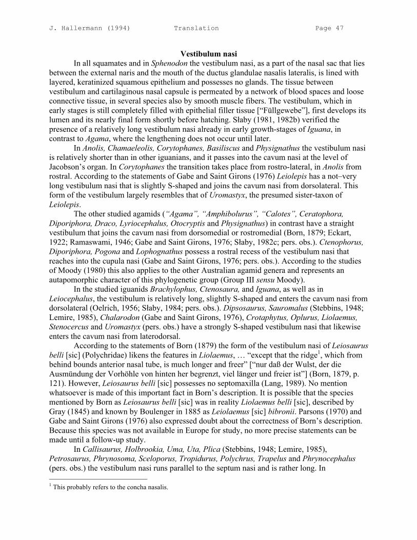
J. Hallermann (1994) Translation Page 47
Vestibulum nasi In all squamates and in Sphenodon the vestibulum nasi, as a part of the nasal sac that lies between the external naris and the mouth of the ductus glandulae nasalis lateralis, is lined with layered, keratinized squamous epithelium and possesses no glands. The tissue between vestibulum and cartilaginous nasal capsule is permeated by a network of blood spaces and loose connective tissue, in several species also by smooth muscle fibers. The vestibulum, which in early stages is still completely filled with epithelial filler tissue [“Füllgewebe”], first develops its lumen and its nearly final form shortly before hatching. Słaby (1981, 1982b) verified the presence of a relatively long vestibulum nasi already in early growth-stages of Iguana, in contrast to Agama, where the lengthening does not occur until later. In Anolis, Chamaeleolis, Corytophanes, Basiliscus and Physignathus the vestibulum nasi is relatively shorter than in other iguanians, and it passes into the cavum nasi at the level of Jacobson’s organ. In Corytophanes the transition takes place from rostro-lateral, in Anolis from rostral. According to the statements of Gabe and Saint Girons (1976) Leiolepis has a not–very long vestibulum nasi that is slightly S-shaped and joins the cavum nasi from dorsolateral. This form of the vestibulum largely resembles that of Uromastyx, the presumed sister-taxon of Leiolepis. The other studied agamids (“Agama”, “Amphibolurus”, “Calotes”, Ceratophora, Diporiphora, Draco, Lyriocephalus, Otocryptis and Physignathus) in contrast have a straight vestibulum that joins the cavum nasi from dorsomedial or rostromedial (Born, 1879; Eckart, 1922; Ramaswami, 1946; Gabe and Saint Girons, 1976; Słaby, 1982c; pers. obs.). Ctenophorus, Diporiphora, Pogona and Lophognathus possess a rostral recess of the vestibulum nasi that reaches into the cupula nasi (Gabe and Saint Girons, 1976; pers. obs.). According to the studies of Moody (1980) this also applies to the other Australian agamid genera and represents an autapomorphic character of this phylogenetic group (Group III sensu Moody). In the studied iguanids Brachylophus, Ctenosaura, and Iguana, as well as in Leiocephalus, the vestibulum is relatively long, slightly S-shaped and enters the cavum nasi from dorsolateral (Oelrich, 1956; Słaby, 1984; pers. obs.). Dipsosaurus, Sauromalus (Stebbins, 1948; Lemire, 1985), Chalarodon (Gabe and Saint Girons, 1976), Crotaphytus, Oplurus, Liolaemus, Stenocercus and Uromastyx (pers. obs.) have a strongly S-shaped vestibulum nasi that likewise enters the cavum nasi from laterodorsal. According to the statements of Born (1879) the form of the vestibulum nasi of Leiosaurus belli [sic] (Polychridae) likens the features in Liolaemus, … “except that the ridge1, which from behind bounds anterior nasal tube, is much longer and freer” [“nur daß der Wulst, der die Ausmündung der Vorhöhle von hinten her begrenzt, viel länger und freier ist”] (Born, 1879, p. 121). However, Leiosaurus belli [sic] possesses no septomaxilla (Lang, 1989). No mention whatsoever is made of this important fact in Born’s description. It is possible that the species mentioned by Born as Leiosaurus belli [sic] was in reality Liolaemus belli [sic], described by Gray (1845) and known by Boulenger in 1885 as Leiolaemus [sic] bibronii. Parsons (1970) and Gabe and Saint Girons (1976) also expressed doubt about the correctness of Born’s description. Because this species was not available in Europe for study, no more precise statements can be made until a follow-up study. In Callisaurus, Holbrookia, Uma, Uta, Plica (Stebbins, 1948; Lemire, 1985), Petrosaurus, Phrynosoma, Sceloporus, Tropidurus, Polychrus, Trapelus and Phrynocephalus (pers. obs.) the vestibulum nasi runs parallel to the septum nasi and is rather long. In 1 This probably refers to the concha nasalis.

J. Hallermann (1994) Translation Page 48
phrynosomatids, Phrynocephalus and Trapelus it joins the cavum antorbitale dorsally; in Tropidurus, Plica and Polychrus in contrast it joins the cavum nasi medially (Lemire, 1985; pers. obs.). In Bradypodion and Chamaeleo the vestibulum is relatively long and, in the latter, S-shaped. In Brookesia in contrast it is short and joins the cavum nasi laterorostrally (Haas, 1937; Malan, 1946; Gabe and Saint Girons, 1976; pers. obs.). This form of the vestibulum may be related to the limited space in the short nasal region of chamaeleons. The prägnante [succinct??] macrophthalmy of this group may be assumed to be causal. Only in Scincidae (except for Chalcides), Anguidae and Serpentes, within Scleroglossa, is the vestibulum very short; it passes into the cavum nasi at the level of the external naris. In most studied gekkotans, cordylids, lacertids, teiids, and amphisbaenians, and in Xantusia and Anniella, the vestibulum is not especially long and enters the cavum nasi rostrally, rostromedially or rostrolaterally. According to the studies of Lemire (1985), Acanthodactylus, Eremias (Lacertidae), and several geckos (Coleonyx, Ptyodactylus, Stenodactylus, Tarentola, Tropiocolotes) constitute exceptions to thereto. In these genera, which live in arid habitats, the vestibulum takes up about half the total length of the nasal capsule and is only in Acanthodactylus slightly S-shaped; the mouth of the vestibulum into the cavum nasi always takes place dorsomedially. In Varanus in contrast the vestibulum is rather long and U-shaped and enters the cavum nasi rostrodorsally. Sphenodon possesses a short vestibulum that passes into the cavum nasi medial to the external naris (Bellairs and Kamal, 1981; Gabe and Saint Girons, 1976; Malan, 1946). A great lengthening of the vestibulum nasi—in connection with a well-developed erectile tissue—is usually present in species that live in dry and hot biotopes (phrynosomatids, Dipsosaurus, Sauromalus; cf. Lemire, 1985). In these cases a lengthened vestibulum serves to humidify and adjust the dry and dusty air to the temperature of the body tissue. Moreover the particles that reach the cavum nasi can be trapped in the vestibulum by the moist surface and washed out by glandular fluids from the glandula nasalis lateralis. On the other hand a greatly elongated vestibulum can also be observed in several montane forms (e.g., Liolaemus, Phymaturus), where it could serve to warm up the inspired air by the net of blood vessels that surrounds the vestibulum. Stebbins (1948) says that the function of a lengthened vestibulum nasi with its keratinized epithelium principally consists in keeping particles in the inspired air out of the cavum nasi; the humidification of inspired air in contrast occurs in the cavum nasi. The close relationship of the duct of the glandula nasalis lateralis to the hind end of the vestibulum nasi, however, speaks for an important role of the glandular secretions in the humidification of inspired air (cf. Duvdevani, 1972). The partial closure of the naris and the decreasing in size of the lumen of the vestibulum by erectile tissue or an external nasal muscle is also of great use in decreasing transpiration. Species that dig in sandy soil face special problems of protecting the interior of the nose from the intrusion of soil particles. This can effectively be prevented by closure of the naris by expanded erectile tissue, as effected for example by the nasal valve (Anniella, Phrynosomatidae: Stebbins, 1948). The form and length of the vestibulum nasi and its juncture with the cavum nasi likewise play an important role in this respect (siphon-like vestibulum in Phrynosomatidae and Phrynocephalus). In species of other phylogenetic groups the interior of the nose is protected from particles in other ways. In the apothecary skink Scincus scincus Hartmann (1989) could observe the following: in front of the external nares of burrowed animals, aggregations of sand built up that were held together by moisture from the inspired air. These were by and by blown away again—especially during activity.

J. Hallermann (1994) Translation Page 49
The appearance of a lengthened and specialized vestibulum nasi, as found in phrynosomatids, was explicated by Stebbins (1948) in a typological evolutionary story [“in einem typologischen Evolutionsbild erläutert”]. In the beginning there was a short vestibulum and a large cavum nasi with a small concha (“primitive reptilian condition”). As an adaptation to dry habitats, slight lengthening of the vestibulum nasi and enlargement of the concha nasalis took place (as in Gerrhonotus [Anguidae]). As the next step Stebbins (1948) presented the beginning of the overlying of the cavum nasi by the vestibulum nasi. This is realized in Coleonyx (Gekkonidae), which lives in desert regions. Further lengthening of the vestibulum nasi and reduction of the concha nasalis would have arisen as a further adaptation to desert regions (Dipsosaurus-type sensu Stebbins, 1948). According to Stebbins, complete overlying of the cavum nasi by the vestibulum, with the transition at its hind end, could be conceived as a further specialization and represented by the “Uma-type” (sensu Stebbins, 1948) among phrynosomatids. This evolutionary picture reminds one strongly of a step ladder. It exemplifies rather well, however, the possible appearance of the specialized nasal organ within Iguania. A comparison with a great number of squamates makes appear highly probable that the lengthened vestibulum nasi represents a derived condition and is functionally related to a way of life in dry/hot biotopes. Optimization of the adaptation to dry/hot habitats and the opening of numerous ecological niches may have resulted from a more effective nasal closure mechanism (erectile tissue, nasal muscle). Further possibilities for survival in desert regions were opened by behavioral adaptations. Numerous lizards turned to a nocturnal way of life and use holes and hiding places to protect themselves from the heat of the day (geckos, skinks, monitor lizards: Stebbins, 1948; Pianka, 1986). In one ecological study a relatively larger portion of desert-dwellers were encountered in full sun among iguanians, in contrast to skinks, geckos, and monitor lizards (Pianka, 1986). Several taxa in Scincidae build subterranean tunnel systems that are also used by other species. Desert-dwelling monitor lizards especially have turned to seasonal activity (Pianka, 1986). A relatively short vestibulum nasi that rostrolaterally joins the front end of the cavum nasi can be taken for the basic pattern of Squamata, just as one finds in other sauropsids (Parsons, 1970; Character 20.0). The basic pattern of Iguania is probably distinguished by a lengthened vestibulum nasi that joins the cavum nasi rostrolaterally (Character 20.1). Winding of the vestibulum nasi into an S-shape and the related changes to the cartilaginous structures, especially underlying by the cartilago parietotectalis (Character 9.1), is probably a synapomorphy of Iguanidae, Crotaphytidae, Opluridae, and Tropiduridae (Character 20.2). The form of the septomaxilla also speaks for a hypothesis of this sort. Caudomedially this bone is bent up dorsally and contacts the roof of the nasal capsule in all examined representatives with an S-shaped vestibulum (Character 30.1). In contrast, the representatives of Tropidurini (sensu Frost, 1992) examined thus far—Plica umbra (Lemire, 1985) and Tropidurus melanopleurus (pers. obs.)—possess a straight vestibulum nasi and a small septomaxilla. One would have to make the additional assumption for these two genera or for Tropidurini that, if Tropiduridae is monophyletic, their straight vestibulum arose from an S-shaped one. The monophyly of Tropiduridae however is only weakly founded (incomplete gular fold and reduced angular; Frost and Etheridge, 1989). If the requirement that Tropiduridae constitutes a clade is omitted, a sister-group relationship between Tropidurus and Corytophanidae results as the most parsimonious hypothesis.

J. Hallermann (1994) Translation Page 50
In Uromastyx, where the vestibulum is likewise S-shaped, the septomaxilla in contrast does not rise dorsally. In its place, however, a ridge that depends from the cartilago parietotectalis is developed, which is in contact ventrally with the septomaxilla. This was termed by me the crista posterior tecti nasi. The twisted vestibulum of Chamaeleo probably likewise arose convergently. It is moreover noteworthy that in no representative of Scleroglossa can an S-shaped vestibulum nasi and a laterally junction be found. The S-shaped twisting of the vestibulum nasi probably arose three independent times within Iguania (Character 20.2). The divergent construction of the nasal capsule of the different groups and the form of the septomaxilla speak for this hypothesis. The greatly elongated, straight vestibulum nasi of Phrynosomatidae and the entrance by the same in the hind part of the cavum nasi can be taken as derived in relation to the basic pattern of Iguania and counted as an autapomorphy of Phrynosomatidae (Character 20.3). In this respect I assume that this form of the vestibulum arose from the postulated condition of the basic pattern of Iguania (Character 20.1). The septomaxilla of Phrynosomatidae is extraordinarily long but not bent up dorsally (Character 30.2). The very similar form of the vestibulum nasi in Phrynocephalus and Trapelus can be seen as a convergent construction, for numerous characters speak for a closer relationship with the other agamas and chamaeleons (Moody, 1980; Frost and Etheridge, 1989). Lemire (1985) also describes a very similar form of the vestibulum nasi in Agama agama and A. bibroni, which I, however, cannot confirm for A. agama (cf. Malan, 1946). The character (20.3) cited above may be a synapomorphy of Trapelus and Phrynocephalus or have a broader distribution within the erstwhile genus Agama. This cannot be decided at the present time. Frost and Etheridge (1989) distinguish five derived conditions of the nasal space that could have developed from the plesiomorphic condition or one of the other derived conditions: 1. “sink trap” of Phrynosomatidae, with a greatly elongated vestibulum and a dorsal entrance; no concha nasalis. This character was also counted here as an autapomorphy of this family (Character 20.3) and turns up convergently in Phrynocephalus. 2. “S-condition”: with the vestibulum nasi twisted into an S-shape, overlying of the cavum nasi by the vestibulum and its dorsolateral entrance. Septomaxilla bent up dorsally and plow-shaped. Apomorphy for Iguanidae s.s. This character has a wider distribution according to my studies (Opluridae, Stenocercus, Phymaturus, Leiocephalus, Liolaemus, Crotaphytus) and can thus count as a synapomorphy of Iguanidae, Opluridae, Crotaphytidae, and Tropiduridae, with a character reversal in Tropidurus (Character 20.2). 3. Fusion of the concha with the roof of the nasal capsule in Tropidurinae1. Stenocercus likewise has the “S-condition” (see above). A reduction of the concha of this sort can be found in numerous other forms and therefore does not represent a synapomorphy of this subfamily2. 4. “Anole condition”: loss of the concha, straight vestibulum. The simplification of the nasal cavity with the reduction of the concha nasalis and the cavum nasi may represent an autapomorphy of Polychridae or a subgroup of this family (Character 11.2, 13.1). (But see the remarks on the studies of Born (1879) on Leiosaurus above.)
1 The genera of these subfamilies were revised taxonomically by Frost (1992). Ophryoessoides, Proctotretus, Stenocercus = Stenocercus; Plica, Strobilurus, Tapinurus, Eastern Tropidurus, Uracentron = Tropidurus; Tropidurus koepckeorum = Plesiomicrolophus; Western Tropidurus = Microlophus; Uranoscodon. 2 The monophyly of Tropidurinae (Stenocercini + Tropidurini sensu Frost, 1992) is according to this only founded on one autapomorphic character: bilobate and bisulcate hemipenes; this also appears within Polychridae.

J. Hallermann (1994) Translation Page 51
5. “Acrodontan condition”: reduced concha, elongated vestibulum, mediodorsal entrance, no bent-up septomaxilla. Exceptions: in chamaeleonids the septomaxilla is lacking and the vestibulum has another course; Physignathus with a short vestibulum. This character applies onto to a part of Acrodonta according to my findings. Moreover, those species of Acrodonta that possess a concha nasalis with cavum conchale either were overlooked by the authors in the literature analysis or were not studied. In contrast to the New World iguanas the genera of agamas were not named as “terminal taxa”; instead, all agamas with the exceptions of Physignathus, Leiolepis and Uromastyx were subsumed as “agamas”. Among the agamas, Leiolepis, Diporiphora, Lophognathus (Gabe and Saint Girons, 1976), Pogona, Ctenophorus and Lyriocephalus (pers. obs.) possess a concha nasalis. Uromastyx and Leiolepis additionally have an S-shaped vestibulum nasi (Lemire, 1985; pers. obs.). It appears more probable that an elongated vestibulum already belonged to the provision of characters of the last common ancestor of Iguania (20.1). Also regarding the Malagasy Opluridae, my own findings do not agree with those presented by Frost and Etheridge (1989). The “primitive condition” of the nose of Oplurus and Chalarodon was viewed as having a straight vestibulum and a concha nasalis. Both species, however, possess a strongly S-shaped vestibulum and a reduced concha nasalis without cavum conchale (cf. Gabe and Saint Girons, 1976; pers. obs.). As well, the statement about Stenocercus and Tropidurus (“nasal vestibule relatively short and straight”, Frost and Etheridge, 1989:47) are not in agreement with my studies. Stenocercus possesses an S-shaped vestibulum, Tropidurus a rather long vestibulum (cf. also Solger, 1876 on Tropidurus and Lemire, 1985 on Plica).
Cavum nasi proprium The boundary between vestibulum nasi and cavum nasi is usually marked by the transition from keratinized squamous epithelium to columnar epithelium. This boundary mostly coincides with the entrance of the duct of the glandula nasalis lateralis (Parsons, 1970). The at first unitary nasal sac is divided during ontogeny by the construction of out-pocketings, denoted by different terms, in different morphological and functional regions (Parsons, 1970; Słaby, 1982b, 1982c; pers. obs.). The development of these compartments depends on different factors. If a voluminous concha nasalis is lacking, then a recessus extraconchalis cannot be constructed (many iguanians; pers. obs.). The size of the cavum antorbitale depends on the position of the internal choana and the size of the planum antorbitale, which once again depends on the size of the eyes and their rostral expansion. The cavum antorbitale is lacking in adult or juvenile Anolis (pers. obs.), Bradypodion, Chamaeleo and Brookesia (Malan, 1946; Haas, 1937; Gabe and Saint Girons, 1976). In young growth-stages of Anolis one finds a caudal out-pocketing of the cavum nasi that can be termed a cavum antorbitale, and thereby during ontogeny the caudal displacement of the choana disappears (Fig. 46, 47). The presence or lack of a cavum antorbitale can only be clarified by analysis of the features of the mucus membranes by means of serial sections. It is probable that Polychrus and Chamaeleolis, which just like Anolis possess a flat planum antorbitale, lack a cavum antorbitale (Character 13.1). The lack of a cavum antorbitale is tied to the reduction of the planum antorbitale and is not listed as an independent character. The cavum antorbitale, which directly follows the region caudal to the internal choana, belongs to the basic pattern of Lepidosauria (Character 13.0).

J. Hallermann (1994) Translation Page 52
If a concha nasalis is developed, the recessus lateralis cavi nasi (recessus extraconchalis) lies dorsolateral to the concha and is lined with respiratory epithelium (Fig. 55). This recess belongs, like the concha nasalis (Character 11.0), to the derived basic pattern of Squamata. The reduction of the extraconchal recess to a simple out-pocketing is certainly related to the reduction of the concha nasalis (Character 11.1). A recessus rostralis cavi nasi—a blind, rostral out-pocketing lined with respiratory epithelium—is present in all studied iguanians. It lies ventromedial—in Petrosaurus and Sceloporus ventrolateral—to the vestibulum nasi and is separated from the choanal groove by connective tissue. The present of this recess was also observed in different iguanians by other authors (Eckart, 1922; Malan, 1946; Gabe and Saint Girons, 1976; Słaby, 1981, 1982b, 1982c). It corresponds to the “anterior blind cavity” of Malan in Agama, Sceloporus and Chamaeleo as well as the “anterior space” of Oelrich in Ctenosaura. Within Scleroglossa a recess of this sort is not present. A rostral out-pocketing of the cavum that lies lateral to the vestibulum nasi but does not undermine it was only described in several geckos (Stenodactylus, Tarentola, Tropiocolotes) (Lemire, 1985). According to the studies of Lemire et al. (1970) on Uromastyx, the recessus rostralis cavi nasi is provided with a large number of mucus cells. The development of this blind recess can probably be seen as related to the lengthening of the vestibulum and its caudally located entrance into the cavum nasi. This would then be a derived condition and an autapomorphy of Iguania (Character 21.1). This hypothesis is strengthened by the observation that the recess of Crotaphytus, Iguana, Anolis, Agama and Chamaeleo is considerably larger in older embryonic stages than in younger ones (Słaby, 1981, 1982b; pers. obs.). Therefore, the recessus rostralis probably represents a secondary [i.e., independent], rostral expansion of the cavum nasi. Medial and lateral out-pocketings of the choanal tube turn up in numerous squamates (“Aulax” sensu Beecker, 1903 = recessus medialis ductus choanalis, recessus lateralis: Parsons, 1970). The functional importance of these out-pocketings lies in the enlargement of the surface area of respiratory epithelium. These recesses could not previously be used for phylogenetic analysis, for their presence and homology was still too poorly known. The opening of the horizontal choanal tube into the descending choanal tube is termed the internal choana (Born, 1879). Haller (1922) named this opening the primitive [primitive] choana. This term, however, is deceptive, for it suggests the interpretation that the internal choana is the primäre [primary] opening of the nasal sac into the oral cavity. However, the [primitive choana] is, in adults, through [“durch”] the opening of the duct of Jacobson’s organ (Matthes, 1934; Bellairs and Boyd, 1950). The caudal opening of the nasal sac into the oral cavity (external choana sensu Born, 1879) is separated during early ontogeny from this primary choana by connective tissue and/or mucus membranes. This tissue can therefore be termed a secondary palate in the wider sense (Fuchs, 1908; Bellairs and Boyd, 1950; Gabe and Saint Girons, 1976). If the descending choanal tube, then, is caudally elongated, one speaks of a ductus nasopharyngeus in the wider sense (Parsons, 1970). In this case, the opening of the choanal tube into the oral cavity consequently does not lie in the same level of section as the internal choana. The ductus nasopharyngeus is usually followed caudally by an unpaired interorbital groove (Gabe and Saint Girons, 1976; pers. obs.) The expansion of the area of olfactory epithelium—like its histological composition—exhibits in part rather large differences within the different squamate groups. In this respect different life habits probably play a more important role than phylogenetic relationship. Thus, the expansion of olfactory epithelium in herbivorous, arboreal, and aquatic forms rather slight, and

J. Hallermann (1994) Translation Page 53
there are only relatively few sensory cells present (Gabe and Saint Girons, 1976). Varanus indeed possesses a large area of olfactory epithelium but a smaller number of sensory cells per unit area than other squamates (Saint Girons, 1975; Gabe and Saint Girons, 1976). In most examined iguanians the external choana (sensu Born, 1879) extends nearly along the entire length of the cavum nasi, though in its rostral section it is only slitlike. The choana is always separated from the duct of Jacobson’s organ by a short secondary palate. Especially in Uromastyx, Polychrus and Corytophanes, the external choana reaches far rostrally. It should be noted that the choana in most species is additionally constricted laterally by a membranous choanal fold (ruga choanalis; Fig. 18, 23). In phrynosomatids, in contrast to other iguanians, the choanal slit is only very narrow, which Lemire (1985) related to their way of life in arid habitats. In Anolis and Chamaeleolis the choana lies in the hind region of the cavum nasi and the secondary palate is lengthened by the long cartilago ectochoanalis. In examined iguanians, Crotaphytus, Pogona and Physignathus have a descending choanal tube, which can be termed the “ductus nasopharyngeus” (Parsons, 1970; Fig. 2), that is caudally elongated and opens ventrally like a slit. In most scleroglossans the choana lies in the hind section of the cavum nasi and passes caudally into a shallow groove in the palatine, which for its part is connected to the unpaired interorbital groove (Göppert, 1903; Gabe and Saint Girons, 1976; pers. obs.). This groove is represented in the osseous skeleton by the Palatinalgrube (“palatinal groove” sensu Estes et al., 1988), which is an autapomorphy of Scleroglossa (Estes et al., 1988). A ductus nasopharyngeus was apparently developed in different ways in various groups. In gekkotans and in Xantusia it is underlain by the broad cartilagines ectochoanales. In Xantusia the ductus nasopharyngeus is functionally closed by partial overlapping of these cartilaginous elements. An unpaired tube (“pseudo conduit nasopharyngé” sensu Gabe and Saint Girons, 1976) is thereby formed. The descending choanal tubes also unite medially into an unpaired tube in numerous skinks and, among snakes, in Xenopeltis, Lichanura, Casarea and Crotalus (Gabe and Saint Girons, 1976). All skinks also possess a partly osseous secondary palate; the palatine, however, builds a complete secondary palate only in a few genera. The nose-throat passages [“Nasen-Rachengänge”] remain largely open like slits in scincids, in contrast to snakes (Greer, 1970; Gabe and Saint Girons, 1976). All other snakes, anniellids and amphisbaenians possess an paired ductus nasopharyngeus that is closed off ventrally by connective tissue (Malan, 1946; Gabe and Saint Girons, 1976). The teiids examined are distinguished by a rostrally closed, but short and double ductus nasopharyngeus (“conduit respiratoire” sensu Gabe and Saint Girons, 1976; Göppert, 1903; Malan, 1946). Lanthanotus, Varanus and Heloderma lack a ductus nasopharyngeus; the choanal slit is unusually large in the latter two species (McDowell, 1972). The function of the ductus nasopharyngeus of squamates consists in the establishment of a connection between choana and glottis. The ventral closure of this groove is achieved by means of the tongue in forms with an incomplete ductus nasopharyngeus (Göppert, 1903). Especially in snakes, which devour large pretty and possess a highly specialized tongue that would not sufficiently establish ventral closure, a completely closed ductus nasopharyngeus arose. Forms that live underground (Amphisbaenia, Anniellidae, Feylininae, Typhlopidae, Leptotyphlopidae) are likewise distinguished by a well-developed ductus nasopharyngeus (Gabe and Saint Girons, 1976). In these forms, however, a conception of the functional relation to their way of life is lacking.

J. Hallermann (1994) Translation Page 54
The caudal lengthening of the descending choanal tube and the related underlying of the choana can, on the basis of ontogenetic observations and phylogenetic considerations, be accepted as derived in relation to the basic pattern of Squamata (Character 22.1). The development of the ductus nasopharyngeus arose in different groups independently of one another and can be seen as an autapomorphy of Gekkota, Scincidae, and Serpentes. The lengthening of the cartilago ectochoanalis (Character 19.1) in Gekkota is tied to the development of a ductus nasopharyngeus and can count as an autapomorphy. The osseous secondary palate of scincids probably arose in relation to the ductus nasopharyngeus and can be taken as an autapomorphic character (Character 24.1). I accept the ventrally completely closed ductus nasopharyngeus of Ophidia, Anniellidae, and Amphisbaenia as derived characters of the basic pattern of these groups (Character 22.2). That far more complete ductus nasopharyngei and osseous secondary palates are present within other sauropsid groups is commonly known from crocodiles and turtles. In Chelonia the osseous secondary palate and ductus nasopharyngeus probably arose predominantly in relation to the creation of a trituration surface and the general consolidation [“Verfestigung”] of the roof of the mouth (Eßwein, in prep.). In crocodiles the reason for the development of an extensive secondary palate and a very long, unpaired ductus nasopharyngeus is to be sought on the one hand in the consolidation of the snout; on the other hand the complete connection between the nose and the windpipe is an important requirement for underwater prey acquisition. Comparison shows that the ductus nasopharyngei of various amniote groups present entirely different pictures. The assumption that a ductus nasopharyngeus was already present in the basic pattern of Sauropsida (or Amniota) can therefore be rejected.
Choanal groove The choanal groove in all examined iguanians with the exception of Anolis and Chamaeleolis is open ventrally and reaches from the external choana to the duct of Jacobson’s organ rostrally (cf. also Malan, 1946; Bellairs and Boyd, 1950; Oelrich, 1956; Gabe and Saint Girons, 1976; Lemire, 1985). The length of the choanal groove depends on the position of the external and internal choana. By definition the choanal groove ends at the dorsal entrance of the descending choanal tube (Fuchs, 1908; Bellairs and Boyd, 1950). This terminology, however, was not used by all authors. Lemire (1985) also termed “sillon choanale” the choanal tube that lies behind the external choana and opens slitlike. If the external choana lies far rostrally, then the choanal groove is only very short; this is the case in most examined iguanians. In Anolis and Chamaeleolis, in contrast, the internal and external choana lie relatively far caudally. Therefore, the choanal groove is relatively long. It is underlain by a broad and long cartilago ectochoanalis, whereby the groove is closed and becomes a tube (Character 19.1, Fig. 23). The choanal groove likewise appears rather long in ventral view in Crotaphytus, for it is extended as a choanal groove that opens slitlike (Fig. 2). The majority of scleroglossan squamates likewise possess a ventrally open choanal groove. In Varanus, Heloderma, amphisbaenians (Amphisbaena, Blanus, Rhineura, but not Trogonophis and Leposternon), examined teiids (Ameiva, Cnemidophorus, Teius, Tubinambis), Xantusia, Feylinia and serpents, in contrast, the choanal groove is closed during ontogeny by connective tissue. The choanal groove ends blindly rostrally in several teiids, while in Varanus and snakes it is completely closed (Busch, 1898; Malan, 1946; Bellairs and Boyd, 1950; McDowell, 1972; Gabe and Saint Girons, 1976). Lanthanotus possesses a short choanal groove. Whether this groove has a rostral connection to the duct of Jacobson’s organ, however, could not
J. Hallermann (1994) Translation Page 55
be determined in the few studies published thus far (McDowell, 1972). In lacertids, several scincids, and cordylids, the choanal groove is not directly connected to the duct of Jacobson’s organ but rather ends a little bit behind it (Lemire, 1985). The following correlations can be determined: taxa in which the choanal groove has no connection to the duct of Jacobson’s organ possess a thin, two-pronged tongue that, as a chemosensory organ, removes substances from the air and from the substrate (Kahmann, 1939; Burghard, 1980). It is in this respect probable that the tongue tips are inserted into the duct of Jacobson’s organ (see, however, the chapter “Jacobson’s organ”). In squamates in which the margin of the tongue forms a smooth or pointed oval [“die eine ausgerandete oder spitzovale Zunge besitzen”], the edges of the tongue probably graze the choanal groove upon retraction, and substances that adhere to the tongue thus arrive in the choanal groove. They are transported rostrally into the ductus vomeronasalis by lacrimal fluid—additionally driven by the movement of cilia. The choanal groove serves, then, as a way to admit gustatory substances into Jacobson’s organ. The lateral underlying of the choanal groove by the cartilago ectochoanalis and by connective tissue prevents, on the one hand, the fluid of the choanal groove from being mixed with the secretions of the oral cavity. On the other hand, substances that adhere to the tongue can arrive in the choanal groove by simple sideways pushing away of the Vomerpolster, for it [the groove] is only laterally closed. In snakes, monitor lizards, teiids, and amphisbaenids, the choanal groove became superfluous as a way to admit gustatory substances, for a highly specialized tongue arose. Sphenodon has no choanal groove. The cavum nasi opens slitlike into the oral cavity along its whole length. The choana reaches rostrally up to the exit of the ductus vomeronasalis. The choana is dorsally constricted by a fold of mucosa (Choanenfalte sensu Fuchs, 1908), which is broader in its caudal section than in its rostral one. The ventral region of the cavum nasi, which lies dorsal to this fold, is lined with respiratory epithelium and is termed the Choanengang [choanal tube] (Beecker, 1903; Fuchs, 1908). The ductus vomeronasalis enters the rostral and ventral section of the choanal tube laterally (Hoppe, 1934; Bellairs and Boyd, 1950). The development of a choanal groove and a short secondary palate of connective tissue can be accepted as a derived character of the basic pattern of Squamata (Character 23.0, 24.0). A lateral out-pocketing of the choanal groove (Winkeltasche [lateral choanal groove] sensu Beecker, 1903) is probably a plesiomorphic character of squamates, for this type of lateral out-pocketing of the choanal tube is present in Sphenodon (Gabe and Saint Girons, 1976). The choanal tube is in this respect related to the sophistication and mode of function of Jacobson’s organ. The secondary closure of the choanal groove in Ophidia, Amphisbaenia, Varanidae, Helodermatidae and Teiidae can be seen as autapomorphies of these groups (Character 23.1). Considering the autapomorphic nature of this character, Teiidae and Amphisbaenia must be given a question mark, for this character is not completely developed in all genera within Amphisbaenia and rather few representatives of Teiidae have been studied. This character may be a synapomorphy of Platynota (sensu Rieppel, 1980 = Varanoidea sensu Estes [et al.], 1988) + Ophidia. Because of the uncertain presence of this character in Lanthanotus, this hypothesis cannot be validated at the present time. In several groups within Squamata the choana is further displaced caudally, as [insofar as?] this can be as the condition of the basic pattern; there arose a ductus nasopharyngeus. The osseous secondary palate of scincids, even when it is not complete, could count as an autapomorphy of this group (Character 24.1).

J. Hallermann (1994) Translation Page 56
Ductus nasolacrimalis The ductus nasolacrimalis in all tetrapods arises as an in-folding of the ectoderm independent from the primordial nasal sac (Bellairs and Boyd, 1950; Medvedeva, 1986). The ductus nasolacrimalis grows progressively from caudal to rostral and during ontogeny acquires a relationship to the nasal sac. In order to be able to understand the complicated relationships of the ductus nasolacrimalis to the choanal groove in older embryos and adults, a short presentation of the development of the palatal region in squamates ought to precede [the character description]. In all squamates and Sphenodon, the anlage of Jacobson’s organ arises as a medial diverticulum in the lower and front region of the nasal sac. The ventral opening of the nasal sac into the oral cavity is the primary (primitive) choana. It is divided in early developmental stages in all squamates. The front opening is the exit opening of Jacobson’s organ (fenestra vomeronasalis externa sensu Oelrich, 1956), the hind one is the definitive [“definitive”] choana. Simultaneously Jacobson’s organ alters its position medioventrally by growth processes and is then clearly separated from the nasal sac. The tissue that rostrally closes the choana can therefore be seen as a type of secondary palate. The choanal tube and the cavum nasi lie dorsally, the choanal groove ventrally (Fuchs, 1908; Bellairs and Boyd, 1950). The distal end of the ductus nasolacrimalis grows medially into the mesenchymal cushion of the secondary palate and connects to the ventral and medial part of the ductus vomeronasalis (Bellairs and Boyd, 1950). As also explicated in the chapter “Ductus nasolacrimalis”, the relationships of the distal end of the ductus nasolacrimalis to the duct of Jacobson’s organ largely agree in all examined late embryonic and adult squamates (cf. also Malan, 1946; Oelrich, 1956; Bellairs and Boyd, 1950; Parsons, 1970; Gabe and Saint Girons, 1976). This applies likewise to those forms in which the choanal groove was secondarily closed. In early embryonic stages of chamaeleonids, however, in which Jacobson’s organ is lacking or rudimentary, the anlage of the ductus nasolacrimalis reaches rather far rostrally to the level of the anlage of Jacobson’s organ. During further ontogeny the ductus nasolacrimalis breaks through to the oral cavity and joins the choanal groove laterally (Malan, 1946; Bellairs and Boyd, 1950). It can therefore be assumed that the distal section of the ductus nasolacrimalis was reduced in chamaeleonids (Character 25.1). This tube therefore no longer joins the duct of Jacobson’s organ medially, for this organ is only rudimentary. In Sphenodon, in contrast, the ductus nasolacrimalis joins the lateral side of the choanal tube, slightly caudal to Jacobson’s organ (Bellairs and Boyd, 1950). Because a secondary palate is not developed in Sphenodon, the ductus nasolacrimalis medially cannot intersect the choanal groove dorsally and join the ductus vomeronasalis. In all other tetrapods—with the exception of Gymnophiona—the ductus nasolacrimalis only has relationships to the cavum nasi and not to Jacobson’s organ or the choanal groove (de Beer, 1937; Parsons, 1970). In Gymnophiona the ductus nasolacrimalis connects the recess of the tentacle apparatus [“Tasche des Tentakelapparates”] with Jacobson’s organ. The latter, however, is connected medially with the cavum nasi (Jürgens, 1971; Billo and Wake, 1987). The connection of the ductus nasolacrimalis to the duct of Jacobson’s organ is probably related to the filling mechanics of the organ (plunger pump device, Broman, 1920). Because the Jacobson’s organ of squamates possesses no glands, in contrast to that of mammals [“Säuger”], rather functioning as a “Wassergeruchsorgan” [water-smelling organ] (sensu Broman, 1920), the close relationship of the lacrimal duct to this organ becomes understandable. The entrance of the
J. Hallermann (1994) Translation Page 57
ductus nasolacrimalis into the medial side of the ductus vomeronasalis can be taken as derived in relation to the basic pattern of Lepidosauria (Character 25.0). In most squamates there are further, proximal connections of the ductus nasolacrimalis to the lateral choanal fissure of the choanal groove, which mostly lie at the level of the internal choana or rostral thereto. Such connections can be found in all studied iguanians, anguids, and several gekkotans. They first appear in later embryonic stages (Bellairs and Boyd, 1950; Gabe and Saint Girons, 1976; pers. obs.). In several autarchoglossans a long union [“eine lange Vereinigung”] of the ductus nasolacrimalis and choanal groove rostral to the internal choana can be observed (“lacrimo-choanal gutter” sensu Bellairs and Boyd, 1950). It even comes to a structural fusion of the epithelia in lacertids and scincids. Further rostrally—at the level of Jacobson’s organ—the ductus nasolacrimalis separates from the choanal groove and joins the ductus vomeronasalis in the described manner. In Anguis, Anniella, Trogonophis and cordylids, the choanal groove and lacrimal duct are connected with one another for a shorter stretch than is the case in lacertids. Xantusia possesses somewhat deviant features [“Verhältnisse”], for here the ductus nasolacrimalis has no connection with the choanal groove and joins the upper section of the duct of Jacobson’s organ medially (Bellairs and Boyd, 1950). The same authors could demonstrate, on the basis of staining experiments, that the secretion of the orbital glands, despite a structural union of the choanal groove with the lacrimal duct and therefore the consequent opening to the oral cavity, arrived pure at the duct of Jacobson’s organ. Thus, the lateral choanal fissure is certainly functionally closed off to the oral cavity. A caudal connection of the ductus nasolacrimalis to the choanal groove enables transport of substances in the choanal groove by lacrimal fluid. The expanded connection of the ductus nasolacrimalis to the choanal groove that is developed in several scincomorphs is a derived character within Squamata. It remains undetermined whether the “lacrimo-choanal gutter” (sensu Bellairs and Boyd, 1950) represents a synapomorphy of Lacertidae and Scincidae or has a broader distribution. A lateral connection of the lacrimal duct with the lateral choanal fissure, occurring only in one place, is probably a plesiomorphic character of the basic pattern of Squamata, for the ductus nasolacrimalis is connected to the choanal groove in a similar way in Sphenodon (Bellairs and Boyd, 1950). Within Squamata, two ductus nasolacrimales on each side [“jederseits”] appear only in Varanus, Heloderma and Lanthanotus (McDowell, 1972; Gabe and Saint Girons, 1976). In Varanus the shorter one joins the cavum nasi a little above the internal choana. The longer one has a lateral connection with the choanal tube at the level of the external choana and then passes to the duct of Jacobson’s organ in the manner described above (Bellairs and Boyd, 1950). Two foramina lacrimales appear only in Varanus and Lanthanotus and were seen by Rieppel (1980) as a synapomorphy of these two genera. An unpaired ductus nasolacrimalis on each side within the nasal capsule can be accepted for the basic pattern of Squamata (Character 26.0). The double ductus nasolacrimalis on each side would then be an autapomorphy of Platynota (sensu Rieppel, 1980; Character 26.1). The assumption that a double ductus nasolacrimalis was the primitive condition (Saint Girons, 1982), because the proximal end of the lacrimal duct is paired in the region of the orbital glands, is less probable. Neither Sphenodon nor other sauropsids possess a double ductus nasolacrimalis. The lacrimal ducts also unite behind the tentacle sac in Gymnophiona, and this joins Jacobson’s organ (Billo and Wake, 1987). With respect to the hypothesis of Saint Girons (1982), therefore, independently occurring fusion of the lacrimal duct in nearly all tetrapod groups would have to be assumed.

J. Hallermann (1994) Translation Page 58
Within Acrodonta the foramen lacrimale is greatly enlarged in all studied agamids (sensu Moody, 1980) and chamaeleonids, in contrast to Uromastyx and Leiolepis. This character was seen as a synapomorphy of Agamidae and Chamaeleonidae (=Agaminae + Chamaeleoninae sensu Frost and Etheridge, 1989) by Frost and Etheridge (1989). Several of these genera (Agama, Cophotis, Ctenophorus, Draco, Lophognathus, Otocryptis, Pogona, Physignathus, Chamaeleo, Bradypodion, Brookesia) that have previously been studied histologically possess a ductus nasolacrimalis with a larger lumen [“einen weitlumigeren Ductus”] than is the case in squamates outside this postulated phylogenetic group (Born, 1879; Eckart, 1920; Malan, 1946; Bellairs and Boyd, 1950; Gabe and Saint Girons, 1976; pers. obs.). This applies as well to all other agamids with the exception of Hydrosaurus, according to the macroscopic studies of Moody (1980). However, not only the size of the lacrimal duct within the nasal capsule may be of import, but also the expanded connection of the canaliculi to the conjunctival space (Saint Girons, 1982). This phenomenon is therefore probably tied to the development of a large foramen lacrimale (Character 27.1). It is moreover notable that most authors do not mention the structure of the ductus nasolacrimalis in the description of the nasal capsule. The size of the ductus nasolacrimalis in Enyalioides, Hoplocercus and Morunasaurus (Hoplocercidae) would be interesting in this context, where a large foramen lacrimale likewise appears (Frost and Etheridge, 1989). No representative of this family has thus far been studied histologically; nor could suitable material be obtained for the present work.
Glandula nasalis lateralis All squamates possess but one nasal gland, the glandula nasalis lateralis. It is located, in those forms that possess a concha nasalis with cavum conchale, in this space (Fig. 55; Parsons, 1970; Bellairs and Kamal, 1981). Shrivastava (1963) mentions the rudiment of a ventral nasal gland in an embryo of Varanus. This could not be demonstrated, however, in any other growth-stage or in any other species (Malan, 1946; Bellairs, 1949). In those iguanians that possess only a reduced concha nasalis or none at all, the glandula nasalis lateralis lies in a shallow pit on the roof of the nasal capsule dorsal to the reduced concha or within the fissura lateralis (e.g., Anolis, Phrynosomatidae; pers. obs.). Numerous little collection canals [“Sammelkanälchen”] unite into one duct that extends rostrally and enters the nasal capsule either through the foramen ductus glandulae nasalis lateralis or through the fissura lateralis. This entrance lies only at the hind end of the vestibulum nasi at the epithelial boundary of the cavum nasi (Parsons, 1970; pers. obs.). Only in Cophotis does the epithelial boundary lie behind the entrance of the duct of the lateral nasal gland (Eckart, 1922). In phrynosomatids the duct of the gland extends caudally, in contrast to most other squamates; the entrance into the vestibulum lies, therefore, far behind the gland (cf. Malan, 1946; Gabe and Saint Girons, 1976; Lemire, 1985; pers. obs.). This results from the great elongation of the vestibulum nasi and its entrance into the cavum antorbitale (Character 20.3). The altered course of the ductus glandulae nasalis lateralis is therefore also related to this (Character 28.1). Both characters are autapomorphies of Phrynosomatidae. Two nasal glands occur in other tetrapod groups, a medial one and a lateral one. Their partly differing positions led to different terms; however, the glands are homologous. In all three amphibian groups the lateral nasal gland joins the vestibulum nasi, while the medial one (ventral gland of Gymnophiona) has a relationship to the out-pocketing of the cavum nasi that has been termed Jacobson’s organ (Matthes 1934). In turtles the medial nasal gland lies between the

J. Hallermann (1994) Translation Page 59
septum nasi and nasal sac in the region of the lateral boundary fold; its duct ends in the region of Jacobson’s epithelium of the cavum nasi (Matthes, 1934; Parsons, 1970). Likewise, archosaurs (crocodiles, birds) probably possess two nasal glands (but cf. Saint Girons, 1988, 1989) that lie close to one another; their two ducts join the septal and lateral wall (respectively) of the vestibulum nasi (Röse, 1893; Bellairs and Shute, 1953; Weber, 1900). Sphenodon, in contrast to all squamates, likewise has two nasal glands whose corresponding entrances are like in other sauropsids. One can therefore accept that the glandula nasalis medialis was completely reduced in the last common ancestor of Squamata (Character 28.0). This position of the lateral nasal gland within the cavum conchale is likewise a derived character of the basic pattern of Squamata and tied with the appearance of a concha nasalis with a cavum conchale (Character 11.0). The size of the glandula nasalis lateralis varies with Iguania, depending on way of life. Moreover, ontogenetic studies have shown that the glandular tissue grows markedly in size after hatching (cf. Lemire, 1985). In desert-dwelling and marine lizards the glandula nasalis lateralis is especially large, and its glandular secretions evince high salt concentrations (Lemire and Vernet, 1982; Lemire, 1985; pers. obs.). Because the sauropsid kidney cannot excrete urine with a higher concentration of salt than is present in the plasma, the glandula nasalis lateralis serves to secrete salt extrarenally. In marine lizards salt excretion can be seen as a mechanism for the maintenance of ion balance as a result of increased salt uptake through food (Schmidt-Nielson et al., 1963). In lizards from arid zones, water resorption in the cloaca and, simultaneously, increased extrarenal salt excretion through the nasal gland result. Here, there are differences in ion concentrations (K+, Na+) that depend on diet (herbivorous vs. insectivorous) (Schmidt-Nielson et al., 1963). Van Lennep (1970) could also establish a correlation between the size of the glandula nasalis lateralis and the aridity of the habitat. Therefore, enlargement of the glandula nasalis lateralis probably arose independently in the various groups. The fine structure of the lateral nasal gland of Uromastyx compares better with the orbital gland of sea turtles than the lateral nasal gland of birds (Van Lennep, 1970). This could be grounded in the same mode of ion excretion (more K+ than Na+). In marine squamates (e.g.: Amblyrhynchus, Hydrophis), however, there is a broad histological concordance with the glandula nasalis lateralis of marine birds (Schmidt-Nielson and Fange, 1958). The various histological constructions of the lateral nasal gland within Squamata were studied by Lemire and Gernot (1974), Gabe and Saint Girons (1971, 1976), Lemire (1985) and Saint Girons and Bradshaw (1987). In particular, the striation of the glandular tissue (“segment stries”; Gabe and Saint Girons, 1976) was especially emphasized by these authors. This striation appears in numerous lizards from nearly all families and is especially conspicuous in several desert-dwelling forms. Because of in-folding of the basal and lateral membranes of the glandular cells at tubular segments [“an tubulären Segmenten”], the tissue appears striated under the light microscope. This section of the gland serves to transport and secrete salt (Saint Girons and Bradshaw, 1987). There is not, however, a correlation with an arid habitat in all cases. Large differences in the fine structure of the gland could be determined in Trapelus mutabilis and Uromastyx acanthinurus, which life in arid regions. These differences were seen as related to the different diets (insectivorous vs. herbivorous) with their various electrolyte composition (Lemire and Gernot, 1982). Lemire (1985) also arrived at the conclusion that the specialized glandular tissue (“segment stries”) arose in relation to the appearance of a salt gland. Its function lies in the production of a hypertonic salt solution and thus in extrarenal excretion. The striation of the glandular tissue is lacking in all studied snakes, several geckos and anguids
J. Hallermann (1994) Translation Page 60
as well as all other vertebrates. Other glands function as salt glands instead, such as the sublingual glands of hydrophiids and acrochordids (Ophidia), the premaxillary gland of homalopsids, the lingual glands of crocodiles and the lacrimal glands of several chelonians (Lemire, 1985). Thus, the “segment stries” arose first within Squamata or in its last common ancestor. Because the “segment stries” is lacking in Ophidia, its presence could be an autapomorphy of “Lacertilia”. Numerous synapomorphic concordances between Platynota (sensu Rieppel, 1980) and Ophidia, however, make this appear improbable. Because this specialized tissue is lacking in most anguimorphs, with the exception of varanids, reduction could also have occurred in Anguimorpha (including Ophidia) and, independently, in several other squamates (Chamaeleonidae, Gekkoninae, Pygopodidae, Feylininae, Cordylidae).
Jacobson’s organ Jacobson’s organ arises during ontogeny as a protuberance [“Ausstülpung”] of the primordial nasal sac. In all studied squamates Jacobson’s organ is spheroidal, in contrast to Sphenodon, where this organ is small and oblong. Dorsal and dorsomedial to the lumen of the organ there is a thick layer of sensory epithelium without Bowmann’s glands. This sensory epithelium is innervated by the nervus vomeronasalis, which extends to the bulbus olfactorius accessorius (Parsons, 1970). In lizards the organ lies ventral or ventromedial to the vestibulum nasi. Jacobson’s organ in snakes, in contrast, is located further caudally, only in the region of the cavum nasi (Gabe and Saint Girons, 1976; Bellairs and Kamal, 1981); the long lamina transversalis anterior, which reaches far caudally, is also related to this. I combine both apomorphic characters (Character 29.2) and assess it as an autapomorphy of Ophidia. In squamates, Jacobson’s organ apparently plays an important role in the reception of chemical substances that are received by the tongue, as the experiments of Kahmann (1939) and Andren (1982) show. Jacobson’s organ acquires great importance especially in the search for food and the location and recognition of mating partners (Burghard, 1980). Halpern (1987) could observe that in snakes (Thamnophis, Vipera) sexual behavior ceases after the nn. vomeronasalis were severed. Olfactory substances can be received not only by direct contact of the tongue with the substrate but also from the air by tongue flicking. As the experiments of Noble and Kumpf (1936) showed, Jacobson’s organ acts as an accessory olfactory organ, for with a destroyed organ, hidden food could only be located with the olfactory organ; however, the experimental animals (Ameiva, Eumeces) required a longer time. A remarkable result of these studies was that Eumeces laticeps, one of the few species within Squamata that practices intensive brood care and even incubates its clutch, could recognize foreign eggs by licking them and remove them from its own clutch (Noble and Mason, 1933). Whether the conspicuous behavior in geckos of licking the eyes with the tongue is related to the reception of olfactory substances and supplying them to Jacobson’s organ is unknown. The faster and more effective way for Jacobson’s to acquire substances that adhere to the surface of the eye would well be to transport them directly by lacrimal fluid, for there is a connection of the ductus nasolacrimalis to Jacobson’s organ. This behavior far more likely serves to clean the cornea. The connection of the distal end of the ductus nasolacrimalis to the ductus vomeronasalis speaks for an important roll of the lacrimal fluid in transporting substances into the lumen of Jacobson’s organ. In this regard, fluid flow in the ductus organi vomeronasalis could be achieved

J. Hallermann (1994) Translation Page 61
by the movement of cilia (Bellairs, 1984). The mode of functioning of a suction pump, whereby the lumen is widened by the release of pressure on the organ by the tongue and the fluid thereby drawn up, appears to me to be a more effective (auxiliary) mechanism (Broman, 1920; Young, 1993). Substances that adhere to the tongue are brushed [“abgestreift”] into the lower end of the duct of Jacobson’s organ and/or the choanal groove. The evolution of tongue shape within Squamata is important in this context to the form-function relationship. Primarily, the tongue principally serves to manipulate food and in the ingestion of water. Touching with the tongue of the substrate, the food, or the partner during mating has been observed in numerous squamates from all families (Gove, 1979; Gravelle and Simon, 19890; pers. obs.). The substrate is touched with the tongue especially in an environment unknown to the animal. I could even observe this in forms like the Rotkehlanolis [?Green Anole, Anolis carolinensis], in which Jacobson’s organ is rather small (cf. Armstrong et al., 1950; Stimie, 1966; Greenberg, 1993). In iguanians, gekkonids, and several scincids, the ventral side of the tongue is studded with soft Papillen (“pallets”, McDowell, 1972) moistened by [sub]lingual glands, by which particles adhere there more easily. During retraction the tongue is usually turned over, and the underside touches the roof of the mouth (McDowell, 1972). In Ctenosaura and other iguanians, the duct of Jacobson’s organ ends on each side in a shallow, triangular, gland-free fossa on the roof of the mouth. This fossa is bounded laterally by a fold of mucosa and medially by a narrow ridge of mucosa (“vomerine raphe”). A groove on the ventral side of the tongue between the pallets corresponds to the “vomerine raphe” on the roof of the mouth so that gustatory substances adhering to the tongue could be deposited in this place and thus carried to Jacobson’s organ. In most scleroglossans this fossa on the roof of the mouth is narrower and the tongue terminally more deeply split (McDowell, 1972). The findings of Schwenk (1985) lead one to suspect, moreover, that especially in several iguanians, the Geschmacksknospen (“taste buds”) on the tongue and in the oral cavity next to the receptor cells in Jacobson’s organ have an important roll in the perception of chemical substances. In autarchoglossans the reception of substances from the air also plays an important roll, for simple flicking of the tongue can first be observed within this group (Schwenk, 1993). Gove (1979) and Burghard (1980) maintained a positive correlation between the frequency of tongue-flicking and the sophistication of Jacobson’s organ. In varanids and serpents the tongue has become a purely sensory organ that serves for receiving chemical substances and for tasting. Substances can also be received from the air in these forms (Bissinger et al., 1979). The anatomy of the tongue of monitor lizards, tegus and snakes permits the insertion of the tips of the tongue into the ducts of Jacobson’s organ (Kahmann, 1939). Gillingham and Clark (1981) in contrast described the function of two bumps of mucosa on the floor of the mouth (“sublingual plicae” sensu McDowell, 1972) in several snakes (Elaphe, Heterodon, Boa, Python, Crotalus, Bitis) that lie directly under the openings of Jacobson’s organ. These bumps, termed “anterior process”, were moved upward during retraction of the tongue and closing of the mouth. They are said to have been inserted as far as the lumen of Jacobson’s organ and thereby to convey olfactory substances, which previously were wiped off on these bumps with the tongue, into the organ. Experiments could show, moreover, that after removal of these processes the experimental animals required considerably longer to find hidden food, although the tongue was undamaged (Gillingham and Clark, 1981). That these processes corresponds topographically and structurally with the ducts of Jacobson’s organ I cannot confirm on the basis of the serial sections of Boa constrictor. The X-ray cinematographic studies by
J. Hallermann (1994) Translation Page 62
Oelofson and Heever (1979) on Varanus and Young (1990) on Boa could not confirm direct insertion of the tips of the tongue into the lumen of Jacobson’s organ. Direct insertion of the tips of the tongue into the ductus vomeronasalis is nevertheless possible, at least in other species. Greater sophistication of Jacobson’s organ in anguimorphs is also appears in the relation of the sensory cells to the support cells in the epithelium of the organ (Gabe and Saint Girons, 1976). Sphenodon had the fewest sensory cells (23.5%), followed by Anolis (47%). The examined agamas (Physignathus, Ctenophorus, Diporiphora, Leiolepis) had a sensory cell proportion of 50–60% in the epithelium of Jacobson’s organ. In contrast, scleroglossan squamates, with 75–80% (several snakes even over 90%) sensory cells, possess a highly sophisticated organ. This applies also to water snakes (Acrochordus, Hydrophis), in which the olfactory epithelium in the cavum nasi is greatly reduced (Gabe and Saint Girons, 1976). The complete isolation of Jacobson’s organ from the nasal cavity, the specialization as an organ for the reception of dissolved olfactory substances received by the tongue, and its specialized filling mechanism probably first appeared in the last common ancestor of Squamata and can be considered derived characters of the basic pattern (Character 29.0). The reduction of the organ is an autapomorphy of Chamaeleonidae (Character 29.1), the displacement of the organ caudally an autapomorphic character of Ophidia (Character 29.2). The question of when Jacobson’s organ appeared for the first time in the phylogenetic history of vertebrates and can first be termed such has been the object of several publications (Zuckerkandl, 1908, 1910; Parsons, 1959a, 1959b; Bertmar, 1981). To what extent it is justifiable to label as “Jacobson’s organ” particular epithelial zones in the cavum nasi whose Bowmann’s glands are lacking and whose nerve fibers extend to the bulbus olfactorius accessorius and to compare these with the organ of the same name in lepidosaurs and mammals, will not be discussed further here. The question of whether a well-differentiated Jacobson’s organ was already present in the basic pattern of Amniota and was reduced in Chelonia, or whether it first appeared later in evolution, after Chelonia split off, as was represented by Olson (1947) and Gaffney (1980) in the theory of Eureptilia, is a difficult one. Another possibility would be that Jacobson’s organ in squamates and mammals became segregated as an out-pocketing of the cavum nasi and differentiated independently of one another. Only in the early ontogeny of crocodiles can a medial diverticulum of the primordial nasal sac be observed that can be seen as an anlage of Jacobson’s organ. This anlage disappears early on; a bulbus olfactorius accessorius is lacking in all archosaurs (Parsons, 1959b).
Dermal bones of the ethmoidal region Several dermal bones of the ethmoidal region were incorporated in phylogenetic analyses by Moody (1980), Etheridge and de Queiroz (1988), and Frost and Etheridge (1989). Here, only those dermal bones will be listed whose Characters were seen as derived with in Iguania by the authors mentioned above. Maxilla In all acrodont iguanians the maxillae of both sides are ventrally in broad contact with one another behind the premaxilla; in other iguanians, in contrast, they are separated by the connection of the vomer to the premaxilla. This is an autapomorphy of Acrodonta (see also Borsuk-Białynicka and Moody, 1984). Moreover, the maxilla in this phylogenetic group extends caudally to behind the fronto-parietal suture.

J. Hallermann (1994) Translation Page 63
Vomer The vomers are paired bones; however, they fuse medially with one another, especially in older individuals (Moody, 1980; pers. obs.). This bone is concave in Uromastyx and Leiolepis, in contrast to all other iguanians (Moody, 1980, 1987). In other squamates it is more or less flat to slightly convex. This character, according to Moody (1980), is a synapomorphy of these two genera (Uromastycidae = Leiolepidinae sensu Frost and Etheridge, 1989). The vomers are very small in Hoplocercus, Morunasaurus and Enyaloides [sic] (autapomorphy of Hoplocercidae). Lacrimal This bone is rather small in all iguanians; sometimes only a small, basal ossification is present that from the outside cannot be seen (Moody, 1980). The lacrimal is lacking in Agama hispida, Calotes versicolor, Cophotis, Draco, Lyriocephalus, Pogona vitticeps, P. barbata, Phrynocephalus, Sitana, Trapelus mutabilis, T. pallidus, T. sanguinolentus, Uromastyx, and Polychrus as well as Phrynosoma, Callisaurus and Holbrookia (Camp, 1923; Moody, 1980; Frost and Etheridge, 1989; pers. obs.). It is not clear whether the bone fuses to another or whether the ossification center disappears. Frost and Etheridge (1989) accepted the lack of a lacrimal as a synapomorphy of Phrynosoma and the “sand lizards” (Callisaurus, Cophosaurus, Holbrookia, Uma). Because variability is high the incorporation of this character was omitted from the phylogenetic analysis. Palatine Dentition on the palatine appears only in a few species within Iguania. Crotaphytus as well as polychrids with the exception of Polychrus possess dentition of this type. The complete reduction of palatine and pterygoid teeth was counted as an autapomorphy of Acrodonta (Moody, 1980). The reduction of the pterygoid teeth in Phrynosomatidae is an autapomorphy of this group (Frost and Etheridge, 1989). Septomaxilla During maceration the septomaxilla, which lies within the nasal capsule, is usually damaged or its position altered. For this reason this bone has thus far for the most part neither been described nor incorporated in phylogenetic analysis (cf. Moody, 1980). In cleared-and-stained preparations, in contrast, this bone can easily be discerned in its spatial relationships to the cartilaginous structures if the dermal bones were previously removed by preparation and a window cut into the roof of the nasal capsule. In the following I provide a tabulated overview of the form of this bone with in Iguania: “Agama”: relatively small, flat bone, divided caudally into posterior process and septum (“bisquamous”) (Malan, 1946; Słaby, 1981; pers. obs.) Ceratophora, Cophotis, Corytophanes, Otocryptis: septomaxilla absent (Eckart, 1922; pers. obs.) Pogona: small, flat, rectangular bone; not split caudally “Phrynocephalus”: septomaxilla relatively long, rising slightly laterally; caudally “bisquamous”

J. Hallermann (1994) Translation Page 64
Physignathus: septomaxilla relatively small; touches septum nasi only rostrally; “bisquamous” (Fig. 16) Uromastyx: septomaxilla slightly convex dorsally; terminating caudally in a lateral spine that rises slightly dorsally; “bisquamous” (Fig. 7) Polychrus: narrow and flat bone; not split Anolis, Chamaeleolis: septomaxilla flat, small, somewhat square; not split (Fig. 23) Basiliscus: septomaxilla flat, pentagonal; laterocaudal spine lies in a fissure of the lamina transversalis anterior (Fig. 13) Sceloporus, Petrosaurus: septomaxilla drawn out [“langgezogen”]; slightly dorsally arched; “bisquamous” only in Sceloporus (Fig. 10) Phrynosoma: septomaxilla lain behind Jacobson’s organ; somewhat square, flat Phymaturus, Liolaemus, Stenocercus, Crotaphytus, Oplurus: septomaxilla flat rostrally; caudally, arched dorsally and rising dorsally; hind edge thus abuts on the roof of the nasal capsule; “bisquamous” (Figs. 3–5, 8, 28) Leiocephalus: septomaxilla terminates caudolaterally in a spine, rises slightly dorsally and “bisquamous”1 Tropidurus: small, flat septomaxilla; caudally “bisquamous” (Figs. 14, 21) Brachylophus: septomaxilla terminates in a rostral spine; caudally bent up; touches the roof of the nasal capsule; not “bisquamous” (Figs. 6, 29) Ctenosaura, Iguana: septomaxilla slightly concave rostrally, caudally “bisquamous”; drawn out into a spine that touches the roof of the nasal capsule (Malan, 1946; Oelrich, 1956). This form also applies to the other iguanids (de Queiroz, 1987). The following interrelations can be determined: a septomaxilla bent up dorsally, whose caudal edge touches the roof of the nasal capsule, appears in all forms with an S-shaped vestibulum nasi. The septomaxilla separates the vestibulum from Jacobson’s organ, which lies medioventrally, and from the rostral recess of the cavum nasi. Uromastyx, in which the septomaxilla does not rise dorsally and does not touch the roof of the nasal capsule, constitutes an exception. If this character had first appeared in the basic pattern of a phylogenetic group comprising Iguanidae, Opluridae, Tropiduridae, and Crotaphytidae, then the septomaxilla of Tropidurus and Leiocephalus would differ from this (see above). The monophyly of
1 Pregill (1992:6) accepted the development of the caudally descending flange of the septomaxilla as an autapomorphy of the genus Leiocephalus. According to the present findings, however, this character already appears in the basic pattern of Squamata. In several species within the genus Leiocephalus the caudolateral process of the septomaxilla was reduced (Pregill, 1992).

J. Hallermann (1994) Translation Page 65
Tropiduridae is founded on the reduction of the angular and the incomplete gular fold (Frost and Etheridge, 1989). These characters also appear in other squamates, which, however, does not speak against these characters being autapomorphies. It must therefore be assumed that within Tropiduridae, namely in the last common ancestor of Tropidurini (sensu Frost, 1992) + Leiocephalus, the septomaxilla became secondarily flat. Paraphyly of Tropiduridae could be postulated as an alternative hypothesis. For the basic pattern of Squamata, Gauthier et al. (1988) accept a caudoventral process of the septomaxilla that touches the dorsal surface of the vomer. This process corresponds to the “ventral squame” (sensu Malan, 1946)—the ventralen Schuppe of my nomenclature [and the septum of Oelrich’s]. The choanal tube is thereby separated from the ductus vomeronasalis. Further on [“Weiterhin”], the septomaxilla has a planar surface in the basic pattern of Squamata (Estes et al., 1988). A convex surface as well as a conspicuous median ridge that abuts on the septum nasi is autapomorphic for Scleroglossa (Estes et al., 1988). The crista lateralis septi nasi was reduced in connection with this (Character 3.1). The septomaxilla never touches the roof of the nasal capsule in scleroglossan squamates (Camp, 1923; Estes et al., 1988). It also speaks little for this conspicuous form having arisen several times within Iguania. Rather, it [the form] is related to the S-shaped course of the vestibulum nasi (Character 20.1). A septomaxilla that caudally is bent up dorsally is a synapomorphy of Iguanidae + Crotaphytidae + Opluridae + Tropiduridae (Character 30.1). In assuming the monophyly of Tropiduridae, one would have to accept a character reversal in Leiocephalus + Tropidurini (sensu Frost, 1992). The elongated septomaxilla of Phrynosomatidae is an autapomorphy of this group (Character 30.2; cf. Frost and Etheridge, 1989) and is tied to the greatly elongated vestibulum nasi (Character 20.3).
Hypothetical conditions of the basic pattern of the ethmoidal region of Squamata (A = apomorphic)
1. Septum nasi without fenestra septi. 2. Short processus praenasalis present. 3. Lateral ridge as a surface for the septomaxilla to lie on (crista lateralis septi nasi); reduction of the roofing cartilage (A). 4/5. Cupula nasi anterior dome-shaped, with a dorsal processus alaris superior and a ventral processus alaris inferior, both small. 6. Foramen apicale in the tectum nasi. 7. No external nasal musculature, but 8. Erectile tissue with inner radial muscle fibers. 9. Tectum nasi: cartilago parietotectalis surrounds the vestibulum nasi dorsally and laterally; the cartilago paranasalis follows caudolaterally; connected ventrally to the lamina transversalis anterior (zona annularis); tectum probably penetrated by a fenestra lateralis and fenestra superior. 10. no fissura lateralis; cartilago paranasalis connected rostrally to the lamina transversalis anterior, caudodorsally to the cartilago parietotectalis, caudally to the planum antorbitale and processus maxillaris; on the lateral edge a groove is developed for the ductus nasolacrimalis. 11. Only a hind concha nasalis with cavum conchale; this [cavum] is filled by the glandula nasalis lateralis (A). 12. Processus maxillaris anterior and posterior; the latter rather long.

J. Hallermann (1994) Translation Page 66
13. Planum antorbitale: slightly dome-shaped; not fused to the septum nasi; connected ventrally to the cartilago paraseptalis. 14. Without rostral processes. 15. Commissura sphenethmoidalis: from the caudodorsal edge of the cartilago parietotectalis to the dorsal edge of the septum interorbitale. 16. Solum nasi: lamina transversalis anterior constitutes the rostral and lateral wall of Jacobson’s organ. 17. Concha of Jacobson’s organ as a process of the lamina transversalis anterior (A). 18. Cartilago paraseptalis from the lamina transversalis anterior to the planum antorbitale, parallel to the septum nasi and separate from it. 19. Cartilago ectochoanalis connected rostrally to the lamina transversalis anterior; underlies the choanal groove and extends up to the front edge of the choana. 20. Vestibulum nasi: straight, about a third of the length of the nasal capsule; lined with keratinized squamous epithelium; joins the cavum nasi rostrally; lies above Jacobson’s organ; external naris lies laterally. 21. Cavum nasi: divides into a caudal cavum antorbitale, a cavum extraconchale (lateral to the concha), a cavum conchale (within the concha) (A) and a choanal tube (respiratory epithelium); no rostral recess. 22. No ductus nasopharyngeus (internal and external choanae at the same level); internal choana lies in the front part of the cavum nasi. 23. Choanal groove: is separated from the choanal tube by the coalescence of lateral and medial ridges of mucosa; extends from the choana up to the duct of Jacobson’s organ; open ventrally (A). 24. Short secondary palate of connective tissue (A). 25. Ductus nasolacrimalis medially joins the front end of the choanal groove at the exit of the ductus vomeronasalis (A). 26. Ductus nasolacrimalis proximally forked, after passage through the foramen lacrimale unpaired; runs lateral to the cartilaginous nasal capsule in a laterally open groove. 27. Small foramen lacrimale in the osseous nasal capsule. 28. Glandula nasalis lateralis: lies in cavum conchale; duct extends rostrally through the aditus conchae, reduction of the glandula nasalis medialis (A). 29. Jacobson’s organ: with sensory epithelium; without Bowmann’s glands, spheroidal and completely separate from nasal sac (A); with its own duct at the caudal end of the organ, which ends in the oral cavity. 30. Septomaxilla: lies within the nasal capsule; covers Jacobson’s organ as a flat bone that medially overlies a cartilaginous ridge of the septum nasi; caudally split; septum apposes the vomer dorsally (A). The basic pattern of the last common ancestor of Iguania differs from the basic pattern of Squamata in the following derived characters: 1.1. Fenestra septi nasi slitlike 10.1. Fissura lateralis present 14.1. Processus rostralis plani antorbitalis developed 16.1. Commissura vomeronasalis posterior developed 20.1. Vestibulum nasi elongated 21.1. Recessus rostralis cavi nasi developed.
J. Hallermann (1994) Translation Page 67
Characters outside the regio ethmoidalis
In this chapter several characters are cited that appear phylogenetically interesting or could be of use to further studies. Several characters will also be discussed that were seen by Frost and Etheridge (1989) as synapomorphies for several subgroups of Iguania. Pila antotica This element is a part of the primary side-wall of the cranium and connects to the basal plate with the pila metoptica and pila accessoria in older embryonic stages in squamates. The appearance of the pila antotica was depicted in the example of Acanthodactylus boskianus (Kamal and Abdeen, 1972). The hind orbital cartilages arise separately from the basal plate and are fused to one another from the beginning. They connect first ventrally to the basal plate and later rostrally to the front orbital cartilage. The hind orbital cartilage is penetrated by the eye-muscles nerves (Nervus III, IV, VI). The gracile “scaffolding” of the primary side-wall of the skull arises by growth processes from the originally unitary cartilaginous mass. The connection between basal plate and pila metoptica is termed the pila antotica (Kamal, 1969). In several species within Iguania the connection to the pila metoptica is reduced, and only short pilae antoticae remain standing on the basal plate as remnants of the hind orbital cartilage. This developmental process was documented by Kamal and Zada (1973) in Agama pallida. This is therefore certainly not the primary condition. I see the lack of a connection between basal plate and pila metoptica (the reduction of the pila antotica) as a derived character of the basic pattern (Character 31.1). A reduction of this kind has certainly taken place several times in the evolution of squamates. Namely, it is lacking not only in numerous forms within Iguania but also in all examined geckos and anguimorphs. Thus, such a character can support an existing hypothesis (see Tab. 2). It could be used as a synapomorphic character of Opluridae + Tropiduridae (cf. Frost and Etheridge, 1989) and of Polychridae + Corytophanidae + Phrynosomatidae + Acrodonta (Character 31.1). In the latter hypothesis, however, one would have to assume a “reversal” of this character in Physignathus + “Amphibolurus”. Foramen epioticum The foramen epioticum lies in the osteocranium at the boundary between epiotic and prootic. It is present in all agamids—with the exception of Moloch and Agama tuberculata (Camp, 1923, Moody, 1980; Frost and Etheridge, 1989; pers. obs.). Studies on its meaning are thus far lacking. According to the studies of Moody (1980), a part of the saccus endolymphaticus is supposed to leave the cavum cranii through this foramen. However, this observation is based in macroscopic studies and does not agree with my light-microscopic findings. In Physignathus and Pogona, in fact, a small vein enters the cavum cranii laterally through the foramen epioticum. I name this vein the vena epiotica. It runs rostrally, medial to the otic capsule and lateral to the meninges, and connects to the vena cerebralis medialis (nomenclature of O’Donoghue, 1921). The latter leaves the cavum cranii rostral to the commissura praefacialis, caudal to the epipterygoid, and unites lateral to this structure with the vena capitis lateralis. The possession of a vena epiotica may be tied to the presence of the foramen epioticum and common to all agamids (sensu Moody, 1980) (Character 32.1). Because the vena epiotica is rather small, its presence can only be determined on the basis of serial sections. Other squamates, and sauropsids more generally, lack a foramen
J. Hallermann (1994) Translation Page 68
epioticum as well as a vena epiotica (Camp, 1923; Bellairs and Kamal, 1981; Estes et al., 1988; pers. obs.). Epibranchiale II A free epibranchiale II, the isolated upper part of the second branchial arch, appears as a small piece of cartilage in numerous squamates (Bruner, 1907; Camp, 1923; Richter, 1933; Kluge, 1967). Only in Coleonyx (Gekkonidae), however, is it connected ventrally to ceratobranchiale II (Kluge, 1962). But this connection arose secondarily, according to the studies of Kluge (1983), and can be counted as a “reversal”. In several species from various families the free epibranchiale II is connected by connective tissue to the processus paroccipitalis of the opisthotic (Camp, 1923). Within Iguania, similar to in Lacerta, there is a rigid connection to the lower part of the opisthotic, the prominentia ampullaris posterior (Bruner, 1907; pers. obs.). Bruner (1907) determined that several muscle fibers attach to the free epibranchiale II. This muscle was termed the m. constrictor venae jugularis internae, for it surrounds the vena capitis lateralis (= v. jugularis interna sensu Bruner, 1907) and extends to epibranchiale II and the processus paroticus. This muscle is also present in forms that do not possess an epibranchiale II. Thus, it could also be observed in Phymaturus and Physignathus, which I have studied. According to the studies of Bruner (1907), contraction of the muscle leads to congestion of the blood [“Blutstauung”]; among other things, the sinus orbitalis and the vestibular sinus are thereby filled. This mechanism apparently plays an important role during shedding. According to the statements of Bruner (1907), protrusion of the eyes can be observed shortly before shedding. On the other hand the swelling of vestibular tissue plays an important role in the closure of the vestibulum nasi. The most parsimonious hypothesis would be to accept a free epibranchiale II in the basic pattern of Lepidosauria, for a free epibranchiale II is also present in Sphenodon (Character 33.0). Then one would only have to assume a secondary connection with the ceratobranchiale II in Coleonyx. An epibranchiale II is lacking in other sauropsids (Estes et al., 1988). The rigid connection of epibranchiale II to the otic capsule may have occurred secondarily (Character 33.1). The lack of an epibranchiale II can also be seen as an apomorphic reduction (Character 33.2). Adding the findings of Richter (1933), this character is a synapomorphy of Agamidae (sensu Moody, 1980) + Chamaeleonidae (+ Polychridae + Corytophanidae + Phrynosomatidae). Uromastyx has a free epibranchiale II; using this character as a synapomorphy of all the families listed above, a character reversal (Character 33.0) could be assumed in Uromastyx. The lack of epibranchiale II was furthermore seen as a synapomorphy of Varanus + Lanthanotus + Ophidia (Estes et al., 1988). Saccus endolymphaticus The saccus endolymphaticus lies within the cavum cranii in most squamates and is uncalcified (Camp, 1923). In all polychrids, however, a large part of the saccus endolymphaticus lies outside the cavum cranii, extends into the neck musculature, and is mostly calcified. It exits between the supraoccipital and parietal (Etheridge, 1959; Etheridge and de Queiroz, 1988; Frost and Etheridge, 1989; pers. obs.). In gekkonines and sphaerodactylines, among gekkonids, and in Brookesia and Cophotis ceylanica, a part of the saccus endolymphaticus likewise lies outside the cavum cranii and in several species extends lateral to the cervical vertebrae into the region of the neck musculature (Kluge, 1967; Etheridge and de Queiroz, 1988). That the saccus endolymphaticus in agamas leaves the cavum cranii through the foramen epioticum, as Moody

J. Hallermann (1994) Translation Page 69
(1980) has expressed, does not agree with my findings (see above). The saccus endolymphaticus in several geckos leaves the cavum cranii in the same way as in polychrids, while in others it leaves through the foramen jugulare (“vagus foramen”, Kluge, 1967). Calcification of the sacci endolymphatici could be observed in several more representatives of Iguania and can be seen as a derived character (see Tab. 2, Character 34.1). There are several theories about the function of calcification of the saccus endolymphaticus. Besides a better pressure balance in the inner ear and a role in the conduction of sound, its function as a calcium deposit used for bone construction and/or egg-shell construction has also been discussed in detail (Camp, 1923; Kluge, 1967; Bauer, 1989). However, it is not clear why the majority of lizards manage without a calcium store of this kind. Sacci endolymphatici reaching into the neck musculature are an autapomorphy of Polychridae (Character 34.2, cf. Frost and Etheridge, 1989). I could observe a calcified saccus endolymphaticus in Basiliscus and Corytophanes, for example; whether it also appears in Laemanctus is not known. If this were the case, this could be an autapomorphy of Corytophanidae or additionally unite it with Polychridae (Character 34.1). In the latter this character was further modified (see above, Character 34.2). A hypothesis of this sort corroborates the sister-group relationship between these two groups that was suggested by Lang (1989) and is supported by two characters. Dermal ossifications [“Hautverknöcherungen”] (“skull rugosity”) on the frontal, prefrontal, and nasal as well as slightly forked hemipenes (after Böhme, 1988) were cited by this author as synapomorphies. Fenestra scapularis Two fenestrae in the scapulocoracoid can be taken as derived characters of the basic pattern of Squamata, according to Etheridge and de Queiroz (1988). In some iguanians as well as within Gekkota and in a few scleroglossan squamates, additional fenestrations appear in the scapula and coracoid. According to Frost and Etheridge (1989), the upper foramen in the scapula is derived within Iguania but the lower one in the coracoid plesiomorphic. This scapular fenestra, according to the authors’ statements, appear in all iguanids, oplurids, tropidurids, leiocephalines, Sceloporus, Callisaurus, Holbrookia and Phrynosoma. A character reversal in Liolaeminae and Petrosaurus was assumed. But according to several studies—as well as the statements of Etheridge and de Queiroz (1988)—Liolaemus also has such a foramen. The function of this membrane-closed fenestration of the scapulocoracoid is unknown. Because nothing argues against it [“Da keine Argumente dagegen sprechen”], one need not assume multiple, independent appearances of this additional opening. This apomorphic character supports the monophyly of (Crotaphytidae) + Iguanidae + Opluridae + Tropiduridae, which I have corroborated using other characters (No. 9.1, 10.2, 20.2, 30.1). The position of Crotaphytidae and Phrynosomatidae is problematic. The possession of a concha with a cavum conchale (Character 11.0) in iguanids and its reduction in other groups would show Crotaphytidae as the sister-group of Opluridae + Tropiduridae. The concha was reduced, however, several times within Iguania, so this character could also have arisen independently in Crotaphytidae. If—as Frost and Etheridge (1989) claim—Phrynosomatidae is the sister-group of Opluridae + Tropiduridae, one would have to accept as a second additional assumption that the junction of the vestibulum nasi and the cavum antorbitale (Character 20.3) arose from the S-shaped vestibulum nasi (Character 20.2). But this is difficult to imagine. I therefore assume that the fenestra scapularis of a part of Phrynosomatidae arose independently.

J. Hallermann (1994) Translation Page 70
Femoral pores Femoral pores belong to the basic pattern of Squamata (Estes et al., 1988). They were reduced in numerous lizards, including Opluridae, Tropiduridae, Corytophanidae, numerous polychrids, and Chamaeleonidae s.s. Frost and Etheridge (1989) saw reduction of the femoral pores as a synapomorphy of Tropiduridae + Opluridae + Polychridae. “Scale organs” Within Iguania there are three different types of dermal sense organs. The “lenticell sense organ”, the “multi hair sense organ”, and the “twisted hair sense organ” (Williams, 1987). The first type appears in the majority of iguanians and scleroglossans. The “multi hair sense organs” only appear within Polychridae and Opluridae as well as in geckos and chamaeleons. The most questionable conclusions that Williams (1987) drew from these results will not be discussed in detail here. In fact, he placed Opluridae and Chamaeleonidae in a sister-group relationship on zoogeographic grounds, with Polychridae being the next relative. Thus, the twisted hair organs in several polychrids and oplurids as well as the acrodonty of chamaeleonids, agamids, and uromastycids are supposed to have arisen through “reticulate evolution”. Moreover, the presence of the plesiomorphic condition (“lenticell”) in Polychrus and the consequences for the phylogenetic position of this genus were not considered. Namely, either a “reversal” of this character in Polychrus or convergent appearance of a saccus endolymphaticus extended into the neck region in Polychrus and the other polychrids would then have to be accepted. Assuming the first hypothesis, the “multi hair scale organs” (= ? spinulate scale organs sensu Frost and Etheridge, 1989) could be seen as a synapomorphy of Opluridae + Polychridae. The “twisted hair organs” in several polychrids and oplurids and in chamaeleonids would have to arisen independently of one another. Postxiphisternal ribs The postxiphisternal ribs are medially separated from one another in the majority of squamates. In polychrids, oplurids, and chamaeleonids, however, they are connected with one another medially (“inscriptional ribs”) and transformed in different ways to “splits” [splints?] or “chevrons” (Etheridge and de Queiroz, 1988; Frost and Etheridge, 1989). The medial fusion of these elements was seen by Frost and Etheridge (1989) as a synapomorphy of Opluridae + Polychridae.
List of characters (0): Condition of the basic pattern of Squamata; (1), (2), (3): derived conditions within Squamata; R = Character reversal Presence as possible aut- or synapomorphy in parentheses 1. Fenestra septi nasi (0) absent, plesiomorphic (Phrynosoma + Sceloporus R, Acrodonta R) (1) slitlike (Iguania, Lyriocephalus R, “Amphibolurus” R) (2) large (Tropiduridae, Brachylophus, Crotaphytus, Chamaeleolis, Uromastyx) 2. Processus praenasalis (0) present, plesiomorphic (1) reduced (Acrodonta)
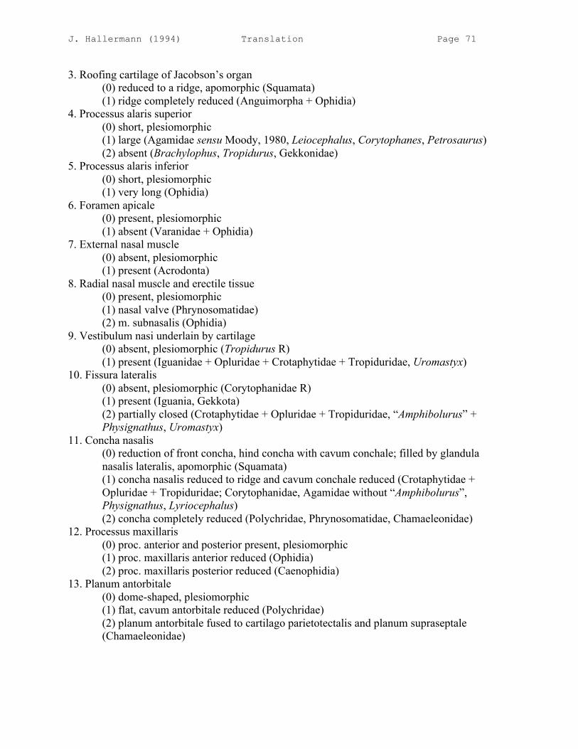
J. Hallermann (1994) Translation Page 71
3. Roofing cartilage of Jacobson’s organ (0) reduced to a ridge, apomorphic (Squamata) (1) ridge completely reduced (Anguimorpha + Ophidia) 4. Processus alaris superior (0) short, plesiomorphic (1) large (Agamidae sensu Moody, 1980, Leiocephalus, Corytophanes, Petrosaurus) (2) absent (Brachylophus, Tropidurus, Gekkonidae) 5. Processus alaris inferior (0) short, plesiomorphic (1) very long (Ophidia) 6. Foramen apicale (0) present, plesiomorphic (1) absent (Varanidae + Ophidia) 7. External nasal muscle (0) absent, plesiomorphic (1) present (Acrodonta) 8. Radial nasal muscle and erectile tissue (0) present, plesiomorphic (1) nasal valve (Phrynosomatidae) (2) m. subnasalis (Ophidia) 9. Vestibulum nasi underlain by cartilage (0) absent, plesiomorphic (Tropidurus R) (1) present (Iguanidae + Opluridae + Crotaphytidae + Tropiduridae, Uromastyx) 10. Fissura lateralis (0) absent, plesiomorphic (Corytophanidae R) (1) present (Iguania, Gekkota) (2) partially closed (Crotaphytidae + Opluridae + Tropiduridae, “Amphibolurus” + Physignathus, Uromastyx) 11. Concha nasalis (0) reduction of front concha, hind concha with cavum conchale; filled by glandula nasalis lateralis, apomorphic (Squamata) (1) concha nasalis reduced to ridge and cavum conchale reduced (Crotaphytidae + Opluridae + Tropiduridae; Corytophanidae, Agamidae without “Amphibolurus”, Physignathus, Lyriocephalus) (2) concha completely reduced (Polychridae, Phrynosomatidae, Chamaeleonidae) 12. Processus maxillaris (0) proc. anterior and posterior present, plesiomorphic (1) proc. maxillaris anterior reduced (Ophidia) (2) proc. maxillaris posterior reduced (Caenophidia) 13. Planum antorbitale (0) dome-shaped, plesiomorphic (1) flat, cavum antorbitale reduced (Polychridae) (2) planum antorbitale fused to cartilago parietotectalis and planum supraseptale (Chamaeleonidae)

J. Hallermann (1994) Translation Page 72
14. Processus rostralis plani antorbitalis (0) absent, plesiomorphic (reduced in Acrodonta?) (1) present (Iguania, “Amphibolurus”, Phrynocephalus) (2) connected to lamina transversalis anterior (Phrynosoma + Sceloporus) 15. Commissura sphenethmoidalis (0) connected to septum interorbitale, plesiomorphic (1) reduced (Anolis + Chamaeleolis, Chamaeleonidae, Gekkonidae, Amphisbaenia, Ophidia) 16. Connection between lamina transversalis anterior and cartilago paraseptalis caudal to Jacobson’s organ = commissura vomeronasalis posterior (0) absent, plesiomorphic (1) developed (Iguania, “Amphibolurus”) (2) reduction of commissura vomeronasalis posterior (Agamidae + Chamaeleonidae, Polychridae + Corytophanidae) 17. Concha of Jacobson’s organ (0) present, apomorphic (Squamata) (1) reduced (Anolis + Chamaeleolis, Chamaeleonidae, Calotes) 18. Cartilago paraseptalis (0) present, separate from septum nasi, plesiomorphic (1) reduced (Polychridae, Chamaeleonidae, Agama, Calotes, Draco, Phrynocephalus, Tropidurus, Ophidia) 19. Cartilago ectochoanalis (0) present, plesiomorphic (1) greatly elongated and broadened, closes off fissura choanalis (Anolis + Chamaeleolis, Gekkota, Xantusia, Ophidia) (2) cartilago hypochoanalis neomorph (Henophidia) 20. Vestibulum nasi (0) short, rostral entrance into cavum nasi, plesiomorphic (Polychridae + Corytophanidae R, Physignathus R) (1) elongated (Iguania, Polychrus R?, Tropidurus R, Varanidae) (2) S-shaped (Iguanidae + Opluridae + Crotaphytidae + Tropiduridae, Uromastycidae, Chamaeleo) (3) greatly elongated, joins cavum antorbitale dorsally (Phrynosomatidae, Phrynocephalus) 21. Recessus rostralis cavi nasi (0) absent, plesiomorphic (1) developed (Iguania) 22. Choanal tube (0) descending choanal tube directly enters oral cavity ventrally, plesiomorphic (1) elongated caudally = ductus nasopharyngeus (Gekkota, Scincidae) (2) ductus nasopharyngeus completely closed off ventrally (Ophidia, Anniellidae, Amphisbaenia) 23. Choanal groove (0) present, apomorphic (Squamata) (1) secondarily closed (Ophidia + Varanidae + Heloderma, Amphisbaenidae?, Teiidae?)

J. Hallermann (1994) Translation Page 73
24. Secondary palate (0) short, of connective tissue, apomorphic (Squamata) (1) incompletely osseous (Scincidae) 25. Ductus nasolacrimalis (0) joins ductus vomeronasalis, apomorphic (Squamata) (1) reduction of distal section of ductus nasolacrimalis (Chamaeleonidae) 26. Ductus nasolacrimalis (0) runs, unpaired on each side, within the nasal capsule, plesiomorphic (1) paired (Varanoidea sensu Estes et al., 1988) 27. Foramen lacrimale (0) small, ductus nasolacrimalis of normal girth, plesiomorphic (1) greatly elongated, ductus nasolacrimalis with large lumen (Agamidae sensu Moody, 1980 + Chamaeleonidae, Hoplocercidae?) 28. Glandula nasalis (0) reduction of glandula nasalis medialis, apomorphic (Squamata); duct of glandula nasalis lateralis extends rostrally, plesiomorphic (1) duct of glandula nasalis lateralis extends caudally (Phrynosomatidae, Phrynocephalus) 29. Jacobson’s organ (0) spheroidal, duct enters oral cavity, apomorphic (Squamata) (1) completely reduced or rudimentary (Chamaeleonidae) (2) Jacobson’s organ lies far back (Ophidia) 30. Septomaxilla (0) lies within the nasal capsule, covers Jacobson’s organ; flat, apomorphic (Squamata, Leiocephalus + Tropidurus R) (1) caudally, it rises dorsally; touches nasal capsule (Iguanidae + Crotaphytidae + Opluridae + Tropiduridae) (2) greatly elongated (Phrynosomatidae) (3) completely reduced (Chamaeleonidae, Corytophanes) 31. Pila antotica (0) connected to pila metoptica, plesiomorphic (1) pila antotica reduced (Tropiduridae + Opluridae, Polychridae + Corytophanidae + Phrynosomatidae + Acrodonta?) 32. Foramen epiotica (0) not developed, plesiomorphic (1) present (Agamidae sensu Moody, 1980) 33. Epibranchiale II (0) present and free, plesiomorphic (Uromastyx R) (1) connected to otic capsule (Tropiduridae + Opluridae, Brachylophus, Petrosaurus R) (2) completely reduced (Agamidae + Chamaeleonidae + ?Polychridae + Corytophanidae + Phrynosomatidae, Varanus + Lanthanotus + Ophidia) 34. Saccus endolymphaticus (0) within cavum cranii, plesiomorphic (1) saccus endolymphaticus (Corytophanidae + Polychridae, Chamaeleo) (2) saccus endolymphaticus calcified, extending into neck musculature (Polychridae)
J. Hallermann (1994) Translation Page 74
Tab. 1: Character distribution in the examined genera (to Character 18) [Matrix] * those characters that within Iguania only appear in the primitive condition were not taken into consideration ** the data of these species were taken in part out of the literature Tab. 2: Character distribution in the examined genera (continuation: Characters 19–34) [Matrix] [Footnotes with asterisks as above] [Translations of German character names of other authors in cladogram (p. 90):] [base of Iguanidae:] herbivory, polycuspid teeth [“Herbivorie, polycuspidate Zähne”] [base of Tropiduridae + Opluridae:] femoral pores lacking, fossa meckeli closed [“Femoralporen fehlen, Fossa Meckeli geschlossen”] [base of Tropiduridae:] gular fold incomplete, angular reduced [“Kehlfalten unvollständig, Angulare red.”] [base of Uromastycidae:] vomer concave [“Vomer konkav”] [base of Acrodonta:] acrodonty [“Acrodontie”] [base of Chamaeleonidae:] slingshot tongue, opposable fingers and toes [“Schleuderzunge, oponierbare Finger u. Zehen”]
Phylogenetic hypothesis The following tree represents a hypothesis of the phylogenetic relationships of the taxa of Iguania examined here. Where sufficient data from other genera were present from the literature, these species were incorporated in the diagram (Ctenosaura, Iguana, Uta, Sceloporus, Bradypodion, Chamaeleo). In the preparation of this phylogenetic diagram, the considerations on the evolutionary development of characters, as elaborated in the discussion, played a role. The aut- and synapomorphic characters were denoted with the same numbers as in the discussion and the table. The character changes within the families were likewise depicted. Probable character reversals were denoted with “R”. Autapomorphies without homoplasies (convergences, parallelisms, character reversals) were underscored. Several apomorphies of other authors, which establish the monophyly of subgroups of Iguania, were added. A question mark stands in those places that are founded solely on apomorphic characters that also appear convergently in other groups. The systematic position of Tropidurus and of Tropidurini (sensu Frost, 1992) within Tropiduridae requires further clarification, for the monophyly of Tropiduridae, like that of Tropidurinae (Stenocercini + Tropidurini sensu Frost, 1992), is not established by any autapomorphic characters common only to this group. A probable possibility similar to that depicted here would be to place Phrynosomatidae as the sister-group to Iguanidae + Crotaphytidae + Opluridae + Tropiduridae. But then the independent reduction of the concha nasalis (11.2), the pila antotica (31.1) and the epibranchiale II (33.2) would have to be assumed for a third time. The assumption that the ridge-shaped concha nasalis in corytophanids reappeared is then dispensed with. The hypothesis envisaged here is therefore more plausible.
J. Hallermann (1994) Translation Page 75
SUMMARY [German] [See following]
SUMMARY
[The English is left to Dr. Hallermann.]
LITERATURE CITED [See original]
APPENDIX
Explanation of abbreviations [See original]
[Figure captions]
Fig. 1: Crotaphytus collaris TL 10.0 mm. Dorsal view of the cartilaginous nasal capsule. On the right side the septomaxilla, nasal sac, and ductus nasolacrimalis were illustrated. Scale bar = 1 mm. Fig. 2: Crotaphytus collaris TL 10.0 mm. Ventral view of the cartilaginous nasal capsule. On the left side the septomaxilla, on the right additionally the nasal sac and ductus nasolacrimalis, were illustrated. Scale bar = 1 mm. Fig. 3: Crotaphytus collaris TL 10.0 mm. Above: Lateral view of the cartilaginous nasal capsule with septomaxilla and outline of dermal bones. Scale bar = 1 mm. Below: View of a parasagittal cut of the cartilaginous nasal capsule with septomaxilla. Note the septum of the septomaxilla (A), which lies on the connection (B) between lamina transversalis anterior and cartilago paraseptalis (= comm. vomeronasalis posterior). Cut surfaces are hatched. Fig. 4: Phymaturus palluma TL 9.6 mm. Dorsal view of the opened right half of the cartilaginous nasal capsule with septomaxilla. Scale bar = 1 mm. Fig. 5: Liolaemus gravenhorstii TL 9.4 mm. Dorsal view of the opened right half of the cartilaginous nasal capsule with septomaxilla. Scale bar = 1 mm. Fig. 6: Brachylophus fasciatus TL 18.6 mm. Dorsal view of the opened right half of the cartilaginous nasal capsule with septomaxilla. Scale bar = 1 mm. Fig. 7: Uromastyx acanthinurus TL 17.0 mm. Dorsal view of the opened right half of the cartilaginous nasal capsule with septomaxilla. Scale bar = 1 mm. Fig. 8: Stenocercus varius TL 8.7 mm. Dorsal view of the opened right half of the cartilaginous nasal capsule with septomaxilla. Scale bar = 1 mm.
J. Hallermann (1994) Translation Page 76
Fig. 9: Oplurus quadrimaculatus TL 15.9 mm. Dorsal view of the opened right half of the cartilaginous nasal capsule with septomaxilla. The septomaxilla was illustrated as transparent. Scale bar = 1 mm. Fig. 10: Petrosaurus thalassinus TL 12.9 mm. Dorsal view of the opened right half of the cartilaginous nasal capsule with septomaxilla. Scale bar = 1 mm. Fig. 11: Basiliscus basiliscus TL 11.9 mm. Dorsal view of the opened right half of the cartilaginous nasal capsule with septomaxilla. Scale bar = 1 mm. Fig. 12: Petrosaurus thalassinus TL 12.9 mm. Ventral view of the left half of the cartilaginous nasal capsule with septomaxilla. Scale bar = 1 mm. Fig. 13: Basiliscus basiliscus TL 11.9 mm. Ventral view of the left half of the cartilaginous nasal capsule with septomaxilla. Scale bar = 1 mm. Fig. 14: Tropidurus melanopleurus TL 10.9 mm. Ventral view of the cartilaginous nasal capsule with septomaxilla. On the right, the dermal bones are additionally illustrated. Scale bar = 1 mm. Fig. 15: Corytophanes cristatus TL 11.0 mm. Ventral view of the cartilaginous nasal capsule with septomaxilla. On the right, the dermal bones are additionally illustrated. Scale bar = 1 mm. Fig. 16: Physignathus cocincinus TL 11.8 mm. Ventral view of the cartilaginous nasal capsule with septomaxilla. On the right, the dermal bones are additionally illustrated. Scale bar = 1 mm. Fig. 17: Brachylophus fasciatus TL 18.6 mm. Ventral view of the cartilaginous nasal capsule with septomaxilla. On the right, the dermal bones are additionally illustrated. Scale bar = 1 mm. Fig. 18: Uromastyx acanthinurus TL 17.0 mm. Ventral view of the cartilaginous nasal capsule with septomaxilla. On the right, the dermal bones are additionally illustrated. Scale bar = 1 mm. Fig. 19: Oplurus quadrimaculatus TL 15.9 mm. Ventral view of the cartilaginous nasal capsule with septomaxilla. On the right, the dermal bones are additionally illustrated. * thin rostral process of the lamina transversalis anterior. Scale bar = 1 mm. Fig. 20: Physignathus cocincinus TL 11.8 mm. Dorsal view of the right half of the cartilaginous nasal capsule with septomaxilla. Scale bar = 1 mm. Fig. 21: Tropidurus melanopleurus TL 13.9 mm. Dorsal view of the opened right half of the cartilaginous nasal capsule. The septomaxilla was illustrated as transparent. Scale bar = 1 mm. Fig. 22: Phymaturus palluma TL 9.6 mm. Ventral view of the left half of the cartilaginous nasal capsule with septomaxilla. Scale bar = 1 mm. Fig. 23: Chamaeleolis barbatus TL 15.5 mm. Ventral view of the cartilaginous nasal capsule with septomaxilla. On the right, the dermal bones are additionally illustrated. Scale bar = 1 mm.
J. Hallermann (1994) Translation Page 77
Fig. 24: Anolis m. marmoratus TL 5.2 mm, stage 3. Lateral view of the cartilaginous nasal capsule. Dermal bones were illustrated as transparent. Scale bar = 1 mm. Fig. 25: Anolis m. marmoratus TL 5.2 mm, stage 3. Ventral view of the cartilaginous nasal capsule. Only ventrally lying dermal bones of the left side are figured. Scale bar = 1 mm. Fig. 26: Physignathus cocincinus TL 11.8 mm. Lateral view of the snout region. The dermal bones, the membranous [“häutige”] naris, and the external nasal muscle are illustrated. Scale bar = 1 mm. Fig. 27: Physignathus cocincinus TL 11.8 mm. Lateral view of the cartilaginous nasal capsule with septomaxilla and outline of the skull. Fig. 28: Oplurus quadrimaculatus TL 15.9 mm. Lateral view of the cartilaginous nasal capsule with septomaxilla and outline of the skull. Scale bar = 1 mm. Fig. 29: Brachylophus fasciatus TL 18.6 mm. Lateral view of the cartilaginous nasal capsule with septomaxilla and outline of the skull. Scale bar = 1 mm. Fig. 30: Corytophanes cristatus TL 11.0 mm. Lateral view of the cartilaginous nasal capsule with septomaxilla and outline of the skull. Scale bar = 1 mm. Fig. 31: Corytophanes cristatus TL 11.0 mm. Dorsal view of the cartilaginous nasal capsule with dermal bones on the left side. Scale bar = 1 mm. Position of the subsequent cross-sectional figures of Crotaphytus collaris (TL 10.0 mm). Fig. 32: Crotaphytus collaris TL 10.0 mm, section 2-4-6. Transverse section in the region of Jacobson’s organ. The vestibulum nasi is still filled up by epithelial filler tissue [“Füllgewebe”]. Note the concha of Jacobson’s organ, the duct, and the lateral ridge of the septum nasi on which the septomaxilla abuts. Scale bar = 0.5 mm. Fig. 33: Crotaphytus collaris TL 10.0 mm, section 3-1-2. Transverse section in the region of the hind end of Jacobson’s organ. The lamina transversalis anterior is hollowed out and is filled with a blind section of the ductus nasolacrimalis. Medial entrance of the ductus nasolacrimalis into the choanal groove. Scale bar = 0.5 mm. Fig. 34: Crotaphytus collaris TL 10.0 mm, section 3-2-7. Transverse section in the region of the rostral recess of the cavum nasi. The posterior process of the septomaxilla separates it from the vestibulum nasi. The ductus nasolacrimalis extends medially dorsal to the choanal groove. Scale bar = 0.5 mm. Fig. 35: Crotaphytus collaris TL 10.0 mm, section 3-6-4. Transverse section in the region of the entrance of the vestibulum into the cavum nasi. The vestibulum nasi is still filled up by epithelial filler tissue. The choanal groove is very narrow but possesses a large lateral recess (lateral choanal fissure). Scale bar = 0.5 mm.

J. Hallermann (1994) Translation Page 78
Fig. 36: Crotaphytus collaris TL 10.0 mm, section 4-4-5. Transverse section in the region of the cavum nasi. The concha nasalis is reduced to a ridge. Note the lateral groove at the outer edge of the cartilago paranasalis, the large fenestra septi nasi, and the position of the internal choana. Scale bar = 0.5 mm. Fig. 37: Crotaphytus collaris TL 10.0 mm, section 5-2-1. Transverse section in the region of the cavum antorbitale and the foramen olfactorium. The ductus nasopharyngeus is functionally closed off. Scale bar = 0.5 mm. Fig. 38: Phymaturus palluma TL 9.6 mm, section 5-3-6. Transverse section in the region of Jacobson’s organ. The vestibulum nasi is still filled up by epithelial filler tissue. Note the broad crest at the septum nasi that is dorsally connected with the cupula nasi, resulting in a small cavity. Scale bar = 0.5 mm. Fig. 39: Phymaturus palluma TL 9.6 mm, section 6-3-3. Transverse section in the front part of the nasal capsule at the level of the recessus rostralis cavi nasi. The ductus nasolacrimalis intersects the choanal groove. The vestibulum nasi has a “double floor”. Scale bar = 0.5 mm. Fig. 40: Corytophanes cristatus TL 11.0 mm, section 3-1-10. Transverse section in the region of Jacobson’s organ. Note the complete cartilaginous covering of the organ. The vestibulum nasi is still filled up by epithelial filler tissue. Scale bar = 0.5 mm. Fig. 41: Corytophanes cristatus TL 11.0 mm, section 3-5-5. Transverse section in the region of the vestibulum nasi. Note the rostral recess of the cavum nasi and the connection of the ductus nasolacrimalis to the choanal groove. Scale bar = 0.5 mm. Fig. 42: Corytophanes cristatus TL 11.0 mm, section 4-2-4. Transverse section in the region of the entrance of the vestibulum into the cavum nasi. A lateral out-pocketing of the choanal tube as well as of the choanal groove is developed. Scale bar = 0.5 mm. Fig. 43: Corytophanes cristatus TL 11.0 mm, section 4-4-4. Transverse section in the region of the internal and external choana. Parts of the glandula nasalis lateralis lie within the nasal capsule. The concha nasalis is reduced to a crest. Scale bar = 0.5 mm. Fig. 44: Corytophanes cristatus TL 11.0 mm, section 5-6-5. Transverse section in the region of the cavum antorbitale and the foramen olfactorium. Scale bar = 0.5 mm. Fig. 45: Anolis m. marmoratus TL 5.2 mm, stage 3, section 1-4-6. Transverse section through the regio ethmoidalis at the level of Jacobson’s organ. Scale bar = 0.5 mm. Fig. 46: Anolis m. marmoratus TL 5.2 mm, stage 3, section 1-6-10. Transverse section through the regio ethmoidalis in the region of the cavum nasi proprium. Scale bar = 0.5 mm. Fig. 47: Anolis m. marmoratus TL 5.2 mm, stage 3, section 1-8-7. Transverse section through the regio ethmoidalis at the level of the choana. Scale bar = 0.5 mm.
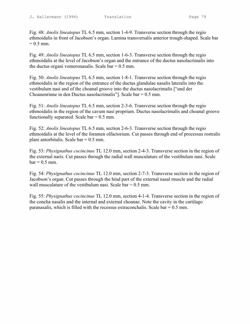
J. Hallermann (1994) Translation Page 79
Fig. 48: Anolis lineatopus TL 6.5 mm, section 1-4-9. Transverse section through the regio ethmoidalis in front of Jacobson’s organ. Lamina transversalis anterior trough-shaped. Scale bar = 0.5 mm. Fig. 49: Anolis lineatopus TL 6.5 mm, section 1-6-3. Transverse section through the regio ethmoidalis at the level of Jacobson’s organ and the entrance of the ductus nasolacrimalis into the ductus organi vomeronasalis. Scale bar = 0.5 mm. Fig. 50: Anolis lineatopus TL 6.5 mm, section 1-8-1. Transverse section through the regio ethmoidalis in the region of the entrance of the ductus glandulae nasalis lateralis into the vestibulum nasi and of the choanal groove into the ductus nasolacrimalis [“und der Choanenrinne in den Ductus nasolacrimalis”]. Scale bar = 0.5 mm. Fig. 51: Anolis lineatopus TL 6.5 mm, section 2-3-6. Transverse section through the regio ethmoidalis in the region of the cavum nasi proprium. Ductus nasolacrimalis and choanal groove functionally separated. Scale bar = 0.5 mm. Fig. 52: Anolis lineatopus TL 6.5 mm, section 2-6-3. Transverse section through the regio ethmoidalis at the level of the foramen olfactorium. Cut passes through end of processus rostralis plani antorbitalis. Scale bar = 0.5 mm. Fig. 53: Physignathus cocincinus TL 12.0 mm, section 2-4-3. Transverse section in the region of the external naris. Cut passes through the radial wall musculature of the vestibulum nasi. Scale bar = 0.5 mm. Fig. 54: Physignathus cocincinus TL 12.0 mm, section 2-7-3. Transverse section in the region of Jacobson’s organ. Cut passes through the hind part of the external nasal muscle and the radial wall musculature of the vestibulum nasi. Scale bar = 0.5 mm. Fig. 55: Physignathus cocincinus TL 12.0 mm, section 4-1-4. Transverse section in the region of the concha nasalis and the internal and external choanae. Note the cavity in the cartilago paranasalis, which is filled with the recessus extraconchalis. Scale bar = 0.5 mm.