introductionpe.sfrnet.org/.../pdf/2011/1/ad57a04c-ee3c-43d8-be3f-8994da00d6fc.pdf · –spring...
TRANSCRIPT


INTRODUCTION
• Pour un diagnostic correct et un traitement adapté, il est indispensable de connaître parfaitement l'anatomie des ligaments de la cheville et du tarse. Les lésions ligamentaires sont la cause la plus fréquente des douleurs aiguës de la cheville. La douleur chronique de la cheville est souvent en rapport avec une lésion ligamentaire à l’origine d’une laxité [1].

INTRODUCTION
• Dans ce travail, nous présentons les résultats de notre expérience sur une séquence 3D optimisée en IRM de la cheville.
• Nous allons revoir l’anatomie des différents ligaments et essayer de les individualiser, ligament par ligament, voire faisceau par faisceau, sur notre séquence 3D.
• Ensuite, quelques cas pathologiques seront illustrés et discutés.

MATERIELS ET METHODES
• Après une étude exploratrice initiale en 2009, une séquence 3D en densité de proton sans Fat Sat1 a été réalisée systématiquement lors des IRM de cheville (IRM 1,5 T, antenne 8 canaux avec des acquisitions axiales initialement et depuis quelque mois, antenne 12 canaux avec des acquisitions sagittales).
1. TR : 1000 ms, TE : 40 ms, voxel : 0,6x0,6x0,7 mm, Temps d'acquisition : 5min 20s,
Matrice: 320x320, Epaisseur de coupe: 0,7 mm

MATERIELS ET METHODES
• Au total, 127 chevilles ont été explorées sur une période de 2 ans. Dans un tiers des cas, un arthroscanner complémentaire a été réalisé.
• Les images obtenues ont été confrontées aux schémas anatomiques et dans certains cas aux images de l'arthroscanner (si réalisé).
RESULTATS
• Nous allons étudier les structures ligamentaires suivantes: – Syndesmose tibio-fibulaire – Ligament collatéral latéral – Ligament collatéral médial – Spring ligament – Ligament long et court plantaire – Ligament interosseux talo-calcanéen – Ligament bifurqué – Ligament calcanéo-cuboïdien latéral – Ligament talo-naviculaire dorsal – Ligament de Lisfranc

Syndesmose tibio-fibulaire
• La syndesmose tibiofibulaire distale joue un rôle important dans la stabilité de la mortaise de la cheville et également dans la transmission des contraintes talocrurales lors de la marche.
• Elle est composée de 4 ligaments [1]: – Ligament tibio-fibulaire antéro-inférieur (et son faisceau
accessoire distal et inconstant ou ligament de Bassett)
– Ligament tibio-fibulaire postéro-inférieur
– Ligament transverse inférieur (ou le faisceau profond du ligament tibio-fibulaire postéro-inférieur)
– Ligament interosseux tibio-fibulaire

Syndesmose tibio-fibulaire Ligament tibio-fibulaire antéro-inférieur et ligament
de Bassett
• Il s’insère sur la tubérosité antérieure du tibia (5 mm en moyenne au-dessus de la surface articulaire) et ses fibres s'étendent dans une direction latéro-distale jusqu’à son insertion distale sur le bord antérieur de la malléole latérale [2].

Fig. 18 Anatomic view of the anterior ligaments of the ankle. 1 Anterior tibiofibular ligament; 2 distal fascicle of the anterior tibiofibular ligament; 3 tibia (anterior tubercle indicated with arrows); 4 anterior talofibular ligament; 5 beveled triangular region of the talus; 6 deep layer of the medial collateral ligament; 7 superficial layer of the medial collateral ligament; 8 notch of Harty
Pau Golanó et al. Anatomy of the ankle ligaments: a pictorial essay. Knee Surg Sports Traumatol Arthrosc. 2010 May; 18(5): 557–569.

Ligament tibiofibulaire antéroinférieur (flèches blues) et son faisceau distal ou ligament de Bassett
(flèches rouges)

Ligament tibiofibulaire antéroinférieur (flèches blues) et son faisceau distal ou ligament de Bassett
(flèches rouges)

Syndesmose tibio-fibulaire Ligament tibio-fibulaire postéro-inférieur et ligament
transverse inférieur • Ce ligament est formé essentiellement par deux éléments
indépendants, la composante superficielle et profonde [2]. • La composante superficielle provient du bord postérieur de
la malléole latérale et se dirige vers le haut et le dedans pour s'insérer sur le tubercule tibial postérieur. Le terme du ligament tibiofibulaire postéroinférieur est habituellement utilisé pour se référer à cette composante superficielle.
• La composante profonde est en forme de cône et est tendue de la région proximale de la fosse malléolaire au bord postérieur du tibia. Cette composante est également connue comme le ligament transverse inférieur, formant un vrai bourrelet [3] pour fournir la stabilité de l’articulation talo-crurale et pour empêcher la translation talienne postérieure [4].
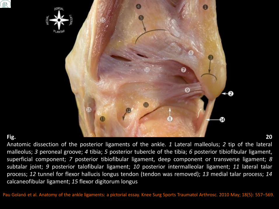
Fig. 20 Anatomic dissection of the posterior ligaments of the ankle. 1 Lateral malleolus; 2 tip of the lateral malleolus; 3 peroneal groove; 4 tibia; 5 posterior tubercle of the tibia; 6 posterior tibiofibular ligament, superficial component; 7 posterior tibiofibular ligament, deep component or transverse ligament; 8 subtalar joint; 9 posterior talofibular ligament; 10 posterior intermalleolar ligament; 11 lateral talar process; 12 tunnel for flexor hallucis longus tendon (tendon was removed); 13 medial talar process; 14 calcaneofibular ligament; 15 flexor digitorum longus
Pau Golanó et al. Anatomy of the ankle ligaments: a pictorial essay. Knee Surg Sports Traumatol Arthrosc. 2010 May; 18(5): 557–569.
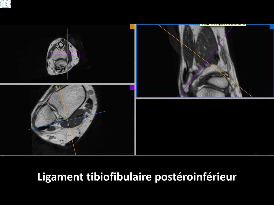
Ligament tibiofibulaire postéroinférieur

Ligament tibiofibulaire postéroinférieur

Ligament tibiofibulaire postéroinférieur (faisceau profond ou ligament transverse inférieur) (flèches
rouges)

Ligament tibiofibulaire postéroinférieur (faisceau profond ou ligament transverse inférieur) (flèches
rouges)
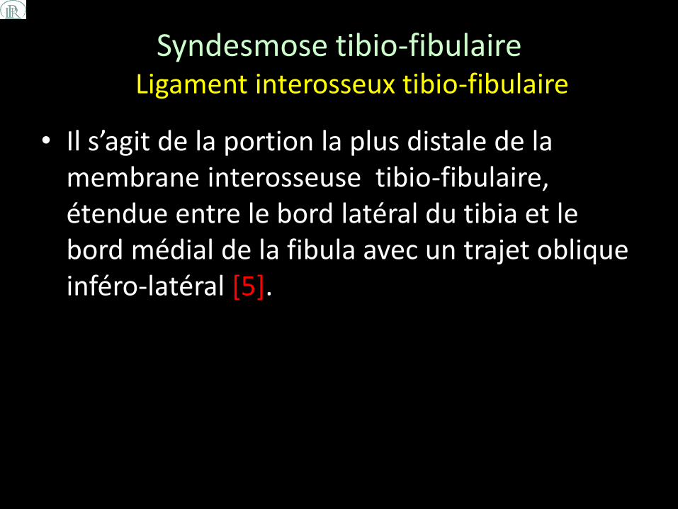
Syndesmose tibio-fibulaire Ligament interosseux tibio-fibulaire
• Il s’agit de la portion la plus distale de la membrane interosseuse tibio-fibulaire, étendue entre le bord latéral du tibia et le bord médial de la fibula avec un trajet oblique inféro-latéral [5].

Anterior (left) and posterior (right) 3D renderings of the syndesmotic ligaments at the ankle demonstrate the anterior inferior tiobiofibular ligament (AITFL), the posterior inferior tibiofibular ligament (PITFL), transverse tibiofibular ligament (TrTFL) and the interosseous membrane (IM)
Leland Y. Tsao, M.D. MRI Web Clinic - July 2010, High Ankle Sprains

Ligament interosseux tibio-fibulaire

Ligament interosseux tibio-fibulaire

Ligament collatéral latéral
• Le complexe du ligament collatéral latéral (LCL) est composé de 3 faisceaux :
– Ligament talo-fibulaire antérieur (TFA)
– Ligament fibulo-calcanéen (FC)
– Ligament talo-fibulaire postérieur (TFP)

Ligament collatéral latéral Ligament talo-fibulaire antérieur
• Le ligament talo-fibulaire antérieur est le ligament le plus souvent touché lors des entorses de la cheville [6].
• Ce ligament joue un rôle important dans la limitation du déplacement antérieur du talus et de la flexion plantaire de la cheville [7].
• Il a le plus souvent un aspect de double bande, comme décrit par Sarrafian [3].
• Le ligament talo-fibulaire antérieur est tendu de la marge antérieure de la malléole externe au corps du talus.

Osteoarticular anatomic dissection of the lateral ligaments of the foot and ankle joint. The anterior talofibular ligament is typically composed of two separate bands. 1 Tip of the lateral malleolus; 2 tibia; 3 anterior tibiofibular ligament; 4 distal fascicle of the anterior tibiofibular ligament; 5 superior band of the anterior talofibular ligament; 6 inferior band of the anterior talofibular ligament; 7 lateral articular surface of the talus; 8 neck of the talus; 9 head of the talus; 10 calcaneofibular ligament; 11 talocalcaneal interosseous ligament; 12 cervical ligament; 13 talonavicular ligament; 14 navicular
Pau Golanó et al. Anatomy of the ankle ligaments: a pictorial essay. Knee Surg Sports Traumatol Arthrosc. 2010 May; 18(5): 557–569.

Ligament talo-fibulaire antérieur

Ligament talo-fibulaire antérieur

Ligament collatéral latéral Ligament calcanéo-fibulaire
• Le ligament calcanéo-fibulaire s’insère sur la partie antérieure de la malléole latérale.
• Dans la position neutre de la cheville, le ligament a un trajet oblique vers le bas et l'arrière et vient s’attacher à la région postérieure de la surface latérale du calcanéum.
• Ce ligament est superficiellement traversé par les tendons fibulaires.
• Sa rupture isolée est rare, elle est souvent associée à une rupture du ligament talo-fibulaire antérieur [2].

Anatomic dissection of the lateral region of the foot and ankle showing the morphology and relationship of the anterior talofibular with the calcaneofibular ligaments. 1 Fibula and tip of the fibula; 2 tibia (anterior tubercle with arrows); 3 anterior tibiofibular ligament; 4 distal fascicle of the tibiofibular ligament; 5 interosseous membrane; 6 foramen for the perforating branch of the peroneal artery; 7 talus; 8 anterior talofibular ligament; 9 calcaneofibular ligament; 10 talocalcaneal interosseous ligament; 11 inferior extensor retinaculum (cut); 12 talonavicular ligament; 13 bifurcate ligament; 14 peroneal tubercle (arrows showing the peroneal tendons sulcus); 15 peroneus longus tendon; 16 peroneus brevis tendon; 17 calcaneal tendon
Pau Golanó et al. Anatomy of the ankle ligaments: a pictorial essay. Knee Surg Sports Traumatol Arthrosc. 2010 May; 18(5): 557–569.

Ligament calcanéo-fibulaire

Ligament calcanéo-fibulaire

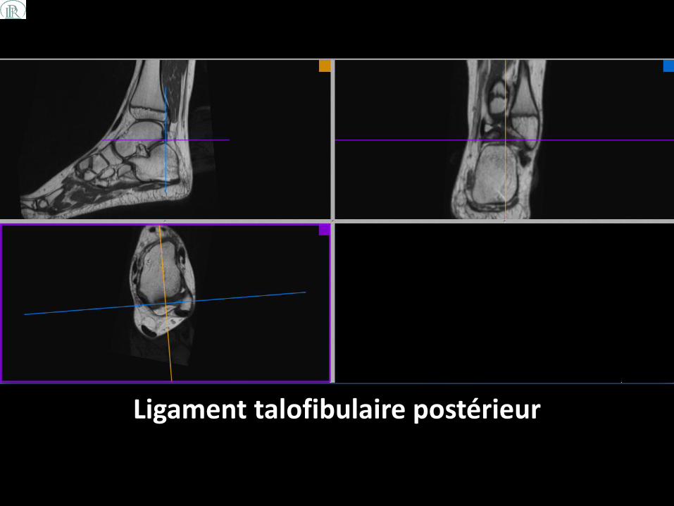
Ligament collatéral latéral Ligament talo-fibulaire postérieur
• Le ligament talo-fibulaire postérieur provient de la fosse malléolaire, située sur la face médiale de la malléole latérale, s’étend presque horizontalement jusqu’à la face postérieur du talus .
• Ce ligament a un aspect multifasciculaire.
• Certaines de ses fibres fusionnent avec les fibres du ligament intermalléolaire postérieur qui est situé entre le ligament transverse inférieur et le ligament talo-fibulaire [2].

Posterior view of the anatomic dissection of the ankle ligaments showing the posterior intermalleolar ligament with its relation to the surrounding anatomy. 1 Fibula; 2 tip of the fibula; 3 peroneal groove of the fibula; 4 tibia; 5 posterior tubercle of the tibia; 6 superficial component of the posterior tibiofibular ligament; 7 deep component of the posterior tibiofibular ligament or transverse ligament; 8 interosseous membrane; 9 posterior talofibular ligament; 10 lateral talar process; 11 tunnel for flexor hallucis longus tendon; 12 flexor hallucis longus retinaculum; 13 calcaneofibular ligament; 14 subtalar joint; 15 flexor digitorum longus tendon (cut); 16 tibialis posterior tendon (cut); 17 posterior intermalleolar ligament: A Tibial insertion (tibial slip in arthroscopic view). B Talar insertion (lateral talar process). C Tibial malleolar insertion through the septum between the flexor digitorum longus and posterior tibial tendons. D Talar insertion (medial talar process) through the joint capsule
Pau Golanó et al. Anatomy of the ankle ligaments: a pictorial essay. Knee Surg Sports Traumatol Arthrosc. 2010 May; 18(5): 557–569.
Ligament talofibulaire postérieur
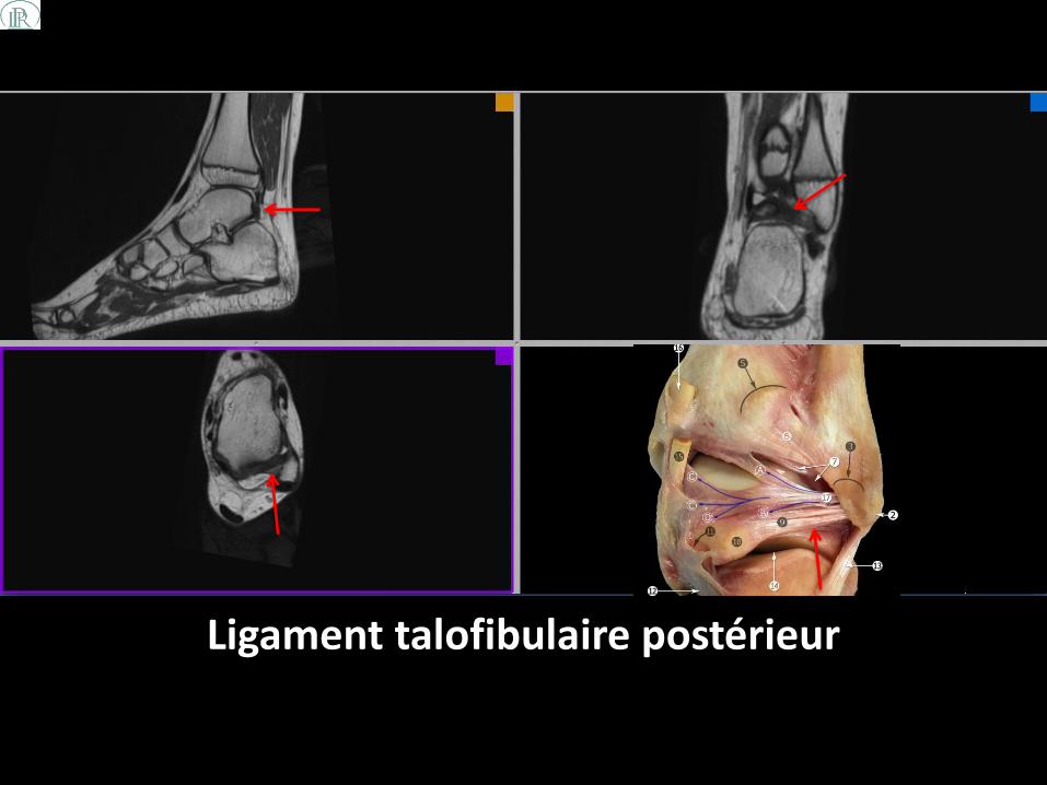
Ligament talofibulaire postérieur

Ligament collatéral médial ou ligament deltoïde
• Le ligament deltoïde est tendu de la malléole médiale à plusieurs os du tarse.
• Il est composé de 2 couches, une couche superficielle et une couche profonde.
• Contrairement à la couche superficielle, la couche profonde est intra-articulaire et est recouverte par la synoviale.

Ligament collatéral médial ou ligament deltoïde
• La composante superficielle: – ligament tibio-naviculaire
– ligament tibio-spring
– ligament tibio-calcanéen
– ligament tibio-talaire postérieur superficiel
• La composante profonde: – ligament tibio-talaire antérieur
– ligament tibio-talaire postérieur profond

Ligament collatéral médial ou ligament deltoïde
• Ces composantes sont variablement présentes, selon les auteurs différents [8-11].
• Ces ligaments sont nommés en fonction de leurs attaches distales au niveau des os du tarse.
• Le ligament tibio-spring est la seule composante sans aucune insertion osseuse distale, car il rejoint le spring ligament [9].

Medial view of the main components of the medial collateral ligament. 1 anterieur tibiotalar ligament; 2 tibionavicular ligament; 3 tibiospring ligament; 4 tibiocalcaneal ligament; 5 deep posterior tibiotalar ligament; 6 medial talocalcaneal ligament; 7 sustentaculum tali; 8 tibialis posterior tendon; 9 spring ligament complex (superomedial calcaneonavicular ligament)
Photo originale: LifeART (and/or) MediClip image copyright (appropriate year) Wolters Kluwer Health, Inc.- Lippincott Williams & Wilkins. All rights reserved
5
4
2
1
9 6
3
7 8

Ligament tibio-talar antérieur

Ligament tibio-talar antérieur

Ligament tibio-naviculaire

Ligament tibio-naviculaire
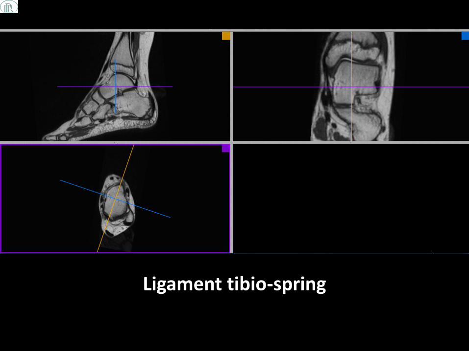
Ligament tibio-spring

Ligament tibio-spring

Ligament tibio-calcanéen

Ligament tibio-calcanéen

Ligament tibio-talaire postérieur
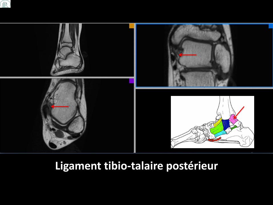
Ligament tibio-talaire postérieur

Spring ligament
• Le spring ligament est un complexe ligamentaire calcanéo-naviculaire plantaire médial de l’arrière pied.
• Il est composé de 2 faisceaux [12]: • Faisceau supéromédial ou ligament calcanéo-naviculaire
supéromédial
• Faisceau inférieur: – Médioplantaire ou ligament calcanéo-naviculaire médioplantaire
oblique
– Inféroplantaire ou ligament calcanéo-naviculaire inféroplantaire longitudinal

Medial and plantar 3D representations of the midfoot demonstrate the 3 major components of the spring ligament complex, the superomedial (SM-CNL), medioplantar oblique (MPO-CNL), and inferoplantar longitudinal (IPL-CNL) calcaneonavicular ligaments. The posterior tibial tendon (PTT) and tibiospring ligament (TS)are also demonstrated.
Pau Michael E. Stadnick, M.D. MRI Web Clinic - January 2008, Spring Ligament Tear

Spring ligament (ligament calcanéo-naviculaire supéromédial)

Spring ligament (ligament calcanéo-naviculaire supéromédial)
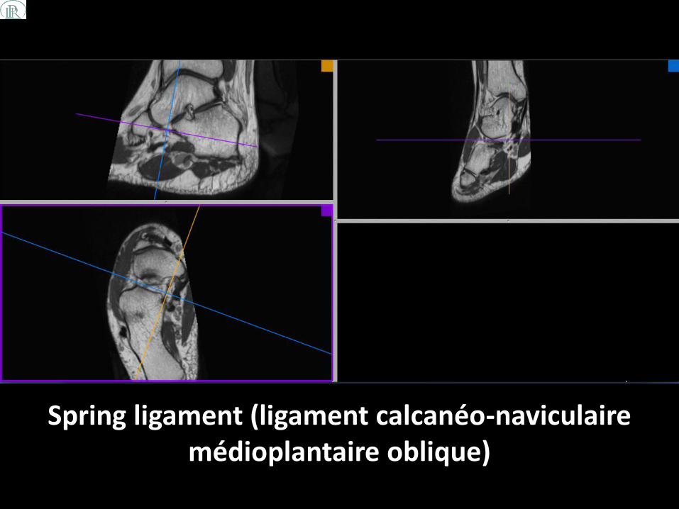
Spring ligament (ligament calcanéo-naviculaire médioplantaire oblique)

Spring ligament (ligament calcanéo-naviculaire médioplantaire oblique)

Spring ligament (ligament calcanéo-naviculaire inféroplantaire longitudinal)

Spring ligament (ligament calcanéo-naviculaire inféroplantaire longitudinal)
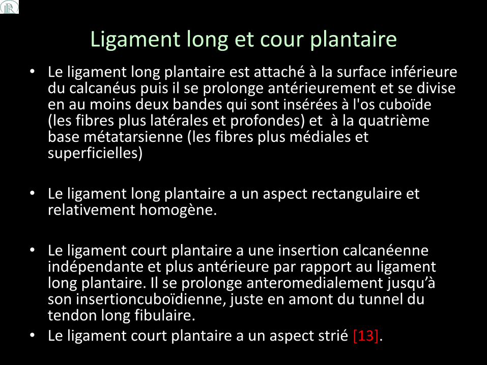
Ligament long et cour plantaire • Le ligament long plantaire est attaché à la surface inférieure
du calcanéus puis il se prolonge antérieurement et se divise en au moins deux bandes qui sont insérées à l'os cuboïde (les fibres plus latérales et profondes) et à la quatrième base métatarsienne (les fibres plus médiales et superficielles)
• Le ligament long plantaire a un aspect rectangulaire et relativement homogène.
• Le ligament court plantaire a une insertion calcanéenne indépendante et plus antérieure par rapport au ligament long plantaire. Il se prolonge anteromedialement jusqu’à son insertioncuboïdienne, juste en amont du tunnel du tendon long fibulaire.
• Le ligament court plantaire a un aspect strié [13].

Sagittal anatomic section shows that most lateral and deep fibers of long plantar ligament (solid arrows) insert on cuboid crest and that medial and superficial fibers extend to metatarsal bases. Open
arrows point to short plantar ligament. Asterisk = peroneus longus tendon.
Melão L et al. Correlation of MRI with gross anatomic findings of long plantar ligament. AJR 2009;193:662-671

Ligament long plantaire
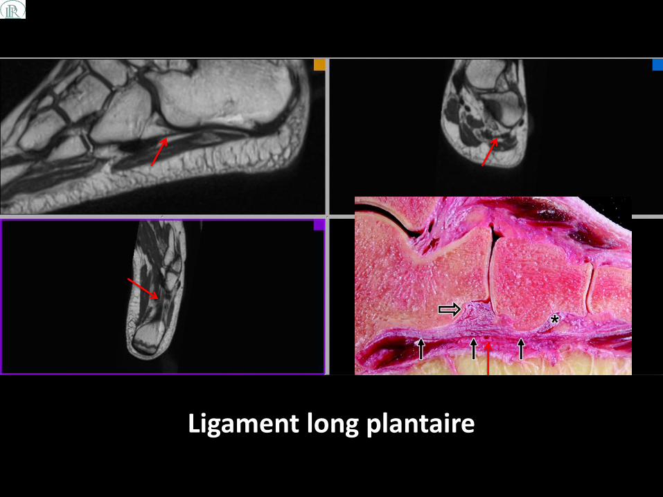
Ligament long plantaire

Ligament court plantaire

Ligament court plantaire

Ligaments du sinus du tarse
• Le sinus du tarse contient 2 structures ligamentaires [14]:
– dans sa portion centrale : ligament interosseux talo-calcanéen
– Dans sa portion antérolatérale : ligament cervical (ou ligament talo-calcanéen antérolatéral)
• Le syndrome du sinus du tarse est causé par une hémorragie ou une inflammation des cavités synoviales du sinus du tarse, avec ou sans rupture ligamentaire.
• Cette pathologie est souvent associée à des lésions du
ligament collatéral latéral [15].
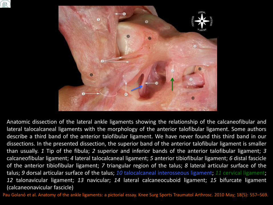
Anatomic dissection of the lateral ankle ligaments showing the relationship of the calcaneofibular and lateral talocalcaneal ligaments with the morphology of the anterior talofibular ligament. Some authors describe a third band of the anterior talofibular ligament. We have never found this third band in our dissections. In the presented dissection, the superior band of the anterior talofibular ligament is smaller than usually. 1 Tip of the fibula; 2 superior and inferior bands of the anterior talofibular ligament; 3 calcaneofibular ligament; 4 lateral talocalcaneal ligament; 5 anterior tibiofibular ligament; 6 distal fascicle of the anterior tibiofibular ligament; 7 triangular region of the talus; 8 lateral articular surface of the talus; 9 dorsal articular surface of the talus; 10 talocalcaneal interosseous ligament; 11 cervical ligament; 12 talonavicular ligament; 13 navicular; 14 lateral calcaneocuboid ligament; 15 bifurcate ligament (calcaneonavicular fascicle)
Pau Golanó et al. Anatomy of the ankle ligaments: a pictorial essay. Knee Surg Sports Traumatol Arthrosc. 2010 May; 18(5): 557–569.

Ligament interosseux talo-calcanéen (sinus du tarse)

Ligament interosseux talo-calcanéen (sinus du tarse)

Ligament cervical (sinus du tarse)
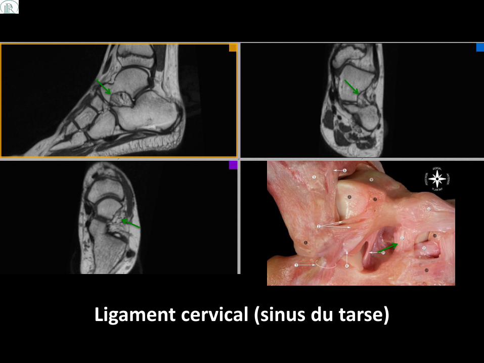
Ligament cervical (sinus du tarse)

Ligaments dorsolatéraux de l’articulation de Chopart
• L’élément clef dorsolatéral est le ligament bifurqué.
• Il s’insère sur le rostre du calcanéus et, de là, se divise en une lame verticale qui rejoint la face latérale de l’os naviculaire et une lame horizontale qui rejoint la face dorsale du cuboïde.
• Il est encadré par deux ligaments moins
puissants[16]: – ligament talonaviculaire dorsal: tendu de la face dorsale du
col du talus à la face dorsale du naviculaire
– ligament calcanéocuboïdien latéral: tendu de la face latérale du calcanéus à la face latérale du cuboïde

1) ligament talonaviculaire dorsal; 2) ligament bifurqué; 3) ligament calcanéocuboïdien latéral.
Congrès thématique de la SIMS: Le pied (juin 2011: Paris). Le Chopart et le carrefour des trois tendons. B. Ferré et al.; p.113-120.

Ligament bifurqué: faisceau calcanéocuboïdien (flèche rouge) et faisceau calcanéonaviculaire
(flèche blue)

Ligament bifurqué: faisceau calcanéocuboïdien (flèche rouge) et faisceau calcanéonaviculaire
(flèche blue)

Ligament talonaviculaire dorsal (flèche rouge) Ligament calcanéocuboïdien (flèche blue)

Ligament talonaviculaire dorsal (flèche rouge) Ligament calcanéocuboïdien latéral (flèche blue)

Ligaments des articulations de Lisfranc
• Les interlignes tarso-métatarsiens sont renforcés par les ligaments dorsaux, plantaires et interosseux.
• Le ligament cunéo-métatarsien médial ou ligament de Lisfranc est l’élément clef de l’interligne.
• Ce ligament s’étend de la face latérale du premier cunéiforme à la face médiale de la base du deuxième métatarsien.
• Il est croisé sur sa face plantaire par le tendon du long fibulaire [17].

Représentation schématique des ligaments interosseux: ligament interosseux de Lisfranc, ligament interosseux cunéo-métatarsien intermédiaire, ligament interosseux cunéo-métatarsien latéral et des ligaments inter-cunéiformes (IC) et inter-métatarsiens (IM).
Congrès thématique de la SIMS: Imagerie du pied et de la cheville (juin 2004: Paris). Les articulations de Lisfranc. F. Thévenin et al.; p. 129–145.
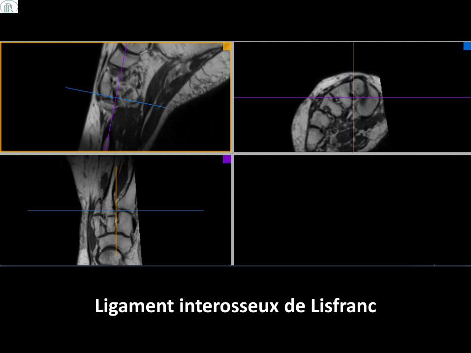
Ligament interosseux de Lisfranc

Ligament interosseux de Lisfranc

PATIENT DE 37 ANS PRESENTANT UNE INSTABILITE CHRONIQUE DE
SA CHEVILLE DROITE
CAS N°1

Séquence DP 3D en faveur d’une lésion de grade III du ligament talo-fibulaire
antérieur

et également une lésion de grade III du ligament fibulo-calcanéen

Arthroscanner retrouvant la lésion de grade III du ligament talo-fibulaire antérieur

Et du ligament fibulo-calcanéen à l’origine d’une opacification des gaines
fibulaires

BILAN D’UNE INSTABILITÉ CHRONIQUE DE LA CHEVILLE DROITE AVEC DES DOULEURS
MALLÉOLAIRES EXTERNES. NOTION DE PLUSIEURS ENTORSES DEPUIS
PLUSIEURS MOIS
CAS N°2
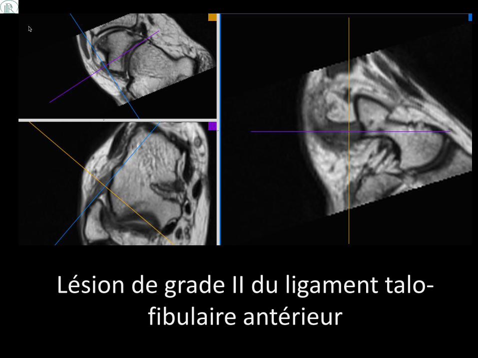
Lésion de grade II du ligament talo-fibulaire antérieur

Lésion ostéochondrale du dôme talien

Arthroscanner retrouvant la lésion ostéochondrale du dôme talien (stade II) ainsi que la lésion de grade
II du ligament talo-fibulaire antérieur

PATIENTE DE 46 ANS PRESENTANT DES DOULEURS MALLEOLAIRES EXTERNES, MAJOREES
SUITE A UNE ENTORSE SEMI RECENTE. NOTION D’ENTORSES A REPETITION
CAS N°3

Epanchement articulaire et net hypersignal du ligament talo-fibulaire antérieur
Ax DP Fat Sat Sag STIR Coro STIR
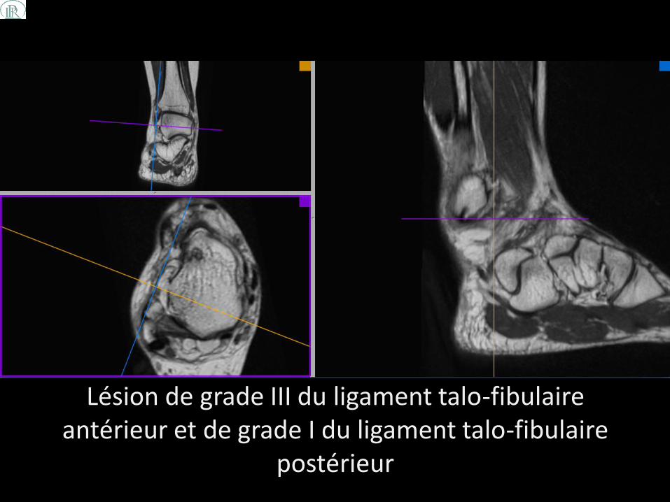
Lésion de grade III du ligament talo-fibulaire antérieur et de grade I du ligament talo-fibulaire
postérieur

PATIENTE PRÉSENTANT DES DOULEURS CHRONIQUES DE LA CHEVILLE GAUCHE, À
PRÉDOMINANCE INTERNE
CAS N°4

Aspect hypersignal du ligament deltoïde associé à un œdème médullaire en regard au niveau de talus
Ax DP FS Sag STIR
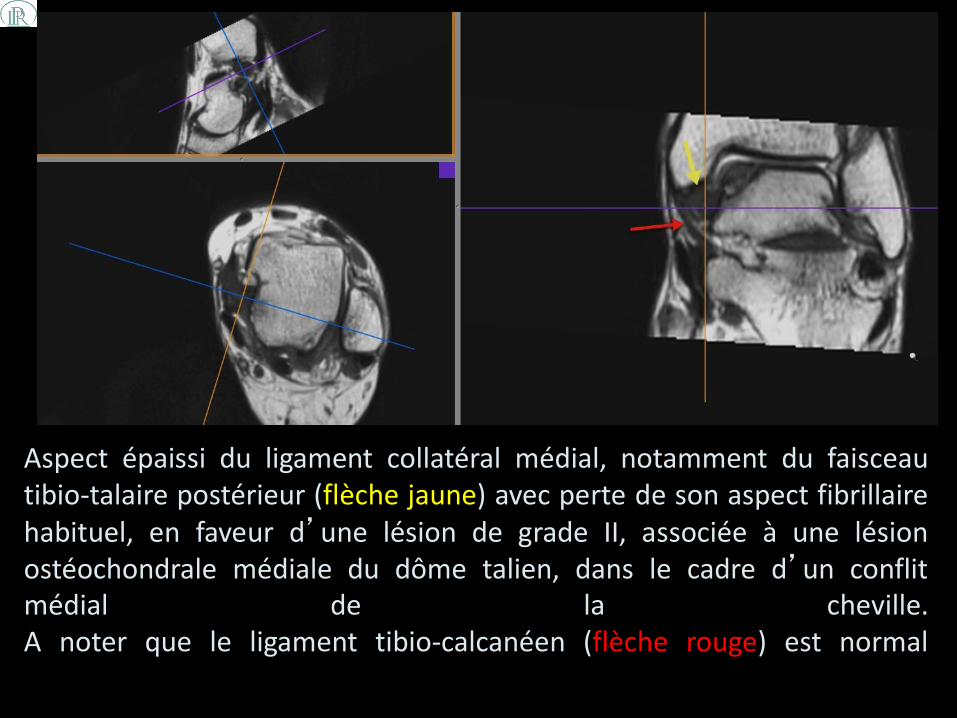
Aspect épaissi du ligament collatéral médial, notamment du faisceau tibio-talaire postérieur (flèche jaune) avec perte de son aspect fibrillaire habituel, en faveur d’une lésion de grade II, associée à une lésion ostéochondrale médiale du dôme talien, dans le cadre d’un conflit médial de la cheville. A noter que le ligament tibio-calcanéen (flèche rouge) est normal

PATIENTE DE 19 ANS PRÉSENTANT DES DOULEURS CHRONIQUES DE LA CHEVILLE DROITE. NOTION DE PLUSIEURS ENTORSES
PRECEDENTES ET D’UN CHOC DIRECT RECENT AU BORD LATERAL DU PIED
CAS N°5

Lésion ostéochondrale du dôme talien, œdème du ligament talo-fibulaire antérieur et œdème médullaire de la portion
antérieure du cuboïde
Ax DP FS Sag STIR Sag T1

Séquence DP 3D permettant de classifier la lésion ostéochondrale à stade III associée à une lésion de grade III du
ligament talo-fibulaire antérieur

et un trait de fracture au niveau du cuboïde à l’origine de l’œdème médullaire
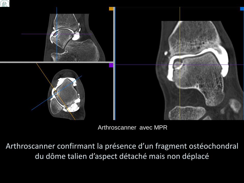
Arthroscanner confirmant la présence d’un fragment ostéochondral du dôme talien d’aspect détaché mais non déplacé
Arthroscanner avec MPR
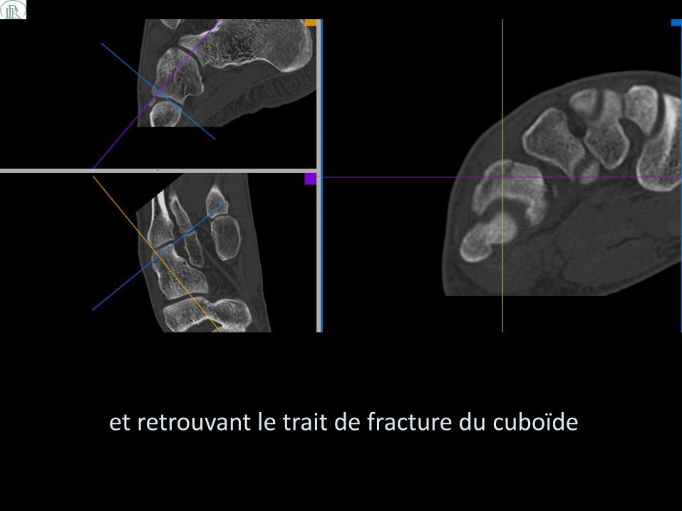
et retrouvant le trait de fracture du cuboïde

CONCLUSION
• La séquence DP 3D sans Fat Sat offre une nouvelle perspective en IRM pour préciser les lésions ligamentaires de cheville.
• Comparativement à l’arthroscanner, cette séquence (en association avec les séquences habituelles) permet d’obtenir une meilleure visibilité de certains faisceaux, notamment les ligaments du tarse.

CONCLUSION
•Nous proposons la réalisation systématique de cette séquence lors des IRM de cheville afin d’étudier de façon optimale les différents faisceaux ligamentaires ce qui permettrait d’éviter:
l’injection de chélate de Gadolinium dans la majorité des cas la réalisation d’un certain nombre d’arthroscanners (geste
invasif avec risques de complications)

REFERENCES 1. Boonthathip M et al. Tibiofibular Syndesmotic Ligaments: MR Arthrography in Cadavers with
Anatomic Correlation. Radiology 2010;254:827-836
2. Pau Golanó et al. Anatomy of the ankle ligaments: a pictorial essay. Knee Surg Sports Traumatol Arthrosc. 2010 May; 18(5): 557–569.
3. Sarrafian SK. Anatomy of the foot and ankle. Descriptive, topographic, functional. 2. Philadelphia: Lippincott; 1993. pp. 159–217.
4. Taylor DC, Englehardt DL, Bassett FH. Syndesmosis sprains of the ankle. The influence of heterotopic ossification. Am J Sport Med. 1992;20:146–150. doi: 10.1177/036354659202000209.
5. Molinari A, Stolley M, Amendola A. High ankle sprains (syndesmotic) in athletes: Diagnostic challenges and review of the literature. Iowa Orthop J. 2009;29:130-138.)
6. Boruta PM, Bishop JO, Braly WG, et al. Acute ankle ligament injuries; a literature review. Foot Ankle. 1990;11:107–113.
7. Bekerom MPJ, Oostra RJ, Golanó P, et al. The anatomy in relation to injury of the lateral collateral ligaments of the ankle: a current concepts review. Clin Anat. 2008;21:619–626. doi: 10.1002/ca.20703.
8. Muhle C, Frank LR, Rand T, et al. Collateral ligaments of the ankle: high-resolution MR imaging with a local gradient coil and anatomic correlation in cadavers. RadioGraphics 1999;19(3):673–683.
9. Stoller DW. Magnetic resonance imaging in orthopaedics and sports medicine 3rd ed.Baltimore, Md: Lippincott Williams & Wilkins, 2006; 786–793.
REFERENCES 10. Pankovich AM, Shivaram MS. Anatomical basis of variability in injuries of the medial malleolus
and the deltoid ligament. I. Anatomical studies. Acta Orthop Scand 1979;50(2):217–223.
11. Klein MA. MR imaging of the ankle: normal and abnormal findings in the medial collateral ligament. AJR Am J Roentgenol 1994;162(2):377–383.
12. Congrès thématique de la SIMS: Imagerie du pied et de la cheville (juin 2004: Paris). Le sprig ligament. M. Cohen et al.; p. 121–128.
13. Melão L et al. Ligaments of the Transverse Tarsal Joint Complex: MRI–Anatomic Correlation in Cadavers. AJR 2009;193:662-671
14. Kjaersgaard-Andersen P, Wethelund J, Helmig P, Soballe K. The stabilizing effect of the ligamentous structures in the sinus and canalis tarsi on the movements in the hindfoot. Am J Sports Med 1988;16: 512 -516
15. Rosenberg Z S et al. MR Imaging of the Ankle and Foot. Radiographics 2000;20:S153-S179
16. Congrès thématique de la SIMS: Le pied (juin 2011: Paris). Le Chopart et le carrefour des trois tendons. B. Ferré et al.; p.113-120.
17. Congrès thématique de la SIMS: Imagerie du pied et de la cheville (juin 2004: Paris). Les articulations de Lisfranc. F. Thévenin et al.; p. 129–145.

Mots clés : cheville, IRM, 3D, ligament
Keywords : ankle, MRI, 3D, ligament
Contact : M.SHEIBANIFAR, Imagerie du Pays de Rance [email protected]
www.imageriedupaysderance.fr