- university of illinois at urbana-champaign
TRANSCRIPT
Effect of Cross-Linking on the Diffusion of Water, Ions, and Small Molecules in Hydrogels
Yanbin Wu, Sony Joseph, and N. R. Aluru*Department of Mechanical Science and Engineering, Beckman Institute for AdVanced Science and Technology,UniVersity of Illinois at Urbana-Champaign, Urbana, IL-61801
ReceiVed: September 12, 2008; ReVised Manuscript ReceiVed: December 16, 2008
The present study reports on molecular dynamics investigations of chemically cross-linked poly(ethyleneglycol) hydrogels with the aim of exploring the diffusion properties of water, ions, and rhodamine within thepolymer at the molecular level. The water structure and diffusion properties were studied at various cross-linking densities with molecular weights of the chains ranging from 572 to 3400. As the cross-linking densityis increased, the water diffusion decreases and the slowdown in diffusion is more severe at the polymer-waterinterface. The water diffusion at various cross-linking densities is correlated with the water hydrogen bondingdynamics. The diffusion of ions and rhodamine also decreased as the cross-linking density is increased. Thevariation of diffusion coefficient with cross-linking density is related to the variation of water content atdifferent cross-linking densities. Comparison of simulation results and obstruction scaling theory for hydrogelsshowed similar trends.
Introduction
Hydrogels, composed of polymer networks and water, havebeen used increasingly in drug delivery systems, tissue engi-neering, contact lenses, and so forth1-5 due to their interestingstructural and mechanical properties. The solidlike character ofthe hydrogel system plays an important role by providingmechanical stability. The hydrogel system also maintainsdynamic behavior typical of liquid phases.5 Two properties ofhydrogels, high water content and rubberlike nature, make themakin to a natural tissue. Biocompatibility and cross-linkedstructure are key properties of hydrogels that allow for variousapplications. Cross-linking allows immobilization of activeagents and biomolecules and helps drug release at a well-definedrate. Cross-linking density is commonly used to tune keyparameters like mesh size and molecular weight between cross-linkers in order to change macroscopic properties such asdiffusion and Young’s modulus. Among the dynamic properties,diffusion of small molecules, such as nutrients, is essential forvitality of living cells in biological systems.
In order to utilize hydrogels for various applications, it isessential to understand their material properties, flexibility,interactions with solutes and transport phenomena. A cross-linked network is difficult to be analyzed by experimentaltechniques of chromatography and fractionation owing to thenetwork’s inability to dissolve. Deeper insight into dynamicprocesses occurring within hydrogels have become possible bytechniques such as high-flux neutron sources and X-raysynchrotrons.6-8 Diffusion in hydrogel has been studied exten-sively using quasi-elastic neutron scattering (QENS),9,10 NMR,11-13
side-by-side diffusion cells,14 fluorescence correlation spectros-copy,15,16 refractive index method,17 and so forth.
Many physical models have been developed to model thediffusion of small solutes in hydrogels.18,19 Solute behavior inhydrogels has been explained in terms of reduction in hydrogelfree volume,20-22 enhanced hydrodynamic drag on the solute,23,24
increased path length due to obstruction,25,26 and a combination
of hydrodynamic drag and obstruction effects.27 The theoreticalrelations are limited and rely on fitting parameters that aretypically not known. With the rapid development of moleculardynamics simulation techniques, it is now possible to study thestructure and dynamics of biomacromolecular systems in anaqueous environment considering explicit water, ion, and solutemolecules.28,29 In recent times, molecular dynamics simulationhas been used to study physical gels,30 poly(vinyl alcohol),10,31
poly(vinyl methyl ether),32 poly(N-isopropylacrylamide),32 poly-acrylamide,33 epoxy-amine networks,34 and so forth. Structureand dynamics of the polymer-water interface in poly(vinylalcohol) (PVA) for a mesh size of 1 nm was studied recently.10
Solvent diffusion coefficient and residence times in hydrophilicsystems indicate that water behaves as a supercooled liquidphase.10 Structural and mechanical properties and diffusion ofglucose and vitamin D in poly(ethylene glycol) (PEG) andpoly(acrylic acid) (PAA) and their double network was inves-tigated by Jang et al.35 Effects of confined water in cages ofdifferent chemical and structural features have also beeninvestigated previously in other natural and man-made structuressuch as vycor glass,36 carbon nanotubes,37,38 boron-nitridenanotubes,39 zeolites,40 cellular membrane channels,41 proteins,29
carbohydrate solution,42 and so forth.Poly(ethylene glycol) (PEG)-based hydrogel network has been
increasingly utilized in tissue engineering applications in recentyears. This is mainly due to their hydrophilicity and resistanceto protein adsorption and biocompatibility. They can also becustomized by modifying the chain length and adding biologicalfunctional groups. Besides, PEG hydrogel is a promisingmembrane material for selective removal of CO2 from a mixturecontaining light gases such as CH4, N2 and H2.43 PEG can beeasily cross-linked using acrylate group as a cross-linker. Inconventional polymerization, the cross-linking density, definedas the number of cross-linkers divided by the number ofmonomers, need not be homogeneous throughout the network,but in poly(ethylene glycol) diacrylate (PEGDA) it is homo-geneous because the molecular weight between the cross-linkersis the same as that of the PEG monomer. This enables PEGDAto be used as an ideal material for studying gel properties. Since
* To whom correspondence should be addressed. E-mail:[email protected].
J. Phys. Chem. B 2009, 113, 3512–35203512
10.1021/jp808145x CCC: $40.75 2009 American Chemical SocietyPublished on Web 02/24/2009
cross-linking has a significant impact on the structural anddynamic properties of the hydrogel, we investigate the structuraland dynamic properties of a hydrogel consisting of cross-linkedPEGDA, water, and small solutes (ions and rhodamine) usingmolecular dynamics (MD) simulations, as they can provide auseful description of water and solute mobility by consideringexplicit water and partial charge for PEG atoms. Rhodamine is
commonly used as a tracer dye in experiments within hydrogelnetworks to determine the transport properties of the network.Studying rhodamine diffusion also helps to understand thediffusion of similar sized biomolecules in PEGDA.
The rest of the paper is organized as follows: first, we presentthe system setup with the force fields used and a description ofthe construction of the cross-linked structure. Then we inves-
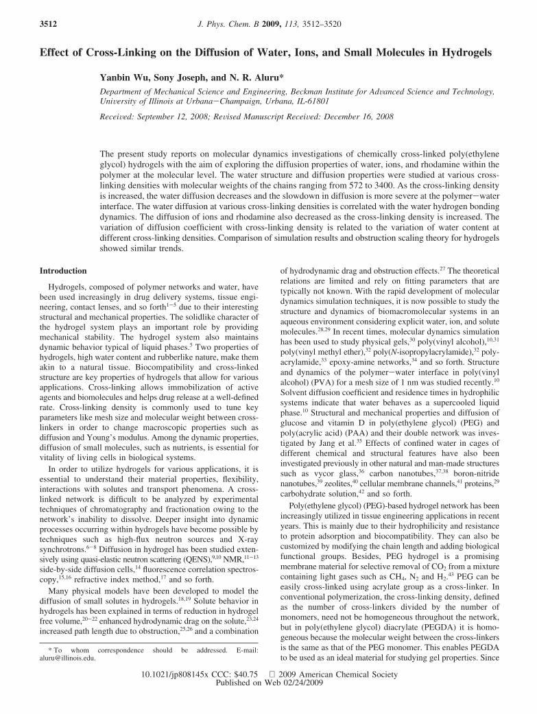
Figure 1. (a) Chemical structure of PEG. (b) Chemical structure of PEGDA. (c) 6 PEGDA chain ends meet and form cyclododecane as cross-linking point. (d) Cross-linked network with 2 × 2 × 2 cells in three dimensions. (e) Chemical structure of rhodamine. (f) Visualization of simulationbox with polymer network, water, rhodamine, and chloride molecules. Yellow, carbon; red, oxygen; white, hydrogen.
TABLE 1: System Composition and Equilibrated Mesh Sizea
prepolymer PEG572 PEG1000 PEG1500 PEG2000 PEG3400
n 13 23 34 45 78cross-linking density(1/n)% 7.69 4.35 2.94 2.22 1.28equilibrated water content/% 74.7 84.2 85.5 89.9 90.9polymer volume content/% 25.3 15.8 14.5 10.1 9.1mesh size/nm 2.28 3.14 3.64 4.47 5.49number of water molecules per cell 291 855 1353 2636 4984number of cells 2 × 2 × 2 2 × 2 × 2 2 × 2 × 2 1 × 1 × 1 1 × 1 × 1simulation box size/nm 4.56 6.28 7.28 4.47 5.49
a n is the degree of polymerization before cross-linking. Mesh size is a function of n.
Water, Ions, and Small Molecules in Hydrogels J. Phys. Chem. B, Vol. 113, No. 11, 2009 3513
tigate the water structure and hydrogen bonding in variousregions divided according to the distance from the polymer.Next, we present the results and discussion on the variation ofdiffusion coefficients of water, ions, and rhodamine as a functionof the cross-linking density. Finally, we compare the variationof diffusion coefficient with cross-linking density from MDsimulation results with the variation predicted from Amsdenobstruction scaling theory.
Methods
Force Field. A force field of the following form has beenemployed
Etotal ) EvdW + EQ + Ebond + Eangle + Edihedral (1)
where Etotal, EvdW, EQ, Ebond, Eangle, and Edihedral are the total, vander Waals (vdW), electrostatic, bond stretching, angle bending,and torsion energies, respectively. In our simulations, we usedthe all atom force field. The force field parameters and chargesfor the PEG chain are taken from Smith et al.,44 Lennard-Jones(LJ) interaction parameters between PEG and water are takenfrom Bedrov et al.,45 rhodamine are taken from Vaiana et al.,46
and the acrylate cross-linker, chloride, and sodium ions are fromthe CHARMM27 force field.47 For water, we used the singlepoint charge/extended (SPC/E) model.48 In order to verify theinteraction parameters between PEG and water, we simulateda system composed only of PEG chains and water and calculatedthe radial distribution function of the oxygen of the PEG withthe oxygen of water and obtained good agreement with theresults of Borodin et al.49 For cross LJ interaction parametersbetween rhodamine and water, we used Lorentz-Berthelotcombination rules and validated by calculating the diffusioncoefficient of rhodamine in bulk water. We obtained a diffusioncoefficient of 0.4243 × 10⁻⁵ cm2/s which is within the rangeof values obtained in various experiments (0.3 × 10⁻⁵ to 0.5 ×10⁻⁵ cm2/s).50-52 For cross LJ interaction parameters betweenions and water and between PEG chain and CHARMM atoms,we followed Patra et al.53 and Zheng et al.,54 respectively, andused Lorentz-Berthelot combination rules.
Simulation System Setup. For our simulations, we used across-linked PEGDA structure with an ideal network withoutany free dangling ends or self-looping or entanglements. Figure1a shows a PEG chain with n monomers. Each PEGDAmolecule (see Figure 1b) is a PEG chain connected to acrylategroups at both ends that serve as cross-linkers. Under the
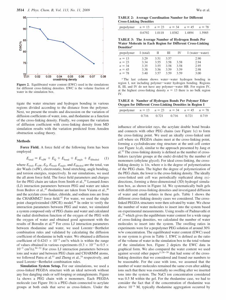
influence of ultraviolet rays, the acrylate double bond breaksand connects with other PEG chains (see Figure 1c) to formthe cross-linking point. We used an ideally cross-linked unitcell where six PEGDA chains meet at the cross-linking point,forming a cyclododecane ring structure at the unit cell center(see Figure 1c,d), similar to the approach presented by Jang etal.35 The cross-linking density is defined as the number of cross-linkers (acrylate groups at the ends) divided by the number ofmonomers (ethylene glycol). For ideal cross-linking, the cross-linking density is 1/n, where n is the degree of polymerizationof the PEG chain. The higher the degree of polymerization ofthe PEG chain, the lower is the cross-linking density. The ideallycross-linked unit cell was periodically replicated along xyz-directions, forming a three-dimensional (3D) hydrogel simula-tion box, as shown in Figure 1d. We systematically built gelswith different cross-linking densities and investigated diffusionof water and small solutes in these gels. Table 1 shows thedifferent cross-linking density cases we considered. The cross-linked PEGDA structures were then solvated by water. We chosethe number of water molecules to insert into the system basedon experimental measurements. Using results of Padmavathi etal.,55 which gives the equilibrium water content for a wide rangeof cross-linking densities, we calculated the number of watermolecules to insert into the system. The swelling ratios inexperiments were for a prepolymer PEG solution of around 30%w/w concentration. The equilibrated water content (EWC) usedin our system is given in Table 1. EWC is defined as the ratioof the volume of water in the simulation box to the total volumeof the simulation box. Figure 2 depicts the EWC data ingraphical form. We also compared the water content we usedagainst several other papers11,56-59 that had some of the cross-linking densities that we considered and found our numbers tobe reasonable. For the case with ions, we assumed that thenumber of water molecules remained the same even after addingions such that there was essentially no swelling after we insertedions into the system. The NaCl ion concentration consideredwas 0.5 M within the gel. For the rhodamine case, we had toconsider the fact that if the concentration of rhodamine wasabove 10-6 M, typically rhodamine aggregation occurred by
Figure 2. Equilibrated water content (EWC) used in the simulationsfor different cross-linking densities. EWC is the volume fraction ofwater in the simulation box.
TABLE 2: Average Coordination Number for DifferentCross-Linking Densities
prepolymer n ) 13 n ) 23 n ) 34 n ) 45 n ) 78
0.6792 1.0118 1.0382 1.4894 1.5985
TABLE 3: The Average Number of Hydrogen Bonds PerWater Molecule in Each Region for Different Cross-LinkingDensitiesa
prepolymer I (total) II III IV I (water-water)
n ) 13 3.29 3.51 3.57 2.90n ) 23 3.34 3.55 3.58 3.58 2.94n ) 34 3.35 3.55 3.58 3.58 2.95n ) 45 3.38 3.56 3.59 3.59 2.98n ) 78 3.40 3.57 3.59 3.59 3.00
a The last column shows water-water hydrogen bonding inregion I, not including polymer-water hydrogen bonding. RegionsII, III, and IV do not have any polymer-water HB. For region IV,at the highest cross-linking density n ) 13 there is no bulk regionIV.
TABLE 4: Number of Hydrogen Bonds Per Polymer EtherOxygen for Different Cross-Linking Densities in Region I
prepolymer n ) 13 n ) 23 n ) 34 n ) 45 n ) 78
0.716 0.721 0.716 0.721 0.719
3514 J. Phys. Chem. B, Vol. 113, No. 11, 2009 Wu et al.
stacking up on its three-ring xanthylium plane (rhodaminestructure is shown in Figure 1e) and this hindered fluorescentyield and diffusion. In experiments, to avoid aggregation a verydilute solution is used, but in simulations, such low concentra-tions would need an extremely large box size and would becomputationally expensive. So in our simulations, we used onlyone rhodamine in the system with the simulation box sizeranging from 4.56 to 7.28 nm, which is much larger than 4 Å,the distance between the planes of rhodamine molecules in adimer structure formed during aggregation.60 The final systemcomposition and equilibrated mesh size for the different casesare summarized in Table 1. Figure 1f shows a snapshot of thesimulation box comprising of the polymer network, water,rhodamine, and chloride ions.
MD simulations were performed with Gromacs 3.3.1.61 Timeintegration was performed using the leapfrog algorithm with atime step of 1.0 fs. The short-range vdW interactions werecomputed using a cutoff scheme (cutoff distance, 1.0 nm). Thelong-range electrostatic interactions were computed by using aparticle mesh Ewald method61 (real space cutoff, 1.0 nm; FFTgridspacing,0.12nm,fourth-orderinterpolation).TheNose-Hooverthermostat62,63 with a time constant of 0.5 ps was used tomaintain the temperature at 300 K. We built the polymernetwork with all PEG chain segments in an all-trans conforma-tion first and then inserted water molecules according toequilibrated water content in the hydrogel. After that, we letthe system equilibrate for 1 ns in an NPT ensemble bymaintaining a pressure of 1 bar (compressibility time constantof 0.2 ps; compressibility of 4.5 × 10⁻⁵ bar-1) with aParrrinello-Rahman barostat.64 The energy, temperature, andbox size of the simulation box reached constant values duringthis equilibration process. Then we further equilibrated thesystem for additional 1 ns of simulation time using an NVTensemble at 300 K. The energy and temperature of thesimulation box reached constant values during this equilibrationprocess. The resulting configuration is used as the starting pointfor further simulations. For collecting sufficient statistics tocompute various properties, the simulations were run for 54 ns.
Polymer-Water Interaction and Water Dynamics
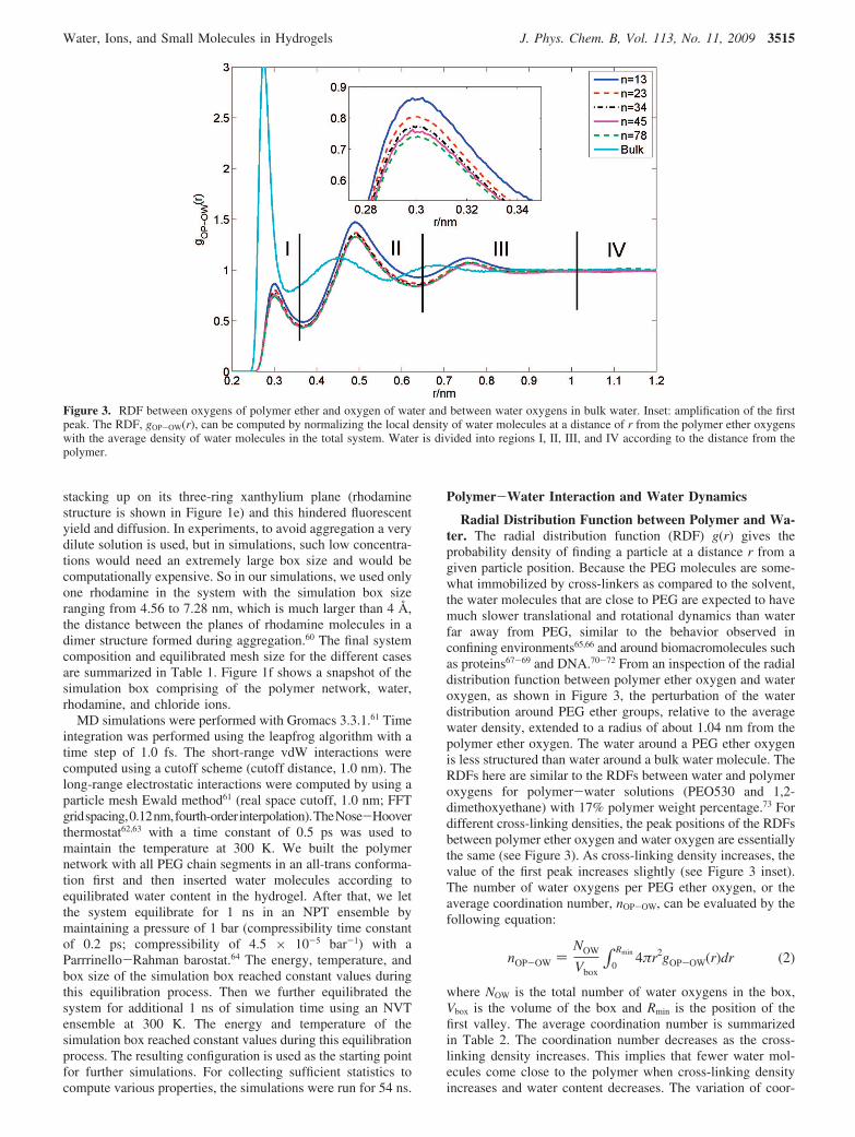
Radial Distribution Function between Polymer and Wa-ter. The radial distribution function (RDF) g(r) gives theprobability density of finding a particle at a distance r from agiven particle position. Because the PEG molecules are some-what immobilized by cross-linkers as compared to the solvent,the water molecules that are close to PEG are expected to havemuch slower translational and rotational dynamics than waterfar away from PEG, similar to the behavior observed inconfining environments65,66 and around biomacromolecules suchas proteins67-69 and DNA.70-72 From an inspection of the radialdistribution function between polymer ether oxygen and wateroxygen, as shown in Figure 3, the perturbation of the waterdistribution around PEG ether groups, relative to the averagewater density, extended to a radius of about 1.04 nm from thepolymer ether oxygen. The water around a PEG ether oxygenis less structured than water around a bulk water molecule. TheRDFs here are similar to the RDFs between water and polymeroxygens for polymer-water solutions (PEO530 and 1,2-dimethoxyethane) with 17% polymer weight percentage.73 Fordifferent cross-linking densities, the peak positions of the RDFsbetween polymer ether oxygen and water oxygen are essentiallythe same (see Figure 3). As cross-linking density increases, thevalue of the first peak increases slightly (see Figure 3 inset).The number of water oxygens per PEG ether oxygen, or theaverage coordination number, nOP-OW, can be evaluated by thefollowing equation:
nOP-OW )NOW
Vbox∫0
Rmin 4πr2gOP-OW(r)dr (2)
where NOW is the total number of water oxygens in the box,Vbox is the volume of the box and Rmin is the position of thefirst valley. The average coordination number is summarizedin Table 2. The coordination number decreases as the cross-linking density increases. This implies that fewer water mol-ecules come close to the polymer when cross-linking densityincreases and water content decreases. The variation of coor-
Figure 3. RDF between oxygens of polymer ether and oxygen of water and between water oxygens in bulk water. Inset: amplification of the firstpeak. The RDF, gOP-OW(r), can be computed by normalizing the local density of water molecules at a distance of r from the polymer ether oxygenswith the average density of water molecules in the total system. Water is divided into regions I, II, III, and IV according to the distance from thepolymer.
Water, Ions, and Small Molecules in Hydrogels J. Phys. Chem. B, Vol. 113, No. 11, 2009 3515
dination number shown here is similar to the phenomenonobserved in 1,2-dimethoxyethane/water solutions with varyingpolymer concentration.74
The water molecules in the gel system can be assigned intodifferent regions according to their distance from the polymerether oxygen atoms. We sampled the solvent in different regions,according to the gOP-OW(r) behavior.10 Since there are threepeaks in the curve, we divided the water into four regions.Region I (r < 0.36 nm) and region II (0.36 nm < r < 0.64 nm)are considered as “close contact” regions. Water at distancesbetween 0.64 and 1.04 nm, where the perturbation in thegOP-OW(r) was minor, is chosen as region III. The remainingwater molecules are considered to be region IV. Water moleculesin regions I-IV are characterized in terms of hydrogen bonding,relaxation times, and diffusion coefficients.
Water Hydrogen Bonding. The hydrogen bonding (HB)structure between water molecules and that between PEG etheroxygen and water was studied by analyzing the trajectory.Hydrogen bonding is defined by adopting the geometric criteriawhere the acceptor-donor (O · · ·O) distance is less than 0.35nm and the angle (O-H · · ·O) is less than 30°. For each cross-linking density across different regions, the total number ofhydrogen bonds is fairly constant except for a small dip in regionI, as shown in Table 3. In region I, the water-water hydrogenbonding is lowered, but that is made up for to some extent bythe hydrogen bonding with the polymer ether oxygen whichacts as an acceptor (see Table 4). The variation of the numberof hydrogen bonds across different regions is similar to thatseen in simulations of PVA hydrogels.10 As cross-linking densityincreases, HB per water molecule decreases for all regions. Forthe highest cross-linking density case (n ) 13), there is no bulkregion and region IV is undefined.
Hydrogen Bond Dynamics. The intermittent time autocor-relation function c(t) expresses the probability that a randomlychosen pair of molecules is bonded at time t, provided that abond existed at time t ) 0, regardless of whether it was bondedin the interim time. c(t) provides valuable insight into therelaxation of the system’s H-bonding network. c(t) is given by
c(t) ) ⟨h(t)h(0)⟩⟨h(0)h(0)⟩ (3)
where h(t) is 1 if molecules are bonded at time t and 0 if not.⟨⟩ denotes average over all pairs of HB at t ) 0 and over many
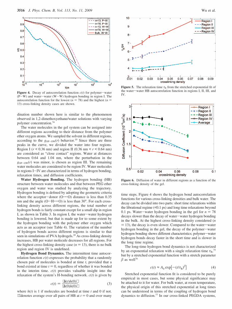
time steps. Figure 4 shows the hydrogen bond autocorrelationfunctions for various cross-linking densities and bulk water. Thedecay can be divided into two parts: short time relaxations withinthe librational regime (<0.1 ps) and long time relaxations beyond0.1 ps. Water-water hydrogen bonding in the gel for n ) 78decays slower than the decay of water-water hydrogen bondingin the bulk. At the highest cross-linking density considered (n) 13), the decay is even slower. Compared to the water-waterhydrogen bonding in the gel, the decay of the polymer-waterhydrogen bonding shows different characteristics; polymer-waterhydrogen bonds decay faster in the short time and is slower inthe long time regime.
The long-time hydrogen bond dynamics is not characterizedby an exponential relaxation with a single relaxation time τR,75
but by a stretched exponential function with a stretch parameter� as well76
c(t) ≈ A0 exp[-(t/τR)�] (4)
Stretched exponential function fit is considered to be purelyempirical in most cases, but some physical significance maybe attached to it for water. For bulk water, at room temperature,the physical origin of this stretched exponential at long timescan be understood in terms of the coupling of hydrogen bonddynamics to diffusion.77 In our cross-linked PEGDA systems,
Figure 4. Decay of autocorrelation function c(t) for polymer-water(P-W) and water-water (W-W) hydrogen bonding in region I. Theautocorrelation function for the lowest (n ) 78) and the highest (n )13) cross-linking density cases are shown.
Figure 5. The relaxation time τR from the stretched exponential fit ofthe water-water HB autocorrelation function in regions I, II, III, andIV.
Figure 6. Diffusion of water in different regions as a function of thecross-linking density of the gel.
3516 J. Phys. Chem. B, Vol. 113, No. 11, 2009 Wu et al.
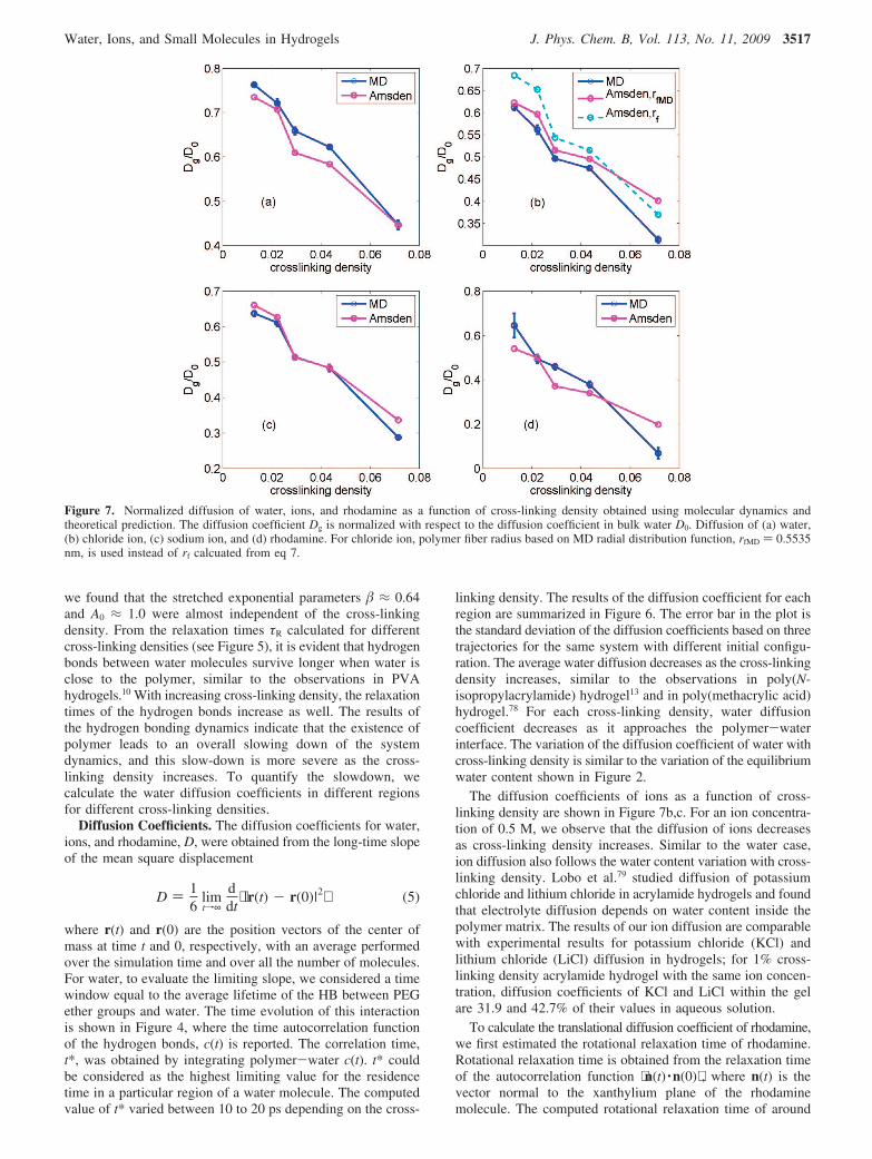
we found that the stretched exponential parameters � ≈ 0.64and A0 ≈ 1.0 were almost independent of the cross-linkingdensity. From the relaxation times τR calculated for differentcross-linking densities (see Figure 5), it is evident that hydrogenbonds between water molecules survive longer when water isclose to the polymer, similar to the observations in PVAhydrogels.10 With increasing cross-linking density, the relaxationtimes of the hydrogen bonds increase as well. The results ofthe hydrogen bonding dynamics indicate that the existence ofpolymer leads to an overall slowing down of the systemdynamics, and this slow-down is more severe as the cross-linking density increases. To quantify the slowdown, wecalculate the water diffusion coefficients in different regionsfor different cross-linking densities.
Diffusion Coefficients. The diffusion coefficients for water,ions, and rhodamine, D, were obtained from the long-time slopeof the mean square displacement
D ) 16
limtf∞
ddt
⟨ |r(t) - r(0)|2⟩ (5)
where r(t) and r(0) are the position vectors of the center ofmass at time t and 0, respectively, with an average performedover the simulation time and over all the number of molecules.For water, to evaluate the limiting slope, we considered a timewindow equal to the average lifetime of the HB between PEGether groups and water. The time evolution of this interactionis shown in Figure 4, where the time autocorrelation functionof the hydrogen bonds, c(t) is reported. The correlation time,t*, was obtained by integrating polymer-water c(t). t* couldbe considered as the highest limiting value for the residencetime in a particular region of a water molecule. The computedvalue of t* varied between 10 to 20 ps depending on the cross-
linking density. The results of the diffusion coefficient for eachregion are summarized in Figure 6. The error bar in the plot isthe standard deviation of the diffusion coefficients based on threetrajectories for the same system with different initial configu-ration. The average water diffusion decreases as the cross-linkingdensity increases, similar to the observations in poly(N-isopropylacrylamide) hydrogel13 and in poly(methacrylic acid)hydrogel.78 For each cross-linking density, water diffusioncoefficient decreases as it approaches the polymer-waterinterface. The variation of the diffusion coefficient of water withcross-linking density is similar to the variation of the equilibriumwater content shown in Figure 2.
The diffusion coefficients of ions as a function of cross-linking density are shown in Figure 7b,c. For an ion concentra-tion of 0.5 M, we observe that the diffusion of ions decreasesas cross-linking density increases. Similar to the water case,ion diffusion also follows the water content variation with cross-linking density. Lobo et al.79 studied diffusion of potassiumchloride and lithium chloride in acrylamide hydrogels and foundthat electrolyte diffusion depends on water content inside thepolymer matrix. The results of our ion diffusion are comparablewith experimental results for potassium chloride (KCl) andlithium chloride (LiCl) diffusion in hydrogels; for 1% cross-linking density acrylamide hydrogel with the same ion concen-tration, diffusion coefficients of KCl and LiCl within the gelare 31.9 and 42.7% of their values in aqueous solution.
To calculate the translational diffusion coefficient of rhodamine,we first estimated the rotational relaxation time of rhodamine.Rotational relaxation time is obtained from the relaxation timeof the autocorrelation function ⟨n(t) ·n(0)⟩ , where n(t) is thevector normal to the xanthylium plane of the rhodaminemolecule. The computed rotational relaxation time of around
Figure 7. Normalized diffusion of water, ions, and rhodamine as a function of cross-linking density obtained using molecular dynamics andtheoretical prediction. The diffusion coefficient Dg is normalized with respect to the diffusion coefficient in bulk water D0. Diffusion of (a) water,(b) chloride ion, (c) sodium ion, and (d) rhodamine. For chloride ion, polymer fiber radius based on MD radial distribution function, rfMD ) 0.5535nm, is used instead of rf calcuated from eq 7.
Water, Ions, and Small Molecules in Hydrogels J. Phys. Chem. B, Vol. 113, No. 11, 2009 3517
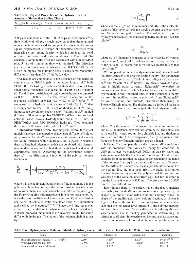
200 ps is comparable to the 100-200 ps in experiments.80 Atime window of 400 ps, a much larger value than the rotationalrelaxation time was used to compute the slope of the meansquare displacement. Diffusion of rhodamine decreases withincreasing cross-linking density, which is similar to the trendobserved for water and ions, as shown in Figure 7d. Toaccurately compute the diffusion coefficient with a linear MSDplot, 54 ns of simulation time was required. The diffusioncoefficient of rhodamine in bulk water was 0.4243 × 10⁻⁵ cm2/s. For the highest cross-linking density considered rhodaminediffusion is less than 5% of the bulk value.
Our results are comparable to the diffusion of molecules ofsimilar size in PEGDA gels in literature. Jang et al.35 builtPEG(1300)DA hydrogel system and studied diffusion of twosmall solute molecules, D-glucose and ascorbic acid (vitaminC). The diffusion coefficient of D-glucose in the gel was reportedas 0.173 ( 0.050 × 10⁻⁵ cm2/s, which is one-third of theD-glucose diffusion in water (0.6 - 0.7 × 10⁻⁵ cm2/s).81-83
D-glucose has a hydrodynamic radius of 3.61-3.8 Å,84,85 thatis comparable to 0.53 ( 0.03 nm of rhodamine 6G.86 Usingfluorescence correlation spectroscopy, Watkins et al.15 studieddiffusion of fluorescent probes Cl-NERF and Texas Red sulfonylchloride, which have a hydrodynamic radius of 0.7 nm, inPEG(700)DA and PEG(1000)DA hydrogel. They obtaineddiffusion coefficients on the order of 10-8 cm2/s.
Comparison with Theory. Over the years, several theoreticalmodels have been developed to characterize diffusion of solutesin hydrogels. Amsden18 compares several theories and modelsfor solute diffusion within hydrogels and suggests Amsden’stheory where hydrodynamic models are combined with obstruc-tion models as one of the best theories that matched severalexperimental results. According to the obstruction scalingtheory,87,88 the diffusion as a function of the polymer volumefraction is
Dg
D0) exp[-π( rs + rf
ksa�-0.75C∞-0.25(1 - 2�)-0.25 + 2rf
)2](6)
where a is the equivalent bond length of the monomer, φ is thepolymer volume fraction, rs is the radius of solute, rf is the radiusof polymer chain, C∞ is the characteristic ratio of polymer, � isthe Flory-Huggins polymer/solvent interaction parameter, Dg
is the diffusion coefficient of solute in gel, and D0 is the diffusioncoefficient of solute in water calculated from MD simulationand verified by literature.38,50-52,89 Since the fitting parameterks = 1 for the different polymers and solutes considered,Amsden proposed this model as a “universal” model for solutediffusion in hydrogels. The radius of the polymer chain is givenby87
rf ) ( MmVlπNA
)1/2
(7)
where l is the length of the monomer unit, Mm is the molecularweight of the monomer, V is the specific volume of the polymer,and NA is the Avogadro number. The solute size rs is thehydrodynamicradiusofthesolutecomputedbytheStokes-Einsteinrelation87
rs )kBT
fπηD0(8)
where kB is Boltzmann’s constant, η is the viscosity of water attemperature T, and f is 4 for solutes whose size approaches thatof the solvent (i.e., water) and 6 for solutes greater in size thanthe solvent.90
We compared our molecular dynamics simulation results withthat from Amsden’s obstruction scaling theory. The parametersused in eq 6 are listed in Table 5. According to Koneshan etal.91 and Valente et al.,92 Stokes’ law breaks down for smallions in highly polar solvents. Nightingale93 extended theempirical correction to Stoke’s law to provide a set of modifiedhydrodynamic radii for small ions. In using Amsden’s theory,we used the modified hydrodynamic radii from Nightingale93
for water, sodium, and chloride ions rather than using theStokes-Einstein relation. For rhodamine, we followed the sameprocedure as Jang et al.35 to compute the hydrodynamic radius
⟨rh⟩-1 ) 1
N2 ∑j*i
N
∑i)1
N
⟨ 1rij
⟩ (9)
where N is the number of atoms in the rhodamine molecule,and rij is the distance between two atom pairs. The solute sizers we used for water, sodium ion, chloride ion, and rhodamineare listed in Table 6. For comparison, the hydrodynamic radiifrom the Stokes-Einstein relation are also listed.
In Figure 7 we compare the results from our MD simulationswith the prediction from Amsden’s theory for water and thedifferent solutes we considered. Diffusion data for water andsodium ion match better than that of chloride ion. The differencecould be from the fact that the equation for calculating the radiusof the polymer fiber, eq 7 does not take the ion size differences,and the different distances of closest approach into account. Forthe sodium ion, the first peak from the radial distributionfunction between oxygen of the polymer and the sodium ionwas close to the value obtained from eq 7, but for the chlorideion, the first peak was at 0.5535 nm. Therefore we used 0.5535nm as rf for chloride ion.
Even though there is no perfect match, the theory matchesreasonably well with MD results. As mentioned previously, theshapes of all the diffusion data are similar, and they follow theshape of the equilibrated water volume fraction, as shown inFigure 2. Unless the solute size and mesh size are comparable,such that the molecular level structure of the polymer networkand the solute structure affect the diffusion, it is the equilibratedwater content that is the key parameter in determining thediffusion coefficient. In experiments, factors, such as concentra-tion of prepolymer solution, defects, rate of gelation, and so
TABLE 5: Physical Properties of the Hydrogel Used inAmsden’s Obstruction Scaling Theory
Mm (g/mol) V (cm3/g) l (nm) a (nm) rf (nm) C∞ �
44 0.845357 0.36 1.54 0.2337 5.287 0.4687
TABLE 6: Hydrodynamic Radii and Modified Hydrodynamic Radii Used in This Work for Water, Ions, and Rhodamine
solute water chloride ion sodium ion rhodamine
bulk diffusion coefficient/× 10-5 cm2/s 2.7038 1.8589 1.3289 0.424350-52
hydrodynamic radius (nm) 0.0914 0.1335 0.1870 0.5819radius used in this work (nm) 0.276 0.332 0.358 0.4873
3518 J. Phys. Chem. B, Vol. 113, No. 11, 2009 Wu et al.
forth, influence the final gel structure and thus the equilibratedwater content and the diffusion data.
Conclusions
To summarize, we have presented molecular dynamicsinvestigations of diffusion of water and small solutes in PEGDAhydrogels of varying cross-linking densities. Diffusion coef-ficient of water and small solutes decreases as cross-linkingdensity increases. The decrease in diffusion of water is correlatedwith the increase in hydrogen bonding relaxation times. Thesimulation results compare well with Amsden’s obstructionscaling theory, if the hydrodynamic radii of the solutes can becomputed more accurately than from the Stokes- Einsteinrelation. The diffusion behavior corresponds quite well with theequilibrated water content in each gel.
Acknowledgment. This work was supported by NSF underGrants 0120978 (Water CAMPWS, UIUC), 0328162 (nano-CEMMS, UIUC), 0523435, and 0810294.
References and Notes
(1) Peppas, N. A.; Huang, Y.; Torres-Lugo, M.; Ward, J. H.; Zhang,J. Annu. ReV. Biomed. Eng. 2000, 2, 9–29.
(2) Lee, K.; Mooney, D. Chem. ReV. 2001, 101, 1869–79.(3) Byrne, M.; Park, K.; Peppas, N. AdV. Drug DeliV. ReV. 2002, 54,
149–161.(4) Langer, R.; Peppas, N. AIChE J. 2003, 49, 2990–3006.(5) Underhill, G.; Chen, A.; Albrecht, D.; Bhatia, S. Biomaterials 2007,
28, 256–270.(6) Gabel, F.; Bicout, D.; Lehnert, U.; Tehei, M.; Weik, M.; Zaccai,
G. Q. ReV. Biophys. 2003, 35, 327–367.(7) Winter, R. Biochem. Biophys. Acta 2002, 1595, 160–184.(8) Koch, M.; Vachette, P.; Svergun, D. Q. ReV. Biophys. 2003, 36,
147–227.(9) Paradossi, G.; Cavalieri, F.; Chiessi, E.; Telling, M. J. Phys. Chem.
B 2003, 107, 8363–8371.(10) Chiessi, E.; Cavalieri, F.; Paradossi, G. J. Phys. Chem. B 2007,
111, 2820–2827.(11) Cruise, G.; Scharp, D.; Hubbell, J. Biomaterials 1998, 19, 1287–
1294.(12) Yasunaga, H.; Kobayashi, M.; Matsukawa, S.; Kirosu, H.; Ando,
I. Annu. Rep. NMR Spectrosc. 1997, 34, 40–105.(13) Tanaka, N.; Matsukawa, S.; Kurosu, H.; Ando, I. Polymer 1998,
39, 4703–4706.(14) Stringer, J.; Peppas, N. J. Controlled Release 1996, 42, 195–202.(15) Watkins, A.; Anseth, K. Macromolecules 2005, 38, 1326–1334.(16) Russell, R.; Axel, A.; Shields, K.; Pishko, M. Polymer 2001, 42,
4893–4901.(17) Weng, L.; Zhou, X.; Zhang, X.; Zhang, L.; Xu, J. Macromol. Rapid
Commun. 2002, 23, 968–971.(18) Amsden, B. Macromolecules 1998, 31, 8382–8395.(19) Masaro, L.; Zhu, X. Prog. Polym. Sci. 1999, 24, 731–775.(20) Yasuda, H.; Peterlin, A.; Colton, C. K.; Smith, K. A.; Merrill, E. W.
Makromol. Chem. 1969, 126, 177–186.(21) Peppas, N.; Reinhart, C. J. Membr. Sci. 1983, 15, 275–287.(22) Lustig, S.; Peppas, N. J. Appl. Polym. Sci. 1988, 36, 735–747.(23) Cukier, R. Macromolecules 1984, 17, 252–255.(24) Phillips, R.; Deen, W.; Brady, J. AIChE J. 1989, 35, 1761–1769.(25) Ogston, A. Trans. Faraday Soc. 1958, 54, 1754–1757.(26) Johansson, L.; Elvingson, C.; Loefroth, J. Macromolecules 1991,
24, 6024–6029.(27) Johnson, E.; Berk, D.; Jain, R.; Deen, W. Biophys. J. 1996, 70,
1017–1023.(28) Hansson, T.; Oostenbrink, C.; van Gunsteren, W. Curr. Opin. Struct.
Biol. 2002, 12, 190–196.(29) Bizzarri, A.; Cannistraro, S. J. Phys. Chem. B 2002, 106, 6617–
6633.(30) Tamai, Y.; Tanaka, H.; Nakanishi, K. Macromolecules 1996, 29,
6750–6760.(31) Tamai, Y.; Tanaka, H. Chem. Phys. Lett. 1998, 285, 127–132.(32) Tamai, Y.; Tanaka, H. Fluid Phase Equilib. 1998, 144, 441–448.(33) Netz, P.; Dorfmuller, T. J. Phys. Chem. B 1998, 102, 4875.(34) Mijovic, J.; Zhang, H. J. Phys. Chem. B 2004, 108, 2557–2563.(35) Jang, S.; Goddard, W.; Kalani, M. J. Phys. Chem. B 2007, 111,
1729–1737.(36) Gallo, P.; Rovere, M.; Spohr, E. J. Chem. Phys. 2000, 113, 11324.
(37) Hummer, G.; Rasaiah, J.; Noworyta, J. Nature 2001, 414, 188–190.
(38) Mashl, R.; Joseph, S.; Aluru, N.; Jakobsson, E. Nano Lett. 2003,3, 589–592.
(39) Won, C.; Aluru, N. J. Am. Chem. Soc. 2007, 129, 2748–2748.(40) Cicu, P.; Demontis, P.; Spanu, S.; Suffritti, G.; Tilocca, A. J. Chem.
Phys. 2000, 112, 8267.(41) Fujiyoshi, Y.; Mitsuoka, K.; de Groot, B.; Philippsen, A.; Grub-
muller, H.; Agre, P.; Engel, A. Curr. Opin. Struct. Biol. 2002, 12, 509–515.
(42) Sonoda, M.; Skaf, M. J. Phys. Chem. B 2007, 111, 11948.(43) Lin, H.; Van Wagner, E.; Freeman, B.; Toy, L.; Gupta, R. Science
2006, 311, 639–642.(44) Smith, G.; Jaffe, R.; Yoon, D. J. Phys. Chem. 1993, 97, 12752–
12759.(45) Bedrov, D.; Smith, G. J. Phys. Chem. B 1999, 103, 3791–3796.(46) Vaiana, A.; Schulz, A.; Wolfrum, J.; Sauer, M.; Smith, J. J. Comput.
Chem. 2003, 24, 632–639.(47) MacKerell Jr, A.; Banavali, N. Biopolymers 2001, 56, 257–265.(48) Berendsen, H.; Grigera, J.; Straatsma, T. J. Phys. Chem. 1987, 91,
6269–6271.(49) Borodin, O.; Bedrov, D.; Smith, G. Macromolecules 2001, 34,
5687–5693.(50) Widengren, J.; Mets, U.; Rigler, R. J. Phys. Chem. 1995, 99, 13368–
13379.(51) Schuster, J.; Cichos, F.; Wrachtrup, J.; von Borczyskowski, C.
Single Mol. 2000, 1, 299–305.(52) Culbertson, M.; Williams, J.; Cheng, W.; Stults, D.; Wiebracht,
E.; Kasianowicz, J.; Burden, D. Anal. Chem. 2007, 79, 4031–4039.(53) Patra, M.; Karttunen, M. J. Comput. Chem. 2004, 25, 678–689.(54) Zheng, J.; Li, L.; Chen, S.; Jiang, S. Langmuir 2004, 20, 8931–
8938.(55) Padmavathi, N.; Chatterji, P. Macromolecules 1996, 29, 1976–1979.(56) Ju, H.; McCloskey, B.; Sagle, A.; Wu, Y.; Kusuma, V.; Freeman,
B. J. Membr. Sci. 2007, 307, 260-267.(57) Lin, H.; Kai, T.; Freeman, B.; Kalakkunnath, S.; Kalika, D.
Macromolecules 2005, 38, 8381–8393.(58) Tan, G.; Wang, Y.; Li, J.; Zhang, S. Polym. Bull. 2008, 61, 91–98.(59) Datta, A. M.Sc. Thesis, Louisana State University, Baton Rouge,
LA, 2007.(60) Chuichay, P.; Vladimirov, E.; Siriwong, K.; Hannongbua, S.; Rosch,
N. J. Mol. Model. 2006, 12, 885–896.(61) Lindahl, E.; Hess, B.; van der Spoel, D. J. Mol. Model. 2001, 7,
306–317.(62) Nose, S. Mol. Phys. 2002, 100, 191–198.(63) Hoover, W. G. Phys. ReV. A 1985, 31, 1695–1697.(64) Parrinello, M.; Rahman, A. J. Appl. Phys. 1981, 52, 7182–7190.(65) Bhattacharyya, K.; Bagchi, B. J. Phys. Chem. A 2000, 104, 10603–
10613.(66) Bagchi, B. Chem. ReV. 2005, 105, 3197–219.(67) Fenimore, P.; Frauenfelder, H.; McMahon, B.; Young, R Proc. Natl.
Acad. Sci. U.S.A. 2004, 101, 14408.(68) Nilsson, L.; Halle, B. Proc. Natl. Acad. Sci. U.S.A. 2005, 102,
13867–13872.(69) Hua, L.; Huang, X.; Zhou, R.; Berne, B. J. Phys. Chem. B 2006,
110, 3704–3711.(70) Pal, S.; Zhao, L.; Zewail, A. Proc. Natl. Acad. Sci. U.S.A. 2003,
100, 8113.(71) Pal, S.; Maiti, P.; Bagchi, B.; Hynes, J. J. Phys. Chem. B 2006,
110, 26396–26402.(72) Andreatta, D.; Lustres, J.; Kovalenko, S.; Ernsting, N.; Murphy,
C.; Coleman, R.; Berg, M. J. Am. Chem. Soc. 2005, 127, 7270–7271.(73) Smith, G.; Bedrov, D. Macromolecules 2002, 35, 5712–5719.(74) Bedrov, D.; Borodin, O.; Smith, G. J. Phys. Chem. B 1998, 102,
5683–5690.(75) Luzar, A.; Chandler, D. Phys. ReV. Lett. 1996, 76, 928–931.(76) Starr, F.; Nielsen, J.; Stanley, H. Phys. ReV. E 2000, 62, 579–587.(77) Luzar, A.; Chandler, D. Nature 1996, 379, 55–57.(78) Yasunaga, H.; Ando, I. Polym. Gels Networks 1993, 1, 83–92.(79) Lobo, V.; Valente, A.; Polishchuk, A.; Geuskens, G. J. Mol. Liq.
2001, 94, 179–192.(80) Deschenes, L.; Bout, D. J. Chem. Phys. 2002, 116, 5850.(81) Marucci, M.; Pettersson, S.; Ragnarsson, G.; Axelsson, A. J. Phys.
D: Appl. Phys. 2007, 40, 2870–2880.(82) Gagnon, M.; Bissonnette, P.; Deslandes, L.; Wallendorff, B.;
Lapointe, J. Biophys. J. 2004, 86, 125–133.(83) Andersson, M.; Axelsson, A.; Zacchi, G. Int. J. Pharm. 1997, 157,
199–208.(84) Hannoun, B.; Stephanopoulos, G. Biotechnol. Bioeng. 1986, 28,
829–835.(85) Schultz, S.; Solomon, A. J. Gen. Physiol. 1961, 44, 1189–1199.(86) Karolin, J.; Geddes, C.; Wynne, K.; Birch, D. Meas. Sci. Technol.
2002, 13, 21–7.
Water, Ions, and Small Molecules in Hydrogels J. Phys. Chem. B, Vol. 113, No. 11, 2009 3519
(87) Amsden, B. Macromolecules 1999, 32, 874–879.(88) Amsden, B. Polymer 2002, 43, 1623–1630.(89) Mills, R.; Lobo, V. Self-diffusion in Electrolyte Solutions: A Critical
Examination of Data Compiled from the Literature; Elsevier PublishingCompany, New York, 1989.
(90) Flynn, G.; Yalkowsky, S.; Roseman, T. J. Pharm. Sci. 1974, 63,479–510.
(91) Koneshan, S.; Lynden-Bell, R.; Rasaiah, J. J. Am. Chem. Soc. 1998,120, 12041–12050.
(92) Valente, A.; Polishchuk, A.; Lobo, V.; Geuskens, G. Eur. Polym.J. 2002, 38, 13–18.
(93) Nightingale, E., Jr. J. Phys. Chem. 1959, 63, 1381–1387.
JP808145X
3520 J. Phys. Chem. B, Vol. 113, No. 11, 2009 Wu et al.