0 ortho notes clinical exam270p
TRANSCRIPT

Always at your side ... • EA. Davis Company
I 2nd Edition
Clinical Examination Pocket Guide

Contacts • Phone/E-Mail
Name
Ph: e-mail:
Name
Ph: e-mail:
Name
Ph: e-mail:
Name
Ph: e-mail:
Name
Ph. e-mail:
Name
Ph: e-mail:
Name
Ph: e-mail:
Name
Ph: e-mail:
Name
Ph: e-mail:
Name
Ph: e-mail:
Name
Ph: e-mail:
Name
Ph: e-mail:
2067_FM_ii-vi.qxd 3/19/09 3:04 PM Page ii

OrthoOrthoNotesNotesClinical Examination Pocket Guide
Dawn Gulick, PhD, PT, ATC, CSCS
Purchase additional copies of this book at yourhealth science bookstore or directly from F. A. Davisby shopping online at www.fadavis.com or by calling 800-323-3555 (US) or 800-665-1148 (CAN)
A Davis Note’s Book
2nd Edition
2067_FM_ii-vi.qxd 3/19/09 3:04 PM Page iii

F. A. Davis Company1915 Arch StreetPhiladelphia, PA 19103www.fadavis.com
Copyright © 2009 by F. A. Davis Company
Copyright © 2009, 2005 by F. A. Davis Company. All rights reserved. This product is protectedby copyright. No part of it may be reproduced, stored in a retrieval system, or transmitted inany form or by any means, electronic, mechanical, photocopying, recording, or otherwise,without written permission from the publisher.
Printed in China by Imago
Last digit indicates print number: 10 9 8 7 6 5 4 3 2 1
Publisher: Margaret BiblisAcquisitions Editor: Melissa DuffieldManager of Content Development: George W. Lang Developmental Editor: Yvonne GillamArt and Design Manager: Carolyn O’Brien
As new scientific information becomes available through basic and clinical research, recom-mended treatments and drug therapies undergo changes. The author(s) and publisher have doneeverything possible to make this book accurate, up to date, and in accord with accepted stan-dards at the time of publication. The author(s), editors, and publisher are not responsible forerrors or omissions or for consequences from application of the book, and make no warranty,expressed or implied, in regard to the contents of the book. Any practice described in this bookshould be applied by the reader in accordance with professional standards of care used in regardto the unique circumstances that may apply in each situation. The reader is advised always tocheck product information (package inserts) for changes and new information regarding doseand contraindications before administering any drug. Caution is especially urged when usingnew or infrequently ordered drugs.
Authorization to photocopy items for internal or personal use, or the internal or personal use ofspecific clients, is granted by F. A. Davis Company for users registered with the CopyrightClearance Center (CCC) Transactional Reporting Service, provided that the fee of $.25 per copy ispaid directly to CCC, 222 Rosewood Drive, Danvers, MA 01923. For those organizations that havebeen granted a photocopy license by CCC, a separate system of payment has been arranged. Thefee code for users of the Transactional Reporting Service is: 8036-2067-5/09 0 � $.25.
2067_FM_ii-vi.qxd 3/19/09 3:04 PM Page iv

ALERTS/ALARMS SHOULDER ELBOW WRIST &
HAND SPINE HIP KNEE ANKLE & FOOT
Place 27⁄8 � 27⁄8 Sticky Notes here for a convenient and refillable note
✓ HIPAA Compliant
✓ OSHA Compliant
Waterproof and Reusable
Wipe-Free Pages
Write directly onto any page of Ortho Notes, 2ewith a ballpoint pen. Wipe old entries off
with an alcohol pad and reuse.
2067_FM_ii-vi.qxd 3/19/09 3:04 PM Page v

Look for our other Davis’s Notes Titles
Coding Notes: Medical Insurance Pocket GuideISBN-13: 978-0-8036-1536-6
Derm Notes: Dermatology Clinical Pocket GuideISBN-13: 978-0-8036-1495-6
ECG Notes: Interpretation and Management GuideISBN-13: 978-0-8036-1347-8
MA Notes: Medical Assistant’s Pocket GuideISBN-13: 978-0-8036-1281-5
Medical Notes: Clinical Medicine Pocket GuideISBN-13: 978-0-8036-1746-9
Mobilization Notes: A Rehabilitation Specialist’s Pocket GuideISBN-13: 978-0-8036-2096-4
Neuro Notes: Clinical Pocket GuideISBN-13: 978-0-8036-1747-6
Provider’s Coding Notes: Billing & Coding Pocket GuideISBN-13: 978-0-8036-1745-2
PsychNotes: Clinical Pocket Guide, 2nd EditionISBN-13: 978-0-8036-1853-4
Rehab Notes: Evaluation and Intervention Pocket GuideISBN-13: 978-0-8036-1398-0
Respiratory Notes: Respiratory Therapist’s GuideISBN-13: 978-0-8036-1467-3
Screening Notes: Rehabilitation Specialists Pocket GuideISBN-13: 978-0-8036-1573-1
Sport Notes: Rehabilitation Specialists Pocket GuideISBN-13: 978-0-8036-1875-6
For a complete list of Davis’s Notes andother titles for health care providers, visit www.fadavis.com
2067_FM_ii-vi.qxd 3/19/09 3:04 PM Page vi

Medical Screening
1
ALERTS/ALARMS
Have you ever experienced or been told you have any of the following
conditions?
Cancer Chronic bronchitisDiabetes PneumoniaHigh blood pressure EmphysemaFainting or dizziness Migraine headachesChest pain AnemiaShortness of breath Stomach ulcersBlood clot AIDS/HIVStroke HemophiliaKidney disease Guillain-Barré syndromeUrinary tract infection GoutAllergies (latex, food, drug) Thyroid problemsAsthma Multiple sclerosisOsteoporosis TuberculosisRheumatic/scarlet fever FibromyalgiaHepatitis/jaundice PregnancyPolio HerniaHead injury/concussion DepressionEpilepsy or seizures Frequent fallsParkinson’s disease Bowel/bladder problemsArthritis
Have you ever had any of the following procedures?
X-ray Blood test(s)CT scan BiopsyMRI EMG or NCVBone scan EKG or stress testUrine analysis Surgery
2067_Tab01_001-049.qxd 3/19/09 1:03 PM Page 1

No
rmal V
ital S
ign
s &
Path
olo
gie
s T
hat In
fluen
ce T
hem
Ag
eIn
fan
tC
hild
Ad
ole
scen
tA
du
lt & E
lderly
Incre
ases D
ue to
:D
ecre
ases D
ue to
:
T98.2°
98.6°98.6°
98.6°In
fection
, exercise, ↓
Hem
atocrit &
h
emo
glo
bin
, n
arcotics,
↑b
loo
d su
gar
↓b
loo
d su
gar,
agin
gH
R80–180
75–14050–100
60–100In
fection
, N
arcotics,
↓H
emato
crit &
acute M
I, h
emo
glo
bin
,↑
K+
↓b
loo
d su
gar,
anxiety, an
emia,
pain
,↓
K+, exercise
RR
30–5020–40
15–2210–20
Infectio
n,
Narco
tics↓
Hem
atocrit &
h
emo
glo
bin
,↑
blo
od
sug
ar, an
xiety, pain
, acute M
I, asthma,
exerciseS
BP
7390
115<130
↑b
loo
d su
gar,
↓H
emato
crit &
CA
D, an
xiety, h
emo
glo
bin
,D
BP
5557
70<85
pain
, exercise ↓
K, n
arcotics,
(SB
P o
nly)
acute MI, anem
ia
ALERTS/ALARMS
2
2067_Tab01_001-049.qxd 3/19/09 1:03 PM Page 2

3
ALERTS/ALARMS
Signs/Symptoms of Emergency Situations
■ SBP ≥180 mm Hg or ≤90 mm Hg■ DBP ≥110 mm Hg■ Resting HR >100 bpm■ Resting RR >30 bpm■ Sudden change in mentation■ Facial pain with intractable headache■ Sudden onset of angina or arrhythmia■ Abdominal rebound tenderness■ Black, tarry, or bloody stools
Generalized Systemic Red Flags
■ Insidious onset with no known mechanism of injury■ Symptoms out of proportion to injury■ No change in symptoms despite positioning or rest■ Symptoms persist beyond expected healing time■ Recent or current fever, chills, night sweats, infection■ Unexplained weight loss, pallor, nausea, B&B changes (constitutional
symptoms)■ Headache or visual changes■ Bilateral symptoms■ Pigmentation changes, edema, rash, nail changes, weakness, numb-
ness, tingling, burning■ Psoas test for pelvic pathology = supine, SLR to 30° & resist hip
flexion; (+) test for pelvic inflammation or infection is lower quadrantabdominal pain; hip or back pain is a (-) test
■ Blumberg’s sign = rebound tenderness for visceral pathology—insupine select a site away from the painful area & place your handperpendicular & push down deep & slow then lift up quickly; (–) = nopain; (+) = pain on release
■ (+) McBurney’s point (appendix) = 1⁄3–1⁄2 the distance between the RASIS & umbilicus
■ (+) Kehr’s sign (spleen) = violent L shoulder pain
2067_Tab01_001-049.qxd 3/19/09 1:03 PM Page 3

ALERTS/ALARMS
4
Visceral Innervation & Referral Patterns
Segmental Innervation Viscera Referral Pattern(s)
C3–5 Diaphragm C-spineT1–5 Heart Anterior neck, chest, left UET4–6 Esophagus Substernal & upper abdominalT5–6 Lungs T-spineT6–10 Stomach Upper abdomen & T-spine
Pancreas Upper abdomen, low T-spine, & upper L-spine
Bile duct Upper abdomen, mid T-spineT7–9 Gallbladder Right UQ, right T-spine
Liver Right T-spineT7–10 Small intestine Mid T-spineT10–11 Testes/Ovaries Lower abdomen & sacrumT10–L1 Kidney L-spine, abdomenT10–L1 Uterus T/L & L/S junctionS2–4 Prostate Sacrum, testes, T/L jctnT11–L2, S2–4 Ureter Groin, suprapubic, medial thigh
Bladder Sacral apex, suprapubic
HeartHeart
Heart Lungs &diaphragmLiver
Liver
Liver
LiverStomach Stomach
Gallbladder
Gallbladder
Appendix
Pancreas
Spleen
Bladder
BladderOvaries, uterus, testicles
Kidney
Smallintestine Colon
2067_Tab01_001-049.qxd 3/19/09 1:03 PM Page 4

5
ALERTS/ALARMS
Lung
Spleen
Heart
Stomach
Colon
ColonSmall
intestine
Rectum
Liver
Gallbladder
Pancreas
Source: From Gulick, D. Screening Notes: Rehabilitation Specialist’s Pocket Guide. FA Davis,Philadelphia, 2006, pages 11-12.
2067_Tab01_001-049.qxd 3/19/09 1:03 PM Page 5

ALERTS/ALARMS
6
Early Warning Signs of Cancer
“CAUTIONS” = Red Flags of Cancer
C = Change in bowel & bladder lasting longer than 7–10 daysA = A sore that fails to heal in 6 weeksU = Unusual bleeding or dischargeT = Thickening/lump (breast or elsewhere)I = Indigestion, difficulty swallowing, early satietyO = Obvious change in wart or mole
■ A = Asymmetrical shape■ B = Border irregularities■ C = Color—pigmentation is not uniform■ D = Diameter >6 mm (bigger than a pencil eraser)■ E = Evolution (change in status)
N = Nagging cough or hoarseness (rust-colored sputum)S = Supplemental signs/symptoms
■ 10–15 lb wt loss in 10–14 days■ Changes in vital signs■ Frequent infections (respiratory or urinary)■ + change in DTRs■ + proximal muscle weakness■ + night pain■ + pathologic fracture■ >45 years old
Cardiovascular Signs to Discontinue Exercise
■ Resting HR <40 or >130■ Irregular pulse; palpitations■ > 6 arrhythmias per hour■ Blood glucose >250 mg/dL■ O2 saturation <90%■ Temp >100°F■ SBP >250 or DBP >120 mm Hg■ Fall in SBP >10 mm Hg■ Cognitive changes
■ Cold, clammy, cyanotic■ PO2 <60; hemoglobin <8 g/dL■ Dyspnea; orthopnea■ Dizziness, syncope■ Bilateral leg or foot edema■ Chest pain (with or without UE
radiation)■ Isolated R biceps or mid-thoracic
pain in females
2067_Tab01_001-049.qxd 3/19/09 1:03 PM Page 6

Signs & Symptoms of Specific Organ Pathology
Pulmonary
■ Cough with or without blood■ Sputum■ SOB or DOE■ Clubbing of nails■ Chest pain■ Wheezing■ Pain with deep inspiration■ Pain ↑ when recumbent & ↓ on involved side■ ↓ O2 saturation■ Signs of a PE
■ Pleural pain■ SOB■ Rapid RR■ Rapid HR■ Coughing up blood
Hepatic
■ R UQ pain■ Weight loss■ Ascites/LE edema■ Carpal tunnel syndrome (bilateral)■ Intermittent pruritus■ Weakness & fatigue■ Dark urine/clay-colored stools■ Asterixis (liver flap) = flapping tremor resulting from the inability to
maintain wrist extension with forearm supported■ Jaundice, bruising, yellow sclera of the eye■ Pain referral to T-spine between scapula, R shoulder, R upper trap,
R subscapular region
7
ALERTS/ALARMS
2067_Tab01_001-049.qxd 3/19/09 1:03 PM Page 7

ALERTS/ALARMS
8
Gastrointestinal
■ Epigastric pain with radiation to the back■ Blood or dark, tarry stool■ Fecal incontinence or urgency■ Tenderness @ McBurney’s point■ Pain/symptoms that change with eating■ Nausea, vomiting, bloating■ Diarrhea or absence of bowel mov’t■ Food may help or aggravate px■ Weight loss, loss of appetite
Renal
■ (+) Murphy’s test = percussion over kidney■ Fever; chills■ Blood in urine (hematuria)■ Cloudy or foul-smelling urine■ Painful or frequent urination■ Pain is constant (stones)■ Back pain at the level of the kidneys■ Costovertebral angle tenderness
Prostate
■ Men >50 yo■ Difficulty starting or stopping urine flow■ Change in frequency■ Nocturia■ Incontinence/dribbling■ PSA level >4 ng/mL■ Sexual dysfunction
2067_Tab01_001-049.qxd 3/19/09 1:03 PM Page 8

Gynecological
■ Cyclic pain■ Abnormal blooding■ Nausea, vomiting■ Vaginal discharge■ Chronic constipation■ Low BP (blood loss)■ Missed or irregular periods
Tasks That May Aggravate & Incriminate VisceralPathology
■ GB = forward bending■ Kidney = lean to affected side■ Pancreas = sit up or lean forward■ Esophagus = swallowing■ GI = eating■ Heart = cold air or exertion■ Renal = side bending away from involved side
Signs & Symptoms of Hyperglycemia
■ Blood glucose >180 mg/dL■ Skin is dry & flushed■ Fruity breath odor■ Blurred vision■ Dizziness■ Weakness■ Nausea■ Vomiting■ Cramping■ Increased urination■ LOC/seizure
9
ALERTS/ALARMS
2067_Tab01_001-049.qxd 3/19/09 1:03 PM Page 9

ALERTS/ALARMS
10
Signs & Symptoms of Hypoglycemia
■ Blood glucose <50–60 mg/dL■ Skin is pale, cool, diaphoretic■ Disoriented or agitated■ Headache■ Slurred speech■ Tachycardic■ LOC
Asthmatic Response(s)
■ Coughing, wheezing■ Substernal chest tightness■ Use of accessory muscles of respiration■ RR >24 bpm■ Peak flow <80% predicted or baseline value■ After an asthma attack, FEV1 peak flow should ↑ by >15% within
5 min of use of inhaler
Signs & Symptoms of Marfan’s Syndrome (inherited autosomal dominant disorder)
■ Disproportionately long arms, legs, fingers, & toes (tall—lower bodylonger than upper body)
■ Long skull with frontal prominence■ Kyphoscoliosis■ Pectus chest (concave)■ Slender ↓ sub-q fat■ Weak tendons, ligaments, & joint capsules with joint hypermobility■ Defective heart valves = murmur■ High incidence of dissecting aortic aneurysm■ Hernia■ Sleep apnea■ Dislocation of eye lens; myopia■ “Thumb sign” = oppose the thumb across the palm, if tip of thumb
extends beyond the palm, the test is (+)
2067_Tab01_001-049.qxd 3/19/09 1:03 PM Page 10

Signs & Symptoms of Depression
■ Sadness; frequent/unexplained crying■ Feelings of guilt, helplessness, or hopelessness■ Suicide ideations■ Problems sleeping■ Fatigue or decreased energy; apathy■ Loss of appetite; weight loss/gain■ Difficulty concentrating, remembering, & making decisions
Signs & Symptoms of Lyme’s Disease
Note: This is a multisystemic inflammatory condition. The transmission ofthe tick spirochete takes ~ 48 hrs. Blood work is used to confirm the disease, not to diagnose it. Clinician should r/o GBS, MS, & FMS.
Early Localized Stage■ Rash with onset of erythema within 7–14 days (range is 3–30 days)■ Rash may be solid red expanding rash or a central spot with rings
(Bull’s-eye)■ Average diameter of rash is 5”–6”■ Rash may or may not be warm to palpation■ Rash is usually not painful or itchy■ Fever■ Malaise■ Headache■ Muscle aches■ Joint pain
Early Disseminated Stage■ ≥ 2 rashes not @ the bite site■ Migrating pain■ Headache■ Stiff neck■ Facial palsy■ Numbness/tingling into extremities■ Abnormal pulse■ Sore throat■ Visual changes
11
ALERTS/ALARMS
2067_Tab01_001-049.qxd 3/19/09 1:03 PM Page 11

ALERTS/ALARMS
12
■ 100°–102° fever■ Severe fatigue
Late Stage■ Arthritis of 1–2 larger joints■ Neurological changes—disorientation, confusion, dizziness, mental
“fog,” numbness in extremities■ Visual impairment■ Cardiac irregularities
Dementia Scales
Score Maximum Task
55
3
5
3
213
11130
Orientation:
What is the (year) (season) (date) (day) (month)?Where are we (state) (country) (town) (building) (floor)?Registration:
Name 3 objects: 1 second to say each. Ask the patientall 3 after you have said them. Give 1 pt for each correct answer. Repeat them untilhe/she learns all 3. Count & record trials: ________Attention & Calculation:
Serial 7s. Score 1 point for each correct answer. Stopafter 5 answers. (Alternative question: Spell “world” backward.)Recall:
Ask for the 3 objects repeated above. Give 1 point foreach correct answer.Language:
Name a pencil & watch.Repeat the following, “No, ifs, ands, or buts.”Follow a 3-stage command: “Take a paper in your hand, fold it in half, & put it on the floor.”Read & obey the following: “Close your eyes.”Write a sentence.Copy the design shown:Total score (Normal ≥24)
2067_Tab01_001-049.qxd 3/19/09 1:03 PM Page 12

13
ALERTS/ALARMS
Deep Tendon Reflexes
Grade Response Jendrassik’s Maneuver
01+2+3+4+
Absent; areflexiaDecreased; hyporeflexiaNormalHyperactive; briskHyperactive with clonus
For UE = patient crosses LEs at ankles& then isometrically abducts LEs
For LE = patient interlocks fingertips &then isometrically pulls elbows apart
Cranial Nerves
Nerve Function Test
I. OlfactoryII. Optic
III. Oculomotor
IV. Trochlear
V. Trigeminal
VI. Abducens
VII. Facial
VIII. Vestibulocochlear (Acoustic)
IX. Glossopharyngeal
X. Vagus
XI. Spinal AccessoryXII. Hypoglossal
SmellVision
Eye movement& pupillaryreactionEye movement
Face sensation& masticationEye movement
Facial muscles& tasteHearing & balanceSwallow, voice,gag reflexSwallow, voice,gag reflexSCM & trapeziusTongue mov’t
Identify odors with eyes closedTest peripheral vision with 1 eye coveredPeripheral vision, eye chart, reactionto light
Test ability to depress & adducteyeFace sensation & clench teeth
Test ability to abduct eye pastmidlineClose eyes & smile; detect varioustastes—sweet, sour, salty, bitterHearing; feet together, eyes open/closed x 5 sec; test for past-pointingSwallow & say “ahh”Use tongue depressor to elicit gagreflex
Rotate/SB neck; shrug shouldersProtrude tongue (watch for lateraldeviation)
2067_Tab01_001-049.qxd 3/19/09 1:03 PM Page 13

ALERTS/ALARMS
14
Neural Tissue Provocation Tests (NTPT)
MEDIAN NERVE TEST
Position: Supine or sitting with contralateral cervicalSB & ipsilateral shoulder depressedTechnique: Extend UE in plane of scapula withelbow extended, forearm supinated, & wrist/fingersextendedInterpretation: + test = pain or paresthesia into mediannerve distribution of UEStatistics: Sensitivity = 94%; specificity = 22%
RADIAL NERVE TEST
Position: Supine or sitting with contralateral cervicalSB & ipsilateral shoulder depressedTechnique: Extend UE with elbow extended, forearmpronated, wrist flexed, & fingers extendedInterpretation: + test = pain or paresthesia into radialnerve distribution of UEStatistics: Sensitivity = 97%; specificity = 33%
ULNAR NERVE TEST
Position: Supine or sitting with ipsilateral shoulderdepressedTechnique: Abduct shoulder to 90° with ER, flexelbow, pronate forearm, extend wrist/fingers in anattempt to place the palm of the hand on the ipsilat-eral earInterpretation: + test = pain or paresthesia into ulnarnerve distribution of UE
2067_Tab01_001-049.qxd 3/19/09 1:03 PM Page 14

Brachial Plexus
15
ALERTS/ALARMS
Axillarynerve
Radialnerve
Musculocutaneousnerve
Mediannerve
Ulnarnerve
C5C4
C6
C7
T1
2067_Tab01_001-049.qxd 3/19/09 1:03 PM Page 15

ALERTS/ALARMS
16
Axillary Nerve
Axillarynerve
Musculocutaneousnerve
Musculocutaneous Nerve
2067_Tab01_001-049.qxd 3/19/09 1:03 PM Page 16

Radial Nerve
17
ALERTS/ALARMS
Radialnerve
2067_Tab01_001-049.qxd 3/19/09 1:03 PM Page 17

ALERTS/ALARMS
18
Median Nerve
Mediannerve
2067_Tab01_001-049.qxd 3/19/09 1:03 PM Page 18

Ulnar Nerve
19
ALERTS/ALARMS
Ulnarnerve
2067_Tab01_001-049.qxd 3/19/09 1:03 PM Page 19

ALERTS/ALARMS
20
Lumbosacral Plexus
L4
L3
L2
L1
L5
S1S2S3S4
Lumbosacraltrunk
Sciaticnerve
Inferiorrectalnerve
Dorsalnerve
of penis
Perinealnerve
Pudendalnerve
Posteriorcutaneousnerve of
thigh
Lateralcutaneousnerve of
thigh
Iliohypogastricnerve
Ilioinguinalnerve
Obturatornerve
Femoralnerve
Genitofemoralnerve
2067_Tab01_001-049.qxd 3/19/09 1:03 PM Page 20
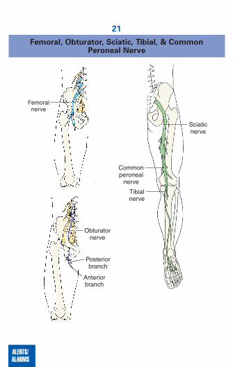
Femoral, Obturator, Sciatic, Tibial, & Common Peroneal Nerve
21
ALERTS/ALARMS
Obturatornerve
Posteriorbranch
Anteriorbranch
Femoralnerve
Sciaticnerve
Tibialnerve
Commonperoneal
nerve
2067_Tab01_001-049.qxd 3/19/09 1:03 PM Page 21

ALERTS/ALARMS
22
Deep & Superficial Peroneal Nerves
Commonperoneal
nerve
Deepperoneal
nerve
Commonperoneal
nerveSuperficialperoneal
nerve
2067_Tab01_001-049.qxd 3/19/09 1:03 PM Page 22

Ph
arm
aco
log
ic S
um
mary
by D
rug
Cla
ssific
atio
n
23
ALERTS/ALARMS
No
nn
arc
otic
An
alg
esic
Ind
icatio
ns =
Pain
, fever
Gen
eric
nam
eA
dvers
e re
actio
ns
(Bra
nd
nam
es)
(Most frequent are
bo
lded)
Inte
ractio
ns
Acetam
ino
ph
en• T
yleno
l• A
nacin
–3• Liq
uip
rin• P
anad
ol
• Acep
hen
• Tempra
Up
set stom
ach, rash
,b
ruisin
g, an
emia
Do
ses >15g are to
xicto
liver & kid
ney;
may b
e fatal
Barb
iturates = ↓
effects & ↑
liver toxicity
Warfarin
= ↑an
ticoag
ulan
t effect
Caffein
e = ↑an
algesic effects
Alco
ho
l = ↑risk o
f liver dam
age (n
ot reco
mm
end
edif co
nsu
min
g ≥
3 glasses o
f alcoh
ol/d
ay
2067_Tab01_001-049.qxd 3/19/09 1:03 PM Page 23

ALERTS/ALARMS
24
An
alg
esic
& N
SA
ID
Ind
icatio
ns =
RA
, OA
, JR
A, p
ain
, fever, p
reven
t thro
mb
osis
, red
uce ris
k o
f MI, T
IA, C
VA
An
ti-infla
mm
ato
ry d
oses a
re >
an
alg
esic
do
ses
Gen
eric
nam
eA
dvers
e re
actio
ns
(Bra
nd
nam
es)
(Most frequent are
bo
lded)
Inte
ractio
ns
Acetylsalicylic acid
(AS
A)
• Asp
irin• E
cotrin
• Em
pirin
• Bayer
• Asp
ergu
mIb
up
rofen
• Mo
trin• N
up
rin• A
dvil
Su
lind
ac• C
lino
ril
Melo
xicam (p
referential
inh
ibitio
n o
f CO
X-2 o
ver C
OX
-1)
Exercise co
ncern
s: Neg
ative effect on
myo
gen
esis & reg
eneratio
n (an
abo
lic effects)
No
t reco
mm
en
ded
for c
hild
ren
Tin
nitu
s, n
au
sea, p
rolo
ng
ed
ble
ed
ing
time, ra
sh
,GI d
istress,b
ruisin
g
GI p
x, dysp
epsia, n
ausea,
dizzin
ess, rash, h
epatitis, h
/a
No
t reco
mm
en
ded
for c
hild
ren
GI p
x,h
/a, rash, co
nstip
ation
,d
izziness, liver d
amag
e, epid
er-m
al necro
sis synd
rom
eS
eizures, cardiac arrhythmias, M
I,hem
orrhage, asthma, erythem
a,anaphylactic reaction, anxiety,abdom
inal pain, coughing
All N
SA
IDs:
• Can
↓card
iop
rotective effects o
f lo
w-d
ose asp
irin• C
an ↑
risk of b
leedin
g w
hen
used
with
gin
kgo
, vitamin
E, w
arfarin,
Plavix, &
hep
arin• C
an ↑
BP
(CO
X-2 in
hib
itors ↑
BP
toa lesser exten
t than
no
nselectives)
• Can
↑n
euro
toxicity w
hen
used
w
ith lith
ium
• Can
pro
du
ce acute ren
al failure
• Are g
astric irritants &
can p
rod
uce
nep
hro
toxicity
2067_Tab01_001-049.qxd 3/19/09 1:03 PM Page 24
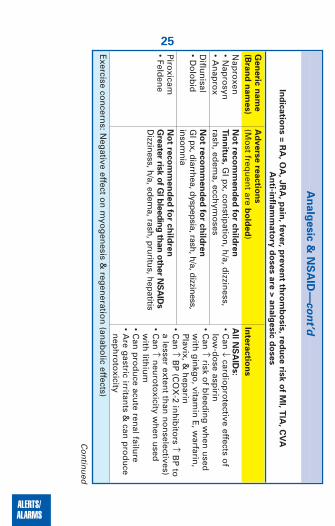
25
ALERTS/ALARMS
An
alg
esic
& N
SA
ID—
co
nt’d
Ind
icatio
ns =
RA
, OA
, JR
A, p
ain
, fever, p
reven
t thro
mb
osis
, red
uce ris
k o
f MI, T
IA, C
VA
An
ti-infla
mm
ato
ry d
oses a
re >
an
alg
esic
do
ses
Gen
eric
nam
eA
dvers
e re
actio
ns
(Bra
nd
nam
es)
(Mo
st frequ
ent are
bo
lded
)In
tera
ctio
ns
Nap
roxen
• Nap
rosyn
• An
apro
xD
iflun
isal• D
olo
bid
Piro
xicam• Feld
ene
Exercise co
ncern
s: Neg
ative effect on
myo
gen
esis & reg
eneratio
n (an
abo
lic effects)
No
t reco
mm
en
ded
for c
hild
ren
Tin
nitu
s,G
I px, co
nstip
ation
, h/a, d
izziness,
rash, ed
ema, ecch
ymo
sesN
ot re
co
mm
en
ded
for c
hild
ren
GI px, diarrhea, dyspepsia, rash, h/a, dizziness,
insomnia
No
t reco
mm
en
ded
for c
hild
ren
Gre
ate
r risk o
f GI b
leed
ing
than
oth
er N
SA
IDs
Dizziness, h/a, edem
a, rash, pruritus, hepatitis
All N
SA
IDs:
• Can
↓card
iop
rotective effects o
flo
w-d
ose asp
irin• C
an ↑
risk of b
leedin
g w
hen
used
with
gin
kgo
, vitamin
E, w
arfarin,
Plavix, &
hep
arin• C
an ↑
BP
(CO
X-2 in
hib
itors ↑
BP
toa lesser exten
t than
no
nselectives)
• Can
↑n
euro
toxicity w
hen
used
w
ith lith
ium
• Can
pro
du
ce acute ren
al failure
• Are g
astric irritants &
can p
rod
uce
nep
hro
toxicity
Continued
2067_Tab01_001-049.qxd 3/19/09 1:03 PM Page 25

ALERTS/ALARMS
26
An
alg
esic
& N
SA
ID—
co
nt’d
Ind
icatio
ns =
RA
, OA
, JR
A, p
ain
, fever, p
reven
t thro
mb
osis
, red
uce ris
k o
f MI, T
IA, C
VA
An
ti-infla
mm
ato
ry d
oses a
re >
an
alg
esic
do
ses
Gen
eric
nam
eA
dvers
e re
actio
ns
(Bra
nd
nam
es)
(Mo
st frequ
ent are
bo
lded
)In
tera
ctio
ns
Ind
om
ethacin
• Ind
ocin
Eto
do
lac• Lo
din
e
Keto
pro
fen• O
rud
is
Exercise co
ncern
s: Neg
ative effect on
myo
gen
esis & reg
eneratio
n (an
abo
lic effects)
H/a
,dro
wsy, d
izziness, n
ausea, G
I px, co
nsti-
patio
n, p
ancreatitis
No
t reco
mm
en
ded
for c
hild
ren
Dysp
ep
sia
, sligh
tly less GI p
x than
oth
erN
SA
ID, n
ausea, d
iarrhea, C
HF, d
izziness, ↑
BP,b
lurred
vision
No
t reco
mm
en
ded
for c
hild
ren
Dysp
ep
sia, h
/a, dizzin
ess, inso
mn
ia, tinn
itus,
perip
heral ed
ema
All N
SA
IDs:
• Can
↓card
iop
rotective effects o
flo
w-d
ose asp
irin• C
an ↑
risk of b
leedin
g w
hen
used
with
gin
kgo
, vitamin
E, w
arfarin,
Plavix, &
hep
arin• C
an ↑
BP
(CO
X-2 in
hib
itors ↑
BP
toa lesser exten
t than
no
nselectives)
• Can
↑n
euro
toxicity w
hen
used
with
lithiu
m• C
an p
rod
uce acu
te renal failu
re• A
re gastric irritan
ts & can
pro
du
cen
eph
roto
xicity
2067_Tab01_001-049.qxd 3/19/09 1:03 PM Page 26

27
ALERTS/ALARMS
An
alg
esic
& N
SA
ID—
co
nt’d
Ind
icatio
ns =
RA
, OA
, JR
A, p
ain
, fever, p
reven
t thro
mb
osis
, red
uce ris
k o
f MI, T
IA, C
VA
An
ti-infla
mm
ato
ry d
oses a
re >
an
alg
esic
do
ses
Gen
eric
nam
eA
dvers
e re
actio
ns
(Bra
nd
nam
es)
(Mo
st frequ
ent are
bo
lded
)In
tera
ctio
ns
Diclo
fenac
• Vo
ltaren• C
ataflamN
abu
meto
ne
• Relafen
Celeco
xib(C
OX
-2 inh
ib)
• Celeb
rex
Exercise co
ncern
s: Neg
ative effect on
myo
gen
esis & reg
eneratio
n (an
abo
lic effects), i.e., may d
elaym
uscle h
ealing
No
t reco
mm
en
ded
for c
hild
ren
Nep
hro
tic p
x,G
I px, h
/a, edem
a, dizzin
ess,h
ypo
glycem
iaN
ot re
co
mm
en
ded
for c
hild
ren
Ab
do
min
al p
ain
, dia
rrhea, d
ysp
ep
sia
,dizziness,h/a, dyspnea, diaphoresisN
ot re
co
mm
en
ded
for c
hild
ren
h/a
,GI p
x, dizzin
ess, ↑B
P, erythema
All N
SA
IDs:
• Can
↓card
iop
rotective effects o
flo
w-d
ose asp
irin• C
an ↑
risk of b
leedin
g w
hen
used
with
gin
kgo
, vitamin
E, w
arfarin,
Plavix, &
hep
arin• C
an ↑
BP
(CO
X-2 in
hib
itors ↑
BP
toa lesser exten
t than
no
nselectives)
• Can
↑n
euro
toxicity w
hen
used
with
lithiu
m• C
an p
rod
uce acu
te renal failu
re• A
re gastric irritan
ts & can
pro
du
cen
eph
roto
xicity
2067_Tab01_001-049.qxd 3/19/09 1:03 PM Page 27

ALERTS/ALARMS
28
*N
arc
otic
An
alg
esic
: APA
P =
Aceta
min
op
hen
Ind
icatio
n =
Pain
Gen
eric
nam
eA
dvers
e re
actio
ns
(Bra
nd
nam
es)
(Mo
st frequ
ent are
bo
lded
)In
tera
ctio
ns
AP
AP
/hyd
roco
do
ne**
• Vico
din
• Lortab
AP
AP
/cod
eine**
• Tylen
ol #3
AP
AP
/oxyco
do
ne
• Perco
cet• T
ylox
Exercise co
ncern
s: Red
uced
exercise capacity d
ue to
respirato
ry dep
ression
especially w
ith C
OP
D;
gu
ard am
bu
lation
to p
revent falls
Diz
zin
ess, n
au
sea, vom
iting, con-fusion, constipation, rash, pruritus,depressionN
au
sea, drow
siness, constipation,nausea, vom
iting, SO
B, pruritus
↓respiration (body builds up
tolerance after 2 wks)
Lig
hth
ead
ed
, diz
zin
ess, n
au
sea,
vo
mitin
g, a
pn
ea, re
sp
irato
ry
dis
tress, h
yp
ote
nsio
n,rash
, co
nstip
ation
, pru
ritus
An
tihistam
ines, an
tipsych
otics, o
ran
tianxiety ag
ents = ↑
CN
S d
epressio
nM
AO
inh
ibito
rs= ↑effects
An
tipsych
otics, an
tianxiety ag
ents, o
ralco
ho
l = ↑C
NS
dep
ression
An
ticho
linerg
ics with
cod
eine = p
aralyticileu
sM
uscle relaxers = ↑
CN
S effects
*A
LL o
pio
ids are ad
dictin
g; w
ithd
rawal sym
pto
ms m
ay app
ear in 6–10 h
ou
rs & last 5 d
ays. Sym
pto
ms m
ay inclu
de b
od
y aches, d
iarrhea,
fever, go
oseflesh
, inso
mn
ia, irritability, lo
ss of ap
petite, n
ausea, vo
mitin
g, ru
nn
y no
se, shiverin
g, &
stom
ach cram
ps.
**Sh
ou
ld n
ot b
e taken w
ith M
AO
inh
ibito
rs.
2067_Tab01_001-049.qxd 3/19/09 1:03 PM Page 28

29
ALERTS/ALARMS
*N
arc
otic
An
alg
esic
: AS
A =
Asp
irin
Ind
icatio
n =
Pain
Gen
eric
nam
eA
dvers
e re
actio
ns
Inte
ractio
ns
(Bra
nd
nam
es)
(Mo
st frequ
ent are
bo
lded
)(A
ll inte
ractio
n e
ffects
of A
SA
ap
ply
)
AS
A/co
dein
e**• E
mp
irin w
ith co
dein
e Take w
ith fo
od
AS
A/o
xycod
on
e• P
ercod
an
Exercise co
ncern
s: Neg
ative effects on
myo
gen
esis & reg
eneratio
n (an
abo
lic effects)
Diz
zin
ess, n
au
sea, ↓
resp
iratio
n,
co
nstip
atio
n, tin
nitu
s, h/a, vo
mit-
ing
, pru
ritus, rash
Lig
hth
ead
ed
, nau
sea, d
izzin
ess,
vom
iting
, eup
ho
ria, pru
ritus,
apn
ea, con
stipatio
n, circu
latory
dep
ression
, hem
orrh
age,
hyp
oten
sion
MA
O inhibitors, insulin, anticoagulants,
methotrexate, or sulfonam
ides = ↑effects
NS
AID
s = pep
tic ulcers
Alco
ho
l = ↑C
NS
dep
ression
Mu
scle relaxants = ↑
CN
S effects, im
pair
jud
gm
ent
Analgesics, phenothiazines, tranquilizers,
or alcohol = ↑C
NS
depressionA
CE
inh
ibito
rs = ↓p
ain relief
An
ticoag
ulan
t or N
SA
ID = ↑
bleed
ing
Meth
otrexate = ↑
toxicity
*A
LL o
pio
ids are ad
dictin
g; w
ithd
rawal sym
pto
ms m
ay app
ear in 6–10 h
ou
rs & last 5 d
ays. Sym
pto
ms m
ay inclu
de b
od
y aches, d
iarrhea,
fever, go
oseflesh
, inso
mn
ia, irritability, lo
ss of ap
petite, n
ausea, vo
mitin
g, ru
nn
y no
se, shiverin
g, &
stom
ach cram
ps.
**Sh
ou
ld n
ot b
e taken w
ith M
AO
inh
ibito
rs.
2067_Tab01_001-049.qxd 3/19/09 1:03 PM Page 29

ALERTS/ALARMS
30
Mu
scle
Rela
xers
/An
tisp
asm
od
ics
Ind
icatio
ns =
Man
ag
e s
pastic
ity (m
uscle
ton
e), re
du
ce m
uscle
gu
ard
ing
Gen
eric
nam
eA
dvers
e re
actio
ns
(Bra
nd
nam
es)
(Mo
st frequ
ent are
bo
lded
)In
tera
ctio
ns
Baclo
fen• Lio
resal
Cariso
pro
do
l• S
om
a (add
ictive)
Cyclo
ben
zaprin
e• Flexeril (u
se no
t reco
mm
end
ed
for > 2–3 w
ks)D
iazepam
• Valiu
m (lo
ng
-term
dep
end
ency)
Tizan
idin
e• Z
anaflex
Exercise co
ncern
s: Interferes w
ith stren
gth
enin
g g
oals
Dro
wsin
ess, n
au
sea, d
izzin
ess,
weakn
ess, c
on
fusio
n, vo
mitin
g,
hig
h fever, h
/a, rash, p
aresthesias
Orth
osta
tic h
yp
ote
nsio
n, d
row
sin
ess,
diz
zin
ess,h/a, vertigo, agitation,
insomnia
Dro
wsin
ess, d
ry m
ou
th, d
izzin
ess,
arrhyth
mias, co
nfu
sion
, transien
tvisu
al hallu
cinatio
ns
Dro
wsin
ess, p
ain
, ph
leb
itis a
t inje
c-
tion
site
,dysarth
ria, con
stipatio
n,
↓H
R, ↓
RR
So
mn
ole
nce, s
ed
atio
n, h
yp
ote
nsio
n,
dry
mo
uth
, UT
I,dizziness, bradycardia,constipation
CN
S d
epressan
ts or alco
ho
l = ↑d
epressio
n
CN
S d
epressan
ts or alco
ho
l = ↑d
epressio
n
CN
S d
epressan
ts or alco
ho
l = ↑d
epressio
nM
AO
inh
ibito
rs or T
ramad
ol = m
ay cause
seizures &
death
CN
S d
epressan
ts or alco
ho
l = ↑d
epressio
nD
igo
xin = risk o
f toxicity
Sm
okin
g = m
ay ↓effects
An
tihyp
ertensives = ↓
BP
Baclo
fen, alco
ho
l, or o
ther C
NS
dep
ressant
= add
itive effectO
ral con
traceptive = ↓
tizanid
ine clearan
ce
2067_Tab01_001-049.qxd 3/19/09 1:03 PM Page 30

31
ALERTS/ALARMS
AC
E In
hib
itors
Ind
icatio
n =
Hig
h B
P
Gen
eric
nam
eA
dvers
e re
actio
ns
(Bra
nd
nam
es)
(Mo
st frequ
ent are
bo
lded
)In
tera
ctio
ns
Cap
top
ril• C
apo
tenE
nalap
ril• V
asotec
Lisino
pril
• Zestril
• Prin
ivilFo
sino
pril
• Mo
no
pril
Qu
inap
ril• A
ccup
rilE
xercise con
cerns: N
o effect o
n exercise cap
acity
Dry
co
ug
h, ra
sh
,dizziness, abdomi-
nal pain, neutropeniaW
eakn
ess, d
ry c
ou
gh
,dizzin
ess, h/a,
hyp
oten
sion
Diz
zin
ess, n
asal c
on
gestio
n, d
ry
co
ug
h, o
rtho
sta
tic h
yp
ote
nsio
n,
dia
rrhea,h
/a, fatigu
e, nau
seaD
izzin
ess, d
ry c
ou
gh
,h/a, fatig
ue,
diarrh
ea, nau
seaS
om
no
lence, p
ruritu
s, dizzin
ess, dry
cou
gh
, hem
orrh
age
An
tacids = ↑
effectsD
igo
xin = ↑
dig
oxin
levelsD
iuretics o
r ph
eno
thiazin
es = hyp
oten
sion
NS
AID
s = ↓an
tihyp
ertensive effects
Insu
lin = ↑
hyp
og
lycemia
Lithiu
m = lith
ium
toxicity
2067_Tab01_001-049.qxd 3/19/09 1:03 PM Page 31

ALERTS/ALARMS
32
AC
E R
ecep
tor B
lockers
Ind
icatio
n =
Hig
h B
P
Gen
eric
nam
eA
dvers
e re
actio
ns
(Bra
nd
nam
es)
(Mo
st frequ
ent are
bo
lded
)In
tera
ctio
ns
Losartan
K+
• Co
zaar
Can
desartan
• Atacan
dIrb
esartan• A
vapro
Exercise co
ncern
s: No
effect on
exercise capacity
Dizzin
ess, h/a, w
eakness, fatig
ue,
chest p
ain, d
iarrhea, an
emia, flu
-likesym
pto
ms
Dizzin
ess, h/a, ru
nn
y no
se, UR
I
An
xiety, chest p
ain, d
iarrhea, d
izzi-n
ess, flu, h
/a, fatigu
e, nau
sea, up
setsto
mach
, sore th
roat, U
TI, vo
mitin
g
Du
e to ↑
K+
levels, sho
uld
no
t be taken
with
K+
sup
plem
ents, salt su
bstitu
tes con
-tain
ing
K+, o
r K+-sp
aring
diu
reticsN
SA
IDs &
AS
A = ↓
antih
yperten
sive effects
2067_Tab01_001-049.qxd 3/19/09 1:03 PM Page 32

33
ALERTS/ALARMS
Ca
++
Ch
an
nel B
lockers
Ind
icatio
n =
An
gin
a
Gen
eric
nam
eA
dvers
e re
actio
ns (M
ost(B
ran
d n
am
es)
frequ
ent are
bo
lded
)In
tera
ctio
ns
Diltiazem
• Card
izem• D
ilacor
• Diltiaz
• Tiazac
Verap
amil
• Calan
Am
lod
ipin
e• N
orvasc
• Am
vazN
ifedip
ine
• Pro
cardia
Exercise co
ncern
s: Dru
gs m
ay cause arth
ralgia/m
yalgia th
at may n
egatively in
fluen
ce exercise capacity
LE
ed
em
a, h
/a, 1°
heart b
lock, arrhythm
ia,bradycardia, nausea,rash, dizziness, fatigue,1°
heart block H
yp
ote
nsio
n, A
V b
lock,
co
nstip
atio
n, d
izziness,
nau
sea, h/a, arrh
ythm
ia,d
yspn
ea
Ed
em
a,h
/a, fatigu
e,n
ausea, flu
shin
g, rash
,LE
edem
a, dizzin
essD
izzin
ess, h
/a, w
eak-
ness, flu
sh
ing
, perip
h-
era
l ed
em
a, n
au
sea
Dig
oxin
= elevated d
igitalis levels
An
esthetics = ↑
anesth
etic effects & d
epressio
n o
f cardiac
con
tractilityC
yclosp
orin
e = ↑cyclo
spo
rine level
Diazep
am = ↑
CN
S d
epressio
nB
eta-blo
ckers = heart failu
reC
ardiac g
lycosid
e = ↑d
igitalis levels
An
tihyp
ertensives = h
ypo
tensio
nC
yclosp
orin
e = ↑levels
Grap
efruit ju
ice = ↑d
rug
levelS
t. Joh
n’s w
ort = ↓
dru
g level
Alco
ho
l = ↑alco
ho
l levelW
hen
com
bin
ed w
ith an
oth
er antih
yperten
sive = hyp
oten
sion
Wh
en co
mb
ined
with
an alp
ha b
locker = h
ypo
tensio
n &
reflextach
ycardia
Verap
amil = ↓
effectsA
ntifu
ng
als or eryth
rom
ycin = ↑
effectsFen
tanyl = severe h
ypo
tensio
nC
imetid
ine = ↑
plasm
a level of n
ifedip
ine
Beta b
lockers = h
ypo
tensio
nG
inkg
o o
r grap
efruit ju
ice = ↑effects
St. Jo
hn
’s wo
rt = ↓d
rug
effect
2067_Tab01_001-049.qxd 3/19/09 1:03 PM Page 33

ALERTS/ALARMS
34
*B
eta
Blo
ck
ers
/An
tihy
pe
rten
siv
es
Ind
ica
tion
s =
An
gin
a, a
rrhyth
mia
s, h
yp
erte
nsio
n
Gen
eric
nam
eA
dvers
e re
actio
ns
(Bra
nd
nam
es)
(Mo
st frequ
ent are
bo
lded
)In
tera
ctio
ns
Pro
pran
olo
l• In
deral
• Inn
oP
ran
Aten
olo
l• T
eno
rmin
Tim
olo
l• B
locad
ren
Meto
pro
lol
• Lop
ressor
• Toprol
Labetalo
l• N
orm
od
yne
• Tran
date
↑LD
L c
ho
leste
rol, b
rad
ycard
ia, fa
tigu
e,
leth
arg
y, h
yp
ote
nsio
n,lig
hth
eaded
,ab
do
min
al cramp
ing
, rash, R
aynau
d’s,
bro
nch
osp
asm in
asthm
atics
↑LD
L c
ho
leste
rol, d
izzin
ess, fa
tigu
e,
hyp
ote
nsio
n, b
rad
ycard
ia,n
ausea, LE
pain
, rash, b
ron
cho
spasm
s, orth
ostatic
hyp
oten
sion
↑LD
L c
ho
leste
rol,b
ron
cho
spasm
s,fatig
ue, b
radycard
ia, extremity p
ain,
weakn
ess, imp
oten
ce ↑
LD
L c
ho
leste
rol, fa
tigu
e, d
izzin
ess,
dep
ressio
n, h
yp
ote
nsio
n,b
radycard
ia,n
ausea, rash
, bro
nch
osp
asms
↑LD
L c
ho
leste
rol, d
izziness, n
ausea,
fatigu
e, hyp
oten
sion
Verap
amil o
r diltiazem
= hyp
oten
sion
Ep
inep
hrin
e = severe perip
heral
vasoco
nstrictio
nIn
sulin
= hyp
og
lycemia
Ph
eno
thiazin
es = ↑ad
verse reaction
sN
SA
IDs = ↓
antih
yperten
sive effectC
a ++ch
ann
el blo
ckers or p
razosin
= ↑
hyp
oten
sion
Card
iac glyco
sides = severe b
radycard
iaIn
sulin
= may alter d
osag
eN
SA
IDs = ↓
antih
yperten
sive effectsN
SA
IDs = ↓
antih
yperten
sive effect
Card
iac glyco
sides = severe b
radycard
iaM
AO
inhibitors, cimetidine, hydralazine,
prazosin, or verapamil = additive effects;
hypotension & bradycardia
Cim
etidin
e = ↑lab
etalol p
lasma levels
Verap
amil = ad
ditive effects
NS
AID
s = ↓an
tihyp
ertensive effect
*Sh
ou
ld n
ot b
e taken w
ith M
AO
inh
ibito
rs
2067_Tab01_001-049.qxd 3/19/09 1:03 PM Page 34

35
ALERTS/ALARMS
An
tilipem
ics
Ind
icatio
ns =
Red
uce L
DL, to
tal c
ho
leste
rol, &
trigly
cerid
e le
vels
Gen
eric
nam
eA
dvers
e re
actio
ns
(Bra
nd
nam
es)
(Mo
st frequ
ent are
bo
lded
)In
tera
ctio
ns
Ato
rvastatin• Lip
itor
Exercise co
ncern
s: Mu
scle weakn
ess & cram
pin
g, m
yalgia
Co
nstip
ation
, mu
scle pain
, flatulen
ce, ↑
liver transam
inase, d
yspep
sia, rh
abd
om
yolysis
An
tacids = ↓
plasm
a level of ato
rvastatinD
igo
xin o
r erythro
mycin
= ↑p
lasma level
of ato
rvastatinB
CP
= ↑p
lasma level o
f BC
PE
rythro
mycin
, niacin
, or an
tifun
gals =
↑risk o
f myo
path
y
*B
eta
Blo
ck
ers
/An
tihy
pe
rten
siv
es—
co
nt’d
Ind
ica
tion
s =
An
gin
a, a
rrhyth
mia
s, h
yp
erte
nsio
n
Gen
eric
nam
eA
dvers
e re
actio
ns
(Bra
nd
nam
es)
(Mo
st frequ
ent are
bo
lded
)In
tera
ctio
ns
Carved
ilol
• Co
reg
Exercise co
ncern
s: As a resu
lt of a b
lun
ting
of H
R, exercise to
20 bp
m ab
ove restin
g H
R; b
eta blo
ckersm
ask symp
tom
s of &
delay reco
very from
hyp
og
lycemia
↑LD
L c
ho
leste
rol, asthenia, dizziness,
fatigue, hypotension, diarrhea, hyper-glycem
ia, wt gain, U
RI
*May p
rod
uce b
ron
cho
con
striction
inp
atients w
ith asth
matic co
nd
ition
s
Cim
etidin
e = ↑carved
ilol p
lasma levels
MA
O inhibitors = bradycardia &
↓B
PC
a ++ch
ann
el blo
ckers = con
du
ction
d
isturb
ances
NS
AID
s = ↓an
tihyp
ertensive effect
2067_Tab01_001-049.qxd 3/19/09 1:03 PM Page 35

ALERTS/ALARMS
36
Diu
retic
s
Ind
ica
tion
s =
Ed
em
a, h
yp
erte
nsio
n
Gen
eric
nam
eA
dvers
e re
actio
ns
(Bra
nd
nam
es)
(Mo
st frequ
ent are
bo
lded
)In
tera
ctio
ns
Furo
semid
e (lo
op
diu
retic)• LasixT
hiazid
e• E
sidrix
• Hyd
rod
iuril
• Lozo
l• Z
aroxo
lynK
+sp
aring
• Ald
acton
e• D
yreniu
mE
xercise con
cerns: D
imin
ished
exercise perfo
rman
ce; limited
mu
scle end
uran
ce; volu
me d
epletio
n;
↑ risk of h
eat-related illn
ess; mu
scle cramp
s 2°h
ypo
kalemia
Deh
ydratio
n, m
uscle cram
ps,
hyp
okalem
ia, hyp
ocalcem
ia (o
steop
oro
sis), cardia arrh
ythm
iasD
izzin
ess,m
uscle w
eakness, cram
ps,
thirst, h
yperg
lycemia, sto
mach
d
iscom
fort
Diz
zin
ess,w
eakness, fatig
ue, h
/a, d
iarrhea, d
ry mo
uth
, mu
scle cramp
s
An
tihyp
ertensives o
r Ca ++
chan
nel b
locker =
↑risk o
f hyp
oten
sion
& arrh
ythm
iasLoop + thiazide diuretic = ↑
risk of hypotension&
arrhythmias
Card
iac glyco
sides = ↑
risk of d
igo
xin to
xicityw
ith K
+lo
ssN
SA
IDs = in
hib
it diu
retic respo
nse
Su
n = p
ho
tosen
sitivity
2067_Tab01_001-049.qxd 3/19/09 1:03 PM Page 36

37
ALERTS/ALARMS
An
tidep
ressan
ts
Ind
icatio
n =
Dep
ressio
n, O
CD
, an
xie
ty
Gen
eric
nam
eA
dvers
e re
actio
ns
(Bra
nd
nam
es)
(Mo
st frequ
ent are
bo
lded
)In
tera
ctio
ns
Am
itriptylin
e• E
lavil
Do
xepin
• Sin
equ
an• A
dap
in• Z
on
alon
Bu
pro
pio
n• W
ellbu
trin• Z
yban
Orth
osta
tic h
yp
ote
nsio
n, ta
ch
ycard
ia,
dry
mo
uth
,stroke, arrhythmia, lethargy,
con
fusio
n, d
ry mo
uth
, urin
ary reten-
tion
, blu
rred visio
n, co
nstip
ation
Dro
wsin
ess, d
izzin
ess, d
ry m
ou
th,
orth
osta
tic h
yp
ote
nsio
n, b
lurre
d
vis
ion
, tach
ycard
ia, d
iap
ho
resis
, co
n-
stip
atio
n, s
eiz
ure
s,co
nfu
sion
, urin
aryreten
tion
Inso
mn
ia, a
gita
tion
, dry
mo
uth
,
trem
or, a
bn
orm
al d
ream
s, h
/a,
excess s
weatin
g, ta
ch
ycard
ia,
nau
sea, c
on
stip
atio
n, v
om
iting
,
diz
zin
ess, rh
initis
, an
ore
xia
, blu
rred
vis
ion
,wt g
ain, seizu
res
Co
ntracep
tives = ↑an
tidep
ressant level &
↑tricyclic-in
du
ced akath
isiaC
lon
idin
e or ep
inep
hrin
e = extreme
hyp
ertensio
nM
AO
inh
ibito
rs = severe excitation
Quinolones = life-threatening arrh
ythm
ias (↑
QTc interval)
Alco
ho
l = CN
S d
epressio
nS
un
= ph
oto
sensitivity
Co
ntracep
tives = ↑an
tidep
ressant level
Clo
nid
ine o
r epin
eph
rine = extrem
e h
yperten
sion
MA
O in
hib
itors = severe excitatio
nQ
uinolones = life-threatening arrhythmias
Alco
ho
l = CN
S d
epressio
nS
un
= ph
oto
sensitivity
MA
O in
hib
itors = ↑
risk of to
xicityN
icotin
e = hyp
ertensio
nLevo
do
pa = ↑
risk of ad
verse reaction
sS
un
= ph
oto
sensitivity
Pred
niso
ne o
r ph
eno
thiazin
e = ↑risk o
fseizu
res
2067_Tab01_001-049.qxd 3/19/09 1:03 PM Page 37

ALERTS/ALARMS
38
An
tidep
ressan
ts—
co
nt’d
Ind
icatio
n =
Dep
ressio
n, O
CD
, an
xie
ty
Gen
eric
nam
eA
dvers
e re
actio
ns
(Bra
nd
nam
es)
(Mo
st frequ
ent are
bo
lded
)In
tera
ctio
ns
Fluo
xetine*
• Pro
zac
Sertralin
e*• Z
olo
ft
Exercise co
ncern
s: Imp
roved
mo
tor p
erform
ance fo
llow
ing
ischem
ic stroke
Nervo
usn
ess, som
no
lence, in
som
nia,
anxiety, d
row
siness, h
/a, tremo
r, d
izziness, w
eakness, n
ausea,
diarrh
ea, dry m
ou
th, an
orexia,
akathisia
Fatig
ue, h
/a, tre
mo
r, diz
zin
ess, in
so
m-
nia
, so
mn
ole
nce, d
ry m
ou
th, n
au
sea,
dia
rrhea, m
ale
sexu
al d
ysfu
nctio
n,
suicid
al beh
avior, akath
isia
Beta b
lockers = h
eart blo
ck, brad
ycardia
MA
O in
hib
itors o
r St Jo
hn
’s wo
rt = seroto
nin
synd
rom
e A
ntip
sycho
tics = ↑co
ncen
tration
of an
tipsy-
cho
tics (extrapyram
idal sig
ns)
Warfarin
= ↑b
leedin
gA
lcoh
ol = ↑
dep
ression
Ben
zod
iazepin
es = ↑effects
MA
O in
hib
itors, trip
tans, iso
niazid
, or
St Jo
hn
’s wo
rt = seroto
nin
synd
rom
eW
arfarin = ↑
bleed
ing
*Sh
ou
ld n
ot b
e taken w
ith M
AO
inh
ibito
rs.
2067_Tab01_001-049.qxd 3/19/09 1:03 PM Page 38

39
ALERTS/ALARMS
Deco
ng
esta
nts
, An
tihis
tam
ines, &
Bro
nch
od
ilato
rs
Ind
icatio
ns =
Bro
nch
osp
asm
s, C
OP
D, e
mp
hysem
a
Gen
eric
nam
eA
dvers
e re
actio
ns
(Bra
nd
nam
es)
(Mo
st frequ
ent are
bo
lded
)In
tera
ctio
ns
Alb
utero
l• P
roven
til• V
ento
lin• B
rethin
e
Pirb
utero
l• M
axair
Salm
eterol
• Sereven
t d
iscus
Exercise co
ncern
s: Dim
inish
ed exercise p
erform
ance; lim
ited m
uscle en
du
rance; system
ic adm
inistratio
nm
ay ↑ hyp
erglycem
ia
Tre
mo
r, nerv
ou
sn
ess, h
/a, h
yp
era
ctiv
-
ity, ta
ch
ycard
ia, n
au
sea, v
om
iting
,
mu
scle cramp
s, hyp
ocalcem
ia, cou
gh
,h
yperg
lycemia
Trem
or, n
ervou
sness, d
izziness,
tachycard
ia, nau
sea, vom
iting
, cou
gh
,h
yperg
lycemia
Naso
ph
ary
ng
itis, U
RI, h
/a, tremo
r, n
ausea, n
ervou
sness, tach
ycardia,
myalg
ia
CN
S stim
ulan
t = ↑C
NS
effectsM
AO
inhibitors or antidepressants = ↑adverse
CV
effectsB
eta blo
ckers = con
traind
icated, m
ay cause
bro
nch
oco
nstrictio
nB
eta blo
ckers = con
traind
icated, m
ay cause
bro
nch
oco
nstrictio
n
MA
O in
hib
itors o
r antid
epressan
ts = ↑effects
Beta b
lockers = co
ntrain
dicated
, may cau
seb
ron
cho
con
striction
M
AO
inh
ibito
rs or an
tidep
ressants = ↑
risk of
severe CV
effects
2067_Tab01_001-049.qxd 3/19/09 1:03 PM Page 39

ALERTS/ALARMS
40
Abbreviations & Symbols Specific to Orthopedics
Please note: This list is not comprehensive and is subject to modification by various
facilities to meet the needs of their patient population.
a . . . . . . . . . . . . . .beforeA . . . . . . . . . . . . .assistanceAAA . . . . . . . . . . .abdominal aortic aneurysmAAROM . . . . . . . .active, assistive range of motionAbd . . . . . . . . . . .abductionABG . . . . . . . . . . .arterial blood gasesACL . . . . . . . . . . .anterior cruciate ligamentA.C. . . . . . . . . . . .before mealsAdd . . . . . . . . . . .adductionADLs . . . . . . . . . .activities of daily livingad lib . . . . . . . . . .as desiredAE . . . . . . . . . . . .above elbowAFib . . . . . . . . . . .atrial fibrillationAFO . . . . . . . . . . .ankle foot orthosisAK . . . . . . . . . . . .above kneeAMA . . . . . . . . . .against medical adviceamb . . . . . . . . . .ambulationANS . . . . . . . . . . .autonomic nervous systemAP . . . . . . . . . . . .anterior-posteriorAPL . . . . . . . . . . .abductor pollicis longusARD . . . . . . . . . . .adult respiratory distressAROM . . . . . . . . .active range of motionASA . . . . . . . . . .aspirinASCVD . . . . . . . .arteriosclerotic cardiovascular diseaseASIS . . . . . . . . . .anterior superior iliac spineATFL . . . . . . . . . .anterior talofibular ligamentA-V . . . . . . . . . . .arterio-venousB . . . . . . . . . . . . .bilateralBBB . . . . . . . . . . .bundle branch blockB&B . . . . . . . . . .bowel & bladderBE . . . . . . . . . . . .below elbowBID . . . . . . . . . . .twice dailyBK . . . . . . . . . . . .below kneeBMI . . . . . . . . . . .body mass indexBMR . . . . . . . . . .basal metabolic rate
2067_Tab01_001-049.qxd 3/19/09 1:03 PM Page 40

BM . . . . . . . . . . . .bowel movementBOS . . . . . . . . . . .base of supportBP . . . . . . . . . . . .blood pressureBRP . . . . . . . . . . .bathroom privilegesBS . . . . . . . . . . . .breath soundsBUN . . . . . . . . . .blood urea nitrogenBx . . . . . . . . . . . .biopsyc . . . . . . . . . . . . . .withCa++ . . . . . . . . . .calciumCA . . . . . . . . . . . .cancerCABG . . . . . . . . .coronary artery bypass graftCAD . . . . . . . . . . .coronary artery diseaseCBC . . . . . . . . . . .complete blood countCC . . . . . . . . . . . .chief complaintCCE . . . . . . . . . . .clubbing, claudication, edemaCHF . . . . . . . . . . .congestive heart failureCHI . . . . . . . . . . .closed head injuryCKC . . . . . . . . . . .closed kinetic chainCN . . . . . . . . . . . .cranial nerveCNS . . . . . . . . . . .central nervous systemc/o . . . . . . . . . . . .complaints ofCO . . . . . . . . . . . .cardiac outputCOPD . . . . . . . . .chronic obstructive pulmonary diseaseCP . . . . . . . . . . . .cerebral palsyCP . . . . . . . . . . . .chest painCPK . . . . . . . . . . .creatine phosphokinaseCPM . . . . . . . . . .continuous passive motion CPP . . . . . . . . . . .closed packed positionCPR . . . . . . . . . . .cardiopulmonary resuscitationCSF . . . . . . . . . . .cerebral spinal fluidCT . . . . . . . . . . . .computed tomographyCTS . . . . . . . . . . .carpal tunnel syndromeCtx . . . . . . . . . . . .cervical tractionCVA . . . . . . . . . . .cerebral vascular accident CXR . . . . . . . . . .chest x-rayD/C . . . . . . . . . . .dischargeDDD . . . . . . . . . . .degenerative disc diseaseDDX . . . . . . . . . . .differential diagnosisDF . . . . . . . . . . . .dorsiflexionDIP . . . . . . . . . . . .distal interphalangealDJD . . . . . . . . . . .degenerative joint disease
41
ALERTS/ALARMS
2067_Tab01_001-049.qxd 3/19/09 1:03 PM Page 41

ALERTS/ALARMS
42
DM . . . . . . . . . . .diabetes mellitusDNR . . . . . . . . . . .do not resuscitateDOB . . . . . . . . . . .date of birthDOE . . . . . . . . . . .dyspnea on exertionDPT . . . . . . . . . . .diphtheria, pertussis, tetanusDSD . . . . . . . . . . .dry sterile dressingDTR . . . . . . . . . . .deep tendon reflexesDVT . . . . . . . . . . .deep vein thrombosisDx . . . . . . . . . . . .diagnosisEAA . . . . . . . . . . .essential amino acidsBL . . . . . . . . . . . .estimated blood lossEEG . . . . . . . . . . .electroencephalogramECK, EKG . . . . . .electrocardiogramEMG . . . . . . . . . .electromyogramENT . . . . . . . . . . .ear, nose, throatEOMI . . . . . . . . . .extra-ocular motion intactEPB . . . . . . . . . . .extensor pollicis brevisER . . . . . . . . . . . .external rotationESR . . . . . . . . . . .erythrocyte sedimentation rateETOH . . . . . . . . . .ethyl alcoholev . . . . . . . . . . . .eversionEx . . . . . . . . . . . .exerciseExt . . . . . . . . . . . .extensionF . . . . . . . . . . . . .frequencyFAQ . . . . . . . . . . .full arc quadsFB . . . . . . . . . . . .feedbackf/b . . . . . . . . . . . .followed byFCU . . . . . . . . . . .flexor carpi ulnarisFDP . . . . . . . . . . .flexor digitorum profundusFEV . . . . . . . . . . .forced expiratory volumeflex . . . . . . . . . . .flexionFOOSH . . . . . . . .fall on outstretched handFPL . . . . . . . . . . .flexor pollicis longusFRC . . . . . . . . . . .functional residual capacityFUO . . . . . . . . . . .fever of unknown originFVC . . . . . . . . . . .forced vital capacityFWB . . . . . . . . . .full weight bearingFx . . . . . . . . . . . .fracturef/u . . . . . . . . . . . .follow-upGB . . . . . . . . . . . .gallbladderGI . . . . . . . . . . . . .gastrointestinal
2067_Tab01_001-049.qxd 3/19/09 1:03 PM Page 42

Grav. 1 . . . . . . . .number of pregnancies (para = births)GSW . . . . . . . . . .gunshot woundGTO . . . . . . . . . . .Golgi tendon organGTT . . . . . . . . . . .glucose tolerance testGU . . . . . . . . . . . .genitourinaryGXT . . . . . . . . . . .graded exercise toleranceH&H . . . . . . . . . .hematocrit & hemoglobinHA . . . . . . . . . . . .headacheHct . . . . . . . . . . . .hematocritHDL . . . . . . . . . . .high density lipoproteinHEENT . . . . . . . .head, ears, eyes, nose, throatHgb . . . . . . . . . . .hemoglobinHIV . . . . . . . . . . .human immunodeficiency virusHNP . . . . . . . . . . .herniated nucleus pulposusH/O . . . . . . . . . . .history ofHOB . . . . . . . . . . .head of bedHP . . . . . . . . . . . .hot packHPI . . . . . . . . . . . .history of present illnessHR . . . . . . . . . . . .heart rateHTN . . . . . . . . . . .hypertensionHx . . . . . . . . . . . .historyI . . . . . . . . . . . . . .independentI + D . . . . . . . . . . .incision & drainageI + O . . . . . . . . . .input & outputICS . . . . . . . . . .intercostal spaceICU . . . . . . . . . . .intensive care unitIDDM . . . . . . . . . .insulin dependent diabetes mellitusI/E ratio . . . . . . . .inspiratory/expiratory ratioIM . . . . . . . . . . . .intramuscularinv . . . . . . . . . . . .inversionIP . . . . . . . . . . . . .interphalangeal jointIPPB . . . . . . . . . . .intermittent positive pressure breathingIR . . . . . . . . . . . . .internal rotationIRDM . . . . . . . . . .insulin resistant diabetes mellitusITB . . . . . . . . . . . .iliotibial bandIV . . . . . . . . . . . . .intravenousJODM . . . . . . . . .juvenile onset diabetes mellitusJRA . . . . . . . . . . .juvenile rheumatoid arthritisJVD . . . . . . . . . . .jugular vein distensionKAFO . . . . . . . . . .knee ankle foot orthosisKUB . . . . . . . . . . .kidney, ureter, bladder
43
ALERTS/ALARMS
2067_Tab01_001-049.qxd 3/19/09 1:03 PM Page 43

ALERTS/ALARMS
44
L . . . . . . . . . . . . .leftLBP . . . . . . . . . . .low back painLBQC . . . . . . . . . .large-base quad caneLCL . . . . . . . . . . .lateral collateral ligamentLDH . . . . . . . . . . .serum lactic dehydrogenaseLE . . . . . . . . . . . .lower extremityLKS . . . . . . . . . . .liver, kidney, spleenLLB . . . . . . . . . . .long leg braceLLC . . . . . . . . . . .long leg castLLQ . . . . . . . . . . .left lower quadrantLMN . . . . . . . . . .lower motor neuronLMP . . . . . . . . . . .last menstrual periodLOC . . . . . . . . . . .loss of consciousnessLOS . . . . . . . . . . .length of stayLP . . . . . . . . . . . .lumbar punctureLTG . . . . . . . . . . .long-term goalLUQ . . . . . . . . . . .left upper quadrantMAFO . . . . . . . . .molded ankle foot orthosisMAL . . . . . . . . . .midaxillary linemax . . . . . . . . . . .maximumMCL . . . . . . . . . . .midclavicular lineMCL . . . . . . . . . . .medial collateral ligamentMCP . . . . . . . . . .metacarpal phalangealMH . . . . . . . . . . .moist heatmin . . . . . . . . . . .minimumMI . . . . . . . . . . . .myocardial infarctionmm . . . . . . . . . . .muscleMMR . . . . . . . . . .measles, mumps, rubellaMMT . . . . . . . . . .manual muscle testmod . . . . . . . . . . .moderateMOI . . . . . . . . . . .mechanism of injuryMRI . . . . . . . . . . .magnetic resonance imagingMRSA . . . . . . . . .methicillin-resistant Staph. aureusMS . . . . . . . . . . . .multiple sclerosisMTrP . . . . . . . . . .myofascial trigger pointMTP . . . . . . . . . . .metatarsal phalangealMVA . . . . . . . . . .motor vehicle accidentMWD . . . . . . . . . .microwave diathermyn/a . . . . . . . . . . . .not applicableN + V . . . . . . . . . .nausea and vomitingNAD . . . . . . . . . .no acute distress
2067_Tab01_001-049.qxd 3/19/09 1:03 PM Page 44

NCV . . . . . . . . . . .nerve conduction velocityng . . . . . . . . . . . .nasogastricNIDDM . . . . . . . .noninsulin dependent diabetes mellitusNKA . . . . . . . . . . .no known allergiesNKDA . . . . . . . . .no known drug allergiesnn . . . . . . . . . . . .nerveNPO . . . . . . . . . . .nothing by mouthNSA . . . . . . . . . . .no significant abnormalityNSAID . . . . . . . . .nonsteroidal anti-inflammatory drugNSR . . . . . . . . . . .normal sinus rhythmNWB . . . . . . . . . .non-weight bearing02 . . . . . . . . . . . . .oxygenOA . . . . . . . . . . . .osteoarthritisOB . . . . . . . . . . . .obstetricsOKC . . . . . . . . . . .open kinetic chainOOB . . . . . . . . . .out of bedOPP . . . . . . . . . . .open packed positionORIF . . . . . . . . . .open reduction, internal fixationOT . . . . . . . . . . . .occupational therapyP + A . . . . . . . . . .percussion and auscultationP + PD . . . . . . . . .percussion + postural drainagep . . . . . . . . . . . . .afterPA . . . . . . . . . . . .posterior-anteriorPAC . . . . . . . . . . .premature atrial contractionPAO2 . . . . . . . . . .alveolar oxygenPaO2 . . . . . . . . . .peripheral arterial oxygen contentPAP . . . . . . . . . . .pulmonary artery pressurePCL . . . . . . . . . . .posterior cruciate ligamentPD . . . . . . . . . . . .postural drainagePDR . . . . . . . . . . .Physicians’ Desk ReferencePE . . . . . . . . . . . .pulmonary embolusPEEP . . . . . . . . . .positive end expiratory pressurePERLA . . . . . . . . .pupils equal reactive to light accommodationPF . . . . . . . . . . . .plantar flexionPFT . . . . . . . . . . .pulmonary function testsPID . . . . . . . . . . . .pelvic inflammatory diseasePIP . . . . . . . . . . . .proximal interphalangealPMH . . . . . . . . . .past medical historyPNF . . . . . . . . . . .proprioceptive neuromuscular facilitationP.O. . . . . . . . . . . .by mouthPOD . . . . . . . . . . .post-op day
45
ALERTS/ALARMS
2067_Tab01_001-049.qxd 3/19/09 1:03 PM Page 45

ALERTS/ALARMS
46
PR . . . . . . . . . . . .pulse ratePRE . . . . . . . . . . .progressive resistive exercisesprn . . . . . . . . . . . .as necessaryPROM . . . . . . . . .passive range of motionPSIS . . . . . . . . . .posterior superior iliac spinept . . . . . . . . . . . . .patientPTB . . . . . . . . . . .patellar tendon bearingPTFL . . . . . . . . . .posterior talofibular ligamentPVC . . . . . . . . . . .premature ventricular contractionPVD . . . . . . . . . . .peripheral vascular diseasePWB . . . . . . . . . .partial weight bearingPx . . . . . . . . . . . .problemq2° . . . . . . . . . . . .every two hoursR . . . . . . . . . . . . .rightRA . . . . . . . . . . . .rheumatoid arthritisRBC . . . . . . . . . . .red blood count/cellsRCL . . . . . . . . . . .radial collateral ligamentRHD . . . . . . . . . . .rheumatic heart diseaseRLQ . . . . . . . . . . .right lower quadrantr/o . . . . . . . . . . . .rule outROM . . . . . . . . . .range of motionROS . . . . . . . . . . .review of systemsRPE . . . . . . . . . . .rate of perceived exertionRR . . . . . . . . . . . .respiratory rateRUQ . . . . . . . . . .right upper quadrantRV . . . . . . . . . . . .residual volumeRx . . . . . . . . . . . .treatments . . . . . . . . . . . . . . .withoutS . . . . . . . . . . . . .supervisionS1 . . . . . . . . . . . .first heart soundS2 . . . . . . . . . . . .second heart soundSAQ . . . . . . . . . . .short arc quadSBQC . . . . . . . . .small base quad caneSC . . . . . . . . . . . .straight caneSC . . . . . . . . . . . .sternoclavicularSCI . . . . . . . . . . . .spinal cord injurySCM . . . . . . . . . .sternocleidomastoidSGOT . . . . . . . . .serum glutamic-oxaloacetic transaminaseSI . . . . . . . . . . . . .sacroiliacSLB . . . . . . . . . . .short leg braceSLP . . . . . . . . . . .speech & language pathology
2067_Tab01_001-049.qxd 3/19/09 1:03 PM Page 46

SLR . . . . . . . . . . .straight leg raisesSOAP . . . . . . . . .subjective, objective, assessment, planSOB . . . . . . . . . . .short of breaths/p . . . . . . . . . . . .status postSPC . . . . . . . . . . .single-point caneSTG . . . . . . . . . . .short-term goalSV . . . . . . . . . . . .stroke volumeSWD . . . . . . . . . .short wave diathermySx . . . . . . . . . . . .symptomsS & S . . . . . . . . . .signs and symptomsTB . . . . . . . . . . . .tuberculosisTBI . . . . . . . . . . . .traumatic brain injuryTENS . . . . . . . . .transcutaneous electrical neuromuscular stimulationTE . . . . . . . . . . . .therapeutic exerciseTFCC . . . . . . . . . .triangular fibrocartilage complexTFL . . . . . . . . . . .tensor fascia lataeTFM . . . . . . . . . . .transverse friction massageTHL . . . . . . . . . . .transverse humeral ligamentTHR . . . . . . . . . . .total hip replacementtid . . . . . . . . . . . .three times dailyTKE . . . . . . . . . . .terminal knee extensionTKR . . . . . . . . . . .total knee replacementTLC . . . . . . . . . . .total lung capacityTMJ . . . . . . . . . . .temporomandibular jointTOS . . . . . . . . . . .thoracic outlet syndromeTPR . . . . . . . . . . .temperature, pulse, respirationTPR . . . . . . . . . . .total peripheral resistanceTTP . . . . . . . . . . .tender to palpationTTWB . . . . . . . . .toe touch weight bearingTURP . . . . . . . . . .transurethral resection of prostateTV . . . . . . . . . . . .tidal volumeTVH . . . . . . . . . . .total vaginal hysterectomyTx . . . . . . . . . . . .treatment or tractionUCHD . . . . . . . . .usual childhood diseaseUCL . . . . . . . . . . .ulnar collateral ligamentUE . . . . . . . . . . . .upper extremityULNT . . . . . . . . . .upper limb neurodynamic test(s)UMN . . . . . . . . . .upper motor neuronURI . . . . . . . . . . .upper respiratory infectionUS . . . . . . . . . . . .ultrasoundUTI . . . . . . . . . . .urinary tract infection
47
ALERTS/ALARMS
2067_Tab01_001-049.qxd 3/19/09 1:03 PM Page 47

ALERTS/ALARMS
48
UV . . . . . . . . . . . .ultravioletVC . . . . . . . . . . . .vital capacityVMO . . . . . . . . . .vastus medialis obliquusV/O . . . . . . . . . . .verbal orderVPC . . . . . . . . . . .ventricular precontractionVS . . . . . . . . . . . .vital signsVTO . . . . . . . . . . .verbal telephone orderWBAT . . . . . . . . .weight bearing as toleratedWBC . . . . . . . . . .white blood count/cellsWBTT . . . . . . . . .weight bearing to toleranceWBQC . . . . . . . . .wide-base quad caneWC . . . . . . . . . . .wheelchairWFL . . . . . . . . . . .within functional limitsWNL . . . . . . . . . .within normal limitsWP . . . . . . . . . . . .whirlpoolXCT . . . . . . . . . . .chemotherapyXRT . . . . . . . . . . .radiation therapyyo . . . . . . . . . . . .years old1° . . . . . . . . . . . . .primary2° . . . . . . . . . . . . .secondary< . . . . . . . . . . . . .less than> . . . . . . . . . . . . .greater than↑ . . . . . . . . . . . . .increase↓ . . . . . . . . . . . . .decrease|| . . . . . . . . . . . . . .parallel
2067_Tab01_001-049.qxd 3/19/09 1:03 PM Page 48

Interpretation of Statistics
Sensitivity■ True positive rate■ Proportion of patients who have a pathology that the test identifies as
positive■ SnNout = Sensitivity, a Negative test rules out the diagnosis■ Calculation = a/(a+c)
Specificity (SpPin)■ True negative rate■ Proportion of patients who have a pathology that the test identifies as
negative■ SpPin = Specificity, a Positive test rules in the diagnosis■ Calculation = d/(b+d)
49
ALERTS/ALARMS
Truth/Gold Standard
Present Absent
(+) Test a b a + b(–) Test c d c + d
a + c b + d a + b + c + d
2067_Tab01_001-049.qxd 3/19/09 1:03 PM Page 49

50
Anatomy
SHOULDER
Anterior scalene muscle
Middle scalene muscle
Brachial plexus
1st rib1st rib
2nd rib2nd rib
3rd rib
4th rib
5th rib
3rd rib
4th rib
5th rib
Clavicle
Subclavian arteryand vein
Cervicalvertebrae
Pectoralis minormuscle
Coracoid process
Clavicle (cut)
Scalene triangle
Brachial plexus
Costoclavicular space Coracopectoral space
2067_Tab02_050-083.qxd 3/19/09 3:26 PM Page 50

51
SHOULDER
Acromion process
Bicepstendon
Long headof biceps
Short headof biceps
Supraspinatus tendon
Coracoid process
Subscapularistendon
Supraspinatus muscle
Infraspinatusmuscle
Teres majormuscle
Teres minormuscle
Lateral headof triceps
Long headof triceps
Acromion process
Acromioclavicular ligament
Coracoclavicular ligament
Trapezoidligament
Conoidligament
Coracoid process
Biceps brachii tendon
Transverseligament
Coracohumeral ligament
Coracoacromialligament
Capsularligaments
Scapula
Humerus
Clavicle
2067_Tab02_050-083.qxd 3/19/09 3:26 PM Page 51

52
SHOULDER
Medical Red Flags
■ Pericarditis
■ Sharp anterior chest & shoulder pain ■ ↑ temp, HR, RR
■ Cardiac ischemia
■ Neck, jaw, left arm, & chest pain■ SOB■ Palpitations■ ↑ BP■ Syncope
■ Pulmonary pathology
■ Neck, shoulder, mid-thorax pain■ Cough■ Fever■ Shallow & ↑ RR
■ Sources of right shoulder/scapula pain
■ Gallstones—8Fs • Fertile = 3rd trimester of pregnancy• Female• Fat• Forty• Fair• Food–fatty intake• Family history• Flatulence
■ Peptic ulcer (lateral border of scapula)■ Diaphragm■ Liver abscess, hepatic tumor
■ Sources of left shoulder pain
■ MI■ Diaphragm■ Ruptured spleen■ Pancreas
2067_Tab02_050-083.qxd 3/19/09 3:26 PM Page 52

53
SHOULDER
Toolbox Test
Shoulder Pain & Disability Index (SPADI)
Pain Scale: How severe is your pain?0 = no pain .....................10 = worse pain imaginable
■ At its worst? 0 1 2 3 4 5 6 7 8 9 10■ When lying on the involved side? 0 1 2 3 4 5 6 7 8 9 10■ Reaching for something on a high shelf? 0 1 2 3 4 5 6 7 8 9 10■ Touching the back of your neck? 0 1 2 3 4 5 6 7 8 9 10■ Pushing with the involved arm? 0 1 2 3 4 5 6 7 8 9 10
Disability Scale: How much difficulty do you have…0 = no pain .....................10 = worse pain imaginable
■ Washing your hair? 0 1 2 3 4 5 6 7 8 9 10■ Washing your back? 0 1 2 3 4 5 6 7 8 9 10■ Putting on an undershirt or pullover sweater? 0 1 2 3 4 5 6 7 8 9 10■ Putting in a shirt that buttons down the front? 0 1 2 3 4 5 6 7 8 9 10■ Putting on your pants? 0 1 2 3 4 5 6 7 8 9 10■ Placing an object on a high shelf? 0 1 2 3 4 5 6 7 8 9 10■ Carrying a heavy object of 10 pounds? 0 1 2 3 4 5 6 7 8 9 10■ Removing something from your back pocket? 0 1 2 3 4 5 6 7 8 9 10Pain Scale Score:
Disability Scale Score: Total Score:
Scoring: Summate the scores & divide by the number of scores possible.If an item is deemed not applicable, no score is calculated. Multiple thetotal score by 100. The higher the score, the greater the impairment.
Source: From Roach, KE, Buudimanmak, E, Songsirideg, N, Yongsuk, L. (1991).
2067_Tab02_050-083.qxd 3/19/09 3:26 PM Page 53

54
SHOULDERQ
uic
k D
AS
H (D
isab
ilities o
f the A
rm, S
ho
uld
er, &
Han
d)
Ple
ase ra
te y
ou
r ab
ility to
do
the
follo
win
g a
ctiv
ities in
the la
st w
eek
by c
irclin
g th
e n
um
ber b
elo
w th
e
No
M
ildM
od
era
te
Severe
ap
pro
pria
te re
sp
on
se.
Diffic
ulty
Diffic
ulty
Diffic
ulty
Diffic
ulty
Un
ab
le
1. Op
en a tig
ht o
r new
jar1
23
45
2. Do
heavy h
ou
seho
ld ch
ores
12
34
5(w
ash w
alls, floo
rs)3. C
arry a sho
pp
ing
bag
or b
riefcase1
23
45
4. Wash
you
r back
12
34
55. U
se a knife to
cut fo
od
12
34
56. R
ecreation
al activities in w
hich
1
23
45
you
take som
e force o
r imp
act th
rou
gh
you
r arm, sh
ou
lder, o
r h
and
(go
lf, ham
merin
g, ten
nis, etc.)
No
t At A
llS
ligh
tlyM
od
era
tely
Qu
ite A
Bit
Extre
mely
7. Du
ring
the p
ast week, to
wh
at 1
23
45
extent h
as you
r arm, sh
ou
lder, o
r h
and
pro
blem
interfered
with
yo
ur n
orm
al social activities w
ith
family, frien
ds, n
eigh
bo
rs, or g
rou
ps?
Continued
2067_Tab02_050-083.qxd 3/19/09 3:26 PM Page 54

55
SHOULDER
Qu
ick D
AS
H (D
isab
ilities o
f the A
rm, S
ho
uld
er, &
Han
d)—
co
nt’d
No
tS
ligh
tly
Mo
dera
tely
Very
Lim
ited
Lim
ited
Lim
ited
Lim
ited
Un
ab
le
8. Du
ring
the p
ast week, w
ere you
1
23
45
limited
in yo
ur w
ork o
r oth
er regu
lar d
aily activities as a result o
f you
r arm
, sho
uld
er, or h
and
pro
blem
?P
lease ra
te th
e s
everity
of th
e
No
ne
Mild
Mo
dera
teS
evere
Extre
me
follo
win
g s
ym
pto
ms in
the la
st w
eek.
9. Arm
, sho
uld
er, or h
and
pain
12
34
510. Tin
glin
g (p
ins &
need
les) in yo
ur
12
34
5arm
, sho
uld
er, or h
and
No
M
ild
Mo
dera
te
Severe
So
Diffic
ult,
Diffic
ulty
Diffic
ulty
Diffic
ulty
Diffic
ulty
I Can
’t Sle
ep
11. Du
ring
the p
ast week, h
ow
mu
ch
12
34
5d
ifficulty h
ave you
had
sleepin
g
becau
se of th
e pain
in yo
ur arm
, sh
ou
lder, o
r han
d?
Qu
ick D
AS
H S
co
re =
[(su
m o
f resp
on
ses/n
um
ber o
f resp
on
ses) –
1 ] ×
25
A Q
uic
k D
AS
H s
co
re c
an
no
t be c
alc
ula
ted
if mo
re th
an
1 ite
m is
no
t an
sw
ere
d
2067_Tab02_050-083.qxd 3/19/09 3:26 PM Page 55

56
SHOULDER
Referral Patterns
Muscle Pain Referral Patterns
Supraspinatus
Infraspinatus
2067_Tab02_050-083.qxd 3/19/09 3:26 PM Page 56

57
SHOULDER
Subscapularis
Teres Minor Biceps Brachii
2067_Tab02_050-083.qxd 3/19/09 3:26 PM Page 57

58
SHOULDER
Palpation Pearls
Rotator Cuff Muscles
Supraspinatus With UE back in maximal extension & IR, palpate from the supraspinatus fossa to the tendon anterior to a-c jointSubscapularis In side-lying, maneuver the relaxed UE to allow you to slide your thumb under the axillary/lateral border of the scapula
Infraspinatus In prone on elbows,palpate posterior-lateral of acromion(just inferior to inferior angle ofacromion)Teres Minor In prone on elbows,palpate just inferior to infraspinatus
Acromion process
Bicepstendon
Long headof biceps
Short headof biceps
Supraspinatus tendon
Coracoid process
Subscapularistendon
Supraspinatus muscle
Infraspinatusmuscle
Teres majormuscle
Teres minormuscle
Lateral headof triceps
Long headof triceps
2067_Tab02_050-083.qxd 3/19/09 3:26 PM Page 58

59
SHOULDER
ROM
Rotational Lack
■ Reach overhead (left figure) as far as possible down the back & markthe most inferior point of the fingers.
■ Reach up the back as far as possible (right figure) & mark the mostsuperior point of the fingers.
■ Measure distance between the marks. This is the rotational lack.
measure
Apley Scratch Test for Quick Screen
3 components:
1. Hand to opposite shoulder2. Hand behind back to opposite scapula3. Hand behind back to inferior angle of opposite scapula
2067_Tab02_050-083.qxd 3/19/09 3:26 PM Page 59
60
SHOULDER
Osteokinematics of the Shoulder
Capsular Patterns
Location of Capsular
Tightness Restrictions in Motion Mobility Deficits
Posterior capsule
Posterior-inferior capsulePosterior-superior capsuleAnterior-superior capsule
Anterior capsule
• ↓ Horizontal adduction, IR, & endrange flexion
• ↓ Posterior glide• ↓ Elevation, IR, & horizontal
adduction• ↓ IR
• ↓ End range flexion & extension,↓ ER & horizontal abduction
• ↓ Abduction, extension, ER, &horizontal adduction
• Weak ER • Poor scapular
stability
• Weak RC• (+) NTPT• Night pain
Normal Abnormal
Normal ROM OPP CPP End-feel(s) End-feel(s)
Elevation 165°–170°
IR/ER 180° total
Scapulo-Humeral Rhythm2:1 (120°:60°)
55°–70°abduction
30°horizontal abduction
Maximalabduction& ER
Flexion = elastic,firm – bony contactAbduction = elasticScaption = elasticIR/ER = elastic/firmHoriz add = softtissueExtension = firmHoriz abd = firm/elastic
Empty = subacromialbursitisHard capsular =frozen shoulder
Capsular = ER > abduction > IR
2067_Tab02_050-083.qxd 3/19/09 3:26 PM Page 60
61
SHOULDER
Gle
no
hu
mera
l Jo
int
Arthrokinematics for Shoulder Mobilization
Concave surface: Glenoid fossa
Convex surface: Humeral head
To facilitate abduction: Humeral head rolls superior& glides inferior/posteriorTo facilitate ER:Humeral head rolls anterior& glides posterior
To facilitate horizontalabduction:Humeral head rolls lateral &glides medial on glenoid
To facilitate elevation: Humeral head spins posteriorTo facilitate IR:Humeral head rolls posterior & glides anteriorTo facilitate horizontal adduction:Humeral head rolls medial & glides lateral on glenoid
Ste
rno
cla
vic
ula
r Jo
int Convex surface:
Medial clavicleConcave surface: Disk & manubrium
Concave surface: Medial clavicle & diskConvex surface: Manubrium
To facilitate depression: Lateral clavicle rolls down-ward & medial clavicleglides superior on disk &manubriumTo facilitate protraction: Medial clavicle & disk rolls& glides anterior onmanubrium
To facilitate elevation:Lateral clavicle rolls upward & medial clavicle glides inferior on disk & manubriumTo facilitate retraction: Medial clavicle & disk rolls & glides posterior on manubrium
2067_Tab02_050-083.qxd 3/19/09 3:26 PM Page 61

62
SHOULDER
Strength & Function
Force Couples of the Shoulder
■ Elevation = trapezius, rhomboid, SA■ Upward rotation = upper/lower trapezius & SA■ Abduction = supraspinatus, subscapularis, & deltoid■ Downward rotation = lower trapezius, latissimus, & pectoralis minor■ Stabilization of the humeral head = RC & long head of biceps
Serratusanterior
Uppertrapezius
Lowertrapezius
Upward scapularrotation
Levatorscapulae
Rhomboids
Latissimus dorsipectoralis major
& minor
Downward scapularrotation
2067_Tab02_050-083.qxd 3/19/09 3:26 PM Page 62
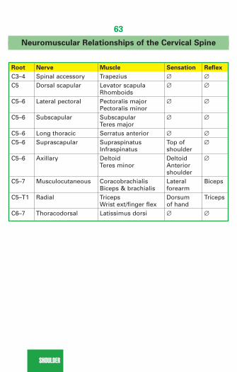
63
SHOULDER
Neuromuscular Relationships of the Cervical Spine
Root Nerve Muscle Sensation Reflex
C3–4 Spinal accessory Trapezius ∅ ∅C5 Dorsal scapular Levator scapula ∅ ∅
RhomboidsC5–6 Lateral pectoral Pectoralis major ∅ ∅
Pectoralis minorC5–6 Subscapular Subscapular ∅ ∅
Teres majorC5–6 Long thoracic Serratus anterior ∅ ∅C5–6 Suprascapular Supraspinatus Top of ∅
Infraspinatus shoulderC5–6 Axillary Deltoid Deltoid ∅
Teres minor Anterior shoulder
C5–7 Musculocutaneous Coracobrachialis Lateral BicepsBiceps & brachialis forearm
C5–T1 Radial Triceps Dorsum TricepsWrist ext/finger flex of hand
C6–7 Thoracodorsal Latissimus dorsi ∅ ∅
2067_Tab02_050-083.qxd 3/19/09 3:26 PM Page 63

64
SHOULDER
Brachial Plexus—Roots, Muscles, & Function
Nerve Root Muscle Function
Radial
Median
Ulnar
C5–8, T1
C6–8, T1
C7–8, T1
Anconeus, brachioradialis,ECRL, ECRB, extensor digitorum, APL, ECU,extensor indices, extensordigiti minimiPronator teres, FCR, palmaris longus, FDS,FPL, pronator quadratus,thenar eminence, lateral 2 lumbricales
FCU, palmaris brevis,hypothenar eminence,adductor pollicis, medial 2 lumbricales, interossei
■ Weak supination, wristextensors, finger flexors,thumb abductors
■ Weak grip due to loss ofwrist stabilization
■ Weak pronation, wristflexion & RD
■ Weak thumb flexion &abduction
■ Weak grip & pinch■ Ape hand■ Weak wrist flexion & UD■ Weak 5th finger flexion■ Weak finger abd/
adduction■ Benediction sign
Special Tests
Neural Tissue Provocation Tests
■ See Alerts/Alarms–page 14.
2067_Tab02_050-083.qxd 3/19/09 3:26 PM Page 64

65
SHOULDER
Shoulder Tests
EMPTY CAN TEST
Purpose: Test supraspinatus musclePosition: SeatedTechnique: Elevate UE 30°–45° in plane of thescapula with IR, resist elevationInterpretation: + test = reproduction of pain &/orweaknessStatistics: Pain: sensitivity = 44%–100% & speci-ficity = 50%–99%Weakness: sensitivity = 77% & specificity = 68%
FULL CAN TEST
Purpose: Test supraspinatus musclePosition: SeatedTechnique: Elevate UE 30°–45° in plane of thescapula with ER, resist elevationInterpretation: + test = reproduction of pain &/orweaknessStatistics: Pain: sensitivity = 66% & specificity = 64%Weakness: sensitivity = 77% & specificity = 74%
DROPPING SIGN
Purpose: Test infraspinatus musclePosition: SeatedTechnique: Shoulder at side with 45°of IR & 90° elbow flexion, resist ERInterpretation: + test = reproductionof pain &/or weaknessStatistics: Sensitivity = 20%–42% &specificity = 69%–100%
Source: From Gulick, D., 2008, page 108.
Source: From Gulick, D., 2008, page 109.
Source: From Gulick, D., 2008, page 109.
2067_Tab02_050-083.qxd 3/19/09 3:26 PM Page 65
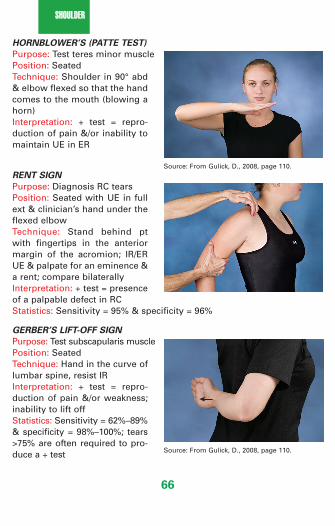
66
SHOULDER
HORNBLOWER’S (PATTE TEST)
Purpose: Test teres minor musclePosition: SeatedTechnique: Shoulder in 90° abd& elbow flexed so that the handcomes to the mouth (blowing ahorn)Interpretation: + test = repro-duction of pain &/or inability tomaintain UE in ER
RENT SIGN
Purpose: Diagnosis RC tearsPosition: Seated with UE in fullext & clinician’s hand under theflexed elbowTechnique: Stand behind ptwith fingertips in the anteriormargin of the acromion; IR/ERUE & palpate for an eminence &a rent; compare bilaterallyInterpretation: + test = presenceof a palpable defect in RCStatistics: Sensitivity = 95% & specificity = 96%
GERBER’S LIFT-OFF SIGN
Purpose: Test subscapularis musclePosition: SeatedTechnique: Hand in the curve oflumbar spine, resist IR Interpretation: + test = repro-duction of pain &/or weakness;inability to lift offStatistics: Sensitivity = 62%–89%& specificity = 98%–100%; tears>75% are often required to pro-duce a + test
Source: From Gulick, D., 2008, page 110.
Source: From Gulick, D., 2008, page 110.
2067_Tab02_050-083.qxd 3/19/09 3:26 PM Page 66

67
SHOULDER
BELLY PRESS OR NAPOLEON SIGN
Purpose: Test subscapularis musclePosition: Seated with hand on bellyTechnique: Press the hand into bellyInterpretation: + test = reproduction ofpain &/or inability to IR; substitutionmay result in UE elevation or wristflexionStatistics: Sensitivity = 25%–40% &specificity = 98%; tears >50% are oftenrequired to produce a + test
BEAR-HUG TEST
Purpose: Test subscapularis musclePosition: Seated with palm of handon opposite shoulder (elbow infront of body)Technique: Resist IR by attemptingto pull hand off the shoulderInterpretation: + test = inability tohold the hand against the shoulder orweakness >20% of contralateral UEStatistics: Sensitivity = 60% & speci-ficity= 92%; tears of 30% can bedetected with this test
HAWKINS/KENNEDY TEST
Purpose: Test for impingementPosition: SeatedTechnique: Place shoulder in 90° of flexion, slight hori-zontal adduction, & maximal IRInterpretation: + test = shoulder pain due to impinge-ment of supraspinatus between greater tuberosityagainst coracoacromial archStatistics: Sensitivity = 72%–92% & specificity =25%–66%
Source: From Gulick, D., 2008, page 111.
2067_Tab02_050-083.qxd 3/19/09 3:26 PM Page 67

68
SHOULDER
NEER’S TEST
Purpose: Test for impingementPosition: SeatedTechnique: Passively take UE into full shoulder flexionwith humerus in IRInterpretation: + test = pain may be indicative ofimpingement of the supraspinatus or long head ofthe bicepsStatistics: Sensitivity = 68%–95% & specificity =25%–68%
IMPINGEMENT RELIEF TEST
Purpose: Confirm impingementPosition: SeatedTechnique: Perform an inferior glide of GH joint while elevating UE toNeer positionInterpretation: + test = reduction or no pain when elevation is accompa-nied by an inferior glide
SULCUS SIGN
Purpose: Assess for inferior instability or AC pxPosition: Sitting with shoulder in neutral & elbowflexed to 90°Technique: Palpate shoulder joint line while usingproximal forearm as a lever to inferiorly distracthumerusInterpretation: + test = ≥ 1 finger-width gap @ theshoulder joint line or AC joint
APPREHENSION TEST
Purpose: Assess for anterior instabilityPosition: SupineTechnique: Abduct the shoulder to 90° & thenbegin to ERInterpretation: + test = pain or apprehensionby the client to assume this position for fearof shoulder dislocation
2067_Tab02_050-083.qxd 3/19/09 3:26 PM Page 68
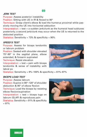
69
SHOULDER
JERK TEST
Purpose: Assess posterior instabilityPosition: Sitting with UE in IR & flexed to 90°Technique: Grasp client’s elbow & load the humerus proximal while pas-sively moving the UE into horizontal adductionInterpretation: + test = a sudden jerk/clunk as the humeral head subluxesposteriorly; a second jerk/clunk may occur when the UE is returned to theabducted positionStatistics: Sensitivity = 73% & specificity = 90%
SPEED’S TEST
Purpose: Assess for biceps tendonitisor labrum problemPosition: Seated with shoulder elevated75°–90° in the sagittal plane, elbowextended, & forearm supinatedTechnique: Resist elevationInterpretation: + test = pain with bicepstendonitis & sense of instability withlabral pxStatistics: Sensitivity = 9%–100% & specificity = 61%–87%
BICEPS LOAD TEST
Purpose: Assess labrumPosition: Supine in 90°–120° of shoulderabduction & 90° of elbow flexionTechnique: Load the biceps by resistingelbow flexion/supinationInterpretation: + test = biceps tugs onlabrum (SLAP) & reproduces pain Statistics: Sensitivity = 91% & specificity= 97%
2067_Tab02_050-083.qxd 3/19/09 3:26 PM Page 69

70
SHOULDER
PAIN PROVOCATION TEST
Purpose: Assess labrumPosition: Supine in 90° shoulderabduction & 90° elbow flexionTechnique: Traction the biceps bypassively taking the forearm intomaximal pronationInterpretation: + test = biceps will tugon labrum & reproduces the pain inthe superior region of the joint line(superior labrum)Statistics: Sensitivity = 17%–100% & specificity = 90%
CRANK TEST
Purpose: Assess labrumPosition: Seated with UE elevated to160° & elbow flexed to 90°Technique: Administer compressiondown the humerus while performingIR/ERInterpretation: + test = pain or clicking Statistics: Sensitivity = 39%–91% &specificity = 67%–93% (greater accu-racy than MRI)
KIM TEST
Purpose: Assess labrumPosition: Seated with UE elevated to ~130° in the plane of the scapula &the elbow flexed to 90°Technique: Apply a compressive force thru the humerusInterpretation: + test = pain or clickingStatistics: Sensitivity = 80%–82% & specificity = 86%–94%
2067_Tab02_050-083.qxd 3/19/09 3:26 PM Page 70

71
SHOULDER
O’BRIEN’S TEST
Purpose: Assess for labrum or AC joint problemPosition: Seated with UE in 90° of elevation, 10° of horiz add, & maximalIR (pronation)Technique: Resist elevation in IR then repeat in ER (supination)Interpretation: + test = pain in IR > ER; pain “inside” shoulder is labrum &pain “on top” of shoulder is ACStatistics: Sensitivity = 47%–100% & specificity = 41%–98%
YERGASON’S TEST
Purpose: Assess THLPosition: Seated with shoulder inneutral, elbow flexed to 90°, & fore-arm supinatedTechnique: Resist elbow flexion withsupinationInterpretation: + test = pain withtenosynovitis; clicking or snappingwith torn THL (with resistance frompronation to supination)Statistics: Sensitivity = 9%–37% & specificity = 86%–96%
2067_Tab02_050-083.qxd 3/19/09 3:26 PM Page 71

72
SHOULDER
AC SHEAR TEST
Purpose: Assess for AC sprainPosition: Seated; UE at sideTechnique: Clinician interlaces fingers & surroundsthe AC joint; squeezing the hands together com-presses the AC jointInterpretation: + test = pain or excessive mov’t isindicative of damage to the AC ligamentsStatistics: Sensitivity = 100% & specificity = 97%
CORACOCLAVICULAR LIGAMENT TEST
Purpose: Assess CC ligamentPosition: Side-lying on the unaffected sideTechnique: Place affected UE behind back, palpate CC ligament while sta-bilizing clavicle; pulling inferior angle of scapula away from ribs to stressthe conoid portion; pulling medial border of scapula away from the ribsstresses the trapezoid portionInterpretation: + test = pain
CROSS-BODY ADDUCTION TEST
Purpose: Assess for AC Position: SeatedTechnique: Shoulder flexed to 90°, horizontallyadduct the UEInterpretation: + test = pain @ AC jointStatistics: Sensitivity = 100% & specificity = 97%
2067_Tab02_050-083.qxd 3/19/09 3:26 PM Page 72

73
SHOULDER
Thoracic Outlet Syndrome (TOS) Compression Sites
Anterior scalene muscle
Middle scalene muscle
Brachial plexus
1st rib1st rib
2nd rib2nd rib
3rd rib
4th rib
5th rib
3rd rib
4th rib
5th rib
Clavicle
Subclavian arteryand vein
Cervicalvertebrae
Pectoralis minormuscle
Coricoid process
Clavicle (cut)
Scalene triangle
Brachial plexus
Costoclavicular space Coracopectoral space
2067_Tab02_050-083.qxd 3/19/09 3:26 PM Page 73

74
SHOULDER
TOS Tests“Rule of the Thumb” = Rotation of the head follows
the direction of the thumb
ADSON’S TEST
Purpose: Assess for TOS @ scalene trianglePosition: SeatedTechnique: While palpating radial pulse, move UEinto abd, ext, & ER, then client rotates head towardthe involved side, takes a deep breath & holds itInterpretation: + test = absent or diminished radialpulse with symptoms reproducedStatistics: Specificity = 74%–89%
WRIGHT’S HYPERABDUCTION TEST
Purpose: Assess for TOS @ coracoid/rib & underpectoralis minorPosition: SeatedTechnique: While palpating radial pulse, passivelyabduct UE to 180° in ER, have client take a deepbreath & hold itInterpretation: + test = absent or diminished radialpulse with symptoms reproducedStatistics: Pulse: sensitivity = 70% & specificity = 53% Pain: sensitivity = 90% & specificity = 29%
MILITARY BRACE (COSTOCLAVICULAR) TEST
Purpose: Assess for TOS @ 1st rib & claviclePosition: SeatedTechnique: While palpating radial pulse, retractshoulders into extension & abduction with the neckin hyperextension (exaggerated military posture)Interpretation: + test = absent or diminished radialpulse or symptoms reproduced
2067_Tab02_050-083.qxd 3/19/09 3:26 PM Page 74

75
SHOULDER
ALLEN’S TEST
Purpose: Assess for TOS @ pectoralisminorPosition: SeatedTechnique: In 90° shoulder abduction &90° elbow flexion, turn head away, takea deep breath & hold itInterpretation: + test = absent or dimin-ished radial pulse with symptomsreproduced
ROOS’ TEST—Elevated Arm Stress Test (EAST)
Purpose: Assess for TOS Position: Seated with UEs at 90° ofshoulder abduction, ER, & elbow flexion.Technique: Open & close hands repeat-edly for 3 minutesInterpretation: + test = reproduction ofsymptoms or sensation of heavinessof the UEs (record time of onset ofsymptoms)Statistics: Sensitivity = 82%–84 & specificity = 30%–100%;
Combination of TOS tests Sensitivity Specificity
• Adson’s + Wright’s (pain)• Adson’s + Roos’• Adson’s + Hyperabd (pain)• Adson’s + Wright’s (pulse)• Wright’s (pain) + Roos’• Wright’s (pain) + Hyperabd (pain)• Wright’s (pulse) + Hyperabd (pulse)
79727254838363
76828894475069
2067_Tab02_050-083.qxd 3/19/09 3:26 PM Page 75

76
SHOULDER
Vascular Components Neural Components
■ (+) Adson’s, Wright’s, Allen’s, Roos’, military press test
■ Hand or arm edema■ Discoloration or UE claudication■ Change in skin temperature or
texture■ Difference of >20 mm Hg in DBP
between UEs■ Poor tolerance of cold & activity
TOS—Differentiation Between Vascular & NeuralComponents
■ Muscle weakness■ Pain with SB of C-spine■ Sensory changes along a neuro-
logical distribution, i.e., radial orulnar nerve
■ (+) Neural tissue provocationtests
2067_Tab02_050-083.qxd 3/19/09 3:26 PM Page 76

77
SHOULDER
Diffe
ren
tial D
iag
no
sis
TO
SC
-dis
cS
ho
uld
er
Cu
bita
l tun
nel
Carp
al tu
nn
el
Pain
Interm
ittent n
eck, S
harp
, con
stant
Sh
ou
lder &
E
lbo
w &
med
ialIn
termitten
tsh
ou
lder, arm
neck &
UE
pro
ximal U
Eh
and
lateral han
dH
eadach
e(+)
(–)(–)
(–)(–)
Nu
mb
ness
Wh
ole U
ER
espective
No
t com
mo
nU
lnar d
istribu
tion
Med
iand
ermato
me
distrib
utio
nE
dem
aP
ossib
leN
orm
alN
orm
alN
orm
alN
orm
alC
olo
rM
ay be ab
no
rmal
May b
e N
orm
alN
orm
alM
ay be
abn
orm
alab
no
rmal
Pro
vocatio
nU
E elevatio
nN
eck po
sition
sA
ctivityE
lbo
w p
ressure
Mu
scle cram
pin
g
w/su
stained
g
raspM
uscle
Weak tricep
s &S
pecific
Weak R
CU
lnar
Med
ianstren
gth
RC
myo
tom
esin
nervatio
ns
inn
ervation
s(+) Tests
NT
PT, A
dso
n’s,
Sp
urlin
g’s,
RC
&
Tinel’s (elb
ow
),P
halen
s, CT
S,
Allen
’s, military
NT
PT
imp
ing
emen
tN
TP
TTin
el’s (wrist)
press, R
oo
s’
2067_Tab02_050-083.qxd 3/19/09 3:26 PM Page 77

78
SHOULDER
Pathology/Mechanism Signs/Symptoms
Breast Cancer
Thoracic Outlet Syndrome—resultsfrom compression of any one ofmany sites 2° postural or muscularimbalances or osseous anomalies.May be due to vascular (only5%–10%) or neural compression;locations of compression include:sternocostovertebral space, sca-lene triangle, costoclavicular space,& coracopectoral space; most com-mon in middle-aged female or aftersurgerySee “Neural vs Vascular Table” on
page 76 for differential diagnosis.
Glenohumeral Dislocation—anterioris most common (90%); mechanismis FOOSH
■ Palpable mass/nodule in breast tissue■ Nipple discharge, retraction, &
local skin dimpling■ Erythema, local rash■ Confirmed with mammogram; biopsy■ Kyphotic posture & forward head■ Awakened @ night with pins &
needles in hand ■ Poorly localized aching pain■ Tenderness in the suprascapular
fossa■ Pain with carrying heavy objects■ (+) Tests: NTPT, Adson’s, Wright’s,
military brace, Roos’ & Allen’s■ DBP > 20 mm Hg difference
between arms■ A/P x-ray needed to r/o cervical
rib (very rare)■ EMG results are controversial■ Need to r/o CTS, radiculopathy,
pronator syndrome■ Prominent acromion, flattened
shoulder silhouette, prominenthumeral head
■ Injured posture: shoulder IR &slightly abducted, elbow flexed,forearm pronated, UE supportedby contralateral limb
■ Sharp, stabbing pain, muscleguarding, humeral head is palpable anteriorly or inferiorly in the armpit
■ (+) Tests: Apprehension test &sulcus sign
■ X-ray—Hill-Sachs lesion may bevisible in A/P view with UE in IR;Bankart lesion in Garth view
■ Need to r/o humeral neck fracturein elderly
Continued
2067_Tab02_050-083.qxd 3/19/09 3:26 PM Page 78

79
SHOULDER
Pathology/Mechanism Signs/Symptoms
Clavicular Fracture—results from afall on the shoulder or a directblow to the clavicleAcromioclavicular Sprain—mayresult from a fall on the acromion& FOOSHSee “Acromioclavicular Sprain
Grades” on page 82.
Labral Tear—may result fromFOOSH, traction force on theshoulder, or a strong biceps contraction
Subacromial Bursitis—chronic irritation resulting from trauma orpoor biomechanics; may occur inmiddle-aged or older clients afteran unusual bout of activity; hx oftendonitis
■ Can’t raise arm■ Visual deformity & TTP■ Confirmed with x-ray■ Pain & crepitus on palpation &
visual deformity■ (+) Tests: Cross body adduction,
O’Brien’s, AC shear, & sulcus/AC tx■ Confirmed with bilateral A/P x-ray
in ER with & without a 10–15 lbweight (stress films)
■ Need to r/o impingement■ Pain with IR & adduction■ Weakness with abduction & flexion■ Client reports a sense of instability■ (+) Tests: Speed’s test, O’Brien’s,
biceps load, pain provocation, &crank
■ Confirmed with CT or MRI; CTdouble contrast is more accuratethan MRI
■ Swift onset of severe pain; localizedto deltoid insertion
■ Noncapsular end-feel with nolimitation in rotation (position ofchoice is adduction)
■ If bursitis exists in isolation (notcommon) then passive ROM ispainful (noncontractile structure)& resistive motions are notpainful (except in 50°–130° rangewhere the contracting tendoncompresses the bursa)
■ (+) Tests: Hawkins/Kennedy,Neer’s & Impingement relief
■ Subacromial bursa warm & TTP(position UE into passive exten-sion to palpate bursa)
■ Imaging is of little value unlesscalcification has occurred; needto r/o RC tear & impingement
Continued
2067_Tab02_050-083.qxd 3/19/09 3:26 PM Page 79

80
SHOULDER
Pathology/Mechanism Signs/Symptoms
Bicipital Tendonitis—chronic irrita-tion resulting from trauma or poorbiomechanicsForward head contributes to abnor-mal scapulo-humeral rhythm
Calcific Tendonitis—cyclic problemof calcification = deposition &resorption with unknown etiology(may be related to tissue hypoxia)Occurs in � > �; R > L; 40–50 yo
Rotator Cuff Strain—results frommechanical compression OR ten-sile overload (eccentric microtears);partial thickness tears occur 25–40 yo& full thickness tears >60 yoRC has limited resiliency for self-repairContributing factors:Posture—forward head influencesGH alignmentAntero-inferior capsule tightness =↓ ERPosterior capsule tightness = ↑ supe-rior & anterior translation of humeralhead
■ Pain ↑ @ night; TTP localized tobiceps tendon @10° of IR (placestendon directly anterior & ~6 cmbelow acromion)
■ Active elevation results in apainful arc; crepitus
■ (+) Speed’s test; (–) Yergason forclick but painful
■ X-ray: bicipital groove view willreveal medial wall angle, spurs,degenerative changes; caudal tilt view will reveal spurring
■ Often associated with RCimpingement
■ ↓ ROM with painful arc 70°–110°& sensation of catching whengoing thru ROM
■ (+) Speed’s test ■ During deposition: chronic mild-
moderate discomfort, throbbingunrelieved by rest
■ During resorption: acute ↑ inpain; sharp & localized
■ Confirmed by A/P film in neutral■ Need to r/o impingement &
adhesive capsulitis■ Painful arc with UE motion; night
pain; deep ache■ Crepitus■ Weakness: abduction +/or ER;
protective shoulder hike■ (+) Special tests depending on
muscle involved—empty/full can(supraspinatus), lift-off or bellypress/Napoleon (subscapularis),hornblowers (teres minor),dropping sign (infraspinatus); (+) O’Brien’s test
■ Strength imbalance (ER MMTshould be 60%–70% of IR)
Continued
2067_Tab02_050-083.qxd 3/19/09 3:26 PM Page 80

81
SHOULDER
Pathology/Mechanism Signs/Symptoms
Supraspinatus Impingement—
results from a progressive loss ofhumeral depressor mechanism(infraspinatus, subscapularis, teres minor, & long head of biceps)2° overuse, cervical px, posturalpx, abnormal biomechanics, orstructural px with acromionMicrotrauma results from IR duringoverhead tennis stroke, swim, throwing; shoulder instability; tightpectoralis minor or weak lower trap& SA allows tipping of scapula withshoulder elevation to ↓ subacromialspace to impingement
Coracoid Impingement—subacromialarch boundaries = acromion, cora-coid, & coracoacromial ligament;houses supraspinatus, long head ofbiceps, subacromial bursa, coraco-humeral ligament; hooked acromion;results from repetitive tasks with UEIR; poor posture
■ X-ray may be normal with smalltears; partial tears = superiorhumeral displacement may beevident with ER; full-thicknesstear = narrowed acromiohumeralinterval & osteophytes on anterior/inferior acromion
■ Diagnostic ultrasound is reliablefor tears > 1 cm
■ Arthrography with contrast =Geyser’s sign (painful)
■ MRI is noninvasive but CT doublecontrast is more accurate thanMRI for full thickness RC tears
■ Pain (especially when sleepingon affected side)
■ Painful arc (60°–120° of elevation)■ Pain & weakness in supraspinatus
& biceps■ “Catching” with flexion in IR■ Pain referral pattern = deltoid inser-
tion & anterior/proximal humerus■ Little to no TTP■ ROM ↓ IR & horizontal adduction■ Posterior capsule tightness; pain
with PROM■ (+) Tests: Neer’s, Hawkins-Kennedy,
Speed’s, empty/full can, & Yocum■ X-rays may reveal ↓ joint space,
arthritis, calcific tendonitis, hookedacromion; early dx is via MRI
■ Should r/o RC tear, TOS, labraltear, & calcific tendonitis
■ Dull pain in the front of the shoul-der provoked by flexion & IR ORabduction & IR
■ Weak downward rotators of scapula■ Forward head & kyphosis
influences GH alignment■ (+) Tests: Neer’s, Hawkins-Kennedy,
& impingement relief
Continued
2067_Tab02_050-083.qxd 3/19/09 3:26 PM Page 81

82
SHOULDER
Pathology/Mechanism Signs/Symptoms
Adhesive Capsulitis—self-limitingdisorder of unknown etiology; highincidence in DM & associated withold Colles fx; proliferation of colla-gen results in thickening of inferiorcapsule & loss of capsular folds;most common in � 40–70 yoSee “Stages & Presentation of
Adhesive Capsulitis” on page 83.
■ X-ray will detect ↓ joint space &hooked acromion
■ Should r/o RC tear, TOS, labraltear, & calcific tendonitis
■ Pain radiating to elbow; nightpain
■ Kyphotic posture, shoulder hiking,low-grade inflammatory response
■ Empty end-feel; ↓ accessorymovement
■ ROM limitations: ER > abduction> IR & reverse scapulohumeralrhythm (scapular 2: humeral 1)
■ Unable to sleep on affected side;MTrP subscapularis
■ Contrast arthrography = 50% reduction in shoulder joint volume(5–10 mL instead of 20–30 mL); plainfilms only reveal osteoporosis 2° todisuse atrophy
Grade Presentation
Normal Acromion to clavicle space should be ~ 0.3–0.8 cmInferior clavicle to coracoid distance should be 1.0–1.3 cm
1st degree AC joint space >0.8 cm & pain with horizontal adduction injury & elevation; (+) AC shear test2nd degree AC joint space 1.0–1.5 cm & CC distance increased by injury 25%–50%3rd degree AC joint space >1.5 cm & CC distance increased by >50% injury with a step deformity
Acromioclavicular Sprain Grades
2067_Tab02_050-083.qxd 3/19/09 3:26 PM Page 82

83
SHOULDER
Stages & Presentation of Adhesive Capsulitis
Stage Clinical Findings Arthroscopic Findings Intervention
I—Freezing
II—Frozen
III—Thawing
• Continualincrease inpain (beforeend-range)
• ↓ A & PROM
• ↓ pain• ↓ A & PROM• Impaired GH
accessory &physiologicalmov’t
• Impaired SH rhythm
• Pain withstretchingonly, ↑ acces-sory & physio-logic motion,return of SH rhythm & ADLs
Erythematous, fibri-nous pannus over the synovium in theaxillary fold
Thickened synoviumwith adhesions devel-oping across the folds
Loss of joint space,humeral head is compressed againstglenoid, & axillary foldis reduced by 50%
Least aggressive:• Modalities• Gentle AROM–
Codman’s• Grade I &
II mobilizationsModerately aggressive:• Modalities• AROM• Gentle PROM• Grade III &
IV mobilizations
Most aggressive:• Modalities• PROM• Grade III &
IV mobilizations• PREs
2067_Tab02_050-083.qxd 3/19/09 3:26 PM Page 83

ELBOW
84
Anatomy
Acromion process
Bicepsbrachiitendon
Long headof biceps
Short headof biceps
Bicepsbrachiitendon
Coracobrachialismuscle
Acromion process
Tricepsbrachiitendon
Long headof triceps brachii
Lateral headof triceps brachii
Medial headof triceps brachii
Medial headof triceps brachii
2067_Tab03_084-098.qxd 3/19/09 3:42 PM Page 84

85
ELBOW
Radialcollateralligament
Annularligament
Ulnarcollateralligament
Radius Ulna
Humerus
Annularligament
Radial collateralligament
Radius
Ulna
Humerus
Biceps brachii tendon
Triceps brachii tendon
Lateral view
Annularligament
Ulnar collateralligament
Ulnar nerve
Radius
Ulna
Humerus
Oblique cord
Bicepsbrachiitendon
Tricepsbrachiitendon
Medial view
2067_Tab03_084-098.qxd 3/19/09 3:42 PM Page 85

ELBOW
86
Referral Patterns
Muscle Pain Referral Patterns
Brachioradialis Biceps brachii
Flexor carpi radialis Flexor carpi ulnaris
2067_Tab03_084-098.qxd 3/19/09 3:42 PM Page 86

87
ELBOW
Muscle Pain Referral Patterns
Extensor carpi ulnaris Extensor carpi radialis longus
Extensor carpi radialis brevis
2067_Tab03_084-098.qxd 3/19/09 3:42 PM Page 87

ELBOW
88
Visual Inspection
■ Carrying angle of the elbow■ 10°–15° valgus in �■ 5°–10° valgus in �
Palpation Pearls
Wrist Extensor Muscles
Extensor carpi radialisbrevis and longus
Extensor digitorumExtensor carpi ulnaris
2067_Tab03_084-098.qxd 3/19/09 3:42 PM Page 88

89
ELBOW
Wrist Flexor Muscles
Pronator teres
Flexor carpi radialis
Flexor carpi ulnarisPalmaris longus
2067_Tab03_084-098.qxd 3/19/09 3:42 PM Page 89
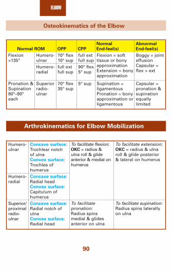
ELBOW
90
Osteokinematics of the Elbow
Normal Abnormal
Normal ROM OPP CPP End-feel(s) End-feel(s)
Flexion >135°
Pronation & Supination 80°–90°each
Humero-ulnarHumero-radial
Superior radio-ulnar
70° flex10° supfull extfull sup
70° flex35° sup
full extfull sup90° flex5° sup
5° sup
Flexion = soft tissue or bonyapproximationExtension = bonyapproximation
Supination = ligamentousPronation = bonyapproximation or ligamentous
Boggy = jointeffusionCapsular = flex > ext
Capsular =pronation &supinationequally limited
Arthrokinematics for Elbow Mobilization
Humero-ulnar
Humero-radial
Superior/proximal radio-ulnar
Concave surface:Trochlear notch of ulnaConvex surface:Trochlea ofhumerusConcave surface:Radial headConvex surface:Capitulum ofhumerusConcave surface:Radial notch ofulnaConvex surface:Radial head
To facilitate flexion: OKC = radius & ulna roll & glideanterior & medial onhumerus
To facilitate pronation:Radius spins medial & glidesanterior on ulna
To facilitate extension:OKC = radius & ulnaroll & glide posterior & lateral on humerus
To facilitate supination:Radius spins laterallyon ulna
2067_Tab03_084-098.qxd 3/19/09 3:42 PM Page 90

91
ELBOW
Strength & Function
Brachial Plexus–Roots, Muscles, & Function
Nerve Root Muscle Functional Deficits
Median
Ulnar
Radial
C6–8, T1
C7–8, T1
C5–8, T1
Pronator teres, FCR,palmaris longus, FDS,FPL, pronator quadra-tus, thenar eminence,lateral 2 lumbricales
FCU, palmaris brevis,hypothenar eminence,adductor pollicis,medial 2 lumbricales,interosseiAnconeus, brachiora-dialis, ECRL, ECRB,extensor digitorum,APL, ECU, extensorindicis, extensor digitiminimi
■ Weak pronation, wristflexion, & RD
■ Weak thumb flexion &abduction
■ Weak grip & pinch■ Ape hand■ Weak wrist flexion & UD■ Weak 5th finger flexion■ Weak finger abd/adductor■ Benediction sign
■ Weak supination, wristextension, finger flexion,thumb abduction
■ Weak grip due to loss ofwrist stabilization
2067_Tab03_084-098.qxd 3/19/09 3:42 PM Page 91
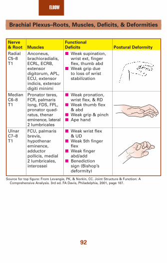
ELBOW
92
Brachial Plexus–Roots, Muscles, Deficits, & Deformities
Nerve Functional
& Root Muscles Deficits Postural Deformity
RadialC5–8 T1
MedianC6–8 T1
UlnarC7–8 T1
Anconeus, brachioradialis,ECRL, ECRB, extensor digitorum, APL,ECU, extensor indicis, extensordigiti minimiPronator teres,FCR, palmarislong, FDS, FPL,pronator quad-ratus, thenareminence, lateral2 lumbricalesFCU, palmarisbrevis,hypothenareminence,adductor pollicis, medial2 lumbricales,interossei
■ Weak supination,wrist ext, fingerflex, thumb abd
■ Weak grip due to loss of wriststabilization
■ Weak pronation,wrist flex, & RD
■ Weak thumb flex& abd
■ Weak grip & pinch■ Ape hand
■ Weak wrist flex & UD
■ Weak 5th fingerflex
■ Weak fingerabd/add
■ Benediction sign (Bishop’sdeformity)
Source for top figure: From Levangie, PK, & Norkin, CC. Joint Structure & Function: AComprehensive Analysis. 3rd ed. FA Davis, Philadelphia, 2001, page 107.
2067_Tab03_084-098.qxd 3/19/09 3:42 PM Page 92
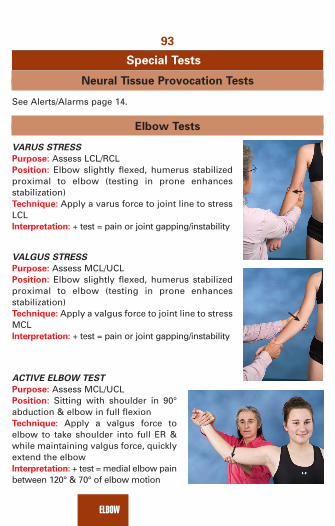
93
ELBOW
Special Tests
Neural Tissue Provocation Tests
See Alerts/Alarms page 14.
Elbow Tests
VARUS STRESS
Purpose: Assess LCL/RCLPosition: Elbow slightly flexed, humerus stabilizedproximal to elbow (testing in prone enhances stabilization)Technique: Apply a varus force to joint line to stressLCLInterpretation: + test = pain or joint gapping/instability
VALGUS STRESS
Purpose: Assess MCL/UCLPosition: Elbow slightly flexed, humerus stabilizedproximal to elbow (testing in prone enhances stabilization)Technique: Apply a valgus force to joint line to stressMCLInterpretation: + test = pain or joint gapping/instability
ACTIVE ELBOW TEST
Purpose: Assess MCL/UCLPosition: Sitting with shoulder in 90°abduction & elbow in full flexion Technique: Apply a valgus force toelbow to take shoulder into full ER &while maintaining valgus force, quicklyextend the elbowInterpretation: + test = medial elbow painbetween 120° & 70° of elbow motion
2067_Tab03_084-098.qxd 3/19/09 3:42 PM Page 93

ELBOW
94
PRONATOR TERES TEST
Purpose: Assess for median nerve entrapmentPosition: UE relaxed in supported positionTechnique: Resist pronation of forearmInterpretation: + test = pain along the palmar aspect of digits 1, 2, & 3(median nerve distribution)
MILL’S TEST
Purpose: Assess for lateral epicondylitisPosition: UE relaxed, elbow extendedTechnique: Passively stretch into wrist flexion &pronationInterpretation: + test = pain @ lateral epicondyle orproximal musculotendinous junction of wristextensors
COZEN’S SIGN
Purpose: Assess for lateral epicondylitisPosition: UE relaxed, elbow extendedTechnique: Resist supination & wrist extension OR resist middle fingerextension (extensor digitorum)Interpretation: + test = pain @ lateral epicondyle or proximal musculo-tendinous junction of wrist extensors
2067_Tab03_084-098.qxd 3/19/09 3:42 PM Page 94

95
ELBOW
PASSIVE TEST
Purpose: Assess for medial epicondylitisPosition: UE relaxed, elbow extendedTechnique: Stretch into wrist extension & supinationInterpretation: + test = pain @ medial epicondyle orproximal musculotendinous junction of wrist flexors
RESISTIVE TEST
Purpose: Assess for medial epicondylitisPosition: UE relaxed, elbow extendedTechnique: Resist pronation & wrist flexionInterpretation: + test = pain @ medial epicondyle orproximal musculotendinous junction of wrist flexors
WARTENBERG’S TEST
Purpose: Assess for ulnar nerve entrapmentPosition: UE relaxed in supported positionTechnique: Resist 5th digit adductionInterpretation: + test = weakness of 5th digit adductors
POSTEROLATERAL or ROTATORY INSTABILITY
Purpose: Assess for elbow instabilityPosition: Elbow extendedTechnique: Apply an axial load with a valgus stress& supinationInterpretation: + test = elbow subluxes with exten-sion & relocates with flexion
2067_Tab03_084-098.qxd 3/19/09 3:42 PM Page 95

ELBOW
96
TINEL’S TEST
Purpose: Assess ulnar nervePosition: Elbow in slight flexionTechnique: Tap groove between olecranon& lateral epicondyleInterpretation: + test = pain & tingling inthe distribution of ulnar nerve (4th & 5thdigits)Statistics: Sensitivity = 28% & specificity =23%
Differential Diagnosis
Pathology/Mechanism Signs/Symptoms
Elbow Dislocation (posterior)—
common in children & young adultsdue to a FOOSH
Radial Head Subluxation—commonin children 2–4 yo resulting from achild being picked up or swung bythe hand or forearm & creating adistraction force
MCL Sprain—elongation/tear of ligament(s); common in throwingathletes 2° valgus stress
■ Pain, inability to flex elbow,deformity, tenderness
■ Confirmed by x-ray■ Need to r/o fx & check distal pulses■ Beware of possible development
of myositis ossificans inbrachialis muscle
■ Child will autosplint in pronation& flexion
■ Radial head is TTP & childreports wrist discomfort from ↑ pressure from radial headbeing displaced distally
■ X-ray if fx is suspected■ Reduction process = thumb in
cubital fossa to serve as a fulcrum,supinate & flex the forearm (will“pop” in)
■ Acute trauma may experience a“pop”
■ TTP @ medial joint line■ Valgus instability■ Confirm with MRI; need to r/o
avulsion
Continued
2067_Tab03_084-098.qxd 3/19/09 3:42 PM Page 96

97
ELBOW
Pathology/Mechanism Signs/Symptoms
Olecranon Bursitis—“student’selbow”—may result from directtrauma or repetitive UE activity
Humerus & Radial Head Fracture—
results from a FOOSHUlnar Neuritis—results from repeti-tive activity or trauma
Osteochondritis Dissecans—resultsfrom repetitive valgus stresses,such as throwing or gymnastics or frequent compressive forces(avascularity of subchondral bone = Panner’s disease)
Reflex Sympathetic Dystrophy or
Complex Regional Pain Syndrome—
may be linked to previous traumabut a large percentage have no precipitating factor
■ Defined mass at the olecranonthat is warm, thick, & “gritty” topalpation
■ ↓ Elbow extension with nonspe-cific TTP
■ MRI used to confirm■ Need to r/o avulsion & RCL/UCL
sprain■ AP & lateral plain film to confirm■ Weak UD, 4th & 5th finger flexion■ Pain with elbow flexion■ (+) Tests: Tinel’s, Wartenberg’s, &
NTPT■ Paresthesia into forearm & 5th
digit■ Need to r/o C-spine pathology &
TOS■ Confirm with MRI■ Lateral elbow pain with ↓ elbow
extension■ Catching/locking of the elbow;
pain with UE WB■ Crepitus with pronation/supination■ X-ray, MRI, CT are helpful in
identifying a loose body■ Abnormal reflexes; varied
manifestations of pain, burning,& edema
■ Nerve adhesions = (+) NTPT(movement is painful)
■ Vasomotor instability & trophicchanges span from warm, redness over dorsum of MP & IPjoints, & excessive moisture tocold temperature, pallor, & dry-ness of hand
■ Osteoporosis■ MRI may or may not be helpful
Continued
2067_Tab03_084-098.qxd 3/19/09 3:42 PM Page 97

ELBOW
98
Pathology/Mechanism Signs/Symptoms
Avulsion/Stress Fracture of
Medial Epicondyle = “Little League
Elbow”—2° repetitive throwingmotion; seen in teenagers withacceleration of UE with elbow flexion & valgus stressMedial Epicondylitis—“Golfer’s
Elbow”—insidious onset 2° torepetitive forces on the elbow;effects pronator teres & FCR
Lateral Epicondylitis—“Tennis
Elbow”—overuse or microtraumato lateral musculature; may resultfrom a small racket grip, a racketthat is too stiff or heavy, or a smallsweet spot; usually involves ECRB
■ Progressive pain & TTP @ medialepicondyle
■ ↓ ROM■ (+) Valgus stress test■ Confirm with x-ray or MRI
■ Pain with resisted wrist flexion &UD &/or passive stretching intowrist extension & supination with RD
■ TTP at proximal musculotendinousjctn of wrist flexors & pronators
■ (+) Passive & resistive tests■ MRI may confirm diagnosis &
r/o fx or avulsion■ Morning stiffness■ Pain with resisted wrist extension,
supination, & RD OR passivestretching into wrist flexion,pronation, & UD
■ (+) Tests: Cozen’s & Mill’s■ TTP at proximal musculotendi-
nous junction of wrist extensors& supinators
■ MRI may confirm diagnosis & r/o fx or avulsion
2067_Tab03_084-098.qxd 3/19/09 3:42 PM Page 98

99
WRIST & HAND
Anatomy
Extensor pollicis longus tendon
Abductor pollicis longus tendon
Adductor pollicis muscle
Extensor pollicis brevis tendon
Opponens pollicis muscle
First dorsalinterosseous muscles
First lumbrical muscle
Long extensor tendon
Terminal tendon of extensor mechanism
Abductor pollicisbrevis muscle
2067_Tab04_099-128.qxd 3/19/09 3:23 PM Page 99

WRIST & HAND
100
Medical Red Flags
■ Digital clubbing ■ Acute pulmonary abscess■ Pulmonary malignancy■ Cirrhosis■ Heart disease■ Ulcerative colitis■ COPD
■ Spoon nails■ Anemia■ Thyroid px■ Syphilis■ Rheumatic fever
■ Eggshell nails = thinning/semitransparent = syphilis■ Nail inflammation, infection, biting■ Paresthia in glove distribution
■ DM■ Lead/mercury poisoning
■ Hand tremor■ Parkinsonism■ Hypoglycemia■ Hyperthyroidism■ ETOH■ MS
■ Causes of CTS■ Hx of statins (cholesterol drugs: Zocor or Lipitor)■ Liver disease■ Hypothyroidism■ Gout■ DM■ Pregnancy/oral contraceptives■ B
6vitamin deficiency
2067_Tab04_099-128.qxd 3/19/09 3:23 PM Page 100

101
WRIST & HAND
Toolbox Tests
Rheumatoid Hand Functional Disability Scale ThatAssesses Functional Handicap
Answer the following questions regarding your ability without the help ofany assistive devices:Answers to 0 = Yes, without difficultythe questions: 1 = Yes, with a little difficulty
2 = Yes, with some difficulty3 = Yes, with much difficulty4 = Nearly impossible to do5 = Impossible
■ Can you hold a bowl?■ Can you seize a full bottle & raise it?■ Can you hold a plate full of food?■ Can you pour liquid from a bottle into a glass?■ Can you unscrew the lid from a jar opened before?■ Can you cut meat with a knife?■ Can you prick things well with a fork?■ Can you peel fruit?■ Can you button your shirt?■ Can you open & close a zipper?■ Can you squeeze a new tube of toothpaste?■ Can you hold a toothbrush efficiently?■ Can you write a short sentence with a pencil or
ordinary pen?■ Can you write a letter with a pencil or ordinary pen?■ Can you turn a round door knob?■ Can you cut a piece of paper with scissors?■ Can you pick up coins from a table top?■ Can you turn a key in a lock?Score:
Scoring: Summate all scores—the higher the score, the greater the disability
Source: From Duruoz, MT, Poiradeau, S, Fermanian, J, et al. Journal of Rheumatology, 23:7,1996.
2067_Tab04_099-128.qxd 3/19/09 3:23 PM Page 101
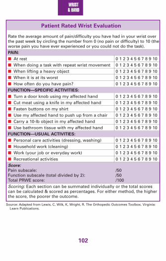
WRIST & HAND
102
Patient Rated Wrist Evaluation
Rate the average amount of pain/difficulty you have had in your wrist overthe past week by circling the number from 0 (no pain or difficulty) to 10 (theworse pain you have ever experienced or you could not do the task).PAIN:
■ At rest 0 1 2 3 4 5 6 7 8 9 10■ When doing a task with repeat wrist movement 0 1 2 3 4 5 6 7 8 9 10■ When lifting a heavy object 0 1 2 3 4 5 6 7 8 9 10■ When it is at its worst 0 1 2 3 4 5 6 7 8 9 10■ How often do you have pain? 0 1 2 3 4 5 6 7 8 9 10FUNCTION—SPECIFIC ACTIVITIES:
■ Turn a door knob using my affected hand 0 1 2 3 4 5 6 7 8 9 10■ Cut meat using a knife in my affected hand 0 1 2 3 4 5 6 7 8 9 10■ Fasten buttons on my shirt 0 1 2 3 4 5 6 7 8 9 10■ Use my affected hand to push up from a chair 0 1 2 3 4 5 6 7 8 9 10■ Carry a 10-lb object in my affected hand 0 1 2 3 4 5 6 7 8 9 10■ Use bathroom tissue with my affected hand 0 1 2 3 4 5 6 7 8 9 10FUNCTION—USUAL ACTIVITIES:
■ Personal care activities (dressing, washing) 0 1 2 3 4 5 6 7 8 9 10■ Household work (cleaning) 0 1 2 3 4 5 6 7 8 9 10■ Work (your job or everyday work) 0 1 2 3 4 5 6 7 8 9 10■ Recreational activities 0 1 2 3 4 5 6 7 8 9 10Score:Pain subscale: /50Function subscale (total divided by 2): /50Total PRWE score: /100Scoring: Each section can be summated individually or the total scorescan be calculated & scored as percentages. For either method, the higherthe score, the poorer the outcome.
Source: Adapted from Lewis, C, Wilk, K, Wright, R. The Orthopedic Outcomes Toolbox. Virginia:Learn Publications.
2067_Tab04_099-128.qxd 3/19/09 3:23 PM Page 102

103
WRIST & HAND
Severity of Symptoms & Functional Status in CarpalTunnel Syndrome
The following questions refer to your symptoms for a typical 24-hour periodduring the past 2 weeks. Circle 1 answer for each question.How severe is the handor wrist pain you haveat night?1. No pain2. Mild pain3. Moderate pain4. Severe pain5. Very severe pain
How often do you havehand or wrist pain dur-ing the daytime?1. Never2. 1 time3. 2–3 times4. 4–5 times5. More than 5 timesDo you have weaknessin your hand or wrist?1. No weakness2. Mild weakness3. Moderate weakness4. Severe weakness5. Very severe
weakness
How often did hand numb-ness or tingling wake youup during a typical night inthe past 2 weeks?1. Never2. 1 time3. 2–3 times4. 4–5 times5. More than 5 times
How often did hand orwrist pain wake you upduring a typical nightin the past 2 weeks?1. Never2. 1 time3. 2–3 times4. 4–5 times5. More than 5 timesHow long, on average,does an episode of painlast during the daytime?1. Never have pain2. Less than 10 minutes3. 10–60 minutes4. More than 60 minutes5. ConstantlyDo you have tinglingsensation in your hand?1. No tingling2. Mild tingling3. Moderate tingling4. Severe tingling5. Very severe tingling
Do you have difficultywith the grasping & useof small objects, suchas keys or pencils?1. No difficulty2. Mild difficulty3. Moderate difficulty4. Severe difficulty5. Very severe difficulty
Do you typically have pain in your hand or wrist during the daytime?1. No pain 2. Mild pain3. Moderate pain4. Severe pain5. Very severe pain
Do you have numbness(loss of sensation) inyour hand?1. No numbness2. Mild numbness3. Moderate numbness4. Severe numbness5. Very severe numbnessHow severe is the numb-ness or tingling at night?1. No numbness/tingling2. Mild numbness/tingling3. Moderate numbness/
tingling4. Severe numbness/tingling5. Very severe numbness/
tingling
Scoring: Summate thescores & divide by 11.The higher the meanscore, the more severethe impairment.Score: _______________
Source: From Levine, et al., Journal of Bone and Joint Surgery, 75A(11): 1585-1592, 1993.
2067_Tab04_099-128.qxd 3/19/09 3:23 PM Page 103

WRIST & HAND
104
Referral Patterns
Muscle Pain Referral Patterns
Flexor digitorum Pronator teres
Flexor pollicis longus 1st dorsal interossei
2067_Tab04_099-128.qxd 3/19/09 3:23 PM Page 104
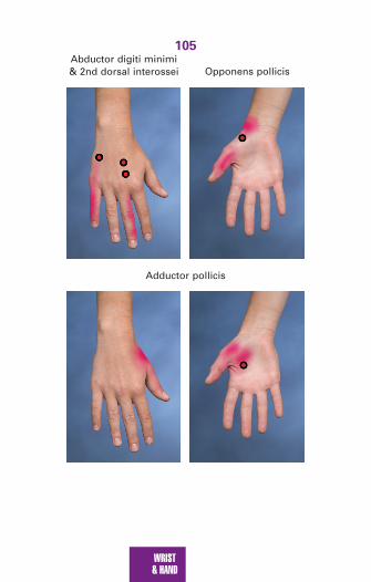
105
WRIST & HAND
Abductor digiti minimi& 2nd dorsal interossei
Adductor pollicis
Opponens pollicis
2067_Tab04_099-128.qxd 3/19/09 3:23 PM Page 105

WRIST & HAND
106
Pathologic Observations
Swan neck Mallet
Boutonnière Dupuytren’s
■ When fist is clenched, all fingers should point to the scaphoid■ Heberden node = DJD of DIP■ Bouchard node = DJD of PIP■ Swan neck = MCP & DIP flexion with PIP hyperextension■ Boutonnière = MCP & DIP extension with PIP flex (ruptured central
extension tendon)■ Mallet finger = flexion of DIP (avulsion)■ Dupuytren’s contracture = flexion of 4th & 5th digits■ Ganglion cyst = defined mass on dorsum of hand■ Pill-rolling tremor = Parkinsonism■ Liver flap = asterixis = flapping tremor resulting from the inability
to maintain wrist extension with the forearm supported in a flexedposition
2067_Tab04_099-128.qxd 3/19/09 3:23 PM Page 106

107
WRIST & HAND
Palpation Pearls
Lunate
Triquetrum
Pisiform
Hamate
CapitateTrapezoid
Trapezium
Scaphoid
2067_Tab04_099-128.qxd 3/19/09 3:23 PM Page 107

WRIST & HAND
108
Wrist Extensor Muscles
Extensor carpi radialisbrevis and longus
Extensor digitorumExtensor carpi ulnaris
2067_Tab04_099-128.qxd 3/19/09 3:23 PM Page 108

109
WRIST & HAND
Wrist Flexor Muscles
Pronator teres
Flexor carpi radialis
Flexor carpi ulnarisPalmaris longus
2067_Tab04_099-128.qxd 3/19/09 3:23 PM Page 109

WRIST & HAND
110
Edema Assessment
Figure-8 Method to Assess Hand Edema (Palmar Surface)
1. Start distal to the lateral styloid process; go medial across the palm ofthe hand to the 5th MCP joint
Lateral styloidprocess
Medial styloidprocess
UlnaRadius
1stMetacarpal
5thMetacarpal
2ndMetacarpal
Source: Gulick, D. Sport Notes: Field & Clinical Examination Guide. FA Davis, Philadelphia,2008, page 171.
2. Over the knuckles to the 2nd MCP joint
Lateral styloidprocess
Medial styloidprocess
UlnaRadius
1stMetacarpal
5thMetacarpal
2ndMetacarpal
Source: Gulick, D. 2008, page 171.
2067_Tab04_099-128.qxd 3/19/09 3:23 PM Page 110

111
WRIST & HAND
3. Across the palm to the medial styloid process
Lateral styloidprocess
Medial styloidprocess
UlnaRadius
1stMetacarpal
5thMetacarpal
2ndMetacarpal
Lateral styloidprocess
Medial styloidprocess
UlnaRadius
1stMetacarpal
5thMetacarpal
2ndMetacarpal
Source: Gulick, D. 2008, page 172.
4. Around the back of the wrist to the lateral styloid process
Source: Gulick, D. 2008, page 172.
2067_Tab04_099-128.qxd 3/19/09 3:23 PM Page 111
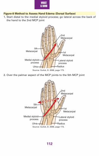
WRIST & HAND
112
Figure-8 Method to Assess Hand Edema (Dorsal Surface)
1. Start distal to the medial styloid process; go lateral across the back ofthe hand to the 2nd MCP joint
Lateral styloidprocess
Medial styloidprocess
Ulna Radius
1stMetacarpal
5thMetacarpal
2ndMetacarpal
Lateral styloidprocess
Medial styloidprocess
Ulna Radius
1stMetacarpal
5thMetacarpal
2ndMetacarpal
Source: Gulick, D. 2008, page 173.
2. Over the palmar aspect of the MCP joints to the 5th MCP joint
Source: Gulick, D. 2008, page 173.
2067_Tab04_099-128.qxd 3/19/09 3:23 PM Page 112

113
WRIST & HAND
3. Across the back of the hand to the lateral styloid process
Lateral styloidprocess
Medial styloidprocess
Ulna Radius
1stMetacarpal
5thMetacarpal
2ndMetacarpal
Lateral styloidprocessMedial styloid
process
Ulna Radius
1stMetacarpal
5thMetacarpal
2ndMetacarpal
Source: Gulick, D. 2008, page 174.
4. Around the front of the wrist to the medial styloid process
Source: Gulick, D. 2008, page 174.
2067_Tab04_099-128.qxd 3/19/09 3:23 PM Page 113
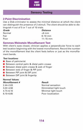
WRIST & HAND
114
Sensory Testing
2-Point DiscriminationUse a Disk-criminator to assess the minimal distance at which the clientcan distinguish the presence of 2 stimuli. The client should be able to dis-tinguish 4 out of 5 or 7 out of 10 trials.Grade Distance
Normal <6 mmFair 6–10 mmPoor 11–15 mm
Semmes-Weinstein Monofilament TestWith client’s eyes closed, clinician applies a perpendicular force to eachtest location beginning with the lowest monofilament. Record the numberof the monofilament that the client feels before or just as the monofila-ment bends.
Test locations:
■ Base of palm/wrist■ Between central palm & distal palm crease■ Between distal palm crease & web of finger■ Between web of finger & PIP joint■ Between PIP joint & DIP joint■ Between DIP joint & fingertip
Normal Values:
Monofilament # Result
2.44–2.83 Normal sensation3.22–4.56 Diminished light touch4.74–6.10 Minimal light touch6.10–6.65 Poor localization
2067_Tab04_099-128.qxd 3/19/09 3:23 PM Page 114

115
WRIST & HAND
Osteokinematics of the Wrist & Hand
Abnormal
Joint Normal ROM Normal End-feel(s) End-feel(s)
Radiocarpal
CMC thumb
MCP 2–5
MCP thumb
IPs 2–5
60°–80° flex60°–70° ext20°–30° RD/UD
70° abd45°–50° flex90° flex
75°–90° flex
100° PIP flex 80° DIP flex
Flex = firm/ligamentous/elasticExt = firm/ligamentous/elasticRD = bonyUD = firm/bonyElastic
Ext = elastic/capsular/ligamentousFlex = elastic/bony/firm/ligamentousAbd = firm/ligamentousFlex = bony/firm/ligamentous/elasticExt = firm/elasticPIP flex =firm/bony/elasticPIP ext = firm/ligamentous/elasticDIP flex = firm/ligamentous/elasticDIP ext = firm/ligamentous/elastic
Capsular =pronation &supinationequallyrestricted
Capsular =abd > ext
2067_Tab04_099-128.qxd 3/19/09 3:23 PM Page 115

WRIST & HAND
116
Arthrokinematics for Wrist & Hand Mobilization
Dis
tal
rad
iou
lnar
Rad
iocarp
al
Concave surface: Radius & radioulnar disk
Convex surface: Proximal carpals
Concave surface: Ulnar notch of radiusConvex surface: Head of ulna
To facilitate wrist flexion:Proximal carpal rolls ante-rior & glides posterior onradius with distal carpalrolling anterior & glidingposterior on the proximalcarpal
To facilitate radial deviation:Proximal carpal rolls lateral & glides medial onradius with distal carpalrolling lateral & glidingmedial on the proximalcarpalTo facilitate pronation:Radius rolls & glides medially over the ulna
To facilitate extension:Proximal carpal rolls posterior & glides anterior & on radius with distal carpal rolling posterior & gliding anterior on theproximal carpalTo facilitate ulnar deviation:Proximal carpal rollsmedial & glides lateral on radius with distalcarpal rolling medial &gliding lateral on the proximal carpalTo facilitate supination:Radius rolls & glides laterally over the ulna
Continued
2067_Tab04_099-128.qxd 3/19/09 3:23 PM Page 116

117
WRIST & HAND
IP 2
–5
MC
P t
hu
mb
MC
P 2
–5
CM
C t
hu
mb
CM
C t
hu
mb Concave surface:
Trapezii
Convex surface: Metacarpal
Concave surface:Base of proximal phalanx
Convex surface: Head of metacarpal
Concave surface: Base of proximal phalanxConvex surface: Head of distal phalanx
To facilitate thumb flexion:Metacarpal rolls & glidesmedial on trapezium
To facilitate thumb abduction:Metacarpal rolls proximal& glides distal on trapeziumTo facilitate flexion:Proximal phalanx rolls &glides anterior onmetacarpalTo facilitate thumb flexion:Distal phalanx rolls &glides anterior on theproximal phalanxTo facilitate flexion:Distal phalanx rolls &glides anterior on theproximal phalanx
To facilitate thumbextension:Metacarpal rolls &glides lateral on trapeziumTo facilitate thumbadduction:Metacarpal rolls distal& glides proximal ontrapeziumTo facilitate extension:Proximal phalanx rolls& glides posterior onmetacarpalTo facilitate thumbextension:Distal phalanx rolls &glides posterior on theproximal phalanxTo facilitate extension:Distal phalanx rolls &glides posterior on theproximal phalanx
Strength & Function
Muscle Function
■ Dorsal interossei = “divide” or separate fingers■ Palmar interossei & lumbricales = “pull” fingers together■ Flexor digitorum superficialis test = with finger in extension, isolate
PIP flexion■ Flexor digitorum profundus test = with finger in extension, isolate DIP
flexion■ Lumbrical test = flex MCP with IPs extended
2067_Tab04_099-128.qxd 3/19/09 3:23 PM Page 117

WRIST & HAND
118
■ Power grips:■ Cylindrical grip = FDP, FDS, FPL, FPB, OP, lumbricales, palmar interossei■ Spherical grip = FDP, FDS, FPL, FPB, OP, lumbricales, dorsal interossei■ Hook grip = FDS, FDP
Tools to Evaluate Grip Strength
■ Hand-held dynamometer■ Jamar device—power grip in various positions■ Pinch meter:
■ Tip-to-tip = anterior interosseous nerve■ Pad-to-pad = median nerve■ 3-jaw chuck = ulnar nerve
■ BP cuff inflated to 20 mm Hg; squeeze & assess pressure change
Objective Tests to Assess Hand Function
■ Minnesota Rate of Manipulation Test■ Minnesota Manual Dexterity Test■ Purdue Pegboard Test■ Modified Moberg Pick-up Test
Quantitative Measure of Ulnar Impaction
■ Assess grip in supinated & pronated wrist positions■ If grip ratio of supination:pronation is
■ = 1, there is no ulnar impaction■ >1, ulnar impaction is present
2067_Tab04_099-128.qxd 3/19/09 3:23 PM Page 118

119
WRIST & HAND
Brachial Plexus–Roots, Muscles, Deficits & Deformities
Nerve &
Root Muscles Functional Deficits Postural Deformity
RadialC5–8T1
MedianC6–8T1
UlnarC7–8T1
Anconeus, brachioradialis,ECRL, ECRB,extensor digitorum, APL,ECU, extensor indicis, extensordigiti minimiPronator teres,FCR, palmarislong, FDS, FPL,pronator quad-ratus, thenareminence, lateral 2 lumbricalesFCU, palmarisbrevis,hypothenareminence,adductor pollicis, medial2 lumbricales,interossei
■ Weak supination,wrist ext, fingerflex, thumb abd
■ Weak grip due toloss of wrist stabilization
■ Weak pronation,wrist flex & RD
■ Weak thumb flex & abd
■ Weak grip & pinch
■ Ape hand
■ Weak wrist flex& UD
■ Weak 5th fingerflex
■ Weak fingerabd/add
■ Benediction sign (Bishop’sdeformity)
Claw hand = median &ulnar
Source for top figure: From Levangie, PK, & Norkin, CC. Joint Structure & Function: AComprehensive Analysis. 3rd ed. FA Davis, Philadelphia, 2001, page 107.
2067_Tab04_099-128.qxd 3/19/09 3:23 PM Page 119

WRIST & HAND
120
Special Tests
Neural Tissue Provocation Tests
■ See Alerts/Alarms page 14.
Wrist & Hand Tests
CLAMP SIGN
Purpose: Assess for scaphoid fxPosition: Wrist in pronation & extension Technique: Grasp web space of the thumbbetween clinician’s thumb & index finger& gently stress the wrist into UD Interpretation: + test = pain in theanatomical snuff boxStatistics: Sensitivity = 52% & specificity= 100%
WATSON’S TEST (Scaphoid shift maneuver)
Purpose: Assess for scaphoid instabilityPosition: Supinated in neutralTechnique: From the radial side, the clini-cian uses his thumb on the palmar side& index finger on dorsal side to applypressure to the distal scaphoid whilemoving the wrist from UD to RDInterpretation: + test = removal of pres-sure will produce a palpable click & dorsalwrist pain
FINKELSTEIN’S TEST
Purpose: Assess for de Quervain’s syndromePosition: Form a fist around the thumbTechnique: Ulnarly deviate the wristInterpretation: + test = pain along EPB & APL
2067_Tab04_099-128.qxd 3/19/09 3:23 PM Page 120

121
WRIST & HAND
WRIST VALGUS TEST
Purpose: Assess UCLPosition: Stabilize radius/ulna proximal to wrist in neutral positionTechnique: Apply a valgus stress to the wrist Interpretation: + test = joint line pain or gapping/instability
PHALANX VARUS/VALGUS TEST
Purpose: Assess MCL & LCLPosition: With finger(s) in neutral, stabilize the proximal phalanxTechnique: Apply a varus/valgus stress via the distal phalanxInterpretation: + test = joint line pain or gapping/instability
PHALEN’S TEST
Purpose: Assess for CTSPosition: Hands relaxedTechnique: Maximally flex the wrists so thedorsal surfaces of the hands are in full con-tact with each other; hold for up to 1 minuteInterpretation: + test = numbness or tinglinginto the median nerve distributionStatistics: Sensitivity = 34%–93% & specificity= 48%–93%
WRIST VARUS TEST
Purpose: Assess RCLPosition: Stabilize radius/ulna proximal to wrist in neutral positionTechnique: Apply a varus stress to the wristInterpretation: + test = joint line pain or gapping/instability
Source: Gulick, D., 2008, page 125.
2067_Tab04_099-128.qxd 3/19/09 3:23 PM Page 121

WRIST & HAND
122
REVERSE PHALEN’S TEST (Prayer Sign)
Purpose: Assess for CTSPosition: Hands relaxedTechnique: Maximally extend the wrists sothe palms of the hands are in full contactwith each other; hold for up to 1 minuteInterpretation: + test = numbness or tinglinginto the median nerve distributionStatistics: Sensitivity = 88% & specificity = 93%
FLICK MANEUVER
Purpose: Assess for CTSPosition: Hands relaxedTechnique: Vigorously shake the hands repeatedlyInterpretation: + test = paresthesia into themedian nerve distributionStatistics: Sensitivity = 37% & specificity = 74%
TINEL’S SIGN
Purpose: Assess for CTSPosition: UE supported in supinationTechnique: Tap the volar surface of thewristInterpretation: + test = tingling into themedian nerve distributionStatistics: CTS: Sensitivity = 27%–79% &specificity = 65%–98%
2067_Tab04_099-128.qxd 3/19/09 3:23 PM Page 122

123
WRIST & HAND
FROMENT’S SIGN
Purpose: Assess for adductor pollicis weakness 2° ulnar nerve paralysisPosition: Client holds a paper between thumb & index fingerTechnique: Clinician tries to tug the paper awayInterpretation: + test = flexion of thumb DIP via FPL will result if theadductor pollicis muscle is impaired by an ulnar nerve px
WARTENBERG‘S TEST
Purpose: Assess ulnar nerve for entrapment at the elbowPosition: UE relaxed in a supported positionTechnique: Resist 5th digit adduction Interpretation: + test = weakness of the 5th digit adduction
MURPHY’S SIGN
Purpose: Assess for lunate dislocationPosition: Make a fistTechnique: Observe alignment of MC jointsInterpretation: + test = 3rd MCP is level with 2nd & 4th, (normally 3rd MCPshould project beyond 2nd & 4th)
ALLEN’S TEST
Purpose: Test for occlusion of radial orulnar arteryPosition: Hand relaxed, supported insupinationTechnique: Clinician compresses both radi-al & ulnar arteries at the wrist while clientclenches hand several times to drain bloodout. With client’s hand open, clinicianreleases pressure on the radial artery—normal hand coloration should return in <5 seconds. Repeat & release ulnar arteryInterpretation: + test = difference between the 2 vessels with respect torefill time or taking >5 seconds for coloration of tissue to return to normal
Wrist tests Sensitivity Specificity
• Flick + Phalen’s 49 62• Flick + Tinel’s 46 68• Phalen’s + Tinel’s 41 72
2067_Tab04_099-128.qxd 3/19/09 3:23 PM Page 123

WRIST & HAND
124
TFCC LOAD TEST
Purpose: Assess TFCCPosition: Wrist in ulnar deviationTechnique: Apply a longitudinal load throughthe 5th metacarpal bone to the TFCCInterpretation: + test = pain @ TFCCStatistics: Sensitivity = 100%
TFCC PRESS TEST/ SUPINATED LIFT TEST
Purpose: Assess TFCCPosition: Elbow flexed at 90° & forearmsupinatedTechnique: Client is asked to lift upagainst resistance (like lifting a table viawrist flexion)Interpretation: + test = compression withUD will ↑ pain @ TFCCStatistics: Sensitivity = 100%
2067_Tab04_099-128.qxd 3/19/09 3:23 PM Page 124

125
WRIST & HAND
Differential Diagnosis
Pathology/Mechanism Signs/Symptoms
Colles’ or Smith’s Fracture—distalradial fractures 2° FOOSH withextreme wrist extension; commonin adults >50 yo, whereas children= greenstick or epiphyseal growthplate
Dupuytren’s Contracture—flexioncontracture with thickening of palmarfascia of 4th & 5th digits; etiology isunknown (if associated with DM,may involved 3rd & 4th digits),epilepsy, & (+) family hx; most common in � >40 yo
Trigger Finger—results when thedemand for manual dexterity & fistclenching tasks exceed the lubricat-ing capacity of the synovial fluid; ↑incidence in DM & people >40 yo
De Quervain’s Syndrome—
tenosynovitis of the abductor pollicis longus & extensor pollicisbrevis > extensor pollicis longus;insidious onset related to pinchingor grasping tasks
■ TTP in anatomical snuffbox ■ Edema & ecchymosis■ Structural deformity with limited
ROM■ Confirmed via PA, oblique & lateral
x-rays (Colles’ fx = distal fragmentangles dorsal & Smith’s fx = distalfragment angles palmar)
■ Nodule in the palmar aponeuro-sis of the ulnar side & tighteningof the natatory ligament
■ Usually no pain but MCPs areunable to extend
■ May reappear again weeks oryears later
■ Confirmed with CT or MRI■ Tender nodules in flexor tendon
@ MC head that moves with thetendon
■ No active finger flexion■ Finger locks in flexion in AM;
extension only can be performedpassively & there is slight painwith clicking/grating when pas-sively moved
■ Diagnosis confirmed with CT orMRI
■ No numbness, tingling, or edema■ AROM of thumb is painful■ Pain radiates into distal radial
forearm■ Pulses are normal■ (+) Finkelstein’s test■ Confirmed with CT or MRI;
should r/o gout
Continued
2067_Tab04_099-128.qxd 3/19/09 3:23 PM Page 125

WRIST & HAND
126
Pathology/Mechanism Signs/Symptoms
Carpal Tunnel Syndrome (CTS)—anoveruse injury related to repetitivetrauma; occurs in � > �; may occurduring pregnancy
Pronator Syndrome—compressionof the median nerve via pronatormuscle
Gamekeeper’s Thumb—ulnar col-lateral ligament injury 2° a forcefulradial deviation of the thumb
Triangular Fibro Cartilage Complex
(TFCC)—injury is the result of force-ful rotation of forearm or FOOSH inpronationGanglion Cyst—most commonmass in the wrist, etiologyunknown, may be associated withrepetitive motions
■ Thenar atrophy but no swellingor trophic changes
■ Night-time numbness of hand(median nerve pattern)
■ Thumb weakness & loss of opposition/abduction—specificallyAPB (beware of substitution ofAPL, innervated by the radialnerve)
■ (+) Tests: Phalen’s, ReversePhalen’s, Flick, Neural provocation,& Tinel sign; (–) TOS
■ Normal pulses (radial & ulnar arter-ies do not pass through tunnel)
■ Sensation of palm is spared■ Need to r/o C-spine problem■ Confirmed with CT or MRI■ Client c/o “heaviness” in the UE■ Pain with overpressure into prona-
tion (median nerve distribution)■ (-) Phalen’s & Tinel’s sign, ↓ NCV■ TTP over pronator teres (~4 cm
distal to cubital crease)■ Mimics CTS but there is no night
pain or weakness■ Confirmed with MRI or CT■ Localized pain & swelling■ TTP @ UCL■ (+) Valgus stress■ Confirmed with MRI, need to r/o
fx & avulsion■ (+) Tests: Load & Press test■ >1 grip ratio of supination:pronation■ TTP @ TFCC ■ Confirmed with MRI or arthrogram■ Defined round mass in the wrist ■ May be painful with motion or
compression■ Not revealed on x-ray, MRI, CT
Continued
2067_Tab04_099-128.qxd 3/19/09 3:23 PM Page 126
127
WRIST & HAND
Pathology/Mechanism Signs/Symptoms
Lunate Dislocation—results fromFOOSH
Tendon Rupture—results from trauma
Raynaud’s Syndrome—cold-inducedreflex digital vasoconstriction &ischemia
Complex Regional Pain
Syndrome—etiology unknown,may occur after traumaSee stages next page.
■ (+) Murphy’s sign■ TTP @ lunate with localized
swelling■ Painful wrist ROM■ May cause paresthesia if median
nerve is involved■ Confirmed with x-ray, need to
r/o fx■ Edema & TTP are tendon specific■ Failure to actively move a joint:
■ EPL = no thumb IP ext (malletfinger)
■ FPL = no thumb IP flex■ ED = no isolated long finger
ext (mallet finger)■ FDP = no DIP flexion (jersey
finger)■ FS = no PIP flexion
■ Confirmed with MRI or CT; needto r/o fx or avulsion
■ Pallor, cyanosis then redness ofdigits (cyclic)
■ (–) TOS test(s)■ Clear C-spine■ ROM, strength, & sensation =
WNL■ Confirmed via Doppler■ Hyperalgesia & hyperhydrosis ■ Capsular tightness & stiffness■ Muscle atrophy & osteoporosis■ Trophic changes & edema■ Vasomotor instability
2067_Tab04_099-128.qxd 3/19/09 3:23 PM Page 127

WRIST & HAND
128
Complex Regional Pain Syndrome
Stage 1
Stage 2
Stage 3
• Burning, aching, tenderness, joint stiffness• Swelling, temperature changes• ↑ nail growth & ↑ hair on hands• ↑ Pain, swelling, joint stiffness• Pain becomes less localized• Change in skin color & texture• Pain radiates all the way up the arm• ↓ NCV• Muscle atrophy
2067_Tab04_099-128.qxd 3/19/09 3:23 PM Page 128

129
SPINE
Anatomy
Ligaments of the neck
Atlas (C1)
Alar ligament
Axis (C2)
Cruciform ligament
Superiorlongitudinal fibers
Transverse ligament of atlas
Inferior longitudinal fibers
Tectorial membrane
SternumClavicle
Acromion process
Lateral pterygoidmuscle
Medial pterygoid muscle
Masseter muscle (cut)
Digastric muscle
Sternocleidomastoidmuscle
Trapezius muscle
Levator scapulaemuscle
Splenius muscle
Scalene muscles
Hyoid bone
Thyrohyoid muscle
Omohyoid muscle
Sternohyoid muscle
Muscles of the neck & face (lateral view)
2067_Tab05_129-155.qxd 3/19/09 3:32 PM Page 129

130
SPINE
Deep muscles of the neck & back
Longissimus capitalis muscle
Splenius capitis muscle
Semispinalis capitis muscle
Serratus posteriorsuperior muscle
Serratus posteriorinferior muscle
Iliocostalis muscle
Longissimus muscle
Spinalis muscle
Erector spinae muscle:
Internal abdominaloblique muscle
Iliac crest of pelvis
Transverse abdominismuscle
Rectus capitis posterior minor muscle
Superior obliquus capitis muscle
Inferior obliquus capitis muscle
Rectus capitis posterior major muscle
Spinalis thoracis muscle
Longissimus thoracis muscle
Iliocostalis lumborummuscle
2067_Tab05_129-155.qxd 3/19/09 3:32 PM Page 130

131
SPINE
Superfiscial muscles of the neck & back
Serratus posteriorinferior muscle
External oblique muscle
Internal oblique muscle
Sternocleidomastoid muscle
Trapezius muscle
Spine ofscapula
Deltoid muscle
Infraspinatusmuscle
Teresminormuscle
Teresmajormuscle
Latissimusdorsi muscle
External obliquemuscle
Gluteus medius muscle
Gluteus maximusmuscle
Iliac crest of pelvis
Erector spinae muscles
2067_Tab05_129-155.qxd 3/19/09 3:32 PM Page 131

132
SPINE
Iliolumbar
Sacroiliac
Sacrospinous
Sacrotuberous
Source: From Cailliet, R. Low Back Pain Syndrome, 3rd ed. FA Davis, Philadelphia, 1983, page 196.
Abdominal muscles
Ligaments of the pelvis
Serratus anteriormuscle
External oblique muscle
Internal oblique muscle
Transverseabdominismuscle
Rectusabdominismuscle
2067_Tab05_129-155.qxd 3/19/09 3:32 PM Page 132

133
SPINE
Spine Medical Red Flags
■ Individuals <20 & >55 yo with persistent night pain, change in B&Bcontrol, (B) LE signs, PMH of CA, nonmechanical pain, SED rate >25
■ Mid-thoracic pain = MI, GB■ Pain from 6th–10th thoracic vertebra = peptic ulcer■ History of prostate CA■ Pulsing LBP = vascular problem (aortic aneurysm)■ Faun’s beard = spina bifida■ Café au lait spots = neurofibromatosis■ Upper back/neck pain that ↑ with deep breathing, coughing, laughing
& ↓ with breath holding; recent hx may include fever URI, flu, MI =pericarditis
■ Enlarged cervical lymph nodes, severe pruritus, irregular fever =Hodgkin’s disease
■ Pain at McBurney’s point = 1⁄3–1⁄2 the distance from (R) ASIS to umbilicus;tenderness = appendicitis
Risk Factors for Chronicity of Spinal Dysfunction
Total work losssecondary LBP
RadiatingLE pain
Depression
Personal px–alcohol,marital, financial
Numbness & paraesthia inthe same distribution
Poor fitness
Smoker
Low job satisfaction
Adversarial legalproceedings
DDDDDeDeeeepeppppressionpressionpressionressionDDDDDDDDDD
Previous LBP
2067_Tab05_129-155.qxd 3/19/09 3:32 PM Page 133

134
SPINE
Toolbox Tests
Neck Disability Index For Chronic Pain
Pain Intensity
__ I have no pain at the moment__ The pain is very mild at the moment__ The pain is moderate at the moment__ The pain is fairly severe at the
moment__ The pain is very severe at the moment__ The pain is the worst imaginable
at the momentPersonal Care (washing, dressing, etc.)__ I can look after myself normally
w/o causing extra pain__ I can look after myself normally
but it causes extra pain__ It is painful to look after myself &
I am slow & careful__ I need some help but manage
most of my personal care__ I need help every day in most
aspect of self care__ I cannot get dressed, wash with
difficulty & stay in BedLifting
__ I can lift heavy weights withoutextra pain
__ I can lift heavy weights but itgives extra pain
__ Pain prevents me from liftingheavy weights off the floor, but Ican manage if they are on a table
__ Pain prevents me from liftingheavy weights but I can manageif they are conveniently placed
__ I can lift only very light weights__ I cannot lift or carry anything at all
Work
__ I can do as much as I want to__ I can only do my usual work but
not more__ I can do most of my usual work,
but not more__ I cannot do my usual work__ I can hardly do any usual work at all__ I can’t do any work at allConcentration
__ I can concentrate fully when Iwant to with no difficulty
__ I can concentrate fully when Iwant to with slight difficulty
__ I have a fair degree of difficultyconcentrating when I want
__ I have a lot of difficulty concen-trating when I want
__ I have a great deal of difficultyconcentrating when I want
__ I cannot concentrate at all
Driving
__ I can drive my car without neckpain
__ I can drive my car as long as Iwant with slight neck pain
__ I can drive my car as long as Iwant with moderate neck pain
__ I can’t drive my car as long as Iwant because of moderate neckpain
__ I can hardly drive at all because of severe neck pain
__ I can’t drive my car at all
Continued
2067_Tab05_129-155.qxd 3/19/09 3:32 PM Page 134

135
SPINE
Neck Disability Index For Chronic Pain—cont’d
Reading
__ I can read as much as I want with no pain in my neck
__ I can read as much as I want with slight pain in my neck
__ I can read as much as I want with moderate pain in my neck
__ I can’t read as much as I wantbecause of moderate pain in myneck
__ I can hardly read at all because of severe pain in my neck
__ I cannot read at all
Headache
__ I have no headaches at all__ I have slight headaches which
come infrequently__ I have moderate headaches
which come infrequently__ I have moderate headaches
which come frequently__ I have severe headaches which
come infrequently__ I have headaches almost all the
timeScore:
Scoring: The items are scored in descending order with the top statement= 0 & the bottom statement = 5. All subsections are added together for acumulative score. The higher the score, the greater the disability.
Recreation
__ I am able to engage in all my recre-ational activities with no neck pain
__ I am able to engage in all myrecreational activities with someneck pain
__ I am able to engage in most butnot all of my usual recreationalactivities because of neck pain
__ I am able to engage in a few ofmy usual recreational activitieswith some neck pain
__ I can hardly do any recreationalactivities because of neck pain
__ I can’t do any recreational activi-ties at all
Sleeping
__ I have no trouble sleeping__ My sleep is slightly disturbed
(<1-hr sleep loss)__ My sleep is mildly disturbed
(1- to 2-hr sleep loss)__ My sleep is moderately disturbed
(2- to 3-hr sleep loss)__ My sleep is greatly disturbed
(3- to 5-hr sleep loss)__ My sleep is completely disturbed
(5- to 7-hr sleep loss)
Source: From the Journal of Manipulation and Physiological Therapeutics. 14(7):561–570, 1991.
2067_Tab05_129-155.qxd 3/19/09 3:32 PM Page 135

136
SPINEO
sw
estry
Lo
w B
ack P
ain
Qu
estio
nn
aire
In every sectio
n, p
lease mark th
e on
e respo
nse th
at mo
st closely d
escribes yo
ur p
rob
lem:
Pain
Inte
nsity
__ I can to
lerate the p
ain w
itho
ut u
sing
pain
killers__ T
he p
ain is b
ad b
ut I m
anag
e with
ou
t pain
killers
__ Pain
killers give co
mp
lete relief from
pain
__ Pain
killers give m
od
erate relief from
pain
__ Pain
killers give very little relief fro
m p
ain__ P
ain killers h
ave no
effect on
the p
ain; I d
on
’t u
se them
Pers
on
al C
are
(washing, dressing, etc.)
__ I can lo
ok after m
yself no
rmally w
itho
ut
causin
g extra p
ain__ I can
loo
k after myself n
orm
ally bu
t it causes
extra pain
__ It is pain
ful to
loo
k after myself &
I am slo
w &
carefu
l__ I n
eed so
me h
elp b
ut m
anag
e mo
st of m
y p
erson
al care__ I n
eed h
elp every d
ay in m
ost asp
ect of self care
__ I cann
ot g
et dressed
, wash
with
difficu
lty &
stay in b
ed
Sta
nd
ing
__ I can stan
d as lo
ng
as I wan
t with
ou
t extra pain
__ I can stan
d as lo
ng
as I wan
t bu
t it given
me
extra pain
__ Pain
preven
ts me fro
m stan
din
g fo
r >1 ho
ur
__ Pain
preven
ts me fro
m stan
din
g >
1⁄2h
ou
r__ P
ain p
revents m
e from
stand
ing
>10 min
utes
__ Pain
preven
ts me fro
m stan
din
g at all
Sle
ep
ing
__ Pain
do
es no
t preven
t me fro
m sleep
ing
well
__ I can sleep
well o
nly b
y usin
g tab
lets__ E
ven w
hen
I take tablets, I h
ave <6 ho
urs sleep
__ Even
wh
en I take tab
lets, I have <4 h
ou
rs sleep__ E
ven w
hen
I take tablets, I h
ave <2 ho
urs sleep
__ Pain
preven
ts me fro
m sleep
ing
at all
Continued
2067_Tab05_129-155.qxd 3/19/09 3:32 PM Page 136

137
SPINE
Osw
estry
Lo
w B
ack P
ain
Qu
estio
nn
aire
—co
nt’d
Liftin
g
__ I can lift h
eavy weig
hts w
itho
ut extra p
ain__ I can
lift heavy w
eigh
ts bu
t it gives extra p
ain__ P
ain p
revents m
e from
lifting
heavy w
eigh
ts o
ff the flo
or, b
ut I can
man
age if th
ey are on
a tab
le__ P
ain p
revents m
e from
lifting
heavy w
eigh
ts b
ut I can
man
age if th
ey are con
venien
tly p
laced__ I can
lift on
ly very ligh
t weig
hts
__ I cannot lift or carry anything at allW
alk
ing
__ Pain
do
es no
t preven
t me w
alking
any
distan
ces__ P
ain p
revents m
e walkin
g m
ore th
an 1 m
ile__ P
ain p
revents m
e walkin
g m
ore th
an 1⁄2
mile
__ Pain
preven
ts me w
alking
mo
re than
1⁄4m
ile__ I can
on
ly walk u
sing
a stick or cru
tches
__ I am in
bed
mo
st of th
e time &
have to
crawl
to th
e toilet
Sex L
ife
__ My sex life is n
orm
al & cau
ses no
extra pain
__ My sex life is n
orm
al bu
t causes so
me extra
pain
__ My sex life is n
early no
rmal b
ut is very p
ainfu
l__ M
y sex life is severely restricted b
y pain
__ My sex life is n
early absen
t becau
se of p
ain__ P
ain p
revents an
y sex life at all
So
cia
l Life
__ My so
cial life is no
rmal &
gives m
e no
extrap
ain__ M
y social life is n
orm
al bu
t increases th
e deg
reeo
f pain
__ Pain
has n
o sig
nifican
t effect on
my so
cial lifeap
art from
limitin
g m
y mo
re energ
etic interests
(e.g., d
ancin
g)
__ Pain
has restricted
my so
cial life & I d
o n
ot g
oo
ut as o
ften__ P
ain h
as restricted m
y social life to
my h
om
e__
I have no social life because of pain
Continued
2067_Tab05_129-155.qxd 3/19/09 3:32 PM Page 137

138
SPINEO
sw
estry
Lo
w B
ack P
ain
Qu
estio
nn
aire
—co
nt’d
Sittin
g
__ I can sit in
any ch
air as lon
g as I like
__ I can o
nly sit in
my favo
rite chair as lo
ng
as I like
__ Pain
preven
ts me sittin
g m
ore th
an 1 h
ou
r__ P
ain p
revents m
e sitting
mo
re than
1⁄2h
ou
r__ P
ain p
revents m
e sitting
mo
re than
10 min
utes
__ Pain
preven
ts me sittin
g at all
Sco
re:
Sco
ring
:Th
e items are sco
red in
descen
din
g o
rder w
ith th
e top
statemen
t = 0 & th
e bo
ttom
statemen
t = 5. T
he su
m o
f the sco
re is mu
ltiplied
by 2.
Resu
lts:0–20% = m
inimal disability; 20%
–40% = m
oderate disability; 40%–60%
= severe disability; 60%–80%
= crippled; 80%
–100% = bed bound or sym
ptom m
agnification
Tra
velin
g
__ I can travel an
ywh
ere with
ou
t extra pain
__ I can travel an
ywh
ere bu
t it gives m
e extra pain
__ Pain is bad but I manage journeys over 2 hours
__ Pain restricts me to journeys < 1 hour
__ Pain
restricts me to
sho
rt necessary jo
urn
eysu
nd
er 30 min
utes
__ Pain
preven
ts me fro
m travelin
g excep
t to th
ed
octo
r or h
osp
ital
So
urce: Fro
m P
hysio
therap
y, 66(8):271–273, 1980.
2067_Tab05_129-155.qxd 3/19/09 3:32 PM Page 138

139
SPINE
Ransford Pain Drawings
Indicate where your pain is located & what type of pain you feel at the pres-ent time. Use the symbols below to describe your pain. Do not indicateareas of pain which are not related to your present injury or condition.
/// Stabbing XXX Burning
000 Pins & Needles = = = Numbness
2067_Tab05_129-155.qxd 3/19/09 3:32 PM Page 139

140
SPINE
Ransford Scoring System
■ Unreal drawings (score 2 points for any of the following)■ Total leg pain■ Front of leg pain■ Anterior tibial pain■ Back of leg & knee pain■ Circumferential thigh pain■ Lateral whole leg pain■ Bilateral foot pain■ Circumferential foot pain■ Anterior knee & ankle pain■ Scattered pain throughout while leg■ Entire abdomen pain
■ Drawings with “expansion” or “magnification” of pain (1–2 points)■ Back pain radiating into iliac crest, groin, & anterior perineum■ Pain drawn outside of diagram
■ Additional explanations, circles, lines, arrows (1 point each)■ Painful areas drawn in (score 1 for small areas & 2 for large areas)
Interpretation: A score of 3 or more points is thought to represent a painperception that may be influenced by psychological factors
Score:
2067_Tab05_129-155.qxd 3/19/09 3:32 PM Page 140

141
SPINE
Present Pain Intensity Index
Instructions: Use the descriptors below to indicate your current level of pain.
0 = No Pain1 = Mild2 = Discomforting3 = Distressing4 = Horrible5 = Excruciating
Short Form McGill Pain Questionnaire
Instructions: Read the following descriptions of pain and mark the numberwhich indicates the level of pain you feel in each category according to thefollowing scale:
1 = None 2 = Mild 3 = Moderate 4 = SevereThrobbingShootingStabbingSharpCrampingGnawingHot-BurningAchingHeavyTenderSplittingTiring-ExhaustingSickeningFearfulPunishing-CruelTotal Score: ______________________________________
The higher the score, the more intense the pain.
2067_Tab05_129-155.qxd 3/19/09 3:32 PM Page 141

142
SPINE
Referral Patterns
Cutaneous Pain Referral Patterns from the Viscera
Viscera Segmental Level Referral Pattern
PharynxHeartBronchi-lungsEsophagusGastricGB
PancreasKidneysBladder
T1–5T2–4T5–6T6–10T7–9
T8–9T10–L1T11–12
Ipsilateral earSternum, neckShoulder, pect, arm L>RNeck, arms, sternum (level of the nipple)Lower thoracic to upper abdomenUpper abdomen (epigastric area), lower scapula, T/LUpper lumbar, upper abdomenUpper lumbar, umbilical areaLower abdomen, lower lumbar, groin
Source: From Gulick, D. Screening Notes: Rehabilitation Specialist’s Pocket Guide. FA Davis,Philadelphia, 2006, page 11.
HeartHeart
Heart Lungs &diaphragmLiver
Liver
Liver
Liver
Stomach StomachGallbladder
Gallbladder
Appendix
Pancreas
Spleen
Bladder
BladderOvaries, uterus, testicles
Kidney
Smallintestine
Colon
2067_Tab05_129-155.qxd 3/19/09 3:32 PM Page 142

143
SPINE
Headaches
Type of Pain Possible Etiology
Acute Trauma, infection, impending CVAChronic Eye strain, ETOH, inadequate ventilationSevere & intense Meningitis, aneurysm, brain tumorThrobbing/pulsating Migraine, fever, hypertension, aortic insufficiencyConstant Muscle contraction/guardingAM pain Sinusitis (with discharge), ETOH, hypertension,
sleeping positionAfternoon pain Eye strain, muscle tensionNight Intracranial disease, nephritisForehead Sinusitis, nephritisTemporal Eye or ear px, migraineOccipital Herniated disk, eye strain, hypertensionParietal Meningitis, constipation, tumorFace Sinusitis, trigeminal neuralgia, dental px, tumorStabbing pain With ear fullness, tinnitus, vertigo = otitis mediaSevere pain With fever, (+) Kernig’s sign = meningitisSevere, sudden pain With ↑ BP = subarachnoid hemorrhageIntermittent pain With fluctuating consciousness = subdural hematoma
2067_Tab05_129-155.qxd 3/19/09 3:32 PM Page 143

144
SPINE
Dermatomes
Source: From Taber’s 20th edition. FA Davis, Philadelphia, 2005, p. 571.
C3
C4C5T1T2T3T4T5T6
T7T8T9
T11
T12
S2 L1
L2
L3
L4
L5
S3
T10
C2
C6
C5
C5
C6
C7
C7
C8
CERVICAL (C)
THORACIC (T)
LUMBAR (L)
SACRAL (S)
C8
C8 T1
T1
C8
S1
2067_Tab05_129-155.qxd 3/19/09 3:32 PM Page 144

145
SPINE
Muscle Pain Referral Patterns
Scalenes
Sternocleidomastoid Trapezius
2067_Tab05_129-155.qxd 3/19/09 3:32 PM Page 145

146
SPINE
Latissimus dorsi
Quadratus Lumborum Gluteus maximus
2067_Tab05_129-155.qxd 3/19/09 3:32 PM Page 146

147
SPINE
Piriformis
Palpation Pearls
Landmarks
Vertebral Level Identification Strategy
C1 1 fingerwidth below mastoid process2 fingerwidths below occipital protuberance
C2 Angle of the mandible3 fingerwidths below occipital protuberance
C3–4 Posterior to hyoid boneC7 Base of neck (prominent posterior spinous process)T2 Superior angle of scapula & jugular notchT7 Inferior angle of the scapulaT10 Xiphoid processT12 12th ribL3 Posterior to umbilicusL4 Iliac crestS2 Level of PSISTip of coccyx Ischial tuberosities
2067_Tab05_129-155.qxd 3/19/09 3:32 PM Page 147

148
SPINE
■ Anterior neck muscles (medial & anterior to lateral & posterior) = sternalbranch of SCM, sternohyoid, clavicular branch of SCM, subclavian vein,anterior scalene, subclavian artery, brachial plexus, middle scalene, pos-terior scalene, levator scapula
■ Posterior neck muscles (medial to lateral) = rectus capitis, semispinalis,splenius capitis, longissimus capitis
■ Posterior thoracic & lumbar spine (medial to lateral) = spinalis,longissimus, iliocostalis
Pathology & Compensatory Strategies That Influence Limb Length
Lengthen of LE Shortening of LE
■ Anterior rotation of SI ■ Posterior rotation of SI■ Extension of hip ■ Hike/flex hip; IR of hip■ ER of hip ■ Circumduct LE■ Supination of foot ■ Flexion of the knee
■ Varus/valgus of knee■ Pronation of foot
Motion Segment(s) Degrees
FB/BB
SB
Rot
ROM
Cervical Normal Ranges
Suboccipital (nod)Mid-cervicalSuboccipital (primarily A/A)Mid-cervicalSuboccipitalMid-cervical
20°–25°30°–35°20°
25°35°45°
2067_Tab05_129-155.qxd 3/19/09 3:32 PM Page 148

149
SPINE
Thoracic Normal Ranges
Motion Degrees
FB 20°–40°BB 15°–30°SB 25°–30°Rot 5°–20°
Motion Degrees
FB = greatest @ L4–5 40°–60°BB 20°–25°SB = greatest @ L3–4 15°–35°Rot = greatest @ L4–S1 5°–20°
Lumbar Normal Ranges
Assessment Methods for Lumbar ROM:■ Schober’s Test = find L4 & mark 5 cm above & 10 cm below. Have
client FB & measure distance between 2 points; Normal > 5 cmincrease
■ Modified Schober’s Test = initial landmark is a mark between the PSIS& then marks at 5 & 10 cm above. Measure the distance between thepoints to reflect the amount of flexion at each lumbar region
■ Inclinometer = (BROM) in standing – place 1 inclinometer on thesacrum & 1 inclinometer over T12 spinous process. Have client FB &the amount of lumbar flexion is calculated by subtracting the sacralangles from the T12 angles
2067_Tab05_129-155.qxd 3/19/09 3:32 PM Page 149

150
SPINE
Osteokinematics of the Spine
Coupled Joint Motions
Basic Principles ■ Hip motion is coupled with innominate motion■ Lumbar motion is coupled with sacral motion ■ Nutation means “to nod” = anterior tilt in sagittal plane■ Counternutation = posterior tilt in sagittal plane
Joint motion Innominate Sacrum
Hip flexionHip extensionHip IRHip ERLumbar FBLumbar BBLumbar rotation
Lumbar SB
Ipsilateral posterior rotationIpsilateral anterior rotationIpsilateral IR or InflareIpsilateral ER or OutflareAnterior rotationSlight posterior rotationIpsilateral posterior rotation& contralateral anterior rotationIpsilateral anterior rotation& contralateral posteriorrotation
∅∅∅∅Nutation then counternutationNutationNutates ipsilaterally
Ipsilateral SB ipsilateral &contralateral SB contralateral
2067_Tab05_129-155.qxd 3/19/09 3:32 PM Page 150

151
SPINE
Arthrokinematics for Spine Mobilization
Atlanto-occipital joint
Atlantoaxial joint
*Intracervical segments
*Intracervical segments
Concave surface:Superior atlasfacetConvex surface:OcciputConcave surface:Inferior atlas facetConvex surface:Superior axis facetFacets are oriented @ 45°in horizontal &frontal planes
Facets are oriented @ 45°in horizontal &frontal planes
To facilitate FB:Occiput rolls anterior & glidesposterior
To facilitate rotation:Atlas pivots onaxis
To facilitate FB:Inferior facet ofsuperior vertebraeglides up & FW onsuperior facet ofinferior vertebrae
To facilitate rotation:Inferior facet ofsuperior vertebraglides posterior &inferior on ipsilat-eral side & anterior& superior on con-tralateral sideTo facilitate protraction:Craniocervical segments extendwhile mid-low cervical segmentsflex
To facilitate BB:Occiput rolls posterior & glidesanterior
To facilitate rotation:Atlas pivots onaxis
To facilitate BB:Inferior facet ofsuperior vertebraeglides down &back on superiorfacet of inferiorvertebraeTo facilitate SB:Inferior facet ofsuperior vertebraglides inferior &posterior & onipsilateral side &superior & anterioron contralateralsideTo facilitate retraction:Craniocervical segments flexwhile mid-low cervical segmentsextend
Continued
2067_Tab05_129-155.qxd 3/19/09 3:32 PM Page 151

152
SPINE
**Thoracic & Lumbar
*Left SB & left rotation are coupled motions in the cervical spine.**Right rotation & left SB are coupled motions in the lumbar spine.
Thoracic facets are oriented inthe frontal planeLumbar facetsare oriented inthe saggital plane
To facilitate flexion:Inferior facet ofsuperior vertebraglides up & FW onsuperior facet ofinferior vertebra
To facilitate rotation:Inferior facet ofcontralateral superior vertebracompressesagainst superiorfacet of inferiorfacet & inferiorfacet of ipsilateralsuperior vertebraseparates fromsuperior facet ofinferior vertebra
To facilitate extension:Inferior facet ofsuperior vertebraglides down & BW on superiorfacet of inferiorvertebraTo facilitate SB:Inferior facet ofsuperior vertebraslides up on thecontralateral sideof SB & down onthe ipsilateral sideof the SB motion
2067_Tab05_129-155.qxd 3/19/09 3:32 PM Page 152

153
SPINE
Posture
Cervical
■ ↑ FH = ↑ compression forces on anterior, lower c-vertebra & posteriorfacets; levator scapula can help to resist these stresses but may resultin MTrP or adaptive shortening
■ Shoulder protraction may result from GH or AC instability
Swayback (↑ kyphosis & ↓ lordosis)
■ Alters the resting position of the scapula & alters the GH rhythm■ Tight hip extensors■ Weak hip flexors or lower abdominals■ Generalized ↓ strength■ Genu recurvatum = ↑ stress on posterior knee & compression of
anterior knee■ Posterior pelvic tilt ■ ↑ stress/elongation of anterior hip joint & posterior t-spine■ Shortening of posterior hip ligaments & anterior t-spine ligaments■ Forward head & shoulders
Lordosis
■ Tight hip flexors or back extensors■ Weak hip extensors or abdominals■ Anterior pelvic tilt ■ ↑ shear forces on lumbar vertebra■ ↑ compression forces on lumbar facets■ Stress & elongation of anterior spinal ligaments■ Narrowing of L-intervertebral foramen
Flatback (↓ kyphosis & ↓ lordosis)
■ Forward head, posterior pelvic tilt, knee flexion■ Tight hip extensors■ Weak hip flexors & back extensors■ Compressive forces in posterior hip jt, anterior L-spine & posterior T-spine
2067_Tab05_129-155.qxd 3/19/09 3:32 PM Page 153

154
SPINE
Neuromuscular Relationships
Motion Nerve
Segment Root Myotome Dermatome Reflex
Occ–C1C1–2
C2–3
C3–4
C4–5
C5–6
C6–7
C7–T1
T1–2
T2–3
C1C2
C3
C4
C5
C6
C7
C8
T1
T2
∅Neck flexion—Rectus capitis & SCMNeck SB—Trapezius &Splenius capitisShoulder elevation—Levator scapula& TrapeziusShoulder abd—Deltoid,Supra/infraspinatus,BicepsElbow flex/Wrist ext—Biceps, ECRL,ECRB, SupinatorElbow ext/Wristflex—Triceps, FCU, FCRThumb ext/UD—EPL, EPB,FCU, ECU∅
∅
Skull vertexTemple, forehead,occiput
Cheek, neck
Clavicle & upperscapula
Anterior arm—shoulder to baseof 1st digit
Anterior arm tolateral forearm,1st & 2nd digit
Lateral forearm,2nd, 3rd, & 4thdigitsMedial arm &forearm to 4th &5th digitsMedial forearmto base of 5thdigitPectoralis & mid-scapula to medialupper arm &elbow
∅∅
∅
∅
Biceps
Brachioradialis
Triceps
Triceps
∅
∅
Continued
2067_Tab05_129-155.qxd 3/19/09 3:32 PM Page 154

155
SPINE
Motion Nerve
Segment Root Myotome Dermatome Reflex
T3–5T5–7T8–12
T12–L1
L1–2
L2–3
L3–4
L4–5
L5–S2
S2–3
S3–4
T3–5T5–7T8–12
L1
L2
L3
L4
L5
S1–2
S3
S4
∅∅∅
Iliacus
Psoas, iliacus, & adductors
Quads
Anterior tibialis
Extensor hallicuslongus
Gluteals, hamstrings, peroneals, gastroc-soleus∅
Bladder & rectum
Upper thoraxCostal marginsAbdominal &lumbar regionsBack to trochanter& inguinal regionBack to mid-anterior thigh tokneeBack & upperbuttock to distalanterior thigh &kneeMedial buttock to lateral thigh,medial tibia & big toePosterior lateralthigh, lateral leg,dorsum of foot, &toes 1, 2, 3Posterior thigh & leg, lateral foot & heel
Groin, medialthigh to kneePerineum & genitals
∅∅∅
∅
Cremasteric
Adductor
Patella
Tib posterior,Med hamstrings
Achilles
∅
∅
2067_Tab05_129-155.qxd 3/19/09 3:32 PM Page 155

156
SPINE
Special Tests
SLUMP TEST
Purpose: Assess neural mobilityPosition: Sitting with trunk in slumpedpostureTechnique: While sustaining neck flexion,sequentially add knee extension of 1 LE &then dorsiflexion; repeat with other LEInterpretation: + test = reproduction ofsymptoms; compare bilaterallyStatistics: Sensitivity = 83% & specificity = 55%
SPURLING’S TEST/ CERVICAL QUADRANT SIGN
Purpose: Assess nerve roots & IVFPosition: SeatedTechnique: Stand behind client with clinician’s fingers interlocked on topof head & compress (axial load) with c-spine in slight extension & lateralflexionInterpretation: + test = referred or reproduction of pain; implicates a varietyof structures related to compromise of the IVFStatistics: Sensitivity = 30%–60% & specificity = 74%–100%
2067_Tab05_156-182.qxd 3/19/09 1:12 PM Page 156

157
SPINE
CERVICAL FORAMINAL DISTRACTION TEST
Purpose: Assess cervical mobility, foraminal size, &nerve root impingementPosition: Supine or sittingTechnique: Clinician imparts a controlled distrac-tion force of the C-spine to ↑ the IVF space &decompress the facet jtsInterpretation: + test = ↓ or centralization of symp-toms implies an effective means of intervention;pain = spinal ligament tear, annulus fibrosistear/inflammation, large disk herniation, muscleguardingStatistics: Sensitivity = 40%–44% & specificity =90%–100%
VERTEBRAL ARTERY TEST
Purpose: Test for integrity of internal carotid arteriesPosition: SupineTechnique: Place hands under client’s occiput to passively extend & SB C-spine then rotate to ~45° & hold x 30 sec; engage client in conversationwhile monitoring pupils & affect; repeat with rotation to opposite directionInterpretation: + test = occlusion of vertebral artery inhibits normal bloodflow & may result in nausea, dizziness, diplopia, tinnitus, confusion, nys-tagmus, unilateral pupil changes
2067_Tab05_156-182.qxd 3/19/09 1:12 PM Page 157
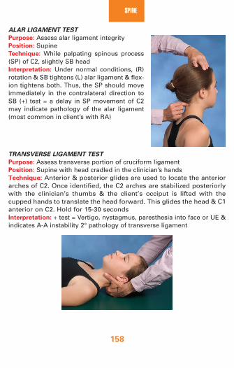
158
SPINE
ALAR LIGAMENT TEST
Purpose: Assess alar ligament integrityPosition: SupineTechnique: While palpating spinous process(SP) of C2, slightly SB headInterpretation: Under normal conditions, (R)rotation & SB tightens (L) alar ligament & flex-ion tightens both. Thus, the SP should moveimmediately in the contralateral direction toSB (+) test = a delay in SP movement of C2may indicate pathology of the alar ligament(most common in client’s with RA)
TRANSVERSE LIGAMENT TEST
Purpose: Assess transverse portion of cruciform ligamentPosition: Supine with head cradled in the clinician’s handsTechnique: Anterior & posterior glides are used to locate the anteriorarches of C2. Once identified, the C2 arches are stabilized posteriorlywith the clinician’s thumbs & the client’s occiput is lifted with thecupped hands to translate the head forward. This glides the head & C1anterior on C2. Hold for 15-30 secondsInterpretation: + test = Vertigo, nystagmus, paresthesia into face or UE &indicates A-A instability 2° pathology of transverse ligament
2067_Tab05_156-182.qxd 3/19/09 1:12 PM Page 158

159
SPINE
LATERAL & AP RIB COMPRESSION
Purpose: Assess ribs for fxPosition: SupineTechnique: With clinician’s hands on the lateral aspect of the rib cage,compress bilaterally; repeat with hands on the front & back of the chestInterpretation: + test = pain due to rib fracture or costochondral separation
RIB MOTION TEST
Purpose: Assess costal mobilityPosition: SupineTechnique: Palpate AP mov’t of ribs as client inhales/exhalesInterpretation: During inspiration, ribs 1–6 should ↑ in AP dimension,while ribs 7–10 should ↑ in lateral dimension via bucket handle action &ribs 8–12 should ↑ in lateral dimension via caliper action; + test = inhibitedrib movement with exhalation suggests an elevated rib; inhibited ribmovement with inhalation suggests a depressed rib
BEEVOR’S SIGN
Purpose: Assess abdominal musculaturePosition: Supine with knees flexed & feet on matTechnique: Head & shoulders are raised off the mat while movement ofthe umbilicus is observedInterpretation: Umbilicus should remain in a straight line. + test depends ondirection of movement. Movement distally = weak upper abdominals,movement proximally = weak lower abdominals, movement up & (R) =weak muscles in (L) lower quadrant, movement down & (L) = weak musclesin the (R) upper quadrant
2067_Tab05_156-182.qxd 3/19/09 1:12 PM Page 159
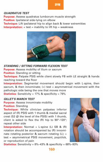
160
SPINE
QUADRATUS TEST
Purpose: Assess quadratus lumborum muscle strength Position: Ipsilateral side-lying on elbowTechnique: Lift ipsilateral hip to align back & lower extremitiesInterpretation: + test = inability to lift hip = weakness
STANDING / SITTING FORWARD FLEXION TEST
Purpose: Assess mobility of ilium or sacrumPosition: Standing or sittingTechnique: Palpate PSIS while client slowly FB with LE straight & handsreaching toward the floorInterpretation: Segmental movement should begin with L-spine, thensacrum, & then innominate; (+) test = asymmetrical movement with thepathologic side being the one that moves more Statistics: Sensitivity = 17% & specificity = 79%
GILLET’S MARCH TEST
Purpose: Assess innominate mobilityPosition: StandingTechnique: While clinician palpates inferioraspect of (R) PSIS with 1 thumb & medial sacralcrest (S2 @ the level of the PSIS) with 1 thumb,client is asked to flex the (R) hip to 90°–120°;repeat other sideInterpretation: Normal = L-spine (L) SB & (R)rotation should be accompanied by (R) innomi-nate rotating posterior & sacrum rotating (L); +test = asymmetrical PSIS movement, pop/click,or reproduction of painStatistics: Sensitivity = 8%–43% & specificity = 68%–93%
2067_Tab05_156-182.qxd 3/19/09 1:12 PM Page 160

161
SPINE
SUPINE TO SIT TEST
Purpose: Assess position of the ilium Position: Supine with both LEs extendedTechnique: Palpate medial malleolus as client performs a long sit-up (Becareful not to rotate the trunk while sitting up)Interpretation: + test = a short-to-long leg position = posterior ilium rota-tion; a long-to-short leg position = anterior ilium rotationStatistics: Sensitivity = 44% & specificity = 64%
LUMBAR QUADRANT TEST
Purpose: Assess nerve roots & IVFPosition: Standing or sitting Technique: Assist the client in extending spine & SB ipsilaterally with rotation contralaterally &then apply overpressure through the shoulders;repeat to other sideInterpretation: + test = radicular symptoms aredue to nerve root compression whereas localpain incriminates the facet joints
PRONE KNEE BENDING
Purpose: Assess neural mobilityPosition: Basic test position = prone withhips extendedTechnique: Add each of the followingmotions to implicate a specific nerves.Interpretation: + test = reproduction ofsymptoms
Modification for nerve bias: Nerve implicated:
Knee flexion Femoral nerve (L2–4)Hip adduction with knee flexion Lateral femoral cutaneous nerveHip abduction, ER, knee extension, Saphenous nerve& ankle dorsiflexion & eversion
2067_Tab05_156-182.qxd 3/19/09 1:12 PM Page 161

162
SPINE
Interpretation: + test = reproduction of symptoms
SLR TEST
Purpose: Assess neural mobilityPosition: Basic SLR test position = hip flexion, adduction, IR with kneeextendedTechnique: Add each of the following motions to implicate specific nerves
Modification for nerve bias: Nerve implicated:
Dorsiflexion Sciatic nerveDorsiflexion, eversion, & toe extension Tibial nerveDorsiflexion & inversion Sural nervePlantarflexion & inversion Common peroneal nerve
Statistics: Sensitivity = 76%–96% & specificity = 10%–45%
2067_Tab05_156-182.qxd 3/19/09 1:12 PM Page 162

163
SPINE
STOOP TEST
Purpose: Differentiate neurogenic vs. vascular intermittent claudicationPosition: StandingTechnique: Client walks briskly until symptoms appear & then flexes for-ward or sitsInterpretation: + test = if symptoms are quickly relieved with FB, claudica-tion is neurogenic; can also perform on a stationary bike
SI POSTERIOR COMPRESSION TEST (Anterior Gapping)
Purpose: Assess for SI pathologyPosition: Supine with clinician’s hands crossed over client’s pelvis onASISsTechnique: Apply a lateral force to the ASISs through the handsInterpretation: + test = reproduction of SI joint painStatistics: Sensitivity = 7%–69% & specificity = 69%–100%
SI POSTERIOR GAPPING TEST (Compression of iliac crests)
Purpose: Assess for SI pathologyPosition: Side-lyingTechnique: Apply a downward force through the anterior aspect of theASIS to create posterior gapping of the SIInterpretation: + test = reproduction of SI joint painStatistics: Sensitivity = 4%–60% & specificity = 81%–100%
HOOVER TEST
Purpose: Assess malingeringPosition: SupineTechnique: Hold client’s heels of (B) LEs in clinician’s hands, ask client tolift 1 leg out of a handInterpretation: + test = client does not lift the leg & there is no downwardforce exerted by the contralateral limb
2067_Tab05_156-182.qxd 3/19/09 1:12 PM Page 163

164
SPINE
Waddell Nonorganic Signs
SI Cluster Tests Sensitivity Specificity
• Standing flexion, PSIS palpation, supine to 82 88long-sit, & prone knee flexion
• Distraction, thigh thrust, Gaenslen’s, 91 78compression, & sacral thrust
• Thigh thrust, distraction, sacral thrust, & 88 78compression
Sign Description
Tenderness—
superficial or nonanatomic
Simulation tests—
axial loading in rotation
Distraction tests –
SLR
Regional
disturbances—
weakness or sensory
Overreaction
Tenderness is not related to a particular structure. Itmay be superficial (tender to a light pinch over awide area) or deep tenderness felt over a wide area(may extend over many segmental levels).These tests give the client the impression that diag-nostic tests are being performed. Slight pressure(axial loading) applied to the top of the head or pas-sive rotation of the shoulders & pelvis in the samedirection produces c/o LBPA (+) clinical test (SLR) is confirmed by testing thestructures in another position. By appearing to testthe plantar reflex in sitting, the examiner may actu-ally lift the leg higher than that of the supine SLR.When the dysfunction spans a widespread region ofthe body (sensory or motor) that cannot be explainedvia anatomical relationships. This may be demonstrat-ed by the client “giving way” or cogwheel resistanceduring strength testing of many major muscle groupsor reporting diminish sensation in a nondermatomalpattern (stocking effect). Disproportionate responses via verbalization, facialexpressions, muscle tremors, sweating, collapsing,rubbing affected area, or emotional reactions.
Note: Any positive test in 3 or more categories results in an overallWaddell Score.
2067_Tab05_156-182.qxd 3/19/09 1:12 PM Page 164

165
SPINE
Differential Diagnosis
Pathology/Mechanism Signs/Symptoms
Torticollis—7 forms of congenitaltorticollis & other causes includehemivertebra, cervical pharyngitis(major cause in 5–10 yo), JRA,trauma
Cervical Sprain—trauma or prolongedstatic positioning
Cervical Strain—single traumaticevent or cumulative trauma; mostoften occurs in 20–40 yo who have faulty posture, overweight,deconditioning
■ Symptoms appear @ 6–8 weeksof age
■ ↓ Contralateral rotation & ipsilateralSB (unilateral)
■ Firm, nontender swelling aboutthe size of an adult thumb nail
■ (–) x-ray■ Complications include visual
issues &/or reflux■ Localized pain; TTP; protective
muscle guarding■ MTrP in cervical, shoulder, &
scapular regions■ ↓ Cervical ROM & stiffness with
activity■ Headache & postural changes—
forward head, kyphosis■ Screen for alar & transverse
ligament px■ Clear vertebral arteries■ Normal DTRs & (–) x-ray■ Pain with contraction & with
stretching■ Pain with prolonged sitting, walk-
ing, standing■ TTP & protective muscle guarding■ Pain appears several hours after
injury; headache■ ↓ Contralateral SB & rotation
(AROM <PROM)■ Clear vertebral arteries■ Normal DTRs■ (–) special tests & (–) x-rays
Continued
2067_Tab05_156-182.qxd 3/19/09 1:12 PM Page 165

166
SPINE
Pathology/Mechanism Signs/Symptoms
Cervical Stenosis—most common30–60 yo; � > �; can be congenitalor developmental, onset is gradual
Cervical Spondylosis—↑ onset withaging but may be accelerated bycumulative trauma, poor bodymechanics, postural changes, orprevious disk injury; most common@ C5–7Cervical Disk Pathology (most
common level is C5–6)—usually theresult of repetitive stresses on theneck as a result of poor posture ormuscle imbalances; most commonin 30–50 yo
Cervical Facet Syndrome—occursas a result of isolated or cumulativetrauma, DDD, aging, or posturalimbalances
■ Unilateral or bilateral symptomsusually span several dermatomes
■ ↑ Pain with cervical BB & ↓ withcervical FB
■ Pain relieved with rest■ Loss of hand dexterity, LOB &
unsteady gait■ (+) Quadrant test■ LMN signs at the level of the
stenosis & UMN signs below thelevel of stenosis
■ X-rays reveal spondylitic bars &osteophytes & ossification of PLL & Ligamentum Flavum
■ ↑ Pain with activity & stiffness @ rest
■ Limited A & PROM; crepitus■ (+) Compression/distraction test■ ↓ Disk height on x-ray; need to
r/o osteophytes■ (+) NTPT—median nerve with con-
tralateral cervical SB, cervical rota-tion <60° & cervical FB <50°
■ (+) Tests: compression, distraction,shoulder depression & Spurling’smaneuver
■ Sensory changes in the respectivedermatome
■ X-rays are of little value■ CT & MRI used to differentiate nucle-
us pulposus from annulus fibrosis■ Pain with hyperextension & rota-
tion of c-spine■ Muscle guarding & stiffness■ Poor movement patterns but no
weakness■ Paresthesia but no changes in DTRs■ Possible (+) NTPT; (+) Quadrant test■ (–) X-ray
Continued
2067_Tab05_156-182.qxd 3/19/09 1:12 PM Page 166

167
SPINE
Pathology/Mechanism Signs/Symptoms
Brachial Plexus Lesion (Plexopathy,
Burner, Stinger)—occurs fromstretching or compression of C-spine or forceful depression ofshoulder
Rib Fracture—mechanism is adirect blow; cough in a frail person
Costochondritis—may be due totrauma, infection, arthritis, or surgery
Compression Fracture—most common in T11–L2, may be relatedto trauma or osteoporosis
Spondylosis / Arthrosis—
degenerative changes that usuallyeffects C5–6, C6–7, L4–5 of clients>60 yo
Spondylolysis—traumatic fracturesof pars or stress fractures due torepeated or sustained extension,seen in young athletes 2° repetitivetrauma (ski jumping, gymnastics);may have a structural predisposition
■ Sharp & burning pain in UE■ Numbness/pins & needles present
in UE■ Transient muscle weakness & ↓
DTR■ Provocation test = ipsilateral cer-
vical SB with compression ORcontralateral SB (stretch)
■ (+) NTPT■ Confirmed with myelogram■ (+) Tests: AP & lateral rib com-
pression■ TTP & pain with deep inspiration■ (+) X-ray is difficult to assess
immediately after injury■ Localized pain in anterior chest
wall■ TTP; pain ↑ with cough that may
radiate into UE■ Acute pain with adjacent muscle
guarding■ Limited BB & rotation■ (+) X-ray■ Onset is slow; pain is unilateral &
↑ with prolonged postures■ Pain ↑ with BB & ↓ with FB but
usually does not radiate■ Confirmed with x-ray; osteo-
phytes, ↓ joint space, & narrowIVF may be present
■ Pain primarily with extension■ Intermittent neurologic signs &
symptoms■ Oblique x-ray reveals fracture of
pars interarticularis without slip-page (Scottie dog with a collar)
Continued
2067_Tab05_156-182.qxd 3/19/09 1:12 PM Page 167

168
SPINE
Pathology/Mechanism Signs/Symptoms
Spondylolisthesis—vertebral subluxation or slippage 2° a longhistory of LB traumaRetrolisthesis = not common butpresents with flexion symptoms
Lumbar Disk Pathology—usuallythe result of repetitive stresses onthe LB using improper bodymechanics or excessive force pos-terior/lateral > lateral; most com-mon in 30–50 yoNote: See “Lumbar Disk Posturing& Pain” on page 173.
Lumbar Sprain—usually resultsfrom a combination of forwardbending with rotation or SB; common in people <30 yo
■ L5 nerve entrapment → sciatica■ Morning stiffness; difficulty
getting OOB■ ↑ Pain with trunk extension■ Poor neuromuscular control—
“Hitching sign” = 2-step processof moving from FB & BB via 1st extending lumbar spine intolordosis & then extending hip
■ Palpable step deformity in WB,gone in NWB
■ (+) Tests: PIVM & compression test■ A/P & lateral x-ray confirms dx■ Posterior-lateral HNP:
■ 1st sign is LBP that slowlydiminishes to leg pain
■ LB flexion 2° ↑ disk pressure■ (+) Thecal signs (pain with
sneezing & coughing)■ (+) SLR; ↓ lumbar lordosis■ Lateral shift in standing that ↓
in supine■ Lateral HNP:
■ No LBP; LE symptoms consis-tent with level of injury
■ ↑ Pain with standing & walking;↓ with sitting
■ (–) SLR■ Standard x-rays are of little value
because they may only detect pre-existing degenerative changes;MRI, CT scan, myelogram &discogram are used for diagnosis
■ Unilateral LBP■ Pain with SB away & rotation
toward affected side■ Referred pain limited to gluteals
& thigh regions
Continued
2067_Tab05_156-182.qxd 3/19/09 1:12 PM Page 168
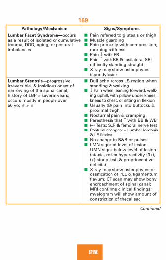
169
SPINE
Pathology/Mechanism Signs/Symptoms
Lumbar Facet Syndrome—occursas a result of isolated or cumulativetrauma, DDD, aging, or posturalimbalances
Lumbar Stenosis—progressive,irreversible, & insidious onset ofnarrowing of the spinal canal; history of LBP × several years;occurs mostly in people over 50 yo; � > �
■ Pain referred to gluteals or thigh■ Muscle guarding■ Pain primarily with compression;
morning stiffness■ Pain ↓ with FB■ Pain ↑ with BB & ipsilateral SB;
difficulty standing straight■ X-ray may show osteophytes
(spondylosis)■ Dull ache across LS region when
standing & walking■ ↓ Pain when leaning forward, walk-
ing uphill, with pillow under knees,knees to chest, or sitting in flexion
■ Usually (B) pain into buttocks &proximal thigh
■ Nocturnal pain & cramping■ Paresthesia that ↑ with BB & WB■ (–) Tests: SLR & femoral nerve test■ Postural changes: ↓ Lumbar lordosis
& LE flexion■ No change in B&B or pulses■ LMN signs at level of lesion,
UMN signs below level of lesion(ataxia, reflex hyperactivity (3+),(+) stoop test, & proprioceptivedeficits)
■ X-ray may show osteophytes orossification of PLL & ligamentumflavum; CT scan may show bonyencroachment of spinal canal;MRI confirms clinical findings;myelogram will show amount ofconstriction of thecal sac
Continued
2067_Tab05_156-182.qxd 3/19/09 1:12 PM Page 169

170
SPINE
Pathology/Mechanism Signs/Symptoms
Trochanteric Bursitis—may resultfrom contralateral gluteus mediusweakness or a change/↑ in activitylevel; direct trauma
Piriformis Syndrome—most commonly due to repeated compressive forces or may resultfrom a change/↑ in activity level; � > �
Ischiogluteal Bursitis—may resultfrom a change/↑ in activity level
Ankylosing Spondylitis (Marie
Stüumpell’s disease)—involvesanterior longitudinal ligament &ossification of disk & thoraciczygapophyseal joints; most com-mon in 15–40 yo; � > �
■ Pain into buttock & lateral thigh■ Pain worse at night & with activity■ TTP over greater trochanter■ Possible “clicking” with AROM &
pain with resisted hip abduction■ Check for leg length discrepancy■ (–) X-ray■ Piriformis TTP■ Ipsilateral LB, buttock, & referred
LE pain■ Pain & weakness with resisted
abduction/ER of thigh■ Pain with stretch into hip flexion,
adduction & IR■ (–) X-ray; need to r/o sprain/strain
or HNP■ Pain into buttock & posterior
thigh that is worse in sitting■ TTP over ischial tuberosity■ (+) Tests: SLR & Patrick test■ (–) X-ray■ Postural changes:
■ Cervical hyperextension ■ Thoracic kyphosis■ ↓ Lumbar lordosis■ Hip & knee flexion contractures
■ Night pain & ↓ rib expansion■ ↑ SED rate■ 5 screening questions:
■ Morning stiffness > 30 minutes■ Improvement with exercise■ Onset of back pain before 40 yo■ Slow onset■ Symptoms >3 months
4+ positive questions is highly corre-lated with AS
Continued
2067_Tab05_156-182.qxd 3/19/09 1:12 PM Page 170

171
SPINE
Pathology/Mechanism Signs/Symptoms
Osteoporosis—results from insuffi-cient formation or excessiveresorption of bone; occurs with ↑age, low body fat, low Ca++ intake,high caffeine intake, bed rest, alcoholism, steroid use
■ Dowager’s hump (dorsal kyphosis)■ Loss of height (2–4 cm/fracture)■ Acute regional back pain (low
thoracic/high lumbar)■ Pain radiating anterior along
costal margins■ Fragile skin■ X-ray does not show bone loss
but will reveal fx■ Bone scan needed for confirmation
Population
Pain location
Pain description
Positional
response
Response to
activity
Pulses & skin
Sensation
Vascular vs. Neurological Claudication
Neurogenical Signs &
Vascular Signs & Symptoms Symptoms
Primarily affects people >40 yoBilateral—hip, thigh, & buttock to calfCramping, aching, squeezing
Pain is present regardless of spinal positionPain brought on by physical exertion (walking, particularly uphill) & relieved within minutes of rest↓ LE pulses; color & skin changesNo burning or sensation changes
Unilateral or bilateral—LB & buttocksNumbness, tingling,burning, weaknessPain ↓ with spinal flexion& ↑ with spinal extensionPain ↑ with walking & ↓with recumbency
Normal pulses & skin
Burning & numbness inLE
2067_Tab05_156-182.qxd 3/19/09 1:12 PM Page 171
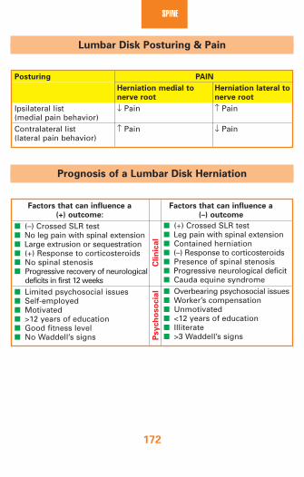
172
SPINE
Lumbar Disk Posturing & Pain
Posturing PAIN
Herniation medial to Herniation lateral to
nerve root nerve root
Ipsilateral list ↓ Pain ↑ Pain(medial pain behavior)Contralateral list ↑ Pain ↓ Pain(lateral pain behavior)
Prognosis of a Lumbar Disk Herniation
Factors that can influence a Factors that can influence a
(+) outcome: (–) outcome
■ (–) Crossed SLR test■ No leg pain with spinal extension■ Large extrusion or sequestration■ (+) Response to corticosteroids■ No spinal stenosis■ Progressive recovery of neurological
deficits in first 12 weeks■ Limited psychosocial issues■ Self-employed■ Motivated■ >12 years of education■ Good fitness level■ No Waddell’s signs P
sych
oso
cia
lC
lin
ical
■ (+) Crossed SLR test■ Leg pain with spinal extension■ Contained herniation■ (–) Response to corticosteroids■ Presence of spinal stenosis■ Progressive neurological deficit■ Cauda equine syndrome■ Overbearing psychosocial issues■ Worker’s compensation■ Unmotivated■ <12 years of education■ Illiterate■ >3 Waddell’s signs
2067_Tab05_156-182.qxd 3/19/09 1:12 PM Page 172

173
SPINE
Diffe
ren
tial D
iag
no
sis
of S
acro
iliac D
ysfu
nctio
ns
Lu
mb
ar
Seate
d
Sit-S
lum
p
Sacra
l Sp
ring
Dia
gn
osis
Sacra
l Base
ILA
Sp
ine
Fle
xio
n T
est
Test
Test
R S
acral flexio
nL S
acral flexio
nB
ilateral sacral flexio
nR
Sacral
extensio
nL S
acral exten
sion
Bilateral sacral
extensio
nL / L FW
sacral to
rsion
R/R
FW
sacral torsio
nL /R
BW
sacral to
rsion
R/ L B
W
sacral torsio
n
Deep
R
Deep
L
Deep
B
Sh
allow
R
Sh
allow
L
Sh
allow
B
Deep
R
Deep
L
Sh
allow
L
Sh
allow
R
Sh
allow
R
Cau
dal
R
Sh
allow
L C
aud
alL
Deep
B
Deep
RC
ranial
RD
eepL
Cran
ialL
Deep
B
Sh
allow
LC
aud
alR
Sh
allow
RC
aud
alL
Deep
RC
aud
alR
Deep
LC
aud
alL
Co
nvex
R
Co
nvex
L
↓Lo
rdo
sis
Co
nvex
L
Co
nvex
R
↑Lo
rdo
sis
Co
nvex
R
Co
nvex
L
Co
nvex
R
Co
nvex
L
(+) R
(+) L
(+) R
(+) L
(+) R
(+) L
(+) L
(+) R
Deep
Rb
asew
ith slu
mp
Deep
Lb
asew
ith slu
mp
Sh
allow
Rb
ase with
extS
hallo
wL
base w
ith ext
Deep
Rb
asew
ith slu
mp
Deep
Lb
asew
ith slu
mp
Sh
allow
Lb
ase with
extS
hallo
wR
base w
ith ext
↓R
ILA sp
ring
on
MTA
↓L ILA
sprin
go
n M
TA
↓R
sacral b
ase on
MTA
↓left sacralb
ase on
MTA
↓L ILA
sprin
go
n LO
A↓
R ILA
sprin
go
n R
OA
↓L sacral
base o
n R
OA
↓R
sacral b
ase on
LOA
ILA = in
ferior lateral an
gle
RO
A = rig
ht o
bliq
ue axis
LOA
= left ob
liqu
e axisM
TA = m
idd
le transverse axis
2067_Tab05_156-182.qxd 3/19/09 1:12 PM Page 173
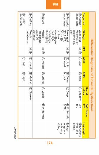
174
SPINED
iffere
ntia
l Dia
gn
osis
of Ilio
sacra
l Dysfu
nctio
ns
Sacra
l S
oft tis
su
e
Dia
gn
osis
Etio
log
yS
FT
AS
ISP
SIS
su
lcu
sT
TP
Leg
len
gth
RA
nterio
rIn
no
min
ate
RP
osterio
r In
no
min
ate
RIn
flare
RO
utflare
RU
pslip
in
no
min
ate
Weak g
lut
med
/max o
rab
do
min
als,g
olf
Prolonged R
LE WB
,fall on
Rischium
,w
eakR
glutm
ed, tightham
strings,short
Rleg
Mu
scleIm
balan
ces,w
eakR
glu
t med
Mu
scleim
balan
ces
(+)R
(+)R
(+)R
(+)R
RLo
w
RU
p &
forw
ard
RM
edial
RLateral
RH
igh
Rh
igh
RD
ow
n
& b
ack
RLateral
RM
edial
RH
igh
RS
hallo
w
LD
eep
RW
ider
RN
arrow
LeftT
FL
RP
iriform
is&
TFL
RP
iriform
is
RS
hortensw
ith lo
ng
sitting
RLeg
leng
then
sw
ith lo
ng
sitting
Continued
2067_Tab05_156-182.qxd 3/19/09 1:12 PM Page 174

175
SPINE
Diffe
ren
tial D
iag
no
sis
of Ilio
sacra
l Dysfu
nctio
ns—
co
nt’d
Sacra
l S
oft tis
su
e
Dia
gn
osis
Etio
log
yS
FT
AS
ISP
SIS
su
lcu
sT
TP
Leg
len
gth
RD
ow
nslip
in
no
min
ateR
Su
perio
r p
ub
ic shear
RIn
ferior
pu
bic sh
ear
Fall on
ischiu
m o
rlan
din
g o
n1 legS
ho
rt leg,
weak g
lut
med
ius &
/or
tigh
t ITB
(+)R
(+)R
RLo
w
Po
ss.R
h
igh
Po
ssR
low
RLo
w
Po
ss.R
hig
h
Po
ssR
lo
w
Rsh
allow
Tigh
t ITB
,ad
du
ctors &
R
qu
adratu
sT
TP
SIJ T
TP
Su
pin
e tosit = sh
ort
to lo
ng
SFT
= Stan
din
g Flexio
n Test
PS
IS = P
osterio
r Su
perio
r Iliac Sp
ine
AS
IS = A
nterio
r Su
perio
r Iliac Sp
ine
TT
P = Ten
der To
Palp
ation
2067_Tab05_156-182.qxd 3/19/09 1:12 PM Page 175

176
SPINE
Anatomy
Temporalartery
Facialnerve
Parotidgland
Common carotidartery
Trachea
Thyroidgland
Facial artery
Submandibularsalivary gland
Parotid duct
Ligaments of the jaw
Joint capsule
Lateral (temporomandibular) ligament
Styloid process
Stylomandibular ligament
Sphenomandibular ligament
Zygomatic arch
2067_Tab05_156-182.qxd 3/19/09 1:12 PM Page 176

177
SPINE
Referral Patterns
Muscle Pain Referral Patterns
Masseter Sternocleidomastoid
Scalene muscle
2067_Tab05_156-182.qxd 3/19/09 1:12 PM Page 177
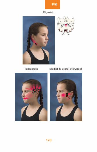
178
SPINE
Digastric
Temporalis Medial & lateral pterygoid
2067_Tab05_156-182.qxd 3/19/09 1:12 PM Page 178

179
SPINE
Palpation Pearls
■ SCM—in supine, find mastoid process & move toward the clavicularnotch, have client raise head & slightly rotate to opposite side
■ Scalenes—stringy muscle above the clavicle between the SCM &traps; to confirm identification, palpate in the general area & haveclient inhale deeply & scalenes should be in the middle of the triangle
■ Masseter—palpate the side of the mandible between the zygomaticarch & the angle of the mandible, have client clench the jaw
■ Suprahyoids—palpate under the tip of the chin & resist mandibulardepression or have the client swallow to confirm identification
■ Anterior digastric—palpate extraorally inferior to body of themandible
■ Posterior digastric—palpate extraorally posterior to the angle of themandible
■ Medial pterygoid—palpate intraorally along medial rim of themandible
■ Lateral pterygoid—palpate intraorally along superior, posterior aspectbehind 3rd maxillary molar
ROM
■ Mandibular depression (opening)—35–50 mm (2–3 knuckles) is functional■ C-deviation = hypomobility toward
side of deviation (lateral pterygoid tension or disk pathology)
■ S-deviation = muscle imbalance or displacement of condyle around disk
■ Mandibular elevation (closing)—palpatequality of movement to resting position
■ Mandibular protrusion = 6–9 mm; must take into account the startingposition if there is an overbite or underbite present
■ Mandibular retrusion = 3–4 mm■ Mandibular lateral excursion = 10–15 mm
2067_Tab05_156-182.qxd 3/19/09 1:12 PM Page 179

180
SPINE
Osteokinematics of the TMJ
Motion Normal End-feel(s) Abnormal End-feel(s)
Opening/ClosingProtrusion/retrusionLateral excursion
Open = tissue stretch/elasticClosed = bone-to-boneTissue stretch/elastic
Tissue stretch/elastic
Hard = osseous abnormality
Springy = disk displacement
Capsular = shortening of periarticular tissues
Arthrokinematics of TMJ
Opening & closing
Protrusion & retrusion
Lateral excursion
Concave surface:Mandibular fossaConvex surface:Mandibularcondyle & disk
Concave surface:Mandibular fossaConvex surface:Mandibularcondyle & disk
Concave surface:Mandibular fossaConvex surface:Mandibularcondyle & disk
To facilitate opening: Condyles rotate anterior for the first 25°, then anterior & inferior gliding of condyle & disk completes the last 15°of movementTo facilitate protrusion:Disk & condyle move down & FW
To facilitate lateral excursion:(R) excursion = (L) condyle & disk glide anterior; while (R) condyle spins aroundvertical axis(L) excursion = (R) condyle & disk glide anterior; while (L) condyle spins aroundvertical axis
To facilitateclosing:Condyles &disk roll pos-terior & glidemedially &superiorTo facilitateretrusion:Disk &condylemove up &BW
2067_Tab05_156-182.qxd 3/19/09 1:12 PM Page 180

181
SPINE
Special Tests
■ CLEAR CRANIAL NERVES – see “Alerts/Alarms” tab page 13.■ AUSCULTATION—used to identify poor joint kinematics or joint/disk
damage; place stethoscope over TMJ, just anterior to tragus of ear,and clinician listens for presence of joint sounds; very sensitive tofinding a problem but not specific in the identification of the structure.
Interpretation: ■ Opening click = click as condyle moves over posterior aspect of
disk in an effort to restore normal relationship; disk is anterior tocondyle; the later the click, the more anterior the disk
■ Reciprocal click = in opening, the disk reduces as the condylemoves under the disk & in closing, a second click is heard as thecondyle slips posteriorly & the disk becomes displaced anteriorly
LATERAL POLE
Purpose: Assess soft tissues of TMJ
Position: Face client with clinician’s index fingers palpating lateral pole ofTMJTechnique: Open & close mouth several timesInterpretation: + test = ↑ or reproduction of symptoms incriminating LCLor TMJ ligament
EXTERNAL AUDITORY MEATUS
Purpose: Assess posterior diskPosition: Face client, clinician inserts little fingers into client’s earsTechnique: While applying forward pressure with fingers, client opens &closes mouth repeatedlyInterpretation: + test = ↑ or reproduction of symptomsStatistics: Sensitivity = 43% & specificity = 75%
DYNAMIC LOADING
Purpose: To mimic TMJ loading to differentiate between TMJ & musclepainPosition: Sitting with roll of gauze between molars on 1-sideTechnique: Client bites down on gauze rollInterpretation: Compression occurs on contralateral side & distraction onipsilateral side of gauze; + test = ↑ or reproduction of symptoms @ TMJ
2067_Tab05_156-182.qxd 3/19/09 1:12 PM Page 181

182
SPINE
Differential Diagnosis
Pathology/Mechanism Signs/Symptoms
Inflammation—may be the resultof acute or repetitive trauma, pro-longed immobilization or surgery*Disk Displacement—may berelated to poor posture, trauma,excessive opening, muscle imbal-ance (anterior displacement ismost common)TMJ Arthritis—gradual onset,poor kinematics or repeated trau-ma of the TMJ that leads to jointerosion
*Disk can result in clicking or locking. Locked open = disk is anterior andwith opening there is a click with the disk being displaced posterior, thenthe joint is locked in the open position; locked closed = disk is anterior tothe condyle so anterior translation is limited & opening is reduced.
■ Capsular tightness with ↓ opening■ Pain with or without movement■ Need to r/o disk displacement■ Muscle guarding■ Localized TTP■ Headache■ Confirmed with MRI
■ Pain, stiffness, crepitus, clicking,grinding
■ ↓ ROM (deviation towardinvolved side)
■ Headache■ Hearing loss & dizziness■ Confirmed with x-ray or MRI;
need to r/o disk problem
2067_Tab05_156-182.qxd 3/19/09 1:12 PM Page 182

183
HIP
Anatomy of the Hip
Muscles of the hip
Gluteus medius muscle
Gluteus maximusmuscle
Gluteus medius muscle (cut)
Femur
Femur
Gluteus minimus muscle
Piriformis muscle
Superior gemellusmuscle
Sacrotuberous ligament
Obturator internusmuscle
Inferior gemellusmuscle Quadratus
femorismuscle
Iliac crestof pelvis
Palpation Pearls
Femoral triangle
■ Superior border = inguinal ligament■ Lateral to medial = sartorius,
femoral nerve, femoral artery,femoral vein, great saphenousvein, pectineus muscle, & adduc-tor longus muscle
■ Piriformis – find mid-pointbetween PSIS & coccyx, piri-formis runs from this point lateral to greater trochanter
Sartorius
Adductorlongus
Inguinalligament
Femoralartery
Femoralnerve
Femoralvein
2067_Tab06_183-198.qxd 3/19/09 1:13 PM Page 183

HIP
184
Medical Red Flags
■ Pain @ McBurney’s point = 1⁄3–1⁄2 the distance from (R) ASIS to umbili-cus; tenderness = appendicitis
■ Blumberg’s sign = rebound tenderness for visceral pathology—insupine select a site away from the painful area, place your hand perpendicular to the abdomen & push down deep & slow; lift upquickly; (–) = no pain; (+) = pain on release
■ Psoas test for pelvic pathology = supine, SLR to 30° & resist hip flexion;(+) test for pelvic inflammation or infection in lower quadrant abdominalpain; hip or back pain is a (–) test
■ Constitutional symptoms■ Enlarged inguinal lymph nodes■ Hip pain in men 18–24 years old of unknown etiology should be
screened for testicular CA■ Systemic causes of hip pain
■ Bone tumors■ Crohn’s disease■ Inflammatory bowel or pelvic inflammatory disease■ Ankylosing spondylitis■ Sickle cell anemia■ Hemophilia■ Urogenital problems
■ Neuromusculoskeletal causes of hip pain■ LB &/or SI■ OA or stress fx■ Hernia■ Muscle weakness■ Sprain/strain■ Labral tear
■ Screen for a sports hernia■ Palpation of marble-sized lump along the path of the inguinal ligament■ Pain with exertion, cough, menstruation■ Radiating pain into groin, ipsilateral thigh, flank, or lower abdomen■ Pain with cutting, turning, striding out
2067_Tab06_183-198.qxd 3/19/09 1:13 PM Page 184
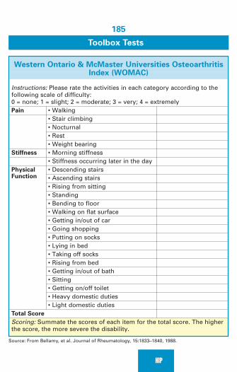
185
HIP
Toolbox Tests
Western Ontario & McMaster Universities OsteoarthritisIndex (WOMAC)
Instructions: Please rate the activities in each category according to thefollowing scale of difficulty: 0 = none; 1 = slight; 2 = moderate; 3 = very; 4 = extremelyPain • Walking
• Stair climbing• Nocturnal• Rest• Weight bearing
Stiffness • Morning stiffness• Stiffness occurring later in the day
Physical • Descending stairsFunction • Ascending stairs
• Rising from sitting• Standing• Bending to floor• Walking on flat surface• Getting in/out of car• Going shopping• Putting on socks• Lying in bed• Taking off socks• Rising from bed• Getting in/out of bath• Sitting• Getting on/off toilet• Heavy domestic duties• Light domestic duties
Total Score
Scoring: Summate the scores of each item for the total score. The higherthe score, the more severe the disability.
Source: From Bellamy, et al. Journal of Rheumatology, 15:1833–1840, 1988.
2067_Tab06_183-198.qxd 3/19/09 1:13 PM Page 185

HIP
186
HARRIS Hip Score
Select the descriptor for each section that best describes your current
condition
Pain—44 possible points
None or ignores it 44Slight, occasional, no compromise in activities 40Mild pain, no effect on average activities, moderate pain with 30unusual activities, may take aspirinModerate pain, tolerable but makes concessions, some limitation 20of ordinary activity, occasional pain medicine stronger than aspirinMarked pain, serious limitation of activity 10Totally disabled, crippled, pain in bed, bedridden 0Function/Gait—33 possible points
Distance Walked Unlimited 114–6 blocks 82–3 blocks 5Indoors only 2Unable to walk 0
Limp None 11Slight 8Moderate 5Severe 0
Support None 11Cane for long walks 7Cane most of the time 5One crutch 3Two canes 2Two crutches 0Not able to walk 0
Continued
2067_Tab06_183-198.qxd 3/19/09 1:13 PM Page 186

187
HIP
HARRIS Hip Score—cont’d
Select the descriptor for each section that best describes your current
condition
Function/Activities—14 possible points
Stairs Normally without rail 4Normally with rail 2In any manner 1Unable to do stairs 0
Shoes & Socks With ease 4With difficulty 2Unable 0
Sitting Comfortable in ordinary chair 1 hr 5On a high chair for 1⁄2 hr 3Unable to sit comfortably 0
Enter Public Transportation 1Deformity—4 points for each of the following present
<30° flexion contracture<10° adduction contracture<10° abduction contracture<3.2 cm leg-length discrepancy
ROM
Flexion 0–45° (1.0 point per degree)+ 0.6 points/degree from 45°–90°+ 0.3 points/degree from 90°–110°
Abduction 0–15° (0.8 points per degree)+ 0.3 points/degree from 15°–20°
ER (in ext) 0–15° (0.4 points per degree)Adduction 0–15° (0.2 points per degree)Total Score
Scoring: The higher the total score, the lower the level of disability.
Source: From Harris, WH. Journal of Bone and Joint Surgery, 51-A(4):737–755, 1969.
2067_Tab06_183-198.qxd 3/19/09 1:13 PM Page 187

HIP
188
Referral Patterns
Muscle Pain Referral Patterns
Gluteus maximus Piriformis Tensor fascia latae
Iliopsoas
2067_Tab06_183-198.qxd 3/19/09 1:13 PM Page 188
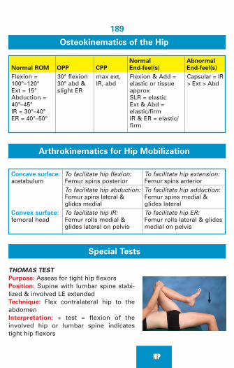
189
HIP
Osteokinematics of the Hip
Normal Abnormal
Normal ROM OPP CPP End-feel(s) End-feel(s)
Flexion = 100°–120°Ext = 15°Abduction = 40°–45°IR = 30°–40°ER = 40°–50°
30° flexion30° abd &slight ER
max ext,IR, abd
Flexion & Add =elastic or tissueapproxSLR = elasticExt & Abd = elastic/firmIR & ER = elastic/firm
Capsular = IR> Ext > Abd
Arthrokinematics for Hip Mobilization
Concave surface: acetabulum
Convex surface: femoral head
To facilitate hip flexion: Femur spins posteriorTo facilitate hip abduction: Femur spins lateral &glides medialTo facilitate hip IR: Femur rolls medial &glides lateral on pelvis
To facilitate hip extension:Femur spins anteriorTo facilitate hip adduction:Femur spins medial &glides lateralTo facilitate hip ER:Femur rolls lateral & glidesmedial on pelvis
Special Tests
THOMAS TEST
Purpose: Assess for tight hip flexorsPosition: Supine with lumbar spine stabi-lized & involved LE extendedTechnique: Flex contralateral hip to theabdomenInterpretation: + test = flexion of theinvolved hip or lumbar spine indicatestight hip flexors
2067_Tab06_183-198.qxd 3/19/09 1:13 PM Page 189

HIP
190
OBER’S TEST
Purpose: Assess for tight ITBPosition: Side-lying with involved hip upTechnique: Extend the involved hip &allow LE to drop into adductionInterpretation: + test = LE fails to adduct
IMPINGEMENT TEST
Purpose: Assess for labral tears & femoroac-etabular impingementPosition: SupineTechnique: Simultaneously flex, adduct & ER hipto end rangeInterpretation: + test = reproduction of pain Statistics: Kappa = 0.58
ELY’S TEST
Purpose: Assess for tight rectus femorisPosition: Side-lying or prone, hip in extension Technique: Flex kneeInterpretation: + test = limited knee flexion with hip extension or inabilityto maintain hip extension when knee is flexed
2067_Tab06_183-198.qxd 3/19/09 1:13 PM Page 190

191
HIP
SCOUR TEST
Purpose: Assess for labral tearPosition: Supine, flex hip to 90°Technique: IR/ER hip with abd/adductionwhile applying a compressive force downthe femurInterpretation: + test = clicking, grinding orpain due to arthritis, acetabular labrum tear,avascular necrosis, or osteochondral defectStatistics: Sensitivity = 75%–79% & specificity = 43%–50%
ANTERIOR LABRAL TEST
Purpose: Assess for labral tearPosition: Supine in PNF D2 flexion (hip in full flex, ER & abd)Technique: Resist movement into ext IR & add (D2 extension)Interpretation: + test = reproduction of pain or click
POSTERIOR LABRAL TEST
Purpose: Assess for labral tearPosition: Supine in flexion with adduction, & IR (similar to PNF D1 patternbut with IR)Technique: Resist movement into ext, abduction, & ER (similar to D1extension but with ER)Interpretation: + test = reproduction of pain or click
2067_Tab06_183-198.qxd 3/19/09 1:13 PM Page 191

HIP
192
LOG ROLL TEST
Purpose: Assess for iliofemoral ligamentlaxityPosition: Supine with LEs extendedTechnique: Roll the LE into maximal ERby applying a medial to lateral forcethrough the thighInterpretation: + test = excessive ER ascompared to the contralateral LEStatistics: Kappa = 0.61
FABER TEST (PATRICK’S)
Purpose: Assess hip/SI & labral pathology Position: Supine -passively flex, abduct &ER the hip (figure-4 position) so that thelateral malleolus of the involved LE is onthe knee of the uninvolved LETechnique: Apply overpressure to flexedkneeInterpretation: + test = hip pain 2° to OA, osteophytes, intracapsular fx, or LBP 2° SI px; tightness without pain is a (–) test; pain experiencedassuming this position may indicate a problem with the sartorius muscleLabral pathology may be suspected if lateral aspect of the knee is >4cmfrom the surface & asymmetricalStatistics: Kappa = 0.63; sensitivity = 41%–77%; specificity = 88%–100%
TRENDELENBURG’S TEST
Purpose: Assess for weakness of gluteus mediusPosition: Standing on involved LETechnique: Flex the contralateral LE; iliac creston WB side should be lower than the NWB sideInterpretation: + test = dropping of the NWBlimb is 2° to abductor weakness (common inepiphyseal problem, Legg-Calve-Perthes, MD)
2067_Tab06_183-198.qxd 3/19/09 1:13 PM Page 192

193
HIP
PIRIFORMIS TEST
Purpose: Assess for tight piriformisPosition: Supine or contralateralside-lyingTechnique: Flex hip to 70°–80° withknee flexed & maximally adduct LE(apply a downward force to the knee)Interpretation: + test = pain in but-tock & sciatica; IR stresses superiorfibers; ER stresses inferior fibers
ORTOLANI’S TEST
Purpose: Assess for congenital hip dislocationPosition: Supine fix hips & knees @ 90° offlexion; clinician’s thumbs are on theinfant’s medial thigh & fingers on the lateral thighTechnique: Firmly traction the thigh whilegently abducting the leg so that femoral headis translated anterior into the acetabulumInterpretation: + test = reduction of thehip; an audible “clunk” may be heard
BARLOW’S TEST
(Opposite of the Ortolani Test)Purpose: Assess for hip dysplasiaPosition: Supine 90/90; clinician’s thumbs areon the infant’s medial thigh & fingers on thelateral thighTechnique: Apply a posterior force thru thefemur as the thigh is gently adductedInterpretation: + test = the examiner’s fingerthat is on the greater trochanter will detect apalpable dislocation
2067_Tab06_183-198.qxd 3/19/09 1:13 PM Page 193

HIP
194
Differential Diagnosis
Pathology/Mechanism Signs/Symptoms
ITB Friction Syndrome—repetitivestress & excessive friction 2° tightITB, pronation with IR of tibia, genuvarum, cycling with cleat in IRProximal problem = hip syndrome Distal problem = runner’s knee
Greater Trochanteric Bursitis—
biomechanical or overuse problem;repetitive inside kicks in soccerresult in forceful adduction andcompression of bursa; contusions
Apophysitis—pelvic fx 2° strenuousmuscle contraction in skeletallyimmature child
■ Pain with downhill running; sense of knee instability
■ (+) Tests: Ober’s, Noble’s, &Renne’s
■ Pain @ 30° of knee flexion in WBresults in ambulating stiff leggedto avoid flexion
■ TTP over lateral femoral epicondyle■ Visible & palpable snapping ■ (–) X-ray; MRI & US may confirm
diagnosis■ Need to r/o trochanteric bursitis
& osteochondritis■ Deep, aching, diffuse pain from
greater trochanter to distal lateralthigh & groin
■ TTP on ITB & pain when rollingon hip when sleeping
■ ROM = WNL except abductionmay be limited by pain
■ No snapping but palpable crepitus may be present
■ (+) Tests: Ober’s & Patrick’s/FABER
■ (–) X-ray (needed to r/o femoralneck stress fx)
■ MRI & US may confirm diagnosis■ TTP & weakness with resisted
muscle contraction @ ASIS, AIIS,PSIS, PIIS—depending on muscleinvolved
■ (+) X-ray for avulsion
Continued
2067_Tab06_183-198.qxd 3/19/09 1:13 PM Page 194

195
HIP
Pathology/Mechanism Signs/Symptoms
Piriformis Syndrome—may resultfrom muscle contracture, trauma,prolonged sitting
Iliopsoas Bursitis/Tendonitis—
irritation & inflammation 2° overuseor unaccustomed activity
Hip Pointer—can result from directtrauma to iliac crest or ASIS result-ing in a contusion
Labral Tear—damage to fibrocarti-lage via degeneration due to repeti-tive hip ER or the application of anexternal rotatory force to the hipwhile in hyperextension & hyperab-duction; highly associated with hip dysplasia; anterior hip pain iscorrelated to weak gluteals &abdominals 2° excessive anteriorfemoral translation
■ Dull ache in buttocks■ Pain ↑ with sitting & walking &
↓ in supine■ Pain with resisted hip ext &
passive IR with adduction■ (–) X-ray needed to r/o stress fx;
MRI needed to r/o spine pathology(LS root lesion, spinal stenosis, SIproblem)
■ Pain in medial groin/thigh withhip flexion & extension
■ Audible snapping when movingfrom hip flex to ext
■ Screen for McBurney’s point &rebound tenderness
■ (–) X-ray; need to r/o avulsion fx■ Confirmed by MRI or US■ TTP @ iliac crest/ASIS■ Pain with resisted hip flexion &
stretching into hip extension■ Pain with ambulation & hip
abduction■ Screen for McBurney’s point &
rebound tenderness■ (–) X-ray; need to r/o fx & avulsion■ Pain with prolonged sitting,
getting in/out of a car, putting onshoes/socks, & twisting activities
■ ↑ Anterior hip pain with hyperext & ER■ Pain with resisted SLR (anterior
lesion)■ Often associated with weak gluteals■ ↓ Hip ROM; clicking/catching
from flexion to extension■ (+) Tests: FABER, impingement,
Scour & labral tests■ Screen for osteoid osteoma &
testicular CA in �■ MRI with contrast is best dx test
(is often inconclusive)
Continued
2067_Tab06_183-198.qxd 3/19/09 1:13 PM Page 195

HIP
196
Pathology/Mechanism Signs/Symptoms
Avulsion Fracture—injury resultsfrom violent muscle contraction
Femoral Neck Stress Fracture—
gradual onset with history ofendurance tasksBeware of eating disorders, amenorrhea, & osteoporosis
Degenerative Joint Disease—usuallyoccurs >55 yo in � > � (3:2)
RA—systemic disorder with bilateralWB symptoms
■ May hear a “pop” ■ Pain with stretch & contraction;
TTP @ apophysis■ (+) Tests: Thomas’ & Ely’s■ May need CT or MRI if x-ray is
inconclusive■ Need to r/o strain & slipped
capital femoral epiphysis■ Groin pain with activity ■ TTP @ greater trochanter■ (+) FABER test■ May need CT or MRI if x-ray is
inconclusive■ Need to r/o trochanteric bursitis
& osteoid osteoma■ Aching pain during WB => groin,
medial thigh & knee■ Loss of movement & function■ Trendelenburg■ (+) FABER test■ X-ray reveals narrow joint space,
spurring & osteophytes; can r/ofx & necrosis
■ Aching pain during WB => groin,medial thigh & distal knee; lossof movement & function 2° pain
■ Trendelenburg ■ (+) Tests: Thomas’, Ely’s & FABER■ X-ray = bilateral demineralization
of femoral head; joint space narrowing; migration of femoralhead into acetabulum
Continued
2067_Tab06_183-198.qxd 3/19/09 1:13 PM Page 196

197
HIP
Pathology/Mechanism Signs/Symptoms
Slipped Capital Femoral
Epiphysis—imbalance of growth &hormones that weakens the epiphy-seal plate; may be 2° ↑ wt gain;occurs in 10–16 yo � 2x > �
Legg-Calvé-Perthes (LCPD)
Disorder—idiopathic osteonecrosisof capital femoral epiphysis; associ-ated with (+) family history &breech birth. Onset occurs over 1–3 monthsbetween 4–13 yo; occurs unilaterally;� > �
Osteoid osteoma—benign tumorfound in long bones; etiologyunknown
Myositis Ossificans—calciumdeposits 2° contusion to the thigh
■ Gradual onset of unilateral hip,thigh & knee pain
■ ↓ Hip IR; hip positioned in flexion,abd, ER
■ Quadriceps atrophy■ Antalgic gait & ↓ limb length■ AP x-ray needed to identify
widening of physis & ↓ ht of epiphysis; lateral view = epiphyseal displacement
■ Need to r/o muscle strain & avulsion
■ Hip or groin pain ( thigh resultingin antalgic gait
■ (+) Trendelenburg■ ↓ ROM (ext, IR & abd); >15° hip
flexion contracture■ Leg length inequality; thigh
atrophy■ Bone scan or MRI needed for
early detection, x-rays mayappear normal for several weeks,1st sign (~4 wks) is radiolucentcrescent image parallel to thesuperior rim of the femoral head
■ Need to r/o JRA & hip inflammation■ Vague hip pain @ night■ ↑ Pain with activity & ↓ with
aspirin■ ↓ ROM & quad atrophy■ May be apparent on x-ray but
confirmed by MRI or CT■ Need to r/o trochanteric bursitis,
femoral neck stress fx■ Localized pain■ Limited knee flexion■ Palpation of a calcific mass
Continued
2067_Tab06_183-198.qxd 3/19/09 1:13 PM Page 197

HIP
198
Pathology/Mechanism Signs/Symptoms
Hip Dislocation—may result from a breech birth, trauma, or when the hip is in a weakened state aftera THR
■ (+) Tests: Ortolani’s & Barlow’s■ (+) X-ray (associated with
torticollis)Congenital ■ Shortened limb, positioned in
flexion & abductionPosterior Traumatic (MVA)■ Groin & lateral hip pain■ Shortened limb, positioned in
flexion, adduction & IRAnterior Traumatic (forced abduction)■ Groin pain & tenderness■ Positioned in extension & ER if
superior/anterior■ Positioned in flexion, abduction
& ER if inferior/anterior
2067_Tab06_183-198.qxd 3/19/09 1:13 PM Page 198

199
KNEE
Knee Anatomy
Anterior
Anterior knee
Patella
Patellatendon
Quadtendon
Vastimedialis
Vastilateralis
Rectusfemoris
Sartorius
Posterior Medial
Popliteus
Sartorius
Gracilis
Semitendinosus
Pes anserinus
Tibialtuberosity
2067_Tab07_199-220.qxd 3/19/09 3:45 PM Page 199

KNEE
200
Medical Red Flags
■ Night pain = tumor or infection■ Cellulitis
■ Recent hx of skin trauma■ Pain, swelling, warmth■ Advancing erythema with reddish streaks■ Chills, fever, weakness
■ DVT risk
■ Immobilization■ Surgery■ Fracture or trauma■ Oral contraceptives■ CHF, CA, DM■ Pregnancy
■ DVT Clinical presentation
■ Leg pain & tenderness■ ↑ Circumference > 1.2 cm■ Tissue warm & firm to palpation■ ↑ Pain with BP cuff inflated to 160 mm Hg■ (+) Homans’ sign
Imaging
Ottawa Knee Rule
X-ray series is only required if the patient presents with any of the follow-ing criteria:■ >55 years old■ Isolated tenderness of the patella■ Tenderness of the head of the fibula■ Inability to flex >90°■ Inability to bear weight (4 steps) both immediately after injury & in
emergency department (regardless of limping) Statistics: Adults: Sensitivity = 98%–100% & specificity = 19%–54%Children: Sensitivity = 92% & specificity = 49%
2067_Tab07_199-220.qxd 3/19/09 3:45 PM Page 200

201
KNEE
Toolbox Tests
Western Ontario & McMaster Universities OsteoarthritisIndex (WOMAC)
Instructions: Please rate the activities in each category according to thefollowing scale of difficulty: 0 = none; 1 = slight; 2 = moderate; 3 = very; 4 = extremelyPain ■ Walking
■ Stair climbing■ Nocturnal■ Rest■ Weight bearing
Stiffness ■ Morning stiffness■ Stiffness occurring later in the day
Physical Function ■ Descending stairs■ Ascending stairs■ Rising from sitting■ Standing■ Bending to floor■ Walking on flat surface■ Getting in/out of car■ Going shopping■ Putting on socks■ Lying in bed■ Taking off socks■ Rising from bed■ Getting in/out of bath■ Sitting■ Getting on/off toilet■ Heavy domestic duties■ Light domestic duties
Total Score
Scoring: Summate the scores of each item for the total score. The higherthe score, the more severe the disability.
Source: From Bellamy, et al. Journal of Rheumatology, 15:1833–1840, 1988.
2067_Tab07_199-220.qxd 3/19/09 3:45 PM Page 201

KNEE
202
Lysholm Knee Rating System
Which items below best describe your knee function today?Limp None 5
Slight or periodic 3Severe & constant 0
Support None 5Cane or crutch needed 2Weight bearing impossible 0
Locking None 15Catching sensation but no locking 10Locking occasionally 6Locking frequently 2Locked joint at examination 0
Instability Never gives way 25Rarely during physical activity 20Frequently during physical activity 15Occasionally during daily activity 10Often during daily activity 5Every step 0
Pain None 25Intermittent during strenuous activity 20Marked during strenuous activity 15Marked with walking >2 km (1.2 miles) 10Marked with walking <2 km (1.2 miles) 5Constant 0
Swelling None 10After strenuous activities 6After ordinary activities 2Constant 0
Continued
2067_Tab07_199-220.qxd 3/19/09 3:45 PM Page 202

203
KNEE
Lysholm Knee Rating System—cont’d
Which items below best describe your knee function today?Stairs No problem 10
Slight problem 6One step at a time 2Impossible 0
Squatting No problem 5Slight problem 4Not >90° knee flexion (halfway) 2Impossible 0
Total Score
Scoring: Summate the scores of each category. The higher the score, thegreater the functional abilities.
Source: From Tegner, Y, Lysholm, J. Rating systems in the evaluation of knee ligament injuries.Clin Orthop Relat Res. 1985 Sep;(198):43–49.
Referral Patterns
Muscle Pain Referral Patterns
Rectus femoris Vasti muscles
2067_Tab07_199-220.qxd 3/19/09 3:45 PM Page 203

KNEE
204
Hamstring muscles Tensor fascia latae
Palpation Pearls
■ Adductor tubercle = attachment of adductor magnus; start on medialfemoral condyle & move proximal between the vastus medialis &hamstring tendons, as the femur dips in, a small point is palpable &often tender
■ Lateral collateral ligament = cross leg so ankle is on contralateralknee (figure-4 position); LCL is palpable at the joint line just proximalto fibular head (firm, pencil-thickness structure)
■ Common peroneal nerve = posterior lateral knee between bicepsfemoris tendon & lateral gastroc muscle belly
■ Popliteus = “unlocker” of the knee; deep muscle only the tendon ispalpable; follow the tibial tuberosity medially around the knee to theposterior aspect & the popliteus tendon is deep to the gastroc/soleus
■ Q-angle = the angle created by the intersection of a line from theASIS to the mid-patella & a line from the mid-patella to the tibialtuberosity. Normal (supine) = 13°–18° for � & 10°–15° for �
2067_Tab07_199-220.qxd 3/19/09 3:45 PM Page 204

205
KNEE
Posterior
Sartorius
Gracilis
Semitendinosus
Bicepsfemoris
Iliotibialtract
Strength & Function
■ Concentric Quad to Hamstring ratio = 5:3 (i.e., hamstrings should be60%–65% of quads)
■ Quad:Hamstring ratio should approach 5:4 at the conclusion of ACLrehabilitation
■ Quad:Hamstring ratio should approach 5:2 at the conclusion of PCLrehabilitation
2067_Tab07_199-220.qxd 3/19/09 3:45 PM Page 205

KNEE
206
Osteokinematics of the Knee
Normal Abnormal
Normal ROM OPP CPP End-feel(s) End-feel(s)
Flexion >130°Rotation = 10°
Maximalextension& tibial ER
Flexion = tissueapproximationExtension =elastic/firmSLR = elastic
Springy block =displacedmeniscusBoggy = ligamentouspathology
25° flexion
■ Femoral condyles begin to contact the patella inferior @ 20° of kneeflexion; progresses superior @ 90° & medial/lateral @ 135° of kneeflexion
■ Structures attached to medial meniscus = MCL & semimembranosus■ Structures attached to lateral meniscus = PCL & popliteus
Arthrokinematics for Knee Mobilization
Concave surface:Tibial plateau
Convex surface:Femoral condyles
To facilitate knee extension: OKC = Tibia rolls & glidesanterior on the femurCKC = Femur rolls anterior& glides posterior on tibia
To facilitate knee flexion:OKC = Tibia rolls & glidesposterior on the femurCKC = Femur rolls poste-rior & glides anterior onthe tibia
2067_Tab07_199-220.qxd 3/19/09 3:45 PM Page 206

207
KNEE
Special Tests
LACHMAN’S TEST
Purpose: Assess for ACL laxityPosition: Supine with knee in 0-30° of flexion (hamstrings relaxed)Technique: Stabilize distal femur & translate proximal tibia forward on thefemurInterpretation: + test = >5 mm of displacement or a mushy, soft end-feel;beware of false (–) test due to hamstring guarding, hemarthrosis, posteriormedial meniscus tearStatistics: Sensitivity = 63%–99% & specificity = 90%–99%
PRONE LACHMAN’S TEST
Purpose: Assess for ACL laxityPosition: Prone with knee flexed to 30°, LE supported & hamstringsrelaxedTechnique: Palpate anterior aspect of the knee while imparting an anteriorforce to posterior-proximal aspect of tibiaInterpretation: + test = >5 mm of displacement or a mushy, soft end-feel Beware of false (–) test due to hamstring guarding, hemarthrosis, posteriormedial meniscus tear
2067_Tab07_199-220.qxd 3/19/09 3:45 PM Page 207

KNEE
208
ANTERIOR DRAWER TEST
Purpose: Assess for ACL laxityPosition: Supine with foot stabilized ontable, knee flexed to 80°–90° & hamstringsrelaxedTechnique: Translate proximal tibia anterioron the femurInterpretation: + test = >5mm of anteriordisplacement; snap or palpable jerk withanterior drawer indicates meniscus pxBeware: Translation may appear excessive with PCL injury if tibia startsfrom a more posterior positionStatistics: Sensitivity = 22%–95% & specificity = 78%–97%
POSTERIOR DRAWER TEST
Purpose: Assess for PCL laxityPosition: Supine with knee flexed to 90° &foot on tableTechnique: Translate proximal tibia poste-riorly on distal femurInterpretation: + test = >5 mm of posteriordisplacementStatistics: Sensitivity = 86%–90% & speci-ficity = 99%
SAG or GODFREY’S TEST
Purpose: Assess for PCL laxityPosition: Supine 90/90, support LEs Technique: Compare the level of thetibial tuberositiesInterpretation: + test = posterior dis-placement of the tibial tuberosity isgreater in the involved lagStatistics: Sensitivity = 79% & speci-ficity = 100%
2067_Tab07_199-220.qxd 3/19/09 3:45 PM Page 208

209
KNEE
CLARKE’S TEST; GRIND TEST; ZOHLER’S TEST
Purpose: Assess for chondromalacia or patellamalaciaPosition: Supine with knee in extension, clini-cian compresses quads at the superior poleof the patella to resist patella movementTechnique: Client contracts quads againstresistanceInterpretation: + test = inability to contractwithout pain
VARUS TEST
Purpose: Assess for LCL laxityPosition: Supine; knee in full extension &then repeat @ 30° flexion Technique: Cup knee with heel of clinician’shand @ medial joint line; use fingers ofother hand to palpate lateral joint line; applya varus stress to the knee through the palmof the medial hand & the forearm/elbow ofthe lateral handInterpretation: + test = pain or excessive gapping of the joint when com-pared with the contralateral side Statistics: Sensitivity = 25%
VALGUS TEST
Purpose: Assess for MCL laxityPosition: Supine; knee in full extension &then repeat @ 30° flexion Technique: Cup knee with heel of clini-cian’s hand @ lateral joint line; use fingersof other hand to palpate medial joint line;apply a valgus stress to the knee throughthe palm of the lateral hand & the fore-arm/elbow of the medial handInterpretation: + test = pain or excessive gapping of the joint when com-pared to the contralateral side Statistics: Sensitivity = 86%–96%
2067_Tab07_199-220.qxd 3/19/09 3:45 PM Page 209

KNEE
210
APLEY’S TEST
Purpose: Assess meniscus (nonspecific for locationof meniscal tear)Position: Prone, knee flexed to 90°; clinician graspsfoot & calcaneusTechnique: While applying a downward force throughthe heel, rotate the tibia internally & externallyInterpretation: + test = pain, popping, snapping, lock-ing, crepitusStatistics: Sensitivity = 13%–58% & specificity =80%–93%
McMURRAY’S TEST
Purpose: Assess meniscusPosition: Supine, with 1 of clinician’s hands to theside of the patella & the other grasping the distal tibiaTechnique: From a position of maximal flexion,extend the knee with IR of the tibia & a varus stressthen returns to maximal flexion & extend the kneewith ER of the tibia & a valgus stressInterpretation: + test = pain or snapping/clicking withIR incriminates the lateral meniscus & ER incrimi-nates the medial meniscus; if pain, snapping, or click-ing occur with the knee in flexion, the posterior hornof the meniscus is involved & if the pain, snapping, orclicking occurs with increasing amounts of kneeextension, the anterior meniscus is involvedStatistics: Sensitivity = 16%–67% & specificity =57%–98%
2067_Tab07_199-220.qxd 3/19/09 3:45 PM Page 210

211
KNEE
THESSALY’S TEST
Purpose: Assess for meniscal tearsPosition: Standing on the involved LE with theknee flexed @ 5°Technique: Clinician holds pt’s outstretchedarms & rotates internally then externally 3x;repeat @ 20° of knee flexionInterpretation: + test = Pt experiences locking orcatchingStatistics: At 5°: Sensitivity = 66%–81% & speci-ficity = 91%–96% and at 20°: Sensitivity =89%–92% & specificity = 96%–97%
PATELLA APPREHENSION (FAIRBANK’S) TEST
Purpose: Assess for subluxing patellaPosition: Supine or seated, 30° knee flex-ion, quads relaxedTechnique: Clinician carefully pushespatella laterallyInterpretation: + test = Pt feels patellaabout to dislocate & contracts quads tokeep this from happening Statistics: Sensitivity = 32%–39% & specificity = 86%
PATELLA TILT TEST
Purpose: Assess for ITB tightness/patella mobilityPosition: Relaxed in supine with knee in extensionTechnique: Clinician attempts to lift the lateral border of patellaInterpretation: + test = inability to lift the lateral border of the patellaabove the horizontal
2067_Tab07_199-220.qxd 3/19/09 3:45 PM Page 211

KNEE
212
NOBLE’S TEST
Purpose: Assess ITB irritationPosition: Supine, start @ 90/90Technique: Apply pressure over the lateralfemoral condyle while extending the kneeInterpretation: + test = pain or clicking @lateral femoral condyle @ 30° of kneeflexion
OBER’S TEST
Purpose: Assess for tight ITBPosition: Side-lying with involved hip upTechnique: Extend the hip & allow LE todrop into adductionInterpretation: + test = LE fails to adductpast anatomic neutral
RENNE’S TEST
Purpose: Assess ITB irritationPosition: StandingTechnique: Apply pressure over the lateralfemoral condyle with AROM of the kneeInterpretation: + test = pain or clicking @ lateralfemoral condyle @ 30° of knee flexion
2067_Tab07_199-220.qxd 3/19/09 3:45 PM Page 212

213
KNEE
PIVOT SHIFT TEST
Purpose: Assess A/L instabilityPosition: SupineTechnique: Knee is taken from full extension to flexion with a valgus stressInterpretation: + test = sudden reduction of the anteriorly subluxed lateraltibial plateau
STUTTER TEST
Purpose: Assess for medial plica irritationPosition: Sitting with knee flexed overthe edge of the tableTechnique: Slowly extend knee with afinger placed lightly in contact with thecenter of the patellaInterpretation: + test = patella stutters asknee moves into extension
PATELLAR BOWSTRING TEST
Purpose: Assess medial plicaPosition: SupineTechnique: Medially displace patella while flexing/extending knee withtibia IRInterpretation: + test = palpable clunk
WILSON’S TEST
Purpose: Assess for osteochondritis of medial femoral condylePosition: Supine with knee flexed to 90°Technique: Extend the knee with IR of the tibiaInterpretation: + test = pain at 30° of flexion in IR that ↓ if the tibia is ER;should r/o meniscal px
2067_Tab07_199-220.qxd 3/19/09 3:45 PM Page 213

KNEE
214
Differential Diagnosis
Pathology/Mechanism Signs/Symptoms
Baker’s Cyst—defect in the posteriorcapsule that is influenced by chronicirritation
Shin Splints/Anterior—an overusesyndrome of tibialis anterior, exten-sor hallicus longus, & extensor dig-itorum longus attributed to runningon unconditioned legs, soft tissueimbalance, alignment abnormali-ties, & excessive pronation toaccommodate rearfoot varus
Shin Splints/Posterior—an overusesyndrome of flexor hallucis longus& flexor digitorum longus; rapid &excessive pronation to compensatefor rearfoot varus; result is ↑ stresson tibialis posterior to deceleratepronation
■ Golf ball–size swelling at semi-membranosus tendon or medialgastroc muscle belly; best palpatedin full knee extension
■ Stiff & tender with limited kneeROM
■ MRI may be helpful; need to r/oDVT & tumor
■ Pain & tenderness over anteriortibialis
■ Pain with resisted dorsiflexion &inversion
■ Pain with stretching into plan-tarflexion & eversion
■ Callus formation under 2ndmetatarsal head & medial side ofdistal hallux
■ Tight gastroc/soleus■ Soreness with heel walking■ (–) X-ray, needed to r/o stress fx■ Callus formation under 2nd>
3rd> 4th MT head & medial sideof distal hallux
■ Pain & soreness over distal1/3–2/3 of posterior/medial shin & posterior medial malleolus
■ Hypermobile 1st MTP■ Pain with resisted inversion &
plantarflexion■ Pain with stretching into dorsi-
flexion & eversion■ (–) X-ray, needed to r/o stress fx
Continued
2067_Tab07_199-220.qxd 3/19/09 3:45 PM Page 214

215
KNEE
Pathology/Mechanism Signs/Symptoms
Compartment Syndrome—a progression of shin splints result-ing in a loss of microcirculation inshin muscle; � > �, R > LBeware: This is an emergency situation
Bursitis—mechanical irritation■ prepatella = common in sport =
falling on knee or maintainingquadruped position (housemaidsknee)
■ infrapatella = clergyman bursitis= kneeling (mechanical irritation)
■ pes anserine = prevalent in long-distance running or middle-agedfemales with OA of the knee
Popliteus Tendonitis—results fromoveruse, downhill running, activi-ties with sudden stops
■ ↑ Soft tissue pressures via fluidaccumulation
■ Ischemia of extensor hallicuslongus
■ Skin feels warm & firm■ Pain with stretch or AROM; foot
drop■ Most reliable sign is sensory
deficit of the dorsum of foot in1st interdigital cleft
■ Pulses are normal until the end & then surgery within 4–6 hoursis required to prevent musclenecrosis & nerve damage
■ Confirmed with MRI & pressureassessment
■ Localized radiating heat ■ Localized egg-shaped swelling■ Radiating pain 2–4 cm below the
involved bursa■ Crepitus■ Discomfort with A & PROM■ Diagnosis confirmed with MRI
■ Posterior lateral knee pain at theend of a workout or runningdownhill (just posterior to LCL)
■ Crepitus over tendon■ Discomfort sitting with legs
crossed & initiating flexionagainst resistance from fullextension
■ MRI may be helpful; need to r/oITB, biceps tendonitis
Continued
2067_Tab07_199-220.qxd 3/19/09 3:45 PM Page 215

KNEE
216
Pathology/Mechanism Signs/Symptoms
Jumper’s Knee = patella tendonitis(most common in skeletally imma-ture) 2° traction overuse injury suchas jumping, kicking, running ordegenerative process 2° microtrauma
ITB Friction Syndrome—repetitivestress & excessive friction 2° tightITB, pronation with IR of tibia, genuvarum, cycling with cleat in IR
Proximal px = hip syndrome Distal px = runner’s kneePlica Syndrome—injury results fromdirect trauma or a significant ↑ inunaccustomed activity (presence ofmedial plica is more common thana lateral plica)
■ TTP at patella tendon insertion &pain with resisted knee extension
■ Localized crepitus & swelling■ ↑ Q-angle■ Need to r/o Osgood-Schlatter’s,
SLJ, & bursitis■ Confirmed with MRI■ Pain with downhill running■ Pain @ 30° of knee flexion in WB
results in ambulating stiff leggedto avoid flexion
■ TTP over lateral femoral condyle■ (+) Tests: Ober’s, Noble’s, & Renne’s■ (–) X-ray■ Need to r/o trochanteric bursitis
& osteochondritis■ MRI & US may confirm diagnosis■ Pain over medial femoral condyle;
palpable cords along medialcondyle, pain at superomedialjoint line
■ Reports of clicking/snapping,locking, “giving way”
■ Full ROM but pain at end rangeflexion
■ False (+) McMurray (pseudolocking)■ (+) Tests: Stutter, plica, theatre
sign & bowstring■ Need to r/o patellofemoral track-
ing px■ X-ray is not helpful, MRI is only
noninvasive procedure thatshows plica
■ Arthroscope may reveal an avas-cular fibrotic edge of the plica
Continued
2067_Tab07_199-220.qxd 3/19/09 3:45 PM Page 216

217
KNEE
Pathology/Mechanism Signs/Symptoms
Chondromalacia (patellofemoral
syndrome–PFS)—softening of thepatella articular cartilage 2° poorbiomechanical alignment/tracking&/or weak hip ER
Patella Subluxation—predisposingfactors include excessive tibial ER,pronation, patella alta, tight lateralretinaculum, weak hip ER, smallmedial patella facet; most commonin adolescent girls with genu val-gum (↑ Q-angle & femoral rotation)
Patella Fracture—results fromdirect trauma
LCL Sprain—injury results fromvarus stress resulting in over-stretching or tearing of the LCL
■ Anterior knee pain; pain withstairs; crepitus
■ VMO atrophy; weak hip ER■ ↑ Knee valgus, ↑ Q-angle■ (+) Tests: Theater, Clarke’s, &
Fairbank’s/apprehension■ Confirmed via MRI■ Effusion shuts down VMO■ (+) Tests: Patella tilt & patella
apprehension■ Tenderness along medial patella
border■ Sitting @ 90/90, patella points lateral
& superior (grasshopper eyes)■ Client c/o knee giving way or
clicking when cutting away fromaffected leg
■ ↑ Q-angle■ X-ray may reveal osteochondral
fragments or fx; multiple viewsare needed to evaluate all articu-lar surfaces
■ Pain & “dome” effusion; palpabledefect
■ Unable to extend knee■ Confirmed with x-ray■ Warm & swollen lateral knee■ TTP @ knee joint line (palpate in
figure-4 position)■ ROM may not be effected■ (+) Varus stress test■ Confirmed with MRI or arthrogram
with contrast■ (–) X-ray but needed to r/o avulsion
or epiphyseal plate injury; Varusstress film may show ↑ joint gapping
Continued
2067_Tab07_199-220.qxd 3/19/09 3:45 PM Page 217

KNEE
218
Pathology/Mechanism Signs/Symptoms
MCL Sprain—injury results fromvalgus stress resulting in over-stretching or tearing of the MCL
ACL Sprain—injury results fromtwisting while changing directions,deceleration with valgus & ER,hyperflexion of the knee with footin plantarflexion
PCL Sprain—injury results fromdashboard blow to anterior shinwith knee flexed @ 90° or falling onthe knee with foot plantarflexed
■ Flexion limited to 90° & kneeextension lag present
■ If deep fibers are torn, knee jointrapidly fills with blood
■ (+) Valgus stress test■ TTP @ knee joint line (possible
palpable defect)■ Confirmed with MRI or arthrogram
with contrast■ (–) X-ray but needed to r/o avul-
sion or epiphyseal plate injury;valgus stress film may show ↑joint gapping
■ Audible pop with immediateswelling (<2 hrs)
■ Intense pain at posterior lateraltibia
■ Unstable in WB■ (+) Tests: Anterior drawer,
Lachman’s, & pivot shift■ KT1000/2000 anterior displace-
ment >5 mm■ (–) X-ray (except for avulsion);
MRI is study of choice■ Bloody arthrocentesis■ Minimal swelling; ecchymosis
may appear days later■ Tenderness in popliteal fossa &
pain with kneeling ■ Pt may be able to continue
to play■ (+) Tests: Posterior drawer,
posterior Lachman’s, & SAG/dropback/Godfrey’s
■ (–) X-ray (except for avulsion);MRI is study of choice
■ Bloody arthrocentesis
Continued
2067_Tab07_199-220.qxd 3/19/09 3:45 PM Page 218

219
KNEE
Pathology/Mechanism Signs/Symptoms
Meniscus Tear—injured via rotatoryforces while WB or hyperextensionof knee; medial femoral/lateral tibialrotation injures medial meniscus &lateral femoral/medial tibial rotationinjures lateral meniscus.Common types of tears:Children = longitudinal & peripheraltearTeenagers = bucket handle tear
DJD—result of aging, poor biome-chanics or repetitive trauma
Osgood-Schlatter’s Disease—tibialapophysitis that may occur fromrapid\growth of femur resulting inavulsion of proximal tibial physis;may have a genetic predisposition;8–15 yo, � > �
■ (–) Varus/valgus stress■ Pain at end range flexion/extension
& WB■ Gradual swelling over 1-3 days;
ecchymosis■ Joint line tenderness■ (+) Tests: McMurray’s & Apley’s
(unreliable in children)■ Anterior horn locks in extension,
posterior in flexion, medial in10°–30° of flexion, lateral >70° offlexion
■ X-ray may r/o fx, tumor, osseousloose bodies
■ MRI may reveal pseudotear; confirm with arthrogram usingcontrast
■ Joint line crepitus■ ↓ Terminal knee extension 2° to
edema (quad atrophy)■ ↓ Stance time during gait■ “Gelling” phenomenon = ↑
viscosity of synovial fluid 2° toinflammation
■ Anteriomedial knee pain & stiffness with immobility
■ X-ray will reveal narrow jointspace, spurring, osteophytes
■ Intermittent aching pain at tibialtubercle & distal patellar tendon
■ Enlarged tibial tuberosity■ Tight quads & hamstrings result-
ing in ↓ AROM■ Effusion results in knee extensor
lag■ (+) Ely test■ (+) X-ray for avulsion of tibial
tuberosity (lateral view)■ Need to r/o avascular necrosis
Continued
2067_Tab07_199-220.qxd 3/19/09 3:45 PM Page 219

KNEE
220
Pathology/Mechanism Signs/Symptoms
Sinding-Larsen Johansson (SLJ)—
results from a traction force on thepatella tendon 2° chronic extensoroverload; 10–14 yo �
Myositis Ossificans—calcificationin a muscle due to trauma, painfulhematoma develop rapidly & calcification occurs in 2–3 wks;ossification occurs in 4–8 wks; maybe neurogenic after a SCI or TBIHeterotropic Ossification—
ossification between rather thanwithin strained muscle fibers resulting from direct trauma
Osteochondritis Dissecans—lesionsof subchondral bone of insidiousonset; possible trauma vs preexist-ing abnormalities of epiphyses;most common in posterolateralmedial femoral condyle; 10–18 yo;� > �
■ Anterior knee pain & TTP at distal pole of the patella withknee extension
■ Antalgic gait■ ↓ Knee ROM■ X-ray (lateral view)= fragmenta-
tion of inferior patella pole■ Warm & TTP over involved site■ ↓ ROM■ Pain with contraction of involved
muscle■ Confirmed with x-ray after
2–3 weeks; earlier with MRI■ ↓ ROM■ Weakness of involved muscle■ TTP, swelling, & hyperemia■ Confirmed with x-ray after
2–3 weeks; earlier with MRI■ Knee effusion■ Crepitus with knee flexion/
extension & effusion■ Poorly localized knee pain■ Antalgic gait■ (+) Wilson’s test■ May have TTP over medial femoral
condyle with knee flexion■ X-ray may not help; need MRI or
bone scan
2067_Tab07_199-220.qxd 3/19/09 3:45 PM Page 220

221
ANKLE & FOOT
Ankle & Foot Anatomy
Medial view of ankle ligaments
Achilles tendon (cut)
Deltoidligament
Sustentaculum tali
Dorsal talonavicular ligament
Dorsal cuneonavicular ligaments
Dorsal tarsometatarsal ligaments
Tibialis anterior tendon
Tibialis posterior tendon
Plantar calcaneonavicular ligament Long plantar ligament
First metatarsal bone
Calcaneal (Achilles) tendon (cut)
Posterior talofibular ligament
Posterior tibiofibular ligament
Anterior talofibular ligament
Anterior tibiofibular ligament
Calcaneofibular ligament
Dorsal talonavicular ligament
Bifurcate ligament
Dorsal cuboideonavicular ligament
Dorsal cuneocuboid ligament
Dorsal cuneonavicular ligaments
Dorsal tarsometatarsal ligaments
Dorsal metatarsal ligaments
Superior peroneal retinaculum
Peroneus longus tendon
Peroneus brevis tendon
Inferior peronealretinaculum
Interosseous talocalcaneal ligament
Long plantar ligament
Lateral view of ankle ligaments
2067_Tab08_221-247.qxd 3/19/09 1:15 PM Page 221

ANKLE & FOOT
222
Medical Red Flags
■ Paresthesia—stocking distribution, associated with:■ DM■ Lead/mercury poison
■ Gout
■ Swelling & TTP @ 1st MTP or ankle■ Pain with A & PROM of foot &/or ankle■ Hypersensitive to touch
■ Lyme’s Disease■ “Bull’s eye” rash (expanding red rings)■ Flu-like symptoms
■ Bilateral ankle edema with ↑ BP with hx of NSAIDS use may be theresult of renal vasoconstriction
Complex Regional Pain Syndrome
Stage 1 ■ Burning, aching, tenderness, joint stiffness■ Swelling, temperature changes■ ↑ nail growth & ↑ hair on foot/feet
Stage 2 ■ ↑ Pain, swelling, joint stiffness■ Pain becomes less localized■ Change in skin color & texture
Stage 3 ■ Pain radiates all the way up the leg■ ↓ Nerve conduction velocity■ Muscle atrophy
2067_Tab08_221-247.qxd 3/19/09 1:15 PM Page 222

223
ANKLE & FOOT
Imaging
Ottawa Ankle Rules
Radiographic series of the ankle is only required if one of the followingare present:
■ Bone tenderness at posterior edge of the distal 6 cm of the medialmalleolus
■ Bone tenderness at posterior edge of the distal 6 cm of the lateralmalleolus
■ Totally unable to bear weight both immediately after injury & (for 4 steps) in the emergency department
Statistics: Adults: Sensitivity = 95%–100% & specificity = 16%Children: Sensitivity = 83%–100% & specificity = 21%–50%
Medial viewLateral view
Posterior edgeor tip of lateral
malleolus
Posterior edgeor tip of medial
malleolus
Base of 5thmetatarsal
Navicular
Ottawa Foot Rules
Radiographic series of the foot is only required if one of the following arepresent:
■ Bone tenderness is at navicular ■ Bone tenderness at the base of 5th MT■ Totally unable to bear weight both immediately after injury & (for
4 steps) in the emergency department
Statistics: Adults: Sensitivity = 93-100% & specificity = 12-21%Children: Sensitivity = 100% & specificity = 36%
2067_Tab08_221-247.qxd 3/19/09 1:15 PM Page 223

ANKLE & FOOT
224
Toolbox Tests
A Performance Test Protocol and Scoring Scale for theEvaluation of Ankle Injuries
Subjective Assessment of the
Injured Ankle Can You Walk Normally?
No symptoms 15 Yes 15Mild symptoms 10Moderate symptoms 5 No 0Severe symptoms 0
Climb Down Stairs?
Can You Run Normally? (2 flights ~ 44 steps)
Yes 15 Under 18 seconds 10No 0 18–20 seconds 5
>20 seconds 0Rising on Heels with Injured Leg Rising on Toes with Injured Leg
>40 seconds 10 >40 seconds 1030–39 seconds 5 30–39 seconds 5<30 seconds 0 <30 seconds 0
Single-limbed Stance with
Injured Leg Laxity of Ankle Joints
>55 seconds 10 Stable (5 mm) 1050–54 seconds 5 Moderate laxity (6–10 mm) 5<50 seconds 0 Severe laxity (>10 mm) 0
Injured Leg Dorsiflexion ROM TOTAL SCORE:
≥10° 105–9° 5<5° 0Scoring: Summate all scoresExcellent = 85–100; Good = 70–80; Fair = 55–65; Poor ≤50
Source: From American Journal of Sports Medicine, 22(4):462–9, 1994 Jul-Aug.
2067_Tab08_221-247.qxd 3/19/09 1:15 PM Page 224

Fo
ot F
un
ctio
n In
dex
Mark th
e ho
rizon
tal lines b
elow
to ad
dress each
task.H
ow
severe
is y
ou
r foo
t pain
?W
orse p
ainN
o p
ainim
agin
able
At its w
orst
In th
e mo
rnin
gW
alking
barefo
ot
Stan
din
g b
arefoo
tW
alking
with
sho
esS
tand
ing
with
sho
esW
alking
in o
rtho
ticsS
tand
ing
in o
rtho
ticsE
nd
of th
e day
Ho
w m
uch
diffic
ulty
do
yo
u h
ave:
So
difficu
ltN
o d
ifficulty
un
able to
Walkin
g in
ho
use
Walkin
g o
utsid
eW
alking
4 blo
cksC
limb
ing
stairs
225
ANKLE & FOOT
Continued
2067_Tab08_221-247.qxd 3/19/09 1:15 PM Page 225

ANKLE & FOOT
226
Fo
ot F
un
ctio
n In
dex—
co
nt’d
Ho
w m
uch
diffic
ulty
do
yo
u h
ave:
So
difficu
ltN
o d
ifficulty
un
able to
Descen
din
g stairs
Stan
din
g tip
toe
Gettin
g o
ut o
f a chair
Clim
bin
g cu
rbs
Walkin
g fast
Becau
se o
f yo
ur fe
et, h
ow
mu
ch
of th
e tim
e d
o y
ou
:
No
ne
All
Stay in
side all d
ayS
tay in b
ed all d
ayLim
it activitiesU
se assistive device
ind
oo
rsU
se assistive device
ou
tdo
ors
To
tal S
co
re:_______________________________________________________________________________________
Sco
ring
:S
um
mate
all s
co
res, e
xclu
de ite
ms th
at a
re n
ot a
pp
licab
le &
mu
ltiple
by 1
00.
Th
e h
igh
er th
e n
um
ber is
, the g
reate
r the im
pairm
en
t.
So
urce: Fro
m Jo
urn
al of C
linical E
pid
emio
log
y, 44(6):561–570, 1991.
2067_Tab08_221-247.qxd 3/19/09 1:15 PM Page 226

227
ANKLE & FOOT
Referral Patterns
Muscle Pain Referral Patterns
Peroneus longus & brevis Peroneus (Fibularis) tertius
longus
brevis
Tibialis anterior
2067_Tab08_221-247.qxd 3/19/09 1:15 PM Page 227

ANKLE & FOOT
228
Flexor hallucis longus Flexor digitorum longus
Extensor digitorum longus Extensor hallucis longus
2067_Tab08_221-247.qxd 3/19/09 1:15 PM Page 228

229
ANKLE & FOOT
Visual Inspection
■ Hammer toe = hyperextension of MTP & DIP with PIP flexion of toes2, 3, 4, 5; associated with hallux valgus; pain is worse with shoes on;corns present
■ Hallux valgus = 1st MTP >20° valgus angle; 1st & 2nd toe overlap■ Index plus foot = 1st MT > 2nd > 3 > 4 > 5■ Index plus-minus foot = 1st MT = 2nd MT > 3 > 4 > 5■ Index minus foot = 1st MT < 2nd > 3 > 4 > 5■ Subtalar neutral = in the prone position with the forefoot passively
dorsiflexed & pronated, it is the position in which the head of thetalus is felt to be equally spaced from the navicular
Palpation Pearls
■ Dorsalis pedis artery = on top of foot between 1st & 2nd metatarsals■ Sustentaculum tali = small ledge just distal to medial malleolus■ Peroneal tubercle = small prominence ~1″ distal to lateral malleolus■ Plantaris = with knee flexed, palpate medial to posterior aspect of the
fibula head, roll over lateral gastroc head and move slightly proximal;palpate for a 1″-wide muscle that runs on an angle from proximal/lateral to distal/medial
■ Tibialis anterior = follow down the lateral tibial shaft to the medialaspect of the medial cuneiform
■ Extensor digitorum longus = while extending the toes, follow the 4 prominent tendons proximal to the ankle—the tendons dive underthe extensor retinaculum and emerge proximally as a thicker mass—follow the muscle belly along the tibia between the tibialis anteriorand the peroneals (fibularis)
2067_Tab08_221-247.qxd 3/19/09 1:15 PM Page 229
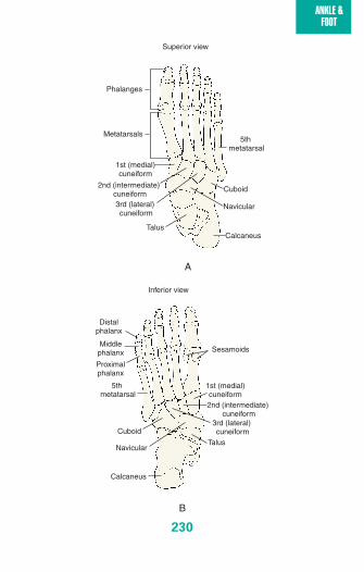
ANKLE & FOOT
230
A
Superior view
Phalanges
Metatarsals5th
metatarsal
1st (medial)cuneiform
2nd (intermediate) cuneiform
3rd (lateral) cuneiform
Cuboid
Navicular
TalusCalcaneus
B
Inferior view
5th metatarsal
1st (medial)cuneiform2nd (intermediate)
cuneiform3rd (lateral) cuneiformCuboid
NavicularTalus
Calcaneus
Distalphalanx
Middlephalanx
Proximalphalanx
Sesamoids
2067_Tab08_221-247.qxd 3/19/09 1:15 PM Page 230

231
ANKLE & FOOT
Extensor digitorum & ext hallucis
Extensordigitorum
longus
Extensorhallucislongus
Extensorretinaculum
Plantaris
Plantaris
Plantaristendon
2067_Tab08_221-247.qxd 3/19/09 1:15 PM Page 231

ANKLE & FOOT
232
Lateral ankle structures
Peroneustertius
Extensordigitorum
longus
Medial malleolus
Tibialis posterior
Flexordigitorum longus
Tibial artery
Tibial nerve
Flexorhallucis longus
Medial ankle structures
2067_Tab08_221-247.qxd 3/19/09 1:15 PM Page 232

233
ANKLE & FOOT
Plantar surface of the foot
Abductorhallucislongus
Flexordigitorum
brevis
Abductordigiti minimi
Feiss Line
Medial malleolus
Navicular1st MTP joint
In NWB, a line is constructed to connect the apex of the medial malleolusto the head of the 1st MTP joint. The navicular bone should be in line withthese 2 structures. In the standing (WB) position, the navicular should notdrop more than 2/3 the distance to the floor.
2067_Tab08_221-247.qxd 3/19/09 1:15 PM Page 233

ANKLE & FOOT
234
Girth
Asse
ssm
en
t
Fig
ure
-8 M
eth
od
to A
ssess A
nkle
Ed
em
a
1. Start d
istal to th
e lateral malleo
lus; g
o m
edial,
just d
istal to n
avicular tu
bero
sity
So
urce: Fro
m G
ulick, D
. Sp
ort N
otes: Field
& C
linical E
xamin
ation
Gu
ide. FA
Davis, P
hilad
elph
ia, 2008, pag
e 169.
Lateralm
alleolus
Medial m
alleolus
Calcaneus
Cuboid
Navicular
Dom
e of talusN
eck of talus
5th Metatarsal
1stM
etatarsal
Lateralm
alleolus
Medial m
alleolus
Calcaneus
Cuboid
Navicular
Dom
e of talusN
eck of talus
5th Metatarsal
1stM
etatarsal
2. Un
der th
e arch to
the p
roxim
al aspect o
f the
head
of th
e 5th m
etatarsal
Continued
2067_Tab08_221-247.qxd 3/19/09 1:15 PM Page 234

235
ANKLE & FOOT
Lateralm
alleolus
Medial m
alleolus
Calcaneus
Cuboid
Navicular
Dom
eof talus
Neck of talus
5th Metatarsal
1stM
etatarsal
Lateralm
alleolus
Medial m
alleolus
Calcaneus
Cuboid
Navicular
Dom
e of talusN
eck of talus
5th Metatarsal
1stM
etatarsal
Fig
ure
-8 M
eth
od
to A
ssess A
nkle
Ed
em
a—
co
nt’d
3. Acro
ss the an
terior tib
ialis tend
on
to th
e distal
aspect o
f the m
edial m
alleolu
s
So
urce: Fro
m G
ulick, D
. 2008, pag
e 170.
4. Over th
e Ach
illes tend
on
back to
the lateral
malleo
lus
2067_Tab08_221-247.qxd 3/19/09 1:15 PM Page 235

ANKLE & FOOT
236
Osteokinematics of the Ankle & Foot
Normal Abnormal
Normal ROM OPP CPP End-feel(s) End-feel(s)
Plantarflexion 30°–50°Dorsiflexion 20°Inversion 10°–30°Eversion 10°–20°1st Extension 35°MTP
2-5 Flexion 75°MTP
10° PF
5°–10°extension
Slightflexion
MaximalDF
Maximalextension
Maximalextension
Elastic (tissuestretch) for allplanes
Capsular
Flex/extension =capsular/elasticAbd/adduction =ligamentous
Empty =sprain/strain
Capsular =extensionlimitedCapsular =flexion limited
2067_Tab08_221-247.qxd 3/19/09 1:15 PM Page 236

237
ANKLE & FOOT
Arthrokinematics for Ankle & Foot Mobilization
Ankle flexion & extension
Ankle inversion & eversion
MTP flexion & extension
Concave surface:Distal tibia/fibulaConvex surface:Talus
Concave surface:Anterior cal-caneal facet &posterior talus Convex surface:Posterior calcaneal facet &anterior talus
Concave surface:PhalanxConvex surface:Metatarsal
To facilitate ankledorsiflexion: OKC—talus rollsanterior & glidesposterior on tibiaCKC—tibia rolls &glides anteriorTo facilitate inversion:OKC—anterior cal-caneal facet rolls &glides medial whileposterior calcanealfacet rolls & glideslateralCKC—talus rollsmedial & glides lat-eral on anterior cal-caneal facet whiletalus rolls & glidesmedial on posteriorcalcaneal facetTo facilitate flexion: Phalanx rolls &glides distal/inferioron metatarsal
To facilitate ankle plantarflexion:OKC—talus rolls posterior & glidesanterior on tibiaCKC—tibia rolls &glides posteriorTo facilitate eversion:OKC—anterior calcaneal facet rolls & glides lateral whileposterior calcanealfacet rolls & glidesmedialCKC—talus rolls lateral & glides medial on anteriorcalcaneal facet whiletalus rolls & glideslateral on posteriorcalcaneal facetTo facilitate extension:Phalanx rolls & glidesproximal/superior onmetatarsal
2067_Tab08_221-247.qxd 3/19/09 1:15 PM Page 237

ANKLE & FOOT
238
Special Tests
ANTERIOR DRAWER
Purpose: Assess for ATF laxityPosition: NWB position in ~ 20° of plan-tarflexion, stabilize the distal tibia/fibulaTechnique: Grasp the posterior aspect ofthe calcaneus/talus & translate the calca-neus/talus anterior on the tibia/fibulaInterpretation: + test = pain & excessivemovement 2° to instability
TALAR TILT
Purpose: Test for laxity of lateral ankleligaments—ATF, CF, PTFPosition: NWB—stabilize the lower leg &palpate respective ligamentTechnique: Grasp calcaneus to apply avarus stress to displace the talus fromthe mortise. Should be performed inplantarflexion (ATF), neutral (CF), & dorsiflexion (PTF)Interpretation: + test = pain or excessive gapping with respect to the con-tralateral limb
SQUEEZE TEST
Purpose: Assess for syndesmotic sprainPosition: Supine with knee extendedTechnique: Begin at the proximal tibia/fibula & firmlycompress (squeeze) the tibia/fibula together, progressdistally toward the ankle until pain is elicited Interpretation: + test = pain at the syndesmosis; thefarther from the ankle the pain is elicited, the moresevere the sprain
Note: Recovery time = 5 + (0.97 x cm from anklejoint that squeeze test is positive) ± 3 days
2067_Tab08_221-247.qxd 3/19/09 1:15 PM Page 238

239
ANKLE & FOOT
ER STRESS TEST (rotate from heel)
KLEIGER’S TEST (rotate from forefoot)
Purpose: Assess for deltoid or syndesmotic sprain Position: Sitting with lower leg stabilized butsyndesmosis not compressedTechnique: Grasp the heel or medial aspect of thefoot & ER in plantarflexion (deltoid lig) & thenrepeat with ER in dorsiflexion (syndesmosis)Interpretation: + test = pain or gapping as com-pared to contralateral limb
WINDLASS TEST
Purpose: Assess for plantar fasciitis Position 1: NWB with knee flexed to 90°Technique 1: Stabilize the ankle in neutral & dor-siflex the great toeInterpretation 1: + test = pain along the mediallongitudinal arch
Position 2: WBTechnique 2: Standing on a stool withequal weight on both foot & toeshanging over the edge of the stool &dorsiflex the great toe Interpretation 2: + test = pain alongthe medial longitudinal arch
2067_Tab08_221-247.qxd 3/19/09 1:15 PM Page 239

ANKLE & FOOT
240
PERONEAL TENDON DISLOCATION
Purpose: Assess for damage to peroneal retinaculumPosition: Prone, knee flexed to 90°Technique: Have the client actively plantarflex & dorsiflex the ankleagainst resistanceInterpretation: + test = tendon subluxing from behind the lateral malleolus
THOMPSON’S TEST
Purpose: Assess for Achilles tendon rupture Position: ProneTechnique: Passively flex the knee to 90° & squeezethe middle 1/3 of the calfInterpretation: Plantarflexion of the foot shouldoccur; + test = failure to plantarflex
HOMAN’S SIGN
Purpose: Assess for thrombophlebitis ofthe lower legPosition: SupineTechnique: Passively dorsiflex the foot& squeeze the calfInterpretation: + test = sudden pain inthe posterior leg or calf
MORTON’S TEST
Purpose: Assess for neuromaPosition: NWBTechnique: Grasp around the transverse metatarsalarch & squeeze the heads of the metatarsals togetherInterpretation: + test = pain between 2nd/3rd or3rd/4th digits that refers to the toes
2067_Tab08_221-247.qxd 3/19/09 1:15 PM Page 240

241
ANKLE & FOOT
BUMP TEST
Purpose: Test for stress fxPosition: NWB—ankle in neutralTechnique: Apply a firm force with thethenar eminence to the heel of the footInterpretation: + test = pain at the site ofthe possible fx
METATARSAL LOAD
Purpose: Assess for metatarsal fracturePosition: NWBTechnique: Grasp the distal aspect of themetatarsal bone & apply a longitudinalforce to load the metatarsalInterpretation: + test = localized pain asthe metatarsal joints are compressed
TINEL’S TEST
Purpose: Assess for tibial nerve damagePosition: NWBTechnique: Tap over posterior tibial nn (medialplantar nerve), just inferior & posterior tomedial malleolusInterpretation: + test = paresthesia into the foot
2067_Tab08_221-247.qxd 3/19/09 1:15 PM Page 241
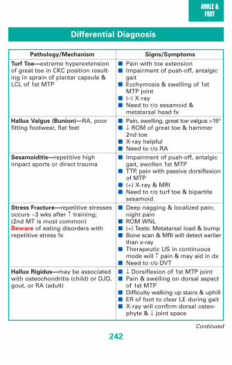
ANKLE & FOOT
242
Differential Diagnosis
Pathology/Mechanism Signs/Symptoms
Turf Toe—extreme hyperextensionof great toe in CKC position result-ing in sprain of plantar capsule &LCL of 1st MTP
Hallux Valgus (Bunion)—RA, poorfitting footwear, flat feet
Sesamoiditis—repetitive highimpact sports or direct trauma
Stress Fracture—repetitive stressesoccurs ~3 wks after ↑ training; (2nd MT is most common)Beware of eating disorders withrepetitive stress fx
Hallux Rigidus—may be associatedwith osteochondritis (child) or DJD,gout, or RA (adult)
■ Pain with toe extension■ Impairment of push-off, antalgic
gait■ Ecchymosis & swelling of 1st
MTP joint■ (–) X-ray ■ Need to r/o sesamoid &
metatarsal head fx■ Pain, swelling, great toe valgus >15°■ ↓ ROM of great toe & hammer
2nd toe■ X-ray helpful■ Need to r/o RA■ Impairment of push-off, antalgic
gait, swollen 1st MTP■ TTP, pain with passive dorsiflexion
of MTP■ (+) X-ray & MRI■ Need to r/o turf toe & bipartite
sesamoid■ Deep nagging & localized pain;
night pain■ ROM WNL■ (+) Tests: Metatarsal load & bump■ Bone scan & MRI will detect earlier
than x-ray■ Therapeutic US in continuous
mode will ↑ pain & may aid in dx■ Need to r/o DVT■ ↓ Dorsiflexion of 1st MTP joint■ Pain & swelling on dorsal aspect
of 1st MTP■ Difficulty walking up stairs & uphill■ ER of foot to clear LE during gait■ X-ray will confirm dorsal osteo-
phyte & ↓ joint space
Continued
2067_Tab08_221-247.qxd 3/19/09 1:15 PM Page 242
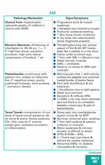
243
ANKLE & FOOT
Pathology/Mechanism Signs/Symptoms
Charcot Foot—hypertrophicosteoarthropathy of midfoot inclient’s with IDDM
Morton’s Neuroma—thickening ofinterdigital nn (25–50 yo; � > �) 2° high heel shoes, excessivepronation, high arch, lateral compression of forefoot, ↑ wt
Plantarfascitis—continuous withgastroc mm; subject to inflamma-tion 2° repetitive stress, poorlycushioned footwear, hard surfaces,↑ pronation, obesity
Tarsal Tunnel—compression of con-tents of tarsal tunnel (posterior tib-ial nerve & artery, tibialis posterior,FDL, FHL) may be 2° trauma,weight gain, excessive pronation,or inflammation
■ Progressive bone & muscle weakness
■ ↓ Sensation but minimal to no pain■ Profound unilateral swelling■ ↑ Skin temp (local); erythema■ X-ray looks like osteomyelitis
(bone fragments present)■ Throbbing/burning into plantar
aspect of 3rd & 4th MT heads;feels like a pebble is in the shoe
■ Callus under involved rays■ ↑ Pain with WB, (+) Morton’s test■ Weak intrinsic muscles■ EMG = unreliable■ Need to r/o stress fx (MRI with
contrast)■ Morning pain that ↓ with activity,
nodules are palpable over proximal-medial border of plantar fascia
■ Pain with dorsiflexion & toeextension
■ ↓ Dorsiflexion due to tight gastroc■ Weak foot intrinsics■ Sensation & reflexes WNL■ (–) EMG; x-ray may show calcaneal
spur but there is no correlationbetween a bone spur & pain ofplantarfascitis
■ Sharp pain into medial/plantaraspect of foot & 1st MTP
■ Burning, nocturnal pain, swelling■ ↑ Pain with walking & passive
d-flexion or eversion■ Motor weakness & intrinsic
atrophy is difficult to detect■ DTRs & ROM = WNL■ (+) Tinel’s sign just below &
behind the medial malleolus■ Abnormal EMG; r/o diabetic
neuropathy & neuroma
Continued
2067_Tab08_221-247.qxd 3/19/09 1:15 PM Page 243
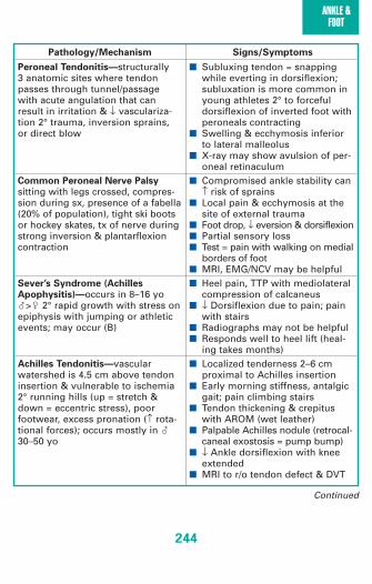
ANKLE & FOOT
244
Pathology/Mechanism Signs/Symptoms
Peroneal Tendonitis—structurally 3 anatomic sites where tendonpasses through tunnel/passagewith acute angulation that canresult in irritation & ↓ vasculariza-tion 2° trauma, inversion sprains,or direct blow
Common Peroneal Nerve Palsy
sitting with legs crossed, compres-sion during sx, presence of a fabella(20% of population), tight ski bootsor hockey skates, tx of nerve duringstrong inversion & plantarflexioncontraction
Sever’s Syndrome (Achilles
Apophysitis)—occurs in 8–16 yo�>� 2° rapid growth with stress onepiphysis with jumping or athleticevents; may occur (B)
Achilles Tendonitis—vascularwatershed is 4.5 cm above tendoninsertion & vulnerable to ischemia2° running hills (up = stretch &down = eccentric stress), poorfootwear, excess pronation (↑ rota-tional forces); occurs mostly in �30–50 yo
■ Subluxing tendon = snappingwhile everting in dorsiflexion;subluxation is more common inyoung athletes 2° to forceful dorsiflexion of inverted foot withperoneals contracting
■ Swelling & ecchymosis inferior to lateral malleolus
■ X-ray may show avulsion of per-oneal retinaculum
■ Compromised ankle stability can↑ risk of sprains
■ Local pain & ecchymosis at thesite of external trauma
■ Foot drop, ↓ eversion & dorsiflexion■ Partial sensory loss■ Test = pain with walking on medial
borders of foot■ MRI, EMG/NCV may be helpful■ Heel pain, TTP with mediolateral
compression of calcaneus■ ↓ Dorsiflexion due to pain; pain
with stairs■ Radiographs may not be helpful■ Responds well to heel lift (heal-
ing takes months)■ Localized tenderness 2–6 cm
proximal to Achilles insertion■ Early morning stiffness, antalgic
gait; pain climbing stairs■ Tendon thickening & crepitus
with AROM (wet leather)■ Palpable Achilles nodule (retrocal-
caneal exostosis = pump bump)■ ↓ Ankle dorsiflexion with knee
extended■ MRI to r/o tendon defect & DVT
Continued
2067_Tab08_221-247.qxd 3/19/09 1:15 PM Page 244

245
ANKLE & FOOT
Pathology/Mechanism Signs/Symptoms
Achilles Tendon Rupture—<30 yo,injury is 2° direct blow to gastroc orforceful contraction; >30 yo, injuryis 2° degeneration (higher inci-dence in people with type “O”blood)
Posterior Tibialis Tendonitis—
inflammatory condition due to poorbiomechanics or overuse; commonin � >40 yo
Shin Splints/Anterior—an overusesyndrome of tibialis anterior, exthallicus longus, & ext digitorumlongus attributed to running onunconditioned legs, soft tissueimbalance, alignment abnormali-ties, & excessive pronation toaccommodate rearfoot varus
Shin Splints/Posterior—an overusesyndrome of flexor hallucis longus& flexor digitorum longus
■ Snap/pop associated with injury■ Palpable gap in tendon (hatchet
sign) if examined early■ Cannot walk on toes, swelling
(within 1–2 hrs) & ecchymosis■ (+) Thompson’s test■ MRI confirms diagnosis■ TTP @ medial ankle ■ Crepitus with AROM■ Pain with passive pronation &
active supination■ Pain with resistive inversion &
plantarflexion■ Pain & tenderness over anterior
tibialis■ Pain with resisted dorsiflexion &
inversion■ Pain with stretching into plan-
tarflexion & eversion■ Callus formation under 2nd metatarsal
head & medial side of distal hallux■ Tight gastroc/soleus muscle■ Soreness with heel walking■ (–) X-ray, needed to r/o stress fx■ Callus formation under 2nd > 3rd
> 4th MT head & medial side ofdistal hallux
■ Pain & soreness over distal 1/3–2/3of posterior/medial shin & posteri-or medial malleolus
■ Hypermobile 1st metatarsal■ Rapid & excessive pronation to
compensate for rearfoot varus;result is ↑ stress on tibialis poste-rior to decelerate foot
■ Pain with resisted inversion &plantarflexion
■ Pain with stretching into dorsi-flexion & eversion
■ (–) X-ray, needed to r/o stress fx
Continued
2067_Tab08_221-247.qxd 3/19/09 1:15 PM Page 245

ANKLE & FOOT
246
Pathology/Mechanism Signs/Symptoms
Compartment Syndrome—a pro-gression of shin splints resulting ina loss of microcirculation in shinmuscle; � > �, R > LBeware: Immediate referral isneeded (Ice but do not compress)
Complex Regional Pain
Syndrome—etiology unknown,may occur after traumaSee stages on page 222.
■ 5 P’s = paresthesia (toes), paresis(drop foot), pain (anterior tibia),pallor, pulseless
■ Skin feels warm & firm■ Pain with stretch or AROM, foot
drop■ Severe cramping, diffuse pain &
tightness■ Most reliable sign is sensory
deficit at the dorsum of foot in1st interdigital cleft
■ Ischemia of extensor hallicuslongus
■ Pulses are normal until the end & then surgery is needed within4–6 hours is required to preventmuscle necrosis & nerve damage
■ ↑ Soft tissue pressures via fluidaccumulation ■ Normal compartment pressure
<10 mm Hg■ > 20 mm Hg is compromised
capillary blood flow■ > 30 mm Hg results in
ischemic necrosis■ (–) X-ray & bone scan; need to
r/o tibial stress fx■ Confirmed with MRI & pressure
assessment■ Hyperalgesia & hyperhidrosis■ Capsular tightness & stiffness■ Muscle atrophy & osteoporosis■ Trophic changes & edema■ Vasomotor instability
Continued
2067_Tab08_221-247.qxd 3/19/09 1:15 PM Page 246

247
ANKLE & FOOT
Pathology/Mechanism Signs/Symptoms
Syndesmotic Sprain—injury toanterior and/or posterior inferiortibiofibular ligament 2° hyperdorsi-flexion & eversionSee stages below
Lateral Sprain—injury to ATF, CF,PTF 2° inversion with plantarflexionSee stages below
■ (+) Tests: Squeeze & ER test■ Pain & swelling over ligament/
interosseous membrane■ Oblique x-ray may show abnormal
widening of joint space■ Recovery time = 5 + (0.97 x cm
from ankle joint that squeeze testis positive) ± 3 days
■ Need to r/o fx & avulsion■ Rich blood supply = significant
swelling within 2 hours■ TTP over involved ligaments,
ecchymosis that drains distal■ Varying levels of instability
(grade 1–3)■ (+) Tests: Talar tilt & anterior
drawer (presence of a dimple justinferior to the tip of the lateralmalleolus)
■ (–) X-ray for fracture but stressfilm may show ↑ joint space
■ Arthrography is only accuratewithin 24 hours
1st degree 2nd degree 3rd degree
■ No hemorrhage■ Minimal swelling■ Point tender■ No varus laxity ■ (–)Anterior drawer■ (–) Talar tilt■ No/little limp■ Difficulty hopping■ Recovery 2–10 days
■ Some hemorrhage■ Localized swelling
(↓ Achilles definition)■ (+) Anterior drawer■ (+) Talar tilt■ No varus laxity■ (+) Limp■ Unable to heel raise,
hop, run■ Recovery 10–30 days
■ Diffuse swelling(no Achilles definition)
■ Tenderness medial & lateral
■ (+) Anterior drawer■ (+) Talar tilt■ (+) Varus laxity■ NWB■ Recovery 30–90 days
Grades of Ankle Sprains
2067_Tab08_221-247.qxd 3/19/09 1:15 PM Page 247

REF
248
References
Akseki D, Ozcan O, Boya H, Pinar H. A new weight-bearing meniscal testand a comparison with McMurray’s test and joint line tenderness.Arthroscopy. 2004;20(9):951-958.
Albeck M. A critical assessment of clinical diagnosis of disc herniation inpatients with monoradicular sciatica. Acta Neurochiropractic.1996;138:40-44.
Albert H, Godskesen M, Westergaard J. Evaluation of clinical tests used inclassification procedures in pregnancy-related pelvic joint pain. EuropeanSpine Journal. 2000;9(2):161-166.
Arky R (Medical Consultant). Physicians’ Desk Reference. Montvocle, NJ:Medical Economics Company. 2008
Barankin B, Freiman A. Derm Notes. Philadelphia, PA: FA Davis; 2006.Barth JRH, Burkhart SS, DeBeer JF. Bear-Hug Test: A New and Sensitive
Test for Diagnosing a Subscapular Tear. Arthroscopy: The Journal ofArthroscopy and Related Surgery. October 2006;22(10):1076-1084.
Beaton DE, Wright JG, Katz JN. Development of the Quick DASH:Comparison of three item-reduction approaches. The Journal of Boneand Joint Surgery. May 2005;87A(5):1038-1046.
Bellamy N, Buchanan WW, et al. Validation study of WOMAC: A health status instrument for measuring clinically important patient relevant outcomes to antirheumatic drug therapy in patients with osteoarthritis ofthe hip or knee. Journal of Rheumatology. 1988;15:1833-1840.
Bellamy N. Pain assessment in osteoarthritis: Experience with the WOMACosteoarthritis index. Seminars in Arthritis and Rheumatism. 1989;18(supplement 2):14-17.
Benner W. Specificity of the Speed’s test: Arthroscopic technique for evalu-ating the biceps tendon at the level of the bicipital groove. Arthroplasty.1998;14(8):789-796.
Biel A. Trail Guide to the Body. 2nd ed. Boulder, CO: Discovery Books; 2001.Blower PW, Griffin AJ. Clinical sacroiliac tests in ankylosing spondylitis and
other causes of low back pain. Annals of Rheumatic Disorders.1984;43:192-195.
Boeree NR, Ackroyd CE. Assessment of the meniscus and cruciate ligaments:an audit of clinical practice. Injury. 1991;22(4):291-294.
Boissannault WG. Primary Care for the Physical Therapist. Elsevier Saunders,2005
Boissannault WG, Bass C. Pathological origins of trunk and neck pain: PartI–Pelvic and abdominal visceral disorders. Journal of Orthopedics andSports Physical Therapy. 1990;12(5):192-207.
2067_REF_248-258.qxd 3/19/09 1:03 PM Page 248

Boissannault WG, Bass C. Pathological origins of trunk and neck pain: PartII–Disorders of the cardiovascular and pulmonary systems. Journal ofOrthopedics and Sports Physical Therapy. 1990;12(5):208-215.
Boissannault WG, Bass C. Pathological origins of trunk and neck pain: PartIII–Diseases of the musculoskeletal system. Journal of Orthopedics andSports Physical Therapy. 1990;12(5):216-221.
Brantigan CO, Roos DB. Diagnosing thoracic outlet syndrome. Hand Clinics.2004;20:27-36.
Broadhurst NA, Bond MJ. Pain provocation tests for the assessment ofsacroiliac joint dysfunction. Journal of Spinal Disorders. 1998;11(4):341-345.
Broomhead A, Stuart P. Validation of the Ottawa ankle rules in Australia.Emergency Medicine. 2003;15(2):126-132.
Bruske J, Bednarski M, Grzelec H, Zyluk A. The usefulness of the Phalen testand the Hoffman-Tinel sign in the diagnosis of carpal tunnel syndrome.Acta Orthopaedica Belgica. 2002;68(2):141-145.
Budiman-Mak E, Conrad KJ, Roach KE. The Foot Function Index: a measureof foot pain and disability. Journal of Clinical Epidemiology.1991;44(6):561-570.
Burkhart S, Morgan CB, Kibler B. Shoulder injuries in overhead athletes:the dead arm revisited. Clinics in Sports Medicine. 2000;19(1):125-158.
Butler DA. Mobilisation of the Nervous System. Melbourne, Australia:Churchill Livingstone; 1991.
Cailliet R. Hand Pain and Impairment. 2nd ed. Philadelphia, PA: FA Davis;1976.
Cailliet R. Neck and Arm Pain. Philadelphia, PA: FA Davis; 1976.Cailliet R. Foot and Ankle Pain. Philadelphia, PA: FA Davis; 1976.Cailliet R. Knee Pain and Disability. Philadelphia, PA: FA Davis; 1976.Cailliet R. Low Back Pain Syndrome. 3rd ed. Philadelphia, PA: FA Davis;
1983.Calis M, Akgen K, Birtane M, Calis H, Tuzun F. Diagnostic values of clinical
diagnostic tests in subacromial impingement syndrome. Annals of theRheumatic Disease. 2000;59:44-47.
Cibulka MT, Koldehoff R. Clinical usefulness of a cluster of sacroiliac jointtests in patients with and without low back pain. Journal of Orthopedicand Sports Physical Therapy. 1999;29(2):83-92.
Ciccone, CD. Pharmacology in Rehabilitation. 2nd ed. Philadelphia, PA: FA Davis; 1990.
Clark KD, Tanner S. Evaluation of the Ottawa ankle rules in children.Pediatric Emergency Care. 2003;19(2):73-78.
249
REF
2067_REF_248-258.qxd 3/19/09 1:03 PM Page 249

REF
250
Colachis S, Ctrohm M. A study of tractive forces and angle of pull on vertebral interspaces in cervical spine. Archives of Physical Medicine.1965;46:820-830.
Crocket HC, Gross LB, Wilk KE, Schwartz ML, Reed J, O’Mara J, Reilly MT,Dugas JR, Meister K, Lynam S, Andrews JR. Osseous adaptations andrange of motion at the glenohumeral joint in professional baseball pitchers. American Journal of Sports Medicine. 2002; 30:20-26.
DeGarceau D, Dean D, Requejo SM, Thordarson DB. The associationbetween diagnosis of plantar fasciitis and windlass test results. Foot andAnkle International. March 2003;24(3):251-255.
Deglin JH, Vallerand AH. Davis’s Drug Guide for Nurse. 7th ed. Philadelphia,PA: FA Davis; 2001.
Duruoz MT, Poiraudeau S, Fermanian J, Menkes CJ, Amor B, Dougados M,Revel M. Development and validation of a rheumatoid hand functional dis-ability scale that assesses functional handicap. Journal of Rheumatology.1996; Jul;23(7):1167-1172.
Dutton M. Orthopaedic Examination, Evaluation, and Intervention. NewYork: McGraw Hill; 2004.
Dworkin SF, Huggins KH, LeResche L, Von Korff M, Howard J, Truelove E,Sommers E. Epidemiology of signs and symptoms in temporomandibulardisorders: clinical signs in cases and controls. Journal of the AmericanDental Association. 1990;120(3):273-281.
Evans PJ, Bell GD, Frank C. Prospective evaluation of the McMurray test.American Journal of Sports Medicine. 1993;21(4):604-608.
Evans PJ. Prospective evaluation of the McMurray test. American Journalof Sports Medicine. 1994;22:567-568.
Evans R. Illustrated Orthopedic Physical Assessment. 2nd ed. St. Louis,MO: Mosby; 2001.
Fairbank JC, Couper J, Davies JB, O’Brien JP. The Oswestry low back paindisability questionnaire. Physiotherapy. 1980;66:271-273.
Farrell K. APTA Home Study Course, Solutions to Shoulder Disorders,11.1.6 Adhesive Capsulitis.
Faught BE. Efficacy of clinical tests in the diagnosis of carpal tunnel.Doctoral dissertation, 2001.
Fitzgerald RH Jr. Acetabular labrum tears: diagnosis and treatment. ClinicalOrthopedics. 1995;311:60-68.
Fowler PJ, Lubliner JA. The predictive value of five clinical signs in theevaluation of meniscal pathology. Arthroscopy. 1989;5:184-186.
Gann N. Orthopedics at a Glance. Thorofare, NJ: Slack Incorporated; 2001.Gillard J, Perez-Cousin M, Hachulla E, Remy J, Hurtevent J-F, Vinckier L,
Thevenon A, Duquesnoy B. Ddiagnosing thoracic outlet syndrome: contribution of provocative tests, ultrasonography, electrophysiology,
2067_REF_248-258.qxd 3/19/09 1:03 PM Page 250
and helical computer tomography in 48 patients. Joint Bone Spine.2001;68416-424.
Goloborod’ko SA. Provocative test for carpal tunnel syndrome. Journal ofHand Therapy. 2004;17:344-348.
Goodman C, Boissonnault W. Pathology: Implications for the PhysicalTherapist. Philadelphia, PA: W.B. Saunders; 1998.
Goodman C, Snyder T. Differential Diagnosis for Physical Therapists:Screening for Referral. 4th ed. Philadelphia, PA: W.B. Saunders; 2008.
Guanche CA, Jones DC. Clinical testing for tears of the glenoid labrum.Arthroscopy. 2003;19(5):517-523.
Gulick DT. Sport Notes. Philadelphia, PA: FA Davis, 2008.Gulick DT. Screening Notes. Philadelphia, PA: FA Davis, 2006Hall CM, Brody LT. Therapeutic Exercise. Philadelphia, PA: Lippincott Williams
and Wilkins; 1998.Hall H. A simple approach to back pain management. Patient Care. 1992;15:
77-91.Hansen PA, Micklesen P, Robinson LR. Clinical utility of the flick maneuver
in diagnosing carpal tunnel syndrome. American Journal of PhysicalMedicine and Rehabilitation. 2004;83:363-367.
Harilainen A. Evaluation of knee instability in acute ligamentous injuries.Annal of Chiropractic Gynaecology. 1987;76:269-273.
Harris H. Harris hip score. Journal of Bone and Joint Surgery (Am).1969;51-A(4):737-55.
Heald SL, Riddle DL, Lamb RL. The shoulder pain and disability index: The con-struct validity and responsiveness of a region-specific disability measure.Physical Therapy, 1997;77:1079-1089.
Hegedus EJ, Goode A, Campbell S, Morin A, Tamaddoni M, Moorman CT,Cook C. Physical examination of the shoulder: a systematic review withmeta-analysis of the individual tests. British Journal of Sports Medicine.2008;42:80-92.
Heller L, Ring H, Costeff H, Solzi P. Evaluation of Tinel’s and Phalen’s signsin diagnosis of the carpal tunnel syndrome. European Neurology.1986;25(1):40-42.
Hertel R, Ballmer FT, Lambert SM, Gerber CH. Lag signs in the diagnosis ofrotator cuff rupture. Journal of Shoulder and Elbow Surgery.1996;5(4):307-313.
Holtby MB, et al. Accuracy of the Speed’s and Yergason tests in detectingbiceps pathology and slap lesions: comparison with arthroscopic find-ings. Arthroscopy. 2004;20(3):231-236.
Holtby MB, Razmjou H. Validity of the supraspinatus test as a single clinicaltest in diagnosing patients with rotator cuff pathology. Journal ofOrthopedics and Sports Physical Therapy. 2004;34:194-200.
251
REF
2067_REF_248-258.qxd 3/19/09 1:03 PM Page 251

REF
252
Hoppenfeld S. Physical Examination of the Spine and Extremities. New York, NY: Appleton-Century-Crofts; 1976.
Institute for Work and Health. http://www.iwh.on.ca/products/dash.php.Accessed February 2008.
Internet Drug Index. Available at: http://www.rxlist.com. Accessed August2004.
Johanson M. APTA Home Study Course, Solutions to Shoulder Disorders,11.1.4 Rotator Cuff Disorders.
Itoi E, Kido T, Sano A, Urayama M, Sata K. Which is more useful, the “fullcan test” or the “empty can test” in detecting the torn supraspinatus tendon? American Journal of Sports Medicine. 1999;27:65-68.
Jonsson T, Althoff B, Peterson L, Renstrom P. Clinical diagnosis of rupturesof the anterior cruciate ligament: a comparative study of the Lachmantest and the anterior drawer sign. American Journal of Sports Medicine.1982;10:100-102.
Kaikkonen A, Kannus P, Jarvinen M. A performance test protocol and scor-ing scale for the evaluation of ankle injuries. American Journal of SportsMedicine. 1994;22(4):462-469.
Karachalios T, Hantes M, Zibis AH, Zachos V, Karantanas AH, Malizos KN.Diagnostic accuracy of a new clinical test (the Thessaly test) for earlydetection of meniscal tears. Journal of Bone and Joint Surgery. May 2005;87A(5):955-962.
Katz JW, Fingeroth RJ. The diagnostic accuracy of ruptures of the anteriorcruciate ligament comparing the Lachman test, the anterior drawer sign,and the pivot shift test in acute and chronic knee injuries. AmericanJournal of Sports Medicine. 1986;14(1):88-91.
Khine H, Dorfman DH, Avner JR. Applicability of Ottawa knee rules for kneeinjuries in children. Pediatric Emergency Care. 2001;17(6):401-404.
Kim S-H, Ha, K-I, Ahn J-H, Kim S-H, Choi H-J. Biceps load test II: a clinicaltest for SLAP lesions of the shoulder. Arthroscopy. 2001;17(2):160-164.
Kim S-H, Park J-S, Jeong W-K, Shin S-K. The Kim test. American Journal ofSports Medicine. 2005;33(8):1188-1192.
Knuttson B. Comparative value of electromyographic, and clinical-neurologicalexaminations in diagnosis of lumbar root compression syndrome. ActaOrthopedic Scandinavia. 1961;Suppl 49:19-49.
Kosteljanetz M, Espersen O, Halaburt H, Miletic T. Predictive values of clini-cal and surgical findings in patients with lumbago-sciatica: a prospectivestudy. Acta Neurochirugica. 1984;73:67-76.
Kuhlman KA, Hennessey WJ. Sensitivity and specificity of carpal tunnelsyndrome signs. American Journal of Physical Medicine andRehabilitation. 1997;76(6):451-457.
2067_REF_248-258.qxd 3/19/09 1:03 PM Page 252
Kurosaka M, Yagi M, Yoshiya S, Muratsu H, Mizuno K. Efficiency of the axially loaded pivot shift test for the diagnosis of a meniscal tear.International Orthopedics. 1999;23:271-274.
Lan LB. The scaphoid shift test. The Journal of Hand Surgery. 1993;18A(2):366-368.
Laslett M, Aprill CN, McDonald B, Young SB. Diagnosis of sacroiliac jointpain: validity of individual provocation tests and composites of tests.Manual Therapy. 2005;10:207-218.
Laslett M, Young SB, Aprill CN, McDonald B. Diagnosing painful sacroiliacjoints: a validity study of a McKenzie evaluation and sacroiliac provoca-tion tests. Australian Journal of Physiotherapy. 2003;49:89-97.
LaStayo P, Weiss S. The GRIT: a quantitative measure of ulnar impactionsyndrome. Journal of Hand Therapy. 2001;14(3):173-179.
Lee JK, Yao L, Phelps CT, Wirth CR, Czajka J, Lozman J. Anterior cruciateligament tears: MR imaging compared with arthroscopy and clinicaltests. Radiology. 1988;166:861-864.
Leroux, et al. Diagnostic value of clinical tests for shoulder impingementsyndrome. Revue du Rhumatisme. 1995;62(6):423-428.
Lester B, Halbrecht J, Levy IM, Gaudinez R. “Press test” for office diagnosisof triangular fibrocartilage complex tears of the wrist. Annals of PlasticSurgery. 1995;35(1):41-45.
Levangie PK. Four clinical tests of sacroiliac joint dysfunction: the associa-tion of test results with innominate torsion among patients with andwithout low back pain. Physical Therapy. 1999;79(11):1043-1057.
Levangie PK, Norkin CC. Joint Structure and Function. 3rd ed. Philadelphia,PA: FA Davis; 2001.
Levine DW, Simmons BP, Koris MJ, Daltroy LH, Hohl GG, Fossel AH, Katz JN.A self-administered questionnaire for the assessment of severity of symp-toms and functional status in carpal tunnel syndrome. Journal of Boneand Joint Surgery. 1993;75(11):1585-1592.
Lewis C, McNerney T. Clinical Measures of Functional Outcomes. Virginia:Learn Publications.
Lewis CL, Sahrmann SA. Acetabular labral tears. Physical Therapy. 2006;86(1):110-121.
Lewis CL, Sahrmann SA, Moran DW. Anterior hip joint force increases withhip extension, decreased gluteal force, or decreased iliopsoas force.Journal of Biomechanics. 2007;40:3725-3731.
Lewis C, Wilk K, Wright R. The Orthopedic OutcomesTool Box. Virginia:Learn Publications.
Lillegard WA, Burcher JD, Rucker KS. Handbook of Sports Medicine. 2nded. Boston, MA: Butterworth-Heinemann; 1999.
253
REF
2067_REF_248-258.qxd 3/19/09 1:03 PM Page 253

REF
254
Liu SH, Henry MH, Nuccion SL. A prospective evaluation of a new physicalexamination in predicting glenoid labral tears. American Journal ofSports Medicine. 1996;24(6):721-725.
Liu SH, Osti L, Henry M, Bocchi L. The diagnosis of acute complete tears of the anterior cruciate ligament. Comparison of MRI, arthrometry andclinical examination. Journal of Bone and Joint Surgery. 1995;77(4):586-588.
Lo IKY, Nonweiler B, Woolfrey M, Litchfield R, Kirkley A. An evaluation ofthe apprehension, relocation, and surprise tests for anterior shoulderinstability. American Journal of Sports Medicine. 2004;32(3):655-661.
Lucchesi GM, Jackson RE, Peacock WF, Cerasani C, Swor RA. Sensitivity ofthe Ottawa rules. Annals of Emergency Medicine. 1995;26(1);1-5.
Ludewig P. APTA Home Study Course, Solutions to Shoulder Disorders,11.1.1 Functional Shoulder Anatomy and Biomechanics.
Lysholm J, Gillquist J. Evaluation of knee ligament surgery results withspecial emphasis on using of a scoring scale. American Journal ofSports Medicine. 1982;10:150-154.
MacDermid JC, Turgeon T, Richards RS, Beadle M, Roth JH. Patient rating ofwrist pain and disability: a reliable and valid measurement tool. Journalof Orthopaedic Trauma. November 1998;12(8):577-586.
MacDermid JC, Wessel J. Clinical diagnosis of carpal tunnel syndrome: asystemic review. Journal of Hand Therapy. 2004;17(2):309-319.
MacDonald PB, Clark P, Sutherland K. An analysis of the diagnostic accura-cy of the Hawkins and Neer subacromial impingement tests. Journal ofShoulder and Elbow Surgery. 2000;9(4):299-301.
Magee D. Orthopedic Physical Assessment. 4th ed. Philadelphia: WBSaunders; 2002.
Malanga GA, Andrus S, Nadler SF, McLean J. Physical examination of theknee: a review of the original test description and scientific validity ofcommon orthopedic tests. Archives of Physical Medicine andRehabilitation. 2003;84:592-603.
Martin RL, Sekiya JK. The interrater reliability of 4 clinical tests used toassess individuals with musculoskeletal hip pain. Journal of Orthopedicsand Sports Physical Therapy. 2008;38(2):71-77
McFarland EG, Kim TK, Savino RM. Clinical assessment of three commontests for superior labral anterior-posterior lesions. American Journal ofSports Medicine. 2002;30(6):810-815.
McKinnis LN. Fundamentals of Orthopedic Radiology. Philadelphia, PA: FA Davis; 1997.
Medscape from Web MD. Available at: www.medscape.com. AccessedAugust 2004.
2067_REF_248-258.qxd 3/19/09 1:03 PM Page 254
Melzack R. The McGill pain questionnaire. In: Pain Measurement andAssessment. New York: Raven Press; 1983:41-48.
Melzack R. The short-form McGill pain questionnaire. Pain. 1987;30:191-197. Mimori K, Muneta T, Nakagawa T, Shinomiya K. A new pain provocation
test for superior labral tears of the shoulder. American Journal of SportsMedicine. 1999;27:137-142.
Nakagawa S, Yoneda M, Hayashida K, Obata M, Fukushima S, Miyazaki Y.Forced shoulder abduction and elbow flexion test: a new simple clinicaltest to detect superior labral injury in the throwing shoulder.Arthroscopy. 2005;21(11):1290-1295.
Narvani A, Tsiridis E, Kendall S, Chaudhuri R, Thomas P. A preliminary reporton prevalence of acetabular labrum tears in sport patients with groin pain.Knee Surgery, Traumatology, and Arthroscopy. 2003;11:403-408.
Neumann D. Kinesiology of the Musculoskeletal System. St. Louis, MO:Mosby; 2002.
Nils J, van Geel C, van der Auwera, van de Velde B. Diagnostic value of fiveclinical tests in patellofemoral pain syndrome. Manual Therapy. 2005
Noble J, Erat K. In defense of the meniscus: a prospective study of 200 menis-cectomy patients. Journal of Bone and Joint Surgery. 1980;62-B:7–11.
Novak CB, Lee GW, Mackinnon SE, Lay L. Provocation testing for cubitaltunnel syndrome. Journal of Hand Surgery. 1994;19A:817-820.
O’Brien SJ, Pagnani MJ, Fealy S, McGlynn SR, Wilson JB. The active com-pression test: A new and effective test for diagnosing labral tears andacromioclavicular joint abnormality. American Journal of SportsMedicine. 1998;26(5):610-613.
Park HB, Yokota A, Gill HS, Rassi GE, McFarland EG. Diagnostic accuracy ofclinical tests for the different degrees of subacromial impingement syn-drome. Journal of Bone and Joint Surgery. 2005;87-A(7):1446-1455.
Partentis MA, Mohr KJ, ElAttrache NS. Disorders of the superior labrum:review and treatment guidelines. Clinical Orthopedics. 2002;400:77-87.
Pfalzer LA, Drouin J. Screening for underlying cancer in acute care physicaltherapy practice. Acute Care Perscpectives. 2001;10(1-2):1-4,10,14,17,36,40,44-55.
Plint AC, Bulloch B, Osmond MH, Stiell I, Dunlap H, Reed M, Tenenbein M,Klassen TP. Validation of the Ottawa ankle rules in children with ankleinjuries. Academic Emergency Medicine. 1999;6(10):1005-1009.
Powell JM, Lloyd GJ, Rintoul RF. New clinical test for fracture of thescaphoid. Canadian Journal of Surgery. July 1988;31(4):237-242.
Ransford AO, Cairns D, Mooney V. The pain drawing as an aid to thepsychoReider B. The Orthopedic Physical Examination. Philadelphia, PA:WB Saunders Company; 1999.
255
REF
2067_REF_248-258.qxd 3/19/09 1:03 PM Page 255

REF
256
Rayan GM, Jensen C. Thoracic outlet syndrome: provocative examinationmaneuvers in a typical population. Journal of Shoulder and ElbowSurgery. 1995;4:113-117.
Roach KE. Development of a shoulder pain and disability index. ArthritisCare and Research. 1991;4:143-149.
Rothstein JM, Roy SH, Wolf SL. The Rehabilitation Specialist’s Handbook.Philadelphia, PA: FA Davis; 1991.
Rubinstein RA Jr, Shelbourne KD, McCarroll JR, VanMeter CD, Rettig AC.The accuracy of the clinical examination in the setting of posterior cruciate ligament injuries. American Journal of Sports Medicine.1994;22:550-557.
Russell A, Maksymovich W, LeClerq S. Clinical examination of the sacroiliacjoints. A prospective study. Arthritis and Rheumatism. 1981;24:1575-1577.
Saal JA. Natural history and nonoperative treatment of lumbar disk hernia-tion. Spine. 1996;21(24S):7S.
Saidoff D, McDonough A. Critical Pathways in Therapeutic Intervention. St. Louis, MO, Mosby; 2002.
Sallay PI, Poggi J, Speer KP, Garrett WE. Acute dislocation of the patella. Acorrelation pathoanatomic study. American Journal of Sports Medicine.1996;24:52-60.
Sandberg R, Balkfors B, Henricson A, Westlin N. Stability tests in knee ligamentinjuries. Archives of Orthopedic Trauma Surgery. 1986;106(1):5-7.
Saunders HD, Ryan RS. Evaluation, Treatment and Prevention ofMusculoskeletal Disorders. Volume 1-Spine. 4th ed. Chaska, MN: TheSaunders Group; 2004.
Scalzitti DA. Screening for psychological factors in patients with low backproblems: Waddell’s nonorganic signs. Physical Therapy. 1997; 77(3):306-312.
Scholten RJPM, Deville WLJM, Opstelten W, Bijl D, Van der Plas CG, BouterLM. The accuracy of physical diagnostic tests for assessing meniscallesions of the knee. Journal of Family Practice. 2001;50(11):938-944.
Sgaglione NA, Pizzo WD, Fox JM, Friedman MJ. Critical analysis of kneeligament rating systems. American Journal of Sports Medicine.1995;23(6): 660-667.
Stankovic R, Johnell O, Maly P, Willner S. Use of lumbar extension, slumptest, physical and neurological examination in the evaluation of patientswith suspected herniated nucleus pulposus. A prospective clinical study.Manual Therapy. 1999;4(1):25-32.
Starkey C, Ryan J. Orthopedic and Athletic Injury Evaluation Handbook.Philadelphia, PA: FA Davis; 2003.
Stiell IG, McKnight RD, Greenberg GH, McDowell I, Nair RC, Wells GA,Johns C, Worthington JR. Implementation of the Ottawa ankle rules.JAMA. March 1994;271(11):827-832.
2067_REF_248-258.qxd 3/19/09 1:03 PM Page 256

Stiell IG, Wells GA, Hoag RH. Implementation of the Ottawa knee rule forthe use of radiography in acute knee injuries. JAMA. 1997;278:2075-2079.
Stiell IG, Greenberg GH, Well GA, McDowell I, Cwinn AA, Smith NA et al.Prospective validation of a decision rule for the use of radiography inacute knee injuries. JAMA. 1996;275:611-615.
Stratford PW, Binkley J. A review of the McMurray test: definition, interpre-tation, and clinical usefulness. Journal of Orthopedic and Sports PhysicalTherapy. 1995;22(3):116-120.
Suenaga E, Noguchi Y, Jingushi S, Shuto T, Nakashima Y, Miyanishi K,Iwamoto Y. Relationship between the maximum flexion-internal rotationtest and the torn acetabular labrum of a dysplastic hip. Journal ofOrthopedic Science. 2002;7:26-32.
Szabo RM, Slater RR. Diagnostic testing in carpal tunnel syndrome. Journalof Hand Surgery. 2000;25(1):184.
Tegner Y, Lysholm J. Rating system in the evaluation of knee ligamentinjuries. Clinical Orthopedics. 1985;198:43-49.
Tennet DT, Beach WR, Meyers JF. Clinical sports medicine update. A reviewof the special tests associated with shoulder examination. AmericanJournal of Sports Medicine. 2003;31:154-160.
Tetro AM, Evanoff BA, Hollstien SB, Gelberman RH. A new provocation testfor carpal tunnel syndrome. Assessment of wrist flexion and nerve com-pression. Journal of Bone and Joint Surgery. 1998;80(3):493-498.
Thompson JC. Netter’s Concise Atlas of Orthopedic Anatomy. Teterboro,NJ: Icon Learning Systems; 2002.
Tigges S, Pitts S, Mukundan S Jr, Morrison D, Olson M, Shahriara A.External validation of the Ottawa knee rules in an urban trauma center in the United States. American Journal of Roentgenology. 1999;172(4):1069-1071.
Tomberlin J. APTA Home Study Course, Solutions to Shoulder Disorders,11.1.2 Physical Diagnostic Tests of the Shoulder: An Evidence-basedPerspective.
Tong HC, Haig AJ, Yamakawa K. Spurling test and cervical radiculopathy.Spine. 2002;27(2):156-159.
Torg JS, Conrad W, Kalen V. Clinical diagnosis of anterior cruciate ligamentinstability in the athlete. American Journal of Sports Medicine. 1976;4:84-93.
Travell J, Simon D. Trigger Point Flip Charts. Baltimore, MD: Williams andWilkins; 1996.
Unverzagt CA, Schuemann T, Mathisen J. Differential diagnosis of a sportshernia in a high-school athlete. Journal of Orthopedics and SportsPhysical Therapy. 2008;38(2):63-70.
257
REF
2067_REF_248-258.qxd 3/19/09 1:03 PM Page 257

REF
258
Ure BM, Tiling T, Kirchner R, Rixen D. Reliability of clinical examination ofthe shoulder in comparison with arthroscopy. Unfallchirurg. 1993;96:382-386.
Van der Wurff P, Meyne W, Hagmeijer RHM. Clinical tests of the sacroiliacjoint. Manual Therapy. 2000;5(2):89-96.
Vernon H, Mior S. The Neck Disability Index: A study of reliability and validity.Journal of Manipulative and Physiological Therapeutics. 1991;14:409-415.
Waddell G. Nonorganic physical signs in low back pain. Spine. 1980; 5(2):117-125.
Waddell G, McCulloch JA, Kummel E, Venner RM. Nonorganic physicalsigns in low-back pain. Spine. 1980; 5:117-125.
Wainner RS, Fritz JM, Irrgang JJ, Boninger ML, Delitto A, Allison S.Reliability and diagnostic accuracy of the clinical examination andpatient self-report measures for cervical radiculopathy. Spine.2003;28(1):52-62.
Wainner RS, Boninger ML, Balu G, Burdett R, Helkowski W. Durkan gaugeand carpal compression test: accuracy and diagnostic test properties.Journal of Orthopedic and Sports Physical Therapy. 2000;30(11):676-682.
Wolf EM, Agrawal V. Transdeltoid palpation (the rent sign) in the diagnosisof rotator cuff tears. Journal of Shoulder and Elbow Surgery.2001;10(5):470-473.
2067_REF_248-258.qxd 3/19/09 1:03 PM Page 258

259
INDEX
For an expanded index go to, http://davisplus.fadavis.com, Keyword, Gulick
Index
A
Abbreviations in orthopedics, 40–48Abdominal anatomy, 132Achilles tendon disorders, 244–245Acromioclavicular sprain, 79, 82Adhesive capsulitis, 82–83Adson’s test, 74Alar ligament test, 158Allen’s test, 75, 123Analgesics, 23–29Angiotensin-converting enzyme
inhibitors and receptor blockers,32
Ankle and footanatomy of, 221, 230–233arthrokinematics and
osteokinematics of, 236–237differential diagnosis and
pathology of, 242–247medical red flags in, 222muscle pain referral patterns in,
227–228tests for, 224–226, 229–235,
238–241Ankylosing spondylitis, 170Anterior cruciate ligament sprain,
218Anterior drawer test, 208, 238Antidepressants, 37–38Antihistamines, 39Antihypertensives, 34–35Antilipemics, 35Antispasmodics, 30Apley’s test, 59, 210Apophysitis, 194, 244Apprehension test, 68Arm questionnaire, 54–55
Arthritis and arthrosis, 167, 182, 196Asthmatic responses, 10Auscultation, 181Avulsion and avulsion fracture, 97,
196Axillary nerve, 16
B
Back. See SpineBaker’s cyst, 214Barlow’s test, 193Bear-hug test, 67Beevor’s sign, 159Belly press, 67Beta-blockers, 34–35Biceps load test, 69Boutonnière hand deformity, 106Brachial plexus, 15, 64, 91, 119, 167Breast cancer, 78Bronchodilators, 39Bump test, 241Bunion, 242Bursitis, 79, 97, 170, 194–195, 215
C
Calcium channel blockers, 33Cancer warning signs, 6Cardiovascular signs to discontinue
exercise, 6Carpal tunnel syndrome, 103, 126Charcot foot, 243Chondromalacia, 217Clamp sign, 120Clarke’s test, 209Claudication, 171Clavicular fracture, 79Claw hand deformity, 119
2067_IND_259-264.qxd 3/19/09 1:02 PM Page 259

INDEX
260
Colles’ fracture, 125Compartment syndrome, 215, 246Complex regional pain syndrome,
97, 127–128, 222, 246Compression spinal fractures, 167Contractures, 106, 125Coracoclavicular ligament test, 72Coracoid impingement, 81–82Costochondritis, 167Costoclavicular test, 74Cozen’s sign, 94Cranial nerves, 13Crank test, 70Cross-body adduction test, 72Cutaneous visceral pain referral
patterns, 142
D
De Quervain’s syndrome, 125Decongestants, 39Degenerative joint disease,
196, 219Dementia scales, 12Depression, 11Dermatomes, 144Disk pathology, 166, 168, 172, 182Dislocations, 78, 96, 127, 197Dropping sign, 66Dupuytren’s hand contracture, 106,
125
E
Early cancer warning signs, 6Edema assessment, 110–113,
234–235Elbow
anatomy of, 84–85arthrokinematics and
osteokinematics of, 90
differential diagnosis and pathology of, 96–98
muscle pain referral patterns in,86–87
strength and function of, 91tests for, 88–89, 93–95
Elevated arm stress test, 75Ely’s test, 190Emergencies, 3Empty can test, 65Epicondylitis, 98Exercise contraindications, 6External auditory meatus test, 181
F
Faber test, 192Facet syndrome, 166, 169Facial anatomy, 129Fairbank’s test, 211Feiss line, 233Femoral nerve, 21Finger anatomy, 99Finkelstein’s test, 120Flatback, 153Flick maneuver, 122Foot. See Ankle and footFoot function index, 225–226Fractures, 79, 97–98, 125, 167, 196,
217, 242Froment’s sign, 123Full can test, 65
G
Gamekeeper’s thumb, 126Ganglion wrist cyst, 126Gastrointestinal pathology, 8Gerber’s lift-off sign, 66Gillet’s march test, 160Glenohumeral dislocation, 78
For an expanded index go to, http://davisplus.fadavis.com, Keyword, Gulick
2067_IND_259-264.qxd 3/19/09 1:02 PM Page 260

Godfrey’s test, 208Gold standard, 49Golfer’s elbow, 98Grind test, 209Grip strength tests, 118Gynecologic pathology, 9
H
Hallux rigidus and hallux valgus, 242Hand. See Wrist and handHarris Hip Score, 186–187Hawkins/Kennedy test, 67Headache, 143Hepatic pathology, 7Hip
anatomy of, 183arthrokinematics and
osteokinematics of, 189differential diagnosis and
pathology of, 194–198medical red flags in, 184muscle pain referral patterns in, 188 tests for, 185–187, 189–193
Hip pointer, 195Homan’s sign, 240Hoover test, 163Hornblower’s test, 66Hyperglycemia and hypoglycemia,
9–10
I
Iliotibial band friction syndrome,194, 216
Impingement, 68, 81–82, 190
J
Jaw anatomy, 129, 176Jerk test, 69Jumper’s knee, 216
K
Kidney pathology, 8Kim test, 70Kleiger’s test, 239Knee
anatomy of, 199arthrokinematics and
osteokinematics of, 206differential diagnosis and
pathology of, 200, 214–220medical red flags in, 200muscle pain referral patterns in,
203–204strength and function of, 205tests for, 201–203, 207–213
Kyphosis, 153
L
Labral hip tests, 191Labral tears, 79, 195Lachman’s test, 207Lateral collateral ligament sprain,
217Legg-Calvé-Perthes disorder, 197Little League elbow, 98Liver pathology, 7Log roll test, 192Lordosis, 153Lumbosacral plexus, 20Lunate dislocation, 127Lung pathology, 7Lyme disease, 11Lysholm Knee Rating System,
202–203
M
Mallet finger deformity, 106Marfan’s syndrome, 10McGill Pain Questionnaire, 141
261
INDEX
For an expanded index go to, http://davisplus.fadavis.com, Keyword, Gulick
2067_IND_259-264.qxd 3/19/09 1:02 PM Page 261

INDEX
262
McMurray’s test, 210Medial collateral ligament sprain,
218Median nerve, 14, 18, 64, 91, 119Medical screening questionnaire, 1Meniscus tear, 219Metatarsal load test, 241Military brace test, 74Mill’s test, 94Morton’s neuroma and test,
240, 243Murphy’s sign, 123Muscle relaxants, 30Musculocutaneous nerve, 16Myositis ossificans, 197, 220
N
Napoleon sign, 67Narcotic analgesics, 28–29Neck, 129–131, 134–135, 148,
165–167. See also SpineNeck Disability Index for Chronic
Pain, 134–135Neer’s test, 68Neural tissue provocation tests, 14Neuritis, 97Neuromuscular relationships, 63,
154–155Noble’s test, 212Nonnarcotic analgesics, 23–27Nonsteroidal anti-inflammatory
drugs, 24–27
O
Ober’s test, 190, 212O’Brien’s test, 71Obturator nerve, 21Ortolani’s test, 193Osgood-Schlatter’s disease, 219
Osteoarthritis index, 185, 201Osteochondritis dissecans, 97, 220Osteoid osteoma, 197Osteoporosis, 171Oswestry Low Back Pain
Questionnaire, 136–138Ottawa rules, 200, 223
P
Pain provocation test, 70Passive elbow test, 95Patella tests and disorders, 211,
213, 217Patient Rated Wrist Evaluation, 102Patrick’s test, 192Patte test, 66Pelvic ligaments, 132Performance scoring scale for ankle
injuries, 224Peroneal nerves, 21–22Phalanx varus/valgus test, 121Phalen’s test, 122Pharmacologic summary, 23–39Piriformis syndrome and test, 170,
193, 195Pivot shift knee test, 213Plantarfasciitis, 243Plica syndrome, 216Posterior cruciate ligament sprain,
218Posterior drawer test, 208Posture and postural disorders, 92,
106, 119, 153, 172Prayer sign, 122Present Pain Intensity Index, 141Pronator syndrome, 126Pronator teres test, 94Prostate pathology, 8Pulmonary pathology, 7
For an expanded index go to, http://davisplus.fadavis.com, Keyword, Gulick
2067_IND_259-264.qxd 3/19/09 1:02 PM Page 262

Q
Quadratus test, 160Quick DASH questionnaire, 54–55
R
Radial head disorders, 96–97Radial nerve, 14, 17, 64, 91–92, 119Ransford pain drawings, 139–140Raynaud’s syndrome, 127Renne’s test, 212Rent sign, 66Resistive elbow test, 95Rheumatoid arthritis, 196Rheumatoid Hand Functional
Disability Scale, 101Rib tests and fracture, 159, 167Roos’ test, 75Rule of the thumb, 74Ruptured tendon, 127, 245
S
Sacroiliac tests, 163–164Sacroiliac dysfunctions, 173Sag test, 208Schober’s test, 149Sciatic nerve, 21Scour test, 191Screening medical questionnaire, 1Segmental innervation, 4Semmes-Weinstein monofilament
test, 114Sensitivity, 49Sesamoiditis, 242Sever’s syndrome, 244Shin splints, 214, 245Short Form McGill Pain
Questionnaire, 141Shoulder
anatomy of, 50–51, 63
arthrokinematics osteokinematicsof, 60–61
differential diagnosis and pathology of, 73–76, 77–83
medical red flags in, 52muscle pain referral patterns in,
56–57strength and function of, 62tests for, 53–55, 58–60, 64–72, 74–75
Shoulder Pain and Disability Index, 53
Sinding-Larsen-Johansson disease, 220
Slump test, 156Smith’s fracture, 125Specificity, 49Speed’s test, 69Spine
anatomy of, 63, 129–132arthrokinematics and
osteokinematics of, 151–152differential diagnosis and
pathology of, 153, 165–175medical red flags in, 133muscle pain referral patterns in,
145–147risk factors for chronic
dysfunction of, 133tests for, 134–141, 147–148,
156–164Spondylitis and related disorders,
166–168, 170Sprains, 79, 82, 96, 165, 168,
217–218, 247Spurling’s test, 156Squeeze test, 238Standing/sitting forward flexion
test, 160Stenosis, 166, 169
263
INDEX
For an expanded index go to, http://davisplus.fadavis.com, Keyword, Gulick
2067_IND_259-264.qxd 3/19/09 1:02 PM Page 263

INDEX
264
Stoop test, 163Straight leg raise test, 162Strains, 165Stress fractures, 98, 196, 242Stutter test, 213Subclavian artery and vein, 50Subluxations, 96, 217Sulcus sign, 68Supine to sit test, 161Swan neck hand deformity, 106Swayback, 153
T
Talar tilt test, 238Tarsal tunnel syndrome, 243Temporomandibular joint, 176–182Tendon rupture, 127, 245Tendonitis, 80, 195, 215, 244–245Tennis elbow, 98Thessaly’s test, 211Thomas test, 189Thompson’s test, 240Thoracic outlet syndrome, 73–75,
76, 78Tibial nerve, 21Tinel’s test, 96, 122, 241Toes, 221, 230, 242Torticollis, 165Transverse ligament test, 158Trendelenburg’s test, 192Triangular fibrocartilage complex,
124, 126Trigger finger, 125Turf toe, 242Two-point discrimination test, 114
U
Ulnar impaction test, 118Ulnar nerve, 14, 19, 64, 91–92, 97, 119
V
Valgus and varus tests, 92–93, 121,209
Vascular claudication, 171Vertebral artery test, 157Visceral anatomy, pathology, and
referral, 4–5, 7–9, 142Vital signs overview, 2
W
Waddell nonorganic signs, 164Wartenburg’s test, 95, 123Watson’s test, 120Western Ontario Osteoarthritis
Index, 185, 201Wilson’s test, 213Windlass test, 239Wright’s hyperabduction test, 74Wrist and hand
anatomy of, 99, 107–109arthrokinematics and
osteokinematics of, 115–117differential diagnosis and
pathology of, 106, 125–128medical red flags in, 100muscle pain referral patterns in,
104–105tests for, 102–103, 107–109,
110–114, 118, 120–125
Y
Yergason’s test, 71
Z
Zohler’s test, 209
For an expanded index go to, http://davisplus.fadavis.com, Keyword, Gulick
2067_IND_259-264.qxd 3/19/09 1:03 PM Page 264