04 - assessment of eyes & ears
TRANSCRIPT

8/10/2019 04 - Assessment of Eyes & Ears
http://slidepdf.com/reader/full/04-assessment-of-eyes-ears 1/62
Assessment of
Eyes and Ears
By B.Lokay, MD, PhDInstitute of Nursing, TSMU
8/10/2019 04 - Assessment of Eyes & Ears
http://slidepdf.com/reader/full/04-assessment-of-eyes-ears 2/62
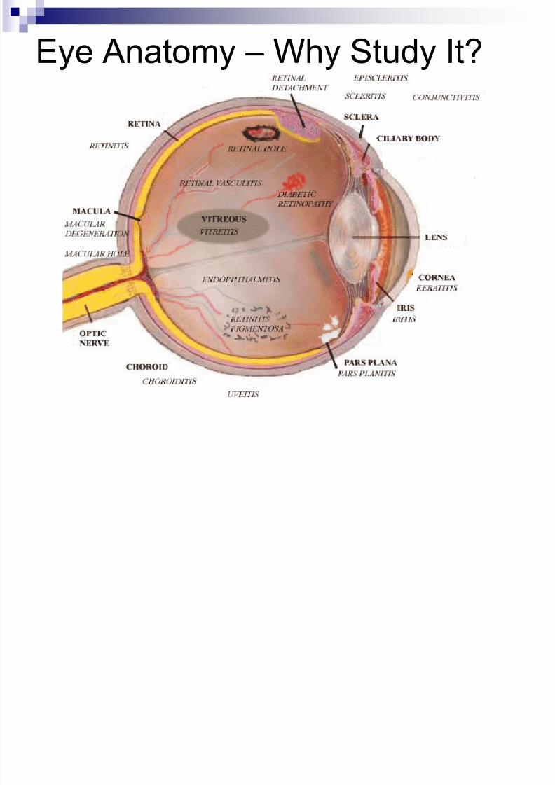
Eye Anatomy – Why Study It?
8/10/2019 04 - Assessment of Eyes & Ears
http://slidepdf.com/reader/full/04-assessment-of-eyes-ears 3/62
Why should you care?
Optometrist – Doctor of optometry, 4 year
undergrad + 4 year optometry school
Ophthalmologists – Medical doctors
In general, optometrists practice primary
and preventive eye care, while
ophthalmologists perform eye surgery
What do nurses do?
8/10/2019 04 - Assessment of Eyes & Ears
http://slidepdf.com/reader/full/04-assessment-of-eyes-ears 4/62
History
Vision difficulty? Halos around lights – in glaucoma
Scotoma – blind spot in visual field – in
glaucoma, optic nerve, and visual pathwaydisorder
Night blindness – Vit A deficiency,glaucoma,
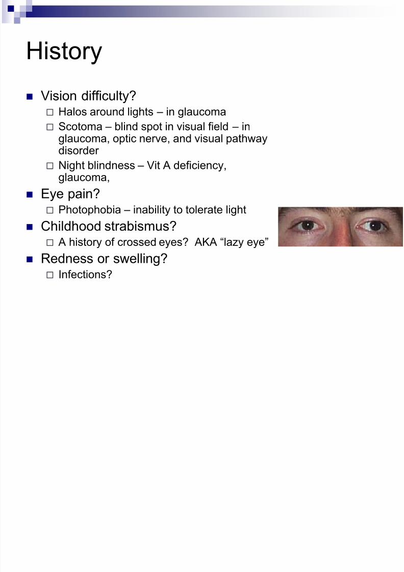
Eye pain?
Photophobia – inability to tolerate light Childhood strabismus?
A history of crossed eyes? AKA “lazy eye”
Redness or swelling? Infections?
8/10/2019 04 - Assessment of Eyes & Ears
http://slidepdf.com/reader/full/04-assessment-of-eyes-ears 5/62
History cont.
Excessive or lack of tearing?
May be due to irritants or obstruction in drainage
Past history of ocular problems? Glaucoma? Family history?
Use of glasses or contact lenses?
When tested last? Any medications?
8/10/2019 04 - Assessment of Eyes & Ears
http://slidepdf.com/reader/full/04-assessment-of-eyes-ears 6/62
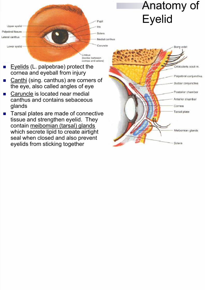
Anatomy of
Eyelid
Eyelids (L. palpebrae) protect the
cornea and eyeball from injury Canthi (sing. canthus) are corners of
the eye, also called angles of eye
Caruncle is located near medialcanthus and contains sebaceous
glands Tarsal plates are made of connective
tissue and strengthen eyelid. Theycontain meibomian (tarsal) glandswhich secrete lipid to create airtightseal when closed and also prevent
eyelids from sticking together
8/10/2019 04 - Assessment of Eyes & Ears
http://slidepdf.com/reader/full/04-assessment-of-eyes-ears 7/62
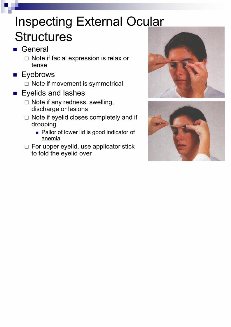
Inspecting External Ocular
Structures General Note if facial expression is relax or
tense
Eyebrows Note if movement is symmetrical
Eyelids and lashes Note if any redness, swelling,
discharge or lesions
Note if eyelid closes completely and ifdrooping
Pallor of lower lid is good indicator ofanemia
For upper eyelid, use applicator stickto fold the eyelid over
8/10/2019 04 - Assessment of Eyes & Ears
http://slidepdf.com/reader/full/04-assessment-of-eyes-ears 8/62
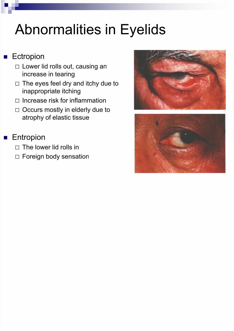
Abnormalities in Eyelids
Ectropion
Lower lid rolls out, causing an
increase in tearing
The eyes feel dry and itchy due toinappropriate itching
Increase risk for inflammation
Occurs mostly in elderly due to
atrophy of elastic tissue
Entropion
The lower lid rolls in
Foreign body sensation
8/10/2019 04 - Assessment of Eyes & Ears
http://slidepdf.com/reader/full/04-assessment-of-eyes-ears 9/62
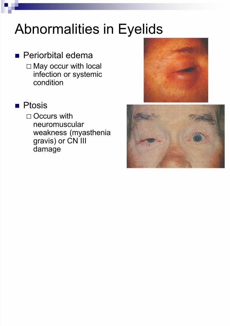
Abnormalities in Eyelids
Periorbital edemaMay occur with local
infection or systemic
condition
PtosisOccurs with
neuromuscularweakness (myastheniagravis) or CN IIIdamage
8/10/2019 04 - Assessment of Eyes & Ears
http://slidepdf.com/reader/full/04-assessment-of-eyes-ears 10/62
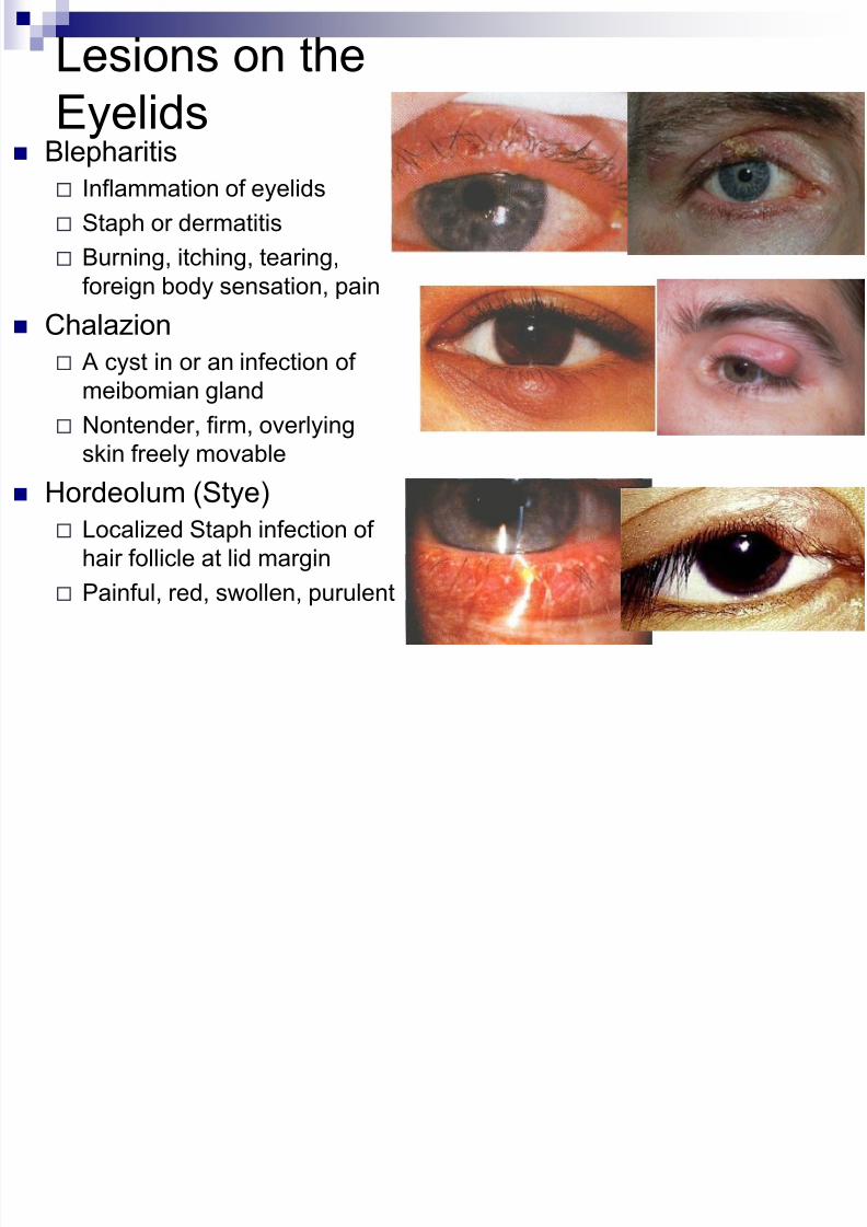
Lesions on the
Eyelids Blepharitis
Inflammation of eyelids
Staph or dermatitis
Burning, itching, tearing,
foreign body sensation, pain
Chalazion
A cyst in or an infection of
meibomian gland
Nontender, firm, overlying
skin freely movable
Hordeolum (Stye)
Localized Staph infection of
hair follicle at lid margin
Painful, red, swollen, purulent
8/10/2019 04 - Assessment of Eyes & Ears
http://slidepdf.com/reader/full/04-assessment-of-eyes-ears 11/62
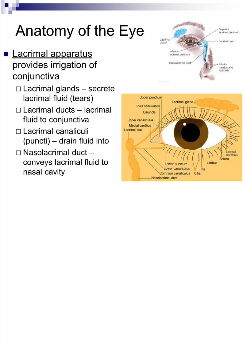
Anatomy of the Eye
Lacrimal apparatus
provides irrigation of
conjunctiva
Lacrimal glands – secrete
lacrimal fluid (tears)
Lacrimal ducts – lacrimal
fluid to conjunctiva
Lacrimal canaliculi
(puncti) – drain fluid into
Nasolacrimal duct –
conveys lacrimal fluid to
nasal cavity
8/10/2019 04 - Assessment of Eyes & Ears
http://slidepdf.com/reader/full/04-assessment-of-eyes-ears 12/62
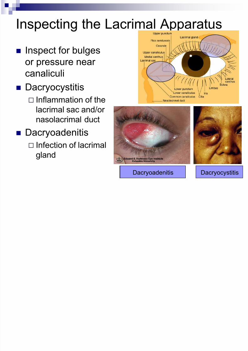
Inspecting the Lacrimal Apparatus
Inspect for bulges
or pressure near
canaliculi
Dacryocystitis Inflammation of the
lacrimal sac and/or
nasolacrimal duct
Dacryoadenitis Infection of lacrimal
gland
DacryocystitisDacryoadenitis
8/10/2019 04 - Assessment of Eyes & Ears
http://slidepdf.com/reader/full/04-assessment-of-eyes-ears 13/62
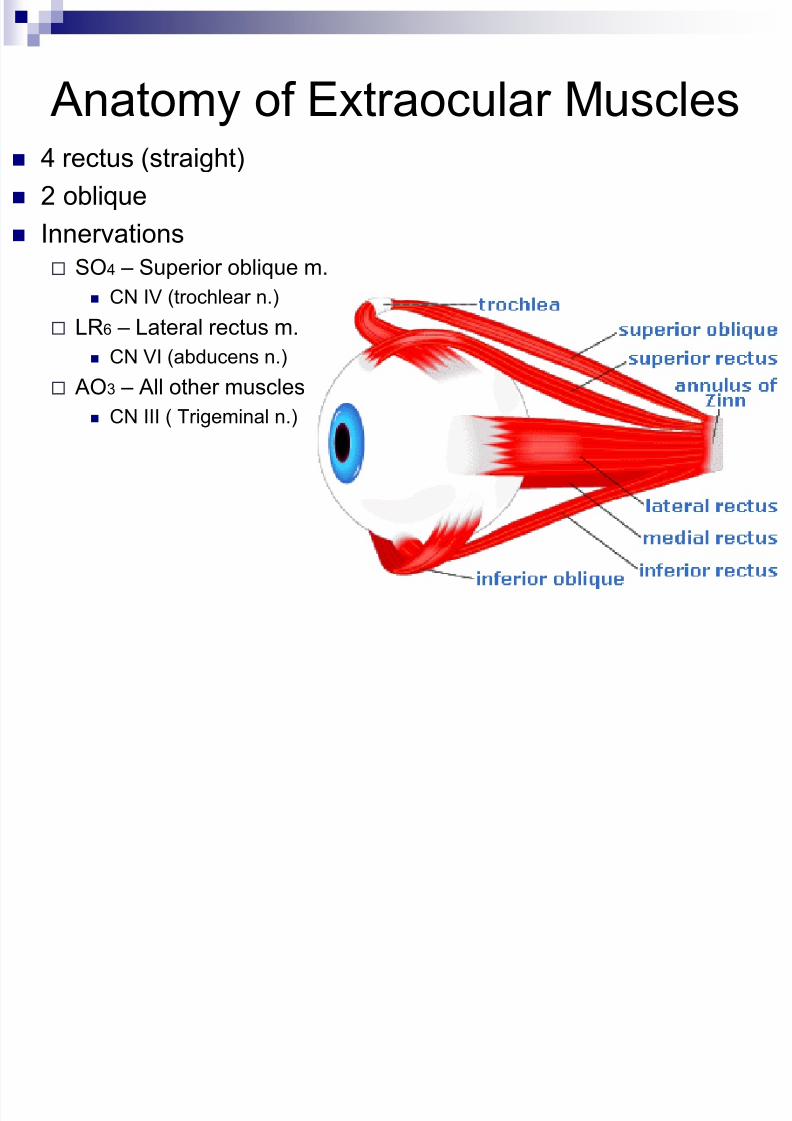
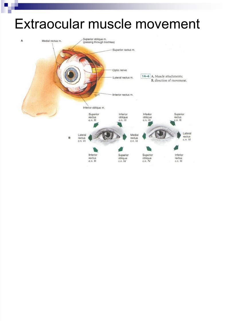
Anatomy of Extraocular Muscles 4 rectus (straight)
2 oblique
Innervations
SO4 – Superior oblique m. CN IV (trochlear n.)
LR6 – Lateral rectus m.
CN VI (abducens n.)
AO3 – All other muscles
CN III ( Trigeminal n.)
8/10/2019 04 - Assessment of Eyes & Ears
http://slidepdf.com/reader/full/04-assessment-of-eyes-ears 14/62
Extraocular muscle movement
8/10/2019 04 - Assessment of Eyes & Ears
http://slidepdf.com/reader/full/04-assessment-of-eyes-ears 15/62
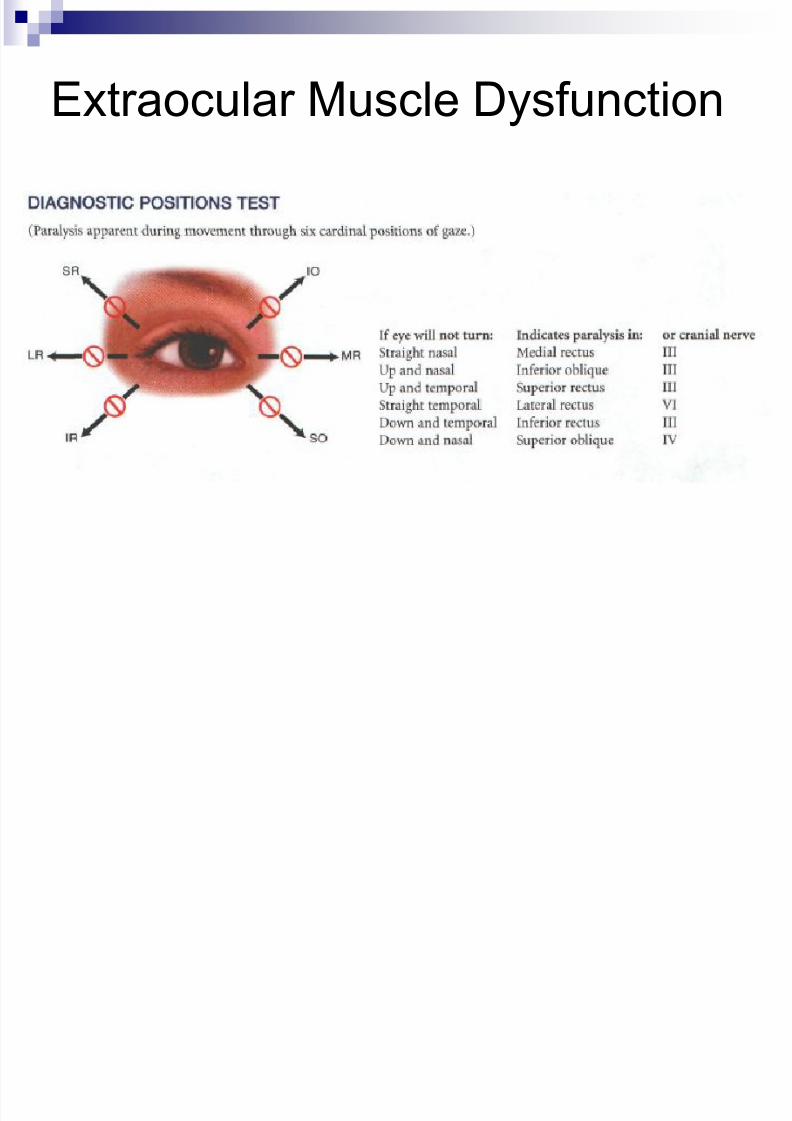
Extraocular Muscle Dysfunction
8/10/2019 04 - Assessment of Eyes & Ears
http://slidepdf.com/reader/full/04-assessment-of-eyes-ears 16/62
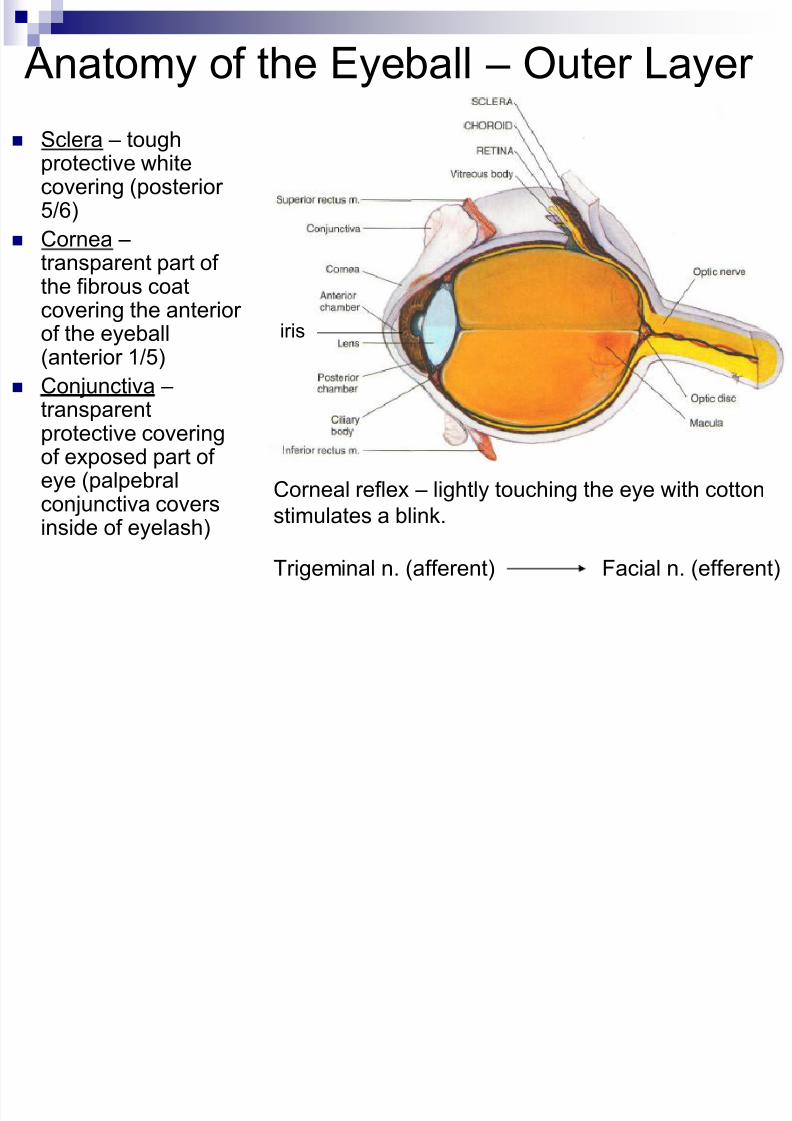
Anatomy of the Eyeball – Outer Layer
Sclera – toughprotective whitecovering (posterior5/6)
Cornea – transparent part ofthe fibrous coatcovering the anteriorof the eyeball(anterior 1/5)
Conjunctiva –
transparentprotective coveringof exposed part ofeye (palpebralconjunctiva coversinside of eyelash)
Corneal reflex – lightly touching the eye with cotton
stimulates a blink.
Trigeminal n. (afferent) Facial n. (efferent)
iris
8/10/2019 04 - Assessment of Eyes & Ears
http://slidepdf.com/reader/full/04-assessment-of-eyes-ears 17/62
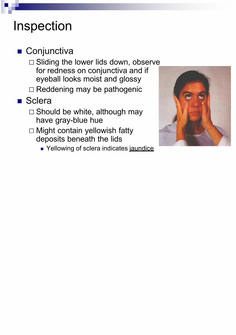
Inspection
Conjunctiva Sliding the lower lids down, observe
for redness on conjunctiva and ifeyeball looks moist and glossy
Reddening may be pathogenic
Sclera Should be white, although may
have gray-blue hue
Might contain yellowish fattydeposits beneath the lids Yellowing of sclera indicates jaundice
8/10/2019 04 - Assessment of Eyes & Ears
http://slidepdf.com/reader/full/04-assessment-of-eyes-ears 18/62
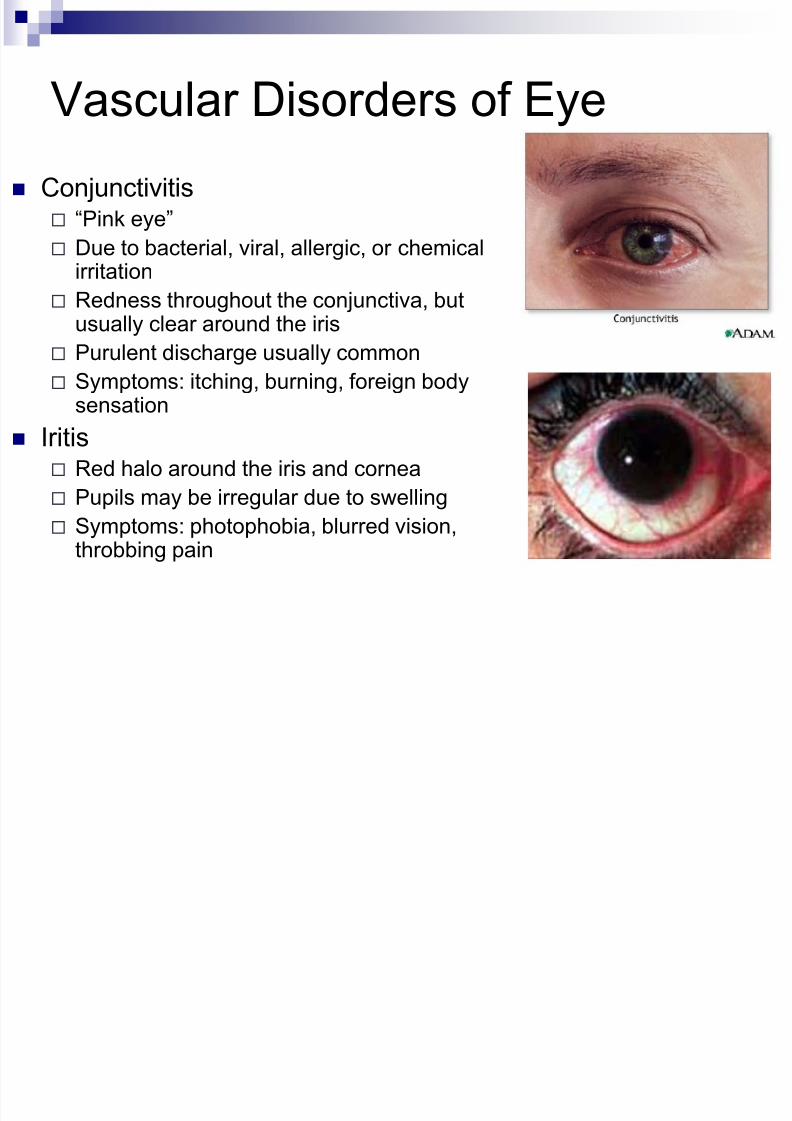
Vascular Disorders of Eye
Conjunctivitis “Pink eye”
Due to bacterial, viral, allergic, or chemical
irritation Redness throughout the conjunctiva, but
usually clear around the iris
Purulent discharge usually common
Symptoms: itching, burning, foreign body
sensation Iritis
Red halo around the iris and cornea
Pupils may be irregular due to swelling
Symptoms: photophobia, blurred vision,
throbbing pain
8/10/2019 04 - Assessment of Eyes & Ears
http://slidepdf.com/reader/full/04-assessment-of-eyes-ears 19/62
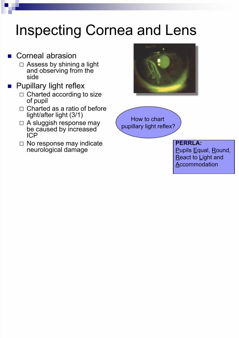
Inspecting Cornea and Lens
Corneal abrasion Assess by shining a light
and observing from theside
Pupillary light reflex Charted according to size
of pupil
Charted as a ratio of beforelight/after light (3/1)
A sluggish response maybe caused by increasedICP
No response may indicateneurological damage
PERRLA:
Pupils Equal, Round,
React to Light and
Accommodation
How to chart
pupillary light reflex?
8/10/2019 04 - Assessment of Eyes & Ears
http://slidepdf.com/reader/full/04-assessment-of-eyes-ears 20/62
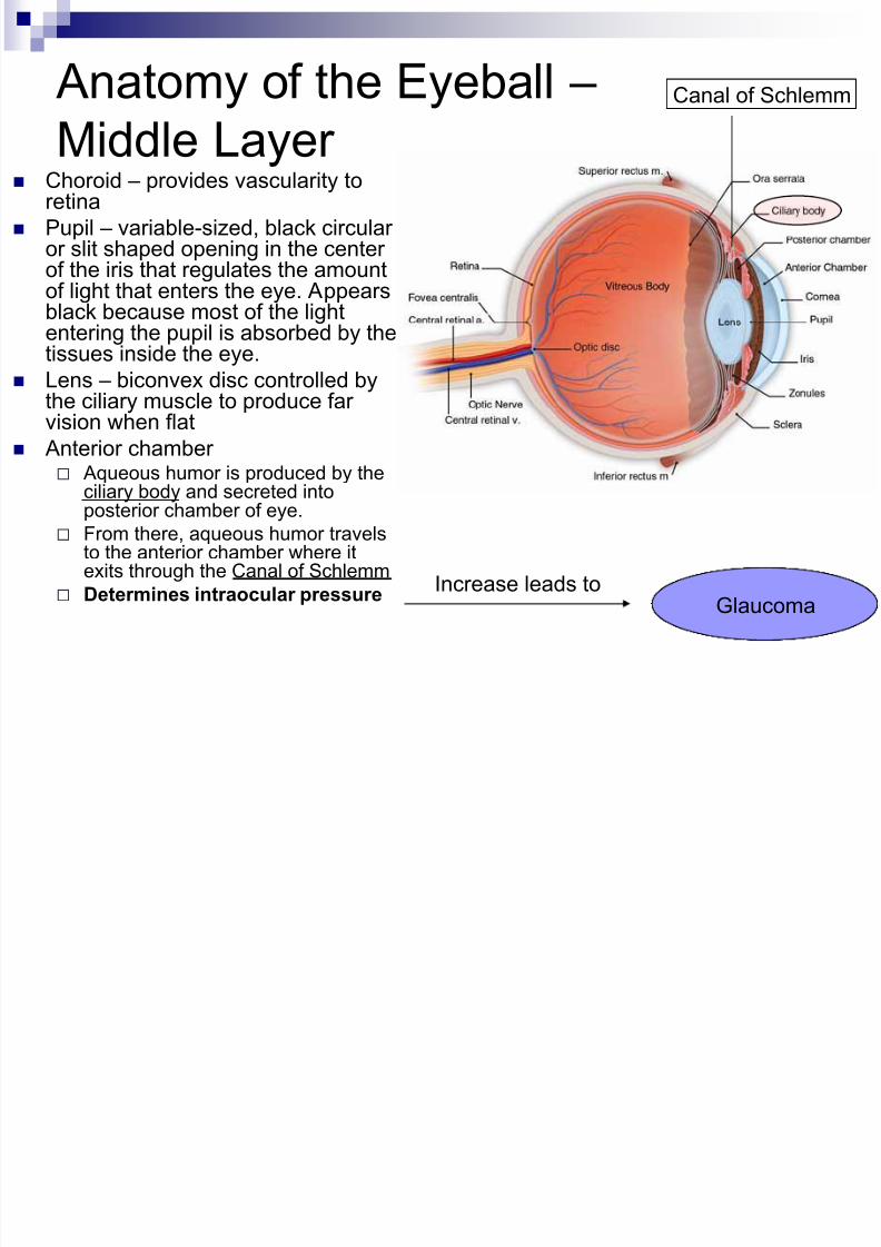
Anatomy of the Eyeball –
Middle Layer Choroid – provides vascularity toretina
Pupil – variable-sized, black circularor slit shaped opening in the centerof the iris that regulates the amountof light that enters the eye. Appears
black because most of the lightentering the pupil is absorbed by thetissues inside the eye.
Lens – biconvex disc controlled bythe ciliary muscle to produce farvision when flat
Anterior chamber Aqueous humor is produced by the
ciliary body and secreted intoposterior chamber of eye.
From there, aqueous humor travelsto the anterior chamber where itexits through the Canal of Schlemm
Determines intraocular pressure
Canal of Schlemm
Increase leads toGlaucoma
8/10/2019 04 - Assessment of Eyes & Ears
http://slidepdf.com/reader/full/04-assessment-of-eyes-ears 21/62
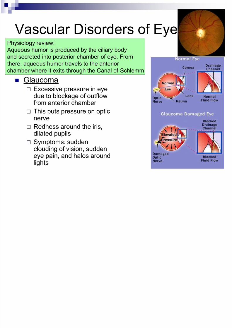
Vascular Disorders of Eye
Glaucoma Excessive pressure in eye
due to blockage of outflowfrom anterior chamber
This puts pressure on opticnerve
Redness around the iris,dilated pupils
Symptoms: suddenclouding of vision, suddeneye pain, and halos around
lights
Physiology review: Aqueous humor is produced by the ciliary body
and secreted into posterior chamber of eye. From
there, aqueous humor travels to the anterior
chamber where it exits through the Canal of Schlemm
8/10/2019 04 - Assessment of Eyes & Ears
http://slidepdf.com/reader/full/04-assessment-of-eyes-ears 22/62
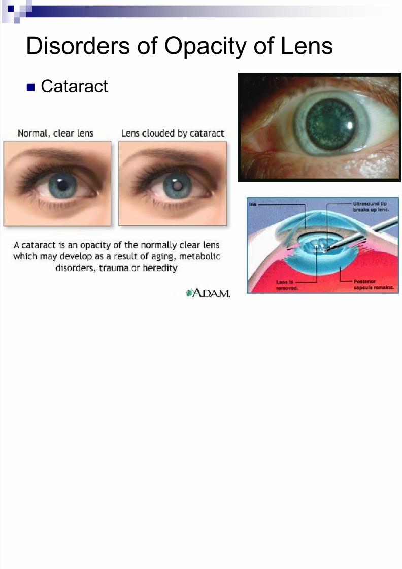
Disorders of Opacity of Lens
Cataract
8/10/2019 04 - Assessment of Eyes & Ears
http://slidepdf.com/reader/full/04-assessment-of-eyes-ears 23/62
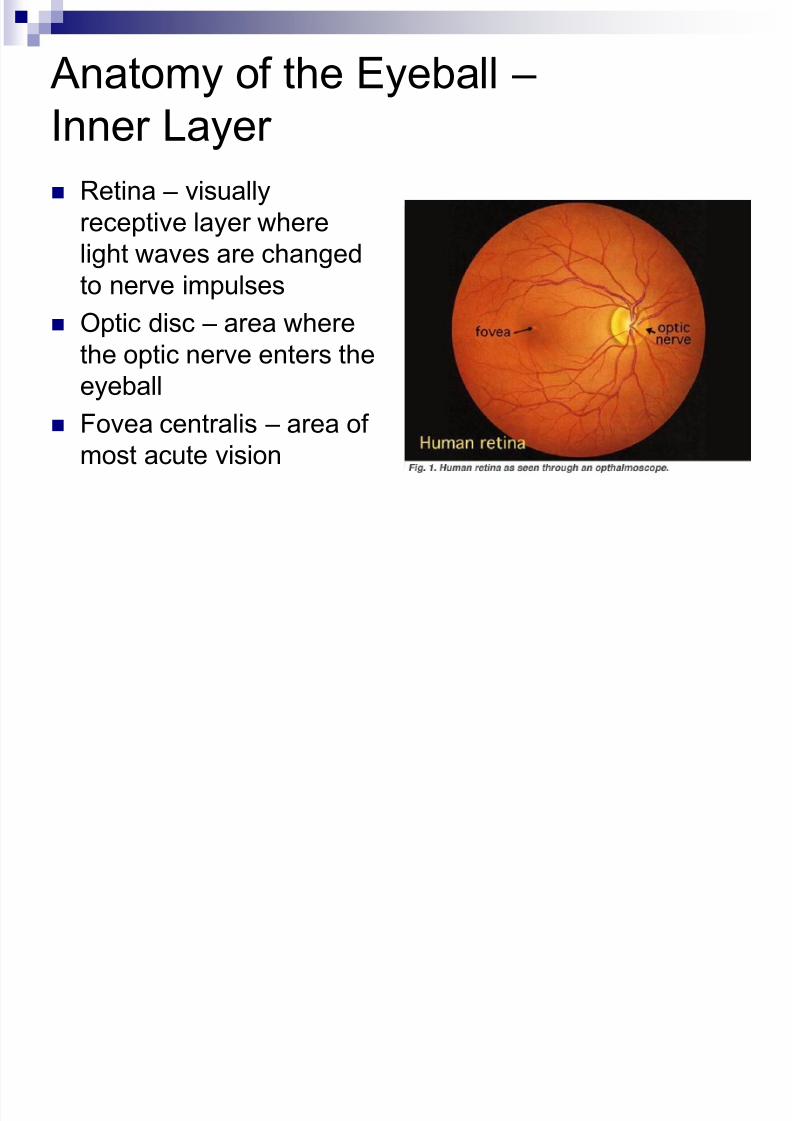
Anatomy of the Eyeball –
Inner Layer Retina – visually
receptive layer where
light waves are changed
to nerve impulses
Optic disc – area where
the optic nerve enters the
eyeball
Fovea centralis – area ofmost acute vision

8/10/2019 04 - Assessment of Eyes & Ears
http://slidepdf.com/reader/full/04-assessment-of-eyes-ears 24/62
Inspecting the Ocular Fundus
Using an ophthalmoscope to inspectthe internal surface of the retina,anterior chamber, lens, and vitreous.
Darken the room to dilate the pupils
Remove eye glasses, contacts maystay in
Ask person to stare at distant object
Hold ophthalmoscope close to youreye and move to within a few inches
of the person’s face A red glow filling the pupil is called
the red reflex and is caused by lightreflecting off the retina
Cataracts appear as opaque blackareas against the red reflex
8/10/2019 04 - Assessment of Eyes & Ears
http://slidepdf.com/reader/full/04-assessment-of-eyes-ears 25/62
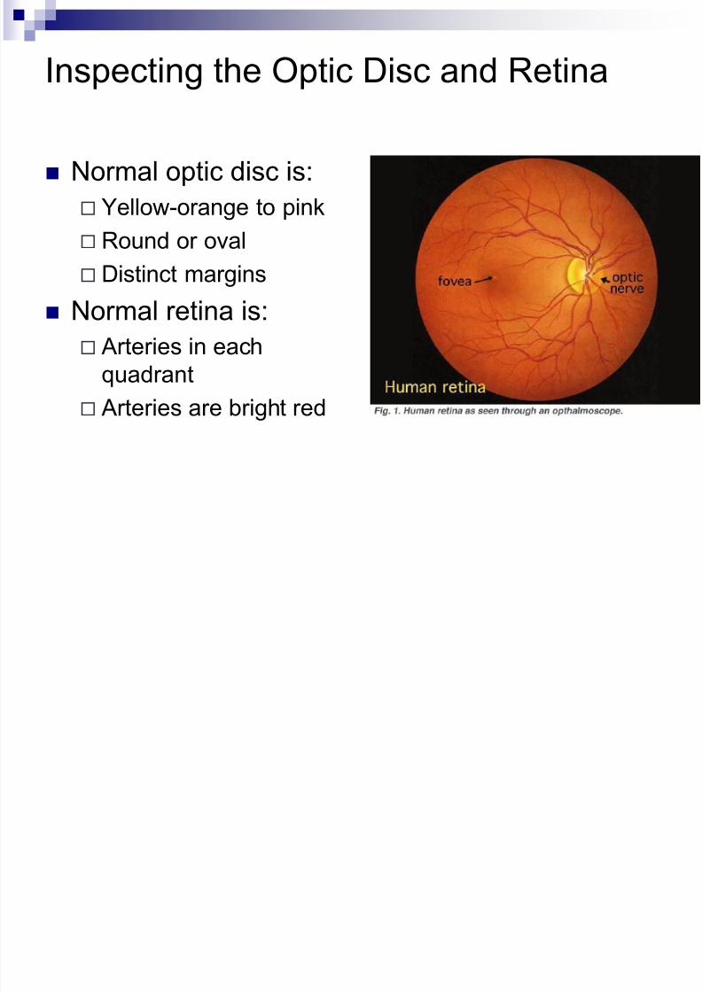
Inspecting the Optic Disc and Retina
Normal optic disc is:
Yellow-orange to pink
Round or oval Distinct margins
Normal retina is:
Arteries in each
quadrant
Arteries are bright red
8/10/2019 04 - Assessment of Eyes & Ears
http://slidepdf.com/reader/full/04-assessment-of-eyes-ears 26/62
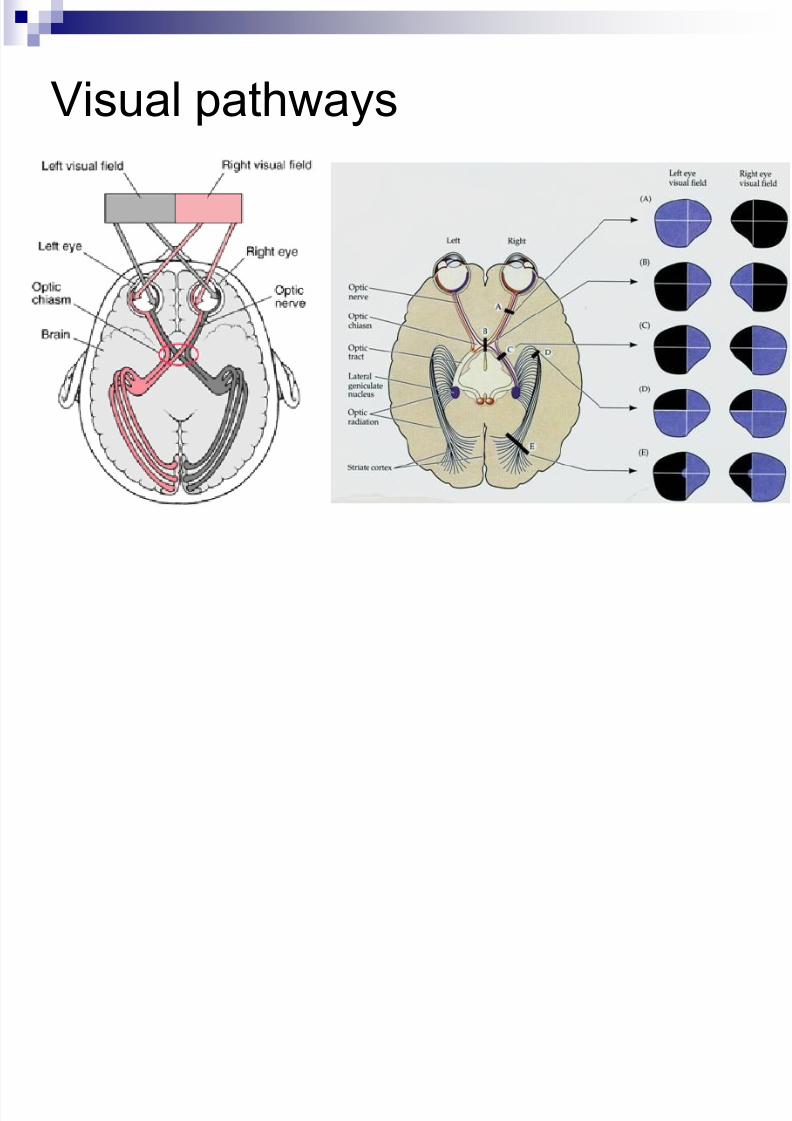
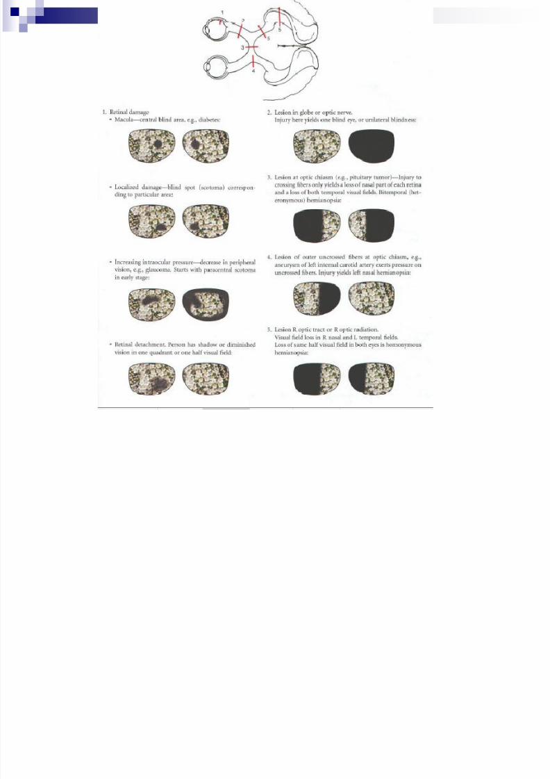
Visual pathways
8/10/2019 04 - Assessment of Eyes & Ears
http://slidepdf.com/reader/full/04-assessment-of-eyes-ears 27/62
8/10/2019 04 - Assessment of Eyes & Ears
http://slidepdf.com/reader/full/04-assessment-of-eyes-ears 28/62
Testing Visual Reflexes Pupillary light reflex
Constriction of pupils when bright light shines on the retina
Direct light reflex – constriction of same sided pupil
Consensual light reflex – simultaneous constriction of both pupils
The impulse is carried afferently by CN II and efferently by CN III
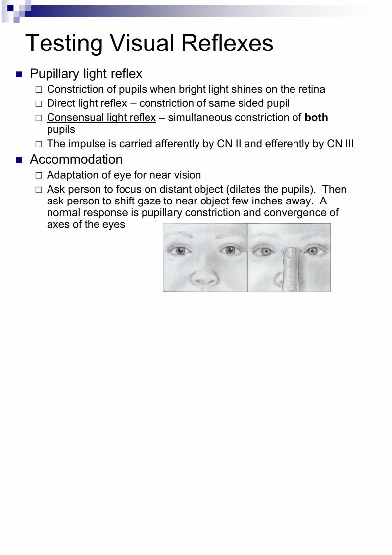
Accommodation Adaptation of eye for near vision
Ask person to focus on distant object (dilates the pupils). Thenask person to shift gaze to near object few inches away. Anormal response is pupillary constriction and convergence ofaxes of the eyes
8/10/2019 04 - Assessment of Eyes & Ears
http://slidepdf.com/reader/full/04-assessment-of-eyes-ears 29/62
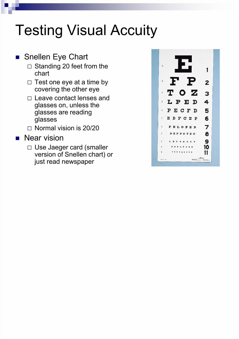
Testing Visual Accuity
Snellen Eye Chart Standing 20 feet from the
chart
Test one eye at a time bycovering the other eye
Leave contact lenses andglasses on, unless theglasses are readingglasses
Normal vision is 20/20
Near vision Use Jaeger card (smaller
version of Snellen chart) or just read newspaper
8/10/2019 04 - Assessment of Eyes & Ears
http://slidepdf.com/reader/full/04-assessment-of-eyes-ears 30/62
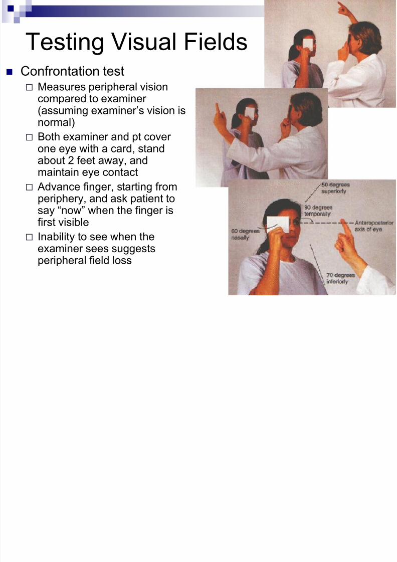
Testing Visual Fields
Confrontation test Measures peripheral vision
compared to examiner(assuming examiner’s vision isnormal)
Both examiner and pt coverone eye with a card, standabout 2 feet away, andmaintain eye contact
Advance finger, starting fromperiphery, and ask patient to
say “now” when the finger isfirst visible
Inability to see when theexaminer sees suggestsperipheral field loss
8/10/2019 04 - Assessment of Eyes & Ears
http://slidepdf.com/reader/full/04-assessment-of-eyes-ears 31/62
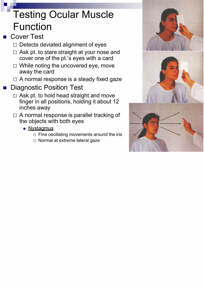
Testing Ocular Muscle
Function Cover Test
Detects deviated alignment of eyes
Ask pt. to stare straight at your nose andcover one of the pt.’s eyes with a card
While noting the uncovered eye, move
away the card A normal response is a steady fixed gaze
Diagnostic Position Test Ask pt. to hold head straight and move
finger in all positions, holding it about 12
inches away A normal response is parallel tracking of
the objects with both eyes
Nystagmus
Fine oscillating movements around the iris
Normal at extreme lateral gaze
8/10/2019 04 - Assessment of Eyes & Ears
http://slidepdf.com/reader/full/04-assessment-of-eyes-ears 32/62
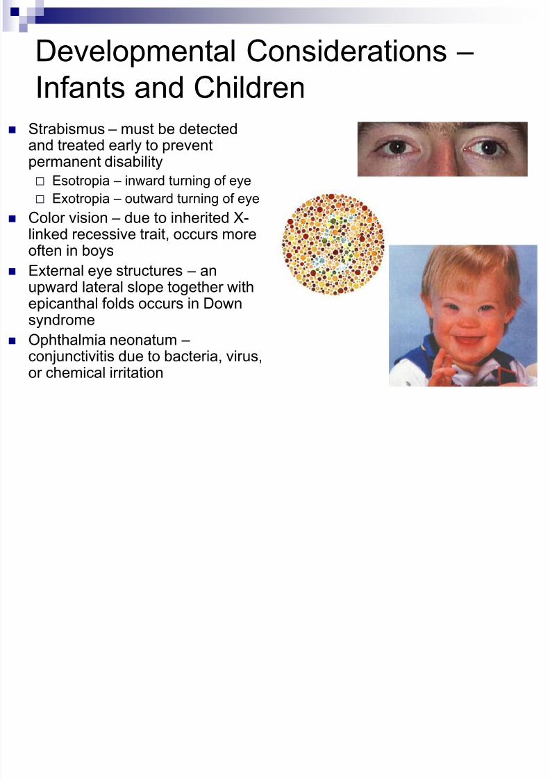
Developmental Considerations –
Infants and Children Strabismus – must be detected
and treated early to preventpermanent disability
Esotropia – inward turning of eye
Exotropia – outward turning of eye
Color vision – due to inherited X-linked recessive trait, occurs moreoften in boys
External eye structures – an
upward lateral slope together withepicanthal folds occurs in Downsyndrome
Ophthalmia neonatum – conjunctivitis due to bacteria, virus,or chemical irritation
8/10/2019 04 - Assessment of Eyes & Ears
http://slidepdf.com/reader/full/04-assessment-of-eyes-ears 33/62
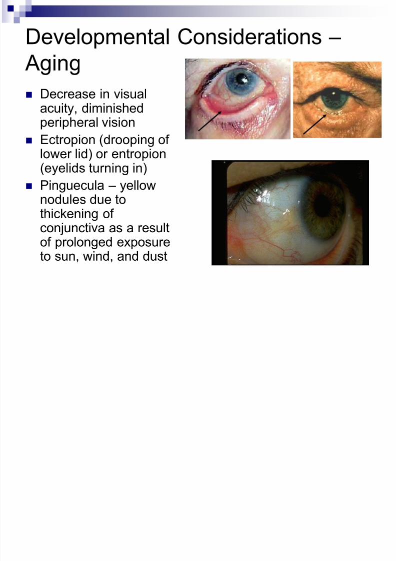
Developmental Considerations –
Aging Decrease in visual
acuity, diminishedperipheral vision
Ectropion (drooping oflower lid) or entropion(eyelids turning in)
Pinguecula – yellownodules due to
thickening ofconjunctiva as a resultof prolonged exposureto sun, wind, and dust
8/10/2019 04 - Assessment of Eyes & Ears
http://slidepdf.com/reader/full/04-assessment-of-eyes-ears 34/62
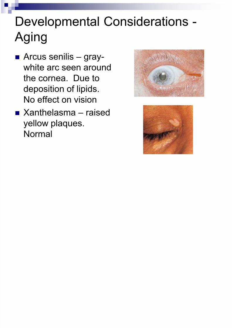
Developmental Considerations -
Aging Arcus senilis – gray-
white arc seen around
the cornea. Due todeposition of lipids.
No effect on vision
Xanthelasma – raised
yellow plaques.Normal

8/10/2019 04 - Assessment of Eyes & Ears
http://slidepdf.com/reader/full/04-assessment-of-eyes-ears 35/62
Ear Anatomy
8/10/2019 04 - Assessment of Eyes & Ears
http://slidepdf.com/reader/full/04-assessment-of-eyes-ears 36/62
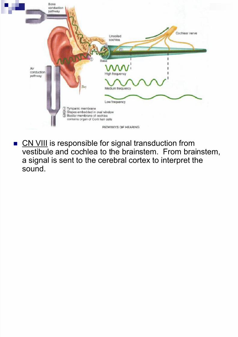
Ear Physiology
External Ear External auditory meatus funnels sound waves, which reflect off the
tympanic membrane to produce vibrations
Cerumen (ear wax) protects the tympanic membrane from foreignsubstances
Middle ear Malleus, incus, and stapes and eustachian tube
Function to: Conduct sound vibrations from tympanic membrane (outer ear) to cochlea
(inner ear)
Protect the cochlea by reducing the amplitude of sounds
Eustachian tube allows equalization of air pressure
Inner ear Vestibule and semicircular canals
Allow brain to sense body position and relation of angle of head to gravity
Cochlea Transfers vibrations from stapes into nerve impulses
8/10/2019 04 - Assessment of Eyes & Ears
http://slidepdf.com/reader/full/04-assessment-of-eyes-ears 37/62
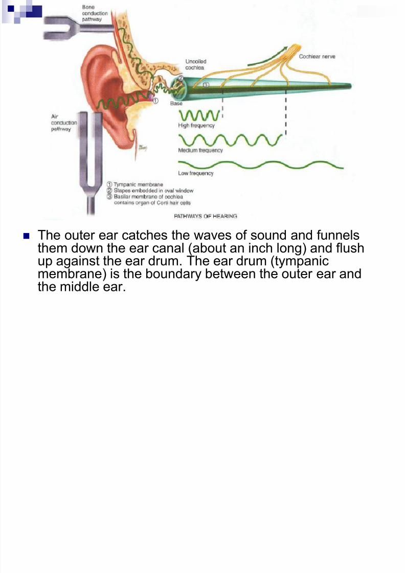
The outer ear catches the waves of sound and funnelsthem down the ear canal (about an inch long) and flushup against the ear drum. The ear drum (tympanicmembrane) is the boundary between the outer ear and
the middle ear.
8/10/2019 04 - Assessment of Eyes & Ears
http://slidepdf.com/reader/full/04-assessment-of-eyes-ears 38/62
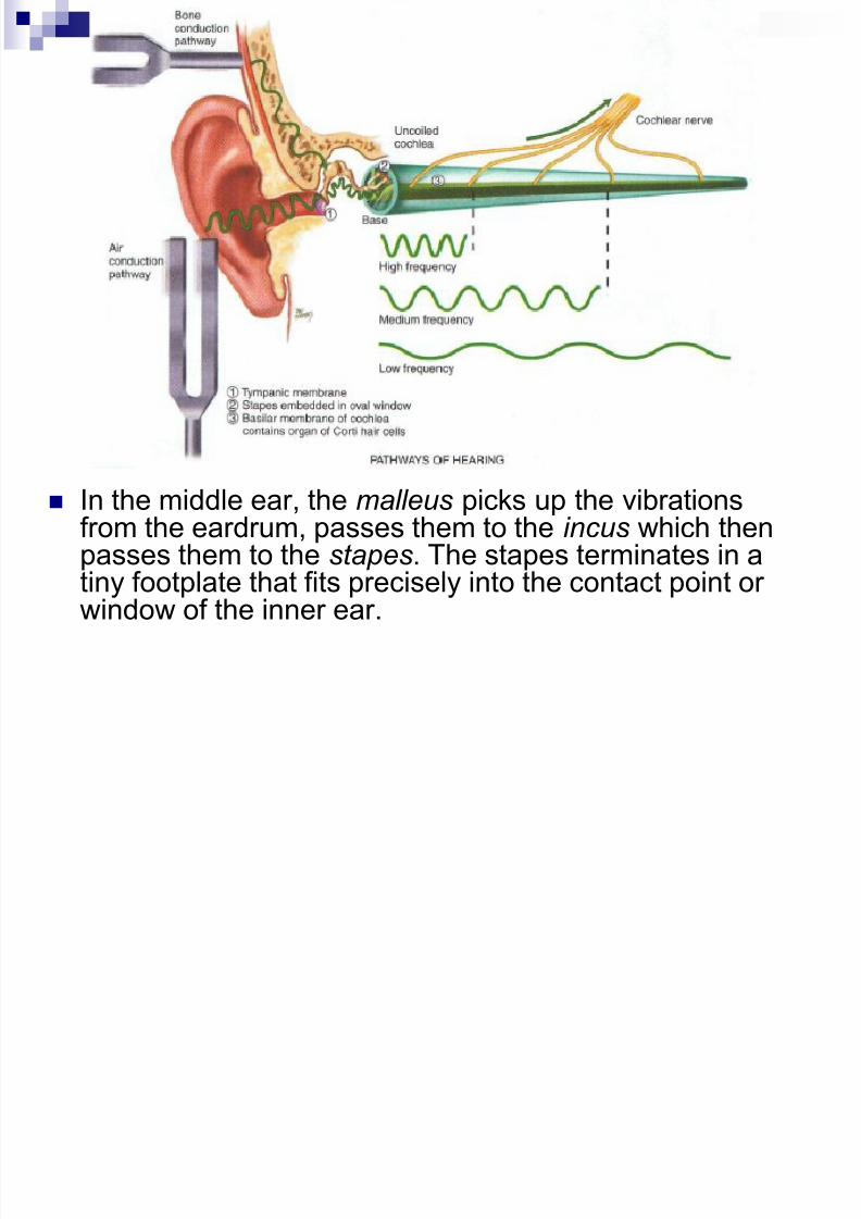
In the middle ear, the malleus picks up the vibrationsfrom the eardrum, passes them to the incus which thenpasses them to the stapes. The stapes terminates in atiny footplate that fits precisely into the contact point or
window of the inner ear.
8/10/2019 04 - Assessment of Eyes & Ears
http://slidepdf.com/reader/full/04-assessment-of-eyes-ears 39/62
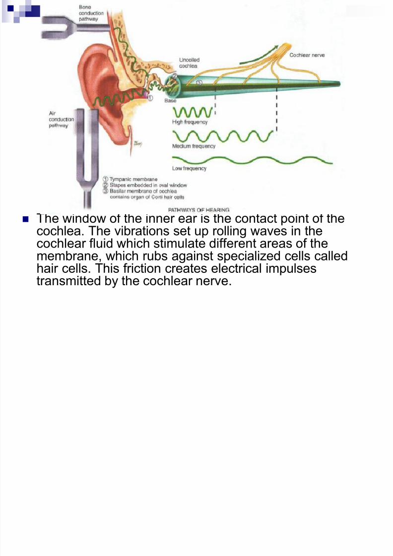
The window of the inner ear is the contact point of thecochlea. The vibrations set up rolling waves in thecochlear fluid which stimulate different areas of themembrane, which rubs against specialized cells calledhair cells. This friction creates electrical impulsestransmitted by the cochlear nerve.
8/10/2019 04 - Assessment of Eyes & Ears
http://slidepdf.com/reader/full/04-assessment-of-eyes-ears 40/62
CN VIII is responsible for signal transduction fromvestibule and cochlea to the brainstem. From brainstem,a signal is sent to the cerebral cortex to interpret the
sound.
8/10/2019 04 - Assessment of Eyes & Ears
http://slidepdf.com/reader/full/04-assessment-of-eyes-ears 41/62
Hearing Loss
Conductive
Mechanical dysfunction of external or middle ear
Partial hearing lossMay be caused by impacted cerumen, foreign bodies,
perforated tympanic membrane, pus or serum in
middle ear, or otosclerosis (hardening of stapes)
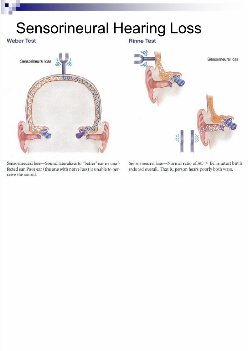
May be fixed Sensorineural
Dysfunction of inner ear, CN VIII, or cerebral cortex
Cannot be fixed
8/10/2019 04 - Assessment of Eyes & Ears
http://slidepdf.com/reader/full/04-assessment-of-eyes-ears 42/62
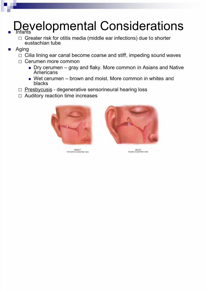
Developmental Considerations Infants
Greater risk for otitis media (middle ear infections) due to shortereustachian tube
Aging
Cilia lining ear canal become coarse and stiff, impeding sound waves
Cerumen more common
Dry cerumen – gray and flaky. More common in Asians and Native
Americans Wet cerumen – brown and moist. More common in whites and
blacks
Presbycusis - degenerative sensorineural hearing loss
Auditory reaction time increases
8/10/2019 04 - Assessment of Eyes & Ears
http://slidepdf.com/reader/full/04-assessment-of-eyes-ears 43/62
Obtaining History
Earaches? (otalgia) Location, character, intensity, associative and alleviating factors
May be directly due to ear disease or maybe referred pain from aproblem in teeth or oropharynx
A viral or bacterial upper respiratory infection may migrate up theeustachian tube and involve the middle ear
Infections? Frequency? Occurred in childhood?
Discharge? (otorrhea) May suggest infection or perforated eardrum Typically with perforation, ear pain drainage
Otitis externa – purulent, sanguineous, or watery
Acute otitis media with perforation – purulent discharge
8/10/2019 04 - Assessment of Eyes & Ears
http://slidepdf.com/reader/full/04-assessment-of-eyes-ears 44/62
More History
Trouble hearing? Gradual our sudden?
Presbycusis – gradual sensorineural hearing impairment in theelderly
Hearing loss due to trauma is often sudden Ringing in ears? (tinnitus)
May be a result of medication
Medications? Some are ototoxic
Vertigo? (spinning) Subjective – person feels like he or she spins
Objective – person feels like room spins
Environmental noise Noise-induced hearing loss
8/10/2019 04 - Assessment of Eyes & Ears
http://slidepdf.com/reader/full/04-assessment-of-eyes-ears 45/62
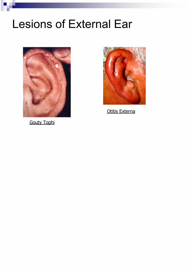
Lesions of External Ear
Gouty Tophi
Otitis Externa
8/10/2019 04 - Assessment of Eyes & Ears
http://slidepdf.com/reader/full/04-assessment-of-eyes-ears 46/62
Assessing External Ear
Size and Shape normal is 4-10cm tall
Skin conditions Note edema, inflammation, lesions
Tenderness Location?
Pain in pinna indicates otitis externa
Pain at mastoid process indicates mastoiditis or lymphadenitis
External Auditory Meatus Atresia – absence or closure of ear canal
Otitis externa may cause purulent discharge
Otitis media may cause rupture of tympanic membrane
If drainage present following trauma, possible basal skullfracture. Perform glucose test (CSF (+) for glucose).
8/10/2019 04 - Assessment of Eyes & Ears
http://slidepdf.com/reader/full/04-assessment-of-eyes-ears 47/62
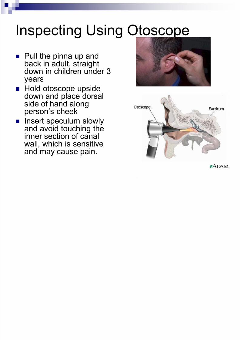
Inspecting Using Otoscope
Pull the pinna up andback in adult, straightdown in children under 3years
Hold otoscope upsidedown and place dorsalside of hand alongperson’s cheek
Insert speculum slowly
and avoid touching theinner section of canalwall, which is sensitiveand may cause pain.
8/10/2019 04 - Assessment of Eyes & Ears
http://slidepdf.com/reader/full/04-assessment-of-eyes-ears 48/62
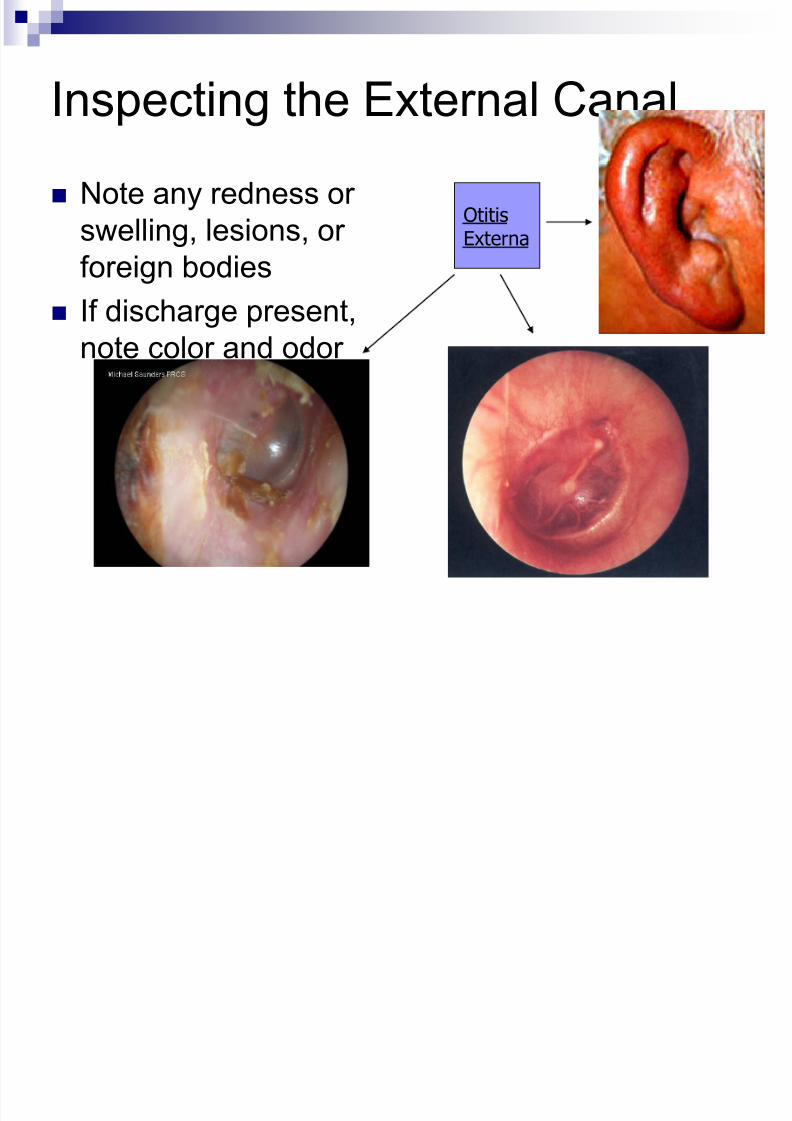
Inspecting the External Canal
Note any redness or
swelling, lesions, or
foreign bodies
If discharge present,
note color and odor
OtitisExterna
8/10/2019 04 - Assessment of Eyes & Ears
http://slidepdf.com/reader/full/04-assessment-of-eyes-ears 49/62
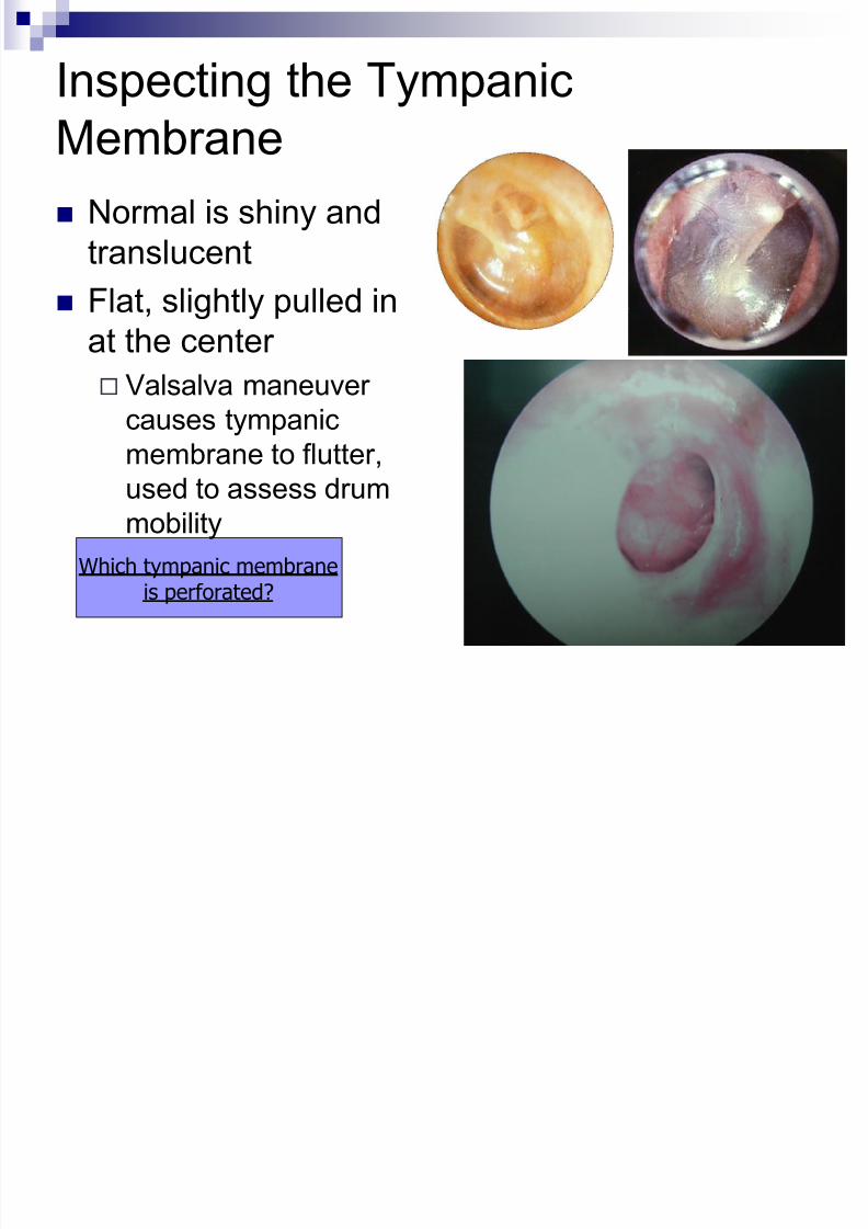
Inspecting the Tympanic
Membrane Normal is shiny and
translucent
Flat, slightly pulled inat the center
Valsalva maneuver
causes tympanic
membrane to flutter,used to assess drum
mobility
Which tympanic membraneis perforated?
8/10/2019 04 - Assessment of Eyes & Ears
http://slidepdf.com/reader/full/04-assessment-of-eyes-ears 50/62
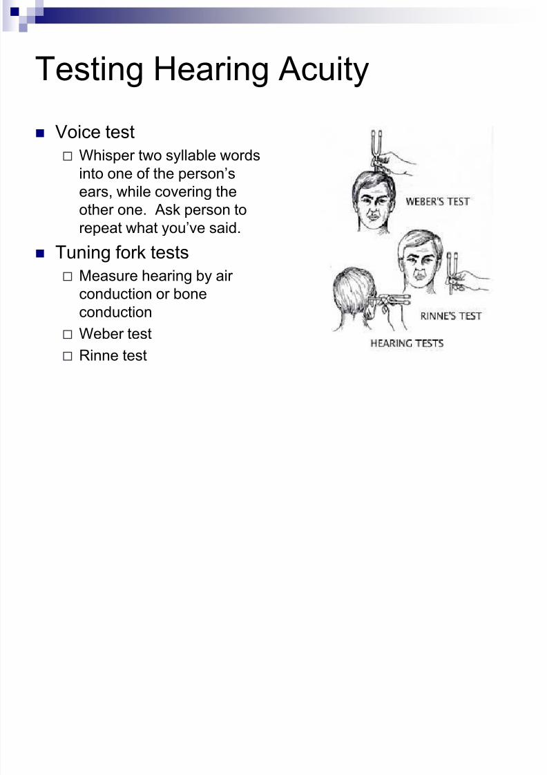
Testing Hearing Acuity
Voice test
Whisper two syllable words
into one of the person’s
ears, while covering theother one. Ask person to
repeat what you’ve said.
Tuning fork tests
Measure hearing by air
conduction or boneconduction
Weber test
Rinne test
8/10/2019 04 - Assessment of Eyes & Ears
http://slidepdf.com/reader/full/04-assessment-of-eyes-ears 51/62
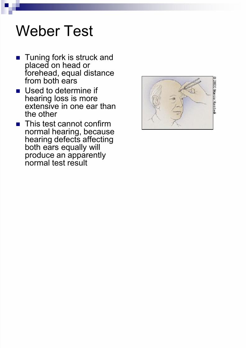
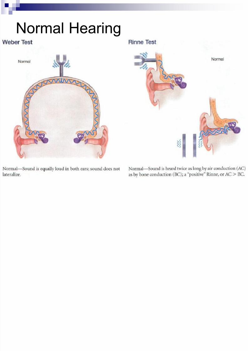
Weber Test
Tuning fork is struck andplaced on head orforehead, equal distancefrom both ears
Used to determine ifhearing loss is moreextensive in one ear thanthe other
This test cannot confirm
normal hearing, becausehearing defects affectingboth ears equally willproduce an apparentlynormal test result
8/10/2019 04 - Assessment of Eyes & Ears
http://slidepdf.com/reader/full/04-assessment-of-eyes-ears 52/62
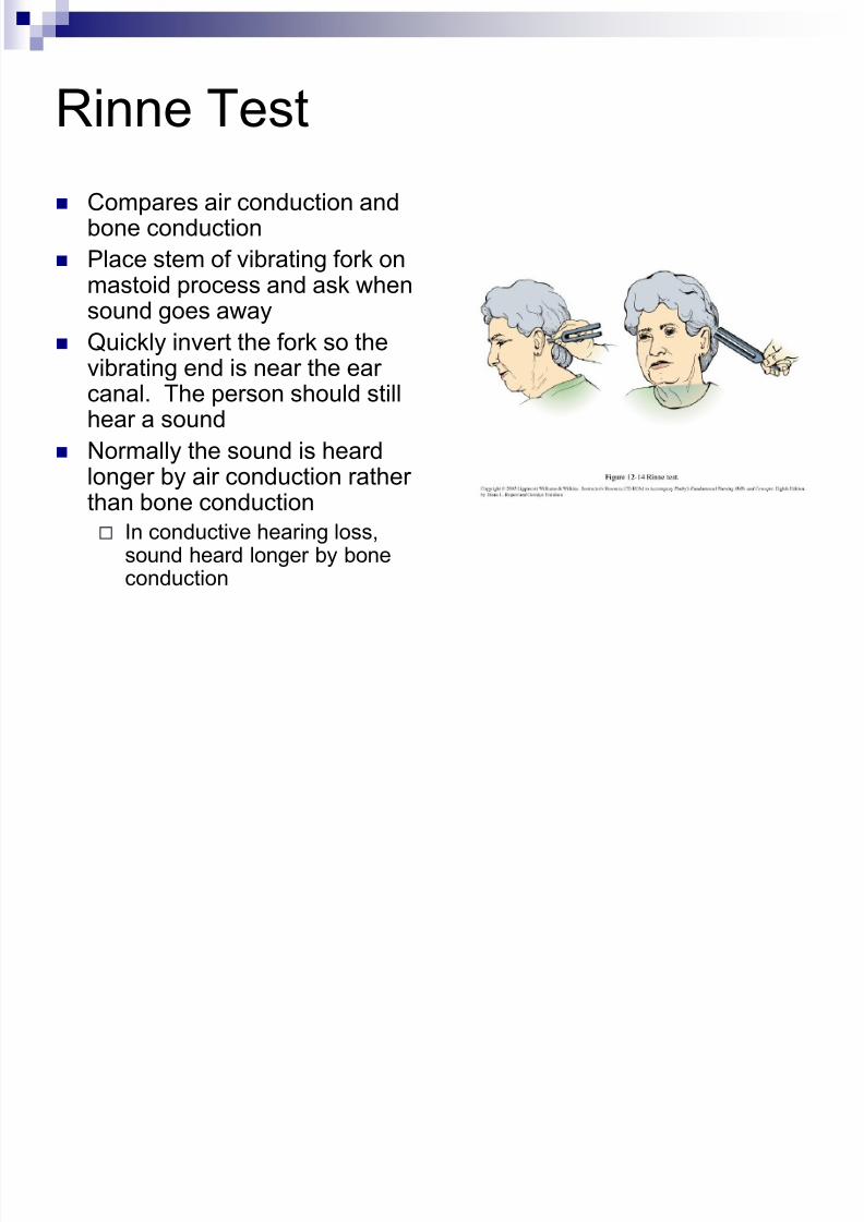
Rinne Test
Compares air conduction andbone conduction
Place stem of vibrating fork onmastoid process and ask when
sound goes away Quickly invert the fork so the
vibrating end is near the earcanal. The person should stillhear a sound
Normally the sound is heardlonger by air conduction ratherthan bone conduction
In conductive hearing loss,sound heard longer by boneconduction
8/10/2019 04 - Assessment of Eyes & Ears
http://slidepdf.com/reader/full/04-assessment-of-eyes-ears 53/62

8/10/2019 04 - Assessment of Eyes & Ears
http://slidepdf.com/reader/full/04-assessment-of-eyes-ears 54/62
Conductive Hearing Loss
8/10/2019 04 - Assessment of Eyes & Ears
http://slidepdf.com/reader/full/04-assessment-of-eyes-ears 55/62
Sensorineural Hearing Loss
8/10/2019 04 - Assessment of Eyes & Ears
http://slidepdf.com/reader/full/04-assessment-of-eyes-ears 56/62
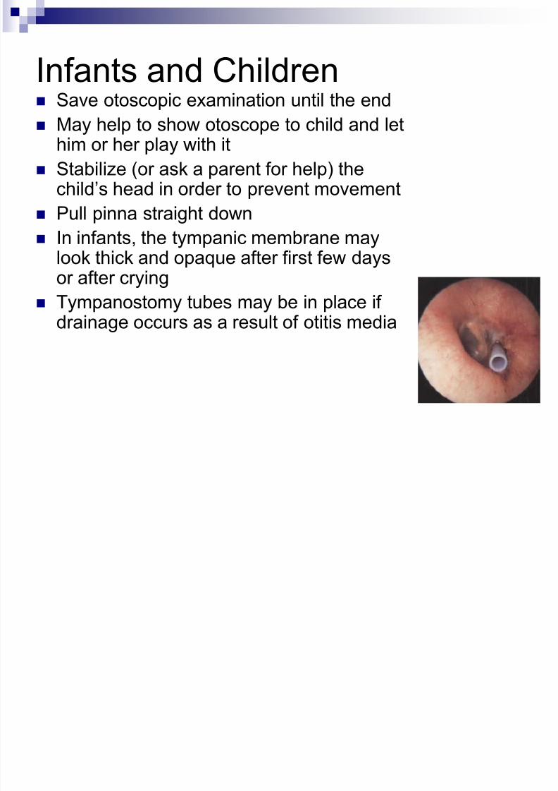
Infants and Children Save otoscopic examination until the end May help to show otoscope to child and let
him or her play with it
Stabilize (or ask a parent for help) thechild’s head in order to prevent movement
Pull pinna straight down
In infants, the tympanic membrane maylook thick and opaque after first few daysor after crying
Tympanostomy tubes may be in place ifdrainage occurs as a result of otitis media
8/10/2019 04 - Assessment of Eyes & Ears
http://slidepdf.com/reader/full/04-assessment-of-eyes-ears 57/62
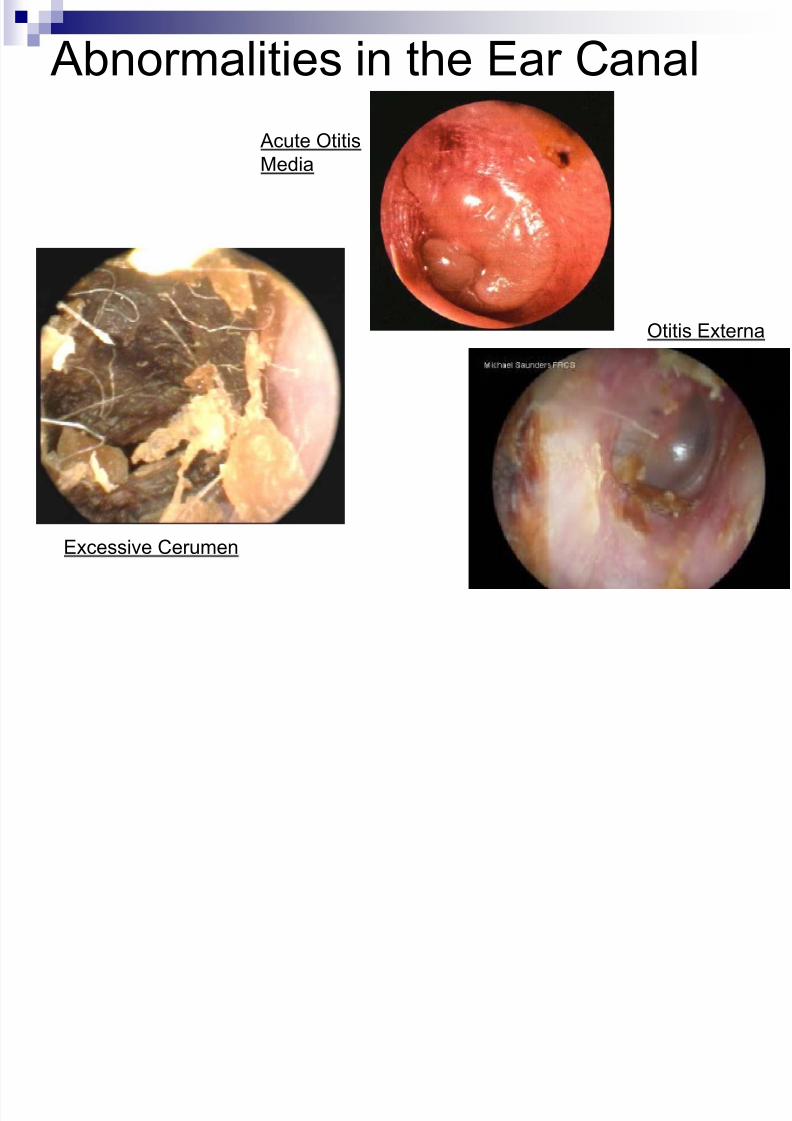
Abnormalities in the Ear Canal
Excessive Cerumen
Acute OtitisMedia
Otitis Externa

8/10/2019 04 - Assessment of Eyes & Ears
http://slidepdf.com/reader/full/04-assessment-of-eyes-ears 58/62
Question 1
A nurse is performing a voice test to assesshearing. Which of the following describes theaccurate procedure for performing this test?
1. Stand 4 feet away from the client to ensure that theclient can hear at this distance
2. Quietly whisper a statement and ask the client torepeat it
3. Whisper a statement with the examiner’s back facingthe client
4. Whisper a statement while the client blocks bothears
8/10/2019 04 - Assessment of Eyes & Ears
http://slidepdf.com/reader/full/04-assessment-of-eyes-ears 59/62
Question 2
A nurse is caring for a client who is
hearing impaired. Which of the following
approaches will facilitatecommunication?
1. Speak frequently
2. Speak loudly3. Speak directly into the impaired ear
4. Speak in a normal tone
8/10/2019 04 - Assessment of Eyes & Ears
http://slidepdf.com/reader/full/04-assessment-of-eyes-ears 60/62
Question 3
A client is diagnosed with a disorderinvolving the inner ear. Which of the
following is the most common clientcomplaint associated with a disorderinvolving this part of the ear?
1. Hearing loss
2. Pruritus3. Tinnitus
4. Burning in the ear
8/10/2019 04 - Assessment of Eyes & Ears
http://slidepdf.com/reader/full/04-assessment-of-eyes-ears 61/62
Question 4
Which of the following statements madeby a parent should make the nurse
suspicious that the tympanic membraneof a young child has ruptured?
1. “She has been crying all night, but she feelsbetter this morning.”
2. “She has some bloody, yellow-looking stuffcoming out of her ear.”
3. “My child does not seem to hear very well.”
4. “My child’s earwax is dark brown.”
8/10/2019 04 - Assessment of Eyes & Ears
http://slidepdf.com/reader/full/04-assessment-of-eyes-ears 62/62
Question 5
While examining the internal ear, the
nurse observes the light reflex on the
tympanic membrane. What does thisfinding indicate?
1. Presence of pus
2. Fluid accumulation3. Scar tissue
4. Normal finding