1,* 1 2 author manuscript nih public access 1 4 3...
TRANSCRIPT

NQR1 controls lifespan by regulating the promotion ofrespiratory metabolism in yeast
María Jiménez-Hidalgo1,*, Carlos Santos-Ocaña1,*, Sergio Padilla1, José M. Villalba2,Guillermo Lopez-Lluch1, Alejandro Martín-Montalvo1, Robin K. Minor4, David A. Sinclair3,Rafael de Cabo4,ϕ, and Plácido Navas1,ϕ1Centro Andaluz de Biología del Desarrollo, Universidad Pablo de Olavide-CSIC and Centre forBiomedical Research on Rare Diseases (CIBERER), ISCIII, E-41013 Sevilla, Spain2Departamento de Biología Celular, Fisiología e Inmunología, Universidad de Córdoba, 14014Córdoba, Spain3Paul F. Glenn Laboratories for the Biological Mechanism of Aging, Department of Pathology,Harvard Medical School, Harvard, MA 02115, USA4Laboratory of Experimental Gerontology, National Institute on Aging, NIH, Baltimore, MD 21224,USA
SummaryThe activity and expression of plasma membrane NADH coenzyme Q reductase is increased bycalorie restriction (CR) in rodents. Although this effect is well established and is necessary forCR's ability to delay aging, the mechanism is unknown. Here we show that the Saccharomycescerevisiae homolog, NQR1, resides at the plasma membrane and when overexpressed extends bothreplicative and chronological lifespan. We show that NQR1 extends replicative lifespan in a SIR2-dependent manner by shifting cells towards respiratory metabolism. Chronological lifespanextension, in contrast, occurs via a SIR2-independent decrease in ethanol production. We concludethat NQR1 is a key mediator of lifespan extension by CR through its effects on yeast metabolismand discuss how these findings could suggest a function for this protein in lifespan extension inmammals.
KeywordsPlasma membrane; coenzyme Q reductase; NQR1; coenzyme Q; replicative lifespan;chronological lifespan; dietary restriction
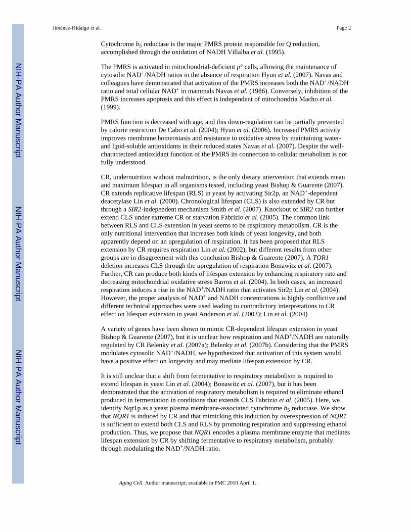
IntroductionThe NAD(P)H-dependent plasma membrane redox system (PMRS) of mammalian cellsregulates fasting-induced apoptosis through the trans-plasma membrane redox system Navaset al. (2007). Figure 1 shows a simple scheme of the basic PMRS components in eukaryoticcells. The PMRS regulates apoptosis in part through maintaining coenzyme Q (Q) in itsreduced state (QH2). QH2 specifically inhibits the magnesium-dependent neutralsphingomyelinase which prevents the initiation of apoptosis Van Maldergem et al. (2002).
ϕ Correspondence: ([email protected]) P.N. and ([email protected]) R.C..*These authors contributed equally to this work
NIH Public AccessAuthor ManuscriptAging Cell. Author manuscript; available in PMC 2010 April 1.
Published in final edited form as:Aging Cell. 2009 April ; 8(2): 140–151. doi:10.1111/j.1474-9726.2009.00461.x.
NIH
-PA Author Manuscript
NIH
-PA Author Manuscript
NIH
-PA Author Manuscript
Cytochrome b5 reductase is the major PMRS protein responsible for Q reduction,accomplished through the oxidation of NADH Villalba et al. (1995).
The PMRS is activated in mitochondrial-deficient ρ° cells, allowing the maintenance ofcytosolic NAD+/NADH ratios in the absence of respiration Hyun et al. (2007). Navas andcolleagues have demonstrated that activation of the PMRS increases both the NAD+/NADHratio and total cellular NAD+ in mammals Navas et al. (1986). Conversely, inhibition of thePMRS increases apoptosis and this effect is independent of mitochondria Macho et al.(1999).
PMRS function is decreased with age, and this down-regulation can be partially preventedby calorie restriction De Cabo et al. (2004); Hyun et al. (2006). Increased PMRS activityimproves membrane homeostasis and resistance to oxidative stress by maintaining water-and lipid-soluble antioxidants in their reduced states Navas et al. (2007). Despite the well-characterized antioxidant function of the PMRS its connection to cellular metabolism is notfully understood.
CR, undernutrition without malnutrition, is the only dietary intervention that extends meanand maximum lifespan in all organisms tested, including yeast Bishop & Guarente (2007).CR extends replicative lifespan (RLS) in yeast by activating Sir2p, an NAD+-dependentdeacetylase Lin et al. (2000). Chronological lifespan (CLS) is also extended by CR butthrough a SIR2-independent mechanism Smith et al. (2007). Knockout of SIR2 can furtherextend CLS under extreme CR or starvation Fabrizio et al. (2005). The common linkbetween RLS and CLS extension in yeast seems to be respiratory metabolism. CR is theonly nutritional intervention that increases both kinds of yeast longevity, and bothapparently depend on an upregulation of respiration. It has been proposed that RLSextension by CR requires respiration Lin et al. (2002), but different results from othergroups are in disagreement with this conclusion Bishop & Guarente (2007). A TOR1deletion increases CLS through the upregulation of respiration Bonawitz et al. (2007).Further, CR can produce both kinds of lifespan extension by enhancing respiratory rate anddecreasing mitochondrial oxidative stress Barros et al. (2004). In both cases, an increasedrespiration induces a rise in the NAD+/NADH ratio that activates Sir2p Lin et al. (2004).However, the proper analysis of NAD+ and NADH concentrations is highly conflictive anddifferent technical approaches were used leading to contradictory interpretations to CReffect on lifespan extension in yeast Anderson et al. (2003); Lin et al. (2004)
A variety of genes have been shown to mimic CR-dependent lifespan extension in yeastBishop & Guarente (2007), but it is unclear how respiration and NAD+/NADH are naturallyregulated by CR Belenky et al. (2007a); Belenky et al. (2007b). Considering that the PMRSmodulates cytosolic NAD+/NADH, we hypothesized that activation of this system wouldhave a positive effect on longevity and may mediate lifespan extension by CR.
It is still unclear that a shift from fermentative to respiratory metabolism is required toextend lifespan in yeast Lin et al. (2004); Bonawitz et al. (2007), but it has beendemonstrated that the activation of respiratory metabolism is required to eliminate ethanolproduced in fermentation in conditions that extends CLS Fabrizio et al. (2005). Here, weidentify Nqr1p as a yeast plasma membrane-associated cytochrome b5 reductase. We showthat NQR1 is induced by CR and that mimicking this induction by overexpression of NQR1is sufficient to extend both CLS and RLS by promoting respiration and suppressing ethanolproduction. Thus, we propose that NQR1 encodes a plasma membrane enzyme that mediateslifespan extension by CR by shifting fermentative to respiratory metabolism, probablythrough modulating the NAD+/NADH ratio.
Jiménez-Hidalgo et al. Page 2
Aging Cell. Author manuscript; available in PMC 2010 April 1.
NIH
-PA Author Manuscript
NIH
-PA Author Manuscript
NIH
-PA Author Manuscript

ResultsCR induces the expression of NQR1
Low glucose concentrations, which some consider to approximate CR in yeast, extendlifespan by increasing respiration Barros et al. (2004) Bonawitz et al. (2007) Lin et al.(2002). Incubating the BY4741 yeast strain in different glucose concentrations led to anincrease in CLS inversely to glucose concentration (Figure S1A). The immunolocalizationof cytochrome c by confocal microscopy demonstrated the increase of mitochondria as theconcentration of glucose was decreased (Figure S2A). These results correlated with theincrease of both cytochrome c and Cox1p by immunoblot in these cells (Figure S2D).Further, oxygen consumption (Figure S2B) and NADH-cytochrome c reductase (complex I+III) activity (Figure S2C) were increased as the concentration of glucose droppedindicating the activation of respiration. These analyses were carried out at 16 h to preventthe glucose inhibition of respiratory-associated activities determined in this work.Corresponding growth curves are included in figure S2E. In the same conditions, NQR1expression was induced earlier and to higher levels in low glucose (0.5%) compared toconcentrations of 2 or 10% glucose as assessed by real time-PCR (Figure 2A). NQR1peptide was also accumulated in cells in yeast growing in low glucose at 10 h and onlyslightly identified at this time when growing in 2% glucose. No expressed protein wasdetected in 10% glucose (Figure 2B). At 24 h, NQR1 was in all cells but the concentrationwas clearly higher at 0.5% glucose (Figure 2B). Relative Nqr1p content in plasmamembrane was normalized to Gas1p in figure 2C, showing a neat increase in CR conditions.To demonstrate if this expression was affecting plasma membrane, enriched fractions wereobtained and both NADH-Q reductase activity and redox state of Q were analyzed. Figure2D shows that NADH-Q reductase activity was higher in plasma membrane of yeast aslower were the concentration of glucose in media. Further, the fraction of QH2 in plasmamembrane was increasing compared to total Q as the glucose concentration in media wasdecreasing (Figure 2E). Low concentration of glucose that mimics CR in yeast induced anincrease of NQR1 expression and its activity in plasma membrane in the same conditionswhere respiration was activated.
NQR1 encodes for a NADH-dependent cytochrome b5 reductase that reduces Q at theplasma membrane
Analysis in silico of yeast genome showed the existence of the YML125c gene that encodeda putative yeast cytochrome b5 reductase (NQR1) Giaever et al. (2002).
In order to analyze a possible function of NQR1 on longevity, this gene was cloned into ayeast expression vector controlled by the GAL promoter (pYES2.1/V5-His-TOPO fromInvitrogen) that allows the fusion of the V5 epitope tag. In 2% galactose, NQR1 wasexpressed uniformly (Figure 3A), and the signal was located mainly on the plasmamembrane and in karmellae (Figure 3B), a special form of endoplasmic reticulum (ER)generated by overexpression of HMG1 (Profant et al., 1999) or plasma membrane AHA1product in yeast Villalba et al. (1992). Cell fractionation analysis performed using a strainharboring the c-myc tag fused to the nuclear copy of NQR1 showed its location at the plasmamembrane (Figure 3C). A high scale study indicated the location of GFP-Nqr1p constructionin ER Giaever et al. (2002), but this could be considered an artifact due to the modificationintroduced in NQR1 after the fusion of a 27 kDa protein as GFP compared with the 34 kDaof Nqr1p. As expected, NQR1 overexpression significantly increased NADH-dependentPMRS activity at the plasma membrane, allowing for increased levels of reduced coenzymeQ (Q) (Figure 3D), and consistent with previous reports, this activity was very sensitive tothe specific flavodehydrogenases inhibitor diphenylene iodonium (DPI) (Figure S3) Rigantiet al. (2004).
Jiménez-Hidalgo et al. Page 3
Aging Cell. Author manuscript; available in PMC 2010 April 1.
NIH
-PA Author Manuscript
NIH
-PA Author Manuscript
NIH
-PA Author Manuscript

Given that NQR1 transfers electrons to Q6 we analyzed the redox state of the endogenous Q6at the plasma membrane in cells overexpressing NQR1 (Figure 4A and 4B). The resultsconfirmed that NQR1 overexpressing cells had double amounts of reduced Q6 (Q6H2) from11.8% to 22.9% of total Q6 when plasma membrane fractions were incubated with NADH.When we induced the overexpression of NQR1, the results were a three fold increase onQ6H2 from 22.7% to 69.1% in the presence of NADH. These results illustrate that NADH isthe natural electron donor in both mammals Sun et al. (1992), and yeast Santos-Ocaña et al.(1998a). No significant changes of NAD+/NADH ratio were detected until 24 hours ofgrowth in control and NQR1 overexpressed cells. However, these cells showed a significantdecrease of total pyridine nucleotide relative to control, 94 nmol/mg DW compared to 53nmol/mg DW (Figure 4D). These results would indicate that recycling of NAD+ levels byNQR1 overexpression in yeast is higher than in wildtype cells, demanding less newbiosynthesis, although the numerous reactions involving NAD+ or NADH at the cytosolmake difficult to interpret these data.
NQR1 overexpression extends both RLS and CLS and increases respirationSince NQR1 plays a role in the regulation of cytosolic NAD+ by increasing the oxidation ofNADH, we set out to perform a longevity analysis in NQR1 overexpressing yeast strains.There are two models that are used to study lifespan in yeast, replicative (RLS) andchronological (CLS) lifespan, which correspond to different aspects of the yeast physiologyMuller et al. (1980). RLS is defined as the number of buds produced by a mother cell andCLS measures the time in which cells are viable in a nutrient exhausted media.
Overexpression of NQR1 in wild type increased mean lifespan from 6.38 to 11.13 days(Figure 5A). Analysis performed on an isogenic sir2Δ strain showed no further changes inmean lifespan, 8.11 to 11.55 days, demonstrating that CLS extension by NQR1 is notdependent on SIR2. Additionally, CLS extension also occured by NQR1 overexpression inother yeast backgrounds such as CEN PK2.1C SIR2∷KanMX4 strains (Figure S1B). It ispossible that lifespan extension could be caused by ER accumulation in karmellae.However, this is not apparently the case because the yeast 3-hydroxy 3-methylglutaryl CoAreductase (HMG1) when overexpressed in yeast also accumulated in karmellae with noalteration of CLS (Figure S1C).
Using the same strains, there was a significant extension in RLS in cells overexpressingNQR1, from 28.07 to 36.75 generations (Figure 5B). However, in this case, the effect ofNQR1 overexpression was greatly attenuated in strains lacking SIR2 (16.52 vs. 20.27generations), indicating that this effect is dependent on Sir2p activity. These results clearlydemonstrate the role of NQR1 in the extension of both CLS and RLS, representing acommon link to both types of lifespan.
Next, we wondered whether the ability of NQR1 to extend lifespan was due to alterations inoxidative metabolism. We found that NQR1 overexpression in yeast caused a significantincrease of O2 consumption, a marker of respiration (Figure 6A), and a decrease on ethanolproduction, a marker of fermentation (Figure 6B). Ethanol decrease is then correlated withincreased respiration rate. Oxidative phosphorylation complexes activities were alsosignificantly increased in these cells (Figure 6C), correlating with the increase of O2consumption. The increased respiratory metabolism was accompanied with a higher growthrate for cells expressing NQR1 (Figure S4)
Respiration is required for NQR1 overexpression effectTo further illustrate the connection between NQR1 and respiration, we used respiratory-defective yeast strains that lacked genes required for Q6 biosynthesis such as COQ2 or
Jiménez-Hidalgo et al. Page 4
Aging Cell. Author manuscript; available in PMC 2010 April 1.
NIH
-PA Author Manuscript
NIH
-PA Author Manuscript
NIH
-PA Author Manuscript

respiratory defective strains unrelated to Q6 biosynthesis such as cor1 and atp2. Thesestrains were transformed with NQR1 and incubated in glucose or galactose (Figure 7A). Allstrains were able to grow in glucose and galactose but cells that overexpressed NQR1 wereonly able to grow on glucose. When the experiment was performed in liquid media, the lackof growth in Nqr1p-overexpressing cells was reproduced but the addition of exogenous Q6to COQ2Δ cells allowed them to resume normal respiration and growth in galactose mediabut not in the cor1 and atp2 mutants, which are unable to restore respiration even after Q6addition (Figure 7B). These results are a clear indication that NQR1 acts through therespiratory metabolism in yeast to promote CLS and RLS extension.
DiscussionDiverse genes have been related to the mechanisms that regulate lifespan in modelorganisms such as Saccharomyces cerevisiae, and some of them are conserved in mammalsBishop & Guarente (2007). Many of these genes have been identified because theirmutations or overexpression mimic totally or in part CR, indicating that different pathwaysare affecting lifespan Kaeberlein et al. (2005). Enzymes involved with respiration,bioenergetics, as well as NAD+ biosynthesis pathways are linked to regulation of lifespan,perhaps, because all these enzymes participate in fitness, nutrient sensing, and energyproduction Lin et al. (2002); Barros et al. (2004); Belenky et al. (2007b); Easlon et al.(2007) The identification of new genes that contribute to balance bioenergetics in yeast,particularly those that facilitate the transition from fermentative to respiratory metabolism,would help us understand the genetics of aging.
The plasma membrane also contributes to the balance of bioenergetics because it containsNADH-dependent dehydrogenases that alter NAD+/NADH ratios Navas et al. (2007), andcan support survival in mitochondria-defective ρ° cells Larm et al. (1994); Hyun et al.(2007). These activities are diminished during aging but their decline is prevented by CR inmammals De Cabo et al. (2004); López-Lluch et al. (2005). Yeast plasma membrane alsocontains a specific NADH-dependent enzyme system that requires Q6 as electron acceptorSantos-Ocaña et al. (1998a); Santos-Ocaña et al. (1998b).
We have shown here that the NQR1-encoded protein localizes in the plasma membrane andreduces Q6 by equivalents from NADH. Overexpression of NQR1 further demonstrated thespecificity of NADH as it was observed for the trans-plasma membrane electron transportsystem in both mammals Sun et al. (1992); Villalba et al. (1995) and yeast Santos-Ocaña etal. (1998a). Cytochrome b5 reductase was identified as responsible for NADH oxidation inliver plasma membrane Navarro et al. (1995). As a consequence, activation of NQR1 at theplasma membrane uses cytosolic NADH that would raise NAD+ level. As a whole, theseresults indicate the regulation of cytosol NAD+ level or better the NAD+/NADH recyclingrate by NQR1 in yeast.
NQR1 has been demonstrated to be an essential protein Giaever et al. (2002) that whenoverexpressed increased both CLS and RLS. CLS extension was caused although SIR2 wasnot present, but this effect was only observed in yeast strains with functional respiration.NQR1 must act through a pathway that depends on NAD+/NADH balance, involvingrespiration, but different to that involving SIR2 Smith et al. (2007), and requiring theincrease of respiration Bonawitz et al. (2007). RLS was extended by NQR1 overexpressionin a SIR2-dependent manner and mimics CR Lin et al. (2002). When overexpressed ingalactose, NQR1 induced a shift from fermentative to respiratory metabolism based on anincrease of oxygen consumption and a decrease of ethanol production, along with a rise ofrespiratory chain maker enzyme activities.
Jiménez-Hidalgo et al. Page 5
Aging Cell. Author manuscript; available in PMC 2010 April 1.
NIH
-PA Author Manuscript
NIH
-PA Author Manuscript
NIH
-PA Author Manuscript

The connection between NQR1 and respiratory bioenergetics is also demonstrated becauseyeast strains can not grow in galactose when this enzyme is overexpressed in the absence ofrespiration. Although yeast can produce mainly fermentation with galactose, being about55% of this sugar accumulated as ethanol Gancedo & Serrano (1989), respiratory shift byNQR1 overexpresion maybe not sufficient to maintain growth in mitochondria deficientstrains. This indicates that probably NADH reoxidation rate was enough to prevent ethanolproduction but without recycling acetaldehyde produced. According to these results,overexpression of NQR1 activates the same respiratory pathway as CR, which also extendsboth CLS and RLS Barros et al. (2004). The opposite is also demonstrating the connectionof NQR1 with respiratory metabolism shift. CR increases the expression of NQR1, raisingQ6 reduction activity at the plasma membrane. In parallel, mitochondrial metabolism shiftedfrom glucose fermentation to respiration.
Taken together, these results further support our hypothesis that NQR1 is required to aproper transition from fermentation to respiration having a positive impact on lifespan.Respiratory metabolism is required to allow lifespan induction by CR (low glucose) Lin etal. (2002); Barros et al. (2004), which also increases NQR1 expression.
We hypothesize that NQR1 would participate in the regulation of bioenergetics transitionfrom fermentation to respiration through the regulation of NAD+ and NADH pools. A modelhas been drawn in figure 8. At low levels of glucose (CR) or during the postdiauxic shift,NQR1 cooperates with ADH2 regulating NAD+/NADH homeostasis. NQR1 oxidizescytosolic NADH by reducing Q at the plasma membrane. NAD+ increase due to this activitywill favor Adh2p-dependent ethanol oxidation promoting CLS extension. Likewise, higherlevels of NAD+ or low levels of NADH due to NQR1 activity, facilitates extension of RLSvia Sirp2 activation.
We demonstrate here that NQR1 encodes for a member of cytochrome b5 reductase familyand it is located at the plasma membrane of yeast. NQR1 specifically requires NADH andQ6 as substrates. Given importance of NAD+ and NADH levels in the modulation oflongevity, we show here that NQR1 is a plasma membrane enzyme able to oxidize theexcess of NADH and decreases the production of ethanol produced during normal growth infermentable carbon sources, regulating the transition from fermentative to respiratorymetabolism. NQR1 over expression causes the extension of both chronological andreplicative lifespan, by regulating this metabolic transition. Thus, activation or increasedlevels of NQR1 in yeast produce a positive impact on longevity. We propose NQR1 as anovel target for pro-longevity interventions and a therapeutic target for the development ofcalorie restriction mimetics.
Experimental ProceduresYeast strains and primers
BY4741 (a, his 3Δ1, leu2Δ0, met15Δ0, ura3Δ0) and BY4741 SIR2∷KanMX4 strains wereobtained from Euroscarf (Frankfurt, Germany). CEN PK2.1C strain (a, his 3-Δ1, leu2-3,112,trp1-289, ura3-52, MAL2-8c, MAL3, SUC3) was a gift from the Dr. Karl D. Entian Proft etal. (1995). Yeast strain with the NQR1 gene labeled with the epitope C-myc was obtainedaccording to the method described previously Longtine et al. (1998). The correct insertion ofc-myc tag was tested by PCR using a reverse primer corresponding to the 3′ flanking regionof the kanamycin cassette (FullK6-R 5′-GAATTCGAGCTCGTTTAAAC-3′) and an internalforward primer located in the NQR1 gene (125iR 5′-TGT TCA TCT GGT CCT TGGTG-3′). The amplicon of the expected size was also analyzed by sequencing (MWG,Ebersberg, Germany). NQR1 gene was cloned from genomic yeast DNA purified from thewild type strain BY4741 by PCR using the forward primer (125FullF 5′-GTT GCC ACC
Jiménez-Hidalgo et al. Page 6
Aging Cell. Author manuscript; available in PMC 2010 April 1.
NIH
-PA Author Manuscript
NIH
-PA Author Manuscript
NIH
-PA Author Manuscript

CAA ACT TAT-3′) and the reverse primer (125RevR 5′-GAC TTG ATC GTC GCC AG-3′)to fuse the V5 epitope in frame or the reverse primer (125FullR 5′-GCA GCC TAC TTTCAA CAC A-3′) to clone the intact NQR1 gene. PCR product was cloned in the vectorpYES2.1 V5-His TOPO (Invitrogen). HMG1 gene was cloned from genomic yeast DNApurified from the wild type strain BY4741 by PCR using the forward primer (HMG1-F 5′-ATGCCGCCGCTATTCAAG-3′) and the reverse primer (HMG1-R 5′-GATTTAATGCAGGTGACGGA-3′). All PCR products were cloned in the vector pYES2.1V5-His TOPO (Invitrogen). The NQR1 and HMG1 expression on this vector requires thegrowth on media containing galactose as carbon source.
Fluorescence methodsEpifluorescence analyses were performed with a Leica (TCS SL) microscopy (Ex 350/Em470-525). Confocal microscopy was performed with a Leica (TCS SP2) equipment (ExArgon 488-514/Em 525). Sample preparation was performed according to Pringle et al.(1991) with minor modifications. As primary antibodies, 1:200 anti-V5 (Invitrogen) andanti-cytochrome c 1:200 (a gift of the Dr. K. Koehler, UCLA) were used. As secondaryantibodies 1:100 anti-rabbit FITC (Molecular Probes) and 1:100 anti-mouse FITC (SantaCruz) were used.
Cell subfractionationPlasma membrane purification was performed according to Serrano (1988), DIGs accordingto Bagnat et al. (2000), mitochondria according to Glick & Pon (1995), karmellae accordingto Villalba et al. (1992), ER, P30, P100 and S100 fractions were purified after successivedifferential centrifugation of S12 fraction obtained from mitochondrial purification as weredescribed previously.
Life span analysesCLS analysis was performed as indicated by Fabrizio & Longo (2003). Briefly cells wereincubated in either minimal medium containing galactose (SDC-ura) or YPD and CLS wasmonitored in expired medium after the fifth day by measuring colony-forming units (CFUs)every 24 hours. In CLS experiments with high glucose the measuring of CFUs was initiatedafter the first day. The number of CFUs at day 5 was considered as the 100% of initialsurvival, required to calculate the age-dependent mortality. RLS analysis was performed bymicromanipulation as described Lin et al. (2000). These assays were carried out in yeaststrains that were previously cultured in galactose to accumulate Nqr1p.
Enzymatic determinationsPlasma membrane redox activities were measured according to the method previouslydescribed Santos-Ocaña et al. (1998b). Mitochondrial respiratory chain activities wereperformed as described Padilla et al. (2004). Oxygen consumption was performed in parallelwith two electrodes using an YSI 5300A Oxygen Biological Monitor at 30°C using amagnetic stirred macro chamber. Ethanol concentration was analyzed enzymatically in cellfree media using a Boehringer Mannheim kit.
Western blots were performed with cell extracts obtained as described previously. Proteinswere separated by SDS-PAGE and were transferred to a nitrocellulose membrane (Biorad)and blocked with 5% blocking reagent (Biorad) in TBS-Tween 20 0.5%. Membranes wereincubated with 1:5000 anti-V5 (Invitrogen), anti-Gas1p 1:10000 (a gift of the Dr. K.Simmons), 1:500 anti-Pma1p (Biomedal, Sevilla, Spain), 1:1000 anti c-myc (Sigma),1:10000 porin (gift of the Dr. G. Schatz), 1:500 anti-Kar2p (Santacruz) and 1:500 anti-Nqr1p. Anti-Nqr1p is a polyclonal antibody obtained in our laboratory using New Zealand
Jiménez-Hidalgo et al. Page 7
Aging Cell. Author manuscript; available in PMC 2010 April 1.
NIH
-PA Author Manuscript
NIH
-PA Author Manuscript
NIH
-PA Author Manuscript

rabbits. A typical protocol involving pre-immune serum extraction, two antigen injectionand serum extraction was performed. As antigen 500 μg of karmellae fraction resuspendedin 500 μl of PBS was used. Anti-NQR1p was purified by affinity after the immobilization ofNQR1p in a nitrocellulose membrane after SDS-PAGE separation performed in a BioradMiniprotean III equipped with a special comb with a lane of 1 ml. The strip containingNqr1p was incubated with the original antiserum. Bound anti-Nqr1p was removed fromNqr1p after 15′ of incubation with glycine buffer pH 2.8. Anti-Nqr1p was tested for westernblot but was shown ineffective for fluorescence methods.
Pyridine nucleotides determinationsExtraction of the NAD+ and NADH nucleotides was performed as described previously (Linet al. 2001) with some modification. Cells were grown in SDc -ura 2% galactose andsamples were collected by duplicate at 0, 3, 10 and 24 hours in 1.5-mL tubes. For eachsample cells were resuspended in 500 μl of cold 50 mM NaOH. Half of the cell solution wasmixed with a same volume of 100 mM HCl. Both acid and alkaline cell solutions wereincubated 30 min at 60°C to perform the hydrolysis. Samples were neutralized with 100 μlof 400 mM Tris-Base (acid hydrolysis) and with 100 μl of HCl 50 mM and 100 μl of 100mM Tris-HCl pH 8. Acid extraction was performed in one tube to obtain NAD+ and alkaliextraction was performed in the other to obtain NADH. After the neutralization sampleswere subjected to centrifugation at 4°C. Supernatant was transferred to new tubes.Neutralized extracts were used for enzymatic cycling reaction as previously described(Theobald et al. 1997). Pyridine nucleotide concentrations were expressed as nmol/mg dryweight cells.
Coenzyme Q6 determinationReduced and total Q6 in plasma membrane samples was performed by HPLC-ECD. Plasmamembrane samples were extracted with isopropanol (1:1 vol) followed by centrifugation andfiltration trough a Millipore PTFE filter. Lipid components were separated by a Beckmann
166-126 HPLC system equipped with a 15-cm Kromasil C-18 column in a column oven setto 40 °C, with a flow rate of 1 ml/min and a mobile phase containing 88:24:10 methanol/ethanol/2-propanol and 13.4 mM lithium perchlorate. Reduced quinones were quantifiedwith an ESA
Coulochem III electrochemical detector (ECD) and a 5010 analytical cell (E1, -500 mV; E2,500 mV) were used. To quantify total Q6 samples were oxidized with a pre-column cell setin oxidizing mode (E, -500 mV). As external standards commercial Q6 (Sigma) was used asthe oxidized form and the same after BH4 treatment as the reduced form.
Real Time PCRYeast total RNA was prepared according the procedures described by the manufacturer withthe Perfect RNA Eukaryotic Mini kit from Eppendorf after a cell wall digestion withZymoliase 20T (Seikagaku Corporation). cDNA was synthesized using the iScript cDNASynthesis kit from Biorad and RT-PCR was performed using the iQ SYBR Green Supermixfrom Biorad. ACT1 gene was used as calibrator gene using as primers ACT1-F-TR 5′-CGCTCC TCG TGC TGT C-3′ and ACT1-R-TR 5′-TGT AGA AGG TAT GAT GCC AGA T-3′.NQR1 expression was measured using as primers 125TR-F 5′-TCC GAA GCC GCT ATTGAA G -3′ and 125TR-R 5′-AGA GAT GGA CTT GAC GAC TTG -3′. Each pointanalyzed was performed with five reactions and at least three reactions were used tocalculate the expression. The expression ratio was calculated according to the 2-ΔΔCP
method Pfaffl (2001).
Jiménez-Hidalgo et al. Page 8
Aging Cell. Author manuscript; available in PMC 2010 April 1.
NIH
-PA Author Manuscript
NIH
-PA Author Manuscript
NIH
-PA Author Manuscript

Statistical methodsStatistical analyses were carried out using the Sigmastat 3.0 (SPSS) statistical package.Survival log rank analyses were calculated for each pair of life span analyses and averagelifespan were shown in the datasets.
Supplementary MaterialRefer to Web version on PubMed Central for supplementary material.
AcknowledgmentsThe work was supported by the Spanish Ministerio de Ciencia y Tecnología, Grant BFU2005-03017/BMC, byAPP2E04053 Grant of the Universidad Pablo de Olavide, and in part by the Intramural Research Program of theNational Institute on Aging, National Institutes of Health.
ReferencesAnderson RM, Latorre-Esteves M, Neves AR, Lavu S, Medvedik O, Taylor C, Howitz KT, Santos H,
Sinclair DA. Yeast Life-Span Extension by Calorie Restriction Is Independent of NAD Fluctuation.Science. 2003; 302:2124–2126. [PubMed: 14605207]
Bagnat M, Keränen S, Shevchenko A, Shevchenko A, Simons K. Lipid rafts function in biosyntheticdelivery of proteins to the cell surface in yeast. Proc Natl Acad Sci USA. 2000; 97:3254–3259.[PubMed: 10716729]
Barros MH, Bandy B, Tahara EB, Kowaltowski AJ. Higher respiratory activity decreasesmitochondrial reactive oxygen release and increases life span in Saccharomyces cerevisiae. J BiolChem. 2004; 279:49883–49888. [PubMed: 15383542]
Belenky P, Bogan KL, Brenner C. NAD+ metabolism in health and disease. Trends Biochem Sci.2007a; 32:12–19. [PubMed: 17161604]
Belenky P, Racette FG, Bogan KL, McClure JM, Smith JS, Brenner C. Nicotinamide ribosidepromotes Sir2 silencing and extends lifespan via Nrk and Urh1/Pnp1/Meu1 pathways to NAD+Cell. 2007b; 129:473–484. [PubMed: 17482543]
Bishop NA, Guarente L. Genetic links between diet and lifespan: shared mechanisms from yeast tohumans. Nat Rev Genet. 2007
Bonawitz ND, Chatenay-Lapointe M, Pan Y, Shadel GS. Reduced TOR signaling extendschronological life span via increased respiration and upregulation of mitochondrial gene expression.Cell Metab. 2007; 5:265–277. [PubMed: 17403371]
De Cabo R, Cabello R, Rios M, Lopez-Lluch G, Ingram DK, Lane MA, Navas P. Calorie restrictionattenuates age-related alterations in the plasma membrane antioxidant system in rat liver. ExpGerontol. 2004; 39:297–304. [PubMed: 15036389]
Easlon E, Tsang F, Dilova I, Wang C, Lu SP, Skinner C, Lin SJ. The dihydrolipoamideacetyltransferase is a novel metabolic longevity factor and is required for calorie restriction-mediated life span extension. J Biol Chem. 2007; 282:6161–6171. [PubMed: 17200108]
Fabrizio P, Gattazzo C, Battistella L, Wei M, Cheng C, McGrew K, Longo VD. Sir2 blocks extremelife-span extension. Cell. 2005; 123:655–667. [PubMed: 16286010]
Fabrizio P, Longo VD. The chronological life span of Saccharomyces cerevisiae. Aging Cell. 2003;2:73–81. [PubMed: 12882320]
Gancedo, C.; Serrano, R. Energy-Yielding metabolism. Academic Press; 1989. p. 205-259.Giaever G, Chu AM, Ni L, Connelly C, Riles L, Veronneau S, Dow S, Lucau-Danila A, Anderson K,
Andre B, Arkin AP, Astromoff A, El-Bakkoury M, Bangham R, Benito R, Brachat S, CampanaroS, Curtiss M, Davis K, Deutschbauer A, Entian KD, Flaherty P, Foury F, Garfinkel DJ, GersteinM, Gotte D, Guldener U, Hegemann JH, Hempel S, Herman Z, Jaramillo DF, Kelly DE, Kelly SL,Kotter P, LaBonte D, Lamb DC, Lan N, Liang H, Liao H, Liu L, Luo C, Lussier M, Mao R,Menard P, Ooi SL, Revuelta JL, Roberts CJ, Rose M, Ross-Macdonald P, Scherens B, SchimmackG, Shafer B, Shoemaker DD, Sookhai-Mahadeo S, Storms RK, Strathern JN, Valle G, Voet M,
Jiménez-Hidalgo et al. Page 9
Aging Cell. Author manuscript; available in PMC 2010 April 1.
NIH
-PA Author Manuscript
NIH
-PA Author Manuscript
NIH
-PA Author Manuscript

Volckaert G, Wang CY, Ward TR, Wilhelmy J, Winzeler EA, Yang Y, Yen G, Youngman E, YuK, Bussey H, Boeke JD, Snyder M, Philippsen P, Davis RW, Johnston M. Functional profiling ofthe Saccharomyces cerevisiae genome. Nature. 2002; 418:387–391. [PubMed: 12140549]
Glick BS, Pon LA. Isolation of highly purified mitochondria from Saccharomyces cerevisiae. MethodsIn Enzymology. 1995; 260:213–223. [PubMed: 8592446]
Hyun DH, Emerson SS, Jo DG, Mattson MP, de Cabo R. Calorie restriction up-regulates the plasmamembrane redox system in brain cells and suppresses oxidative stress during aging. Proc NatlAcad Sci U S A. 2006; 103:19908–19912. [PubMed: 17167053]
Hyun DH, Hunt ND, Emerson SS, Hernandez JO, Mattson MP, de Cabo R. Up-regulation of plasmamembrane-associated redox activities in neuronal cells lacking functional mitochondria. JNeurochem. 2007; 100:1364–1374. [PubMed: 17250676]
Kaeberlein M, Kirkland KT, Fields S, Kennedy BK. Genes determining yeast replicative life span in along-lived genetic background. Mech Ageing Dev. 2005; 126:491–504. [PubMed: 15722108]
Larm JA, Vaillant F, Linnane AW, Lawen A. Up-regulation of the plasma membrane oxidoreductaseas a prerequisite for the viability of human Namalwa ° cells. J Biol Chem. 1994; 296:30097–30100. [PubMed: 7982910]
Lin SJ, Defossez PA, Guarente L. Requirement of NAD and SIR2 for life-span extension by calorierestriction in Saccharomyces cerevisiae. Science. 2000; 289:2126–2128. [PubMed: 11000115]
Lin SJ, Ford E, Haigis M, Liszt G, Guarente L. Calorie restriction extends yeast life span by loweringthe level of NADH. Genes Dev. 2004; 18:12–16. [PubMed: 14724176]
Lin SJ, Kaeberlein M, Andalis AA, Sturtz LA, Defossez PA, Culotta VC, Fink GR, Guarente L.Calorie restriction extends Saccharomyces cerevisiae lifespan by increasing respiration. Nature.2002; 418:344–348. [PubMed: 12124627]
Longtine MS, McKenzie A 3rd, Demarini DJ, Shah NG, Wach A, Brachat A, Philippsen P, Pringle JR.Additional modules for versatile and economical PCR-based gene deletion and modification inSaccharomyces cerevisiae. Yeast. 1998; 14:953–961. [PubMed: 9717241]
López-Lluch G, Rios G, Lane MA, Navas P, de Cabo R. Mouse liver plasma membrane redox systemactivity is altered by aging and modulated by calorie restriction. AGE. 2005
Macho A, Calzado MA, Munoz-Blanco J, Gomez-Diaz C, Gajate C, Mollinedo F, Navas P, Munoz E.Selective induction of apoptosis by capsaicin in transformed cells: the role of reactive oxygenspecies and calcium. Cell Death Differ. 1999; 6:155–165. [PubMed: 10200562]
Muller I, Zimmermann M, Becker D, Flomer M. Calendar life span versus budding life span ofSaccharomyces cerevisiae. Mech Ageing Dev. 1980; 12:47–52. [PubMed: 6986516]
Navarro F, Villalba JM, Crane FL, McKellar WC, Navas P. A phospholipid-dependent NADH-Coenzyme Q reductase from liver plasma membrane. Biochem Biophys Res Commun. 1995;212:138–143. [PubMed: 7611997]
Navas P, Sun IL, Morre DJ, Crane FL. Decrease of NADH in HeLa cells in the presence of transferrinor ferricyanide. Biochem Biophys Res Commun. 1986; 135:110–115. [PubMed: 3954760]
Navas P, Villalba JM, de Cabo R. The importance of plasma membrane coenzyme Q in aging andstress responses. Mitochondrion. 2007; 7(Suppl):S34–40. [PubMed: 17482527]
Padilla S, Jonassen T, Jimenez-Hidalgo MA, Fernandez-Ayala DJM, Lopez-Lluch G, Marbois B,Navas P, Clarke CF, Santos-Ocana C. Demethoxy-Q, An Intermediate of Coenzyme QBiosynthesis, Fails to Support Respiration in Saccharomyces cerevisiae and Lacks AntioxidantActivity. J Biol Chem. 2004; 279:25995–26004. [PubMed: 15078893]
Pfaffl MW. A new mathematical model for relative quantification in real-time RT-PCR. Nucleic AcidsRes. 2001; 29:e45. [PubMed: 11328886]
Pringle JR, Adams AE, Drubin DG, Haarer BK. Immunofluorescence methods for yeast. MethodsEnzymol. 1991; 194:565–602. [PubMed: 2005809]
Proft M, Kötter P, Hedges D, Bojunga N, Entian KDCQsyf. CAT5, a new gene necessary forderepression of gluconeogenic enzymes in Saccharomyces cerevisiae. EMBO Journal. 1995;14:6116–6126. [PubMed: 8557031]
Riganti C, Gazzano E, Polimeni M, Costamagna C, Bosia A, Ghigo D. Diphenyleneiodonium inhibitsthe cell redox metabolism and induces oxidative stress. J Biol Chem. 2004; 279:47726–47731.[PubMed: 15358777]
Jiménez-Hidalgo et al. Page 10
Aging Cell. Author manuscript; available in PMC 2010 April 1.
NIH
-PA Author Manuscript
NIH
-PA Author Manuscript
NIH
-PA Author Manuscript

Santos-Ocana C, Córdoba F, Crane FL, Clarke CF, Navas P. Coenzyme Q6 and iron reduction areresponsible for the extracellular ascorbate stabilization at the plasma membrane of Saccharomycescerevisiae. The Journal Of Biological Chemistry. 1998a; 273:8099–8105. [PubMed: 9525912]
Santos-Ocana C, Villalba JM, Córdoba F, Padilla S, Crane FL, Clarke CF, Navas PCQsyf. Geneticevidence for coenzyme Q requirement in plasma membrane electron transport. Journal OfBioenergetics And Biomembranes. 1998b; 30:465–475. [PubMed: 9932649]
Serrano R. H+-ATPase from plasma membranes of Saccharomyces cerevisiae and Avena sativa roots:purification and reconstitution. Methods Enzymol. 1988; 157:533–544. [PubMed: 2906717]
Smith DL Jr, McClure JM, Matecic M, Smith JS. Calorie restriction extends the chronological lifespanof Saccharomyces cerevisiae independently of the Sirtuins. Aging Cell. 2007; 6:649–662.[PubMed: 17711561]
Sun IL, Sun EE, Crane FL, Morré DJ, Lindgren A, Löw H. Requirements for coenzyme Q in plasmamembrane electron transport. Proc Natl Acad Sci USA. 1992; 89:11126–11130. [PubMed:1454789]
Van Maldergem L, Trijbels F, DiMauro S, Sindelar PJ, Musumeci O, Janssen A, Delberghe X, MartinJJ, Gillerot Y. Coenzyme Q-responsive Leigh's encephalopathy in two sisters. Ann Neurol. 2002;52:750–754. [PubMed: 12447928]
Villalba JM, Navarro F, Córdoba F, Serrano A, Arroyo A, Crane FL, Navas P. Coenzyme Q reductasefrom liver plasma membrane: Purification and role in trans-plasma-membrane electron transport.Proc Natl Acad Sci USA. 1995; 92:4887–4891. [PubMed: 7761418]
Villalba JM, Palmgren MG, Berberian GE, Ferguson C, Serrano R. Functional expression of plantplasma membrane H(+)-ATPase in yeast endoplasmic reticulum. J Biol Chem. 1992; 267:12341–12349. [PubMed: 1534807]
Jiménez-Hidalgo et al. Page 11
Aging Cell. Author manuscript; available in PMC 2010 April 1.
NIH
-PA Author Manuscript
NIH
-PA Author Manuscript
NIH
-PA Author Manuscript

Figure 1. The plasma membrane redox systemScheme of the plasma membrane redox system where electrons move from internal electrondonor such as NADH to external oxidants through the reduction of Q to QH2.
Jiménez-Hidalgo et al. Page 12
Aging Cell. Author manuscript; available in PMC 2010 April 1.
NIH
-PA Author Manuscript
NIH
-PA Author Manuscript
NIH
-PA Author Manuscript

Figure 2. Low glucose conditions induce the expression and activity of NQR1(A) NQR1 shows an early expression during low glucose culture conditions. Wild type cells(BY4741) were inoculated in YPD media containing several glucose concentrations (0.5, 2and 10%). At the indicated points samples were taken to measure NQR1 expression by RT-PCR. Data corresponds to the average of two independent experiments and each point wascalculated with at least three reactions. Data are normalized using ACT1 expression. Controlpoint (0 h) corresponds with the cells used to inoculate the different cultures that weregrown in YPD 10% 16 h (B) Plasma membrane samples purified from BY4741 cellscultured in YPD at several glucose concentrations (0.5, 2 and 10%) were analyzed bywestern blot using a polyclonal anti-NQR1p antibody. The analysis with anti-Gas1ppolyclonal antibody was performed as loading control. Cultures were inoculated at 0.1 UOD 660nm/ml and cells were harvested at 10 and 24 hours to purify the plasma membrane.(C) Densitometric analysis of Nqr1p expression normalized with the Gas1p expression.Arbitrary units were calculated for each sample and the highest level was used as 100% ineach blot. (D) The same plasma membrane samples were subjected to the measure of theactivity of plasma membrane redox system NADH-Q reductase. Activity is expressed as theaverage ± SD of at least three measurements. a Data are significantly different comparedwith other data (p< 0.001). b Data are significantly different compared with YPD 10% data(p< 0.001) (E) The same plasma membrane samples were subjected to HPLC-ECD tomeasure the levels of Q6. The HPLC peaks corresponding to the reduced Q6 (Q6H2) wereintegrated and compared with total Q6 in order to calculate the proportion against total Q6. aData are significantly different compared with other data (p< 0.001).
Jiménez-Hidalgo et al. Page 13
Aging Cell. Author manuscript; available in PMC 2010 April 1.
NIH
-PA Author Manuscript
NIH
-PA Author Manuscript
NIH
-PA Author Manuscript

Figure 3. The protein coded by NQR1 is located at the plasma membrane(A) Fluorescence microscopy indicates a double localization in plasma membrane andkarmellae. Wild type cells transformed with the pNQR1 or pYES2 vector were cultured ingalactose (0.05% or 2%) and glucose 2%. pNQR1 is the pYES2 empty vector harboring thecoding sequence of the YML125c gene (NQR1) fused with V5 epitope controlled by theGAL promoter. Cells were subjected to epifluorescence microscopy analysis using V5antibody as primary and anti-mouse labeled with FITC as secondary. Cell nuclei werelabeled with DAPI. All images were obtained with the same magnification (×1000). (B)Wild type cells BY4741 over expressing NQR1 analyzed with confocal microscopy. Ispossible appreciate with more detail the karmellae structure and the plasma membrane. (C)Mitochondrial localization of NQR1p after over expression is an artifact of mitochondrialpurification process. The nuclear copy of the NQR1 gene was labeled with the c-Mycepitope after the insertion of a kanamycin cassette fused in frame. Cell subfractionationperformed with labeled cells was analyzed by western blotting using as primary antibodiesGas1p (plasma membrane and DIGs), c-Myc, Pma1p (plasma membrane), Porin(mitochondrial outer membrane) and Kar2p (ER). Western blots corresponds to TF (totalfraction), PM (plasma membrane), DIGs (detergent insensitive glycolipid-enrichedfractions), Mito (mitochondria), P30 (pellet from 30,000 rpm centrifugation), P100 (pellet
Jiménez-Hidalgo et al. Page 14
Aging Cell. Author manuscript; available in PMC 2010 April 1.
NIH
-PA Author Manuscript
NIH
-PA Author Manuscript
NIH
-PA Author Manuscript

from 100,000 rpm centrifugation) and S100 (supernatant from 100,000 rpm centrifugation).(D) Plasma membranes samples from wild type yeast strains strain harboring the controlvector (pYES2) and the same vector containing the gene NQR1 (pNQR1) and cultured inseveral amounts of galactose (0.05% or 2%) were used to measure three typical activities ofthe plasma membrane redox system. Results are expressed as the average of at least threeassays ± SD. a Data are significantly different compared with other data (p< 0.001), b Dataare significantly different compared only with pYES2 2% data (p< 0.001).
Jiménez-Hidalgo et al. Page 15
Aging Cell. Author manuscript; available in PMC 2010 April 1.
NIH
-PA Author Manuscript
NIH
-PA Author Manuscript
NIH
-PA Author Manuscript

Figure 4. The protein coded by NQR1 requires NADH as electron donor and increases thereduced Q6 at the plasma membrane(A) Plasma membrane samples from control (pYES2) and NQR1 over expressing cells(pNQR1) were subjected to lipid extraction and separation by HPLC to analyze the levels ofQ6 in both redox states. Some samples were previously incubated in presence of 0.2 mMNADH. (B) The areas of HPLC peaks corresponding to the reduced Q6 (Q6H2) wereintegrated and compared with total Q6 in order to calculate the proportion against total Q6.PM: Plasmas membrane sample. Data correspond to the integration of three chromatogramsfrom the same plasma membrane sample. Data from plasma membrane obtained from cellsexpressing NQR1 (pNQR1) were significantly higher (p<0.001) that control samples. Data
Jiménez-Hidalgo et al. Page 16
Aging Cell. Author manuscript; available in PMC 2010 April 1.
NIH
-PA Author Manuscript
NIH
-PA Author Manuscript
NIH
-PA Author Manuscript

from plasma membrane samples incubated with NADH were significantly higher (p<0.001)that non incubated samples. (C) The wild type yeast strain BY4741 harboring the controlvector (pYES2) and the same vector containing the gene NQR1 (pNQR1) were cultured in2% galactose. At the indicated times were collected samples to quantify separately the levelof pyridine nucleotides. Results correspond to the pool of pyridine nucleotides and areexpressed as the average of three assays ± SD and are representative of a set of twoexperiments. Inside the bars is indicated the ratio NAD+/NADH. a The pool of pyridinenucleotides at 10 and 24 hours was significantly higher in pYES2 cells compared to pNQR1cells (p<0.001).
Jiménez-Hidalgo et al. Page 17
Aging Cell. Author manuscript; available in PMC 2010 April 1.
NIH
-PA Author Manuscript
NIH
-PA Author Manuscript
NIH
-PA Author Manuscript

Figure 5. NQR1 over expression produces the extension of chronologic and replicative life span(A) Chronological life span assay. Liquid cultures containing galactose as carbon sourcewere inoculated with the same number of cells. After 5 days of growth, samples of eachculture were taken to measure the cell viability in YPD plates. The first day was used ascontrol (100%). Average life was BY4741:pYES2 (6.38), BY4741:pNQR1 (11.13), BY4741SIR2∷KMX4:pYES2 (8.11), BY4741 SIR2∷KMX4:pNQR1 (11.55). The survival log rankanalysis shown a p<0.001 with the exception of BY4741:pNQR1/SIR2∷KMX4:pNQR1 thatwas p=0.244. The plots show a representative experiment repeated 3 times with similarresults. (B) Replicative life span analysis. Average life was BY4741:pYES2 (28.07),BY4741:pNQR1 (36.75), BY4741 SIR2∷KMX4:pYES2 (16.52), BY4741
Jiménez-Hidalgo et al. Page 18
Aging Cell. Author manuscript; available in PMC 2010 April 1.
NIH
-PA Author Manuscript
NIH
-PA Author Manuscript
NIH
-PA Author Manuscript

SIR2∷KMX4:pNQR1 (20.27) The survival log rank analysis shown a p<0.001 with theexception of BY4741 SIR2∷KMX4:pYES2/BY4741 SIR2∷KMX4:pNQR1 that was p=0.004.The analysis was carried out twice independently with more than 45 cells for each straineach time. Representative results are shown.
Jiménez-Hidalgo et al. Page 19
Aging Cell. Author manuscript; available in PMC 2010 April 1.
NIH
-PA Author Manuscript
NIH
-PA Author Manuscript
NIH
-PA Author Manuscript

Figure 6. Respiratory metabolism is enhanced after NQR1 over expression(A) Oxygen consumption was measured in wild type cells (BY4741 strain) harboring thecontrol vector (pYES2) or pNQR1 in a continuous method in parallel. (B) Same cells asprevious experiment were used to measure the ethanol production during the growth. Wasmeasured the ethanol concentration in the culture media. (C) Mitochondrial activitiesmeasured in wild type cells (BY4741) harboring the control vector (pYES2) or pNQR1.Results show average values and SD for at least three assays and were expressed as nmol/mg mitochondrial protein.min. Data from mitochondria obtained from cells expressingNQR1 (pNQR1) were significantly higher (p<0.001) that control samples. CS, citratesynthase; I, Complex I, NADH-DCPIP reductase; II, Complex II Succinate-DCPIPreductase; III, bc1 complex, decyl ubiquinone-cytochrome c reductase; IV, complex IVcytochrome c oxidase and I+III, NADH-cytochrome c reductase.
Jiménez-Hidalgo et al. Page 20
Aging Cell. Author manuscript; available in PMC 2010 April 1.
NIH
-PA Author Manuscript
NIH
-PA Author Manuscript
NIH
-PA Author Manuscript

Figure 7. NQR1 overexpression requires a respiratory metabolism(A) Several strains harboring the control plasmid pYES2 or the pNQR1 plasmid that werespotted in 10-fold dilutions in SDc - ura plates with 2% glucose or 2% galactose in order toanalyze the requirement of Q6 to produces life span extension. BY4741, wild type; ΔCOQ2,defective in the Q6 synthesis; ΔCOR1, defective in the bc1 complex; ΔATP2, defective in theATP synthase complex. (B) The previous experiment was carried out in liquid media inorder to complement the respiration deficiency with 2 μM of exogenous Q6. The symbol (+)indicates the rate of growth.
Jiménez-Hidalgo et al. Page 21
Aging Cell. Author manuscript; available in PMC 2010 April 1.
NIH
-PA Author Manuscript
NIH
-PA Author Manuscript
NIH
-PA Author Manuscript

Figure 8. Function of NQR1 to scavenging NADH produced during fermentative to respiratorytransitionA model to approach the role of NQR1 in NADH oxidation to promote NAD+ raise. This isconnected to NAD+ reduction of ADH2 to decrease ethanol accumulation.
Jiménez-Hidalgo et al. Page 22
Aging Cell. Author manuscript; available in PMC 2010 April 1.
NIH
-PA Author Manuscript
NIH
-PA Author Manuscript
NIH
-PA Author Manuscript