1. 2 first to view cells in 1665, robert hooke used a microscope to examine a thin slice of cork...
TRANSCRIPT

1

2
First to View CellsFirst to View CellsIn 1665, Robert
Hooke used a microscope to examine a thin slice of cork (dead plant cell walls)
What he saw looked like small boxes

3
First to View CellsFirst to View CellsHooke is responsible
for naming cellsHooke called them
“CELLS” because they looked like the small rooms that monks lived in called Cells
4
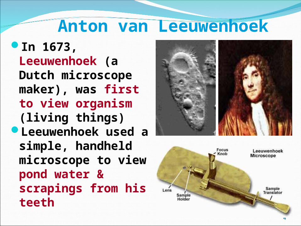
Anton van LeeuwenhoekIn 1673, Leeuwenhoek
(a Dutch microscope maker), was first to view organism (living things)
Leeuwenhoek used a simple, handheld microscope to view pond water & scrapings from his teeth

5
Beginning of the Cell Beginning of the Cell TheoryTheoryIn 1838, a German
botanist named Matthias Schleiden concluded that all plants were made of cells
Schleiden is a cofounder of the cell theory

6
Beginning of the Cell TheoryBeginning of the Cell Theory
In 1839, a German zoologist named Theodore Schwann concluded that all animals were made of cells
Schwann also cofounded the cell theory
7
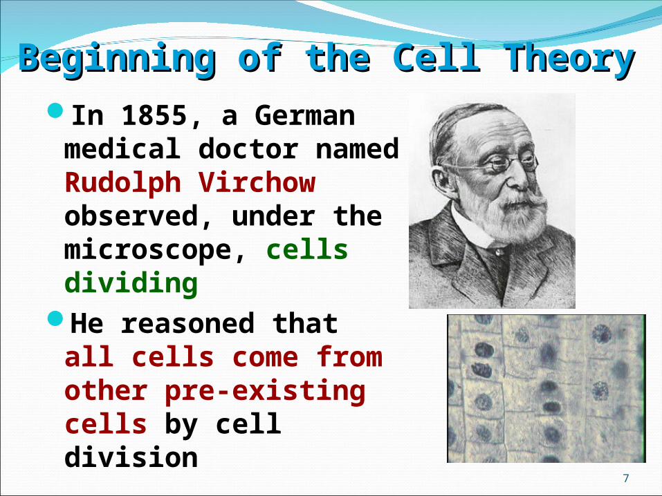
Beginning of the Cell TheoryBeginning of the Cell TheoryIn 1855, a German medical
doctor named Rudolph Virchow observed, under the microscope, cells dividing
He reasoned that all cells come from other pre-existing cells by cell division

Cell TheoryCell TheoryCellsCells are the basic living units of organization and function
All cells come from other cellscome from other cellsWork of Schleiden, Schwann, Schleiden, Schwann, and Virchowand Virchow contributed to this theory
Each cell is a microcosm of life

9
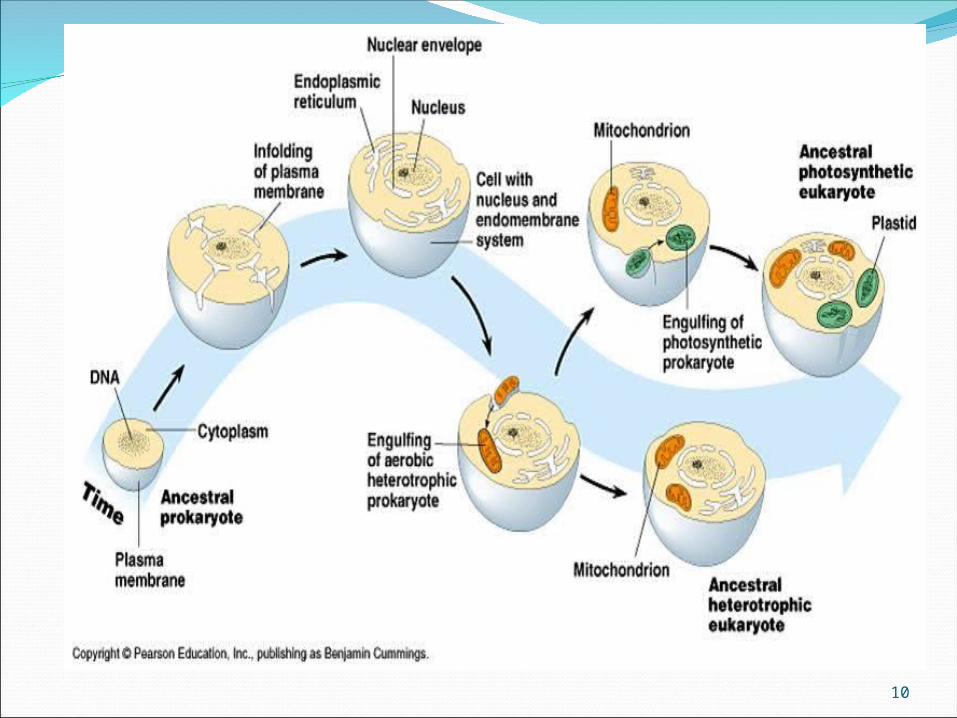
ENDOSYMBIOTIC THEORYENDOSYMBIOTIC THEORYIn 1970, American biologist, Lynn
Margulis, provided evidence that some organelles within cells were at one time free living cells themselves
Supporting evidence included organelles with their own DNA
Chloroplast and Mitochondria
10

11
Number of CellsNumber of CellsAlthough ALL living things are made of cells,
organisms may be:Unicellular – composed of one cellMulticellular- composed of many cells that may
organize into tissues, etc.

Biological Size and Cell Biological Size and Cell DiversityDiversity
12
13
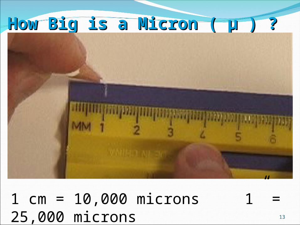
How Big is a Micron ( µ ) ?How Big is a Micron ( µ ) ?
1 cm = 10,000 microns 1” = 25,000 microns
14
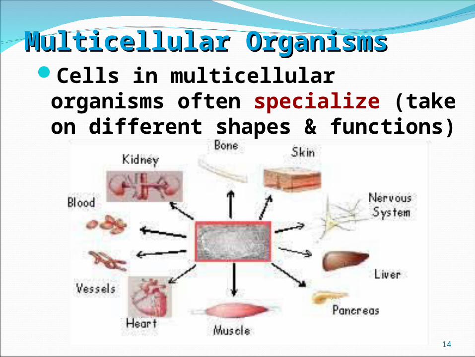
Multicellular OrganismsMulticellular OrganismsCells in multicellular organisms often
specialize (take on different shapes & functions)

Cell surface area-to-volume ratioPlasma membrane must be large
enough relative to cell volume to regulate passage of materials
Volume increases faster than surface area so cells must divide
Cell size and shape related to functionEx: Nerve cells, skin cells, sperm cells
15

Cell Surface Area-to-Volume Cell Surface Area-to-Volume RatioRatio
16
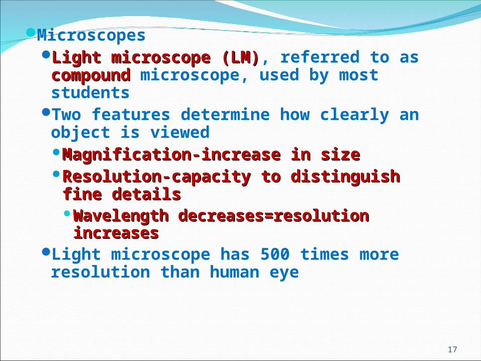
MicroscopesLight microscope (LM)Light microscope (LM), referred to as compoundcompound microscope, used by most students
Two features determine how clearly an object is viewedMagnification-increase in sizeMagnification-increase in sizeResolution-capacity to distinguish fine Resolution-capacity to distinguish fine detailsdetailsWavelength decreases=resolution Wavelength decreases=resolution increasesincreases
Light microscope has 500 times more resolution than human eye
17
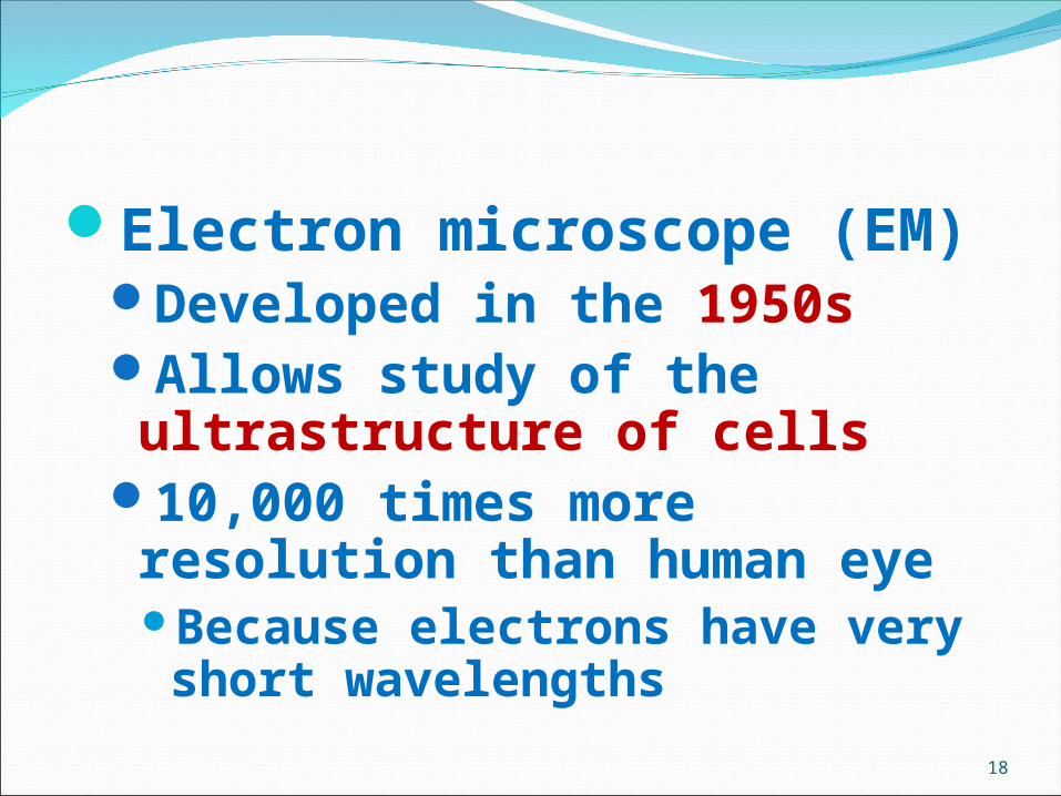
Electron microscope (EM)Developed in the 1950sAllows study of the ultrastructure of cells
10,000 times more resolution than human eyeBecause electrons have very short wavelengths
18

Types of electron microscopeTransmission electron microscope (TEM)TEM)
Used to view internal cell structuresinternal cell structuresSpecimen is embedded in plastic, cut thin, placed on a metal grid and shot with a beam of electrons..
Scanning electron microscope (SEM)SEM)Specimen is coated with gold or other Specimen is coated with gold or other metalmetal
Produces 3-D picture of cell surface3-D picture of cell surface
***Can’t***Can’t be used to view living cellsview living cells
19
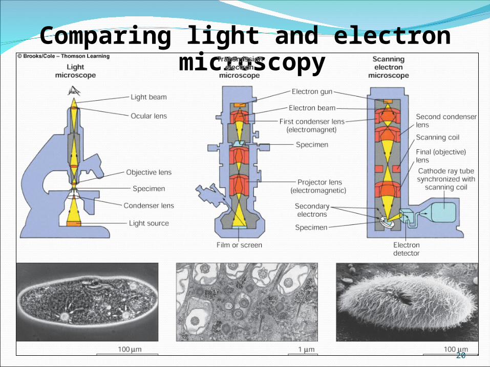
Comparing light and electron microscopy
20

Cell fractionationCell fractionationUsed to isolate & study of organelles
Cells broken apart and the resulting cell extract spun in a centrifuge
Centrifugal force separates extractPelletPellet – heavier cell organelles
Found at the bottom of the tubeSupernatant Supernatant – liquid poured off
lighter particles (dissolved molecules and ions)
21

Cell fractionation
22

Two Basic Types of CellsProkaryotes: Includes bacteria
(archaeabacteria and Eubacteria)
Eukaryotes: All other known organisms (protists, fungi, plants and animals)
23
24
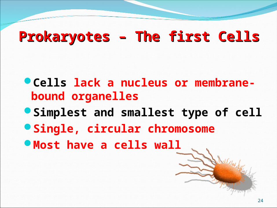
Prokaryotes – The first CellsProkaryotes – The first Cells
Cells lack a nucleus or membrane-bound organelles
Simplest and smallest type of cellSingle, circular chromosomeMost have a cells wall

25
ProkaryotesProkaryotesNucleoid region (center)
contains the DNASurrounded by cell
membrane & cell wall (peptidoglycan)
Contain ribosomes (no membrane) in their cytoplasm to make proteins
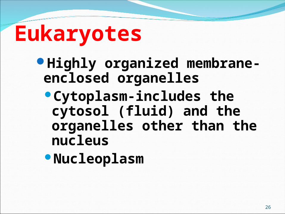
Highly organized membrane-enclosed organellesCytoplasm-includes the cytosol (fluid) and the organelles other than the nucleus
Nucleoplasm
Eukaryotes
26
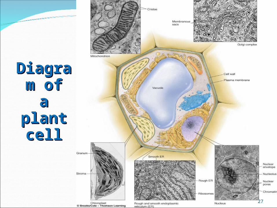
DiagraDiagram ofm of
a plant a plant cellcell
27

DiagraDiagram ofm ofan an
animal animal cellcell
28
29

The CellThe Cell All Living things are composed of All Living things are composed of cellscells All Cells have/contain the following:All Cells have/contain the following: Cell Membrane - Lipid BilayerCell Membrane - Lipid Bilayer - Separates inside from outside - Separates inside from outside
30

Functions of cell or plasma membranesMaintains homeostasisMaintains homeostasisDivides cell into compartmentsDivides cell into compartments,
allowing for specialized activitiesInteracting membranes form
endomembrane systemendomembrane systemVesiclesVesicles transport materials between
compartments (ER Golgi, Golgi plasma membrane…)
31

32
Cell or Plasma MembraneCell or Plasma Membrane
Outsideof cell
Insideof cell(cytoplasm)
Cellmembrane
Proteins
Proteinchannel Lipid bilayer
Carbohydratechains
Composed of double layer of phospholipids and proteins
Surrounds outside of ALL cellsControls what enters or leaves the cellLiving layer

33
The Cell Membrane is Fluid
Molecules in cell membranes are constantly moving and changing

34
Endomembrane System
Includes nuclear membrane connected to ER connected to cell membrane (transport)

CytoplasmCytoplasmCytoplasmCytoplasm - everything but DNA/Nucleus - everything but DNA/Nucleus
35

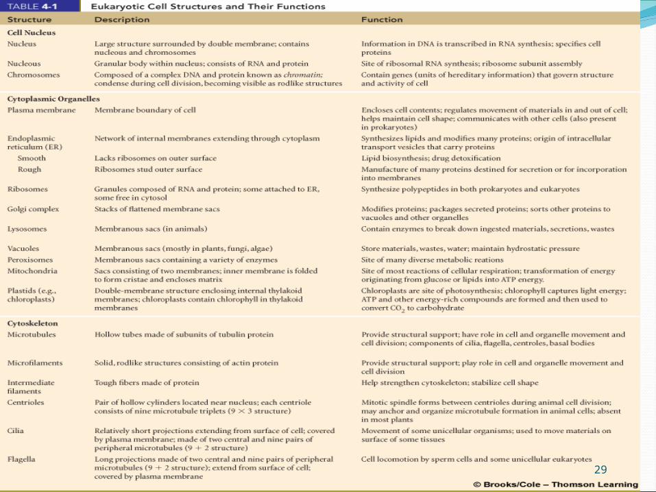
Major Organelles of the Eukaryotic CellMajor Organelles of the Eukaryotic Cell
The Nucleus “eukaryotic” means “true nucleus”
Contains & protects the cell’s DNA
Helps coordinate the division of cells
Surrounded by a Nuclear EnvelopeEnvelope is double layered with an
Inner & Outer membraneHas perforations called Nuclear
Pores which allow large molecules to pass in/out of the nucleus
Contains a NucleolusRibosomes are made in this
regionContains DNA packaged in
structures called chromosomes ChromosomChromosomeses
36
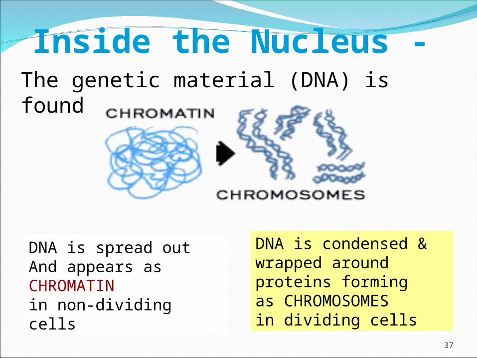
37
Inside the Nucleus -The genetic material (DNA) is found
DNA is spread out And appears as CHROMATINin non-dividing cells
DNA is condensed & wrapped around proteins forming as CHROMOSOMES in dividing cells

The cellThe cellnucleusnucleus
38
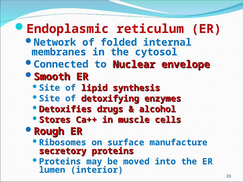
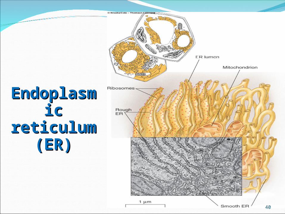
Endoplasmic reticulum (ER)Network of folded internal membranes in the
cytosolConnected to Nuclear envelopeNuclear envelopeSmooth ER Smooth ER
Site of lipid synthesislipid synthesisSite of detoxifying enzymesdetoxifying enzymesDetoxifies drugs & alcoholDetoxifies drugs & alcoholStores Ca++ in muscle cellsStores Ca++ in muscle cells
Rough ERRough ERRibosomes on surface manufacture secretory secretory
proteinsproteinsProteins may be moved into the ER lumen (interior)
39
EndoplasEndoplasmicmic
reticulum reticulum (ER)(ER)
40
41
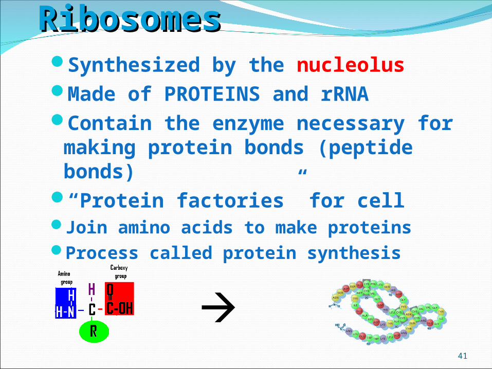
RibosomesRibosomesSynthesized by the nucleolusMade of PROTEINS and rRNAContain the enzyme necessary for making
protein bonds (peptide bonds)“Protein factories” for cellJoin amino acids to make proteinsProcess called protein synthesis

Golgi complexCisternae (sacs)Cisternae (sacs) that process, sort, and
modify proteins In animal cellsanimal cells, Golgi complex also
manufactures lysosomes lysosomes GlycoproteinsGlycoproteins
Transported to the ciscis face (receiving side)
Golgi modifies modifies carbohydrates and lipids and packages into vesiclespackages into vesicles that pinch off the trans face pinch off the trans face (shipping side)
42
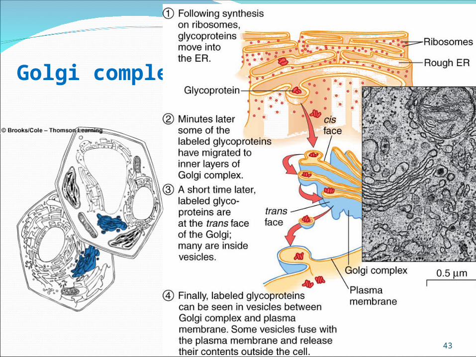
Golgi complex
43

44
Golgi AnimationGolgi Animation
Materials are transported from Rough ER to Golgi to the cell membrane by VESICLES

LysosomeLysosomess
Small Vessicles which serve to digest particles and clean-up cells
Contain Lysozyme – a powerful digestive enzyme
Digests food particles
Destroys worn-out organelles and invading bacteria
Self-Destructs worn-out cells
45

PeroxisomesInvolved in lipid metabolism and
detoxificationContain enzymes (catalase) that produce
and degrade hydrogen peroxide H2O2 H2O + O2
46

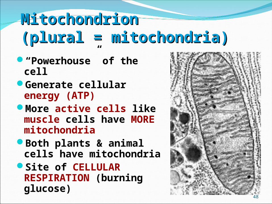
Mitochondria Mitochondria Sites of aerobic respirationaerobic respirationOrganelles enclosed by a double membraneHas its own genome own genomePlays important role in apoptosis apoptosis
(programmed cell death)(programmed cell death)CristaeCristae (internal folds) and matrix matrix
(innermost space) contain enzymes for aerobic respirationNutrients broken down and energy packaged
in ATPATPCarbon dioxide Carbon dioxide and water water by-products
47
48
MitochondrionMitochondrion(plural = mitochondria)(plural = mitochondria)
“Powerhouse” of the cellGenerate cellular energy (ATP)More active cells like muscle
cells have MORE mitochondriaBoth plants & animal cells have
mitochondriaSite of CELLULAR RESPIRATION
(burning glucose)

Mitochondria
49

50
MITOCHONDRIASurrounded by a DOUBLE
membrane
Folded inner membrane called CRISTAE (increases surface areafor more chemical Reactions)
Has its own DNA
Interior called MATRIX
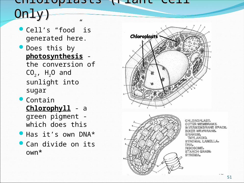
Chloroplasts (Plant Cell Only)Chloroplasts (Plant Cell Only)Cell’s “food” is
generated here.Does this by
photosynthesis - the conversion of CO2, H2O and sunlight into sugar
Contain Chlorophyll - a green pigment - which does this
Has it’s own DNA*Can divide on its
own*n*
51

Chloroplasts Plastids that carry out photosynthesisInner membrane of chloroplast encloses the
stroma (gel-like liquid)Contains stacks of interconnected sacs
called thylakoidsStack of thylakoids called granaDuring photosynthesis, chlorophyll traps
light energy (sunlight)Energy converted to chemical energy in ATP
52

Chloroplast
53

Cellular respiration and photosynthesis
54
TheCytoskelet
on
55
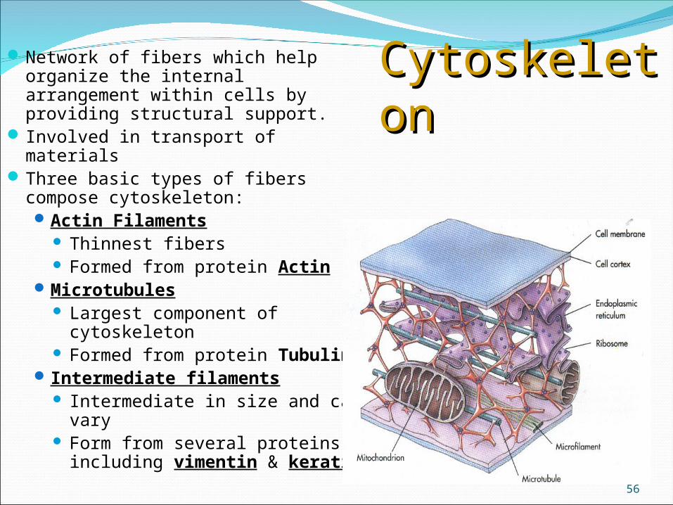
CytoskeletoCytoskeletonn
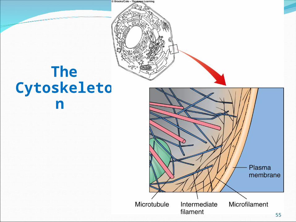
Network of fibers which help organize the internal arrangement within cells by providing structural support.
Involved in transport of materialsThree basic types of fibers
compose cytoskeleton:Actin Filaments
Thinnest fibers Formed from protein Actin
Microtubules Largest component of
cytoskeleton Formed from protein Tubulin
Intermediate filaments Intermediate in size and can
vary Form from several proteins
including vimentin & keratin 56
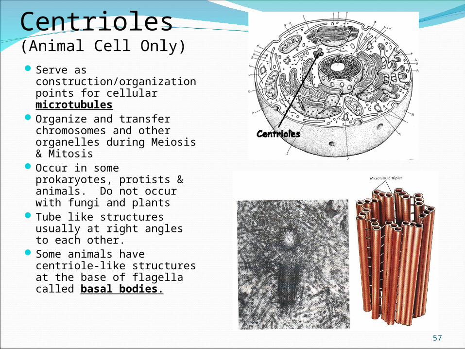
Centrioles(Animal Cell Only)Serve as
construction/organization points for cellular microtubules
Organize and transfer chromosomes and other organelles during Meiosis & Mitosis
Occur in some prokaryotes, protists & animals. Do not occur with fungi and plants
Tube like structures usually at right angles to each other.
Some animals have centriole-like structures at the base of flagella called basal bodies.
57

Cilia and flagella Cilia and flagella Thin, movable structures that project
from cell surfaceFunction in movementMicrotublesMicrotubles anchored in cell by basal basal
bodybody
58

59
Cilia & FlagellaCilia & Flagella
Made of protein tubes called microtubules
Microtubules arranged (9 + 2 arrangement)
Function in moving cells, in moving fluids, or in small particles across the cell surface

Glycocalyx (cell coat) Cell coat formed by polysaccarides extending
from plasma membraneProtects the cell Used in cell recognition, adhesion or
communicationMany animal cells are also surrounded by an
extracellular matrix (ECM)Gel of carbohydrates and fibrous proteins
Most bacteria, fungi, and plant cell walls made of carbohydrates
60

Extracellular matrixIntegrins- membrane receptors important in cell movementFibronectins- help organize the matrix and help cells attach
61
Plantcell walls
62
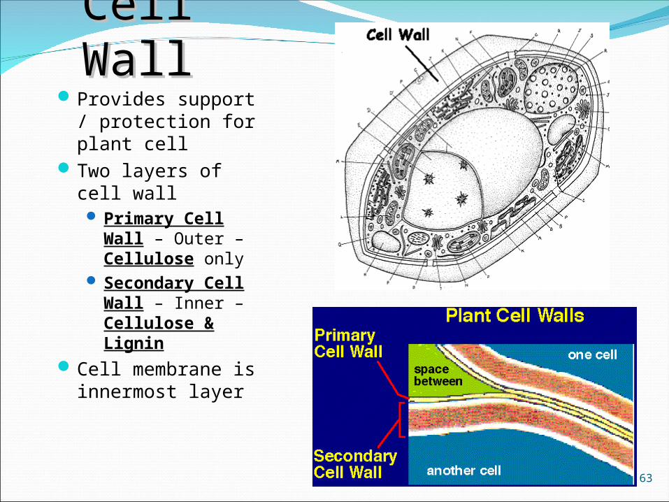
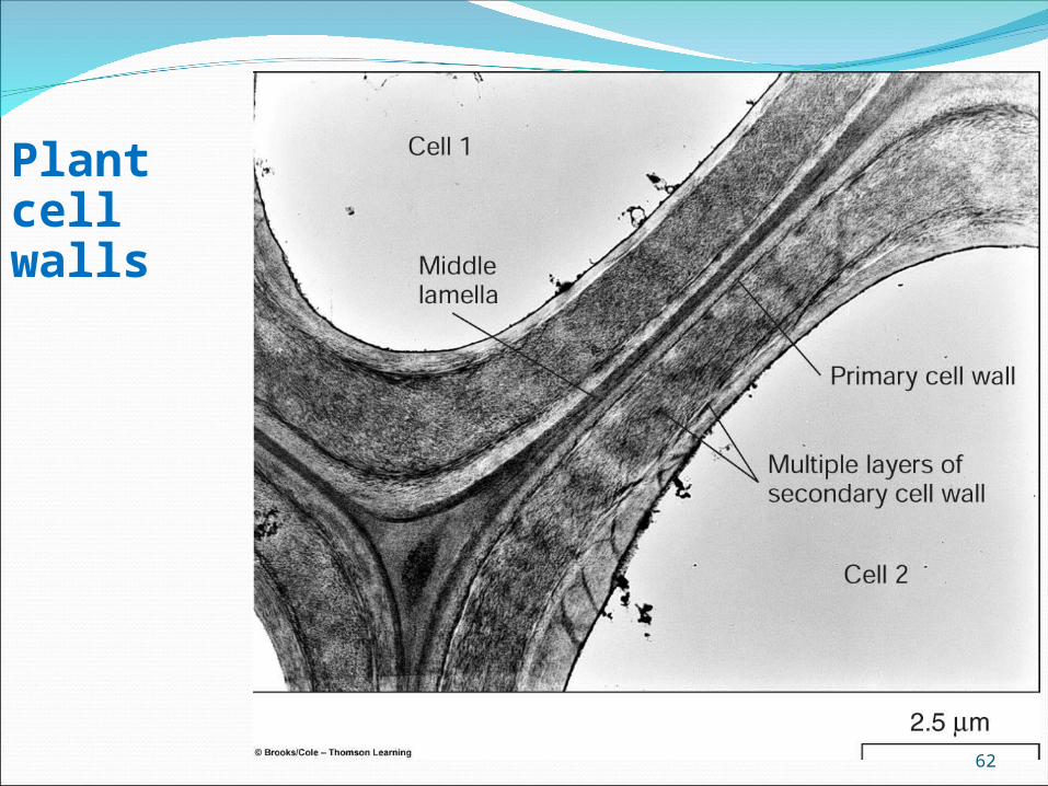
Cell WallCell WallProvides support /
protection for plant cell
Two layers of cell wall Primary Cell
Wall – Outer – Cellulose only
Secondary Cell Wall – Inner – Cellulose & Lignin
Cell membrane is innermost layer
63

64
• Nonliving layer• Found in plants, fungi,
& bacteria• Made of cellulose in
plants• Made of peptidoglycan
in bacteria• Made of chitin in Fungi
Cell wallCell WallCell Wall
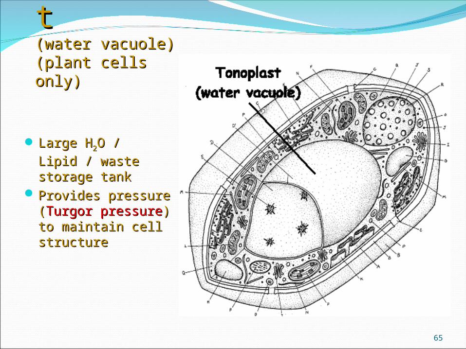
TonoplastTonoplast(water vacuole)(water vacuole)(plant cells only)(plant cells only)
Large HLarge H22O / Lipid / O / Lipid /
waste storage tankwaste storage tank Provides pressure Provides pressure
((Turgor pressureTurgor pressure) to ) to maintain cell maintain cell structurestructure
65

66
Plant Cell OrganellesPlant Cell Organelles

67