1 abd & chest 2 rt 124 – spring image review pt 2
TRANSCRIPT
1
ABD & CHEST 2
Rt 124 – Spring
Image Review pt 2
2
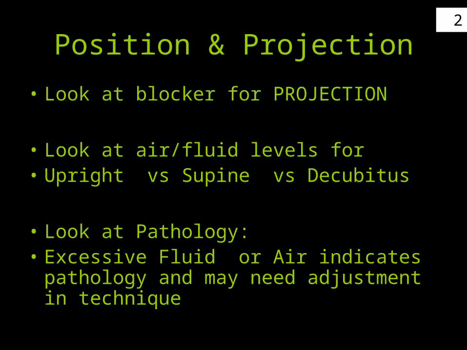
Position & Projection
• Look at blocker for PROJECTION
• Look at air/fluid levels for• Upright vs Supine vs Decubitus
• Look at Pathology:• Excessive Fluid or Air indicates pathology
and may need adjustment in technique

3Projection ?
AP
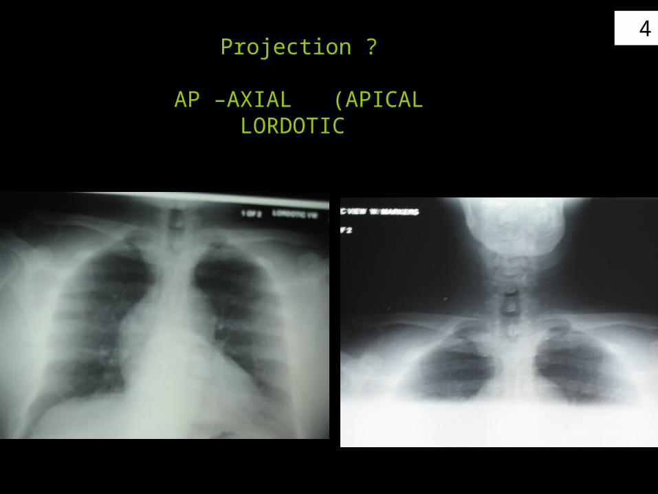
4Projection ?
AP –AXIAL (APICAL LORDOTIC
5
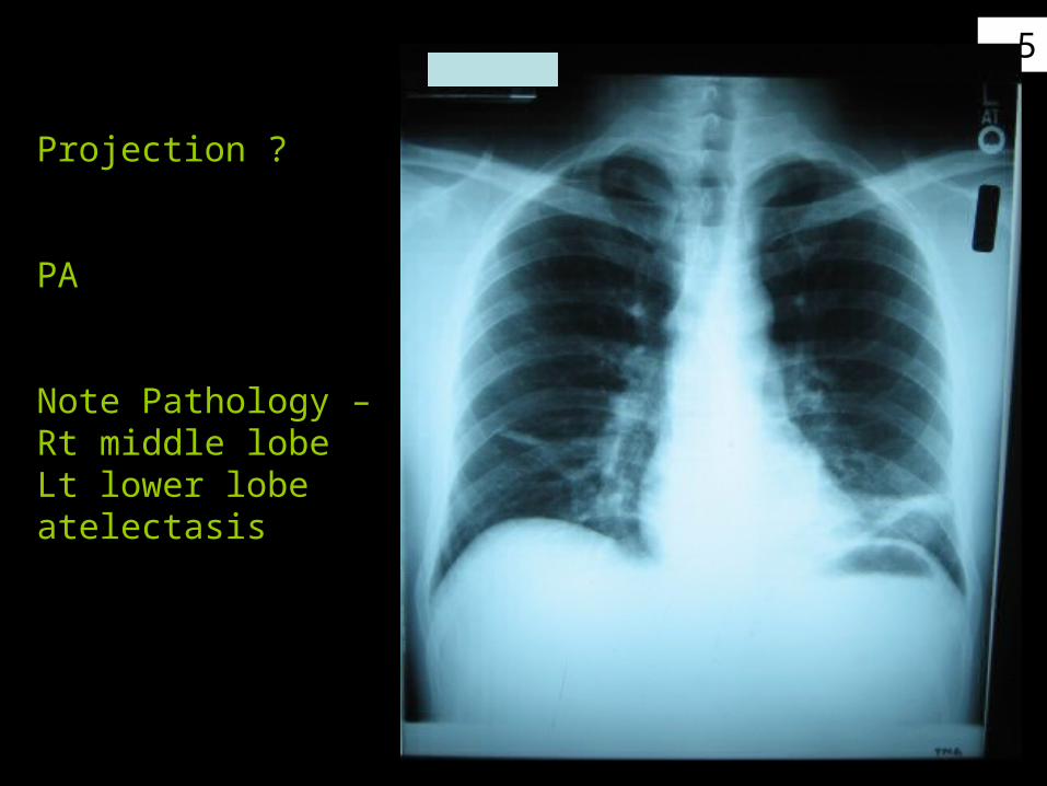
Projection ?
PA
Note Pathology –Rt middle lobeLt lower lobeatelectasis
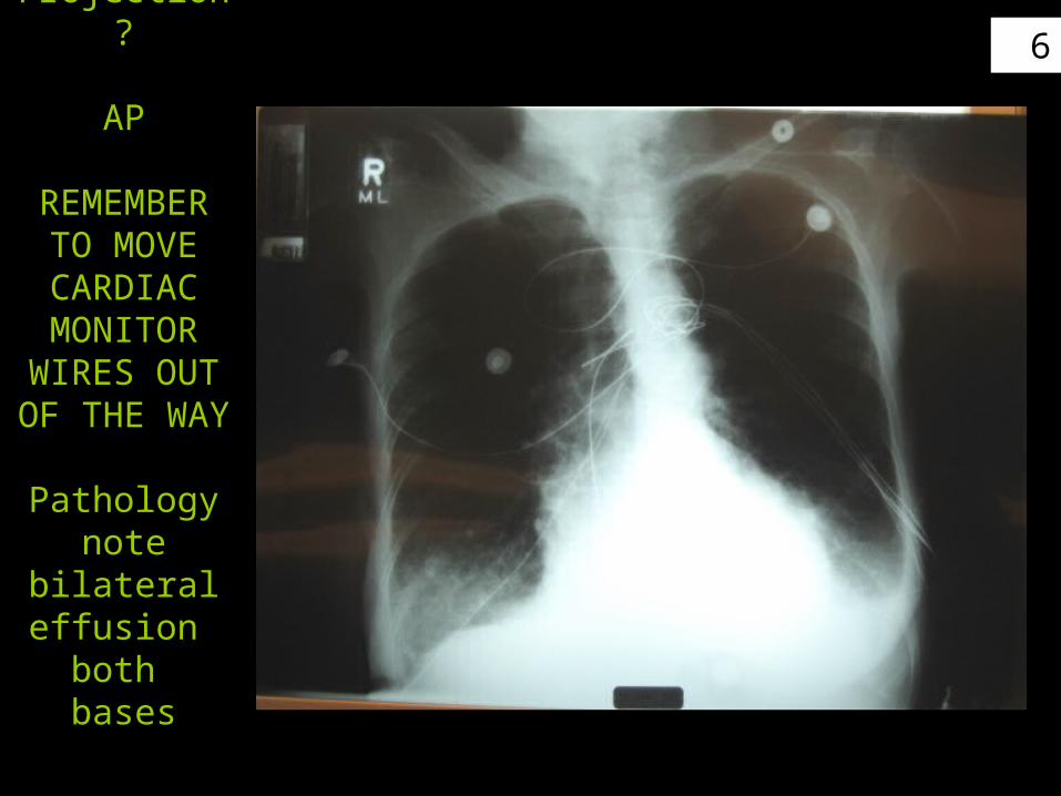
6Projection ?
AP
REMEMBER TO MOVE CARDIACMONITOR
WIRES OUT OF THE WAY
Pathologynote bilateral
effusion both bases
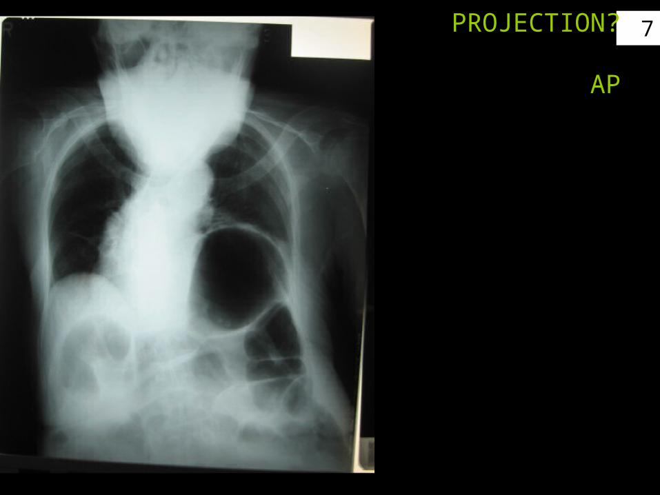
7PROJECTION?
AP
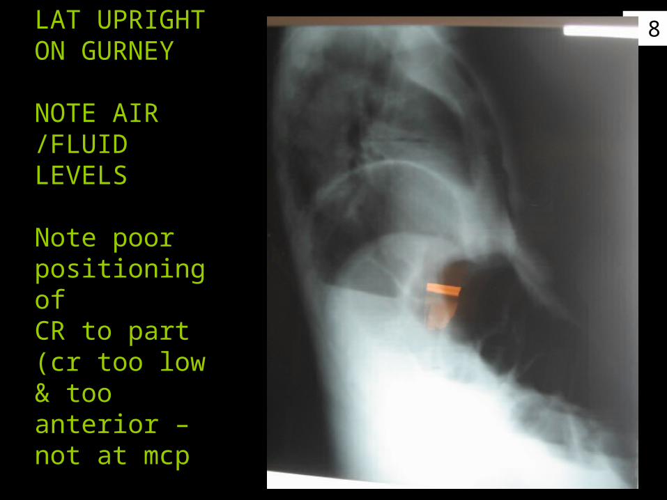
8
LAT UPRIGHTON GURNEY
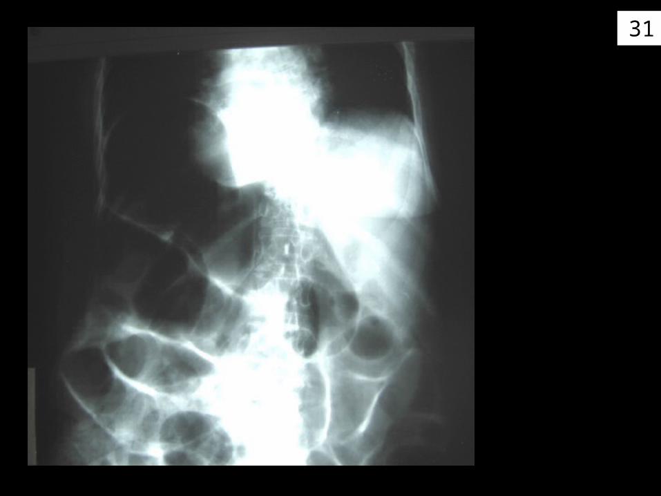
NOTE AIR /FLUID LEVELS
Note poor positioning ofCR to part(cr too low & too anterior – not at mcp
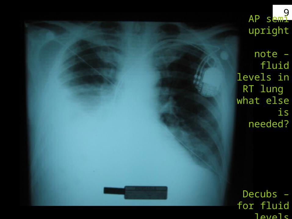
9
Position ?Projection?
AP semi upright
note – fluid
levels in RT lung
what else is needed?
Decubs –for fluid levels
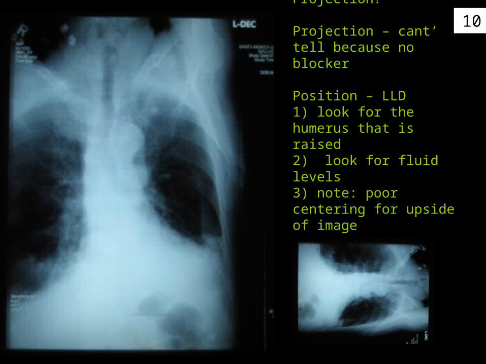
10Position / Projection?
Projection – cant’ tell because no blocker
Position – LLD1) look for the humerus that is raised2) look for fluid levels3) note: poor centering for upside of image

11
Position / Projection?
Projection – AP blocker lower RTPosition – RLD 1) look for the humerus that is raised2) look for fluid levels3) poor marker placement – label of image
look for fluid levels
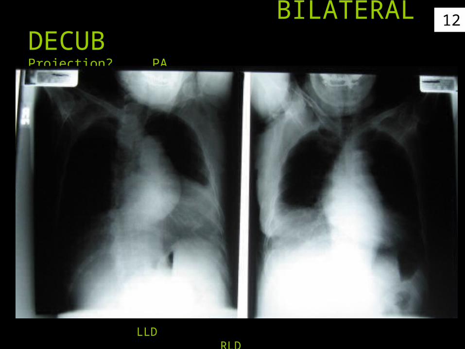
12 BILATERAL DECUBProjection? PA AP
LLD RLD
13
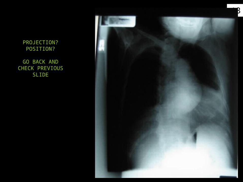
PROJECTION?POSITION?
GO BACK AND CHECK PREVIOUS
SLIDE

14
PROJECTION?POSITION?
GO BACK AND CHECK PREVIOUS
SLIDE

15
ABDOMEN -
SUPINE – UPRIGHT - LLD

16
KUB
WHAT IS THE CRITIQUE TO
JUDGE PROPER
TECHNIQUE?

17Upper abd - should center higher to include more diaphram
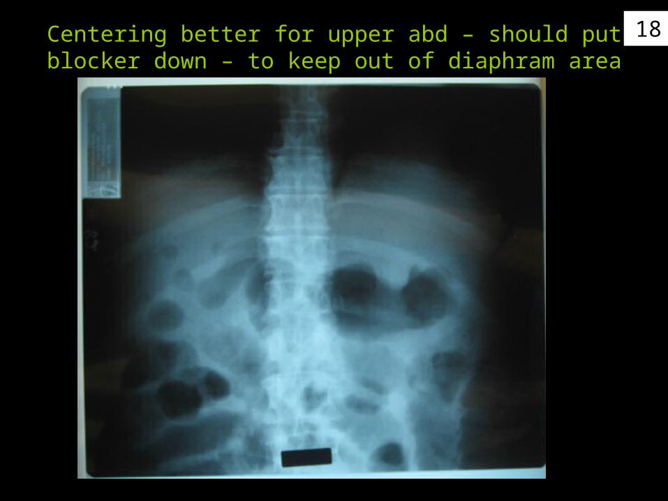
18Centering better for upper abd – should put blocker down – to keep out of diaphram area
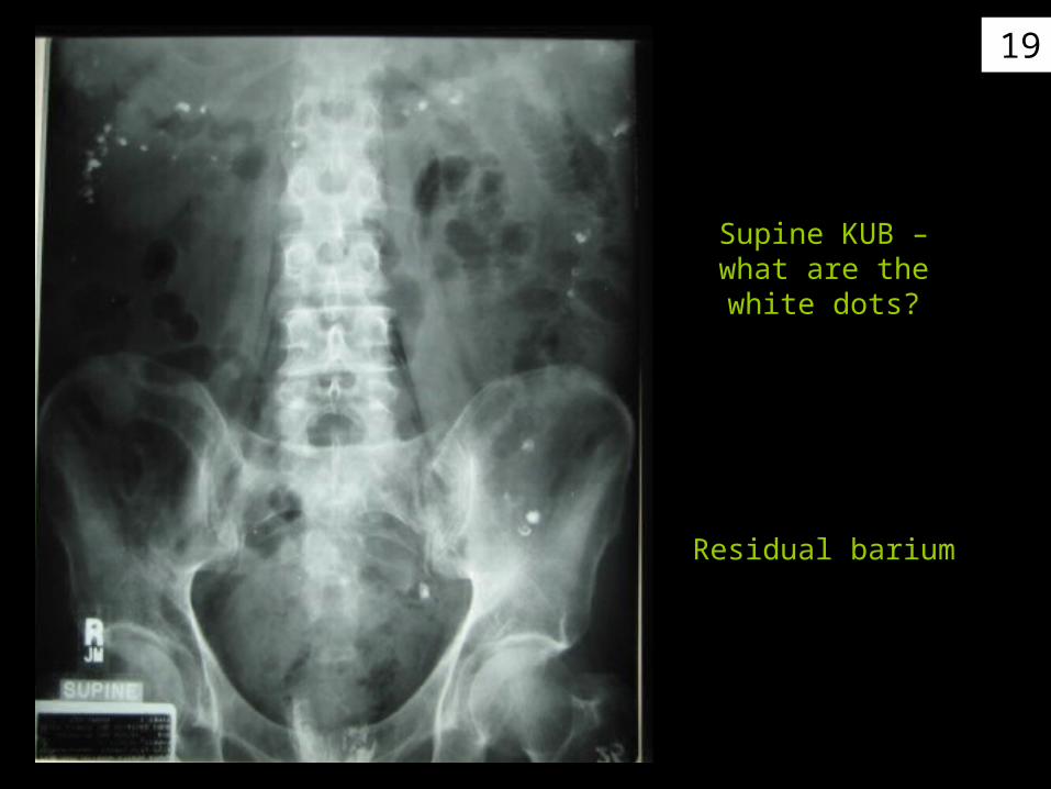
19
Supine KUB –what are the white
dots?
Residual barium
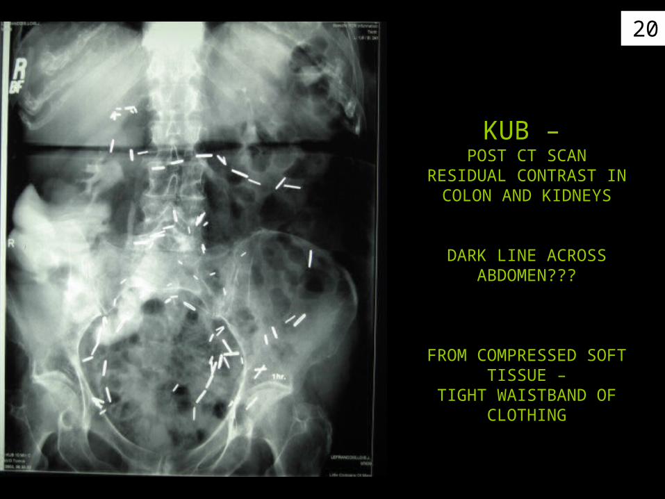
20
KUB – POST CT SCAN
RESIDUAL CONTRAST IN COLON AND KIDNEYS
DARK LINE ACROSS ABDOMEN???
FROM COMPRESSED SOFT TISSUE –
TIGHT WAISTBAND OF CLOTHING

21Case example of
SUPINE – upper & KUB Upright
(repeated – diaphram clipped)
Should have collimated to upper abd – not exposed lower abd twice

22
KUB
FLAT PLATESUPINE ABD
INCLUDESENTIRE ABD
(TAKEN AT 48” SID)
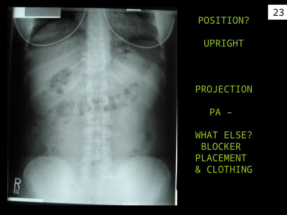
23POSITION?
UPRIGHT
PROJECTION
PA –
WHAT ELSE?BLOCKER
PLACEMENT & CLOTHING

24KUBSUPINE
25
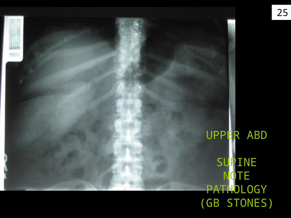
UPPER ABD
SUPINENOTE
PATHOLOGY (GB STONES)

26
UPRIGHT ABD
CRITIQUEWHAT IS THE DARK
LINE IN THE CENTER
27
PATHOLOGY&
Positioning

28Obstruction
lg bowel

29
Example:may need4 films inquadrant
to include allof abd
structures(obstruction)

30Free air in the abdomen
31
32
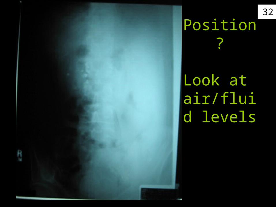
Position?
Look at air/fluid levels
33
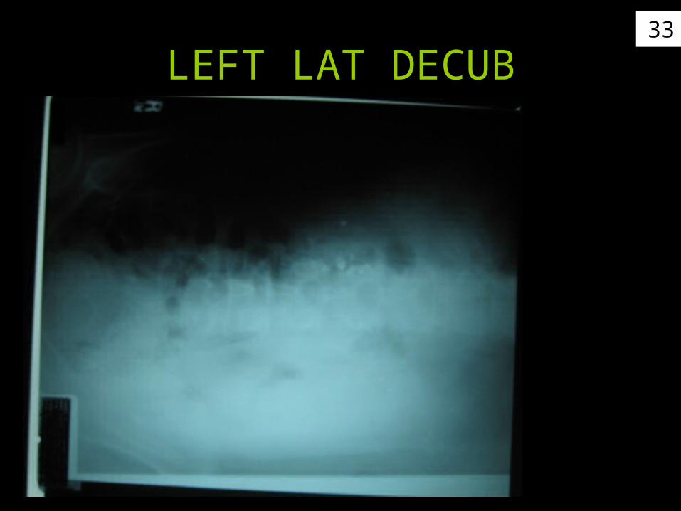
LEFT LAT DECUB
34
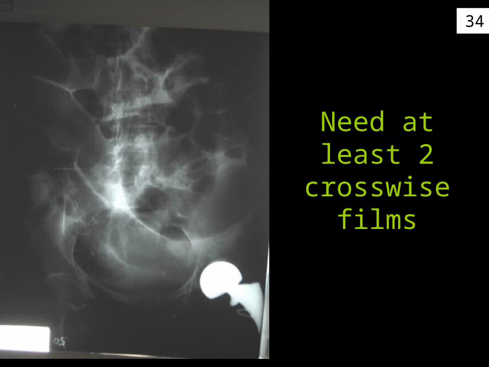
Need at least 2 crosswise
films
35
CRITIQUE IMAGESFOR POSITIONING
COLLIMATION &CENTRAL RAY PLACEMENT

36Critique: If taken for AP chest –
CR is < too cephalic – moving clavicles above apex
37
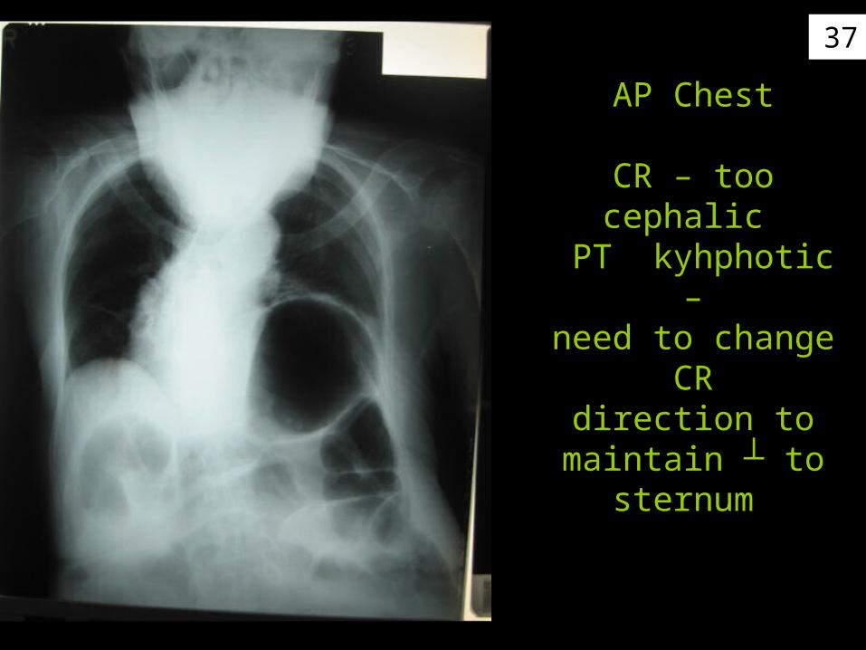
AP Chest
CR – too cephalic PT kyhphotic –
need to change CRdirection to
maintain ┴ to sternum
38
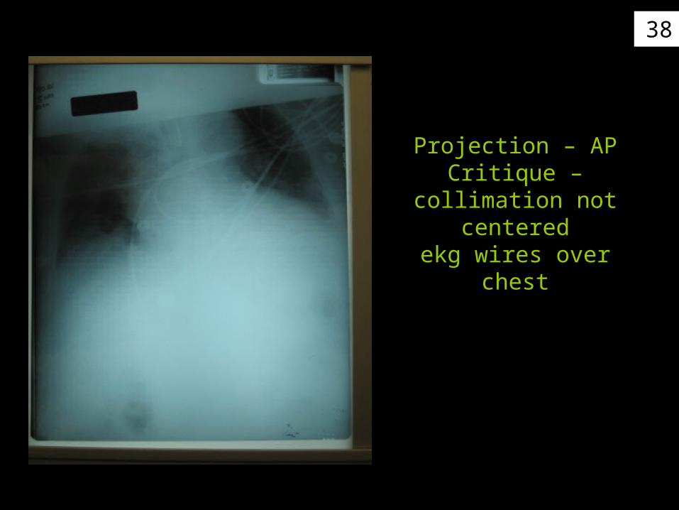
Projection – APCritique –
collimation not centeredekg wires over chest
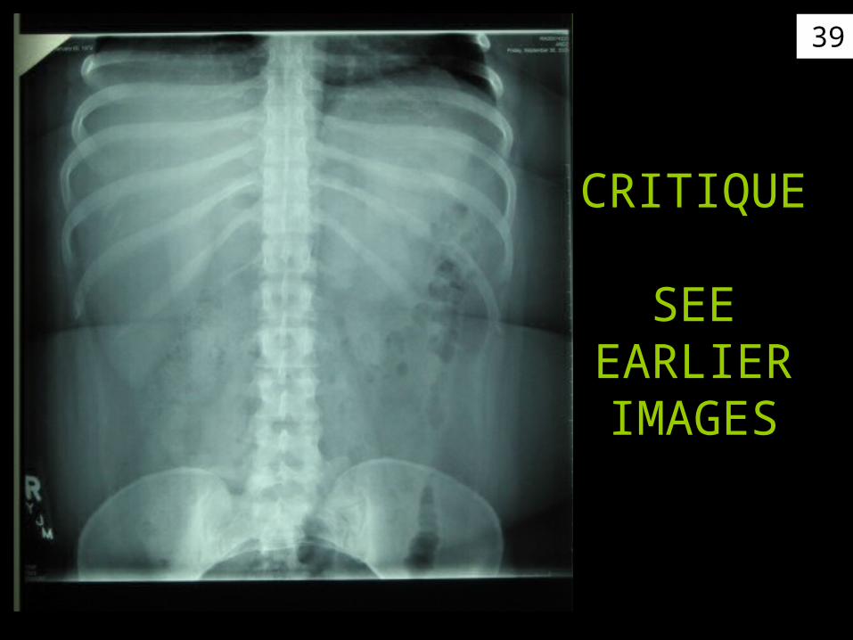
39
CRITIQUE
SEE EARLIER IMAGES

40
Lat gurney chest prop arms up with
sponges get ST of arms off of
chest
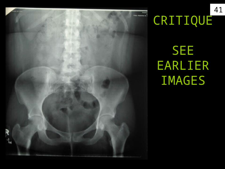
41
CRITIQUE
SEE EARLIER IMAGES
42
Also review images on first presentation
Written test on Tues
Lab on Thursday

43
More pathology& positioning
We will cover in more detailin GI section
44
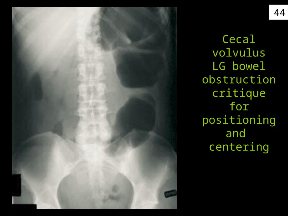
Cecal volvulusLG bowel
obstructioncritique forpositioning
and centering
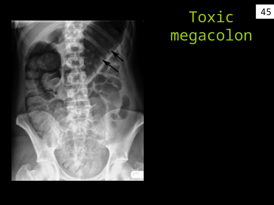
45Toxicmegacolon

46
Projection?
Postion?
PA – according to blocker
Supine – no air fluid levels

47
Projection?
AP

48
Projection?
PA

49
What is thisstep ladder
sign indicate for pathology?
Obstructionsee air-fluid
levels Position?Upright!

50Small bowel obstruction-remember to
include all areas of the abdomen
what could have improved this
image?
2 cross wise14 x 17

51
Critique for positioning& projection
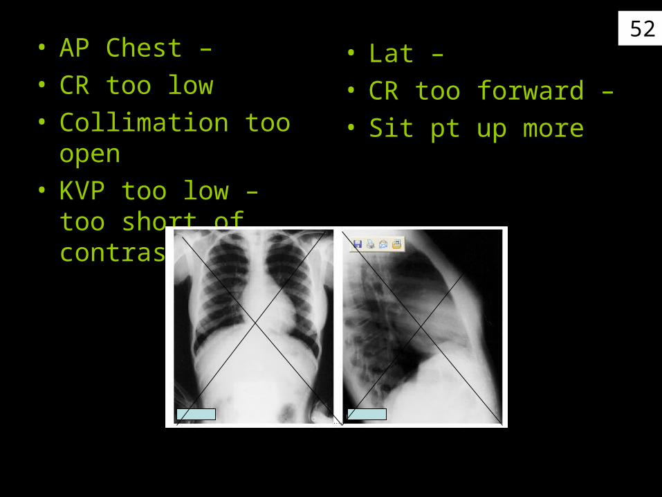
52• AP Chest –• CR too low• Collimation too open• KVP too low – too
short of contrast
• Lat –• CR too forward –• Sit pt up more
53
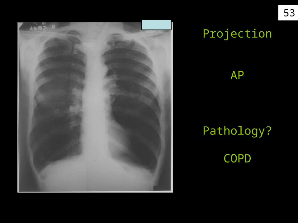
Projection
AP
Pathology?
COPD