1 essentials of human anatomy essentials of human anatomy dr fadel naim ass. prof. faculty of...
TRANSCRIPT

1
Essentials of Human AnatomyEssentials of Human Anatomy
Dr Fadel NaimAss. Prof. Faculty of Medicine
IUG
Blood Vessels

Blood Vessels
• An efficient mode of transport for oxygen, nutrients, and waste products to and from body tissues.
• Heart is the mechanical pump that propels the blood through the vessels.
• Heart and blood vessels form a closed-loop system.• Blood is continuously pumped to and from the
tissues.• Are not rigid and immobile.• Can pulsate and change shape in accordance with
the body’s needs.

Blood Vessels
• ArteriesArteries• Carry blood away from ventricles of heart
• ArteriolesArterioles• Receive blood from arteries• Carry blood to capillaries
• CapillariesCapillaries• Sites of exchange of substances between blood and body cells
• VenulesVenules• Receive blood from capillaries
• VeinsVeins• Carry blood toward atrium of heart

Three Main Classes of Blood Vessels
• Arteries become progressively smaller as they divide and get further from the heart.
• Veins become progressively larger as they merge and get closer to the heart.
• Anastomosis: Site where two or more vessels merge to supply the same body region. – arterial anastomoses: alternate route – Veins tend to form many more anastomoses
than do arteries.
Three Main Classes of Blood Vessels
• End arteries– Arteries that do not form anastomoses– Only one route– E.g.: renal artery, splenic artery
• Functional end arteries
– Have small anastomoses
– E.g.: coronary arteries

Blood Vessel Tunics• Tunica Intima, or Tunica Interna
– innermost layer– composed of:
• an endothelium (simple squamous epithelium)• subendothelial layer (areolar CT)
• Tunica Media– middle layer of the vessel wall – composed of:
• circularly arranged smooth muscle cells – Sympathetic innervation:
• Increase: vasoconstriction (narrowing of the blood vessel lumen)
• Decrease: vasodilation (widening of the blood vessel lumen)

Blood Vessel TunicsTunica Externa, or Tunica Adventitia
– outermost layer– composed of:
• areolar connective tissue that contains elastic and collagen fibers– helps anchor the vessel to other tissues – Term adventitia is used to specify outer layer in blood vessels
that are buried in CT• Vasa vasorum : blood vessels that supply large blood
vessels– In the externa
• Arteries vs Veins: – Media largest in arteries, externa largest in veins– Lumen is smallest in arteries– Artery wall have more elastic and collagen fibers
• Capillaries: only the Interna

Arteries
• In the systemic circulation, carry oxygenated blood to the body tissues.
• Pulmonary arteries carry deoxygenated blood to the lungs.
• Three basic types of arteries: – elastic arteries, muscular arteries, and arterioles– as an artery’s diameter decreases
• corresponding decrease in the amount of elastic fibers• relative increase in the amount of smooth muscle
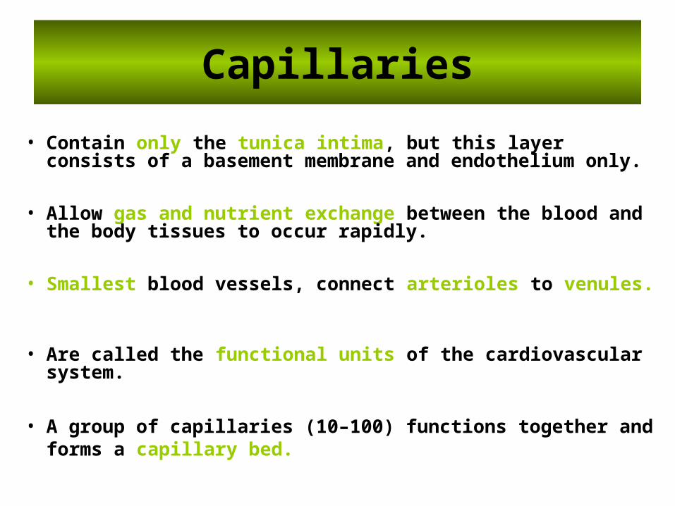
Capillaries
• Contain only the tunica intima, but this layer consists of a basement membrane and endothelium only.
• Allow gas and nutrient exchange between the blood and the body tissues to occur rapidly.
• Smallest blood vessels, connect arterioles to venules.
• Are called the functional units of the cardiovascular system.
• A group of capillaries (10–100) functions together and forms a capillary bed.
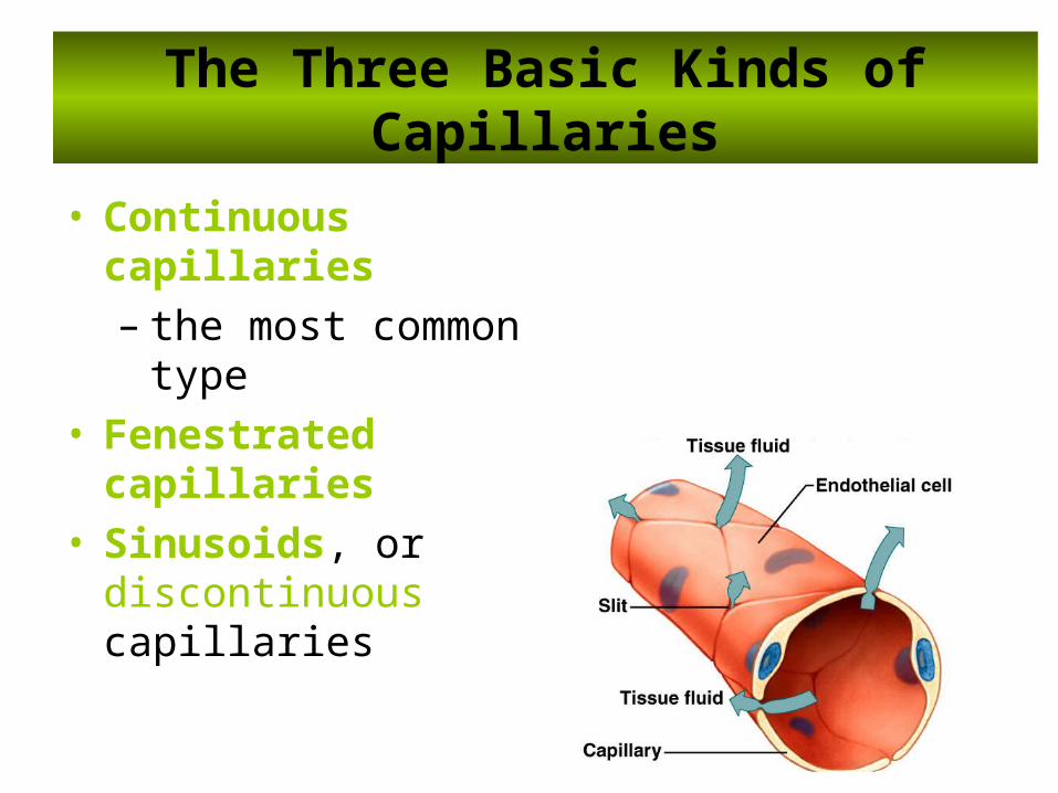
The Three Basic Kinds of Capillaries
• Continuous capillaries – the most common type
• Fenestrated capillaries• Sinusoids, or discontinuous
capillaries
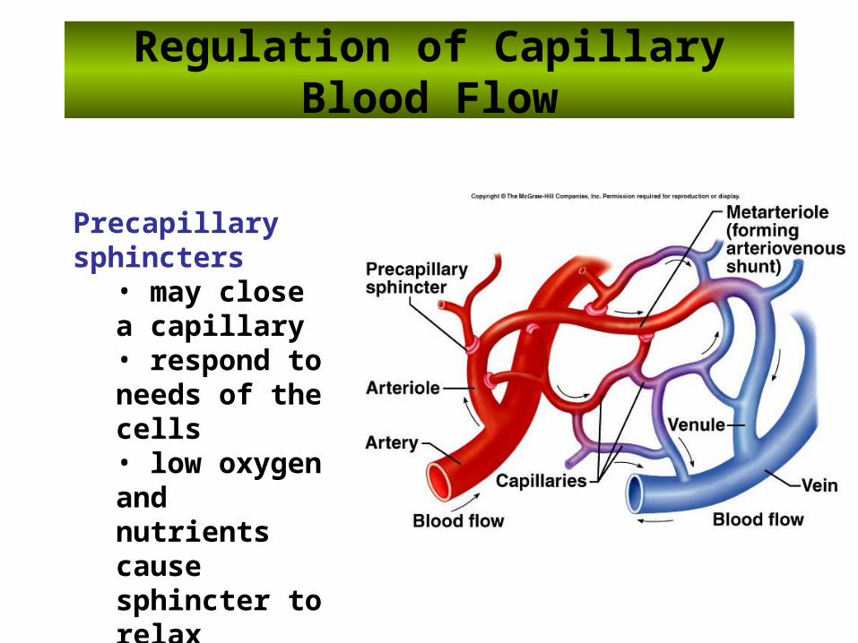
Regulation of Capillary Blood Flow
Precapillary sphincters • may close a capillary• respond to needs of the cells• low oxygen and nutrients cause sphincter to relax
Veins
• Drain capillaries and return the blood to the heart.
• Walls are relatively thin and the vein lumen is larger.
• Systemic veins carry deoxygenated blood to the right atrium of the heart, while pulmonary veins carry oxygenated blood to the left atrium of the heart.
• Blood pressure is substantially reduced by the time blood reaches the veins.
• Hold about 60% of the body’s blood at rest. • Veins function as blood reservoirs.

From Venules to Veins
• Venules merge to form veins.
• Venule becomes a “vein” when its diameter is greater than 100 micrometers.
• Blood pressure in veins is too low to overcome the forces of gravity.
• To prevent blood from pooling in the limbs, most veins contain one-way numerous valves to prevent blood backflow in the veins.
• As blood flows superiorly in the limbs, the valves close to prevent backflow.
• Numerous valves along its length to assist in moving blood back to the heart.

From Venules to Veins
Many deep veins pass between skeletal muscle groups.
• As the skeletal muscles contract, veins are squeezed to help pump the blood toward the heart.
• This process is called the skeletal muscle pump.

Venous flow occurs by:
1. muscle contraction
2. respiratory pump
3. valve assistance
4. Ventricular relaxation

Arteriole
• smallest arterioles only have a few smooth muscle fibers• capillaries lack muscle fibers

Pulse
• alternate expanding and recoiling of the arterial wall that can be felt
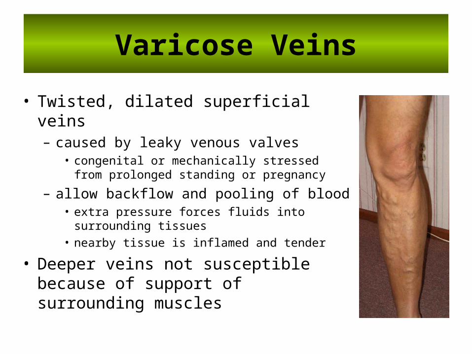
Varicose Veins
• Twisted, dilated superficial veins– caused by leaky venous valves
• congenital or mechanically stressed from prolonged standing or pregnancy
– allow backflow and pooling of blood• extra pressure forces fluids into surrounding
tissues• nearby tissue is inflamed and tender
• Deeper veins not susceptible because of support of surrounding muscles
GREAT SAPHENOUS VEIN CUT DOWN
• Usually performed at the ankle• Disadvantage that phlebitis is a potential complication• Also at the groin in the femoral triangle,
– Phlebitis is relatively rare– Larger diameter of the vein – Use of large-diameter catheters – Rapid infusion of large volumes of fluids.

The Great Saphenous Vein In Coronary Bypass Surgery
• In occlusive coronary disease caused by atherosclerosis, the diseased arterial segment can be bypassed by inserting a graft consisting of a portion of the great saphenous vein.
• The venous segment is reversed so that its valves do not obstruct the arterial flow.
• The great saphenous vein can also be used to bypass obstructions of the brachial or femoral arteries

Venipuncture of the Upper Limb
• Because of the prominence and accessibility of the superficial veins of the upper limb, they are commonly used for venipuncture
• These veins may be embedded with the subcutaneous tissue (fat), making them difficult to see
• By applying a tourniquetapplying a tourniquet to the arm, the venous return is occluded and the veins distend and are usually visible and/or palpable.

Venipuncture of the Upper Limb
• The median cubital veinThe median cubital vein is commonly used for venipuncture for:– Drawing blood – Inserting a catheter for right cardiac catheterization
• The dorsal venous network and the cephalic and basilic veins arising from it are commonly used for intravenous feedingintravenous feeding

FEMORAL ARTERY CATHETERIZATION
• A long, fine catheter can be inserted into the femoral artery as it descends through the femoral triangle.
• The catheter is guided under fluoroscopic view along the external and common iliac arteries into the aorta.
• The catheter can then be passed into the inferior mesenteric, superior mesenteric, celiac arteries, or renal arteries.
• Contrast medium can then be injected into the artery under examination and a permanent record obtained by taking a radiograph.
• Pressure records can also be obtained by guiding the catheter through the aortic valve into the left ventricle.

Measuring Blood Pressure
• Arterial blood pressure measurement using sphygmomanometersphygmomanometer.
• A cuff is placed around the arm and inflated with air until it compresses the brachial artery against the humerus and compresses the brachial artery against the humerus and occludes itoccludes it.
• A stethoscopeA stethoscope is placed over the artery in the cubital over the artery in the cubital fossafossa, the pressure in the cuff is gradually released
• The examiner detects the sound of blood beginning to spurt through the artery.
• The first audible spurt indicates systolic blood pressure.The first audible spurt indicates systolic blood pressure. • As the pressure is completely released, the point at which the
pulse can no longer be heard is the diastolic blaod pulse can no longer be heard is the diastolic blaod pressure. pressure.
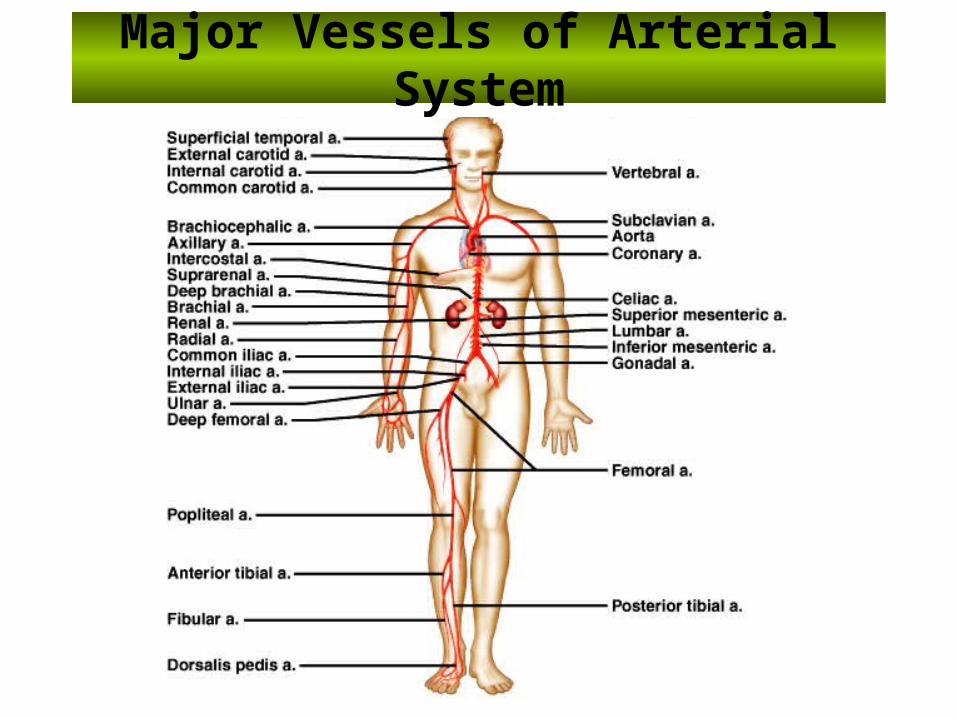
Major Vessels of Arterial System

Major Branches of the Aorta
• Coronary arteries• Brachiocephalic• Left common carotid• Left subclavian• Celiac• Superior mesenteric artery• Renals• Ovarian / testicular• Inferior mesenteric

Minor Branches of the Aorta
• Pericardial Thoracic
• Bronchial Lumbar
• Esophageal Suprarenal
• Mediastinal
• Intercostal
• Phrenic

Major Blood Vessels of the Heart
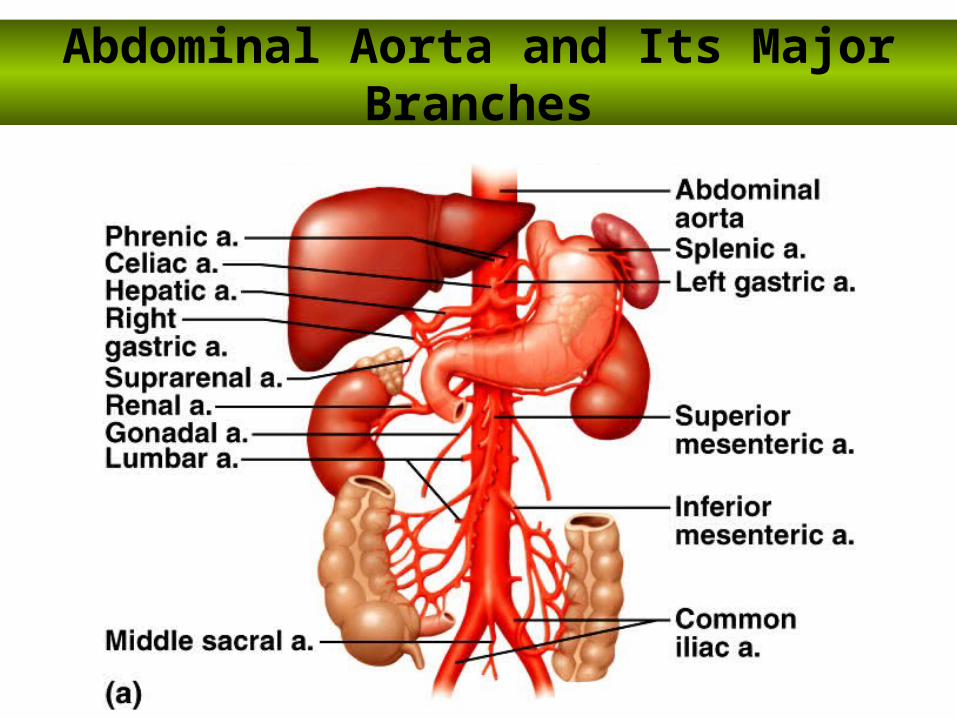
Abdominal Aorta and Its Major Branches
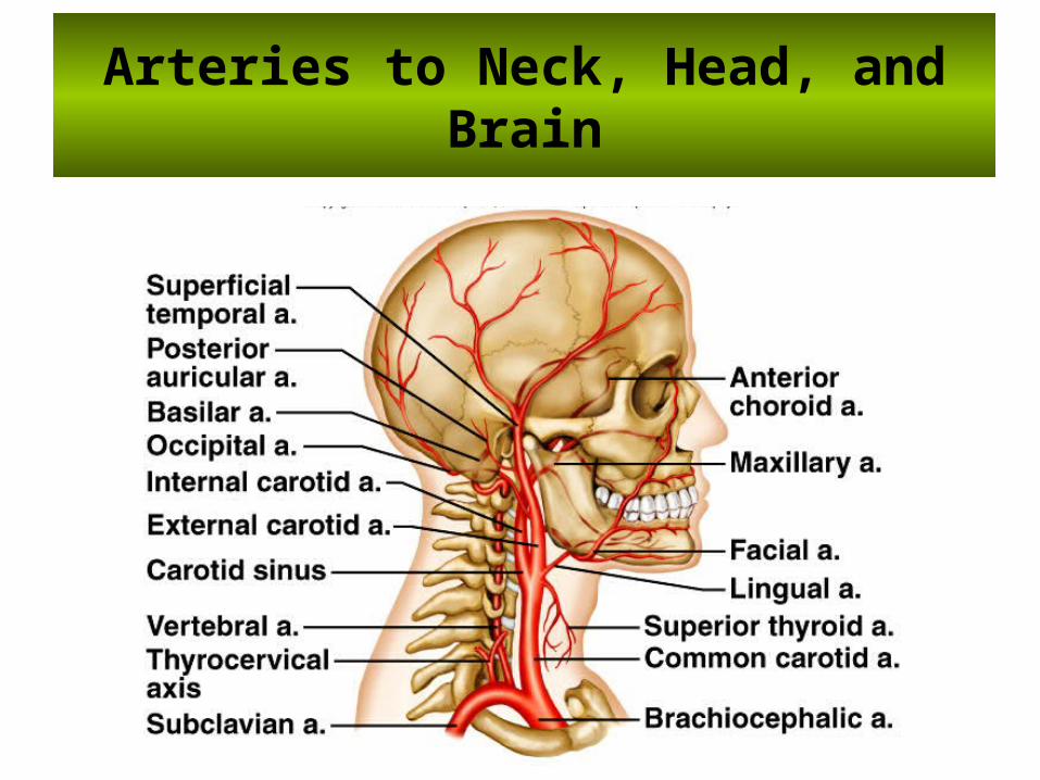
Arteries to Neck, Head, and Brain

Cerebral Arterial Circle• Circle of WillisCircle of Willis• formed by anterior and posterior cerebral arteries, which join the internal carotid arteries

Arteries to Shoulder and Upper Limb

Arteries to Thoracic Wall

Arteries to Pelvic Region
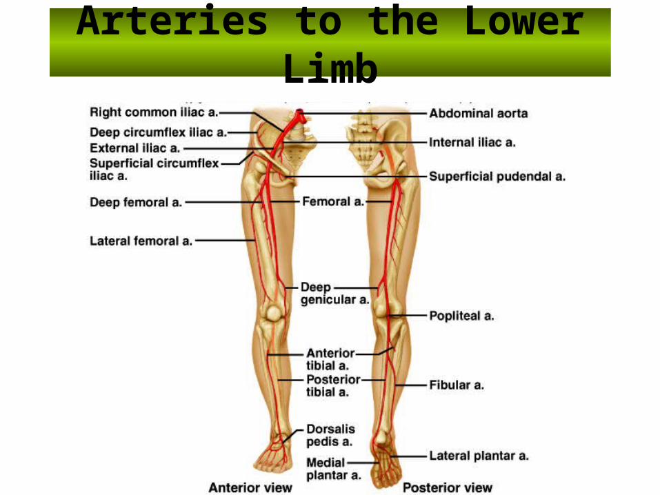
Arteries to the Lower Limb
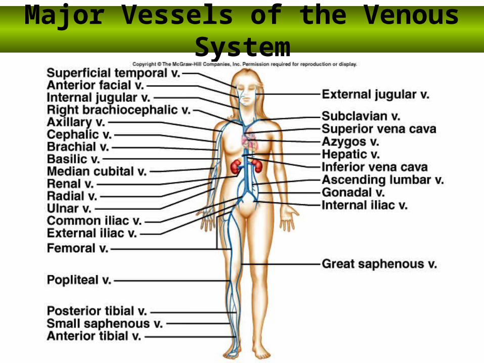
Major Vessels of the Venous System

Major Veins of the Brain, Head and Neck
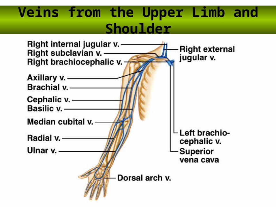
Veins from the Upper Limb and Shoulder
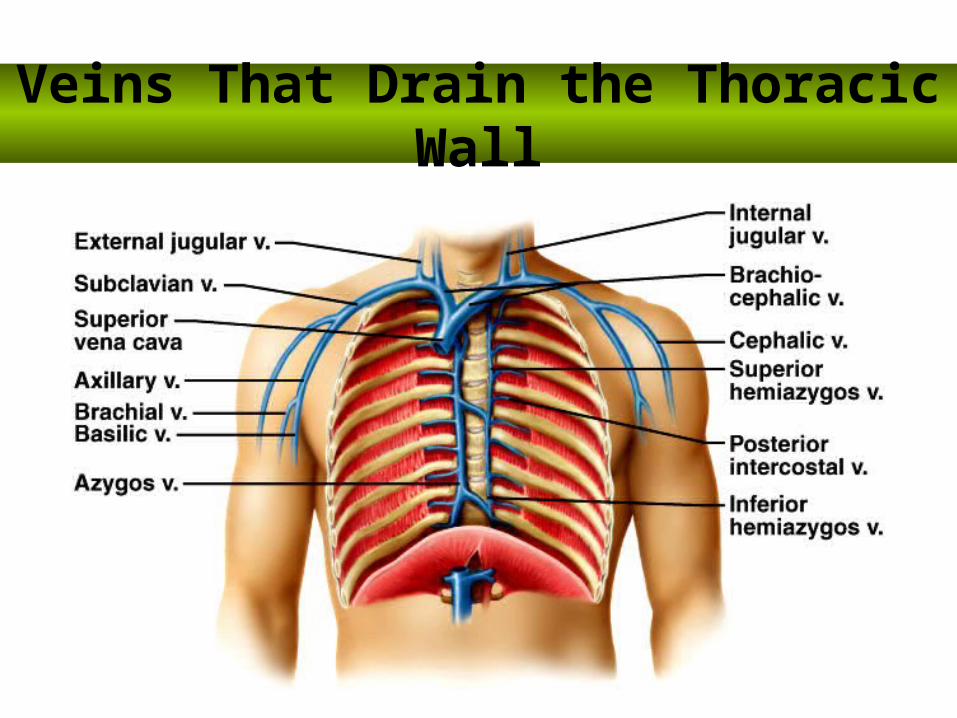
Veins That Drain the Thoracic Wall
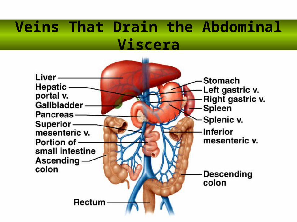
Veins That Drain the Abdominal Viscera

Veins of the Lower Limb and Pelvis

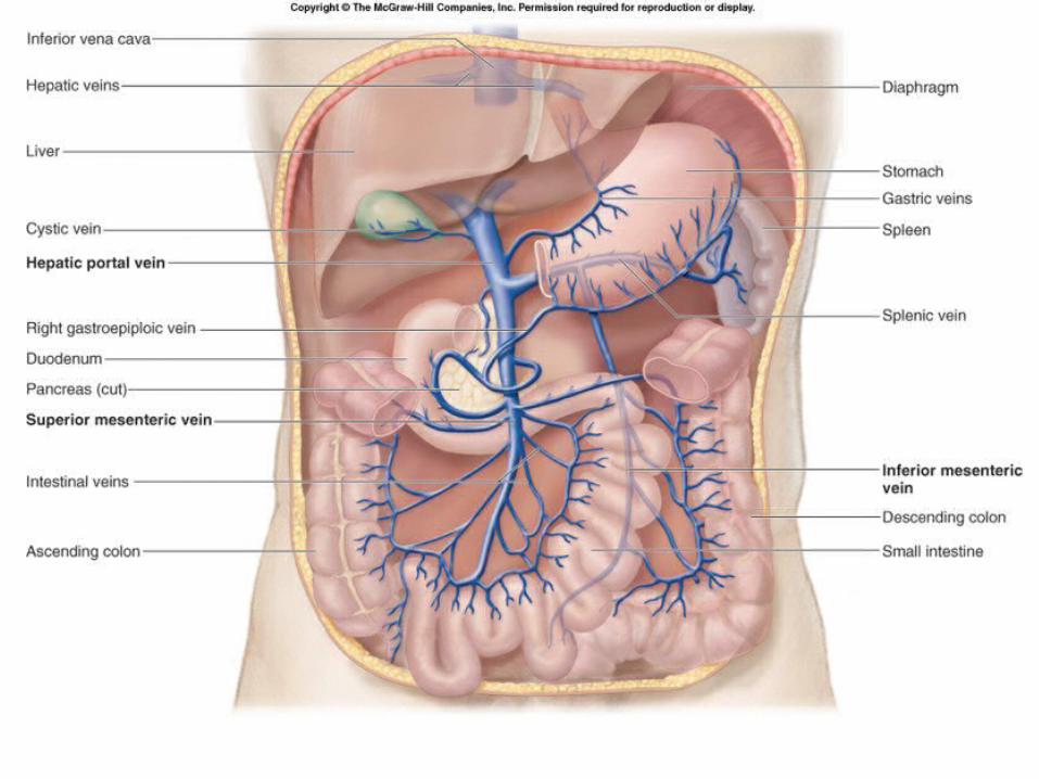
Hepatic Portal System
• A venous network that drains the GI tract and shunts the blood to the liver for processing and absorption of transported materials.
• Blood exits the liver through hepatic veins that merge with the inferior vena cava.
• Is needed because the GI tract absorbs digested nutrients, and these nutrients must be processed and/or stored in the liver.

THE END