1 spectral signatures of non-adiabatic dynamics richard n. dixon school of chemistry, university of...
TRANSCRIPT
1
Spectral signatures of non-adiabatic dynamics
Richard N. Dixon
School of Chemistry, University of Bristol, Bristol BS8 1TS, UK
International Symposium on Molecular Spectroscopy61st Meeting - - June 19-23, 2006
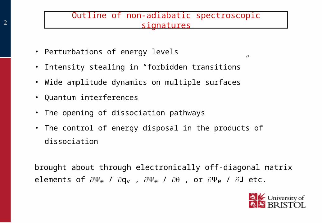
2 Outline of non-adiabatic spectroscopic signatures
• Perturbations of energy levels
• Intensity stealing in “forbidden transitions”
• Wide amplitude dynamics on multiple surfaces
• Quantum interferences
• The opening of dissociation pathways
• The control of energy disposal in the products of dissociation
brought about through electronically off-diagonal matrix elements of
e / qv , e / , or e / J etc.
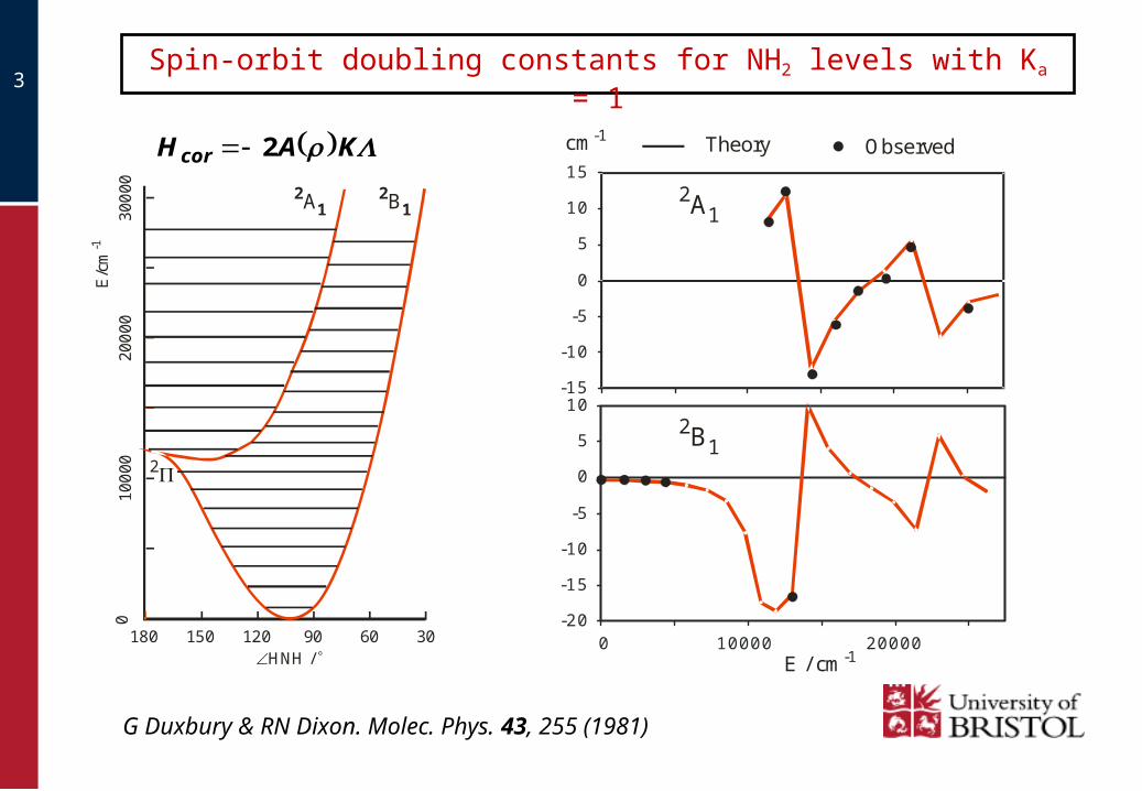
3Spin-orbit doubling constants for NH2 levels with Ka = 1
G Duxbury & RN Dixon. Molec. Phys. 43, 255 (1981)
180 150 120 90 60 30
010
000
200
0030
000
E/c
m-1
HNH /
2A12B1
2
-20
-15
-10
-5
0
5
10
0 10000 20000
-15
-10
-5
0
5
10
15
ObservedTheorycm-1
2A1
2B1
E / cm-1
KAHcor 2
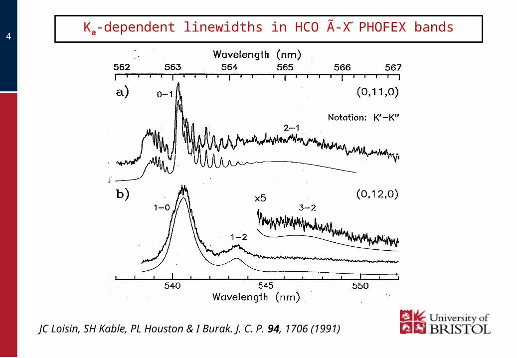
4Ka-dependent linewidths in HCO A-X PHOFEX bands
JC Loisin, SH Kable, PL Houston & I Burak. J. C. P. 94, 1706 (1991)
5
2
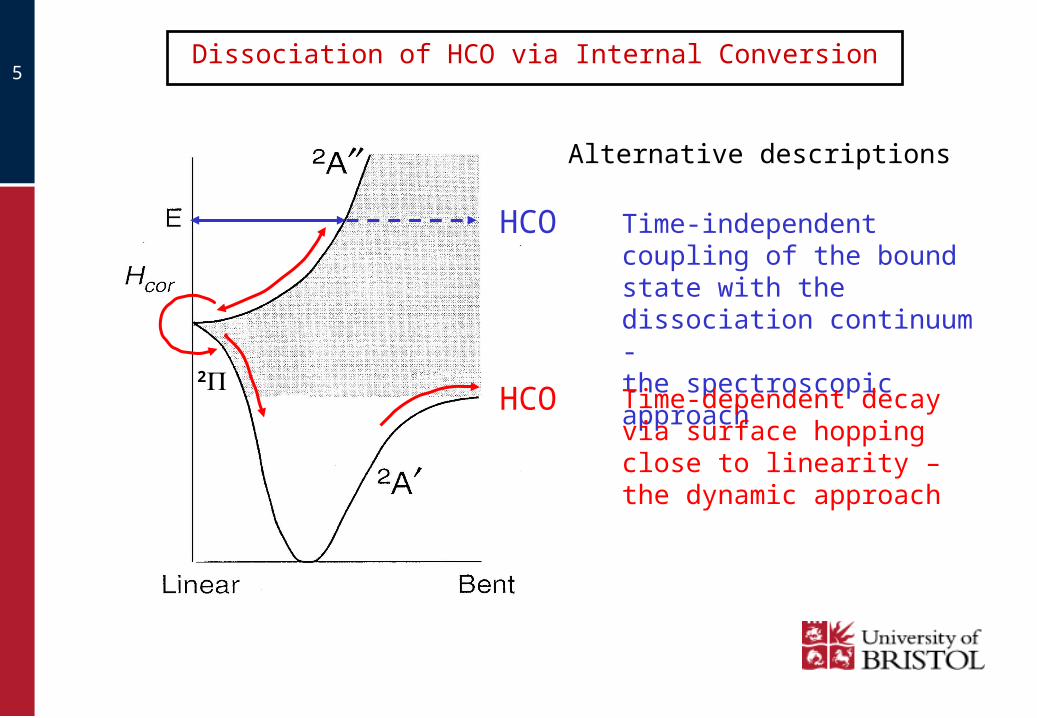
Dissociation of HCO via Internal Conversion
Time-independent coupling of the bound state with the dissociation continuum -the spectroscopic approach
Time-dependent decay via surface hopping close to linearity – the dynamic approach
HCO
HCO
Alternative descriptions

6Renner-Teller induced pre-dissociation widths
(time-dependent theory)
R.N. Dixon. Chem. Soc Rev.. 23, 375, (1994)
7
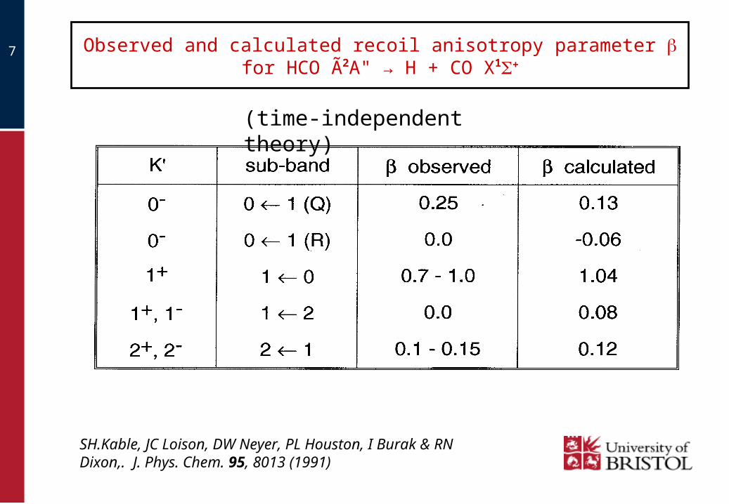
SH.Kable, JC Loison, DW Neyer, PL Houston, I Burak & RN Dixon,. J. Phys. Chem. 95, 8013 (1991)
(time-independent theory)
Observed and calculated recoil anisotropy parameter for HCO A2A" → H + CO X1+
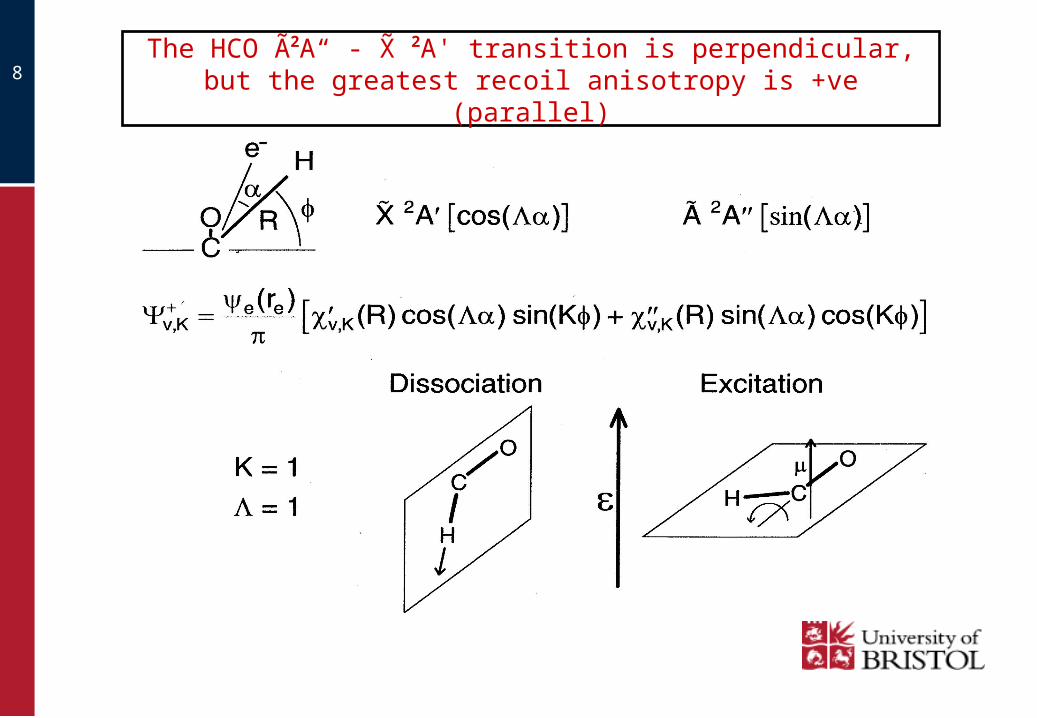
8 The HCO A2A“ - X 2A' transition is perpendicular,but the greatest recoil anisotropy is +ve (parallel)

9
Molecular beam of R-H
Rydberg taggedH-atoms
Molecular Beam
“Rydberg Tagging ”366 nm
“Rydberg Tagging ”366 nm
Photolysisbeam
High resolution H-atom (Rydberg state) photofragment translational spectroscopy
Detector
n=60-90
n=2
n=1
Lyman-α
(121.6 nm)
Tagging (366nm)
Ionisation LimitLyman α
2
121
td
mm
mTKERFragment
HH
Record the time-of-flight spectrum Transform to Total Kinetic Energy Release
10
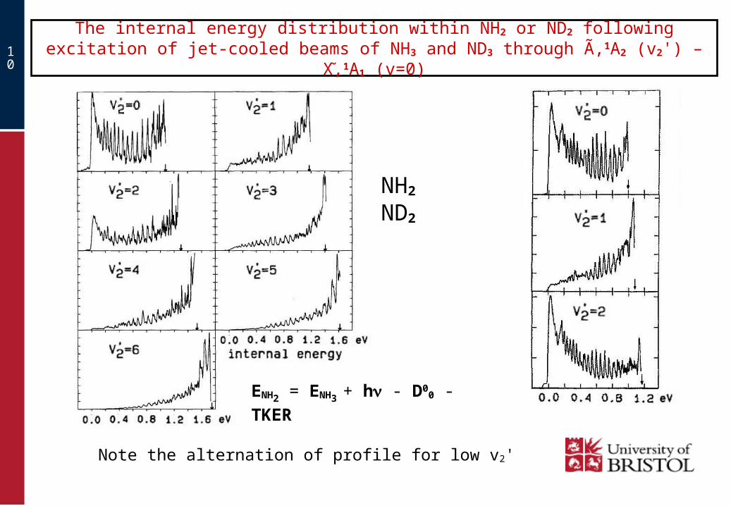
The internal energy distribution within NH2 or ND2 following excitation of jet-cooled beams of NH3 and ND3 through A,1A2 (v2') – X,1A1 (v=0)
Note the alternation of profile for low v2'
NH2 ND2
ENH2 = ENH3 + h - D00 - TKER

11
NH2 internal energy spectra from photolysis of NH3 at 47110 cm-1 (210
band) in perpendicular polarisation
Note the predominance of levels with high Ka N

12
The planar excited state evolves over a low barrier from having 3s Rydberg character at short R to having an NH anti-bonding character at long R
2-D cuts of the potentials for the X and A states of NH3
13
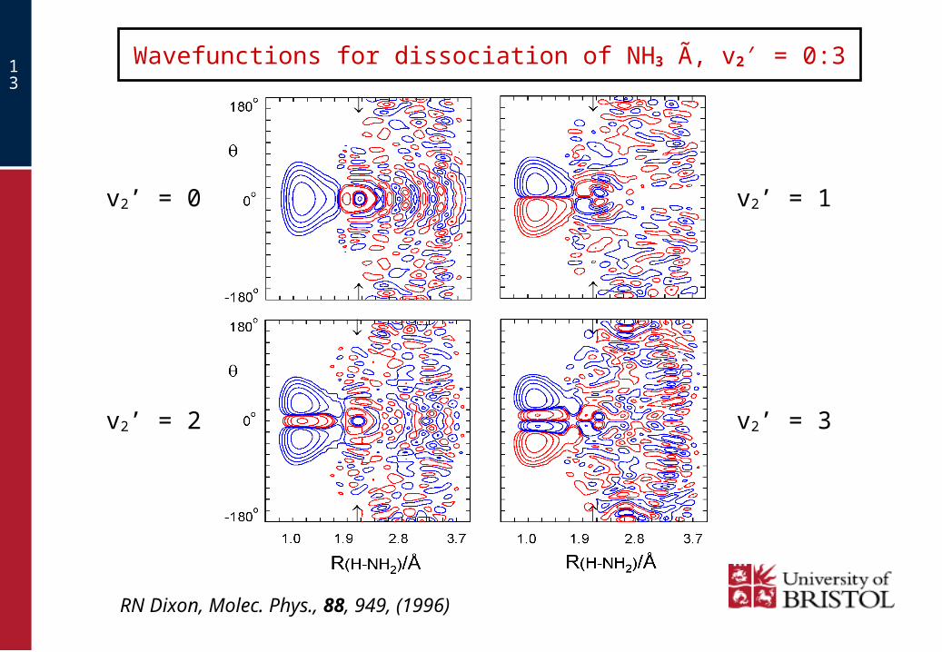
Wavefunctions for dissociation of NH3 A, v2′ = 0:3
v2’ = 0
v2’ = 2
v2’ = 1
v2’ = 3
RN Dixon, Molec. Phys., 88, 949, (1996)
14
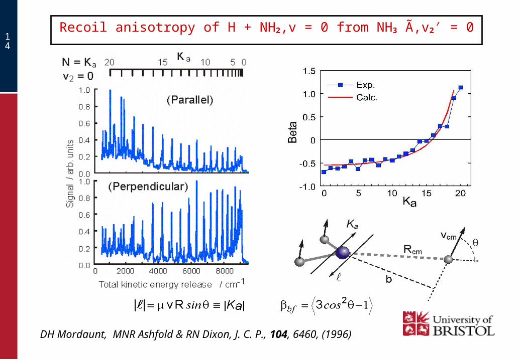
Recoil anisotropy of H + NH2,v = 0 from NH3 A,v2′ = 0
DH Mordaunt, MNR Ashfold & RN Dixon, J. C. P., 104, 6460, (1996)

15
The photochemistry of H2O plays an important role in atmospheric chemistry and in interstellar masers, particularly for excitation at the Lyman- wavelength.
Three electronic states of the water molecule are implicated in this process.
The major branching is to yield H + OH in a wide range of states.
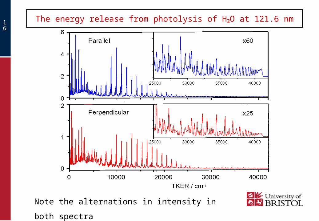
The photodissociation of H2O at Lyman-(121.6 nm)
16
The energy release from photolysis of H2O at 121.6 nm
Note the alternations in intensity in both spectra
17
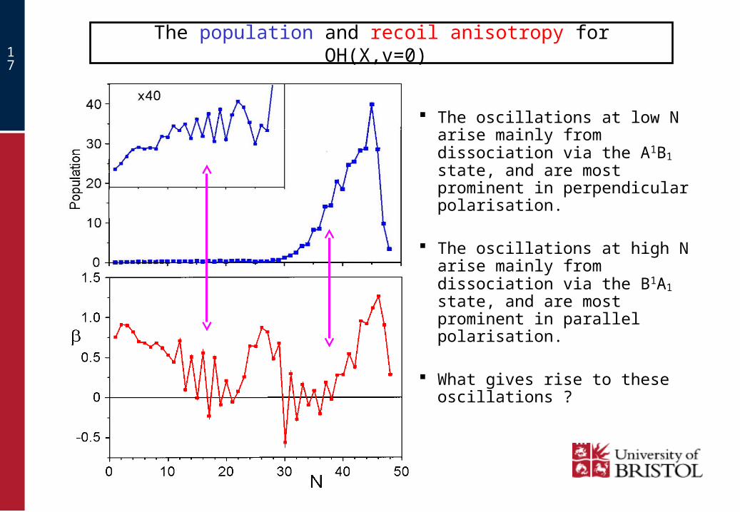
The population and recoil anisotropy for OH(X,v=0)
The oscillations at low N arise mainly from dissociation via the A1B1 state, and are most prominent in perpendicular polarisation.
The oscillations at high N arise mainly from dissociation via the B1A1 state, and are most prominent in parallel polarisation.
What gives rise to these oscillations ?
18
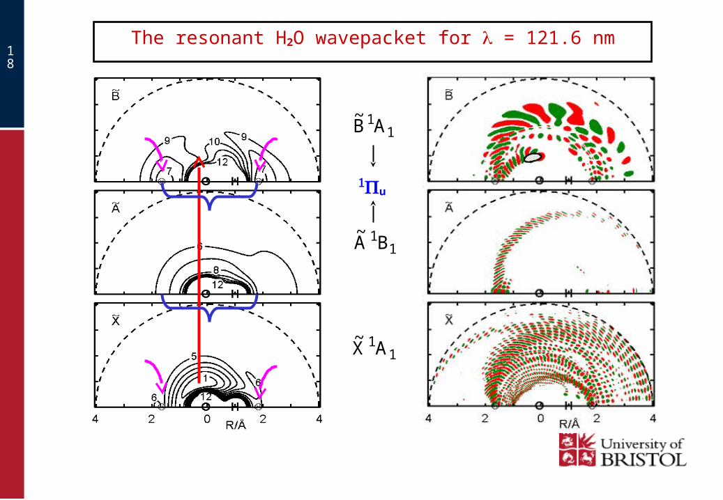
The resonant H2O wavepacket for = 121.6 nm
11AB
~
11BA
~
11AX
~
u
19
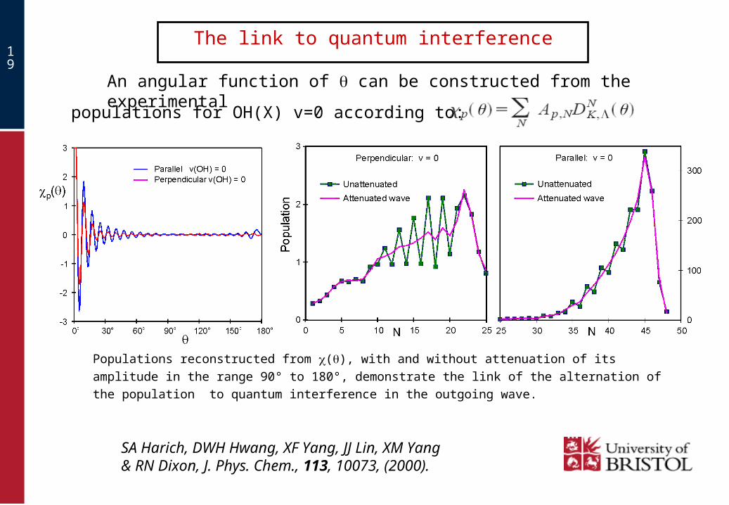
The link to quantum interference
An angular function of can be constructed from the experimental
populations for OH(X) v=0 according to:
SA Harich, DWH Hwang, XF Yang, JJ Lin, XM Yang & RN Dixon, J. Phys. Chem., 113, 10073, (2000).
Populations reconstructed from (), with and without attenuation of its amplitude in the
range 90° to 180°, demonstrate the link of the alternation of the population to quantum
interference in the outgoing wave.
20
Dissociation dynamics of some heteroaromatic molecules
Domcke and co-workers have investigated the excited state potential
energy surfaces of chromophores of biological interest using multi-
reference ab initio methods.
They have found characteristic features of these heteroaromatic
molecules:
• Strongly absorbing diabatically bound singlet * excited states.
• Optically weak but photochemically reactive * excited states.
• Conical intersections which provide a mechanism for ultrafast
deactivation of excited states.
Experimental and/or accurate ab initio vibration frequencies often
available both for parent molecules and their dissociation products.
A. Sobolewski, & W. Domcke, Chem. Phys., 259, 181, (2000)

21
General Features of 1* States
En
erg
y / e
V
R(X-H) / Å 1.0 1.5 2.0
4.0
3.0
6.0
5.0
* states
S0
1*
• 1* state above the 1* states in phenol
and indole.
• Low oscillator strengths.
• Rydberg at short range; dissociative in the R(X-H)
coordinate at long range.
• Form conical intersections with
lower energy singlet states.
En
erg
y / e
V
R(X-H) / Å 1.0 1.5 2.0
4.0
3.0
6.0
5.0 1*
* states
S0
• 1* state below the 1* states in pyrrole and
imidazole.
22
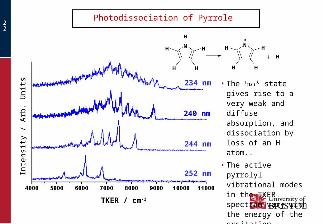
Photodissociation of Pyrrole
TKER / cm-1
Inte
nsity
/ A
rb.
Uni
ts
N
H
H
HH
HN H
HH
H
+ H
234 nm
244 nm
252 nm
• The 1* state gives rise to a very weak and diffuse absorption, and dissociation by loss of an H atom..
• The active pyrrolyl vibrational modes in the TKER spectrum vary with the energy of the excitation.
240 nm

23
3000 4000 5000 6000 7000 8000 9000 10000
TKER / cm-1
Inte
nsi
ty
v=0
14(b2)Combination
bands
=244 nm
Pyrrole – assignment of modes at 244 nm
21(b1)
20(b1)
Dissociation via the diffuse 1A2 () state leads to population of pyrrolyl modes of all three non-totally symmetric classes, reflecting their presumed retention following vibronically induced excitation.
16(b2)
9(a2)
These modes have differing polarisation behaviours.

24
The disposal of the available energy upon dissociation
Excitation of any of the three disappearing modes results in V → T transfer, leading to population of Pyrrolyl in v = 0..
The rapid dissociation favours adiabatic retention of modes excited in Pyrrole. Consequently the internal energy of Pyrrolyl rises with increase in the excitation energy such that the mean TKER remains close to E, the fall in the potential energy from the small exit channel barrier to the dissociation asymptote.
N
H
H
HH
HN H
HH
H
+ H
Mode conserved from pyrrole to pyrrolyl.
Disappearing mode.
B Cronin, MGD Nix, RH Qadiri & MNR Ashfold, PCCP, 6, 5031, 2004

25
The 1A2(*) S1-state of 2,5-Dimethyl Pyrrole
• The S1←S0 spectrum shows some resolved vibrational structure, indicating a higher barrier to dissociation than in Pyrrole.
• The methyl torsional modes are vibronically active in S1←S0. • The band origin and dissociation energies are lower than in Pyrrole.• The fragmentation process is more structured than in Pyrrole.
B Cronin, MGD Nix, AL Devine, RN Dixon & MNR Ashfold, Phys Chem Chem Phys, 8, 599, 2006
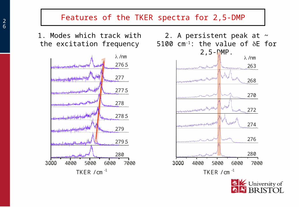
26
Features of the TKER spectra for 2,5-DMP
1. Modes which track with the excitation frequency
2. A persistent peak at ~ 5100 cm-1: the value of E for 2,5-DMP.
4000 5000 6000 7000
276.5
/ nm
277
277.5
278
278.5
279
279.5
280
TKER / cm-1
4000 5000 6000 7000
TKER / cm-1
263
268
270
272
274
276
280
/ nm

27
Features, and adiabatic potentials for 2,5-DMP
3. Strong features are accompanied by three lower KE satellite peaks.
4000 5000TKER / cm-1
263
270
276
280
/ nm
Adiabatic potentials
• Part of Eint for a promoting mode must be channelled into a disappearing mode to reduce the barrier and permit dissociation.
• The S1 and S0 states can be coupled at the circled conical intersection by a2 vibrational modes.

28
• Part of this wavepacket will remain on the 1A1 surface and decay
via the S0 continuum.
• Coupling back to the 1A2 surface will lead to population of two
quanta of some a2 mode. (19 or 20), or a combination..
• A second portion will evolve onto the 1A1 surface.
• The 1A1 and 1A2 states
are coupled by a2
vibrational modes.
1A1 (S0)
1A2
(a2)
(a2)2(a2)
Dynamical coupling at the Conical Intersection
• One portion of the wavepacket
will be unaffected by this
interaction (v(a2) = 0).
29
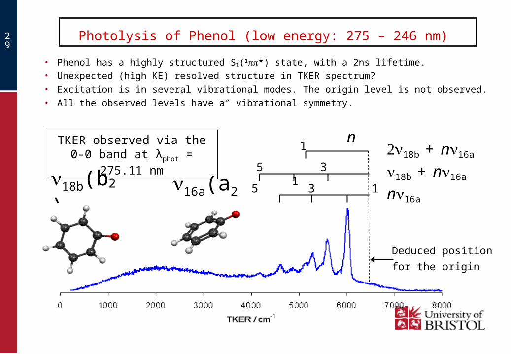
Photolysis of Phenol (low energy: 275 – 246 nm)
Deduced position
for the origin
n16a
18b + n16a
TKER observed via the 0-0 band at λphot = 275.11 nm
16a(a2)18b(b2)
• Phenol has a highly structured S1(1*) state, with a 2ns lifetime.
• Unexpected (high KE) resolved structure in TKER spectrum?• Excitation is in several vibrational modes. The origin level is not observed.• All the observed levels have a″ vibrational symmetry.
5 3 1
5 3 1
n18b + n16a 1

30
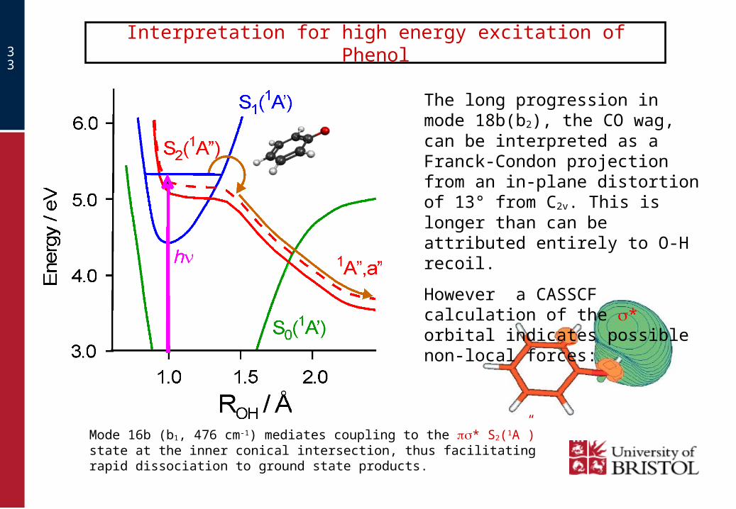
Interpretation for low energy excitation of Phenol
1.The lower energy levels of the *S1 state are predissociated by internal conversion (IC) to high levels of the S0 ground state having predominant vibration in OH stretching (13-16 quanta).
2.Mode 16a (a2. 372 cm-1) in odd quanta mediates coupling to the * S2(1A”) state at the outer conical intersection, thus facilitating dissociation to ground state products.
Phenoxyl is only populated in a” vibrational levels, but:
the disappearing OH a“ torsional mode is not active.

31
Phenol low energy excitation: 275 – 270 nm
Some modes excited in Phenol S1 become inter-converted in carrying through to Phenoxyl. This may result from a Duschinsky rotation of these normal coordinates, either in the IC step to S0, or at the S0 to S2 conical intersection.
Excitation
10
0154
00
01
10 (OH)4 τ
11(OH)τ
2016a
106a
32
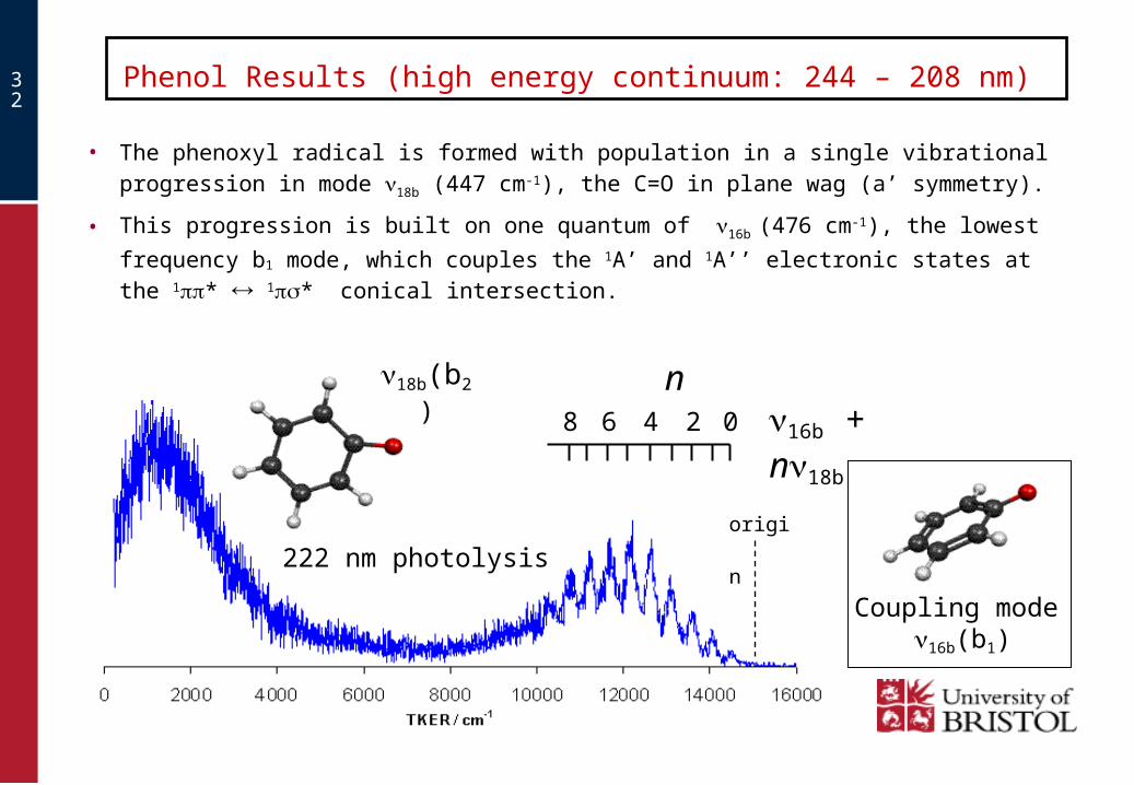
Phenol Results (high energy continuum: 244 – 208 nm)
0246
222 nm photolysis
Coupling mode16b(b1)
• The phenoxyl radical is formed with population in a single vibrational progression
in mode 18b (447 cm-1), the C=O in plane wag (a’ symmetry).
• This progression is built on one quantum of 16b (476 cm-1), the lowest frequency
b1 mode, which couples the 1A’ and 1A’’ electronic states at the 1* 1* conical intersection.
16b + n18b8
n18b(b2)
origin
33
Interpretation for high energy excitation of Phenol
Mode 16b (b1, 476 cm-1) mediates coupling to the * S2(1A”) state at the inner conical intersection, thus facilitating rapid dissociation to ground state products.
The long progression in mode 18b(b2), the CO wag, can be interpreted as a Franck-Condon projection from an in-plane distortion of 13° from C2v. This is longer than can be attributed entirely to O-H recoil.
However a CASSCF calculation of the * orbital indicates possible non-local forces:

34
Reconciliation of the TKER for low and high energy
The recognition that different promoting modes are involved in photodissociation of phenol following low and high energy excitation to S1 was critical to reconciling the TKER values for the full data set.
The plot of the predicted position of the TKER for v=0 against the excitation energy then gives a single straight line of unit slope.
This leads to: D0(O-H) = 30015 ± 40 cm-1.
MNR Ashfold, B Cronin, AL Devine, RN Dixon & MGD Nix, Science, 312, 163, 2006.

35
Summary of non-adiabatic effects
NH2
HCO
NH3
H2O
Pyrrole
2,5-DMP
2
Phenol
Perturbations
Intensity stealing
Wide amplitude dynamics
Quantum interference
Facilitate dissociation
Affect energy disposal
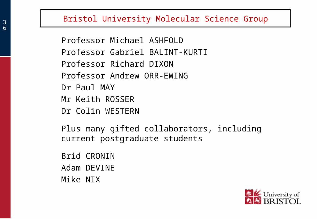
36
Bristol University Molecular Science Group
Professor Michael ASHFOLD
Professor Gabriel BALINT-KURTI
Professor Richard DIXON
Professor Andrew ORR-EWING
Dr Paul MAY
Mr Keith ROSSER
Dr Colin WESTERN
Plus many gifted collaborators, including current postgraduate students
Brid CRONIN
Adam DEVINE
Mike NIX

37