1 spine imaging & myleography radiographic exam of the cns structures within the verterbral...
TRANSCRIPT

1 Spine
Imaging &
MYLEOGRAPHYRADIOGRAPHIC EXAM OF THE CNS
STRUCTURES WITHIN THE VERTERBRAL CANAL
RT 255 – SPRING (2010 rev)
Pt info:

2 Myelography
• Requires contrast introduction into the subarachnoid space by spinal puncture =
• INTRATHECAL INJECTION• Puncture made at L2-L3 or L3-L4 space
– May also be introduced into cisterna magna at C1 and occipital bone
New Notes-
• These pathologies are demonstrated radiographically as a deformity in the subarachnoid space or an obstruction of the passage of the contrast within the subarachnoid space.
• It is also useful in identifying a stenosis or narrowing of the subarachnoid space by watching the dynamic flow patterns of the CSF.
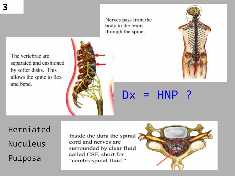
3
Dx = HNP ?
Herniated
Nuculeus
Pulposa
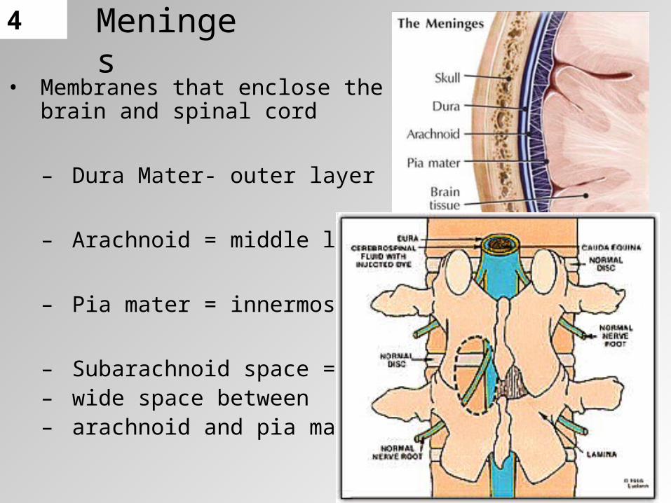
4Meninges
• Membranes that enclose the brain and spinal cord
– Dura Mater- outer layer
– Arachnoid = middle layer
– Pia mater = innermost layer
– Subarachnoid space = – wide space between – arachnoid and pia mater

5Subarachnoid space
– Wide space between arachnoid and pia mater• Filled with CSF• Bathes brain & spinal cord with nutrients• Cushions against shocks and blows• Where contrast is injected for myelograms
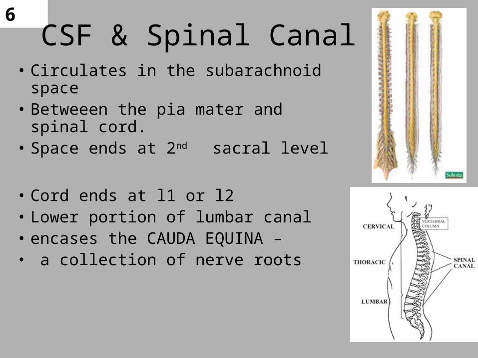
6CSF & Spinal Canal
• Circulates in the subarachnoid space • Betweeen the pia mater and spinal cord.• Space ends at 2nd sacral level
• Cord ends at l1 or l2• Lower portion of lumbar canal • encases the CAUDA EQUINA –• a collection of nerve roots

7Myelography
• Contrast is generally water-soluble, nonionic, iodinated medium
OMNIPAQUE
ISOVUE
•OLD – OIL BASED (Panopaque)•then – Metrizimide (water sol)
•NOW•NON-IONIC water based
•ISOVUE “M” 200 OR 300 M•INTRATHECAL INJECTION !!
•FOLLOW UP WITH •CT SCAN W/IN 1 HR
CONTAST MEDIA
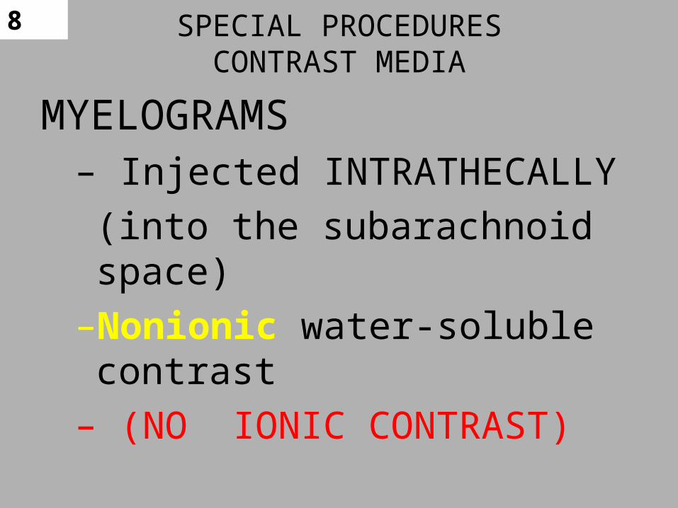
8 SPECIAL PROCEDURESCONTRAST MEDIA
MYELOGRAMS– Injected INTRATHECALLY
(into the subarachnoid space)
–Nonionic water-soluble contrast
– (NO IONIC CONTRAST)
9
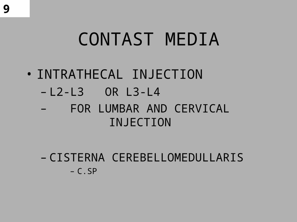
CONTAST MEDIA
• INTRATHECAL INJECTION– L2-L3 OR L3-L4– FOR LUMBAR AND CERVICAL
INJECTION
– CISTERNA CEREBELLOMEDULLARIS – C.SP
10
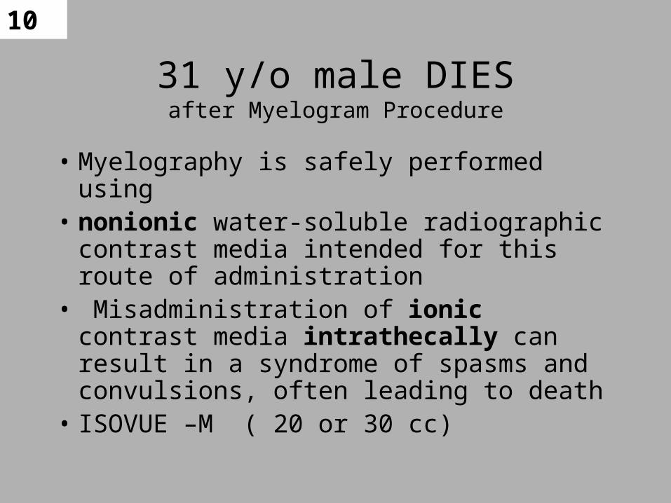
31 y/o male DIESafter Myelogram Procedure
• Myelography is safely performed using• nonionic water-soluble radiographic contrast
media intended for this route of administration
• Misadministration of ionic contrast media intrathecally can result in a syndrome of spasms and convulsions, often leading to death
• ISOVUE –M ( 20 or 30 cc)
11
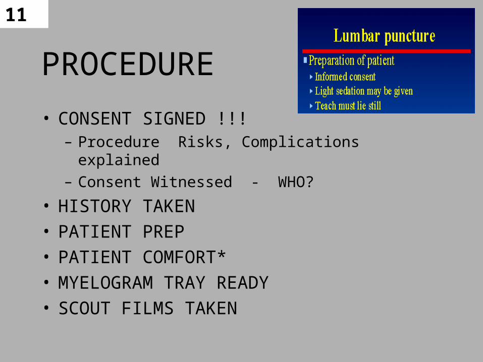
PROCEDURE
• CONSENT SIGNED !!! – Procedure Risks, Complications explained
– Consent Witnessed - WHO?
• HISTORY TAKEN• PATIENT PREP• PATIENT COMFORT*• MYELOGRAM TRAY READY• SCOUT FILMS TAKEN
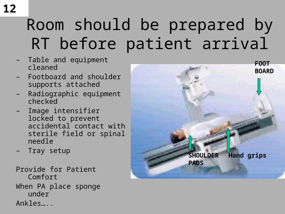
12
Room should be prepared by RT before patient arrival
– Table and equipment cleaned– Footboard and shoulder
supports attached– Radiographic equipment
checked– Image intensifier locked to
prevent accidental contact with sterile field or spinal needle
– Tray setup
Provide for Patient Comfort When PA place sponge underAnkles…..
FOOTBOARD
SHOULDERPADS
Hand grips
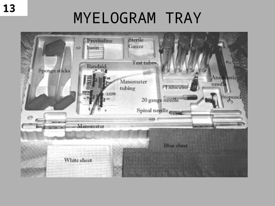
13MYELOGRAM TRAY

14 Additional Supplies• Blankets• Sterile towels• Non-ionic iodinated contrast media• Sterile gloves for DR• Shields for PT, DR, anyone else in room,
and yourself• Varying sizes of spinal needles and needles• Extra syringes and tubing • Cleaning liquid – skin prep (Betadine*)

15
Syringes and Spinal Needles
Syringes
Spinal Needles(covered)
More Spinal Needles (uncovered)
16
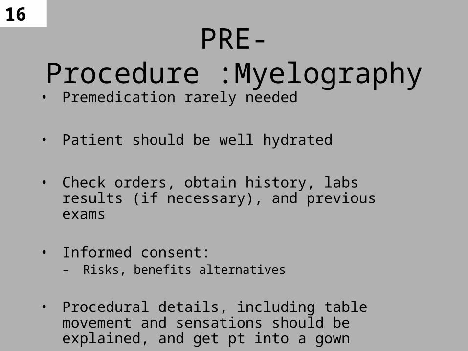
PRE- Procedure :Myelography• Premedication rarely needed
• Patient should be well hydrated
• Check orders, obtain history, labs results (if necessary), and previous exams
• Informed consent:– Risks, benefits alternatives
• Procedural details, including table movement and sensations should be explained, and get pt into a gown
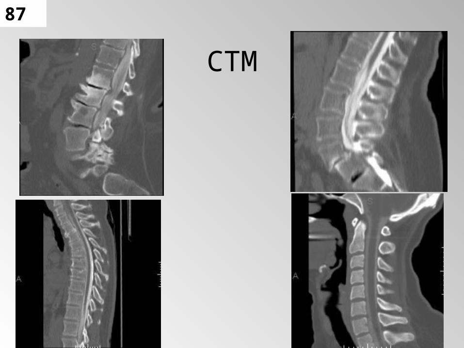
17
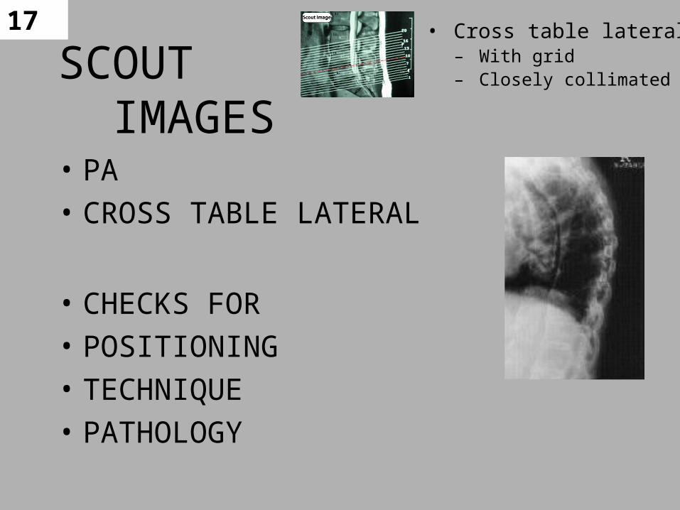
SCOUT IMAGES• PA
• CROSS TABLE LATERAL
• CHECKS FOR
• POSITIONING
• TECHNIQUE
• PATHOLOGY
• Cross table lateral – With grid– Closely collimated

18
Critique?
What are the projections for the scout?
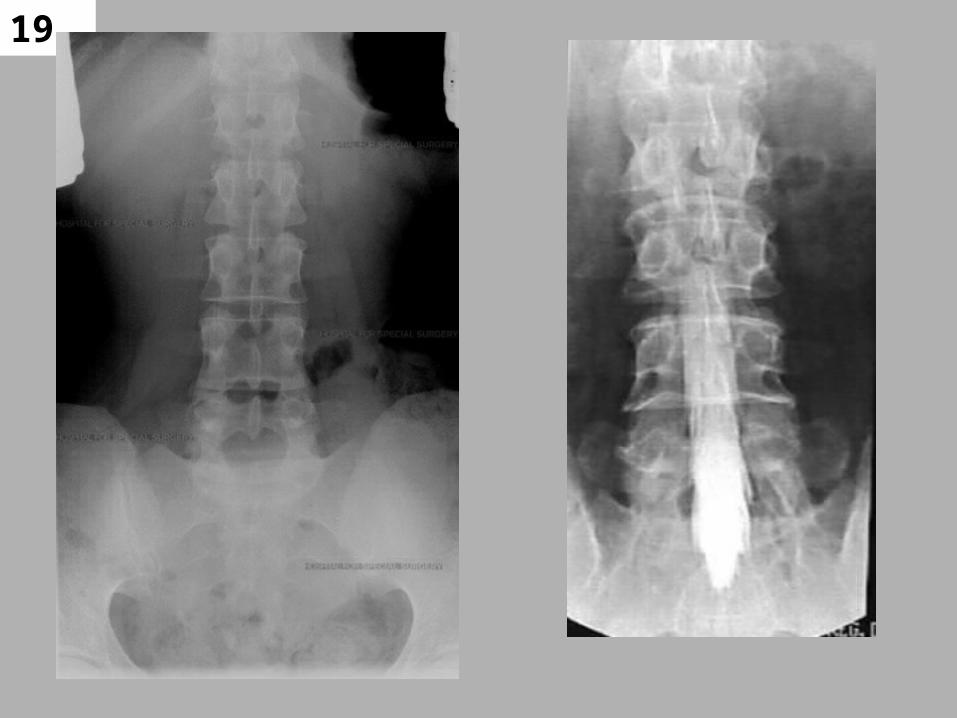
19
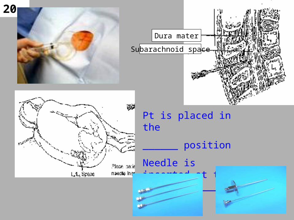
20
Dura mater
Subarachnoid space
Pt is placed in the
______ position
Needle is inserted at the level of _______
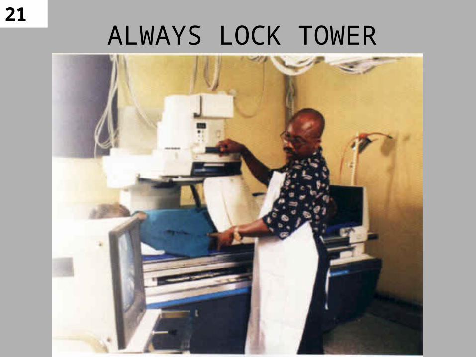
21ALWAYS LOCK TOWER

22 Room must be R & F with
Table movement capabilities
23
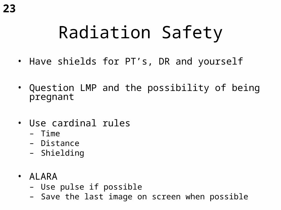
Radiation Safety
• Have shields for PT’s, DR and yourself
• Question LMP and the possibility of being pregnant
• Use cardinal rules– Time– Distance– Shielding
• ALARA– Use pulse if possible– Save the last image on screen when possible
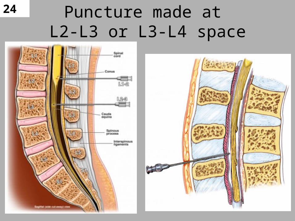
24 Puncture made at L2-L3 or L3-L4 space
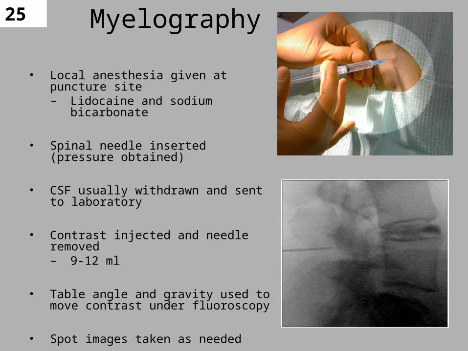
25 Myelography
• Local anesthesia given at puncture site– Lidocaine and sodium bicarbonate
• Spinal needle inserted (pressure obtained)
• CSF usually withdrawn and sent to laboratory
• Contrast injected and needle removed– 9-12 ml
• Table angle and gravity used to move contrast under fluoroscopy
• Spot images taken as needed

26Prone & Lateral Flexion
• Prone– Sponge under abdomen
for flexion of spine
• Lateral flexion is not commonly used– Widens interspace for
easier introduction of needle
–CSP injection only used when pathology present in LSP
27
Spot Films
• Central ray vertical or horizontal using CR or film screen cassettes
• Images are taken at– Site of blockage– Level of distortion
• If conus medullaris is area of concern:– Lay pt supine– Central ray at T12- L1– Use 10x12 cassette and collimate tightly

28Myelogram For
L.SP or C.SP –
Contrast usually injected in Lumbar
Table placed
TRENDELBURG
To move contrast
To Cervical Area
29Myelography
• If contrast is moved into cervical area, head is positioned in acute extension to prevent contrast from entering ventricular system
– Acute extension compresses cisterna magna and is the only position that will prevent contrast from entering ventricles
– Keep pt at 30 degrees post contrast injection –– Or may get a MIGRANE
30
Myelography
• Usually performed as outpatient basis
• Common for CT myelography (CTM) to be used with conventional Myelogram
• MRI often used instead
• Myelography and CTM still used for patients with contraindications for MRI
– Pacemakers and metal fusion rods
31
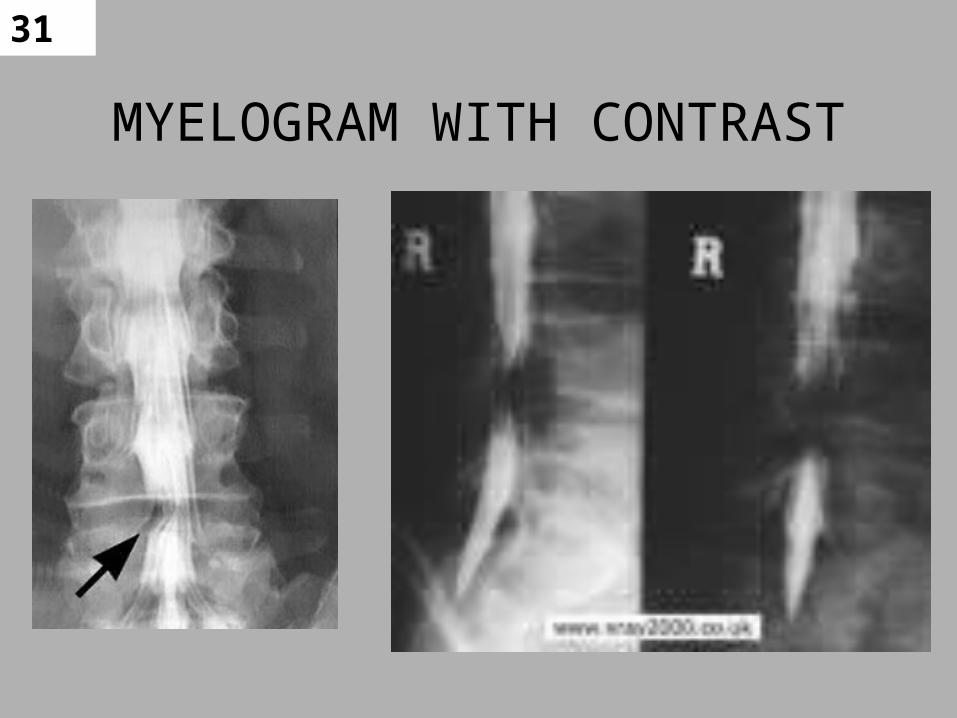
MYELOGRAM WITH CONTRAST
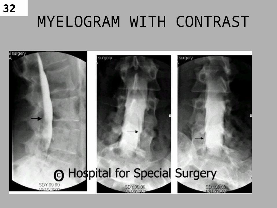
32MYELOGRAM WITH CONTRAST

33
34
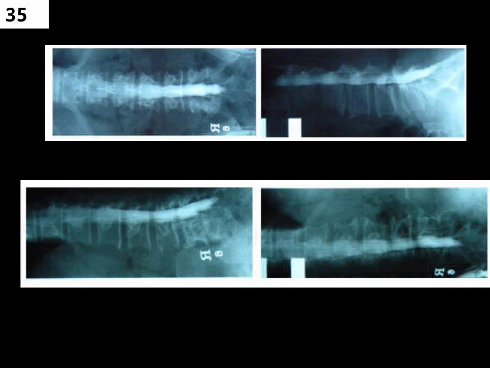
35
36
Post procedure: Myelography
– Monitoring required
– Head and shoulders elevated 30 to 45 degrees
– Bed rest for several hours
– Fluid encouraged
– Puncture site checked before release
37
Possible Complications from Myelography
• Vomiting
• Vertigo
• Neck Pain
• MIGRANES
• Spinal Headache
– Due to loss of CSF during puncture
– Increased severity upright
– Decreased pain when recumbent.
38
RISK
• The main risk with a myelogram is the potential for a spinal headache.
• The spinal headache usually resolves in one to two days with rest and fluids, and seems to be more common for patients with a history of migraine headaches.

39
CONTRAINDICATIONS
• ?? CONTRAST ALLERGY/reaction
• CEREBRAL ANERUYSMS,
• AV MALFORMATIONS
• MYELOGRAM CAN INCREASE INTRACRANAIL PRESSURE
• RECENT LUMBAR PUNCTURE 1 week

40Contraindications & Considerations
• Heparin stopped 4 hours before – Can be restarted 2 hrs after procedure– Usually given as IP
• Coumadin stopped 3-4 days before – Usually OP– Labs usually indicated
• PT < 15.0 seconds (clotting time)– Preferable to reschedule exam if below 15
• Platelets >100,000– If below 50,000 a platelet transfusion may be
indicated before procedure

41More Severe Complications
• Nerve root damage
• Meningitis
• Epidural abscess
• Contrast reaction (anaphylactic shock)
• CSF leak
• Hemorrhage

42
Treatment for Spinal Headache• Initial treatment
– Tylenol
– Horizontal position
– Forced fluids
– Caffeine
• Persistent headache– If a fever occurs,
contact MD• May be indicative of
meningitis
– Beyond 48 hrs w/o fever (24 hrs if severe)• Blood patch
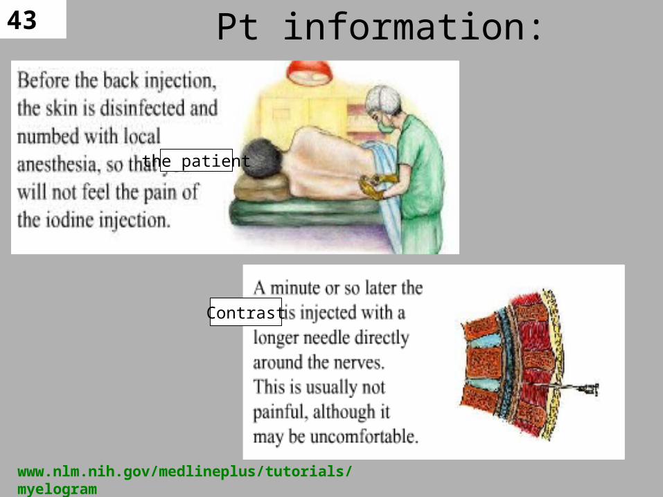
43 Pt information:
the patient
Contrast
www.nlm.nih.gov/medlineplus/tutorials/ myelogram
44
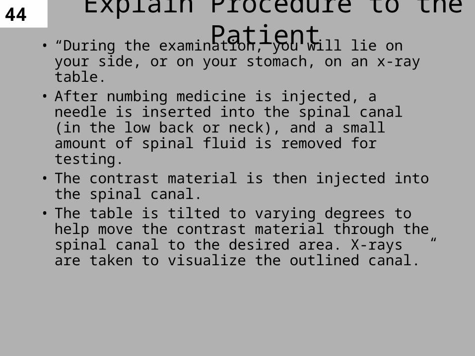
• “During the examination, you will lie on your side, or on your stomach, on an x-ray table.
• After numbing medicine is injected, a needle is inserted into the spinal canal (in the low back or neck), and a small amount of spinal fluid is removed for testing.
• The contrast material is then injected into the spinal canal.
• The table is tilted to varying degrees to help move the contrast material through the spinal canal to the desired area. X-rays are taken to visualize the outlined canal.”
Explain Procedure to the Patient
45
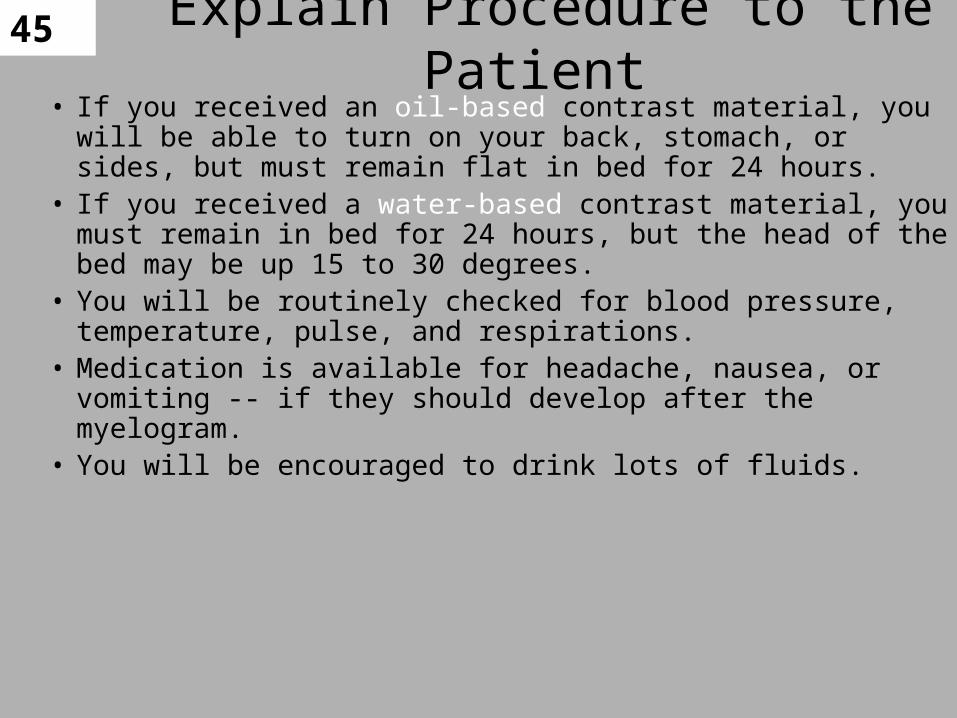
• If you received an oil-based contrast material, you will be able to turn on your back, stomach, or sides, but must remain flat in bed for 24 hours.
• If you received a water-based contrast material, you must remain in bed for 24 hours, but the head of the bed may be up 15 to 30 degrees.
• You will be routinely checked for blood pressure, temperature, pulse, and respirations.
• Medication is available for headache, nausea, or vomiting -- if they should develop after the myelogram.
• You will be encouraged to drink lots of fluids.
Explain Procedure to the Patient

46
1. “If you experience continuous mild to severe headaches, there may be a small leak of spinal fluid. This generally is not dangerous.
2. If symptoms do not resolve the leak can be sealed over with a blood patch. This is done by the anesthesiologist.
3. If you are on any antidepressant medication, please check with your doctor.”
Explain Procedure to the Patient
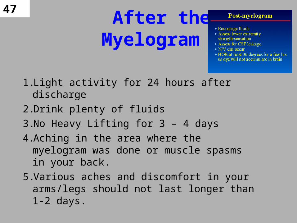
47 After the Myelogram
1.Light activity for 24 hours after discharge
2.Drink plenty of fluids
3.No Heavy Lifting for 3 – 4 days
4.Aching in the area where the myelogram was done or muscle spasms in your back.
5.Various aches and discomfort in your arms/legs should not last longer than 1-2 days.

48Blood Patch for Clot
• Sterily injecting a small amount of patient’s blood into the epidural space– Clot will occur over puncture
site
– Usually will stop headache immediately
– 1st patch is 70% effective
– 2nd patch is 95% effective
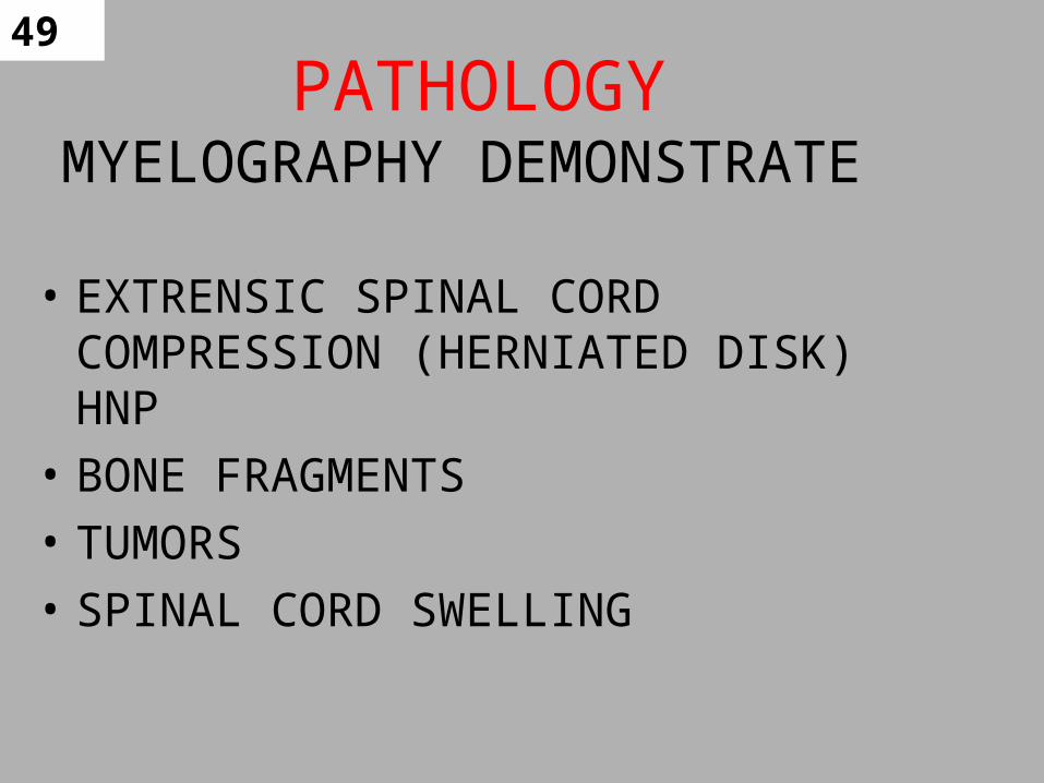
49 PATHOLOGYMYELOGRAPHY DEMONSTRATE
• EXTRENSIC SPINAL CORD COMPRESSION (HERNIATED DISK) HNP
• BONE FRAGMENTS
• TUMORS
• SPINAL CORD SWELLING

50 ENCROACHMENTS ON SPINAL CANAL
• Radiographically as a deformity of the Subarachnoid Space or
• Obstruction of passage
of column of contrast
within space
• Narrowing Identified

51
52
53
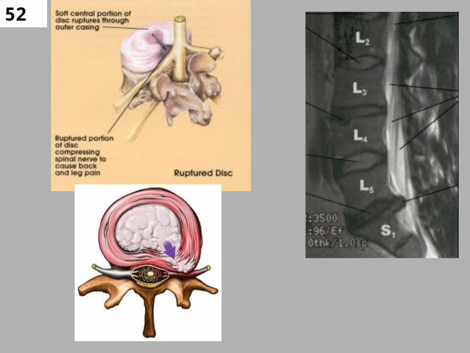
Disc Herniation
• Repetitive tears to the annulus thins the wall (similar to a bald spot on a tire) and may lead to a disc rupture (similar to a tire blowout)
• The disc rupture presses a spinal nerve(s) against the bony surface of a vertebra.
• This condition is often referred to as a ruptured disc.
• Even pressure from everyday activities can push the disc through the torn or cracked annulus and pinch a spinal nerve root(s).

54HNP / Ruptured Disks
• weakening of the ligament structures, and degeneration of the lumbar discs
• When we increase the stresses of the disc with lifting, bending or twisting, low back pain may occur.
• Larger or repetitive stresses, such as shoveling the snow, may produce tears in the annulus (fibrous casing that contains the disc).
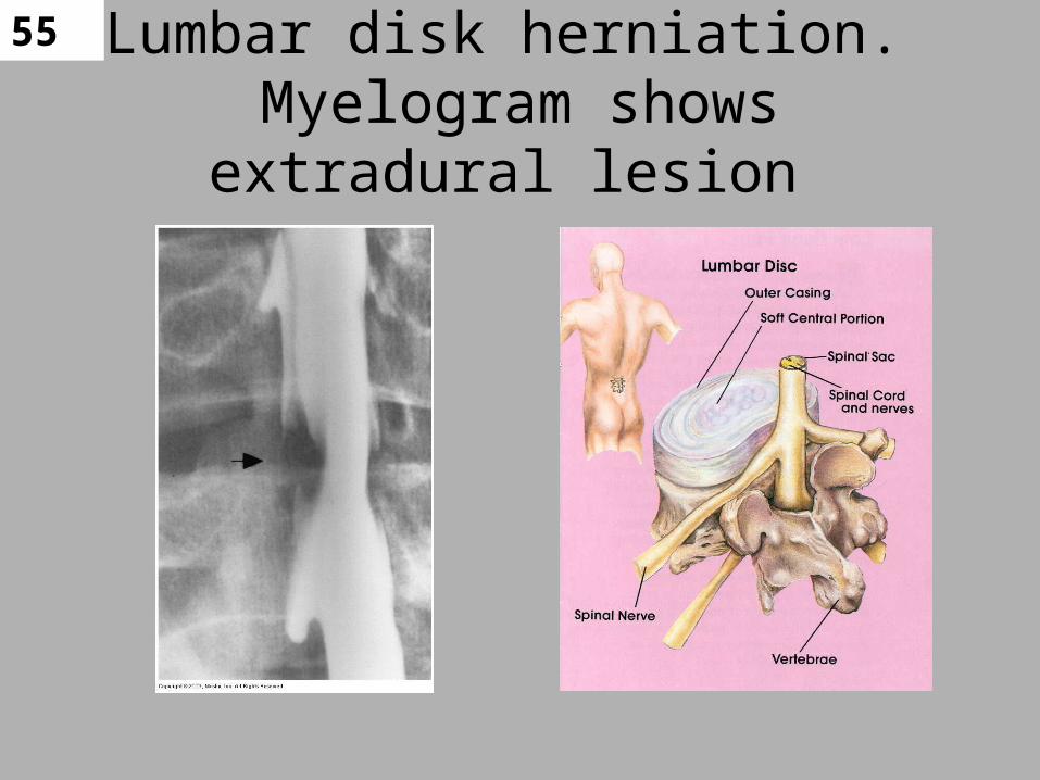
55Lumbar disk herniation.
Myelogram shows extradural lesion
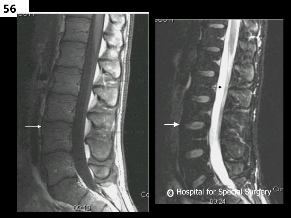
56
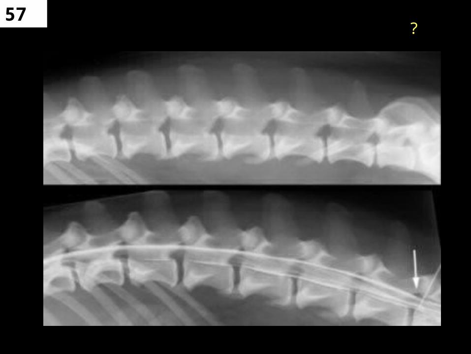
57?
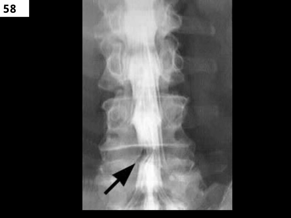
58

59Degenerative disc disease
is the normal manifestation of the aging process
• The discs function as a shock absorber and cushion between the vertebrae.
• As we age, the water content of the disc decreases, and the disc becomes dry.
• The lumbar spine supports the weight of the entire spinal column.
• significant motion occurs between the lumbar vertebrae,
• These two factors influence the process leading to narrowing or collapse the disc space in the lumbar spine
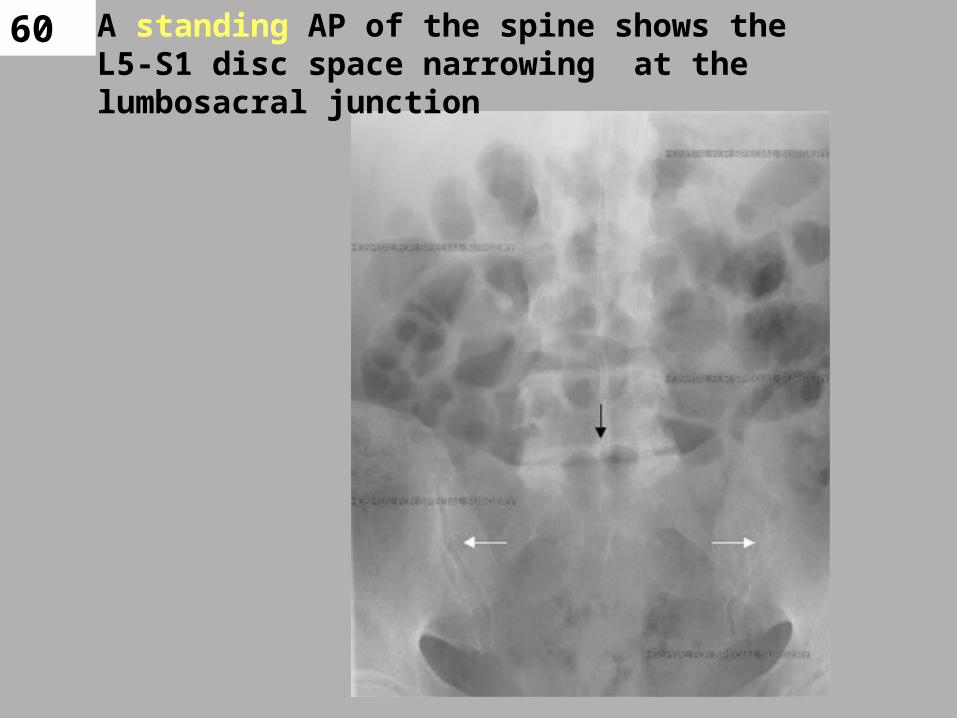
60 A standing AP of the spine shows the L5-S1 disc space narrowing at the lumbosacral junction

61Spondylolisthesis
• there is a slipping of one vertebral body in any direction in relation to the one below it.
• A forward, or anterior displacement is most common
• Degenerative spondylolisthesis is more prevalent in females than in males

62Spondylolisthesis.
Lateral view of lower lumbar spine shows anterior
slippage of L4 with respect to L5
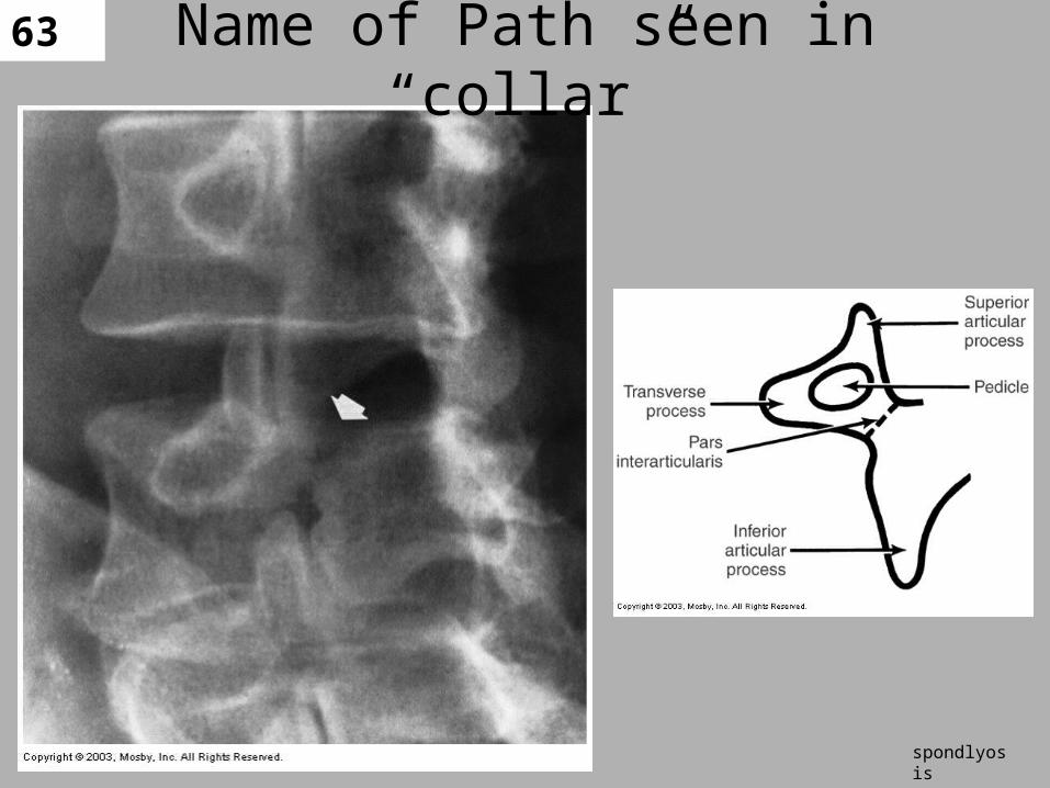
63Name of Path seen in “collar”
spondlyosis
64
PAIN MANAGEMENT(C-ARM)
• Cortisone Injections: • can be injected into the epidural space and
offer temporary relief by decreasing inflammation.
• Many patients respond well to conservative management with decreased or manageable pain levels and improved activity levels.
• This positive response may be temporary, or may last for quite a few years.
65
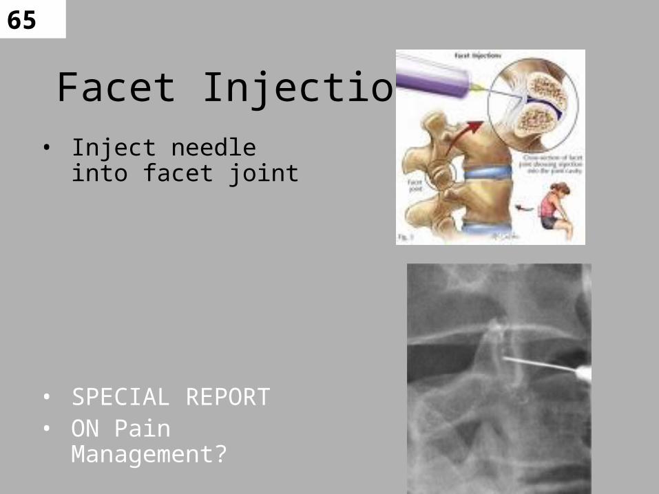
Facet Injections• Inject needle into facet
joint
• SPECIAL REPORT• ON Pain
Management?
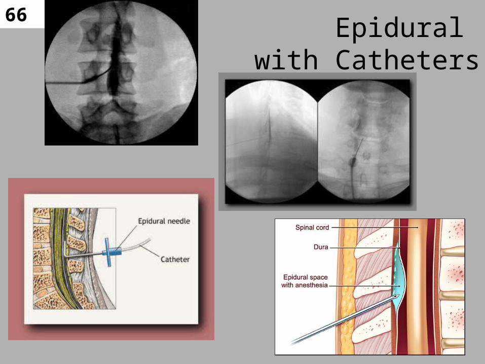
66Epidural
with Catheters

67
SCS With Generator and Transmitter

68
Osteoporosis & Fractures
• 28 million people are at risk, due to fragile bone structure, for an
• osteoporotic fracture:• 700,000 spinal fractures -one every 45 seconds
• 300,000 hip fractures • 250,000 wrist fractures • 250,000 other types of fractures. • SPECIAL REPORT ON BONE DEXA SCAN??

69
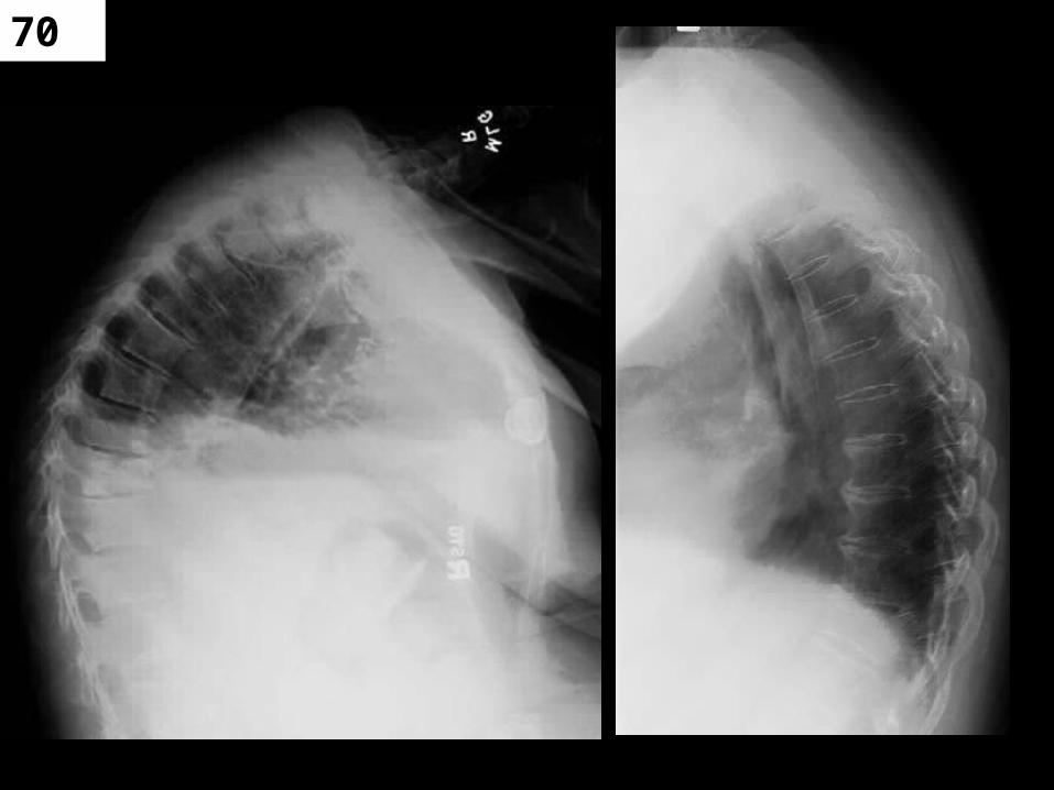
70
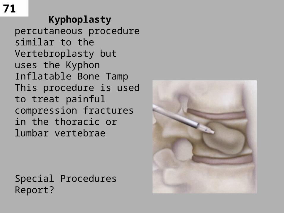
71Kyphoplasty
percutaneous procedure similar to the Vertebroplasty but uses the Kyphon Inflatable Bone TampThis procedure is used to treat painful compression fractures in the thoracic or lumbar vertebrae
Special Procedures Report?

72
Vertebroplasty under Fluoro
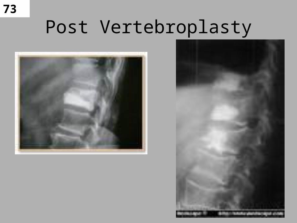
73
Post Vertebroplasty
74
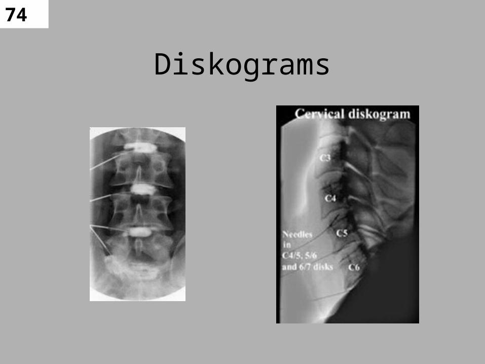
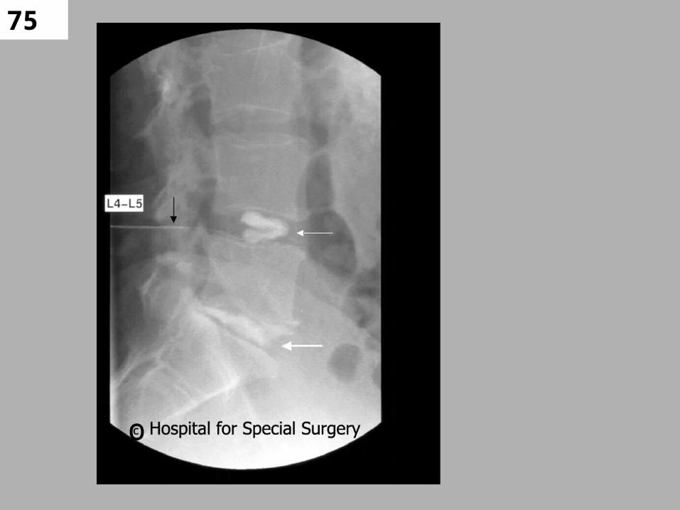
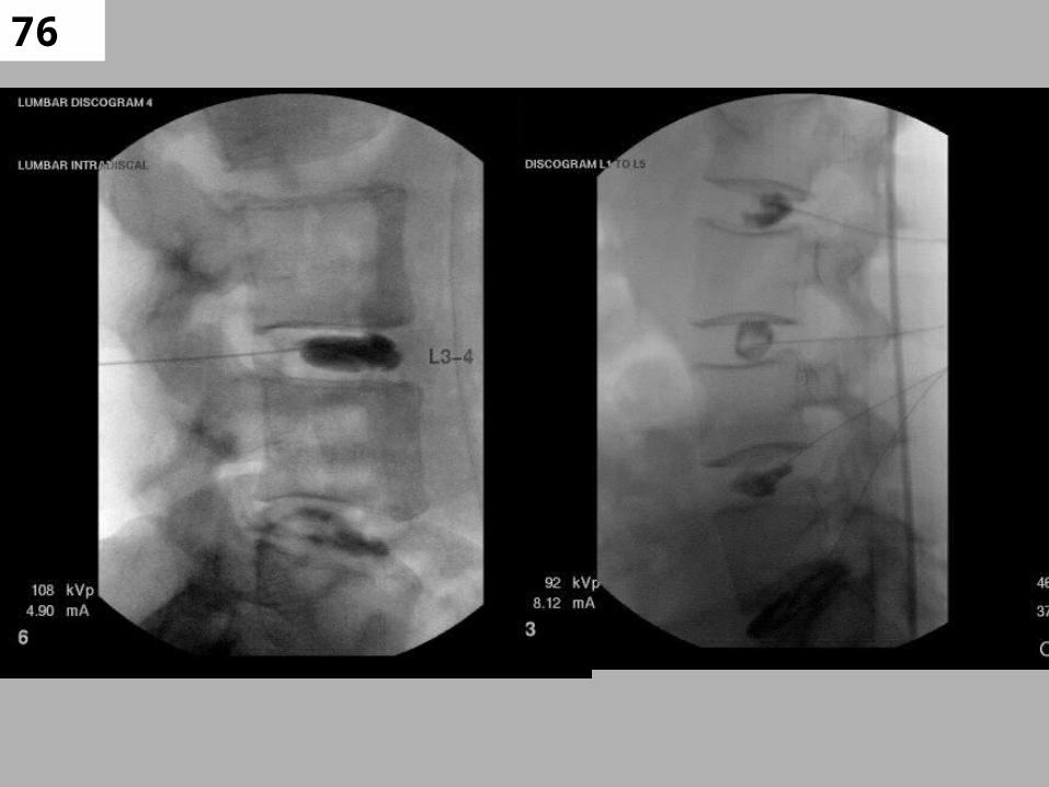
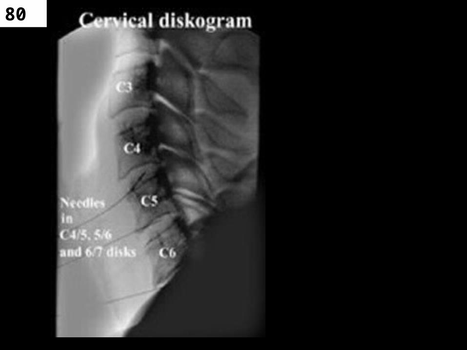
Diskograms
75
76

77
Lumbar Diskograms
78
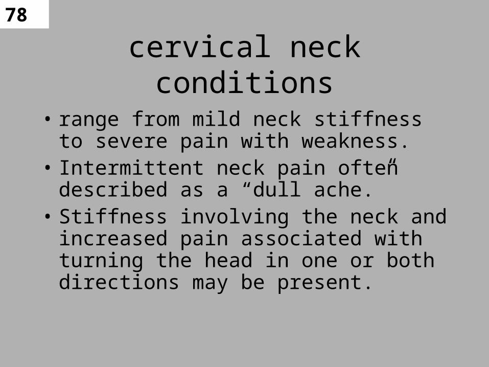
cervical neck conditions
• range from mild neck stiffness to severe pain with weakness.
• Intermittent neck pain often described as a “dull ache.”
• Stiffness involving the neck and increased pain associated with turning the head in one or both directions may be present.

79
Neck Pain
• Pain radiating into the shoulder or shoulder blade.
• Pain and/or numbness extending into the arm, hand, and fingers.
• Weakness of the arm or hand grip, which may be accompanied by muscle atrophy (the reduction in size of a muscle)
• Weakness involving the legs or loss of coordination
80
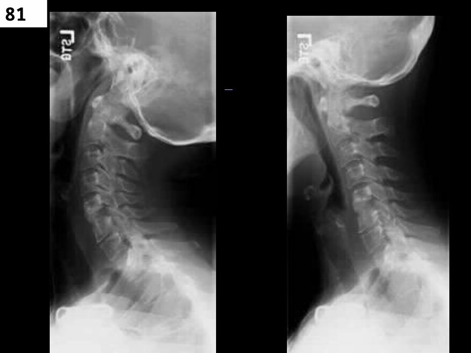
81

82
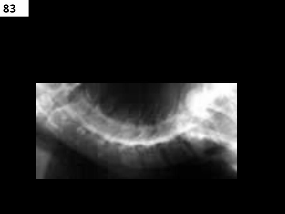
83
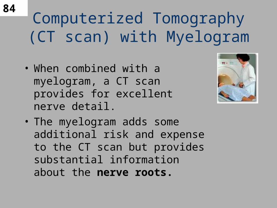
84Computerized Tomography (CT scan) with Myelogram
• When combined with a myelogram, a CT scan provides for excellent nerve detail.
• The myelogram adds some additional risk and expense to the CT scan but provides substantial information about the nerve roots.

85
CT of Spine
• Useful in diagnosis of vertebral column hemangiomas and lumbar spine stenosis
• Often used post-trauma to assess Axis and Atlas fractures and for better demonstration of C7-T1
• Clearly demonstrates size, number and locations of fracture fragments of C, T and L spine.
86
CTM
• Performed after intrathecal injection
• Can be performed at any level of vertebral column
• Multiple slices taken (1.5 – 3mm) – Gantry is tilted
• Windowing allows for density and contrast changes
• Can obtain images with small amounts of contrast– Can be done 4 hours after initial injection
87
CTM

88
MRI of Spinal Cord and CSF flow
• Non-invasive
– Provides anatomic detail of brain, spinal cord, intravertebral disc spaces, and CSF within subarachnoid space
– Does not require intrathecal injection
– Does not have bone artifacts
89
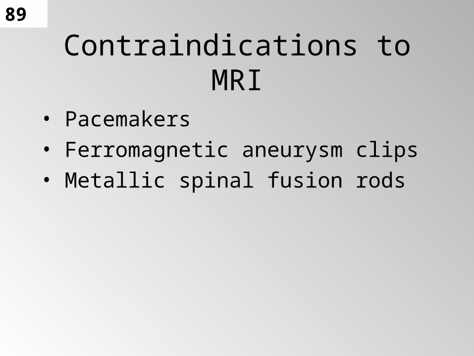
Contraindications to MRI
• Pacemakers
• Ferromagnetic aneurysm clips
• Metallic spinal fusion rods
90
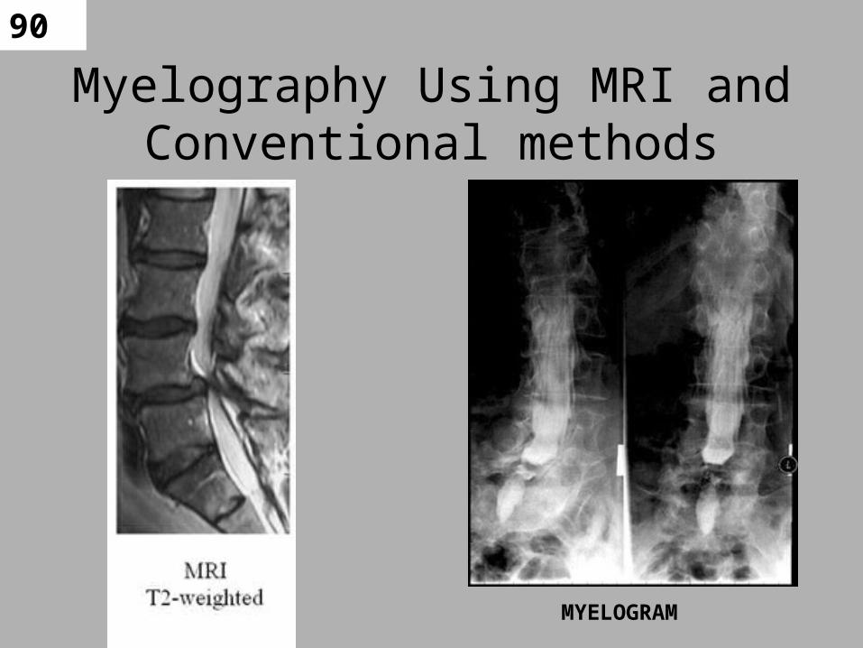
Myelography Using MRI and Conventional methods
MYELOGRAM
91
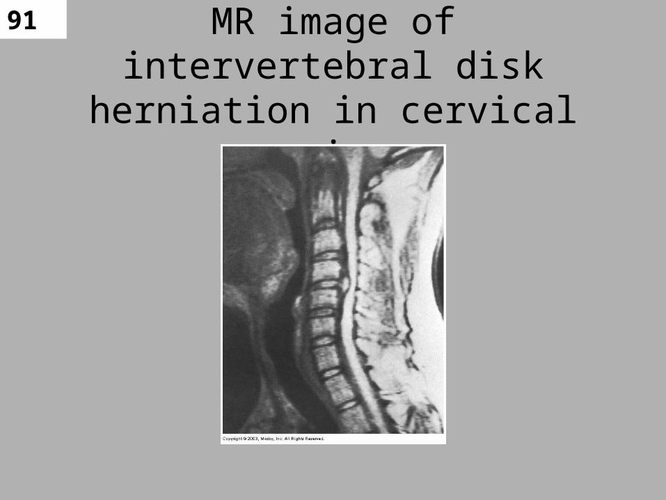
MR image of intervertebral disk herniation in cervical region
92 Preference of MRI
• MRI is the preferred modality for
• Spinal cord– Allows direct visualization of spinal cord, nerve
roots, and surrounding CSF– Can be done in various planes– Aid in diagnosis and treatment of neurodisorders

93
Usefulness of MRI
• Assessing demyelinating disease– Such as MS
• Spinal cord compression
• Postradiation therapy changes of spinal cord tumors
• Herniated disks
• Congenital abnormalities of vertebral column
• Metastatic disease
• Paraspinal masses

94
MRI vs. CT
• MRi superior to CT for imaging of posterior fossa– CT has artifacts from bone– MRI is free from bone artifacts
• MRI has inability to image calcified structures.
• CT is superior for calcifications

95
RADIOGRAPHIC IMAGES AND
PATHOLOGY
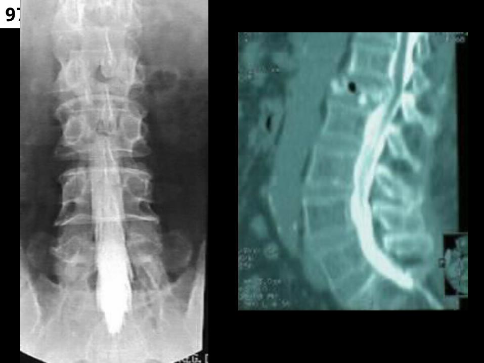
WHAT DO YOU SEE?

96
97
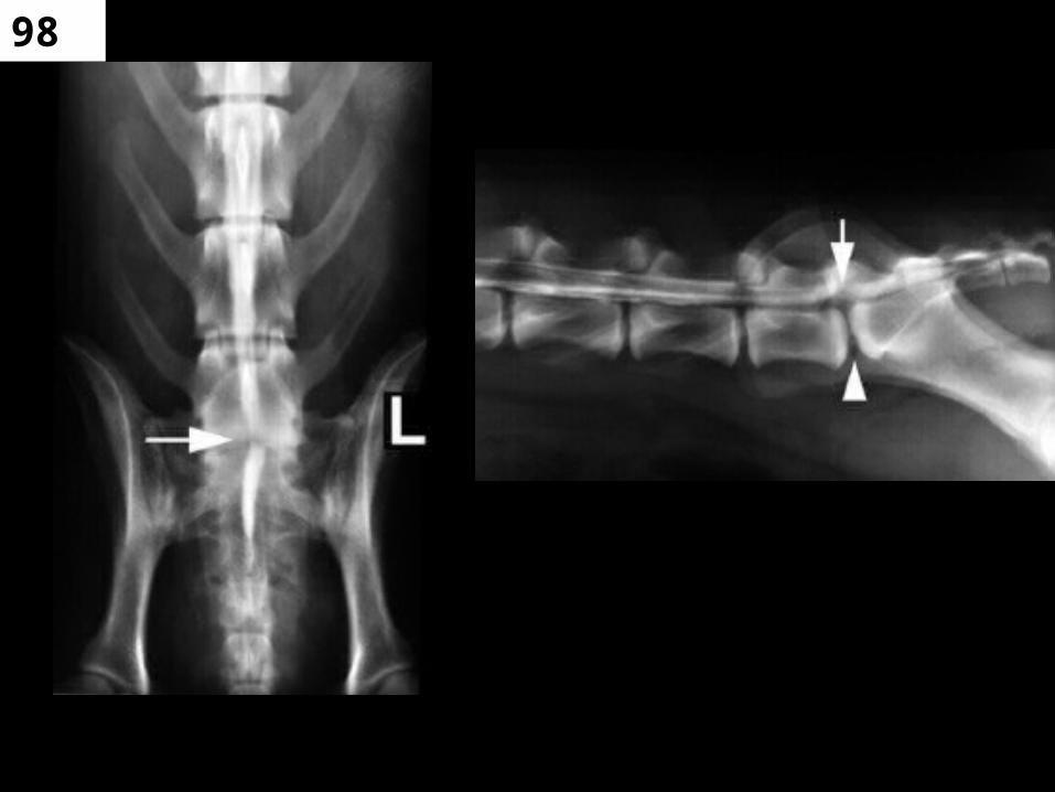
98
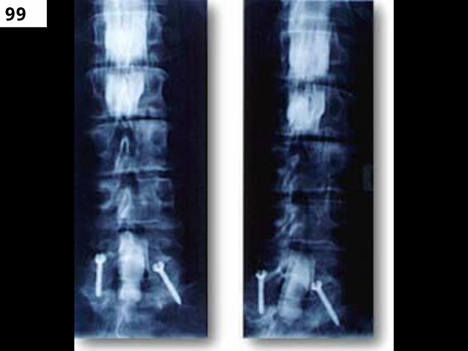
99

100

101
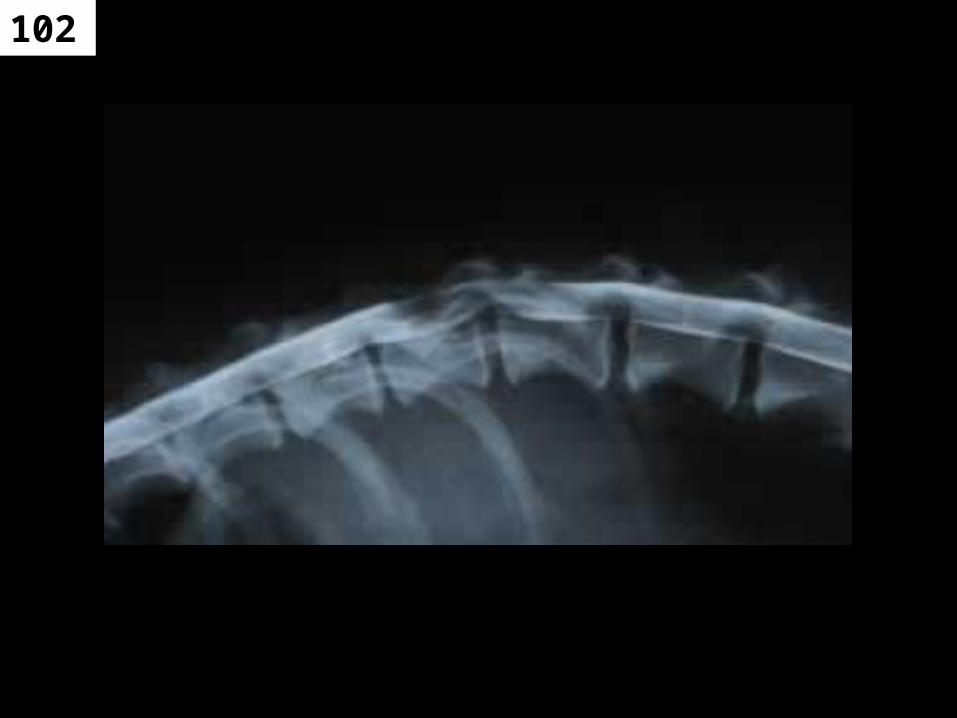
102