1 yan chao-gan ph. d [email protected] state key laboratory of cognitive neuroscience and learning,...
TRANSCRIPT

1
YAN Chao-Gan严超赣Ph. D
State Key Laboratory of Cognitive Neuroscience and Learning,
Beijing Normal University, China
Data Processing of Resting-State fMRI
(Part 1)
2
Outline
• Overview
• Data Preparation
• Preprocess
• ReHo, ALFF, fALFF Calculation
• Functional Connectivity
• Utilities

3
Overview
Based on Matlab, SPM, REST, MRIcroN’s dcm2nii

4
Setup
E:\ITraWork\100402Trainning\Softs\DPARSF_V1.0_100201
NO Chinese character or space in the path.

5
DPARSF's standard procedure
Convert DICOM files to NIFTI images. Remove First 10 Time Points. Slice Timing. Realign. Normalize. Smooth (optional). Detrend. Filter. Calculate ReHo, ALFF, fALFF (optional). Regress out the Covariables (optional). Calculate Functional Connectivity (optional). Extract AAL or ROI time courses for further analysis (optional).

6
Outline
• Overview
• Data Preparation
• Preprocess
• ReHo, ALFF, fALFF Calculation
• Functional Connectivity
• Utilities
7
Data preparation
Arrange the information of the subjects

8
Data preparation
Information of subjects

9
Data preparation
Arrange the information of the subjects
Arrange the MRI data of the subjects
Functional MRI data
Structural MRI data
DTI data
10
被试信息整理
原始数据整理

11
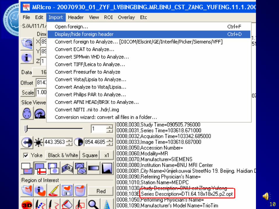
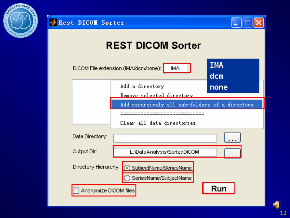
Sort DICOM data
12
IMAdcmnone

13
Data preparation
Arrange each subject's fMRI DICOM images in one directory, and th
en put them in "FunRaw" directory under the working directory.
Subject 1’s DICOM filesFunRaw directory, please name as thisSubject 1’s directoryWorking directory
14
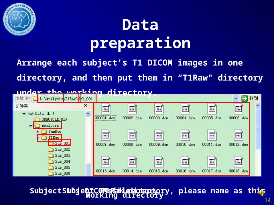
Data preparation
Arrange each subject's T1 DICOM images in one directory, and then
put them in “T1Raw" directory under the working directory.
Subject 1’s DICOM filesT1Raw directory, please name as thisSubject 1’s directoryWorking directory

15
Data preparation
Set the parameters in DPARSF
Set the working directorySet the time points (volumes)The detected subjects’ IDSet the TR

16
Outline
• Overview
• Data Preparation
• Preprocess
• ReHo, ALFF, fALFF Calculation
• Functional Connectivity
• Utilities

17
Preprocess
• DICOM -> NIFTI
• Remove First 10 Time Points
• Slice Timing
• Realign
• Normalize
• Smooth
• Detrend
• Filter: 0.01-0.08

18
DICOM->NIFTI
MRIcroN’s dcm2niigui
SPM5’s DICOM Import

19
DICOM->NIFTI
DPARSF

20
Preprocess
• DICOM -> NIFTI
• Remove First 10 Time Points
• Slice Timing
• Realign
• Normalize
• Smooth
• Detrend
• Filter: 0.01-0.08

21
Remove First 10 Time Points
DPARSF
22
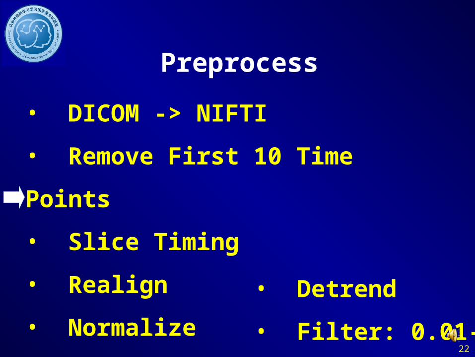
Preprocess
• DICOM -> NIFTI
• Remove First 10 Time Points
• Slice Timing
• Realign
• Normalize
• Smooth
• Detrend
• Filter: 0.01-0.08

23
Slice Timing
Why?

24
Slice Timing
Why?
Huettel et al., 2004

25
Slice Timing
1:2:25,2:2:24252 2-(2/25)25
26
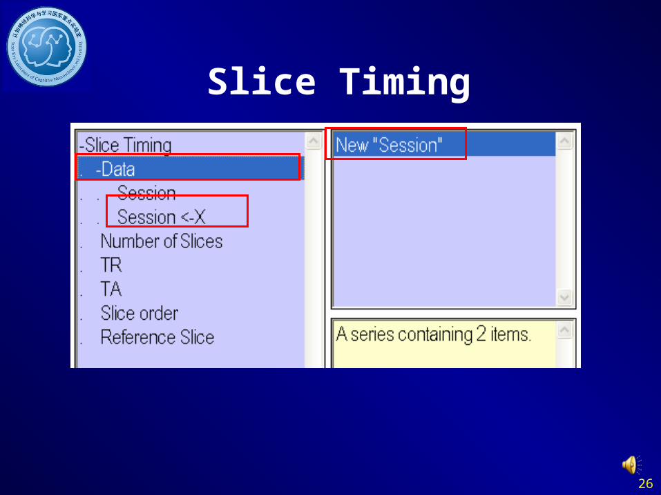
Slice Timing

27
Slice Timing
DPARSF
1:2:25,2:2:24

28
Slice TimingIf you start with NIFTI images (.hdr/.img pairs) before slice timing,
you need to arrange each subject's fMRI NIFTI images in one
directory, and then put them in "FunImg" directory under the
working directory.
FunImg directory, please name as this

29
Preprocess
• DICOM -> NIFTI
• Remove First 10 Time Points
• Slice Timing
• Realign
• Normalize
• Smooth
• Detrend
• Filter: 0.01-0.08

30
Realign
Why?

31
Realign

32
Realign
DPARSF

33
Realign
Check head motion:
Excluding Criteria: 2.5mm and 2.5 degreeNone
Excluding Criteria: 2.0mm and 2.0 degreeSub_013
Excluding Criteria: 1.5mm and 1.5 degreeSub_013
Excluding Criteria: 1.0mm and 1.0 degreeSub_007Sub_012Sub_013Sub_017Sub_018

34
Preprocess
• DICOM -> NIFTI
• Remove First 10 Time Points
• Slice Timing
• Realign
• Normalize
• Smooth
• Detrend
• Filter: 0.01-0.08

35
Normalize
Why?
Huettel et al., 2004
36
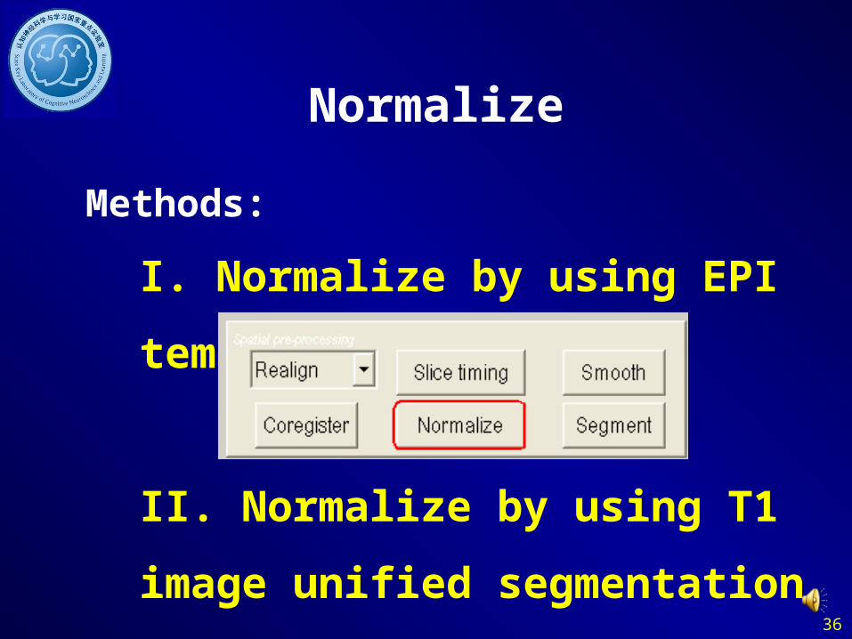
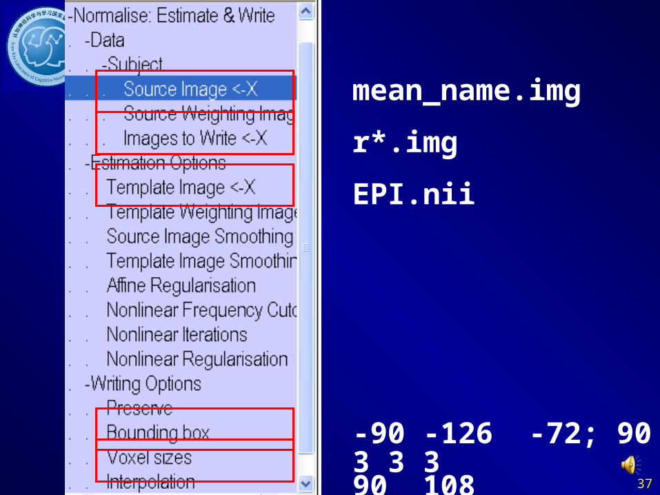
Normalize
Methods:
I. Normalize by using EPI templates
II. Normalize by using T1 image unif
ied segmentation
37
mean_name.img
r*.img
EPI.nii
-90 -126 -72; 90 90 1083 3 3

38
Normalize I

39
Normalize
Methods:
• Normalize by using EPI templates• Normalize by using T1 image unifi
ed segmentation
Structural image was coregistered to the mean functional image after the motion correction
The transformed structural image was then segmented into gray matter, white matter, cerebrospinal fluid by using a unified segmentation algorithm
Normalize: the motion corrected functional volumes were spatially normalized to the MNI space using the normalization parameters estimated during unified segmentation (*_seg_sn.mat)

40
Normalize II: Coregister
mean_name.img T1.img

41
Normalize II:
T1_Coregisted.img
Light Clean
ICBM space template
– East Asian brains
– European brains

42
Normalize II:Segment
New “Segment”

43
New “Normalize: Write”
New “Subject”
name_seg_sn.mat
-90 -126 -72; 90 90 1083 3 3
r*.img
Normalize II:
44
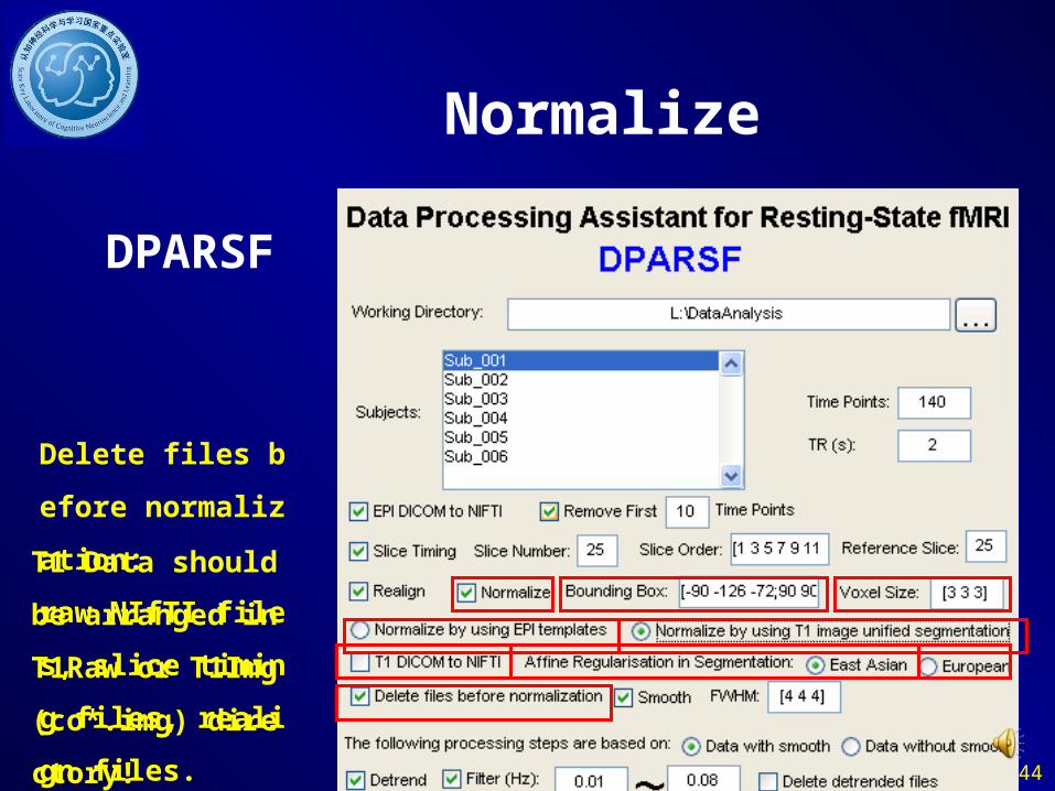
Normalize
DPARSF
T1 Data should be a
rranged in T1Raw
or T1Img (co*.img)
directory!
Delete files before n
ormalization:
raw NIfTI files, slic
e timing files, realig
n files.

45
Normalize
Check Normalization with DPARSF{WROKDIR}\PicturesForChkNormalization
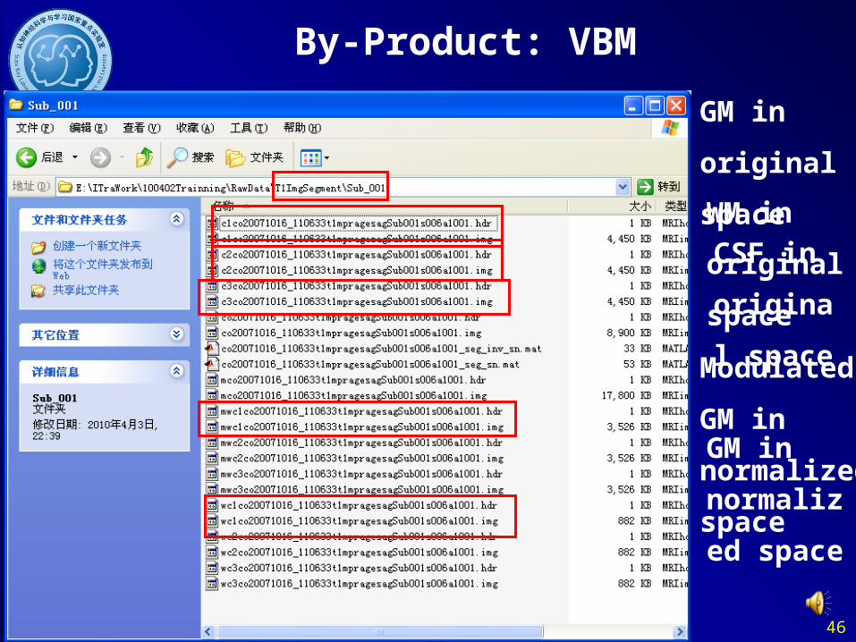
46
GM in
original
spaceWM in
original
space
GM in
normalized
space
Modulated
GM in
normalized
space
CSF in
original
space
By-Product: VBM

47
Preprocess
• DICOM -> NIFTI
• Remove First 10 Time Points
• Slice Timing
• Realign
• Normalize
• Smooth
• Detrend
• Filter: 0.01-0.08
48
Smooth
Why?
• Reduce the effects of the bad n
ormalization
• …

49
w*.imgFWHM kernel
Smooth

50
Smooth
DPARSF
ALFF, fALFF, Funt
ional Connectivity:
Data with smooth
ReHo:
Data without smoot
h
Without former steps:
Data arranged in Fu
nImgNormalized dir
ectory.

51
Preprocess
• DICOM -> NIFTI
• Remove First 10 Time Points
• Slice Timing
• Realign
• Normalize
• Smooth
• Detrend
• Filter: 0.01-0.08

52
Detrend

53
Preprocess
• DICOM -> NIFTI
• Remove First 10 Time Points
• Slice Timing
• Realign
• Normalize
• Smooth
• Detrend
• Filter: 0.01-0.08

54
滤波
Why? • Low frequency (0.01–0.08 Hz) fluctuations (LF
Fs) of the resting-state fMRI signal were of physi
ological importance. (Biswal et al., 2005)
• LFFs of resting-state fMRI signal were suggeste
d to reflect spontaneous neuronal activity (Logot
hetis et al., 2001; Lu et al., 2007).
Biswal B, Yetkin FZ, Haughton VM, Hyde JS (1995) Functional connectivity in the motor cortex of resting human brain using echo-planar MRI. Magn Reson Med 34: 537–541. Logothetis NK, Pauls J, Augath M, Trinath T, Oeltermann A (2001) Neurophysiological investigation of the basis of the fMRI signal. Nature 412: 150–157. Lu H, Zuo Y, Gu H, Waltz JA, Zhan W, et al. (2007) Synchronized delta oscillations correlate with the resting-state functional MRI signal. Proc Natl Acad Sci U S A 104: 18265–18269.

55
Filter

56
Detrend and Filter
DPARSF
If you want to calcu
late fALFF, please
do not delete the det
rended files
Without former steps:
Data arranged in Fu
nImgNormalized or
FunImgNormalizedS
moothed directory.

57
Outline
• Overview
• Data Preparation
• Preprocess
• ReHo, ALFF, fALFF Calculation
• Functional Connectivity
• Utilities

58
ReHo (Regional Homogeneity)
Note: Please do not smooth your data in preprocessing, just smooth your data after ReHo calculation.
Zang et al., 2004
Zang YF, Jiang TZ, Lu YL, He Y, Tian LX (2004) Regional homogeneity approach to fMRI data analysis. Neuroimage 22: 394–400.

59
ReHo
If the resolution of your data is not 61*61*73, please resample your mask file at first.

60
Choose one of your functional image. e.g. your normalized functional image or image after Detrend and Filter.
Choose the mask file or ROI
definition file. e.g.
BrainMask_05_61x73x61.img Resample Mask
Resample other kind of data
Data Resample

61
Data Resample

62
Data Resample
0 – Nearest Neighbor
1 – Trilinear
2- 2nd degree b-spline
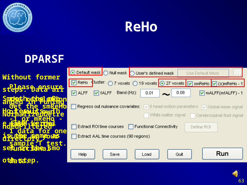
63
Please ensure the
resolution of your
own mask is the
same as your
functional data.
Without former steps:
Data arranged in Fu
nImgNormalizedDet
rendedFiltered
directory.
ReHo
DPARSF
Get the smReHo -1
or mReHo - 1 data f
or one sample T test.
Smooth the mReHo
results. The FWHM
kernel is the same a
s set in the smooth s
tep.

64
Zang et al., 2007
Zang YF, He Y, Zhu CZ, Cao QJ, Sui MQ, et al. (2007) Altered baseline brain activity in children with ADHD revealed by resting-state functional MRI. Brain Dev 29: 83–91.
ALFF(Amplitude of Low
Frequency Fluctuation )
65
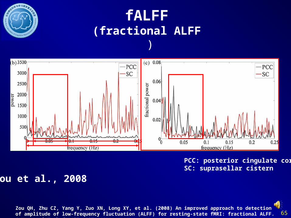
fALFF(fractional ALFF )
Zou et al., 2008
Zou QH, Zhu CZ, Yang Y, Zuo XN, Long XY, et al. (2008) An improved approach to detection of amplitude of low-frequency fluctuation (ALFF) for resting-state fMRI: fractional ALFF. J Neurosci Methods 172: 137-141.
PCC: posterior cingulate cortexSC: suprasellar cistern
66
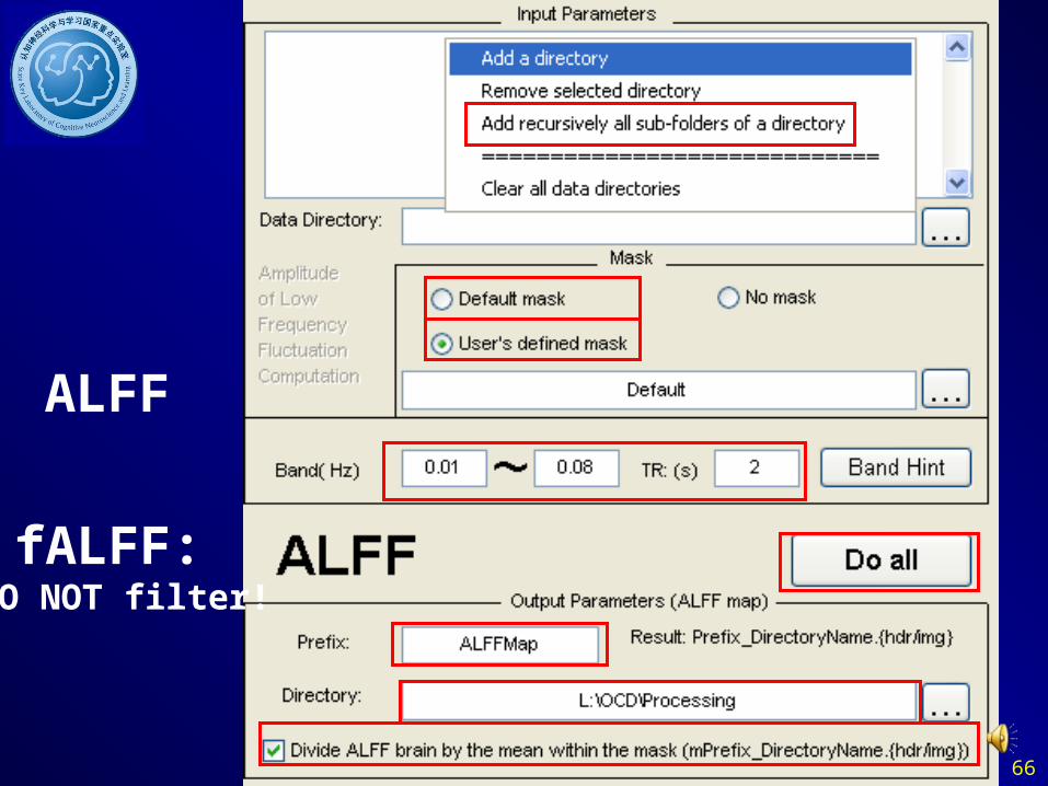
ALFF
fALFF: DO NOT filter!

67
Without former steps:
Data arranged in Fu
nImgNormalizedSmo
othedDetrendedFilte
red
or FunImgNormalize
dSmoothedDetrende
d directory.
Please ensure the
resolution of your
own mask is the
same as your
functional data.
ALFF and fALFF
DPARSF
Get the mALFF - 1
or (mfALFF - 1) da
ta for one sample T
test.
Please DO NOT del
ete the detrended fil
es before filter. DP
ARSF will calculate
d the fALFF based
on data before filter.

68
Outline
• Overview
• Data Preparation
• Preprocess
• ReHo, ALFF, fALFF Calculation
• Functional Connectivity
• Utilities

69
Regress out nuisance covariates
• Head motion parameters: rp_name.txt
• Global mean signal
• White matter signal
• Cerebrospinal fluid signal

70
Extract
Covariates
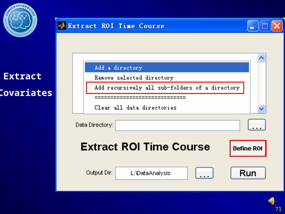
71
Extract
Covariates

72
Extract
Covariates

73
Extract
Covariates

74
Extract
Covariates

75
Extract
Covariates
Extract one subject’s
Covariates

76
Extract
Covariates
Extract multi subjects’
Covariates

77
Extract
Covariates

78
Extract
Covariates

79
Regress out nuisance Covariates
Extract Covariates• Head motion parameters: rp_name.txt• Global mean signal• White matter signal• Cerebrospinal fluid signal
• Combine the covariates for future using in REST
RPCov=load('rp_name.txt'); BCWCov=load('ROI_FCMap_name.txt'); Cov=[RPCov,BCWCov]; save('Cov.txt', 'Cov', '-ASCII', '-DOUBLE','-TABS');

80
Regress out
Covariates

81
Extract
Covariates CovList.txt:
Covariables_List:X:\Process\Sub3Cov.txtX:\Process\Sub2Cov.txtX:\Process\Sub1Cov.txt
CovList.txt:

82
Without former steps:
Data arranged in Fu
nImgNormalizedDet
rendedFiltered
or FunImgNormalize
dSmoothedDetrende
dFiltered
directory.
rp*.txt
DPARSF
CsfMask_07_61x73
x61.img
BrainMask_05_61x
73x61.img
Regress out nuisance Covariates
WhiteMask_09_61x
73x61.img

83
Without former steps:
Data arranged in Fu
nImgNormalizedDet
rendedFiltered
or FunImgNormalize
dSmoothedDetrende
dFiltered
directory.
DPARSF
Regress out Covariates
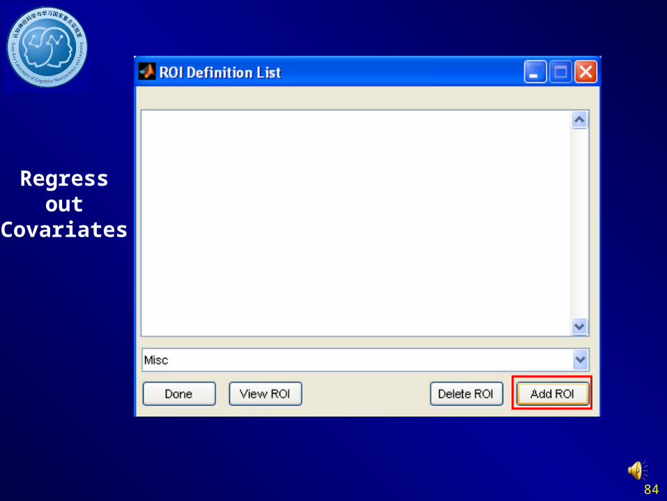
84
Regress out
Covariates

85
Please ensure
the resolution
of your ROI
file is the same
as your
functional data.
Regress out
Covariates
86
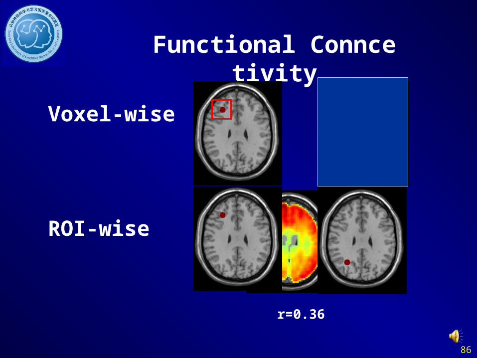
Functional Conncetivity
Voxel-wise
ROI-wise
r=0.36
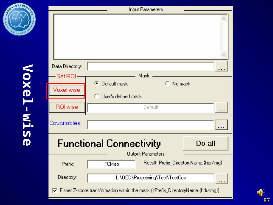
87
Voxel-w
ise

88
Voxel-w
ise
Please ensure
the resolution
of your ROI
file is the same
as your
functional data.
SeedList.txt:
Seed_Time_Course_List:X:\Process\Sub3Seed.txtX:\Process\Sub2Seed.txtX:\Process\Sub1Seed.txt
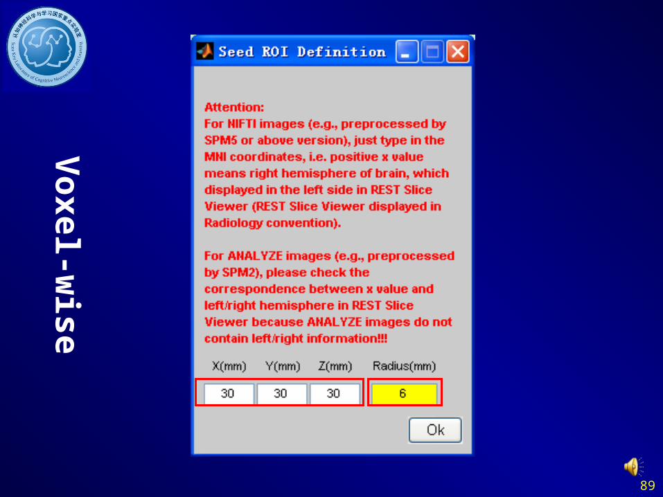
89
Voxel-w
ise

90
Voxel-w
ise

91
Voxel-w
ise

92
Voxel-w
ise
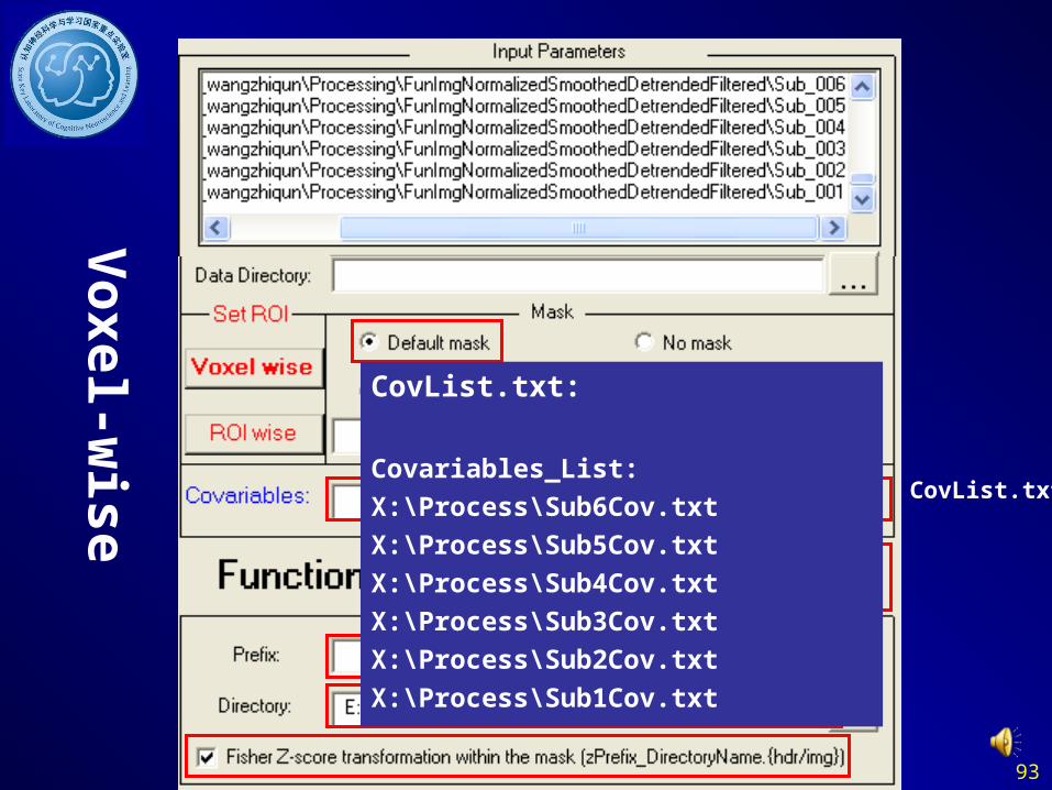
93
Voxel-w
ise
CovList.txt
CovList.txt:
Covariables_List:X:\Process\Sub6Cov.txtX:\Process\Sub5Cov.txtX:\Process\Sub4Cov.txtX:\Process\Sub3Cov.txtX:\Process\Sub2Cov.txtX:\Process\Sub1Cov.txt

94
RO
I-wise

95
RO
I-wise

96
RO
I-wise
CovList.txt
CovList.txt:
Covariables_List:X:\Process\Sub6Cov.txtX:\Process\Sub5Cov.txtX:\Process\Sub4Cov.txtX:\Process\Sub3Cov.txtX:\Process\Sub2Cov.txtX:\Process\Sub1Cov.txt

97
RO
I-wise
98
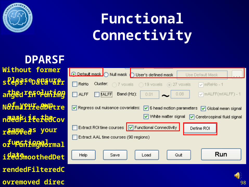
Without former steps:
Data arranged in Fu
nImgNormalizedDet
rendedFilteredCovre
moved
or FunImgNormalize
dSmoothedDetrende
dFilteredCovremove
d directory.
Please ensure the
resolution of your
own mask is the
same as your
functional data.
Functional Connectivity
DPARSF
99
Fu
nction
al C
onn
ectivity
100
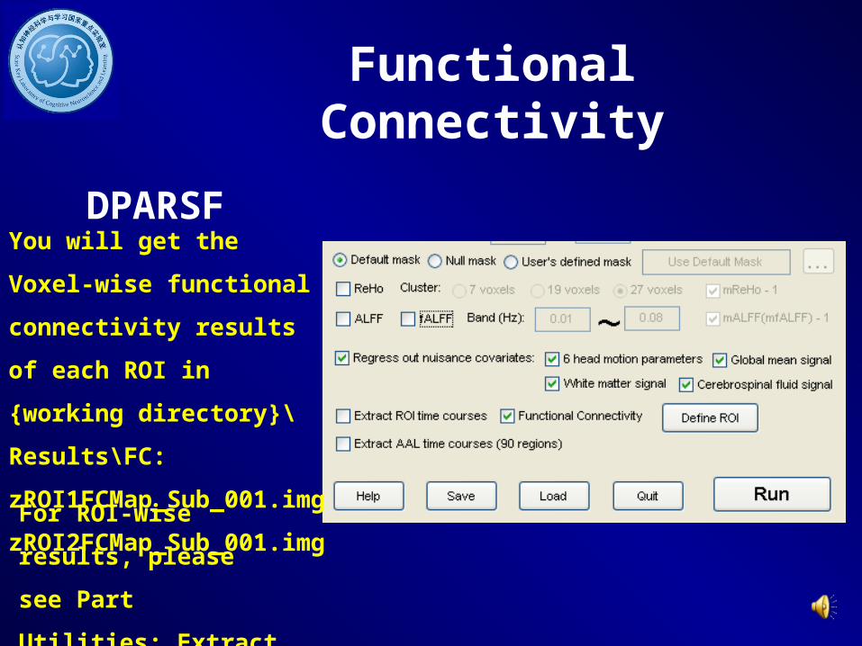
You will get the Voxel-wise
functional connectivity results
of each ROI in {working
directory}\Results\FC:
zROI1FCMap_Sub_001.img
zROI2FCMap_Sub_001.img
For ROI-wise results,
please see Part Utilities:
Extract ROI time courses.
Functional Connectivity
DPARSF

101
Outline
• Overview
• Data Preparation
• Preprocess
• ReHo, ALFF, fALFF Calculation
• Functional Connectivity
• Utilities

102
Without former steps:
Data arranged in Fu
nImgNormalizedDet
rendedFilteredCovre
moved
or FunImgNormalize
dSmoothedDetrende
dFilteredCovremove
d directory.
Extract ROI time courses
DPARSF
103
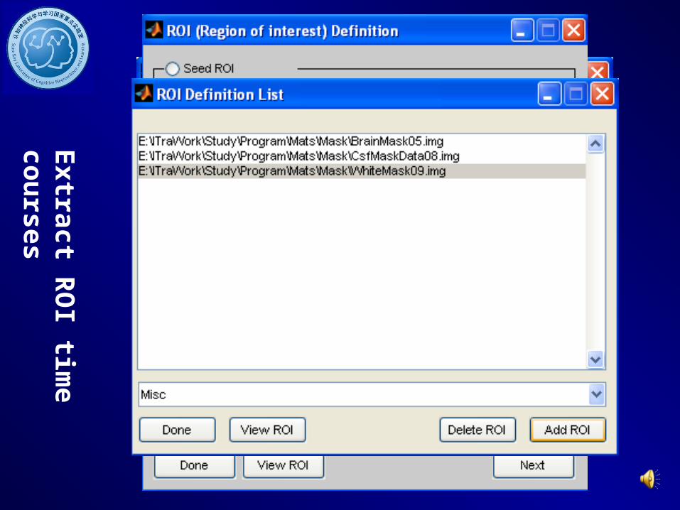
Extract R
OI tim
e courses
104
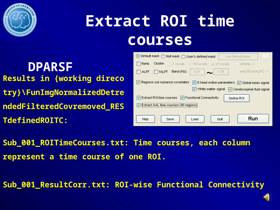
Results in {working direcotry}\FunI
mgNormalizedDetrendedFilteredCo
vremoved_RESTdefinedROITC:
Extract ROI time courses
DPARSF
Sub_001_ROITimeCourses.txt: Time courses, each column represent a time
course of one ROI.
Sub_001_ResultCorr.txt: ROI-wise Functional Connectivity

105
Without former steps:
Data arranged in Fu
nImgNormalizedDet
rendedFilteredCovre
moved
or FunImgNormalize
dSmoothedDetrende
dFilteredCovremove
d directory.
Extract AAL time courses
DPARSF

106
Results in {working direcotry}\FunI
mgNormalizedDetrendedFilteredCo
vremoved_AALTC:
Extract AAL time courses
DPARSF
Sub_001_AALTC.mat: Time courses of each AAL region.

107
Normalization by usin
g T1 image segmentati
on: co*.img
Realign without Slice
Timeing: a*.img
Change prefix of Images
DPARSF

108
Normalization by usin
g T1 image segmentati
on: co*.img
Change prefix of Images
DPARSF
aa*.img -> ra*.img
ra

109
Save parameters to
*.mat
Save and Load Parameters
DPARSF
Load parameters
from *.mat

110
www.restfmri.net
Further Help
Further questions:

111
Thanks to
DONG Zhang-YeGUO Xiao-JuanHE YongLONG Xiang-YuSONG Xiao-WeiYAO LiZANG Yu-FengZHANG HanZHU Chao-ZheZOU Qi-HongZUO Xi-Nian……
All the group members!
SPM Team: Wellcome Department of Imaging Neuroscience, UCL
MRIcroN Team: Chris Rorden
……

112
Thanks for your attention!