1.0 introduction - nathan luedtke laboratory 1.0 introduction the central dogma of biology states...
TRANSCRIPT

1
1.0 Introduction
The central dogma of biology states that from DNA, RNA is transcribed to serve
as an information messenger that is translated into proteins.1 Messenger RNA
(mRNA) accounts, however, for only a small fraction of the RNA present within a
cell. Far from being a passive carrier of genetic code, RNA exhibits vast
structural and functional diversity and is intimately involved in a wide range of
biological activities, including information storage and chemical catalysis.
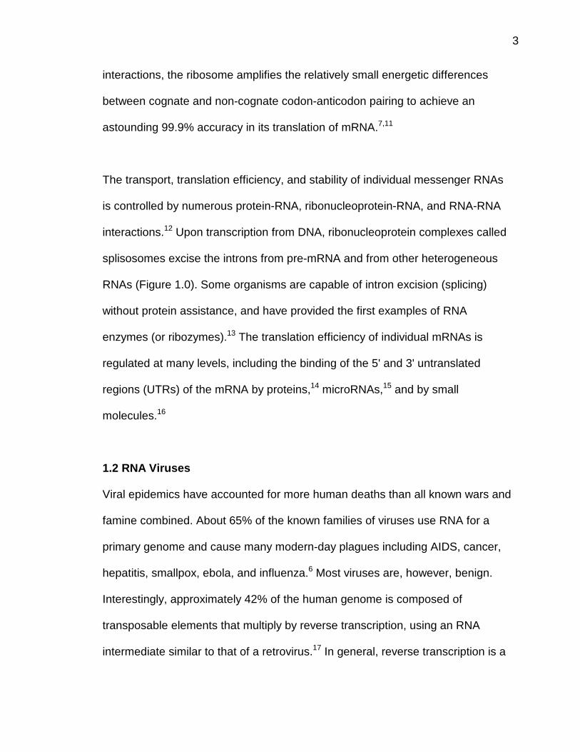
Figure 1.0: Molecular recognition of RNA often precedes catalytic events that areessential to a wide range of cellular activities including: (a) initiation of DNAreplication,2 (b) extension of the telomeric regions of chromosomes,3 (c) splicingof pre-mRNA,4 and iron chelation.5 In addition, RNA serves as the primarygenome of most pathogenic viruses.6
A more “modern” interpretation of the central dogma of biology is that RNA has
structural and functional characteristics that are, in many ways, similar to both

2
DNA and proteins. RNA is, therefore, an intermediate between DNA and proteins
in more than one respect.
1.1 Translation
Gene expression relies upon an interplay between recognition events and
catalytic activities that are mediated by RNA-protein complexes. Ribosomal RNA
(rRNA) accounts for the vast majority of total cellular RNA (80%) and provides
both the molecular scaffold and enzymatic activities needed for protein
translation.7 The key step of translation occurs in the ribosome’s A-site, where
codon-anticodon recognition decodes mRNA. Upon a correct codon-anticodon
match between mRNA and the anticodon loop of tRNA, the ribosome’s peptidyl
transferase activity catalyzes the formation of a new peptide bond between the
amino acid-charged tRNA in the A-site and the growing protein chain on the
tRNA in the P-site.7 Studies have shown that prokaryotic ribosomes that are
stripped of protein are still capable of limited peptidyl transferase activity.8 In
accordance with this result, recent crystal structures show that the peptidyl
transferase active site is composed entirely of rRNA.9 A single, unusually basic
adenosine may be the key player in the mechanism of peptidyl transfer.10
Transfer RNAs, at 15% of total cellular RNA, are the most common type of
“soluble” RNA (i.e. lacking any associated proteins). The binding of tRNA to the
ribosomal A-site is mediated by extensive RNA-RNA interactions (including
rRNA-tRNA, and mRNA-tRNA binding). Through these, and other important
3
interactions, the ribosome amplifies the relatively small energetic differences
between cognate and non-cognate codon-anticodon pairing to achieve an
astounding 99.9% accuracy in its translation of mRNA.7,11
The transport, translation efficiency, and stability of individual messenger RNAs
is controlled by numerous protein-RNA, ribonucleoprotein-RNA, and RNA-RNA
interactions.12 Upon transcription from DNA, ribonucleoprotein complexes called
splisosomes excise the introns from pre-mRNA and from other heterogeneous
RNAs (Figure 1.0). Some organisms are capable of intron excision (splicing)
without protein assistance, and have provided the first examples of RNA
enzymes (or ribozymes).13 The translation efficiency of individual mRNAs is
regulated at many levels, including the binding of the 5' and 3' untranslated
regions (UTRs) of the mRNA by proteins,14 microRNAs,15 and by small
molecules.16
1.2 RNA Viruses
Viral epidemics have accounted for more human deaths than all known wars and
famine combined. About 65% of the known families of viruses use RNA for a
primary genome and cause many modern-day plagues including AIDS, cancer,
hepatitis, smallpox, ebola, and influenza.6 Most viruses are, however, benign.
Interestingly, approximately 42% of the human genome is composed of
transposable elements that multiply by reverse transcription, using an RNA
intermediate similar to that of a retrovirus.17 In general, reverse transcription is a
4
highly error-prone process allowing viral elements to evolve rapidly under
selective pressures (such as anti-viral drugs). An additional 8% of the human
genome is composed of repetitive genomic elements known as “retrovirus-like
elements”.17 Their structures very closely resemble those of retroviruses, carrying
the open reading frames common to all retroviruses (Gag, Pol, Env), flanked by
5' and 3' long terminal repeats. Overall, the human genome is composed of
approximately 50% self-repeating parasitic sequences. Compare this with the
unique (non-repeated) genes, representing only ~5% of the human genome!17
1.3 Small Molecules That Modulate RNA Activity
The ability of RNA to facilitate the essential biochemical activities needed for
information storage, signal transduction, replication, and enzymatic catalysis has
distinguished it as a candidate for being the central biomolecule in a prebiotic
world.18 If such an “RNA world” ever did exist, then small molecule-RNA
interactions certainly played a key role in the regulation of RNA replication,
processing, as well as other enzymatic and regulatory activities.19
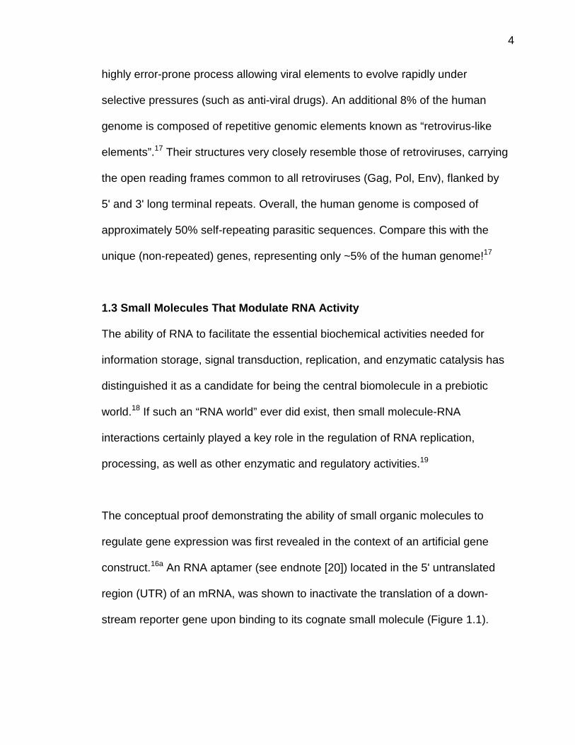
The conceptual proof demonstrating the ability of small organic molecules to
regulate gene expression was first revealed in the context of an artificial gene
construct.16a An RNA aptamer (see endnote [20]) located in the 5' untranslated
region (UTR) of an mRNA, was shown to inactivate the translation of a down-
stream reporter gene upon binding to its cognate small molecule (Figure 1.1).
5
The mechanism proposed for the small molecule-dependent translation
inactivation involves a structural rearrangement of the 5'-UTR into a rigid
complex that cannot be scanned by the ribosomal pre-initiation machinery.
Recent studies have shown that natural systems use small molecule-RNA
binding (accompanied by RNA structural rearrangements) to directly modulate
mRNA translational efficiencies.16 b,c
Figure 1.1: The mature mRNA of an artificial gene construct is actively translatedin the absence of small-molecule binding (Top). Upon binding the 5'-UTR by itscognate small molecule, the translation of the gene is deactivated (Bottom).16a
Recent studies indicate that similar mRNA-small molecule control mechanismsoccur in vivo and appear, therefore, to represent a normal aspect ofmetabolism16b,c
1.4 Magnesium (II)
Much like proteins, the primary sequence of an RNA directs its folding into a
unique 3-D structure.21 Correct RNA folding, however, typically relies upon the
binding of divalent metal ions (especially Mg2+). The Mg2+ induced folding of the

6
Tertrahymena thermophila group I intron has become an important paradigm for
RNA folding.22 In the absence of Mg2+, it occupies an ensemble of highly dynamic
secondary structures that are dominated by duplex regions interrupted by internal
bulges and stem loops. The group 1 intron secondary structure can be predicted
from its nucleotide sequence using base-pairing and nearest neighbor rules.23
Upon Mg2+ binding it collapses into a more rigid, enzymatically active, tertiary
structure with fewer conformations available. In at least one region of the group 1
intron, Mg2+ binding induces a rearrangement of the RNA secondary structure
itself.24 These cation-mediated “higher-order” folding interactions remain a major
obstacle in the prediction of a 3-dimensional RNA structure given only its primary
sequence.
Mg2+ exhibits a low to moderate affinity to many unrelated RNAs. Mg2+ binding
affinities (Kd) range from 0.01 mM through 10 mM in the presence of 0.1 – 0.2 M
of monovalent ions.25 Given the 3-dimensional structure of an RNA, an
electrostatic contour map can be calculated, allowing for the theoretical
prediction of Mg2+ binding sites.26 The accuracy of such predictions is
complicated by issues related to induced fit and by the limited understanding of
the characteristics of the cations themselves. Crystal structures of tRNAPhe, for
example, indicate that different cations bind at different RNA sites, depending
upon the identity of the ion.27 Few of the metal cation binding sites overlap with
one another (as would be predicted by electrostatic contour mapping).27
Tremendous diversity in the position, size, and affinity provided by RNA
7
coordination sites, suggests that metal ion-RNA complexes may have exhibited
diverse catalytic activities in a prebiotic “RNA world”.
RNA-cation binding interactions are essential for the proper folding and catalytic
function of RNA.22 There are some cases, however, where small molecules other
than metal cations can be used to facilitate the folding and enzymatic activity of
RNA. Linear polyamines (like spermine) and aminoglycosides (Figure 1.2) can
displace Mg2+ from RNA, and have been shown to directly facilitate the
enzymatic activities of the hairpin and hammerhead ribozymes even in the
absence of divalent metal ions.28 Structurally complex and semi-rigid polycations
may have once served as RNA scaffolds, similar to the ribosomal proteins of
today.
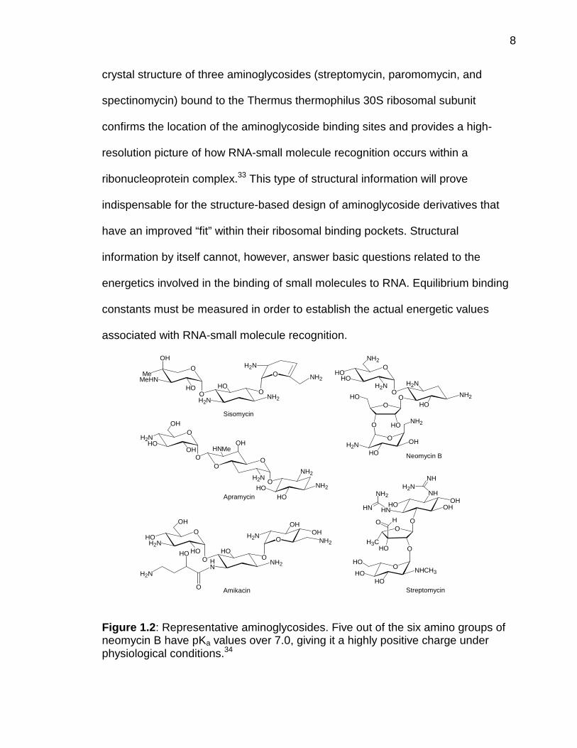
1.5 Aminoglycosides
Aminoglycoside antibiotics are a diverse family of natural products that interfere
with prokaryotic protein biosynthesis (Figure 1.2). Their ability to non-specifically
bind to RNA through electrostatic interactions was described over 20 years
ago.29 The aminoglycosides are also capable, however, of site-specific
recognition of prokaryotic rRNA. Early footprinting experiments indicated that
aminoglycosides bind to discrete locations within the ribosome.30 Later
experiments showed that aminoglycosides increase the affinity of tRNA to the
30S ribosomal A-site,31 thus providing an attractive mechanism to explain their
ability to selectively decrease the fidelity of prokaryotic translation.32 A recent
8
crystal structure of three aminoglycosides (streptomycin, paromomycin, and
spectinomycin) bound to the Thermus thermophilus 30S ribosomal subunit
confirms the location of the aminoglycoside binding sites and provides a high-
resolution picture of how RNA-small molecule recognition occurs within a
ribonucleoprotein complex.33 This type of structural information will prove
indispensable for the structure-based design of aminoglycoside derivatives that
have an improved “fit” within their ribosomal binding pockets. Structural
information by itself cannot, however, answer basic questions related to the
energetics involved in the binding of small molecules to RNA. Equilibrium binding
constants must be measured in order to establish the actual energetic values
associated with RNA-small molecule recognition.
OHO
HO
NH2
H2N
O
NH2
OH
OH2N
O HN
OH
OH
HO
OHO
H2N
NH2
OHH2NHO
NH2
O
HO
OO
NH2
HOO
HOO
HO
H2N
OMe
HO
NH2
H2N
O
NH2O
OH
MeHN
OH2N
HO
HNHO OH
NHCH3HOHO
HOO
NH
OO
OHOH3C
H2NNH
HN
NH2OH
O H
Amikacin
Sisomycin
Neomycin B
StreptomycinO
H2N
HO
O
OH
O
HOHO
H2NNH2
NH2
OH2N
HO
OH
O
HNO
OH Me
Apramycin
Figure 1.2: Representative aminoglycosides. Five out of the six amino groups ofneomycin B have pKa values over 7.0, giving it a highly positive charge underphysiological conditions.34
9
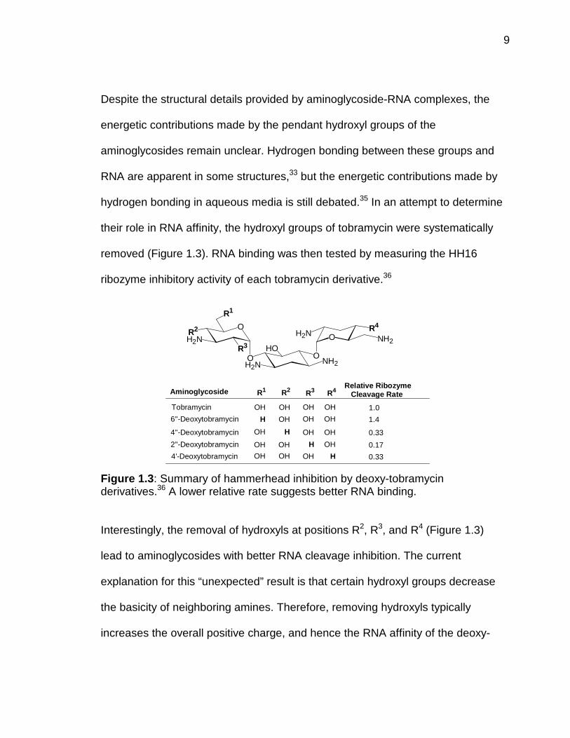
Despite the structural details provided by aminoglycoside-RNA complexes, the
energetic contributions made by the pendant hydroxyl groups of the
aminoglycosides remain unclear. Hydrogen bonding between these groups and
RNA are apparent in some structures,33 but the energetic contributions made by
hydrogen bonding in aqueous media is still debated.35 In an attempt to determine
their role in RNA affinity, the hydroxyl groups of tobramycin were systematically
removed (Figure 1.3). RNA binding was then tested by measuring the HH16
ribozyme inhibitory activity of each tobramycin derivative.36
Figure 1.3: Summary of hammerhead inhibition by deoxy-tobramycinderivatives.36 A lower relative rate suggests better RNA binding.
Interestingly, the removal of hydroxyls at positions R2, R3, and R4 (Figure 1.3)
lead to aminoglycosides with better RNA cleavage inhibition. The current
explanation for this “unexpected” result is that certain hydroxyl groups decrease
the basicity of neighboring amines. Therefore, removing hydroxyls typically
increases the overall positive charge, and hence the RNA affinity of the deoxy-
O
NH2O
OR2
H2N
H2NO
HO
H2NNH2
R3
R4
R1
OH
OH
2''-Deoxytobramycin
OH
Aminoglycoside
OH
R3
OH
R2
H OH6''-Deoxytobramycin
Tobramycin
H
OH
OH
R1
4'-Deoxytobramycin OH
4''-Deoxytobramycin H
OH
R4
OH
H
0.17
OH
OH
OH OH
OH
Relative RibozymeCleavage Rate
1.0
0.33
1.4
0.33

10
derivatives. These de-hydroxylated tobramycin derivatives have not yet been
tested for the binding of other RNAs, so the roles of the hydroxyls in RNA
specificity remain unclear.
1.6 Ligand Specificity
For the purposes of this thesis, specificity will be defined as the binding affinity
(Keq) of a small molecule to a particular RNA site, divided by its average affinity to
“all” other potential binding sites:
specificity = Keq(interaction of interest)average Keq(other sites of interaction)
Specificity is proportional to occupancy of the “desired” RNA site, versus the
occupancy of all other potential binding sites. For practical reasons, specificity is
a relative term, where the affinity between a small molecule and its RNA “target”
is weighted by its affinity to “other” nucleic acids. The reported specificity is,
therefore, always dependent on the selection of the competitor or “non-specific”
nucleic acids used for the comparison.
High specificity is a prerequisite for the effective modulation of RNA activity in
vivo. Since non-specific binding sites are typically present at much higher
concentrations as compared to the desired target, the bioavailability of a small
molecule may suffer even if it has a moderate affinity to “other” sites. The binding
11
of the small molecules to tRNA, rRNA, DNA, proteins, phospholipids, etc., may
also cause undesired biological “side effects” including toxicity and mutagenicity.
Aminoglycosides, for example, are not ideal antibiotics. Their promiscuous
binding of RNA and/or membrane components may be related to the multiple
therapeutic side effects and the low-moderate bacteriacidal potency exhibited by
these compounds.37,38 Aminoglycosides bind to and inhibit the function of a wide
range of unrelated RNAs with moderate activities (IC50 = 0.1 – 100 µM).19,28c,39
Aminoglycosdies, therefore, exhibit a low specificity for most of these RNA sites.
Aminoglycosides do, however, show excellent specificity for RNA over DNA
(Section 6.0). For this reason, we have used aminoglycosides as “scaffolds” for
the synthesis of new small molecules targeted towards specific RNA sites. These
derivatives are found to exhibit dramatically different RNA specificities and
altered biological activities when compared to their aminoglycoside precursors.
1.7 Goals
There are still no "rules" for the structure-based design of small molecules that
are targeted to a specific RNA tertiary fold. One obstacle is that there are still
very few examples of small molecules that bind to natural RNA structures with
high specificity. There are other potential reasons as well. For example, RNA is a
highly dynamic molecule known to occupy multiple conformations. The structural
details of an RNA do not typically entail the potential structural changes it can
12
adopt upon ligand binding. This adds additional complexity to the structure-based
design of RNA ligands.40
Despite recent progress in the understanding of how small molecules recognize
RNA,41 the following fundamental questions remain largely unanswered:
1. How do electrostatic interactions affect the RNA affinity and specificity ofaminoglycoside-based ligands?
2. How does one design small molecules that exhibit high affinity and highspecificity for a pre-determined RNA target?
To help answer these questions, we have addressed a number of goals:
1. Design and synthesize new small molecules that are targeted to a pre-determined RNA site.
2. Rapidly characterize the affinity and specificity of new RNA ligands usingfluorescence-based methodologies.
3. Conduct experiments in a systematic fashion so that trends in RNA-smallmolecule recognition can be identified.
To evaluate the “higher-order” biological impacts of RNA binding, we have
chosen the HIV-1 Rev-RRE interaction as our model system. This way, new RNA
ligands that show promising activities may eventually prove themselves as future
antiviral agents. Our work, along with the efforts by many other groups,
contributes to the growing body of knowledge that will aid in the future design,
synthesis, and application of small molecules directed to RNA.