1010c lab... · web viewexercise 5 –cell structure and membrane function exercise 8 –...
TRANSCRIPT

Laboratory Manual for General Biology I (BSC 1010C)
Lake-Sumter Community CollegeScience Department
Leesburg

Table of Contents
Note to Students.........................................................................................................................................3
Exercise 1 - Measurements and Lab Techniques.........................................................................................4
Exercise 2 - Functional Groups, Organic Molecules, Buffers, and Dilutions...............................................13
Exercise 3 - Qualitative Analysis of Biological Molecules...........................................................................23
Exercise 4 - The Microscope......................................................................................................................31
Exercise 5 - Cell Structure and Membrane Function..................................................................................46
Exercise 6 - Enzyme Activity......................................................................................................................56
Exercise 7 - Respiration.............................................................................................................................63
Exercise 8 - Photosynthesis.......................................................................................................................67
Exercise 9 - Cell Division............................................................................................................................73
Exercise 10 - DNA Fingerprinting...............................................................................................................81
Exercise 11 - Genetics................................................................................................................................96
A significant portion of this lab manual is used with the kind permission of the Science Department at Seminole State College, Sanford, Florida.
Lake-Sumter Community College, Leesburg Laboratory Manual for BSC 1010C 2

Note to Students
Students should read and study the exercises before coming to the laboratory and should supply themselves with the necessary materials including the text book, lecture notes, laboratory manual, calculators, pens, and pencils. All students are required to wear appropriate clothing to lab as outlined by the lab instructor as well as follow all safety precautions during laboratory exercises.
Lake-Sumter Community College, Leesburg Laboratory Manual for BSC 1010C 3

Exercise 1 - Measurements and Lab Techniques
IntroductionIn scientific experiments, observation and accurate measurements are essential. The investigations in this exercise will familiarize you with some of the methodologies and equipment in use in biology laboratories.
Your objective is to learn to correctly select and use equipment to obtain accurate results, while avoiding damage to the equipment or yourself.
Materials
Equipmentmeter sticksmetric rulersblocks of various sizesirregularly shaped objects (fossils, rocks, bones, etc.)500 ml graduated cylinderstriple beam balances
Part A: The Metric SystemScientific measurements are expressed in the units of the metric system or its modern day successor, the International System of Units (SI). We will use this system exclusively throughout this course.
The metric system was invented by the French vicar Gabriel Moutin in 1670 and officially adopted as the standard for weights and measures in France in 1795. Since then it has spread throughout much of the rest of the world. Although the United States traditionally uses the English system, its use has become more common in recent years. You may have even noticed canned goods and drinks in grocery stores are given in metric as well as English units.
Just like in the English system, the metric system has three categories of units. For distance, it is meter, for volume, liter, and for mass, gram. The metric system makes use of prefixes to change the value of the unit in multiples of 10 (Table 1.1)
Lake-Sumter Community College, Leesburg Laboratory Manual for BSC 1010C 4

Exercise 1 – Measurements and Lab Techniques
Table 1.1. Metric System UnitsExponential multiplier Length Volume Mass
103 kilometer (km) kiloliter (kl) kilogram (kg)102 hectometer (hm) hectoliter (hl) hectogram (hg)101 decameter (dam) decaliter (dal) decagram (dag)
100 = 1 meter (m) liter (l) gram (g)10-1 decimeter (dm) deciliter (dl) decigram (dg)10-2 centimeter (cm) centiliter (cl) centigram (cg)10-3 millimeter (mm) milliliter (ml) milligram (mg)10-4
These units have no prefixes10-5
10-6 micron (µ) microliter (µl) microgram (µg)10-7
These units have no prefixes10-8
10-9 nanometer (nm) nanoliter (nl) nanogram (ng)Use this mnemonic device to remember the order of the prefixes:
kids have dropped over dead converting many blank blank metric blank blank numbers
Conversion between related units is accomplished by moving the decimal point the appropriate number of places left or right (Fig. 1.1).
Fig. 1.1 Metric Unit Conversion Staircase
Move “up” the staircase to larger units, “down” to smaller ones. As example, to convert 37.35 decimeters (dm) to millimeters (mm), move the decimal point 2 places to the right (3735).
Lake-Sumter Community College, Leesburg Laboratory Manual for BSC 1010C 5
m, l, g
nano (n)
micron (µ)
milli (m)
centi (c)
deci (d)
deca (dam)
hecto (h)
kilo (k)

Exercise 1 – Measurements and Lab Techniques
Fill in the basic metric unit for each measurement in Table 1.2
Table 1.2 Basic Metric UnitsMeasurement Basic Metric Unit
LengthVolume
Mass
Carry out the metric conversions in Table 1.3.
Table 1.3 Practice Metric Conversions550 ml __________ l3.7 g __________ mg
20 km __________ m78.4 cm __________ mm212 µl __________ ml
67.5 dam __________ µm500 µm __________ mm
Part B: Length MeasurementsLength measurements are made with a metric ruler. When using a linear device, you should extend your answer at least to the finest divisions on the device. For example, if you have a meter stick with markings to the millimeter, you could measure your height to the nearest millimeter (e.g., 1754 millimeters or 1.754 meters). The size of objects falling between marked divisions may be interpolated. Interpolation is an estimation how the distance an object extends between the smallest marks on the device.
Part B1: Metric HeightProcedure
1. Obtain a meter stick2. Find a partner and stand them with their back against a wall or door frame3. Make a small mark at the level of the top of their head4. Measure this height in centimeters making the most accurate measurement you can with the
meter stick5. Repeat the procedure with yourself and record your height here __________ cm
Part B2: Calculating Surface Area to Volume Ratios (SA : Vol)
Procedure1. Use the dimensions given in table 1.4 for various block sizes, calculate total surface area and
volume and enter in Tables 1.5 and 1.6
Lake-Sumter Community College, Leesburg Laboratory Manual for BSC 1010C 6
h
l
w

Exercise 1 – Measurements and Lab Techniques
Table 1.4 Block Dimensionsl (cm) w (cm) h (cm)
SmallMedium
Large
Calculating Surface Area:Surface area of a rectangular block = 2 (l x w) + 2 (l x h) + 2 (w x h).Use the data in Table 1.4 to fill in Table 1.5.
Table 1.5 Surface Area CalculationsSmall Medium Large
calculations calculations calculations
SA __________ cm2 SA = __________ cm2 SA = __________ cm2
Calculating Volume:Volume of a rectangular block = l x w x hFill in Table 1.6
Table 1.6 Volume CalculationsSmall Medium Large
calculations calculations calculations
Vol __________ cm3 Vol __________ cm3 Vol __________ cm3
Lake-Sumter Community College, Leesburg Laboratory Manual for BSC 1010C 7

Exercise 1 – Measurements and Lab Techniques
Calculating Surface Area : Volume (SA : Vol)Divide the surface area (cm2) by the volume (cm3) recording your answer in Table 1.7
Table 1.7 Surface Area, Volume, and SA: VolSurface area (cm2) Volume (cm3) SA : Vol
SmallMedium
Large
Use the data from Table 1.7 to construct a bar plot in Fig. 1.2.
Fig. 1.2 Relationship Between SA : Vol and Block Size
The plot just constructed provides a visual illustration of the changes in SA : Vol with blocks of different volumes.Describe the kind of relationship you see:
This SA : Vol ratio is very important in biology and helps to explain why cells have typically not grown larger than microscope size. The SA : Vol affects the movement of materials in and out of cells. Very small cells have high ratios and can usually supply most all the cell’s transportation requirements through diffusion. But, as you noticed in this procedure an object’s ratio decreases relatively quickly as it grows in size. This larger size means less surface area is available per unit of volume. The result is as cells grow larger, diffusion is not longer sufficient to meet all the cells needs. Cells must either divide to maintain that larger ratio or develop elaborate internal transport mechanisms. These topics will be discussed further in later sections of this course.
Lake-Sumter Community College, Leesburg Laboratory Manual for BSC 1010C 8

Exercise 1 – Measurements and Lab Techniques
Part C: Measuring Volume of Irregular Shaped SolidsCalculation of the volume of regularly shaped objects like rectangular blocks or spheres is straightforward. However, how can we obtain the volume of something like a piece of bone, or rock, or a fossil? Their irregular shapes preclude the use of any formula. However, two important facts are useful to remember
o A submerged object will displace an amount of water equal to its volumeo 1 ml = 1 cm3
Procedure1. Obtain a 500 ml graduated cylinder2. Fill cylinder to about the midway mark with tap water3. Note the level of water in the cylinder in ml
Reading a graduated cylinderGraduated cylinders are marked off in volume unitsLarger units are indicated (e.g., 10 ml, 20 ml, 50 ml, etc.)Smaller units are not marked but are indicated
You must pay attention to these smaller, unmarked units to get an accurate reading for volume
Due to capillary attraction, a liquid in a graduated cylinder will not form a flat surface. Instead, it curves up the sides forming a dip or meniscus. By convention, we always read the volume of the liquid from the bottom of the meniscus (Fig. 1.3)
Fig. 1.3 Graduated cylinder readings (record you readings in the blanks)
___ ml ___ ml ___ ml
4. Being careful not to splash out any of the water in the cylinder, submerge the irregularly shaped object. Make sure it is completely underwater. Objects that float should be held underwater
5. Make note of the level of water in the graduated cylinder again6. Subtract the initial volume of water from this final reading (express your answer in cm3)7. Record your data in Table 1.8
Table 1.8 Water Displacement DataIrregularly shaped object Volume (cm3)
Lake-Sumter Community College, Leesburg Laboratory Manual for BSC 1010C 9

Exercise 1 – Measurements and Lab Techniques
Part D: Measuring Mass and DensityProcedure
1. Use a triple-beam balance to determine the mass (in grams) of the objects listed in Table 1.92. Calculate the volume of these objects using the methods described previously3. Calculate density of each object
Density = mass (g) / volume (ml or cm3)4. Record your answers in Table 1.9
Table 1.9 Mass, Volume, and Density of Various ObjectsMass (g) Volume
(cm3 or ml)Density
(g / cm3 or ml)irregularly shaped object _______________________small blockmedium block
The density of water is 1 g /ml or cm3.
In comparing the densities of the objects in Table 1.9 to the density of water,Which objects float?
The densities of these objects are __________ than that of water.Which objects sink?
The densities of these objects are __________ than that of water.
Lake-Sumter Community College, Leesburg Laboratory Manual for BSC 1010C 10

Exercise 1 – Measurements and Lab Techniques
Practice Problems
1. Calculate the surface area and volume of a rectangular solid measuring 8.6 cm in length, 2.4 cm in width, and 3.8 cm in height (use appropriate units). The mass of this block is 121.6 g. What is its density and will it sink or float in water?
2. Calculate the surface area and volume of a rectangular solid measuring 43 mm in length, 12 mm in width, and 19 mm in height (report your answer in cm2 and cm3). The mass of this block is 8.5 g. What is its density and will it sink or float in water?
Lake-Sumter Community College, Leesburg Laboratory Manual for BSC 1010C 11

Exercise 1 – Measurements and Lab Techniques
3. Initial volume of water in a graduated cylinder is 0.26 l. Completely submersing an irregularly shaped object into the water raises the water level to 512 ml. What is the volume of the object (express your answer in cm3)? The mass of this object is 60 g. What is its density and will it sink or float in water?
4. A principle of ecology known as Bergmann’s rule states an organism of a given species will be larger in colder latitudes than those in warmer ones. For example, grey squirrels (Sciurus carolinensis) in Florida are significantly smaller than their counterparts in New York. Using what you have learned about changes in surface area with volume and its implications for membrane transfer, provide a scientifically reasonable explanation for this observation.
Lake-Sumter Community College, Leesburg Laboratory Manual for BSC 1010C 12

Exercise 2 - Functional Groups, Organic Molecules, Buffers, and Dilutions
IntroductionAn overwhelming majority of the elements listed on the periodic table are naturally occurring. A much smaller proportion of those are found in living systems in anything other than trace amounts. Six of those elements are most abundant (CHNOPS):
Carbon (C) Hydrogen (H) Nitrogen (N)Oxygen (O) Phosphorus (P) Sulfur (S)
Other elements of biological significance include sodium, potassium, calcium, magnesium, iron, and chlorine. Atoms of these elements combine through bonding in a variety of ways to form molecules.
This exercise will examine some of the basic combinations of atoms that form molecules. Basic principles of pH and buffers, as well as dilutions will also be covered.
Materials
Equipmentspectrophotometersmolecular model kitscuvettescuvette racksKimwipesTest tubes and racks10 ml pipettespipette pumps50 ml beakersmarking pencils
Reagents and SolutionsBogen’s Universal Indicator1M NaOH1M HClpH 4 buffered solutionpH 4 unbuffered solutioncolored dye stock solution, 100%distilled waterunknown dye solutions
Part A: Functional Groups and Biologically Important MoleculesMost biological molecules are held together by covalent bonds. Covalent bonds result in relatively stable molecules that do not dissociate in aqueous (water) environments. These stable molecules can serve as monomers (building blocks or subunits) for the synthesis of larger dimers (2 monomers) or polymers (chains of many monomers).
Biological molecules are classified according to their functional groups. Functional groups are clusters of atoms bonded to carbon backbones and are most commonly involved in chemical reactions. They impart particular characteristics to larger molecules to which they are attached. For example, any molecule with a carboxyl group behaves as an organic acid like fatty acids or amino acids. Those with a hydroxyl group are considered alcohols (e.g. glycerol). Carbohydrates contain a carbonyl group (either an aldehyde if it’s at the end of the molecule or a ketone if not) along with a number of hydroxyl groups. Table 2.1 illustrates some of the more biologically important functional groups. In this table, each line represents one covalent bond. Single and double bonds can exist. Each functional group bonds to a carbon backbone, often symbolized by the letter “R” (e.g. R-OH would be a molecule containing a hydroxyl functional group). Each functional group must have at least one covalent bond available for attachment to this carbon backbone.
Lake-Sumter Community College, Leesburg Laboratory Manual for BSC 1010C 13

Exercise 1 – Measurements and Lab Techniques
Table 2.1 Biologically Important Functional Groups
Hydroxyl Carbonyl CarboxylAldehyde Ketone
Amine Phosphate Sulfhydryl
Procedure1. Fill in Table 2.2 using the periodic chart in your text.
Table 2.2 Elements Represented in Molecular Model Kits
Element Atomic Symbol
Atomic Number
# of Valence Electrons
# of e-s needed to fill valence shell
Carbon
Hydrogen
Nitrogen
Oxygen
Phosphorus
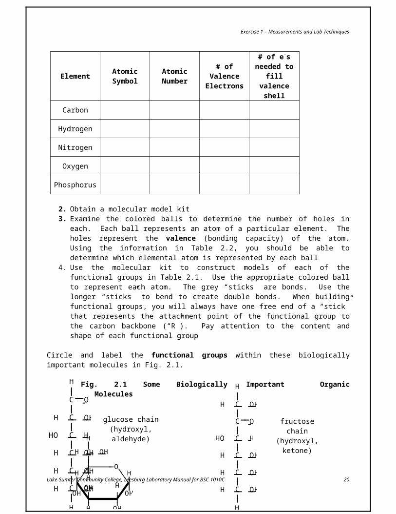
2. Obtain a molecular model kit3. Examine the colored balls to determine the number of holes in each. Each ball represents an
atom of a particular element. The holes represent the valence (bonding capacity) of the atom.
Lake-Sumter Community College, Leesburg Laboratory Manual for BSC 1010C 14
__OH __ C
O
H____
__
__ C
O
____
__ __ C
O
OH____
__
O
O__ P
O
OH____
__
__
__H
__ N
H
H__
__ __ S H__

Exercise 1 – Measurements and Lab Techniques
Using the information in Table 2.2, you should be able to determine which elemental atom is represented by each ball
4. Use the molecular kit to construct models of each of the functional groups in Table 2.1. Use the appropriate colored ball to represent each atom. The grey “sticks” are bonds. Use the longer “sticks” to bend to create double bonds. When building functional groups, you will always have one free end of a “stick” that represents the attachment point of the functional group to the carbon backbone (“R”). Pay attention to the content and shape of each functional group
Circle and label the functional groups within these biologically important molecules in Fig. 2.1.
Fig. 2.1 Some Biologically Important Organic Molecules
Lake-Sumter Community College, Leesburg Laboratory Manual for BSC 1010C 15
C__
H
O
H
OH
____
C
C
C
C
C
____
____
____
__
__
__
__
__
__
__
__
__
__
OH
OH
OH
HO
H
H
H
H
H
H
OHC
C
C
____
____
__
__
__
__
__
__
OH
OH
H
H
H
H
glucose chain(hydroxyl, aldehyde)
__
__
H
C
____
____ C
H
N __OH
O
H
__
H
__
C__
H
O
H
OH
____
C
C
C
C
C
____
____
____
__
__
__
__
__
__
__
__
__
__
OH
OH
OH
HO
H
H
H
H
H
fructose chain(hydroxyl, ketone)
glycerol(hydroxyl)
glycine(amine, carboxyl)
O
OH
H____
OH
H
____
OHH
H
____C____
____
H
OHH
H
____C____
____
OH
____
OHH
H
OH
____
OHOH
OH
C
H HH
H
H____
____
____
____
____ O
____
fructose ring(hydroxyl)
glucose ring(hydroxyl)

Exercise 1 – Measurements and Lab Techniques
Part B: BuffersThe pH of blood and other body fluids is relatively insensitive to the addition of acids or bases. This is due to the presence of buffers in living systems which help to maintain homeostasis by maintaining normal pH levels. The pH of a solution can be determined in a variety of ways, including the use of pH meters, litmus paper, and chemical reagents. In this exercise, we will use the chemical reagent Bogen’s Universal Indicator to determine pH of specific solutions.
Bogen’s Universal Indicator changes color at specific pH end points:Pink = pH 4Yellow = pH 6Green = pH 7Blue = pH 9Violet > pH 9
In order to determine the effect of buffers on pH, we will attempt to raise the pH of an unbuffered acid solution by adding small amounts of a base. For comparison, we will repeat this procedure with a buffered acid solution. Once both solutions are basic, we will attempt to return them to the original pH by adding small amounts of acid.
Procedure1. Obtain two 50 ml beakers and label them A and B2. Pipette 10 ml of an unbuffered pH 4 solution into beaker A3. Pipette 10 ml of a buffered pH 4 solution into beaker B4. Add 3 drops of Bogen’s Universal Indicator to each beaker5. Note the color. __________ Is this color expected? __________6. Slowly add 1M sodium hydroxide (NaOH) one drop at a time to beaker A, swirling the beaker
between each drop. Do until you detect a permanent color change to violet7. Record the number of drop required to change the color to violet in Table 2.38. Repeat the last two steps with beaker B
The test you just performed illustrated the effect of a buffer when you attempted to increase the pH (make it more basic). Did the buffered solution require more or less (circle one) drops to change the pH? Do you suppose buffers would resist pH changes in either direction? __________
Continue the procedure from above9. Slowly add 1M hydrochloric acid (HCl) one drop at a time to beaker A, swirling the beaker
between each drop. Do until you detect a permanent color change to pink10. Record the number of drops required to change the color to pink in Table 2.311. Repeat the last two steps with beaker B
Table 2.3 The Effect of Buffer on pH ChangeBeaker Contents # drops to violet # drops back to pink
A unbuffered, pH 4 solution
Lake-Sumter Community College, Leesburg Laboratory Manual for BSC 1010C 16

Exercise 1 – Measurements and Lab Techniques
B buffered, pH 4 solution
Part C: Dilutions
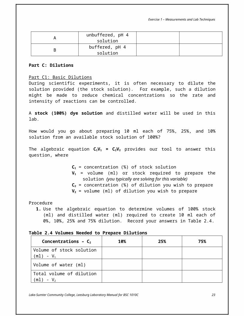
Part C1: Basic DilutionsDuring scientific experiments, it is often necessary to dilute the solution provided (the stock solution). For example, such a dilution might be made to reduce chemical concentrations so the rate and intensity of reactions can be controlled.
A stock (100%) dye solution and distilled water will be used in this lab.
How would you go about preparing 10 ml each of 75%, 25%, and 10% solution from an available stock solution of 100%?
The algebraic equation C1V1 = C2V2 provides our tool to answer this question, where
C1 = concentration (%) of stock solutionV1 = volume (ml) or stock required to prepare the solution (you typically are
solving for this variable)C2 = concentration (%) of dilution you wish to prepareV2 = volume (ml) of dilution you wish to prepare
Procedure1. Use the algebraic equation to determine volumes of 100% stock (ml) and distilled water (ml)
required to create 10 ml each of 0%, 10%, 25% and 75% dilution. Record your answers in Table 2.4.
Table 2.4 Volumes Needed to Prepare Dilutions
Concentrations – C2 10% 25% 75%
Volume of stock solution (ml) - V1
Volume of water (ml)
Total volume of dilution (ml) - V2
2. Obtain 3 test tubes and a test tube rack3. Prepare the three dilutions from Table 2.4 by pipetting the correct amount of stock in the test
tube first and then diluting the stock with the correct amount of distilled water. There should be the same amount of liquid in each test tube when you are finished
4. Obtain 5 cuvettes on a cuvette rack5. Transfer distilled water (0% dye solution) to the first cuvette up to about ¾ full. Distilled water is
used as a blank solution to calibrate the spectrophotometer
Lake-Sumter Community College, Leesburg Laboratory Manual for BSC 1010C 17

Exercise 1 – Measurements and Lab Techniques
6. One at a time and in order of increasing concentration, transfer enough of the other 4 solutions so that each cuvette is approximately ¾ full
7. Set the spectrophotometer to a wavelength of 450nm8. Read the % light transmittance for each dye solution you prepared and record your results in
Table 2.5Table 2.5 % Light Transmittance Associated with Various Concentrations of Dye
Dye Solution % Concentration of Dye % Light Transmittance
1 0 (DH2O only) 100
2 10
3 25
4 75
5 100 (stock)
Unknown A, B, C, D (circle yours)
What relationship exists between concentration of dye and % light transmittance?
Part C2: The Standard Curve
Procedure1. Plot the 0%, 10%, 25%, 75%, and 100% data from Table 2.5 on Fig. 2.22. Attempt to draw a “best fit” line through the scatter of data points. Do not simply connect the
dots. Make your line pass through the “average” spread of the dots. This line represents a standard curve and illustrates the relationship between percent concentration of a dye solution and percentage of light transmitted. Use this standard curve to complete Part C3
Lake-Sumter Community College, Leesburg Laboratory Manual for BSC 1010C 18

Exercise 1 – Measurements and Lab Techniques
Fig. 2.2 Standard Curve Relating Dye Concentration to % Light Transmittance
Describe the kind of relationship you see:
Part C3: Determination of Unknown Dye Concentration
Procedure1. Select a cuvette of unknown dye concentration (letters A-D) from the samples available2. Record the letter of your unknown in Table 2.53. Use the calibrated spectrophotometer to read the % transmittance of your unknown dye
concentration solution. Record in Table 2.54. Determine the concentration of your unknown by finding the value of % transmittance on the Y-
axis of Fig. 2.2 and drawing a perpendicular line down from that point to where it crosses the X-axis. That intersection point is the percent dye concentration of your unknown. Record that in Table 2.5
5. Return your unknown cuvette to your instructor and tell them your result
Lake-Sumter Community College, Leesburg Laboratory Manual for BSC 1010C 19
% Li
ght
Tran
smitt
ance
Dye Concentration (%)
Exercise 1 – Measurements and Lab Techniques
6. Rinse out the rest of the cuvettes and place them on the cuvette rack. Do not scrub them with a test tube brush as it will scratch and render them useless
Lake-Sumter Community College, Leesburg Laboratory Manual for BSC 1010C 20

Exercise 2 –Functional Groups, Organic Molecules, Buffers, and Dilutions
Practice Problems and Review Questions
1. Given a stock solution of 2.0% dextrose, how would you prepare 10 ml of each of the following solutions?
a. 0.1% dextrose solution
b. 1.0% dextrose solution
c. 0.5% dextrose solution
2. Given a stock solution of 5.0% sodium chloride (NaCl), how would you prepare 20 ml of each of the following solutions?
a. 2.0% sodium chloride solution
b. 0.5% sodium chloride solution
c. 3.0% sodium chloride solution
Lake-Sumter Community College, Leesburg Laboratory Manual for BSC 1010C 21

Exercise 2 –Functional Groups, Organic Molecules, Buffers, and Dilutions
3. Given a stock solution of 10% dextrose, how would you prepare 5 ml of a 0.9% dextrose solution?
4. Given a stock solution of 0.9% dextrose, how would you prepare 5 ml of a 0.5% dextrose solution?
5. Given a stock solution of 0.5% dextrose, how would you prepare 5 ml of a 0.004% dextrose solution?
6. How would you prepare 25 ml of a 15% dye solution beginning with a 20% stock dye solution?
7. How would you prepare 9 liters of a 50% dye solution beginning with a 60% stock dye solution? Express your answer in ml.
8. How would you prepare 600 ml of a 20% starch solution beginning with a 50% stock starch solution? Express your answer in liters.
9. You have 10 ml of a 60% stock dye solution. What is the maximum amount of a 12% dye solution you could prepare?
Lake-Sumter Community College, Leesburg Laboratory Manual for BSC 1010C 22
Exercise 2 –Functional Groups, Organic Molecules, Buffers, and Dilutions
10. How would you go about preparing the 12% dye solution in question 9?
11. What are buffers and why are they biologically important?
12. List the functional groups present in each of these molecules
glucose
fructose
glycine
glycerol
13. List some possible polymers that can be formed from each of these monomers
glucose
fructose
glycine
glycerol
Lake-Sumter Community College, Leesburg Laboratory Manual for BSC 1010C 23

Exercise 3 - Qualitative Analysis of Biological Molecules
IntroductionMacromolecules are large molecules formed from aggregates of smaller ones. Biological macromolecules are typically classified as carbohydrates, lipids, proteins, and nucleic acids. It is possible to identify macromolecules and monomers by using chemical indicators.
Reagents used as chemical indicators express their results either qualitatively or quantitatively by determining the presence or relative amount of a substance in a solution. The example in Table 3.1 should help you understand the basic difference between qualitative and quantitative analyses.
The reagents used in this exercise provide qualitative results. Each reagent exhibits a visible color change in the presence of a specific substance; however, it does not provide an amount (quantitative) result. A qualitative test will also be used to track the step-by-step hydrolysis of the polymer starch, a polysaccharide, into its glucose (monosaccharide) monomers.
Table 3.1 A Case Study Illustrating the Difference Between Qualitative and Quantitative AnalysesCase Study You are given a beaker containing 100 ml of an aqueous solution
QuestionA B
Are proteins present in this solution?
How many mg of protein are dissolved in this 100 ml solution?
Thinking
Would smelling, tasting, or touching the solution help determining if it has proteins or not? (not a good idea in lab)
The best thing to do is add a protein indicator. If the solution changes color, then proteins are present.
Changing the solution’s color indicated proteins are present, but it does not detect exactly how much protein is present.
An analytical test giving the answer in numbers, not just by presence or absence, needs to be done.
Response A qualitative analysis must be performed.
A quantitative analysis must be performed.
Lake-Sumter Community College, Leesburg Laboratory Manual for BSC 1010C 24

Exercise 3 –Qualitative Analysis of Biological Molecules
Materials
Equipmenttest tubes and racks10 ml pipettespipette pumps10 ml graduated cylindersmarking pencils (Sharpie)filter paper disksPetri dishwater baths at 95C
Reagents and Solutions1% dextrose (glucose)6% starch (amylose)1 M NaOHapple juicechicken brothegg whitewhole milkvegetable oildistilled waterBenedict’sIKI
BiuretSudan IV
Part A: Detection of CarbohydratesCarbohydrates are molecules consisting of one (monosaccharide), two (disaccharide), or many (polysaccharide) simple sugars. Examples of carbohydrates include glucose, sucrose, glycogen, maltose, and starch (amylose).
In this exercise, you will experiment with two carbohydrate reagents:Benedict’s reagent – usually light blue in color, forms a yellow-green, orange, or red precipitate when boiled in the presence of reducing sugars such as simple sugars (e.g. glucose)Iodine-Potassium Iodide (IKI) – amber colored, forms a dark purple or black precipitate in the presence of starch.
Read the information on the following pages (Parts A1, A2, and A3) and fill in the first three columns of Table 3.2 before performing the experiments.
Part A1: Detection of Simple Sugars
Procedure1. Obtain a test tube rack and six test tubes per group2. Label the test tubes 1 through 6. #1 and #2 will be used in this part3. Use a 10 ml pipette to transfer 1 ml of the dextrose (glucose) solution to test tube #14. Use a different (why?) pipette to transfer 1 ml of the starch solution (swirl to mix before
transferring) to test #25. Use a 10 ml graduated cylinder to measure and transfer 1 ml of Benedict’s reagent to each test
tube. Swirl to mix6. Note the color of each solution7. Gently heat the contents of each test tube in a 95C water bath for two minutes8. Observe and record any color change in Table 3.2
Lake-Sumter Community College, Leesburg Laboratory Manual for BSC 1010C 25

Exercise 3 –Qualitative Analysis of Biological Molecules
Part A2: Detection of StarchProcedure
1. Use a pipette to transfer 1 ml of dextrose solution to test tube #32. Use a different pipette to transfer 1 ml of starch solution (swirl to mix before transferring) to
test tube #43. Add one drop of IKI reagent to each test tube and swirl gently4. Observe and record any color change in Table 3.2
Part A3: Identification of a Carbohydrate UnknownIf you were given an unknown solution and had to perform both the simple sugar (Part A1) and the starch (Part A2) tests in the same test tube, which test would you perform first? The following experiment will help to answer this question.
Procedure1. Use a pipette to transfer 1 ml of dextrose to both test tubes #5 and #62. Use a different pipette to transfer 1 ml of starch to both test tubes #5 and #63. In test tube #5, perform the Benedict’s test first4. Make note of any color changes5. After the Benedict’s test perform the IKI test in test tube #56. In test tube #6, perform the IKI test first7. Make note of any color changes8. After the IKI test perform the Benedict’s test in test tube #69. Make note of any color changes10. Record your observation in Table 3.211. From the results of test tubes #5 and #6, determine which test you should run first if you were
limited to using just one test tube and had to test for both simple sugars and starch. Only one of these two test tubes will allow you to see the results of both tests correctly
Which test would you perform first and why?
12. Obtain a simple sugar / starch unknown (labeled A, B, C, and D) and test it using the proper sequence of Benedict’s and IKI reagent
13. Record the letter of your unknown and any color changes in Table 3.2
What (water, glucose, starch, or both) was in your unknown?
Lake-Sumter Community College, Leesburg Laboratory Manual for BSC 1010C 26

Exercise 3 –Qualitative Analysis of Biological Molecules
Table 3.2 Qualitative Analysis of Simple Sugars, Starch, and a Carbohydrate UnknownTest Tube
Test Solution Reagent Hypothesis Results
1
2
3
4
5 Benedict’s 1st
IKI 2nd
6 IKI 1st
Benedict’s 2nd
Unknown (_____)
Lake-Sumter Community College, Leesburg Laboratory Manual for BSC 1010C 27

Exercise 3 –Qualitative Analysis of Biological Molecules
Part B: Detection of LipidsA lipid is a non-polar (hydrophobic) organic molecule which is insoluble in water. One type of lipid are fats, also called triglycerides or triacylglycerols. A fat molecule is composed on one glycerol and three fatty (palmitic) acid molecules. Sudan IV-lipid complex will produce an orange spot on filter paper to which lipid has been added.
Procedure1. Obtain a blank filter paper disk2. Mark the disk with a pencil following the pattern as shown in this figure
A – apple juiceC – chicken brothE – egg whiteM – whole milkO – vegetable oilW – distilled water (control)
3. Make a hypothesis as to which of the above substances you would expect to contain lipids4. Record this hypothesis in Table 3.55. Transfer a small drop of each substance to the appropriate circle on the filter paper6. Allow the filter paper to dry7. Once dry, soak the filter paper for 3 minutes in a petri dish containing Sudan IV reagent. Leave
the dish on the counter where it was originally to avoid spillage8. Remove the filter paper disk with forceps and gently rinse with distilled water over the sink for
one minute9. Hold the filter paper over something white for contrast and observe the results10. Examine the color for the six spots and indicate whether the substances contained lipid using
the by indicating “-“ for negative (no color change; no lipid) and “+” for positive (color change; lipid)
11. Record your results in Table 3.512. Compare your results to your hypothesis
Table 3.5 Sudan IV Test for LipidsSubstance Tested Hypothesis Result
Apple juiceChicken broth
Egg white (albumin)Whole milk
Vegetable oilDistilled water
Lake-Sumter Community College, Leesburg Laboratory Manual for BSC 1010C 28
M
E
C
OW
A

Exercise 3 –Qualitative Analysis of Biological Molecules
Part C: Detection of ProteinsProteins are polymers of amino acids in which the carboxyl functional group of one amino acid forms a peptide bond with the amine functional group of another amino acid.
__
__
H
C
____
____ C
H
N __OH
O
H
__
H
__
R
__
__
H
C__
______ C
H
N __OH
O
H__
H
__
R__
__
H
C
____
____ C
H
N __OH
O
H
__
H
__
R
__
__
H
C
____
____ C
H
N __OH
O
H
__
H
__
R
+
H2O
peptide bond
Biuret reagent, which is pale blue, contains copper sulfate (CuSO4). The Biuret reaction is based on the complex formation of cupric ions with proteins. In this reaction, copper sulfate is added to a protein solution in strong alkaline solution. A purplish-violet color is produced, resulting from the complex formation between the cupric ions and the peptide bond.
Procedure1. Obtain a test tube and rack and six clean test tubes per group2. Mark the test tubes with the same symbols used in the lipid experiment (Part C)3. Make a hypothesis as to which of the above substances you would expect to contain proteins4. Record this hypothesis in Table 3.65. Transfer 1 ml (approximately 20 drops) of the appropriate solution to properly marked test tube6. Dispense 1 ml of 1M NaOH into each test tube7. Swirl gently to mix8. Add 0.5 ml of 1% Biuret reagent to each test tube9. Swirl gently to mix10. Look for any instant change in color from blue to violet. This is the positive test for proteins11. Record your results in Table 3.6 using the same symbols (- and +) as described in Part C12. Compare your results to your hypothesis
Table 3.6 Biuret Test for ProteinsSubstance Tested Hypothesis Result
Apple juiceChicken broth
Egg white (albumin)Whole milk
Vegetable oilDistilled water
Lake-Sumter Community College, Leesburg Laboratory Manual for BSC 1010C 29

Exercise 3 –Qualitative Analysis of Biological Molecules
Practice Problems and Review Questions
1. Explain the difference between a qualitative and quantitative analysis test.
2. What substance is used as a control in the a. Sudan IV test?
b. Biuret test?
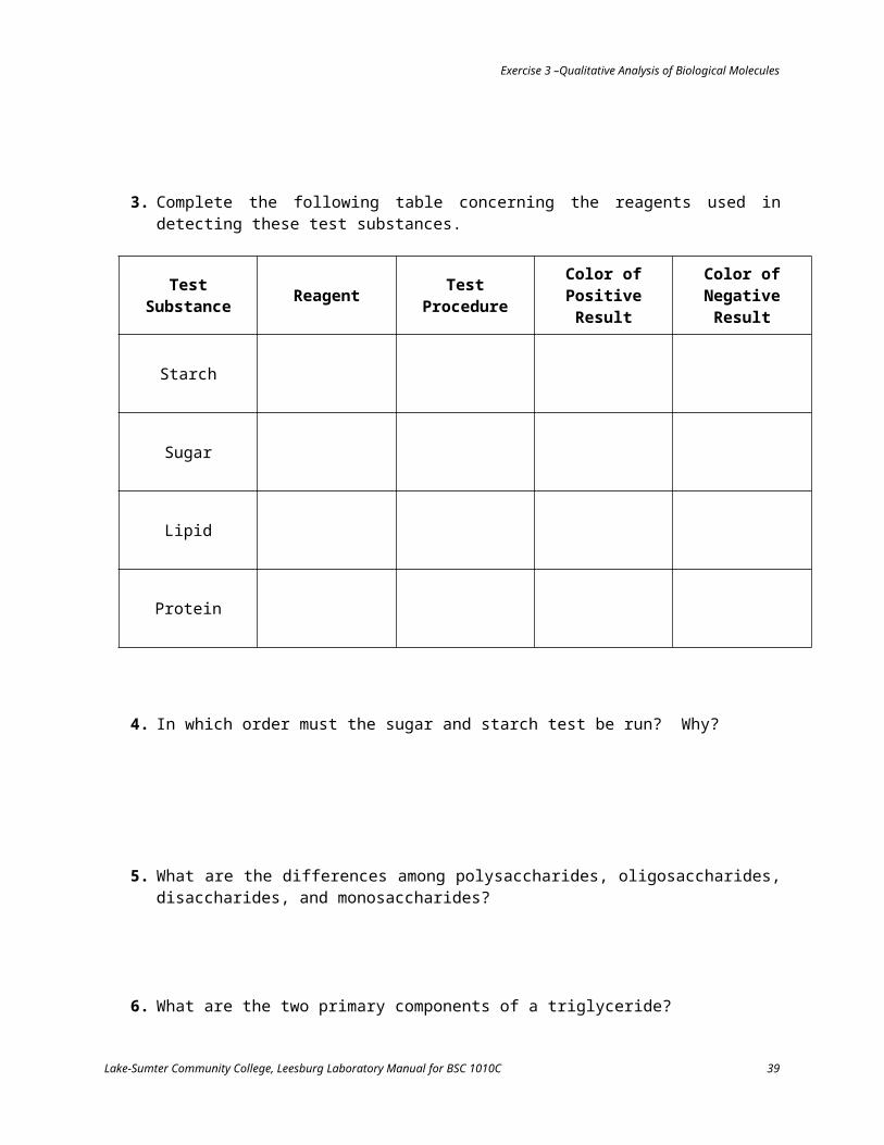
3. Complete the following table concerning the reagents used in detecting these test substances.
Test Substance Reagent Test Procedure Color of Positive Result
Color of Negative Result
Starch
Sugar
Lipid
Protein
4. In which order must the sugar and starch test be run? Why?
Lake-Sumter Community College, Leesburg Laboratory Manual for BSC 1010C 30

Exercise 3 –Qualitative Analysis of Biological Molecules
5. What are the differences among polysaccharides, oligosaccharides, disaccharides, and monosaccharides?
6. What are the two primary components of a triglyceride?
7. What are the monomers that make up proteins?
8. List and briefly describe the four levels of protein structure.
9. How do proteins of foods differ from those of the organism consuming them?
10. Name a molecule of living systems other than protein which contains nitrogen.
11. What is hydrolysis?
Lake-Sumter Community College, Leesburg Laboratory Manual for BSC 1010C 31

Exercise 4 - The Microscope
IntroductionThe microscope is an essential tool in modern biology. It allows us to view structural details of organs, tissue, and cells not visible to the naked eye.
This laboratory exercise is designed to demonstrate some of the potential uses of various types of light microscopes and to help you become familiar with proper microscopic techniques.
Materials
Equipmentcompound microscopedissecting microscopemicroscope slidescoverslipsdropperslens paperforcepstoothpicks
Biological SpecimensAllium (onion)pond water
Prepared Slidesnewspaper printcolored threadsParamecium
Reagents
IKImethylene blueDetain (or Protoslo)
Part A: Care and Use of the Compound Microscope
ALWAYS CARRY THE MICROSCOPE UPRIGHT WITH TWO HANDS, ONE ON THE BASE, THE OTHER ON THE ARM
MAKE SURE YOUR WORKBENCH IS FREE OF CLUTTER BEFORE YOU PLACE THE MICROSCOPE ON THE BENCH
DO NOT DRAG OR SHOVE THE MICROSCOPE ACROSS THE LAB BENCH – ALWAYS LIFT TO MOVE OR TURN IT
The steps on the next few pages represent the correct procedure for viewing a specimen under a compound microscope. Your instructor will demonstrate the proper use of the microscope as well as describe its features. Refer to Fig. 4.1 to familiarize yourself with the parts of the microscope as you study each step in the procedure.
Lake-Sumter Community College, Leesburg Laboratory Manual for BSC 1010C 32

Exercise 4 –The Microscope
Fig. 4.1 The Compound Light Microscope
Lake-Sumter Community College, Leesburg Laboratory Manual for BSC 1010C 33

Exercise 4 –The Microscope
Viewing a Specimen with a Compound Light Microscope
Procedure1. Clean the slide and coverslip by rubbing them gently with lens paper2. Use the coarse focus adjustment knob to maximize the working distance (the distance between
the stage and the objective lens)3. Rotate the revolving nosepiece into position with the scanning power (4x) objective lens in the
viewing position4. Center the slide holder of the mechanical stage on the microscope stage5. Place the slide between the stage clip and push it all the way back to the bar6. Plug in the microscope and turn on the light switch7. Using the mechanical stage drive knobs, center the coverslip and specimen over the stage
aperture8. While carefully watching the slide on the stage, use the coarse focus adjustment knob to move
the specimen towards the scanning objective lens until it stops. The stage will come close to the lens but will not touch it
9. Adjust the interpupillary distance until you see a single circle while looking through the microscope with both eyes open. This circle of light is called the field of view
10. While looking through the ocular lenses, turn the coarse and fine focus adjustment knobs of the microscope until you see something you believe is the specimen. Stop. Move the slide back forth using the mechanical stage drive knobs. The item you thought was specimen should likewise be moving back and forth
11. Cover of close the eye that is not looking through the ocular containing the diopter ring. Viewing with only that eye focus using the coarse and fine focus adjustment knobs. Adjust the light using the iris diaphragm adjustment lever and/or the light adjustment. Then close your other eye adjusting the diopter ring on that ocular lens to bring the object into focus
12. Adjust the condenser to the highest position13. Using the mechanical stage drive knobs, center the specimen of choice in the viewing area14. These microscopes are parfocal (if one lens is in focus, all other lenses are, at least, close to
focus). In order to change to the next highest magnification, simply rotate the nosepiece to the low power (10x) objective lens
15. These microscopes are also parcentral (if an object is in the center of the field of view for one lens, it will be, at least, close to the center of the field of view at other lenses)
16. Using the mechanical stage drive knobs, re-center the specimen in the viewing area17. With the low power (10x) objective, use the coarse and fine focus adjustment knobs to focus
the view of the specimen and the iris diaphragm adjustment lever to increase the light intensity on the specimen
18. Re-center the specimen in the field of view. Rotate the nosepiece to the high power (40x) objective lens. Use the FINE FOCUS ADJUSTMENT KNOB ONLY to focus and the iris diaphragm adjustment lever to increase the light intensity on the specimen. If needed, use the light adjustment to provide additional light
19. When removing the slide, rotate the nosepiece so the scanning power (4x) objective is in the viewing position, then use the coarse focus adjustment knob to maximize the working distance
20. After you have completed the laboratory activity, turn the light switch off. Clean all microscope lenses (objective and ocular) with lens cleaner and lens paper
Lake-Sumter Community College, Leesburg Laboratory Manual for BSC 1010C 34

Exercise 4 –The Microscope
21. Prepare the microscope for storage using the checklist below. Be surea. The scanning power (4x) objective is in the viewing positionb. The mechanical stage has been positioned so the stage arm is flush with the right side
of the stagec. The cord is wrapped securely around the microscope armd. The stage has been adjusted all the way downe. The condenser has been adjusted all the way upf. The light adjustment is turned all the way down and the light is turned off
Part B: MagnificationThere is a set of three objective lenses on your microscope. The magnification (or power) of each objective lenses is engraved on the side of the objective. The ocular lens is also normally engraved with its magnification (typically 10x).
To determine the total magnification of a specimen, use the following formula:
Total Magnification = Ocular Magnification x Objective Magnification
Procedure1. Use Table 4.1 to record the magnification values for each objective lens and the ocular lens on
the microscope2. Calculate total magnification (using the formula above) for each objective lens and record n
Table 4.1
Table 4.1 Total Magnification of Microscope
Objective Lens NameMagnification
Objective Lens Ocular Lens Total
Scanning
Low Power
High Power
Part C: Working Distance and Diameter of the Field of View
Part C1: Working DistanceWorking distance is the distance between the stage and objective lens (Fig 4.2). Because objective lenses vary in lengths, the working distance will change as you switch from one objective lens to the next.
In a microscope, as magnification increases, working distance ______________________.
Lake-Sumter Community College, Leesburg Laboratory Manual for BSC 1010C 35

Exercise 4 –The Microscope
Fig. 4.2 Working Distances with Various Objective Lenses
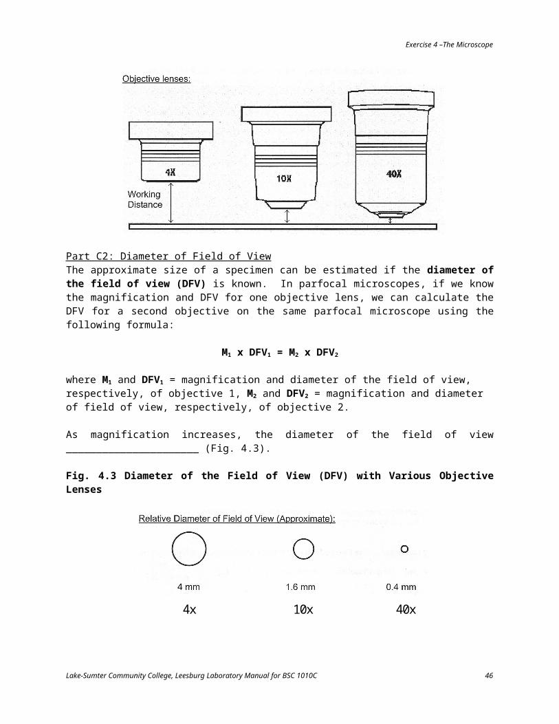
Part C2: Diameter of Field of ViewThe approximate size of a specimen can be estimated if the diameter of the field of view (DFV) is known. In parfocal microscopes, if we know the magnification and DFV for one objective lens, we can calculate the DFV for a second objective on the same parfocal microscope using the following formula:
M1 x DFV1 = M2 x DFV2
where M1 and DFV1 = magnification and diameter of the field of view, respectively, of objective 1, M2 and DFV2 = magnification and diameter of field of view, respectively, of objective 2.
As magnification increases, the diameter of the field of view ______________________ (Fig. 4.3).
Fig. 4.3 Diameter of the Field of View (DFV) with Various Objective Lenses
4x 10x 40x
Lake-Sumter Community College, Leesburg Laboratory Manual for BSC 1010C 36

Exercise 4 –The Microscope
Fill in Table 4.2 for your microscope using the values given for the scanning objective and the above formula.
Table 4.2 Diameter of Field of View (DFV) for the Compound Microscope
Objective Lens Magnification DFV (mm) DFV (µ)
Scanning 4 4__________
Low Power 10__________ __________
High Power 40__________ __________
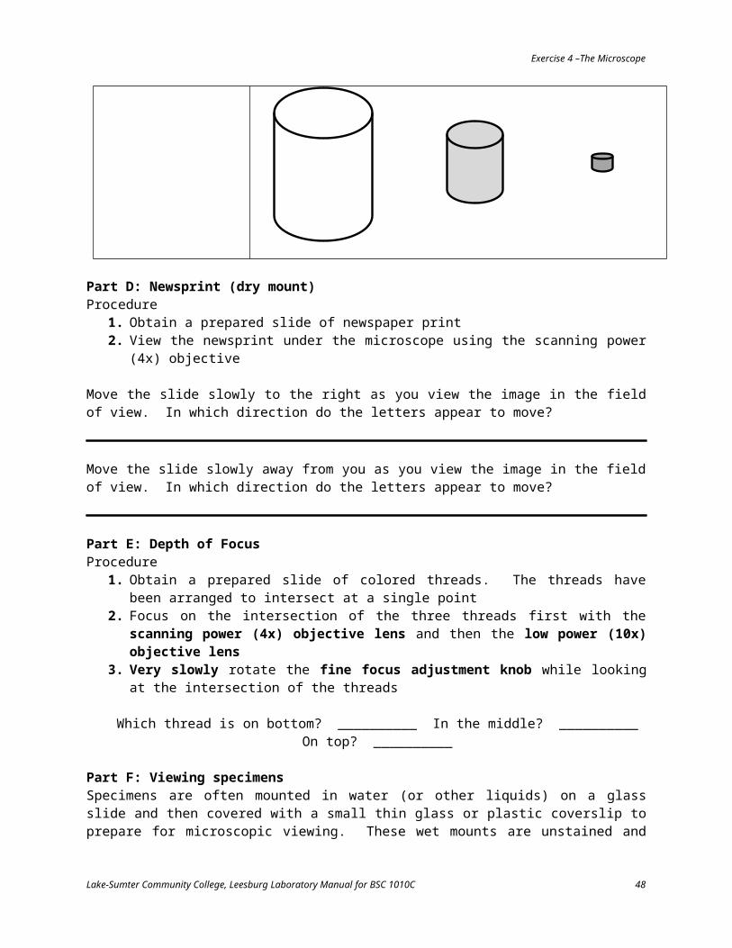
Part C3: Depth of FocusThe depth of focus for a particular objective refers to the power of the objective to produce an in-focus image from objects that are slightly different distances away from the objective lens. As magnification power increases, the depth of focus decreases.
When viewing specimens under a microscope, it is beneficial to keep in mind that as magnification power increases the microscope’s field of view becomes smaller, thinner, and darker (Table 4.3).
Table 4.3 Changes in a Microscope’s Field of View as a Function of Magnification Power
Scanning Low Power High Power
Diameter of Field of View (DFV)
Depth of Focus
Light
Lake-Sumter Community College, Leesburg Laboratory Manual for BSC 1010C 37
Gets Smaller
Gets Thinner
Gets Darker

Exercise 4 –The Microscope
Part D: Newsprint (dry mount)Procedure
1. Obtain a prepared slide of newspaper print2. View the newsprint under the microscope using the scanning power (4x) objective
Move the slide slowly to the right as you view the image in the field of view. In which direction do the letters appear to move?
Move the slide slowly away from you as you view the image in the field of view. In which direction do the letters appear to move?
Part E: Depth of FocusProcedure
1. Obtain a prepared slide of colored threads. The threads have been arranged to intersect at a single point
2. Focus on the intersection of the three threads first with the scanning power (4x) objective lens and then the low power (10x) objective lens
3. Very slowly rotate the fine focus adjustment knob while looking at the intersection of the threads
Which thread is on bottom? __________ In the middle? __________ On top? __________
Part F: Viewing specimensSpecimens are often mounted in water (or other liquids) on a glass slide and then covered with a small thin glass or plastic coverslip to prepare for microscopic viewing. These wet mounts are unstained and sometimes difficult to see. Replacement staining can add color and contrast enhancing the detail of the specimen.
It is important to be able to estimate the sizes of different specimens under the microscope. Already knowing the diameter of the field of view for a particular objective (Table 4.2), we can utilize the following formula to estimate size:
size of cell=DFV# of cells across DFV
Lake-Sumter Community College, Leesburg Laboratory Manual for BSC 1010C 38
Exercise 4 –The Microscope
At which magnification do you think you are able to get the most accurate estimate of cell number and thus the most accurate estimation of cell size? __________. Why?
Part F1: Paramecium
Procedure1. Obtain a prepared slide of the single-celled protozoan, Paramecium2. Use the correct focusing technique to find the Paramecium at high power3. Estimate the # of Paramecium cells required to fill across the DFV end-to-end4. Use the formula to calculate Paramecium length in microns5. Estimate the # of Paramecium cells required to fill across the DFV side-by-side6. Use the formula to calculate Paramecium width in microns
Paramecium cells arranged end-to-end Paramecium cells arranged side-to-side
Paramecium Length __________ (in microns) Paramecium Width __________ (in microns)
Part F2: Allium (onion) epidermis (wet mount)
Procedure1. Prepare a wet mount of Allium (onion) epidermis2. Place one or two drops of water on a clean slide3. Peel the epidermis (thin skin) off the inside of a piece of sliced onion using forceps4. Place the epidermis carefully in the water on the slide5. Place a coverslip over the epidermis6. Observe the cells under the microscope and sketch what you see
Lake-Sumter Community College, Leesburg Laboratory Manual for BSC 1010C 39

Exercise 4 –The Microscope
7. Stain the onion cells with IKI using the replacement staining technique
a. Place a few drops of IKI on the slide against one edge of the coverslipb. Place the smooth edge of a single layer of paper towel up against the opposite edge of the
coverslip. The paper towel will pull the water out from underneath the coverslip. In turn, the water as it exits will drag the IKI stain underneath the coverslip
c. Continue this process, adding more IKI if necessary, until the stain covers the area under the coverslip
d. Examine under the microscope
8. Observed the cells under the microscope again and sketch what you see
9. Can you see more or less detail after staining compared to the unstained cells? _____________10. Estimate the length and width of an onion cell (in microns)
Onion Cell Length __________ (in microns) Onion Cell Width __________ (in microns)
Lake-Sumter Community College, Leesburg Laboratory Manual for BSC 1010C 40

Exercise 4 –The Microscope
Lake-Sumter Community College, Leesburg Laboratory Manual for BSC 1010C 41

Exercise 4 –The Microscope
Part F3: Cheek Cells (wet mount)
Procedure1. Place one or two drops of water on a clean slide2. Obtain a clean toothpick and collect cheek cells by gently scraping the inside of your cheek3. Swirl the tip of the toothpick in the water on the slide (immediately discard your toothpick)4. Stain your cheek cells with methylene blue stain5. Place a coverslip over your cheek cells6. Observe and sketch the stained cheek cells. Identify the nucleus, cytoplasm, and cell membrane
7. How do these cells differ from onion cells?
8. Estimate the diameter of one of your cheek cells (in microns)
Cheek Cell Diameter __________ (in microns)
Part G: Pond WaterAlthough staining cells makes it easier to see their detail, most staining techniques also kill any live specimens. Thus, looking at microorganisms can be a challenge. Living microorganisms are also difficult to see clearly because many of them are motile and must be chased around the slide while you are focusing.
Procedure1. Place a drop of pond water on a clean microscope slide. Try to obtain a sample that is near any
floating debris and organisms tend to congregate there. Be careful not to shake the jar2. Add a coverslip3. Examine under the microscope4. Try to keep motile microorganism in focus by following them around as they move on the slide.
If they move too quickly, carefully lift up the coverslip and add a drop of Detain (or Protoslo)5. Draw a few of the critters you see in space provided
Lake-Sumter Community College, Leesburg Laboratory Manual for BSC 1010C 42

Exercise 4 –The Microscope
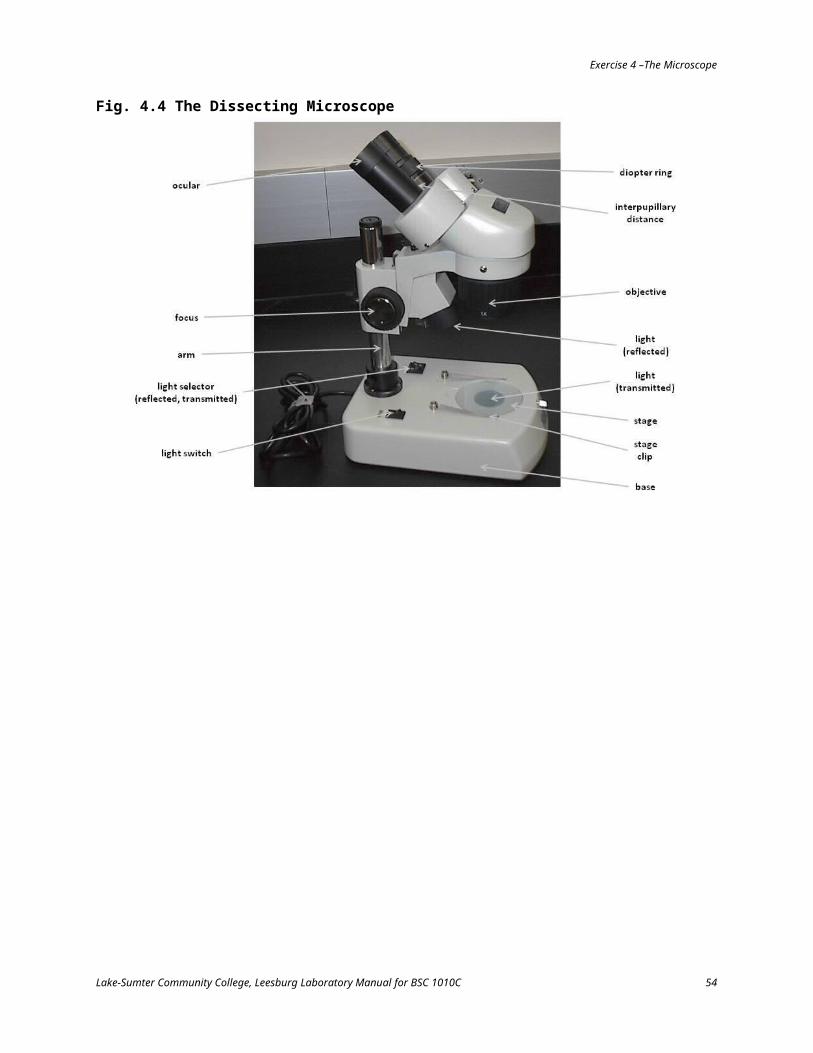
Part H: The Dissecting MicroscopeIt is possible to have too much magnification when viewing some specimens. For example, how would you use your compound light microscope to view an entire earthworm? Larger specimens may require lower magnification. For this, biologists use dissecting microscopes (Fig. 4.4). Fill in Table 4.4 and notice the diameter of the field of view in these microscopes is substantially larger than that in compound light microscope. Your instructor will describe the use and features of this microscope.
Lake-Sumter Community College, Leesburg Laboratory Manual for BSC 1010C 43

Exercise 4 –The Microscope
Fig. 4.4 The Dissecting Microscope
Lake-Sumter Community College, Leesburg Laboratory Manual for BSC 1010C 44

Exercise 4 –The Microscope
Table 4.4 Diameter of the Field of View for the Dissecting Microscope
Objective Lens Magnification DFV (mm) DFV (µ)
Lowest Power 2 10 10,000
Highest Power 4 5__________
Procedure1. Obtain a dissecting microscope using two hands to carry it2. Identify the parts as per Fig. 4.4 and their functions3. Observe the various objects made available in lab using the dissecting microscope
Using the information in Table 4.4, complete this sentence for the dissecting microscope
As magnification increases, DFV __________
Have you seen this relationship before? __________ Where? __________________________
Lake-Sumter Community College, Leesburg Laboratory Manual for BSC 1010C 45
Exercise 4 –The Microscope
Practice Problems and Review Questions
1. What is the total magnification of an object if the ocular lens magnification is 20x and the objective lens magnification is 45x?
2. Which objective lens is in place if the object you are viewing is magnified 1000x assuming an ocular lens magnification of 10x?
3. What is the diameter of the field of view (DFV) of a 1000x objective lens if the DFV of a 400x objective lens is 500 µ? Express your answer in mm.
4. What is the DFV of a 40x objective lens if the DFV of a 10x objective lens is 3 mm? Express your answer in µ.
5. When viewing an organism using the 40x objective lens from question 4, you estimate 6 organisms could fit across the DFV if they were laid end-to-end and 20 could fit is stacked side-by-side. What is the length and width of this organism (in microns)?
6. What is the DFV of a 25x objective lens if the DFV of a 100x objective lens is 1.5 mm?
7. Using the 100x objective lens from question 6, you estimate 12 organisms could fit across the DFV if they were laid end-to-end and 30 could fit is stacked side-by-side. What is the length and width of this organism (in microns)?
Lake-Sumter Community College, Leesburg Laboratory Manual for BSC 1010C 46

Exercise 4 –The Microscope
8. What is the magnification of an objective lens with a DFV or 0.8 mm if the DFV of a 100x objective lens is 2 mm?
Lake-Sumter Community College, Leesburg Laboratory Manual for BSC 1010C 47
Exercise 5 - Cell Structure and Membrane Function
IntroductionThe cell is the lowest level of biological organization performing all activities of life. Therefore, it is the fundamental unit of structure in living things. As such, the characteristics of cells are of monumental concern to the understanding of biology. The structure of cellular components reflects adaptation to accomplish those functions necessary for life. The collective functions of individual cells allow for the activity and behavior of the entire organism of which those cells are a part.
In this laboratory exercise, you will use a compound light microscope to examine cells and observe cellular activity. You will also conduct experiments illustrating some of the basic mechanisms of cellular transport.
Materials
Equipmentcompound light microscopemicroscope slidescoverslipstest tubes and racksbeakersdroppersdialysis tubingdental floss or string
scissorstriple beam balances95C water bath
Biological SpecimensElodea
Reagents and SolutionsBenedict’sIKI10.0% saline solutionconcentrated glucoseconcentrated starch
Part A: Cellular TransportCellular transport mechanisms are typically divided into two categories: Passive Transport and Active Transport. The basic differences between them is summarized in Table 5.1
Table 5.1 Basic Differences Between Passive and Active TransportPassive Transport Active Transport
Types and Direction of Involves the movement of water Involves the movement of
Lake-Sumter Community College, Leesburg Laboratory Manual for BSC 1010C 48

Exercise 4 –The Microscope
Transported Substances
or solute through a semi-permeable membrane down their concentration gradient (i.e., from regions of higher concentration of water or solutes to regions of lower concentration).
solutes through a semi-permeable membrane against their concentration gradient (i.e., from regions of lower concentration of solutes to regions of higher concentration).
Cellular Energy Does NOT require cellular energy in the form of ATP.
Requires cellular energy in the form of ATP.
Membrane Transport Proteins
Passive transports systems include two types of diffusion and osmosis:
Simple diffusion – membrane transport proteins not required
Facilitated diffusion – membrane transport proteins required
Osmosis – specific to the passive transport of water from an area of higher water concentration to an area of lower water concentration (lower to higher concentration of solutes). Water moves through protein channels known as aquaporins.
Requires membrane transport proteins.
Solutions are often described using the terms hypotonic, hypertonic, and isotonic. Tonicity is a comparative term related to the concentration of solutes in a solution. It may be defined as the ability of a solution to cause a cell to gain or lose water. Hypotonic solutions contain less solute by % (i.e., more water) when compared to hypertonic solutions, which contain more solutes by % (i.e., less water). With a hypotonic solution that is separated from a hypertonic one by a selectively permeable membrane that allows water molecules to pass through but not solutes, the net movement of water molecules will be from a region of high water concentration (i.e., low solute – hypotonic) to a region of lower water concentration (i.e., high solute – hypertonic). Isotonic solutions are equal to one another in solute concentration; therefore, a concentration gradient does not exist and water moves in equal rate back and forth across the membrane.
This exercise will explore some of these basic principles of cellular transport.
Part A1: Passive Transport in a Model Cell
Lake-Sumter Community College, Leesburg Laboratory Manual for BSC 1010C 49

Exercise 4 –The Microscope
Procedure1. Obtain a piece of dialysis tubing2. Working quickly so the dialysis tubing won’t dry out, fold one end and tie off with floss or string3. Open the other end of the tubing by sliding your fingers back and forth across the top4. Place 10 ml of concentrated glucose and 10 ml of concentrated starch into the bag5. Squeeze out the excess air from the bag before folding its other end and tying off6. Rinse the bag gently under running water at the sink and blot dry with a paper towel. Make
sure the bag is not leaking7. Weigh the bag to the nearest 0.1 g and record as initial mass of the bag in Table 5.28. Fill a beaker with distilled water9. Add just enough IKI to the beaker water to turn it light yellow10. Place the dialysis bag in the beaker. The bag should be fully submerged11. Let your beaker sit no less than 30 minutes
This model cell system consists of four different molecules which could possibly move through the small holes in the dialysis bag. What are they?
1. _______________ 2. _______________ 3. _______________ 4. _______________
Based on the molecular size of these four molecules, develop a hypothesis to describe which molecules will move into the bag, which will move out and why. Record your hypothesis in Table 5.3
12. After your bag has soaked for the appropriate amount of time (no less than 30 minutes), remove it from the beaker and gently blot dry with a paper towel
What color is the solution in the bag? _______________ What color is the solution in the beaker? _______________
13. Weigh the bag again to the nearest 0.1 g and record in Table 5.214. Calculate the change in the mass of the bag by subtracting the initial mass from the final mass of
the bag and record in Table 5.215. Calculate the % mass change of the bag using this formula and record in Table 5.2
% mass change of bag= final bag mass - initial bag massinitial bag mass
x 100
Table 5.2 Mass and Time of Dialysis Bag ExperimentMass Time (hr : min)
Final __________ g _____ : _____Initial __________ g _____ : _____
Change in Mass of Bag __________ g% Mass Change of Bag __________ %
16. Pour about 1 ml of the contents of the bag into a test tube17. Test the bag contents with Benedict’s reagent18. Pour about 1 ml of the contents of the beaker into a test tube19. Test the beaker contents with Benedict’s
Lake-Sumter Community College, Leesburg Laboratory Manual for BSC 1010C 50

Exercise 4 –The Microscope
20. Fill in Table 5.3
Table 5.3 Results of Dialysis Bag Experiment
Molecular Component of
the Dialysis Bag System
Net Movement of Molecules Across the Dialysis Bag (In / Out / None)
Hypothesis
Final Results Based on
Observations and Testing
Explanation
Part A2: Osmosis in Elodea Elodea is a common aquatic plant related to Hydrilla. It has leaves of only two layers of thickness.
In this exercise, the thin leaves of Elodea will be useful in exploring some of the principles of osmosis. As seen under the compound microscope, the movement of cytoplasm with the Elodea leaf cells along the perimeter of the cell called cyclosis or cytoplasmic streaming will be observed.
Procedure1. Using forceps, remove one leaf from an Elodea plant2. Prepare a wet mount of the leaf using distilled water3. Observe the leaf at high power under the microscope4. Identify the parts of an Elodea leaf (Fig. 5.1)
Where are the chloroplasts located? _______________ Do you see cyclosis (cytoplasmic streaming)? _______________
5. Draw your Elodea cell and label the visible parts
Lake-Sumter Community College, Leesburg Laboratory Manual for BSC 1010C 51

Exercise 4 –The Microscope
6. Using the replacement staining technique, replace the distilled water under your coverslip with the saline solution
7. After 5-10 minutes, observe the cells again and make note of any changes that have occurred8. Draw the cell again
Where are the chloroplasts located now?
What cellular structure (not visible previously) has receded from the cell wall?
What happened to the volume of the central vacuole to cause this change?
In what type (hypotonic, hypertonic, isotonic) of environment is the Elodea cell in?
Fig. 5.1 Elodea Cell
Lake-Sumter Community College, Leesburg Laboratory Manual for BSC 1010C 52

Exercise 4 –The Microscope
Part A3: Osmoregulation in ProtistsSome single-celled organisms live in a fresh water environment that is hypotonic to their cellular fluid which means they are continually taking on water through osmosis. They stay alive because they possess abilities to regulate internal water pressure using contractile vacuoles. These contractile vacuoles remove excess water from the cell. Contractile vacuoles typically appear a “fluid-filled bubbles” in the cytoplasm that slowly get large and then suddenly disappear.
Procedure1. Using the web, find and view pictures and video clips of contractile vacuole function in
Paramecium. Your instructor may also make some clips available online or have you view them in class
2. Draw a Paramecium and label the contractile vacuole
Lake-Sumter Community College, Leesburg Laboratory Manual for BSC 1010C 53
Exercise 4 –The Microscope
What is its function?
Part B: Structure and Motility in ProtistsMost groups of protists are capable of movement. This motility is made possible by one of three types of structures. Organisms like Amoeba move by means of pseudopodia (“false foot”) which are extensions of the cytoplasm. Paramecium and similar organisms move using cilia, fine hair-like structures covering the cell membrane. Organisms typically have many, many cilia. Other protists, such as Euglena, move using flagella, which are whipped back and forth. Organisms usually have one or just a few flagella. Finally, some protists lack the ability to move at all. Procedure
1. Using the web, find and view pictures and video clips of protist structure and movement. Your instructor may also make some clips available online or have you view them in class
2. Using online resources and the text book, draw and label the following parts for Amoeba, Paramecium, and Euglena
cell membranecytoplasmpseudopod (Amoeba)cilia (Parmecium)flagella (Euglena)
contractile vacuolenucleuschloroplast (Euglena)food vacuole
Lake-Sumter Community College, Leesburg Laboratory Manual for BSC 1010C 54

Exercise 4 –The Microscope
Amoeba
Paramecium
Euglena
Lake-Sumter Community College, Leesburg Laboratory Manual for BSC 1010C 55

Exercise 5 –Cell Structure and Membrane Function
Practice Problems and Review Questions
1. If the initial mass of a dialysis bag was 8.2 g and final mass was 10.9 g, what is the % mass change of the bag?
2. If the initial mass of a dialysis bag was 10.6 g and final mass was 11.1 g, what is the % mass change of the bag?
3. If the initial mass of a dialysis bag was 9.9 g and final mass was 8.8 g, what is the % mass change of the bag?
4. A pre-weighed dialysis bas which contained a solution of 10% glucose was placed in a beaker containing a solution of 20% glucose. After one hour, the bag was weighed again. Calculate the % mass change of this dialysis bag from the following information:
Mass of bag before experiment: 15.3 gMass of bag after experiment: 12.7 g
5. Was the beaker solution in question 4 hypertonic, hypotonic, or isotonic to the dialysis bag contents?
6. What are the major differences between the following pairs of cells?
prokaryotic and eukaryotic
Lake-Sumter Community College, Leesburg Laboratory Manual for BSC 1010C 56

Exercise 5 –Cell Structure and Membrane Function
plant and animal
protists and generalized animal cells
7. How was the dialysis bag in your experiment an example of a semi-permeable membrane?
8. Define these terms:
hypertonic
hypotonic
isotonic
9. Complete the following sentence: When two aqueous solutions are separated by a semi-permeable membrane, the net water movement is always from a ________tonic to a ________tonic solution.
Lake-Sumter Community College, Leesburg Laboratory Manual for BSC 1010C 57
Exercise 6 - Enzyme Activity
IntroductionEnzymes are biological catalysts that regulate the rate of chemical reactions. Their 3-dimensional conformation and therefore their function can be affected by several variables.
In this laboratory exercise, you will manipulate various factors that affect an enzyme’s activity. The enzyme is catalase, which is found in most all living organisms. Catalase decomposes hydrogen peroxide (H2O2), a toxic compound, into water and oxygen:
2H2O2 + catalase 2H2O + O2
The amount of oxygen created is directly proportional to the rate of the enzymatic reaction. Therefore, measuring the amount of oxygen produced provides a measure of the speed at which the reaction is proceeding.
The effect of external factors such as substrate concentration, temperature, and pH will be examined.
Materials
Equipmenttest tubes and racksmetric rulersgraduated cylindersdroppersmarking pens (Sharpies)thermometersice water bathwarm (40C) water bathhot (95C) water bath
Reagents and Solutionscatalase (sheep’s blood)3% hydrogen peroxide (H2O2)pH 3 buffered H2O2 solutionpH 5 buffered H2O2 solutionpH 7 buffered H2O2 solutionpH 9 buffered H2O2 solutionpH 11 buffered H2O2 solution
Part A: Enzyme Activity as a Function of Substrate ConcentrationAn enzyme requires a substrate which it converts into product.
Drawing from what you have learned about enzymes so far, develop a hypothesis regarding the effect of substrate (H2O2) concentration on enzymatic reaction rate.
Hypothesis: As substrate concentration increases, reaction rate will _______________.
Procedure1. Obtain six test tubes and a test tube rack per group2. Add 3 drops of blood and add to each of the six tubes3. Using a dropper, add 3 drops of H2O2 to test tube #14. After timing for 60 seconds, mark the maximum height of the bubble column with a Sharpie5. Doing each tube one at a time, repeat steps 3 and 4 to the remaining five test tubes increasing
by three drops the amount of H2O2 in each test tube (i.e., test tube #2 receives 6 drops of H 2O2, test tube #3 receives 9 drops of H2O2, etc.)
Lake-Sumter Community College, Leesburg Laboratory Manual for BSC 1010C 58

Exercise 5 –Cell Structure and Membrane Function
6. Upon completion, return to each test tube and measure in mm the distance from the bottom of the test tube to the height of the mark you made
7. Fill in Table 6.1 and plot the results as a bar chart in Fig. 6.1
Table 6.1 Reaction Rates for Catalase at Various Substrate ConcentrationsTest Tube Blood (# drops) H2O2 (# drops) Height of Bubble Column (mm)
1 3 32 3 63 3 94 3 125 3 156 3 18
Fig. 6.1 Reaction Rate of Catalase as a Function of Substrate Concentration
Make a general statement regarding the effect of substrate concentration on enzymatic reaction rate:
Lake-Sumter Community College, Leesburg Laboratory Manual for BSC 1010C 59
Heig
ht o
f Bub
ble
Colu
mn
(mm
)
# of Drops of Substrate (H2O2)
Exercise 5 –Cell Structure and Membrane Function
Part B: Enzyme Activity as a Function of TemperatureTemperature is a measure of the speed at which molecules are moving. As temperature increases, the molecular movement speed does so as well. Increasing temperature increases the probability and rate at which enzyme and substrate come together, thereby increasing the reaction rate. However, enzymes are subject to denaturation at excess temperatures. A denatured enzyme’s active site conformation is changed not allowing the substrate to bind. The result is that at these temperatures, the reaction will decrease. An enzyme’s optimum temperature is that point which has the greatest reaction rate but does not denature the enzyme.
Drawing from what you have learned about enzymes so far, develop a hypothesis regarding the effect of temperature on enzymatic reaction rate.
Hypothesis: As temperature increases, reaction rate will _______________ and then _______________.
Procedure1. Obtain eight test tubes and a test tube rack per group2. Using a pipette, measure 1 ml of H2O2 into four of the eight test tubes3. Using a dropper, add 3 drops of blood to each of the other four test tubes4. Place one test tube each of H2O2 and catalase into each of the water baths (make note of which
tubes are yours for later retrieval). Leave the remaining pair of test tubes in the rack5. Allow all tubes to acclimate for 15 minutes6. After 15 minutes, proceeding one pair of tubes at a time, pour the H2O2 into the catalase7. After timing for 60 seconds, mark the maximum height of the bubble column with a Sharpie8. Upon completion, return to each test tube and measure in mm the distance from the bottom of
the test tube to the height of the mark you made9. Fill in Table 6.2 and plot the results as a bar chart in Fig. 6.2
Table 6.2 Reaction Rates for Catalase at Various Temperatures
Temperature (C) Catalase (# drops) H2O2 (ml) Height of Bubble Column (mm)
Cold 3 1Room 3 1Warm 3 1
Hot 3 1
Lake-Sumter Community College, Leesburg Laboratory Manual for BSC 1010C 60

Exercise 5 –Cell Structure and Membrane Function
Fig. 6.2 Reaction Rate of Catalase as a Function of Temperature
Make a general statement regarding the effect of temperature on enzymatic reaction rate:
What is the optimum temperature for catalase? __________
Lake-Sumter Community College, Leesburg Laboratory Manual for BSC 1010C 61
Heig
ht o
f Bub
ble
Colu
mn
(mm
)
Temperature (C)

Exercise 6 –Enzyme Activity
Part C: Enzyme Activity as a Function of pHAnother variable that can affect enzyme conformation and therefore activity levels is pH. Like with temperature, enzymes also have an optimum pH.
Drawing from what you have learned about enzymes so far, develop a hypothesis regarding the effect of pH on enzymatic reaction rate.
Hypothesis: As pH moves away from optimum, reaction rate will _______________.
Procedure1. Obtain five test tubes and a test tube rack per group2. Using a pipette, add 1 ml of buffered (3, 5, 7, 9, 11) H2O2 to each of the five tubes3. Using a dropper, add 3 drops of blood to test tube #14. After timing for 60 seconds, mark the maximum height of the bubble column with a Sharpie5. Doing each tube one at a time, repeat steps 3-5 above to the remaining four test tubes 6. Upon completion, return to each test tube and measure in mm the distance from the bottom of
the test tube to the height of the mark you made7. Fill in Table 6.3 and plot the results as a bar chart in Fig. 6.3
Table 6.3 Reaction Rates for Catalase at Various Temperatures
Test Tube pH Catalase (# drops) H2O2 (ml) Height of Bubble Column (mm)
1 3 3 12 5 3 13 7 3 14 9 3 15 11 3 1
Lake-Sumter Community College, Leesburg Laboratory Manual for BSC 1010C 62

Exercise 6 –Enzyme Activity
Fig. 6.3 Reaction Rate of Catalase as a Function of pH
Make a general statement regarding the effect of pH on enzymatic reaction rate:
What is the optimum pH for catalase? __________
Lake-Sumter Community College, Leesburg Laboratory Manual for BSC 1010C 63
Heig
ht o
f Bub
ble
Colu
mn
(mm
)
pH

Exercise 6 –Enzyme Activity
Practice Problems and Review Questions
1. What is meant by an organic catalyst?
2. List and describe the affect of the three major factors that cause changes in rate of enzymatic activity.
3. Why did certain temperatures and pH exhibit little or no activity at all?
4. During the reaction, you may have noticed a slight bit of heat given off. Explain the source of this heat.
5. What is the general equation for all enzymatic reactions?
6. Fill in these blanks
The rate of enzymatic reaction is _______________ (directly / inversely) proportional to substrate concentration.
At optimum, enzymatic reaction rate is __________ (greatest / least).
Lake-Sumter Community College, Leesburg Laboratory Manual for BSC 1010C 64

Exercise 7 - Respiration
IntroductionRegardless of species, all living organisms carry on the process of respiration. The equation for respiration is
C6H12O6 + 6O2 6CO2 + 6H2O + energy
In this process, the energy released from food molecules is coupled into the synthesis of ATP. The energy in ATP is then used to power metabolic reactions. Respiration can occur with oxygen (aerobic) or without oxygen (anaerobic) respiration. In anaerobic respiration, only glycolysis occurs while in aerobic respiration glycolysis is followed by the Kreb’s cycle and a process known as oxidative phosphorylation. During glycolysis, 2 ATPs for each glucose molecules are produced. This is the total ATP production for anaerobic respiration. Aerobic respiration increases that output approximately 19 fold (38 ATPs).
In this laboratory exercise, you will examine aspects of respiration by qualitative examination of carbon dioxide production.
Materials
Equipmentlarge test tubes and racksbean seeds
ungerminatedgerminated
Erlenmeyer flasksrubber stoppersthistle funnelsglass tubing
Reagents and Solutionsphenol red
Part A: Carbon Dioxide ProductionSeeds contain stored food material in the form of carbohydrates. When a seed germinates, the carbohydrate is broken down liberating energy (ATP) needed for growth of the enclosed embryo.
For this procedure, dry bean seeds have been soaking for some time in water to begin the germination process. Another set of beans was not soaked and is, therefore, not germinating.
Procedure1. Obtain two respiration flask setups (Fig. 7.1)2. Place about 30 ml of ungerminated seed into one of the flasks. Repeat this for the other flask
with germinated seeds3. Fit rubber stoppers securely into the flasks and the thistle funnels4. Add enough water to each test tube to cover the ends of the glass tubing coming out of the flask5. Set the flasks aside for approximately 1 hour6. After this time, add enough phenol red to each test tube to turn the water a medium red color.
Phenol red is a pH indicator that changes color in response to changes in pH. Red indicates a neutral pH. A yellow color signifies an acid
Lake-Sumter Community College, Leesburg Laboratory Manual for BSC 1010C 65

Exercise 6 –Enzyme Activity
7. Pour water through the thistle funnels into each flask forcing the gas in the flask out through the glass tubing and into the test tube. Carbon dioxide (CO2) when bubbled through water forms a mild acid called carbonic acid (H2CO3). Any CO2 given off by the seeds in the flask will interact with the water in the test tube creating carbonic acid changing the phenol red solution to yellow
8. Record your results in Table 7.1
Fig. 7.1 Respiration Procedure Setup
Table 7.1 Carbon Dioxide (CO2) Production in Bean SeedsUngerminated Germinated
colorCO2 present?
Which set of seeds was respiring?
How do you know?
Lake-Sumter Community College, Leesburg Laboratory Manual for BSC 1010C 66

Exercise 7 –Respiration
Practice Problems and Review Questions
1. What is the chemical equation for respiration?
2. Define or describe the following:
aerobic
anaerobic
NADH
FADH2
ATP
3. How many ATPS are produced during
glycolysis (total)
glycolysis (net)
Krebs cycle
aerobic respiration (eukaryotes)
anaerobic respiration (net)
Lake-Sumter Community College, Leesburg Laboratory Manual for BSC 1010C 67

Exercise 7 –Respiration
4. Some wines are marketed as “sparkling” wines. Sparkling wines are considered so due to a dissolved gas that makes them fizzy. Apply what you know about the process of respiration to explain the type and origin of this gas.
5. Animals are either homeothermic or poikilothermic (homeo – same, poikilo – varied, thermic – warm), more commonly known as “warm-blooded” or “cold-blooded”, respectively. Homeothermic animals include mammals and birds. Examples of poikilothermic species would include the invertebrates, reptiles, fish, amphibians, etc. Using what you have learned about respiration and enzymes, explain why poikilotherms move more slowly in the winter than the summer.
6. Very small mammals such as shrews and many rodent species have very high metabolic rates compared to larger mammals like ourselves or elephants. Based on what you’ve learned about temperature’s effect on the rate of enzymatic reactions, as well as the relationship between surface area and volume, provide an explanation.
Lake-Sumter Community College, Leesburg Laboratory Manual for BSC 1010C 68
Exercise 8 - Photosynthesis
IntroductionThe equation for photosynthesis is essentially the reverse of respiration.
6CO2 + 6H2O C6H12O6 + 6O2
In this process, energy from the sun is used to reduce carbon dioxide (CO 2) into glucose (C6H12O6). Photosynthesis is best understood as a set of two linked sets of chemical reactions. The light reactions require sunlight and water as inputs and output energy in the form of ATP, NADPH as a reducing agent. Oxygen is also formed during the light reactions. The outputs of the light reactions then become inputs for the dark reactions. Dark reactions, as the name suggests, do not require sunlight. Instead, the dark reactions reduce carbon dioxide into glucose using the ATP and the electrons carried by NADPH from light reactions.
In this laboratory exercise, you will examine aspects of photosynthesis by spectral examination of the pigment chlorophyll. A qualitative analysis of carbon dioxide production as evidence of the process of photosynthesis will also be conducted.
Materials
Equipmentlarge test tubes and racksring standstest tube clampsspectrophotometerkimwipescuvetteslight sourcerazor blades or scalpelbeakersdrinking straws
Biological SpecimensElodea
Reagents and Solutionsdilute chlorophyllphenol red
Part A: Absorption Spectrum of Photosynthetic PigmentsThe absorption spectrum is the pattern of light absorption of a particular pigment. In this exercise, a spectrophotometer will be used to determine the absorption spectrum of chlorophyll.
Procedure1. Fill a spectrophotometer cuvette with a dilute chlorophyll solution2. Fill a second cuvette with the chromatography solvent to act at a control3. Prepare the spectrophotometer4. Measure the light absorbency of the chlorophyll extract for wavelengths 375 nm to 725 nm in 25
nm increments (Table 8.1)5. Plot light absorbency as the dependent variable (y-axis) and wavelength as the independent
variable (x-axis) in Fig. 8.1
Lake-Sumter Community College, Leesburg Laboratory Manual for BSC 1010C 69

Exercise 7 –Respiration
Table 8.1 Light Absorbance of ChlorophyllWavelength
(nm) Absorbance Wavelength (nm) Absorbance Wavelength
(nm) Absorbance
375 500 625400 525 650425 550 675450 575 700475 600 725
Fig. 8.1 Absorption Spectrum of Chlorophyll
Lake-Sumter Community College, Leesburg Laboratory Manual for BSC 1010C 70
Wavelength (nm)
% Li
ght A
bsor
banc
e

Exercise 8 –Photosynthesis
Part B: The Uptake of Carbon Dioxide During PhotosynthesisDuring the dark reactions of photosynthesis, plants take up carbon dioxide and use NADPH to reduce it to carbohydrate (glucose).
Hypothesis: The amount of carbon dioxide in the water surrounding aquatic plant, will _______________.
Procedure1. Fill two test tubes with tap water. Add enough phenol red to each test tube to turn the water a
medium red color (phenol red is a pH indicator: yellow is acid, red is neutral)2. Using a drinking straw, exhale bubbles into both test tubes until the phenol red solution turns
yellow. Carbon dioxide dissolving in water creates a weak acid known as carbonic acid (H2CO3)3. Obtain a healthy green sprig of Elodea and place into one of the test tubes4. Place both test tubes side-by-side in a test tube rack in front of a lamp5. Fill the beaker with tap water and place in between the lamp and the test tubes6. Periodically, check the color of the solutions until you detect a change from yellow to red (this
generally takes anywhere between 45-60 minutes)7. Record your results in Table 8.2
Table 8.2 Carbon Dioxide (CO2) Uptake by Elodea
Start Time: ____ : ____Test Tube Initial color Final color
with Elodeawithout Elodea
Lake-Sumter Community College, Leesburg Laboratory Manual for BSC 1010C 71

Exercise 8 –Photosynthesis
Practice Problems and Review Questions
1. What is the chemical equation for photosynthesis?
2. Define or describe the following:
light reactions
dark reactions
NADPH
Rubisco
absorption spectrum
3. What are the three stages of the Calvin Cycle?
Lake-Sumter Community College, Leesburg Laboratory Manual for BSC 1010C 72

Exercise 8 –Photosynthesis
4. How many peaks were there in the chlorophyll absorption spectrum? At which wavelengths did they occur? At which wavelengths, was the least amount of light absorbed? In viewing this data, provide a reasonable hypothesis as to why the leaves of most plants are green.
5. In the Elodea and carbon dioxide experiment, what does the gradual change in color from yellow to red indicate about the pH of the solution? What does this say about the amount of carbon dioxide in the solution? Where did the carbon dioxide go?
Lake-Sumter Community College, Leesburg Laboratory Manual for BSC 1010C 73

Exercise 8 –Photosynthesis
6. Many Florida waterways have extensive aquatic vegetation as well as an abundance of photosynthetic algae. Would you expect the pH of these waterways to fluctuate through a 24 hour period? If so, how would it change?
7. What was the purpose of the empty tube in the Elodea and carbon dioxide experiment?
8. You may have noticed small bubbles in the solution with the Elodea. What gas would you expect to be in these bubbles?
Lake-Sumter Community College, Leesburg Laboratory Manual for BSC 1010C 74

Exercise 9 - Cell Division
IntroductionThere are two distinct types of cell division in eukaryotes. In mitosis, genetically identical daughter cells are created while in meiosis the resultant cells not only differ from each other genetically but only contain half the original genetic information.
This laboratory exercise will demonstrate the steps the mitosis and meiosis using demonstration and simulation. Additionally, Allium (onion) cells in various stages of mitosis will be examined under the microscope.
Materials
Equipmentcompound microscopestwo colors of pop beads
magnetic “centromeres” Prepared SlidesAllium root tip mitosiswhitefish mitosis
Part A: The Cell Cycle
Part A1: Observation of the Cell Cycle in Allium (onion)
The cell cycle is an ordered sequence of events in the life of a eukaryotic cell from its origin at the division of the parent cell until it itself divides. In this exercise, interphase and the mitotic phases (prophase, metaphase, anaphase, telophase) of Allium will be viewed under the microscope and/or through images available on the web.
Procedure1. Obtain a compound light microscope2. Obtain a prepared Allium root tip slide3. Focus your observations on the region of cell division (meristematic region). This area of rapidly
dividing cells is located just behind the root tip (the rounded portion of the root tip which consists of relatively larger cells). Center this area in the field of view and use the high power objective to make your final observation
4. Draw and label the stages in Fig. 9.1
Lake-Sumter Community College, Leesburg Laboratory Manual for BSC 1010C 75

Exercise 9 – Cell Division
Fig. 9.1 Sketches of the Stages of Mitosis in Allium
Lake-Sumter Community College, Leesburg Laboratory Manual for BSC 1010C 76
2 daughter cells

Exercise 9 – Cell Division
Part A2: Observation of the Cell Cycle in Animal Cells (whitefish)
A good place to observe the stages of mitosis in an animal is in a cross section of a blastula. A blastula is a stage during embryonic development of animals in which cells are rapidly dividing.
Procedure1. Obtain a compound light microscope2. Obtain a prepared whitefish slide3. Draw and label the stages in Fig. 9.2
How do the cell cycle phases of animal cellular division differ from those in plants?
Lake-Sumter Community College, Leesburg Laboratory Manual for BSC 1010C 77

Exercise 9 – Cell Division
Fig. 9.2 Sketches of the Stages of Mitosis in Whitefish
Lake-Sumter Community College, Leesburg Laboratory Manual for BSC 1010C 78
2 daughter cells

Exercise 9 – Cell Division
Part A3: Simulation of Mitosis with Pop Beads
Procedure1. Use the pop beads provided to assemble 4 chromosomes (2 homologous pairs) consisting of two
sister chromatids each. Construct your chromosomes like this: yellow (paternal) – 2 long, 2 short and red (maternal) – 2 long 2 short (Fig. 9.3)
2. Manipulate the chromosomes constructed to illustrate each of the stages of mitosis
Fig. 9.3 Initial Arrangement of Pop Beads in Mitosis Simulation
Lake-Sumter Community College, Leesburg Laboratory Manual for BSC 1010C 79

Exercise 9 – Cell Division
Part B: MeiosisMeiosis is a cellular division in which a diploid (2n) cell divides to produce haploid (n) cells which become gametes or sex cells. It is necessary to reduce the chromosome number by half in these sex cells so when fertilization (the union of the gametes) occurs in sexual reproduction the resulting zygote (offspring) will have the correct diploid number of chromosomes.
Procedure1. Use the same set of pop beads and initial arrangement (Fig. 9.3) from the previous exercise2. Manipulate the chromosomes constructed to illustrate each stage of meiosis3. Disconnect and reconnect pop beads to simulate crossing-over in prophase I thereby creating
genetically non-identical daughter cells (Fig. 9.4)
Fig. 9.4 Crossing-Over Sequence for Meiosis Simulation
Lake-Sumter Community College, Leesburg Laboratory Manual for BSC 1010C 80
chiasma formation during crossing over
in prophase I
recombined chromatids formed in
anaphase I
synapsis and tetrad arrangement during
prophase I

Exercise 9 – Cell Division
Practice Problems and Review Questions
1. Define or describe the following:
cytokinesis
spindle fiber
synapsis
homologous chromosomes
chromosome vs. chromatid
2. How does metaphase of mitosis differ from metaphase I of meiosis?
3. During mitosis, a cell with 52 chromosomes will produce daughter cells with how many chromosomes?
4. During metaphase I of meiosis you observe a cell with 84 chromatids. How many chromosomes will there be in the cells at the completion of meiosis?
Lake-Sumter Community College, Leesburg Laboratory Manual for BSC 1010C 81

Exercise 9 – Cell Division
5. When, during the life of an organism, would you expect to find the most intensive mitotic activity?
6. Describe a chiasma. When are they formed? What is the end result of the formation of chiasma?
7. What is the benefit of the genetic variation created during meiosis as it pertains to environmental pressures and the process of natural selection?
Lake-Sumter Community College, Leesburg Laboratory Manual for BSC 1010C 82

Exercise 10 - DNA Fingerprinting
IntroductionDeoxyribonucleic acid (DNA) is a double stranded genetic molecule consisting of many monomers called nucleotides, hence DNA is a polynucleotide. The two strands of DNA are connected to one another by hydrogen bonds between the nitrogenous bases of each strand. The DNA base pair sequence and DNA quantity (base pair total) vary from species to species. There would be less, but still measurable differences among conspecifics. Indeed, no two organisms of the same species, unless they are clones or identical twins, share exactly the same base pair sequence. It is the differences in these base pair sequences that allows for the identification of genetic similarities between DNA from two sources using a process known as DNA fingerprinting.
This laboratory exercise will investigate the basic concepts in DNA fingerprinting involving techniques such as polymerase chain reaction (PCR) and the analysis of short tandem repeats (STR’s).
Materials
Equipmentscissorsmarking pen or pencils
Part A: Polymerase Chain Reaction (PCR)The most current form of DNA fingerprinting begins with a technique known as polymerase chain reaction (PCR). The advantage of PCR is that only a tiny amount of DNA is needed and the sample can be old, stored under less than ideal conditions or even partially degraded. PCR involves the following steps:
1. The DNA sample is placed in a small test tube with a solution of deoxyribonucleotides, small pieces of DNA to act as primers, and the enzyme DNA polymerase. The mixture is then placed in a thermal cycling device, which will raise and lower the temperature of the tube at precisely timed intervals.
2. Denaturing – occurs to the DNA when the mixture is raised to 94 C. The hydrogen bonds between the nitrogenous bases break down from the heat and the two complementary strands of DNA separate.
3. Annealing – the primers attach themselves to the long pieces of DNA through complimentary base pairing (A-T; G-C) when the temperature is lowered to 65C.
4. Extending – DNA polymerase extends the new strands of DNA from the primers as the temperature is raised to 72C.
5. The process (denaturing, annealing, extending) is repeated.
The PCR process amplifies the original amount of DNA in a very short period of time (~ 2 minutes per cycle) so investigators will have much more DNA to use for subsequent analyses with little delay.
Lake-Sumter Community College, Leesburg Laboratory Manual for BSC 1010C 83

Exercise 10 – DNA Fingerprinting
Procedure1. On the following page is a “sample” of DNA and some primers2. Use scissors to cut out all the pieces and lay them on the table3. Starting with the two strands next to one another and lined up so the complementary base pairs
are aligned4. Denaturing - slide the two strands of DNA away from one another5. Annealing - move two of the primers in between the DNA strands, lining them up to form
complementary base pairs6. Extending - use a marking pen or pencil to write in the complementary bases to the new DNA
strand until both strands are complete
After one round of PCR, one molecule of DNA consisting of two complementary strands yields _____ molecules of DNA for a total of _____ strands.
How many DNA molecules would exist after 2 PCR cycles? _____ 5 cycles? _________________10 cycles? _________________20 cycles? _________________30 cycles? _________________
Write a formula to calculate the number of DNA molecules which will be created for a given number of PCR cycles.
Are all the DNA molecules created identical? _____
Lake-Sumter Community College, Leesburg Laboratory Manual for BSC 1010C 84

Exercise 10 – DNA Fingerprinting
Lake-Sumter Community College, Leesburg Laboratory Manual for BSC 1010C 85

Exercise 10 – DNA Fingerprinting
Part B: Short Tandem Repeat (STR) AnalysisOnce the amplification of the original DNA through PCR has occurred, analysis of the DNA fingerprint can begin. Although estimates of the differences in DNA between individuals are very small (~ 1/10 of one percent), the sheer volume of DNA an individual possesses results in about 3 million bases pairs of unique sequence (i.e., each person differs by about 3 million DNA base pairs). The analysis of short tandem repeats examines some of this individual variation.
Humans, like other eukaryotes, contain interruptions in the protein-coding genes, called introns. Because introns do not contain protein synthesis information, the base sequences in these regions tend to be repetitive. The same sequence of four, five, or six bases repeats itself over and over again. For example, intron 3 of the human α fibrinogen (a blood clotting protein) gene contains a sequence of bases “TTTC”, which repeats:
TTTCTTTCTTTCTTTCTTTCTTTCTTTCTTTCTTTCTTTCTTTCTTTCTTTCTTTCTTTC
Because these repetitive sequences are short (4-6 bases) and occur side-by-side (in tandem) they are termed short tandem repeats (STR’s). The objective of DNA fingerprinting is to determine how many times a sequence of an STR is repeated in a DNA sample.
How many times does the STR “TTTC” repeat itself in the above DNA sequence? _____
For intron 3 in the human α fibrinogen gene, an individual has between a 5 and 20% chance of sharing the same number of repeats with another individual. Although that seems like a relatively low chance, it must be remembered that the human population is quite large, so there would still be quite a few people in this group. Therefore, knowing the number of STR’s for just one particular intron is not sufficient enough to develop a unique DNA fingerprint. To achieve that, more than one STR must be examined.
For example, intron 1 of the human tyrosine hydroxylase gene repeats the sequence “AATG”. Like the human α fibrinogen gene, only about 5-20% of people will repeat the “AATG” sequence the same number of times. Combining both these STR percentages narrows the field considerably.
As an illustration, assume that 1 out of every 20 people (1/20 or 5%) repeats the intron 3 of the human α fibrinogen gene STR (“TTTC”) 14 times and that 1 out of every 5 people (1/5 or 20%) repeats the intron 1 of the human tyrosine hydroxylase gene STR (“AATG”) 10 times. The chances that two individuals will share the same number of repeats for both STR’s would be calculated as such:
Chance of sharing the same number of repeats in both STR’s = chance of sharing the same number of repeats in the first STR x chance of sharing the same number of repeats in the second STR
For this example:
Chance of sharing the same number of repeats in both STR’s = 1/20 x 1/20 = 1/400
So, an individual has a 1 in 400 chance (0.25%) of sharing the same number of repeats in both STR’s. The more STR’s examined, the more that chance decreases until, within statistical certainty, a unique DNA fingerprint is assembled.
Lake-Sumter Community College, Leesburg Laboratory Manual for BSC 1010C 86

Exercise 10 – DNA Fingerprinting
Since 1997 the Federal Bureau of Investigation (FBI) has set standards for DNA fingerprinting analysis for forensic and law enforcement purposes. To meet those standards, 13 specific genes areas (loci; singular locus) are evaluated. These loci are found on autosomes (non-sex chromosomes). A 14th locus is used to determine the sex of the individual and measures STR’s on the X and Y chromosomes. Requiring a match of at least 14 loci virtually guarantees a conclusive identification as the odds of two, unrelated person matching all 14 loci is approximately 1 in 1,000,000,000,000,000 (1 quadrillion).
After PCR, the scientist or technician must find out how many repeats occur at each loci examined. There are number of ways this is accomplished, but most methods compare the DNA molecules produced by PCR to DNA fragments of known lengths. DNA fragments are created by cutting the DNA sample with special molecules known as restriction enzymes.
Part C: Restriction EnzymesThe preparation of a sample of DNA for fingerprinting involves treating the DNA with specific restriction enzymes (endonucleases). Restriction enzymes cut both strands of DNA at specific locations known as recognition sites (Table 10.1). The original intent of these enzymes is to provide protection for bacteria against some invading bacteriophages. Bacteriophages are viruses that attack bacteria. Cutting DNA with restriction enzymes results in smaller DNA fragments (restriction fragments) of various base pair lengths.
Table 10.1 Recognition Sites for Various Restriction EnzymesRestriction Enzyme BstEII EcoRI HindIII
Recognition Site
---GGTNACC------GGTNACC------CCANTGG------CCANTGG---
---GAATTC------GAATTC------CTTAAG------CTTAAG---
---AAGCTT------AAGCTT------TTCGAA------TTCGAA---
Digested DNA Fragments
---GXXXXGTNACC------CCANTGXXXXG---(N = any nucleotide)
---GXXXXAATTC------CTTAAXXXXG---
---AXXXXAGCTT------TTCGAXXXXA---
A DNA Restriction Map provides the exact locations of the recognition sites for a particular restriction enzyme on a DNA sample. The map indicates distances from the origin where the enzyme cut the DNA. Size of each DNA fragment produced by restriction enzyme treatment can be measured in base pair (bp) units. Figure 10.2 illustrates a map of a DNA molecule of 50,000 bp in length. This DNA molecule has been treated with three different restriction enzymes.
Procedure1. Examine Fig. 10.2 and notice that each enzyme (A, B, and C) cuts the DNA sample at different
recognition sites (for example, enzyme A cuts the DNA at 15,010 bp, again at 24,650 bp, and a third time at 30,003 bp)
2. Calculate the base pair (bp) size for each DNA fragment and write that number above the fragment
Lake-Sumter Community College, Leesburg Laboratory Manual for BSC 1010C 87

Exercise 10 – DNA Fingerprinting
Fig. 10.2 Restriction Map of 50,000 Base Pair DNA Sample
uncut DNA
enzyme A
enzyme B
enzyme C
Lake-Sumter Community College, Leesburg Laboratory Manual for BSC 1010C 88

Exercise 10 – DNA Fingerprinting
Part D: Gel Electrophoresis and Fingerprint AnalysisElectrophoresis means to “carry with an electric current.” The digested DNA fragments created by the restriction enzymes are loaded into an agarose gel in which an electric current will flow through. Because DNA is a negatively charged molecule (due to the presence of large numbers of phosphate groups in its backbone), it will migrate through the gel towards the positive pole of the electrophoresis chamber. This procedure will separate the digested DNA fragments based upon molecular size. Smaller fragments will migrate further through the gel than larger ones. The different fragments can then be stained to make them visible. The resultant pattern of stained DNA “bands” or fragments constitutes a DNA fingerprint (see Fig. 10.3 as example).
Fig. 10.3 Sample DNA Fingerprint
Lake-Sumter Community College, Leesburg Laboratory Manual for BSC 1010C 89

Exercise 10 – DNA Fingerprinting
Part D1: Restriction Maps
Procedure1. Compare the Restriction Map in Fig. 10.2 to the DNA fingerprint in Fig. 10.4. Does the number
of bands or fragment created by each restriction enzyme in the fingerprint coincide with the number of fragments illustrated in the Restriction Map? __________
2. Label each of the DNA bands in Fig. 10.4 with its actual size in base pairs. Remember, in electrophoresis fragments are separated upon size, with smaller fragments moving farther from the wells at the negative end of the chamber than larger ones
Lake-Sumter Community College, Leesburg Laboratory Manual for BSC 1010C 90

Exercise 10 – DNA Fingerprinting
Fig. 10.4 DNA Fingerprint of 50,000 Base Pair DNA Sample
Lake-Sumter Community College, Leesburg Laboratory Manual for BSC 1010C 91

Exercise 10 – DNA Fingerprinting
-
+
uncut DNA
enzyme A
enzyme B
enzyme C
Part D2: Using a DNA Fingerprint to Determine the Number of STR’s
Lake-Sumter Community College, Leesburg Laboratory Manual for BSC 1010C 92
Exercise 10 – DNA Fingerprinting
The examination of banding patterns in gel electrophoresis can be used to determine the number of STR’s in a given DNA sample. Intron 3 of human α fibrinogen and intron 1 of the human tyrosine hydroxylase genes may result in a banding pattern such as Fig. 10.5.
Fig 10.5 STR Banding Patterns for Two Human Genes
α fibrinogen locus tyrosine hydroxylase locus
72 bp
68 bp
64 bp
60 bp
56 bp
52 bp
48 bp
44 bp
40 bp
size markers
-
+
Why is there only one band for the human α fibrinogen loci? (recall an organism can be homozygous or heterozygous for a particular gene)
To determine the number of STR’s for each band, divide the size of the DNA fragment by the number of bases that repeat (usually 4-6) in that particular STR. For example, a DNA fragment of an STR which repeats the 5 bases “AGGTA” that is 55 base pairs long would repeats that STR 55/5 or 11 times.
Lake-Sumter Community College, Leesburg Laboratory Manual for BSC 1010C 93

Exercise 10 – DNA Fingerprinting
For each allele in Fig. 10.5, how many times does the STR in intron 3 of the human α fibrinogen gene repeat?_____ _____
For each allele in Fig. 10.5, how many times does the STR in intron 1 of the human tyrosine hydroxylase gene repeat? _____ _____
An example of a DNA fingerprint from 14 different loci is presented in Table 10.2.
Table 10.2 Sample DNA Fingerprint from 14 LociLocus D3S1358 vWA FGA D8S1179 D21S11 D18S51 D5S818Genotype 15, 18 16, 16 19, 24 12, 12 29, 31 12, 13 11, 11
Locus D13S17 D7S820 D16S539 THO1 TPOX CSF1PO AMELGenotype 10, 11 10, 10 11, 11 9, 10 8, 8 11, 12 X, Y
Is the person with this DNA fingerprint male or female? __________ {10.9}
Which loci are heterozygous?
Which loci are homozygous?
Procedure1. Assume the DNA fingerprint in Table 10.2 above comes from a blood sample collected at a crime
scene. Law enforcement authorities currently hold three suspects. A sample of DNA has been collected from each suspect and their DNA fingerprints for the loci in Table 10.2 have been determined (Table 10.3)
Lake-Sumter Community College, Leesburg Laboratory Manual for BSC 1010C 94

Exercise 10 – DNA Fingerprinting
Table 10.3 DNA Fingerprints for Three SuspectsSuspect “A”
Locus D3S1358 vWA FGA D8S1179 D21S11 D18S51 D5S818Genotype 15, 18 16, 17 20, 24 12, 14 29, 31 10, 12 11, 13
Locus D13S17 D7S820 D16S539 THO1 TPOX CSF1PO AMELGenotype 12, 12 13, 13 11, 12 9, 9 7, 8 12, 12 X, Y
Suspect “B”Locus D3S1358 vWA FGA D8S1179 D21S11 D18S51 D5S818Genotype 15, 15 16, 16 19, 25 12, 12 30, 31 10, 11 11, 13
Locus D13S17 D7S820 D16S539 THO1 TPOX CSF1PO AMELGenotype 12, 13 9, 10 11, 11 9, 10 9, 9 11, 14 X, Y
Suspect “C”Locus D3S1358 vWA FGA D8S1179 D21S11 D18S51 D5S818Genotype 15, 18 16, 16 19, 24 12, 12 29, 31 12, 13 11, 11
Locus D13S17 D7S820 D16S539 THO1 TPOX CSF1PO AMELGenotype 10, 11 10, 10 11, 11 9, 10 8, 8 11, 12 X, Y
Based on these data, which of these suspects is the most likely culprit? __________
How would the answer change if it was found out Suspect “C” had recently been the recipient of a bone marrow transplant? (hint: what are the functions of bone marrow?)
Lake-Sumter Community College, Leesburg Laboratory Manual for BSC 1010C 95

Exercise 10 – DNA Fingerprinting
Practice Problems and Review Questions
1. Define or describe the following:
DNA
STR
PCR
locus (loci)
annealing
2. In DNA, __________ binds to thymine, guanine binds to __________ through __________ bonds.
3. How many loci are required by the FBI to positively identify an individual? Is it reasonably possible for two, non-related persons to share the same genotypes at all these loci? Why or why not?
Lake-Sumter Community College, Leesburg Laboratory Manual for BSC 1010C 96

Exercise 10 – DNA Fingerprinting
4. The following set of DNA fingerprints is from an actual crime scene. It contains a set of size markers and banding patterns from blood samples collected from the victim, two suspects, and blood found on the victim’s clothing. From what is shown here, determine which suspect’s blood more likely matches the blood found on the victim’s clothes. Justify your decision.
victi
m
susp
ect
susp
ect
bloo
d sta
in o
n vi
ctim
’s clo
thes
size
mar
kers
susp
ect 1
susp
ect 2
size
mar
kers
victi
m
bloo
d st
ain
on
victi
m’s
clot
hes
Lake-Sumter Community College, Leesburg Laboratory Manual for BSC 1010C 97

Exercise 10 – DNA Fingerprinting
5. DNA fingerprinting can also be used in issues of family relationships. Since each person receives half their chromosomes from each parent, half of a person’s DNA fingerprint should match the mother and half should match the father.
Consider the following DNA fingerprints as evidence in a paternity case. Is it reasonably possible the male in question is the baby’s biological father? __________
How would the answer change if the male in question had an identical twin?
MotherLocus D3S1358 vWA FGA D8S1179 D21S11 D18S51 D5S818Genotype 13, 18 15, 15 18, 24 12, 12 28, 31 13, 13 11, 11
Locus D13S17 D7S820 D16S539 THO1 TPOX CSF1PO AMELGenotype 10, 10 11, 11 10, 11 9, 9 10, 11 11, 11 X, X
BabyLocus D3S1358 vWA FGA D8S1179 D21S11 D18S51 D5S818Genotype 15, 18 15, 16 19, 24 12, 12 29, 31 12, 13 11, 11
Locus D13S17 D7S820 D16S539 THO1 TPOX CSF1PO AMELGenotype 10, 11 10, 11 11, 11 9, 10 8, 10 11, 12 X, X
Alleged FatherLocus D3S1358 vWA FGA D8S1179 D21S11 D18S51 D5S818Genotype 13, 15 16, 16 18, 19 12, 14 29, 30 12, 12 10, 11
Locus D13S17 D7S820 D16S539 THO1 TPOX CSF1PO AMELGenotype 11, 12 10, 10 11, 11 10, 10 8, 8 11, 12 X, Y
6. Calculate the number of repeats in the DNA fragments for the STR’s given.
a. STR: “AAGCTA” DNA fragment length: 72 bp # of repeats: _____
b. STR: “TTAT” DNA fragment length: 52 bp # of repeats: _____
c. STR: “TAGGG” DNA fragment length: 105 bp # of repeats: _____
d. STR: “AAGCT” DNA fragment length: 60 bp # of repeats: _____
e. STR: “GAGGCT” DNA fragment length: 144 bp # of repeats: _____
Lake-Sumter Community College, Leesburg Laboratory Manual for BSC 1010C 98

Exercise 11 - Genetics
IntroductionMany features or characteristics of plants and animals are acquired during its lifetime. For example, a person may get a tattoo and although the mark is, for all intents and purposes, permanent on their skin their children will not be born with tattoos. Other characteristics, however, are inheritable and can be passed from one generation to the next. These inheritable characteristics are encoded in DNA in units called genes.
Genes may exist as alternative forms known as alleles. An allele of a gene is nothing more than a gene choice (blue or brown for eye color, unattached or attached for earlobes, widow’s peak or no widow’s peak, etc.). Some alleles are dominant because, when present, they control the appearance or phenotype of the organism. Other alleles are recessive and only phenotypically expressed when no dominant allele for that gene is present.
Diploid organisms receive one set of chromosomes each from the maternal (female) and the paternal (father) parent. If the alleles for a given gene inherited from the parents are the same, the organism is considered homozygous for that trait and heterozygous if the alleles are different. The actual combination of alleles inherited by the offspring for a trait is known as its genotype.
This laboratory exercise will investigate how dominant and recessive alleles affect an organism and demonstrate how these effects occur in a predictable pattern.
Materials
Equipmentears of purple : yellow genetic cornstraight pinsPTC taste papercolor vision diagnostic charts
Part A: Monohybrid Cross in Corn (Zea mays)Corn kernels are actually the fruit of the corn plant, each containing an individual corn embryo. Around the embryo are a number of structures providing nourishment and protection. Between the endosperm (where starch is stored) and the pericarp (covering of the kernel) is a layer of cells called the aleurone. The color of the aleurone is controlled by several genes. One gene produces a purple aleurone, the other the yellow kernels with which we are most familiar. The purple allele is dominant, the yellow allele being recessive. The dominant purple allele can be symbolized by “R” while “r” represents the recessive yellow allele. Therefore, corn kernels with the homozygous genotype “RR” are purple as are those kernels who are heterozygous (“Rr”). Only kernels homozygous for the yellow allele (“rr”) will express the recessive phenotype, yellow.
Lake-Sumter Community College, Leesburg Laboratory Manual for BSC 1010C 99

Exercise 11 – Genetics
Procedure1. Obtain an ear of genetic corn. Notice it contains a mixture of purple and yellow kernels. This
ear of corn represents the F2 generation from the following cross:
RR, Rr, rr
Rr x Rr
The parents (“Rr” and “Rr”) are the F1 generation and were obtained from the P generation cross of a homozygous dominant (“RR”) and recessive (“rr”) individual. The offspring (“RR, ‘Rr”, “rr”) are then the F2 generation. This cross results in a mixture of phenotypes in the F 2
generation. Most of the kernels are purple, a fewer number are yellow . Punnett square diagrams are used to diagram genetic crosses. Using this mating, fill in the Punnett square in Table 11.1
Table 11.1 Monohybrid Cross Punnett Square for Kernel Color in Corn (Zea mays)F1 gametes R r
Rgenotype __________
phenotype __________
genotype __________
phenotype __________
rgenotype __________
phenotype __________
genotype __________
phenotype __________
Based upon the Punnett square in Table 11.1, about what proportion of the kernels in the ear of corns should be purple? _____
What proportion should be yellow? _____
2. Place a straight pin at the end of the row being counted to serve as a place holder. Count the number of purple kernels and yellow kernels in one row only. Enter data in Table 11.2
Table 11.2 Kernel Counts in F2 Generation Corn (Zea mays) – One Row# of kernels Number Proportion*
purpleyellowtotal
*divide the number of kernels of each color by the total number of kernels in that row
Is the proportion of purple to yellow kernels close to the predicted 3:1 (75% purple, 25% yellow) ratio? __________
Lake-Sumter Community College, Leesburg Laboratory Manual for BSC 1010C 100
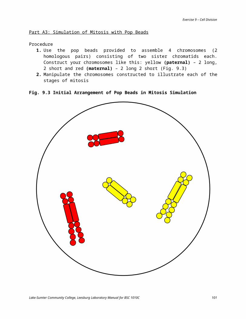
Exercise 11 – Genetics
While the numbers might be close (and even if they are not) to the expected phenotypic ratio of 3:1, statistical theory says the bigger the sample size the closer our actual data should fit theoretical predictions.
3. Increase the sample size for this dataset by counting all rows in the ear of corn and fill in Table 11.3
Table 11.3 Kernel Counts in F2 Generation Corn (Zea mays) – All Rows# of kernels Number Proportion*
purpleyellowtotal
*divide the number of kernels of each color by the total number of kernels in that row
Did counting more kernels make the numbers come more closely to the expected 3:1 ratio of purple to yellow color? __________
Part B: Phenylthiocarbimide (PTC) Taste TestPhenylthiocarbimide is an organic compound which interacts with the taste buds on the tongue and mouth of some people but not in others. Therefore, some people can taste PTC and are considered “tasters.” Other people cannot taste PTC and are considered “non-tasters.” Being able to taste is the dominant condition (“T”), non-tasters the recessive (“t”).
Procedure1. Obtain a piece of paper treated with a small amount of PTC and place it on the tongue. Tasters
will sense a very strong flavor, non-tasters will taste only “paper”2. Collect results on the number of tasters and non-tasters from the population in class and use
that data to fill in Table 11.4
Table 11.4 PTC TastingPhenotypes Number Proportion*
tasternon-taster
total*divide the number of phenotypes by the total class size
What is the genotype of a taster? __________ What is the genotype of a non-taster? __________
What is the predicted phenotypic ratio for a cross between two heterozygous (Tt) individuals? __________
Were the resultant proportions close to this ratio? __________
Lake-Sumter Community College, Leesburg Laboratory Manual for BSC 1010C 101

Exercise 11 – Genetics
If the proportions were not close (and even if they were), provide an explanation as to why they would be (hint: sample size).
Part C: Multiple Alleles – Human Blood TypesSome genes have more than two alleles. Although each organism can only have two copies of a gene (why? _________________), within an entire population there may be several alleles. One example of this is seen with the ABO blood types in human beings. Blood types are produced by multiple alleles. On the membranes of red blood cells are proteins which can stimulate an immune response. These proteins are called antigens. The dominant allele “IA” produces the A antigen protein, the dominant allele “IB” produces the B antigen protein. The recessive allele “i” produces no protein. When a genetic trait has more than one dominant allele, a situation of codominance may exist. The genetic trait for human ABO blood typing contains three alleles (IA, IB, i).
Procedure1. Fill in Table 11.5 with the possible genotypes for the listed phenotypes
Table 11.5 Human ABO Blood Typing Genotypes and PhenotypesGenotype(s) Phenotype
_____ _____ or _____ _____ A
_____ _____ or _____ _____ B
_____ _____ AB
_____ _____ O
Part D: X-Linked CharacteristicsIn humans, all somatic cells (typical body cells) contain 23 pairs of chromosomes. Of these, 22 pairs are autosomes, the last pair are the sex chromosomes. The sex chromosomes are related to the gender of the individual and are called X and Y. Women have two X chromosomes, men have an X and a Y. Haploid sex cells (gametes) produced by women (ova) have only the X chromosome, male gametes (sperm) have either X or Y. Sex of the offspring is determined by the male.
The X chromosome is large and carries many genes such as those for essential muscle proteins and retinal pigments. The Y chromosome, on the other hand, is quite small and carries only a few genes, mostly related to male gender development. A defective gene on the X chromosome will be phenotypically expressed in a male because there is no other X chromosome to compensate. However, a woman with the same defective gene will not express it phenotypically if her other X chromosome is normal. She will be a carrier though in that she has the defective gene but does not express it.
Red-green colorblindness is caused by a mutation in a gene for retinal pigments on the X chromosome. The defective allele is recessive to the normal one so a woman with one normal X chromosome (X) and one colorblind carrying chromosome (XC) will have normal color vision because the X chromosome is dominant for this trait over the XC chromosome. Men, however will be colorblind if they possess the XC
chromosome since there is no other X to be dominate over it and will exhibit red-green colorblindness.
Lake-Sumter Community College, Leesburg Laboratory Manual for BSC 1010C 102

Exercise 11 – Genetics
Procedure1. Obtain a color vision diagnostic chart2. Test for colorblindness in yourself and among lab partners
What is your gender? __________ (if you don’t know, excuse yourself to the restroom to find out)
What is your sex chromosome genotype? __________
Do you exhibit red-green colorblindness? __________
Do any lab partners exhibit colorblindness? __________
If so, what are their sexes and genotypes? ____________________
Part E: Dihybrid CrossesDihybrid crosses are used to examine the inheritance patterns in more than one gene (di = two). As an example, consider fur color and texture in guinea pigs. Among these organisms, black fur color is dominant (“B”) over white (“b”) and rough fur coat is dominant (“R”) over smooth (“r”).
ProcedureUse a Punnett Square to answer the questions regarding the offspring in the F 1 generation from the P generation crosses indicated
1. BBRR x bbrr
What will the genotype be of all of the offspring from this cross?
What will be the phenotypes of all of the offspring from this cross?
Lake-Sumter Community College, Leesburg Laboratory Manual for BSC 1010C 103

Exercise 11 – Genetics
2. Bbrr x bbRr
What proportion of the guinea pigs will be
black, rough? __________ black, smooth? __________
white, rough? __________ white, smooth? __________
3. BbRr x bbRr
What proportion of the guinea pigs will be
black, rough? __________ black, smooth? __________
white, rough? __________ white, smooth? __________
Lake-Sumter Community College, Leesburg Laboratory Manual for BSC 1010C 104

Exercise 11 – Genetics
4. BbRr x BbRr
What proportion of the guinea pigs will be
black, rough? __________ black, smooth? __________
white, rough? __________ white, smooth? __________
Lake-Sumter Community College, Leesburg Laboratory Manual for BSC 1010C 105

Exercise 11 – Genetics
Practice Problems and Review Questions
1. Define or describe the following:
monohybrid cross
dihybrid cross
testcross
allele
dominant allele
recessive allele
genotype
phenotype
homozygous
heterozygous
Lake-Sumter Community College, Leesburg Laboratory Manual for BSC 1010C 106

Exercise 11 – Genetics
2. What are the expected phenotypic and genotypic ratios in a monohybrid cross between two heterozygous individuals?
3. What is the expected phenotypic ratio in a dihybrid cross between two organisms that are heterozygous for both traits?
4. List all the possible types of gametes that can be produced from an organism with the genotype AaBb.
5. Humans as well as many other mammals show a genetic condition known as albinism. A recessive allele interferes with the ability to produce the brown pigment melanin which colors eyes and hair in addition to protecting the skin from the harmful effects of UV light. People who are homozygous recessive produce little or no melanin and have very pale eyes, white skin, and yellow or white hair. Normal pigmentation is produced by a dominant allele (“A”). The albino allele is recessive (“a”). Use Punnett Squares to calculate genotypic and phenotypic proportions for the parental generation crosses indicated and denote which F1 genotypes (if any) will be carriers.
a. AA x aa
b. Aa x aa
Lake-Sumter Community College, Leesburg Laboratory Manual for BSC 1010C 107

Exercise 11 – Genetics
c. Aa x Aa
d. AA x Aa
e. aa x aa
6. A woman with normal pigmentation, whose mother was an albino, mates with an albino. What is the probability their first child will be an albino?
7. Normal parents have an albino child. Give the genotypes for the parents and the child.
Lake-Sumter Community College, Leesburg Laboratory Manual for BSC 1010C 108

Exercise 11 – Genetics
8. A person is a taster as is their mother. The father is a non-taster. What is this person’s genotype?
9. A non-taster has two taster parents. What is the genotype of this person and their parents?
10. For ABO blood typing in humans, for which phenotypes is the genotype definitive?
11. A person has blood type O. Their mother is B and father is A. What is the genotype of this person and their parents?
12. A paternity suit involves a child whose blood type is AB. The mother is blood type B, the alleged father is O. Make a ruling on this case as to whether it is reasonably possible this is the biological father.
13. Is it possible for a female human to be colorblind? What would her genotype be?
14. Can two normal vision parents produce a colorblind son? Explain with a diagram.
15. Can two normal vision parents produce a colorblind daughter? Explain with a diagram.
Lake-Sumter Community College, Leesburg Laboratory Manual for BSC 1010C 109

Exercise 11 – Genetics
16. Can two colorblind parents, produce a normal vision son? Explain with a diagram.
17. A normal vision woman, whose father was colorblind, mates with a colorblind man. What proportion of their sons would be colorblind? What proportion of their daughters would be colorblind and what proportion would be carriers? If one of their normal vision sons mates with a homozygous, normal vision woman, would it be possible for them to have a colorblind child?
18. A guinea pig that is heterozygous for fur color and texture mates with another guinea pig heterozygous for both traits. They produce a total of 96 offspring. Use a Punnett Square to diagram this cross then answer the questions below.
How many of the 96 offspring will phenotypically be
black with rough fur? __________
black with smooth fur? __________
white with rough fur? __________
white with smooth fur? __________
Lake-Sumter Community College, Leesburg Laboratory Manual for BSC 1010C 110

Answers
Lake-Sumter Community College, Leesburg Laboratory Manual for BSC 1010C 111