(12) united states patent pardridge et al. (45) date of ... · 6,041,775 a 3/2000 century 6,060,069...
TRANSCRIPT

(12) United States Patent Pardridge et al.
USOO8834874B2
US 8,834,874 B2 Sep. 16, 2014
(10) Patent No.: (45) Date of Patent:
(54) METHODS AND COMPOSITIONS FOR INCREASING DURONATE 2-SULFATASE ACTIVITY IN THE CNS
(75) Inventors: William M. Pardridge, Pacific Palisades, CA (US); Ruben J. Boado, Agoura Hills, CA (US)
(73) Assignee: Armagen Technologies, Inc., Calabasas, CA (US)
(*) Notice: Subject to any disclaimer, the term of this patent is extended or adjusted under 35 U.S.C. 154(b) by 136 days.
(21) Appl. No.: 12/901,481
(22) Filed: Oct. 8, 2010
(65) Prior Publication Data
US 2011 FO1 10935 A1 May 12, 2011
Related U.S. Application Data (60) Provisional application No. 61/250,378, filed on Oct.
9, 2009, provisional application No. 61/256,049, filed on Oct. 29, 2009.
(51) Int. Cl. A6 IK39/00 (2006.01) C07K 16/00 (2006.01) C07K 6/8 (2006.01) C07K 6/28 (2006.01) A61 K38/00 (2006.01)
(52) U.S. Cl. CPC ............. C07K 16/18 (2013.01); C07K 2319/33
(2013.01); A61 K38/00 (2013.01); C07K 16/2869 (2013.01); C07K 2317/565 (2013.01)
USPC .................. 424/133.1; 424/134.1; 530/387.1; 530/388.26
(58) Field of Classification Search None See application file for complete search history.
(56) References Cited
U.S. PATENT DOCUMENTS
4,801,575 A 1/1989 Pardridge 4.902,505 A 2/1990 Pardridge et al. 4.946,778 A 8, 1990 Ladner et al. 5,154,924 A 10, 1992 Friden 5, 180,820 A 1/1993 Barde et al. 5, 182,107 A 1/1993 Friden 5,229,500 A 7, 1993 Barde et al. 5,438,121 A 8, 1995 Barde et al. 5.453,361 A 9/1995 Yancopoulos et al. 5,527,288 A 6, 1996 Gross et al. 5,527,527 A 6, 1996 Friden 5,562.903. A 10, 1996 CO et al. 5,610,279 A 3, 1997 Brockhaus et al. 5,618,920 A 4/1997 Robinson et al. 5,656.284 A 8, 1997 Balkin 5,672,683 A 9, 1997 Friden et al. 5,693,762 A 12/1997 Queen et al. 5,824,782 A 10, 1998 Holzer et al. 5,837,231 A 11/1998 Low et al.
5,848,991 A 12/1998 Gross et al. 5,977,307 A 11/1999 Friden et al. 5.997,501 A 12/1999 Gross 6,015,662 A 1/2000 Hackett 6,041,775 A 3/2000 Century 6,060,069 A 5, 2000 Hill et al. 6,153,190 A 11/2000 Young et al. 6,165,783 A 12/2000 Weiss et al. 6,248,262 B1 6/2001 Kubotera et al. 6,284,262 B1 9, 2001 Place 6,287,792 B1 9/2001 Pardridge et al. 6,322,808 B1 1 1/2001 Trautman et al. 6,329,508 B1 12/2001 Friden 6,348,210 B1 2/2002 Gale 6,361,760 B1 3/2002 Murata 6,372,250 B1 4/2002 Pardridge 6,375,975 B1 4/2002 Modi 6,531,309 B1 3/2003 Hu et al. 6,582,945 B1 6/2003 Raso 6,583,272 B1 6/2003 Bailon 6,709,833 B2 3/2004 Fukul et al. 6,743,427 B1 6/2004 Schenk 6,858,206 B2 2/2005 Kakkis 7,053,202 B2 5, 2006 Okeefe et al. 7,078,376 B1 7/2006 Thompson 7,214,658 B2 5, 2007 Tobinick 7,226,758 B1 6/2007 Lin et al. 7,294,704 B2 11/2007 Simon et al. 7,309,687 B1 12/2007 Brines et al. 7,388,079 B2 6/2008 Pardridge et al. 7,741,446 B2 6/2010 Pardridge et al. 8,053,569 B2 11/2011 Pardridge et al.
(Continued)
FOREIGN PATENT DOCUMENTS
EP O613007 A2 8, 1994 EP O613007 A3 10, 1995 JP 6-2281.99 8, 1994 WO WO99/OO150 A2 1, 1999 WO WO99/OO951 A1 1, 1999
(Continued) OTHER PUBLICATIONS
Aronovich et al (Am J Hum Genet 58: 75-85, 1996).* Ai, et al., 2003. Intraputamenal Infusion of GDNF in Aged Rhesus Monkeys: Distribution and Dopaminergic Effects. The Journal of Comparative Neurology 461: 250-261. Airavaara, et al. Effects of repeated morphine on locomotion, place preference and dopamine in heterozygous glial cell line-derived neurotrophic factor knockout mice. Genes Brain Behay. Apr. 2007;6(3):287-98.
(Continued)
Primary Examiner — Daniel E. Kolker Assistant Examiner — Aditi Dutt
(74) Attorney, Agent, or Firm — Wilson Sonsini Goodrich & Rosati
(57) ABSTRACT
Provided herein are methods and compositions for treating a Subject Suffering from a deficiency in iduronate 2-sulfatase in the CNS. The methods include systemic administration of a bifunctional fusion antibody comprising an antibody that crosses the blood brain barrier (BBB) and an iduronate 2-sul fatase.
16 Claims, 20 Drawing Sheets
US 8,834,874 B2 Page 2
(56) References Cited
U.S. PATENT DOCUMENTS
8,124,095 B2 2/2012 Pardridge et al. 8,142,781 B2 3/2012 Pardridge et al. 8,227,212 B2 7/2012 von Figura et al. 8.497,246 B2 7/2013 Pardridge et al.
2002fOO52311 A1 2002.0137684 A1 2002fO169109 A1 2003/01291.86 A1 2003. O165853 A1
5, 2002 Solomon et al. 9/2002 Tchistiakova et al. 11/2002 Plata-Salaman et al. 7/2003 Beliveau et al. 9/2003 Pardridge et al.
2004f0072291 A1 4/2004 Carr et al. 2004/0101904 A1 5/2004 Pardridge et al. 2004.0102369 A1 5, 2004 Wu et al. 2004/0229250 A1* 1 1/2004 Figura et al. ...................... 435/6 2004/0248.197 A1 12/2004 Holtzman et al. 2005. O142141 A1 6/2005 Pardridge 2007/0081992 A1 4/2007 Pardridge et al. 2007/0082380 A1 4/2007 Pardridge et al. 2007/0275882 A1 1 1/2007 Meijer et al. 2007/028094.0 A1 12, 2007 Winkles et al. 2008.0003211 A1 1/2008 Fogh et al. 2008/005.1564 A1 2/2008 Pardridge et al. 2008. O152645 A1 6/2008 Pardridge et al. 2008. O170994 A1 7/2008 Pardridge et al. 2008.0171055 A1 7/2008 Pardridge et al. 2008/0292639 A1 11, 2008 Shen et al. 2009.0053219 A1 2/2009 Pardridge et al. 2009 OO68206 A1 3/2009 Pardridge et al. 2009, O156498 A1 6/2009 Pardridge et al. 2009/0238789 A1 9/2009 Guyon et al. 2010, OO77498 A1 3/2010 Pardridge 2010/0098693 A1 4/2010 Pardridge 2010/0172919 A1 7, 2010 Grimm et al. 2010/0261647 Al 10/2010 Pardridge et al. 2010/0290985 A1 1 1/2010 Pardridge et al. 2011 0110935 A1 5/2011 Pardridge et al. 2012/0269807 A1 10/2012 Pardridge et al. 2013, O142794 A1 6/2013 Pardridge et al. 2013/0287773 Al 10/2013 Pardridge et al.
FOREIGN PATENT DOCUMENTS
WO WO99/OO150 A3 4f1999 WO WO99,66951 A1 12/1999 WO WOOOf 15759 A1 3, 2000 WO WOO3,O74081 A1 9, 2003 WO WOO3,O74081 A1 12/2003 WO WO 2004/050016 A2 6, 2004 WO WO 2006/081171 A1 8, 2006 WO WO 2007/022416 A2 2, 2007 WO WO 2007/044323 A2 4/2007 WO WO 2007/022416 A3 5/2007 WO WO-2008-022349 2, 2008 WO WO-2009-018122 2, 2009 WO WO 2007/044323 A3 5, 2009 WO WO 2009,070597 A2 6, 2009
OTHER PUBLICATIONS
Al Sawaf, et al. Neurological findings in Hunter disease: pathology and possible therapeutic effects reviewed. J. Inherit Metab Dis. Aug. 2008:31(4):473-80. Albayrak, et al. 1997. Effect of transient focal ischemia on blood brain barrier permeability in the rat: Correlation to Cell Injury. Acta Neuropathol 94: 158-163. Alberts, et al. Molecular Biology of the Cell. 3rd Edition. Garland Publishing Inc. New York. 1994; pp. 1206-1207. AMGEN www.amgen.com. Accessed Dec. 16, 2005. Bachis, et al. 2003. Brain-Derived Neurotropic Factor Inhibits Human Immunodeficiency Virus-1/gp 120- Mediated Cerebellar Granule Cell Death by Preventing gp120 Internalization. The Journal of Neuroscience 23 (13): 5712-5722. Barth et al. Boron neutron capture therapy of brain tumors: an emerg ing therapeutic modality. Neurosurgery. Mar. 1999:44(3):433-50; discussion 450-1.
Beck, et al. 1994. Brain-Derived Neurotropic Factor Protects Against Ischemic Cell Damage in Rat Hippocampus. Journal of Cerebral Blood Flow and Metabolism 14: 689-692. Bifare, et al. 2005. Brain-Derived Neurotropic Factor Protects against Multiple Forms of Brain Injury in Bacterial Meningitis. The Journal of Infectious Diseases 191: 40-45. Biogen Idec. www.idecpharm.com/site/home.html. Accessed Dec. 16, 2005. Boado et al. Genetic engineering, expression, and activity of a fusion protein of a human neurotrophin and a molecular Trojan horse for delivery across the human blood-brain barrier. Biotechnology and Bioengineering. 2007:97:1376-1386. Boado et al. Humanization of anti-human insulin receptor antibody for drug targeting across the human blood-brain barrier. Biotechnol ogy and Bioengineering. 2007:96:381-391. Boado, et al. Genetic engineering of a lysosomal enzyme fusion protein for targeted delivery across the human blood-brain barrier. Biotechnol Bioeng. 2008; 99(2):475-84. Boado, et al. Engineering and expression of a chimeric transferrin receptor monoclonal antibody for blood-brain barrier delivery in the mouse. Biotechnol Bioeng. Mar. 1, 2009; 102(4): 1251-8. Boado, et al. GDNF fusion protein for targeted-drug delivery across the human blood-brain barrier. Biotechnol Bioeng. Jun. 1, 2008; 100(2):387-96. Boado, et al. Humanization of anti-human insulin receptor antibody for drug targeting across the human blood-brain barrier. Biotechnol Bioeng. 2007: 96(2):381-91. Braun, et al. Metabolic correction and cross-correction of mucopolysaccharidosis type II (Hunter syndrome) by retroviral-me diated gene transfer and expression of human iduronate-2-sulfatase. Proc. Natl. Acad. Sci. 1993: 90:1 1830-1 1834. Brines, et al. Erythropoetin crosses the blood-brain barrier to protect against experimental brain injury, Proc Natl Acad Sci USA. 2000; 97: 10526-10531. Brummell, et al. Probing the combining site of an anti-carbohydrate antibody by saturation-mutagenesis: role of the heavy-chain CDR3 residues. Biochemistry. 1993; 32(4): 1180-7. Buchli, et al. Inhibition of Nogo: a key strategy to increase regenera tion, plasticity and functional recovery of the lesioned central ner vous system. Ann Med. 2005:37(8):556-67. Burgess, et al. Possible dissociation of the heparin-binding and mitogenic activities of heparin-binding (acidic fibroblast) growth factor-1 from its receptor-binding activities by site-directed mutagenesis of a single lysine residue. JCell Biol. Nov. 1990; 111(5 Pt 1):2129-38. Chen, et al. In vitro Scanning Saturation mutagenesis of all the speci ficity determining residues in an antibody binding site. Protein Engi neering. 1999; vol. 12, No. 4, 349-356. Cheng, et al. 1997. Marked Age-dependent Neuroprotection by Brain-derived Neurotropic Factor Against Neonatal Hypoxic Ischemic Brain Injury. Annals of Neurology 41 (4): 521-529. Cheng, et al. 2004. Neuroprotection for Ischemic Stroke: Two Decades of Success and Failure. The Journal of the American Society for Experimental Neuro Therapeutics 1:36-45. Coloma, et al. 1999. Transport Across the Primate Blood-Brain Bar rier of a Genetically Engineered Chimeric Monoclonal Antibody to the Human Insulin Receptor. Pharmaceutical Research 17 (3): 266 274. Coloma, et al. Design and production of novel tetravalent bispecific antibodies. Nat Biotechnol. Feb. 1997:15(2): 159-63. Coloma, et al. The hinge as a spacer contributes to covalent assembly and is required for function of IgG. J. Immunol. Jan. 15, 1997;158(2):733-40. Cowen, et al. 2004. Neuropeptides: implications for alcoholism. Journal of Neurochemistry 89: 273-285. Dawson, et al. 2001. A comparative assessment of the efficacy and side-effect liability of the neuroprotective compounds in experimen tal stroke. Brain Research 892: 344-350. Deguchi, et al. Retention of biologic activity of human epidermal growth factor following conjugation to a blood-brain barrier drug delivery vector via an extended poly(ethylene glycol) linker. Bioconjug Chem. Jan.-Feb. 1999; 10(1):32-7.
US 8,834,874 B2 Page 3
(56) References Cited
OTHER PUBLICATIONS
Duchnowska, et al. Central nervous system metastases in breast cancer patients administered trastuzumab. Cancer Treat Rev. Jun. 2005:31(4):312-8. Duffy, et al. 1987. Blood-brain barrier transcytosis of insulin in developing rabbits. Brain Research 420:32-38. Ehrenreich, et al. Erythropoetin therapy for acute stroke is both safe and beneficial. Mol Med. Aug. 2002:8(8):495-505. European search report and search opinion dated Dec. 2, 2010 for Application No. 07841 110.5. European search report dated Feb. 23, 2010 for Application No. 6825.389.7. Ferber, D. Bridging the blood-brain barrier; new methods improve the odds of getting drugs to the brain cells that need them. PLoS Biol. Jun. 2007:5(6):e 169: 1191-1194. Fillebeen, et al. Receptor-mediated transcytosis of lactoferrin through the blood-brain barrier, J Biol Chem. Mar. 12, 1999:274(11):7011-7017. Flomen, et al. Determination of the organisation of coding sequences within the iduronate sulphate sulphatase (IDS) gene. Hum. Mol. Genet. 1993; 2(1):5-10. Forough, et al. Differential transforming abilities of non-secreted and secreted forms of human fibroblast growth factor-1. J Biol Chem. Feb. 5, 1993:268(4):2960-8. Friden, et al. Blood-brainbarrierpenetration and in vivo activity of an NGF conjugate. Science. Jan. 15, 1993:259(5093):373-377. Fu, et al. Neuroprotection in stroke in the mouse with intravenous erythropoietin-Trojan horse fusion protein. Brain Res. Jan. 19. 2011; 1369:203-7. Epub Oct. 31, 2010. Gennaro, 2000. Remington: The Science and Practice of Pharmacy. 20 ed. Gillies, et al. Bi-functional cytokine fusion proteins for gene therapy and antibody-targeted tratment of cancer. 2002, Cancer Immunology and Immunotherapy, vol. 51, pp. 449-460. Green-Sadan, et al. Transplantation of glial cell line-derived neurotrophic factor-expressing cells into the striatum and nucleus accumbens attenuates acquisition of cocaine self-administration in rats. Eur J Neurosci. Oct. 2003;18(7):2093-8. He, et al. Identification and characterization of the molecular lesion causing mucopolysaccharidosis type I in cats. Mol Genet Metab. 1999: 67(2):106-12. He, et al. Autoregulation of glial cell line-derived neurotrophic factor expression: implications for the long-lasting actions of the anti-ad diction drug, Ibogaine. FASEB J. Nov. 2006:20(13):E1820-E1827; 2420-2422. He, et al. Glial cell line-derived neurotrophic factor mediates the desirable actions of the anti-addiction drug ibogaine against alcohol consumption. J Neurosci. Jan. 19, 2005:25(3):619-28. Hetman, et al. 1999. Neuroprotection by Brain-derived Neurotropic Factor Is Mediated by Extracellular Signal-regulated Kinase and Phoshatidylinositol 3-Kinase. The Journal of Biological Chemistry 274 (32): 22569-22580. Hoshaw, etal. 2005. Central administration of IGF-I and BDNF leads to long-lasting antidepressant-like effects. Brain Research 1037: 204-208. Ibanez, et al. An extended Surface of binding to Trk tyrosine kinase receptors in NGF and BDNF allows the engineering of a multifunctional pan-neurotrophin. EMBO J. Jun. 1993; 12(6):2281 93. Ibanez, Structure-function relationships in the neurotrophin family. J Neurobiol. Nov. 1994:25(11): 1349-61. International search report and written opinion dated Feb. 22, 2011 for PCT/US 2010/0521.13. International search report dated Feb. 27, 2009 for PCT Application No. USO8,71121. International search report dated Jul. 1, 2008 for PCT Application No. USO6,38587. International search report dated Sep. 7, 2010 for PCT Application No. US 10-27882.
International search report dated Sep. 16, 2008 for PCT Application No. US2007f76316. Jefferies, et al. Analysis of lymphopoietic stem cells with a monoclonal antibody to The rat transferrin receptor. Immunology. Feb. 1985:54(2):333-41. Jethwa, et al. 2004. Neuromedin U has a physiological role in the regulation of food intake and partially mediates the effects of leptin. American Journal of Physiology-Endocrinology and Metabolism 289: E301-E305. Jiang, et al. 2005. BDNF Variation and Mood Disorders: A Novel Functional Promoter Polymorphism and Val66Met are Associated with Anxiety but Have Opposing Effects. Neuropsychopharmacol ogy 30: 1353-1361. Juul, et al. Erythropoietin concentrations in cerebrospinal fluid of nonhuman primates and fetal sheep following high-dose recombi nant erythropoietin, Biol. Neonate. 2004:85:138-144. Kakkis, et al. Overexpression of the human lysosomal enzyme alpha L-iduronidase in Chinese hamster ovary cells. Protein Expr Purif. 1994; 5(3): 225-32. Kido, et al. 2000. Neuroprotective effects of brain-derived neurotropic factor in eyes with NMDA-induced neuronal death. Brain Research 884: 59-67. Kim, et al. 2003. Continuous Brain-derived Neurotropic Factor (BDNF) Infusion. After Methylprednisolone Treatment in Severe Spi nal Cord Injury. Journal of Korean Medical Science 19: 113-122. Krewson, et al. 1995. Distribution of nerve growth factor following direct delivery to brain interstitium. Brain Research 680: 196-206. Kurihara, et al. 1999. Imaging Brain Tumors by Targeting Peptide Radiopharmaceuticals through the Blood-Brain Barrier. Cancer Research 59: 6159-6163. Lai, et al. Structural determinants of Trk receptor specificities using BDNF-based neurotrophin chimeras. J Neurosci Res. Dec. 1, 1996:46(5):618-29. Lazar, et al. Transforming growth factor alpha: mutation of aspartic acid 47 and leucine 48 results in different biological activities. Mol Cell Biol. Mar. 1998; 8(3): 1247-52. Lee, et al. 2002. Imaging Brain Amyloid of Alzheimer Disease in Vivo in Transgenic Mice With an AB Peptide Radiopharmaceutical. Journal of Cerebral Blood Flow and Metabolism 22: 223-231. Lewin, B. Genes IV. Oxford University Press. 1990. p. 810. Lin, et al. Structure-function relationships in glucagon: properties of highly purified des-His-1-, monoiodo-, and (des-Asn-28. Thr 29)(homoserine lactone-27)-glucagon. Biochemistry. Apr. 22. 1975;14(8): 1559-63. Marvin, et al. Recombinant approaches to IgG-like bispecific anti bodies. Acta Pharmacol Sin. Jun. 2005:26(6):649-58. McGrath, et al. Bifunctional fusion between nerve growth factor and a transferrin receptor antibody. J Neurosci Res. Jan. 15. 1997:47(2): 123-33. McLendon et al. Radiotoxicity of systemically administered 21 1At labeled human/mouse chimeric monoclonal antibody: a long-term survival study with histologic analysis. IntJ Radiat Oncol Biol Phys. Sep. 1, 1999:45(2):491-9. Menzies, et al. 1993. Contributions of ions and albumin to the for mations and resolution of ischemic brain edema. Journal of Neurosurgery 78: 257-266. Messer, et al. Role for GDNF in biochemical and behavioral adapta tions to drugs of abuse. Neuron. Apr. 2000:26(1):247-57. Mori, et al. 2004. Differential expression patterns of TrkB ligands in the macaque monkey brain. Developmental Neuroscience 15:2507 2511. Muenzer, et al. A phase II/III clinical study of enzyme replacement therapy with idursulfase in mucopolysaccharidosis II (Hunter syn drome). Genet Med. Aug. 2006:8(8):465-73. Muenzer, et al. Advances in the treatment of mucopolysaccharidosis type I.N Engl J Med. May 6, 2004:350(19):1932-4. NCBI Reference Sequence: NM 000202.5 Homo sapiens iduronate 2-sulfatase (IDS), transcript variant 1, mRNA. 1992. http://www. incbi.nlm.nih.gov/nuccore/NM 000202.5. Nutt, et al. 2003. Randomized, double-blind trial of glial cell line derived neurotropic factor (GDNF) in PD. Neurology 60: 69-73. Office Action dated Jan. 15, 2008 for U.S. Appl. No. 1 1/245,710. Office Action dated Jan. 15, 2009 for U.S. Appl. No. 1 1/841,623.

US 8,834,874 B2 Page 4
(56) References Cited
OTHER PUBLICATIONS
Office Action dated Jan. 23, 2009 for U.S. Appl. No. 1 1/245,546. Office action dated Feb. 16, 2011 for U.S. Appl. No. 1 1/893,281. Office action dated Feb. 16, 2011 for U.S. Appl. No. 12/150,983. Office Action dated Mar. 10, 2010 for U.S. Appl. No. 12/179,806. Office action dated Mar 18, 2011 for U.S. Appl. No. 12/574,571. Office action dated Apr. 6, 2011 for U.S. Appl. No. 1 1/245,710. Office Action dated Apr. 13, 2007 for U.S. Appl. No. 1 1/245,710. Office action dated May 12, 2010 for U.S. Appl. No. 1 1/893,281. Office action dated May 13, 2011 for U.S. Appl. No. 12/688,842. Office Action dated Jun. 3, 2008 for U.S. Appl. No. 1 1/245,710. Office Action dated Jun. 17, 2009 for U.S. Appl. No. 1 1/841,541. Office action dated Jun. 27, 2011 for U.S. Appl. No. 1 1/245,546. Office Action dated Jul. 2, 2008 for U.S. Appl. No. 1 1/245,546. Office Action dated Jul. 2, 2009 for U.S. Appl. No. 1 1/245,710. Office Action dated Jul. 31, 2009 for U.S. Appl. No. 12/179,806. Office Action dated Aug. 20, 2009 for U.S. Appl. No. 12/323,232. Office action dated Sep. 15, 2010 for U.S. Appl. No. 12/150,983. Office Action dated Sep. 20, 2007 for U.S. Appl. No. 1 1/245,710. Office Action dated Sep. 24, 2009 for U.S. Appl. No. 1 1/841,623. Office Action dated Oct. 12, 2010 for U.S. Appl. No. 1 1/245,710. Office action dated Oct. 13, 2009 for U.S. Appl. No. 1 1/893,281. Office Action dated Oct. 15, 2007 for U.S. Appl. No. 1 1/245,710. Office Action dated Oct. 20, 2009 for U.S. Appl. No. 1 1/245,546. Office Action dated Oct. 30, 2009 for U.S. Appl. No. 1 1/841,594. Office Action dated Nov. 8, 2007 for U.S. Appl. No. 1 1/245,546. Office Action dated Nov. 10, 2008 for U.S. Appl. No. 1 1/245,710. Office Action dated Nov. 13, 2006 for U.S. Appl. No. 1 1/245,710. Office Action dated Dec. 16, 2009 for U.S. Appl. No. 1 1/841,541. Office Action dated Feb. 2, 2010 for U.S. Appl. No. 1 1/245,710. Office Action dated Mar. 26, 2010 for U.S. Appl. No. 12/323,232. Office Action dated Mar. 26, 2010 for U.S. Appl. No. 1 1/841,594. Office Action dated Mar. 7, 2011 for U.S. Appl. No. 12/558.348. Office Action dated Jul. 1, 2010 for U.S. Appl. No. 1 1/245,546. Padlan, et al. Identification of specificity-determining residues in antibodies. FASEB J. 1995; 9(1):133-9. Pardridge, 2001. Brain drug targeting: The future of brain drug devel opment. Cambridge University Press. Pardridge, 2002. Neurotrophins, neuroprotection and the blood-brain barrier. Current Opinion in Investigational Drugs 3 (12): 1753-1757. Pardridge, 2003. Blood-Brain Barrier Drug Targeting: The Future of Brain Drug Development. Molecular Interventions 3: 90-105. Pardridge, 2005. The Blood-Brain Barrier and Neurotherapeutics. NeuroRx: The Journal of the American Society for Experimental NeuroTherapeutics 2 (1): 1-2. Pardridge, 2001. Neuroprotection in stroke: is it time to consider large-molecule drugs? Drug Discovery Today 6: 751-753. Pardridge, 2005. The Blood-Brain Barrier. Bottleneck in Brain Drug Development. NeuroRX: The Journal of the American Society for Experimental NeuroTherapeutics 2:3-14. Pardridge, et al. 1987. Human Blood-Brain Barrier Transferrin Receptor. Metabolism 36: 892-895. Pardridge, et al. 1993. Transport of Human Recombinant Brain Derived Neurotrophic Factor (BDNF) Through the Rat Blood-Brain Barrier in Vivo Using Vector-Mediated Peptide Drug Delivery. Phar maceutical Research 11 (5): 738-746. Pardridge, et al. 1995. Human Insulin Receptor Monoclonal Anti body Undergoes High Affinity Binding to Human Brain Capillaries in Vitro and Rapid Transcytosis Through the Blood-Brain Barrier in Vivo in the Primate. Pharmaceutical Research 12 (6): 807-816. Pardridge, et al. 1998, Combined Use of Carboxyl-Directed Protein Pegylation and Vector-Mediated Blood-Brain Barrier Drug Delivery System Optimizes Brain Uptake of Brain-Derived Neurotrophic Fac tor Following Intravenous Administration. Pharmaceutical Research 15 (4): 576-582. Pardridge, et al. Drug and gene targeting to the brain with molecular Trojan horses. Nat Rev Drug Discov. Feb. 2002;1(2): 131-9. Park, et al. Production and characterization of fusion proteins con taining transferrin and nerve growth factor. J Drug Target. 1998;6(1):53-64.
Pencea, et al. 2001. Infusion of Brain-Derived Neurotrophic Factor into the Lateral Ventricle of the Adult Rat Leads to New Neurons in the Parenchyma of the Striatum, Septum, Thalamus, and Hypothala mus. The Journal of Neuroscience 21 (17): 6706-6717. Penichet, et al. An antibody-avidin fusion protein specific for the transferrin receptor serves as a delivery vehicle for effective brain targeting: initial applications in anti-HIV antisense drug delivery to the brain. JImmunol. Oct. 15, 1999; 163(8):4421-4426. Preston, et al. 1997. Evidence for pore-like opening of the blood brain barrier following forebrain ischemia in rats. Brain Research 761: 4-10. Raghavan, et al. Analysis of the pH dependence of the neonatal Fc receptor?immunoglobulin G interaction using antibody and receptor variants. Biochemistry. Nov. 14, 1995:34(45): 14649-57. Ratliff-Schaub, et al. 2005. Randomized controlled trial of transdermal secretion on behavior of children with autism. Autism 9 (3): 256-265. Robinson, et al. 1999. The structures of the neurotrophin 4 homodimer and the brain-derived neurotrophic factor f neurotrophin 4 heterodimer reveal a common Trk-binding site. Protein Science 8: 2589-2597 Rudikoff, et al. Single amino acid Substitution altering antigen-bind ing specificity. Proc Natl AcadSci U S A. Mar. 1982:79(6):1979-83. Ruiz-Leon, et al. 2003. Induction of Tyrosine Kinase Receptor B by Retinoic Acid Allows Brain-Derived Neurotrophic Factor-Induced Amyloid Precursor Protein Gene Expression in Human SHSY5Y Neuroblastoma Cells. Neuroscience 120: 1019-1026. Sakane, et al. 1997. Carboxyl-directed Pegylation of Brain-derived Neurotrophic Factor Markedly Reduces Systemic Clearance with Minimal Loss of Biologic Activity. Pharmaceutical Research 14 (8): 1085-1091. Schabitz, et al. 1997. Intraventricular Brain-Derived Neurotrophic Factor Reduces Infarct Size After Focal Cerebral Ischemia in Rats. Journal of Cerebral Blood Flow and Metabolism 17: 500-506. Schlachetzki, et al. Expression of the neonatal Fc receptor (FcRn) at the blood-brain barrier. J Neurochem. Apr. 2002;81(1):203-6. Schwartz, et al. A Superactive insulin: B10-aspartic acidinsulin(hu man). Proc Natl AcadSci U S A. Sep. 1987:84(18):6408-11. Scott, etal. Human alpha-L-iduronidase: cDNA isolation and expres sion. Proc Natl AcadSci U S A. Nov. 1, 1991:88(21):9695-9. Selmayr, et al. Induction of tumor immunity by autologous B lymphoma cells expressing a genetically engineered idiotype. Gene Ther. May 1999;6(5):778-84. Shin, et al. Transferrin-antibody fusion proteins are effective in brain targeting, Proceedings of the Natinal Academy of Sciences, 1995. vol.92, pp. 2820-2824. Siren, et al., Erythropoetin prevents neuronal apoptosis after cerebral ischemia and metabolic stress. Proc Natl Acad Sci U S A. Mar. 27. 2001;98(7):4044-9. Skolnick, et al. From genes to protein structure and function: novel applications of computational approaches in the genomic era. Trends Biotechnol. Jan. 2000:18(1):34-9. Review. Spina, et al. 1992. Brain-Derived Neurotrophic Factor Protects Dopamine Neurons Against 6-Hydroxydopamine and N-Methyl-4- Phenylpyridinium Ion Toxicity: Involvement of the Glutathione Sys tem. Journal of Neurochemistry 59 (1): 99-106. Strauss, et al. 2005. Brain-derived neurotrophic factor variants are associated with childhood-onset mood disorder: confirmation in a Hungarian sample. Molecular Psychiartry 10: 861-867. Sukegawa-Hayasaka, et al. Effect of Hunter disease (mucopolysac charidosis type II) mutations on molecular phenotypes of iduronate 2-sulfatase: enzymatic activity, protein processing and structural analysis. J. Inherit. Metab. Dis. 2006; 29:755-761. Takahashi, et al. 1991 Inhibition of cell growth and tumorigenesis of human glioblastoma cells by a neutralizing antibody against human basic fibroblast growth factor. Federation of European Biochemical Societies 288 (1,2): 65-71. The BDNF Study Group (Phase III). 1999. A controlled trial of recombinant methionyl human BDNF in ALS. Neurology 52: 1427 1433. Thoenen, et al. 2002. Neurotrophins: from enthusiastic expectations through Sobering experiences to rational therapeutic approaches. Nature Neuroscience Supplement 5: 1046-1050.

US 8,834,874 B2 Page 5
(56) References Cited
OTHER PUBLICATIONS
Tomatsu, et al. Murine model (Galns(tm(C76S)slu)) of MPS IVA with missense mutation at the active site cysteine conserved among sulfatase proteins. Mol Genet Metab. Jul. 2007:91(3):251-8. Triguero et al. Capillary depletion method for quantification of blood-brain barrier transport of circulating peptides and plasma pro teins. J Neurochem. 1990; 54(6):1882-8. Tsukahara, et al. 1994. The Role of Brain-derived Neurotrophic Factor in Transient Forebrain Ischemia in the Rat Brain. Neurosurgery 34 (2): 323-331. Voznyi, et al. A fluorimetric enzyme assay for the diagnosis of MPS II (Hunter disease). J. Inherit. Metab. Dis. 2001; 24.675-680. Weich, et al. Interleukin-3/erythropoietin fusion proteins: in vitro effects on hematopoietic cells. Exp Hematol. May 1993:21(5):647 55. Whittaker, et al. Characterization of the functional insulin binding epitopes of the full-length insulin receptor, J Biol Chem. 2005:280(22):20932-6. Wraith, et al. Mucopolysaccharidosis type II (Hunter syndrome): a clinical review and recommendations for treatment in the era of enzyme replacement therapy. Eur J Pediatr. Mar. 2008;167(3):267 77. Wraith, J. Enzyme replacement therapy in mucopolysaccharidosis type I: progress and emerging difficulties. J Inherit Metab Dis. Apr. 2001:24(2):245-50. Wu, et al. 1999. Neuroprotection with noninvasive neurotrophin delivery to the brain. Proceedings of the National Academy of Sci ences of the USA: Neurobiology 96: 254-259. Wu, et al. Drug targeting of a peptide radiopharmaceutical through the primate blood-brain barrier in vivo with a monoclonal antibody to the human insulin receptor. J Clin Invest. Oct. 1, 1997: 100(7): 1804 12. Yamashita, et al. 1997. Post-Occlusion Treatment with BDNF Reduces Infarct Size in a Model of Permanent Occlusion of the Middle Cerebral Artery in Rat. Metabolic Brain Disease 12 (4): 271-280. Yan, et al. 1994. Distribution of Intracerebral Ventricularly Admin istered Neurotrophins in Rat Brain and Its Correlation with Trk Receptor Expression. Experimental Neurology 127: 23-36. Yan, et al. Enduring vulnerability to reinstatement of methamphetamine-seeking behavior in glial-cell-line-derived neurotrophic factor mutant mice. FASEB J. Jul. 2007:21(9): 1994 2004. Yip, et al. Three-dimensional structural interactions of insulin and its receptor. J Biol Chem. Jul. 25, 2003:278(30):27329-32. Zhang, et al. 2001. Conjugation of brain-derived neurotrophic factor to a blood-brain barrier drug targeting system enables neuroprotec tion in regional brain ischemia following intrvenous injection of the neurotrophin. Brain Research 889: 49-56. Zhang, et al. 2001. Neuroprotection in Transient Focal Brain Ischemia. After Delayed Intravenous Administration of Brain-De rived Neurotrophic Factor Conjugated to a Blood-Brain Barrier Drug Targeting System. Stroke 32: 1378-1384. Zhang, et al. 2003. Global Non-Viral Gene Transfer to the Primate Brain Following Intravenous Administration. Molecular Therapy 7 (1): 11-18. Zhang, et al. Mediated efflux of IgG molecules from brain to blood across the blood-brain barrier. J Neuroimmunol. Mar. 1, 2001; 114(1- 2): 168-72. Zito, et al. Sulphatase activities are regulated by the interaction of Sulphatase-modifying factor 1 with SUMF2, EMBO Rep. 2005; 6(7):655-660. Aharoni, et al. Directed evolution of mammalian paraoxonases PON1 and PON3 for bacterial expression and catalytic specializa tion. Proc Natl Acad Sci U S A. Jan. 13, 2004: 101(2):482-7. Epub Dec. 26, 2003. Altschul, et al. Optimal sequence alignment using affine gap costs. Bulletin of Mathematical Biology. 1986; 48(5-6):603-16. Altschul, et al. Basic Local Alignment Search Tool. J. Mol. Biol. 1990:215:403-410.
Altschul, et al. Gapped BLAST and PSI-BLAST: a new generation of protein database search programs. Nucleic Acids Res. 1977:25:3389 402. Arndt, et al. Generation of a highly stable, internalizing anti-CD22 single-chain Fv fragment for targeting non-Hodgkin's lymphoma. Int J. Cancer. Dec. 10, 2003: 107(5):822-829. Ausubel, et al. Current Protocols in Molecular Biology. John Wiley & Sons, New York, 1995 Supplement. Baloh, et al. Functional mapping of receptor specificity domains of glial cell line-derived neurotrophic factor (GDNF) family ligands and production of GFRalphal RET-specific agonists. JBiol Chem. Feb. 4. 2000:275(5):3412-20. Batzer, et al. Enhanced evolutionary PCR using oligonucleotides with inosine at the 3'-terminus. Nucleic Acid Res. 1991; 19:5081. Boado, et al. AGT-181: expression in CHO cells and pharmacokinet ics, Safety, and plasma iduronidase enzyme activity in Rhesus mon keys. Oct. 2009: 144(2):135-41. Boado, et al. CHO cell expression, long-term stability, and primate pharmacokinetics and brain uptake of an IgG-paroxonase-1 fusion protein. Biotechnol Bioeng. Jan. 2011; 108(1): 186-96. Boado, et al. Drug delivery of antisense molecules to the brain for treatment of Alzheimer's disease and cerebral AIDS. J Pharm Sci. Nov. 1998:87(11): 1308-15. Boado, et al. Fusion Antibody for Alzheimer's Disease with Bi Directional Transport Across the Blood-Brain Barrier and Abeta Fibril Disaggregation. Bioconjug Chem. 2007:18(2):447-55. Boado, et al. Reversal of lysosomal storage in brain of adult MPS-I mice with intravenous Trojan horse-iduronidase fusion protein. Mol Pharm. Aug. 1, 2011;8(4): 1342-50. Epub Jun. 17, 2011. Boado, et al. Selective targeting of a TNFR decoy receptor pharma ceutical to the primate brain as a receptor-specific IgG fusion protein. J Biotechnol. Mar. 2010:146(1-2):84-91. Carnicella, et al. GDNF is a fast-acting potent inhibitor of alcohol consumption and relapse. Proc Natl Acad Sci U S A. Jun. 10, 2008; 105(23):8114-9. Casset, et al. A peptide mimetic of an anti-CD4 monoclonal antibody by rational design. Biochem Biophys Res Commun. Jul. 18, 2003:307(1): 198-205. Cassol, et al. Stability of dried blood spot specimens for detection of human immunodeficiency virus DNA by polymerase chain reaction. J. Clin Microbiol. Dec. 1992:30(12):3039-42. Chothia, et al. Canonical structures for the hypervariable regions of immunoglobulins. J Mol Biol. Aug. 20, 1987; 196(4):901-17. Chung etal. Antibodies against West Nile Virus nonstructural protein NSI prevent lethal infection through Fc gamma receptor-dependent and -independent mechanisms. JVirol. Feb. 2006:80(3): 1340-51. Colman, P.M. Effects of amino acid sequence changes on antibody antigen interactions. Res Immunol. Jan. 1994: 145(1):33-6. Crow, et al. Biochemical and histopathological studies on patients with mucopolysaccharidoses, two of whom had been treated by fibroblast transplantation. J. Clin Pathol. 1983:36(4):415-30. De Pascalis, et al. Grafting of "abbreviated” complementarity-deter mining regions containing specificity-determining residues essential for ligand contact to engineer a less immunogenic humanized monoclonal antibody. J Immunol. Sep. 15, 2002; 169(6):3076-84. Deakin, et al. Enzymatically active paraoxonase-1 is located at the external membrane of producing cells and released by a high affinity, saturable, desorption mechanism. J Biol Chem. Feb. 8, 2002:277(6):4301-8. Epub Nov. 28, 2001. Deane, et al. IgG-assisted age-dependent clearance of Alzheimer's amyloid beta peptide by the blood-brain barrier neonatal Fc receptor. J Neurosci. 2005; 25(50): 11495-503. Dreier, et al. Recombinant immunocytokines targeting the mouse transferrin receptor: construction and biological activities. Bioconjug Chem. Jul.-Aug. 1998:9(4):48.2-9. Duffy, et al. 1988. Human blood-brain barrier insulin-like growth factor receptor. Metabolism. Feb:37(2): 136-40. Durrington, et al. Paraoxonase and atherosclerosis. Arterioscler Thromb. Vasc Biol. Apr. 2001:21(4):473-80. Eketall, et al. Distinct structural elements in GDNF mediate binding to GFRalphal and activation of the GFRalphal-c-Ret receptor com plex. EMBO J. Nov. 1, 1999; 18(21):5901-10.
US 8,834,874 B2 Page 6
(56) References Cited
OTHER PUBLICATIONS
Elliott, et al. Control of rHuEPO biological activity: the role of carbohydrate. Exp Hematol. Dec. 2004:32(12): 1146-55. EP Appl. No. 08796594.3 Search Report and opinion dated Mar. 16. 2012. Eslamboli, et al. Continuous Low-Level Glial Cell Line-Derived Neurotrophic Factor Delivery Using Recombinant Adeno-Associ ated Viral Vectors Provides Neuroprotection and Induces Behavioral. Recovery in a Primate Model of Parkinson's Disease. J. Neurosci. 2005:25:769-77.
Frenkel, et al. Modulation of Alzheimer's beta-amyloid neurotoxic ity by site-directed single-chain antibody. J Nel Jroimmunol. Jul. 1, 2000: 106(1-2):23-31. Fukuchi, et al. Amelioration of amyloid load by anti-Abeta single chain antibody in Alzheimer mouse model. Biochem Biophys Res Commun. May 26, 2006:344(1):79-86. Fukuda et al. In vitro evolution of single-chain antibodies using mRNA display. Nucleic Acids Research, 2006; 34(19): e127. Golden, et al. Human blood-brain barrier leptin receptor. Binding and endocytosis in isolated human brain microvessels. J Clin Invest. Jan. 1, 1997:99(1): 14-8. Grasso, et al. Neuroprotection by erythropoietin administration after experimental traumatic brain injury. Brain Res. Nov. 28, 2007: 1182:99-105. Habgood, et al. Changes in blood-brain barrier permeability to large and Small molecules following traumatic brain injury in mice. Eur J Neurosci. Jan. 2007:25(1):231-8. Haisma, etal. Construction and characterization of a fusion protein of single-chain anti-CD20 antibody and human beta-glucuronidase for antibody-directed enzyme prodrug therapy. Blood. Jul. 1, 1998:92(1): 184-90. Hansson et al. Prediction of Alzheimer's disease using the CSF Abeta42/Abeta40 ratio in patients with mild cognitive impairment. Dement Geriatr Cogn Disord. 2007:23(5):316-20. Henikoffet al. Predicting the effects of amino Acid substitutions on protein function. Annu Rev Genomics Hun, Genet. 2006:7:61-80. Henikoff, et al. Amino acid substitution matrices from proteinblocks. Proc Natl Acad Sci U S A. 1992; 89(22): 10915-9. Holliger, et al. Engineered antibody fragments and the rise of single domains. Nat Biotechnol. Sep. 2005:23(9): 1126-36. Huston, et al. Protein engineering of antibody binding sites: recovery of specific activity in an anti-digoxin single-chain Fv analogue pro duced in Escherichia coli. Proc Natl Acad Sci U S A. 1988; 85(16):5879-83. Iwasaki, et al. Protective effect of interleukin-3 and erythropoietin on motor neuron death after neonatal axotomy. Neurol Res. Oct. 2002:24(7):643-6. Jethwa, et al. 2004. Neuromedin U has a physiological role in the regulation of food intake and partially mediates the effects of leptin. American Journal of Physiology—Endocrinology and Metabolism 289: E301-E305. Josse, et al. Identification of residues essential for human paraoxonase (PON1) arylesterase/organophosphatase activities. Biochemistry. Mar. 2, 1999;38(9):2816-25. Josse, et al. Oligomeric states of the detergent-solubilized human serum paraoxonase (PON1). J Biol Chem. Sep. 6, 2002:277(36):33386-97. Josse, et al. The active site of human paraoxonase (PON1). J Appl Toxicol. Dec. 2001:21 Suppl 1:S7-11. Kabat, et al., Sequences of Proteins of Immunological Interest. 5th Ed. Public Health Service, National Institutes of Health, Bethesda, Md. 1991;pp. 647-649. Karlin, et al. Applications and statistics for multiple high-scoring segments in molecular sequences. Proc. Natl. Acad. Sci. USA. 1993:90:5873-87. Kashmiri, et al. SDR grafting a new approach to antibody human ization. Methods. May 2005:36(1):25-34. Kastin, etal. Glial cell line-derived neurotrophic factor does not enter normal mouse brain. Neuroscience Letters. 2003:340:239-41.
Kim, et al. Decreased paraoxonase-1 activity is a risk factor for ischemic stroke in Koreans. Biochem Biophys Res Commun. Dec. 7. 2007:364(1): 157-62. Kitagawa, etal. Reduction of Ischemic Brain Injury by Topical Appli cation of Glial Cell Line-Derived Neurotrophic Factor After Perma nent Middle Cerebral Artery Occlusion in Rats. Stroke. 1998:29:1417-22. Kobayashi, et al. Intracerebral Infusion of Glial Cell Line-Derived Neurotrophic Factor Promotes Striatal Neurogenesis. After Stroke in Adult Rats Stroke. 2006:37:2361-67. Koehne, et al. Vascular endothelial growth factor and erythropoietin concentrations in cerebrospinal fluid of children with hydrocephalus. Childs Nerv Syst. Apr. 2002:18(3–4): 137-41. Lang, et al. Randomized controlled trial of intraputamenal glial cell line-derived neurotrophic factor infusion in Parkinson disease. Annals of Neurology. 2006; 59:459-66. Lapchak, et al. Glial cell line-derived neurotrophic factor attenuates behavioural deficits and regulates nigrostriatal dopaminergic and peptidergic markers in 6-hydroxydopamine-lesioned adult rats: com parison of intraventricular and intranigral delivery. Neuroscience. 1997;78:61-72. Lee, et al. Drug targeting to the brain using avidin-biotin technology in the mouse; (blood-brain barrier, monoclonal antibody, transferrin receptor, Alzheimer's disease). J Drug Target. 2000;8(6):413-24. Lenz, et al. Stoichiometric and catalytic scavengers as protection against nerve agent toxicity: a mini review. Toxicology. Apr. 20. 2007:233(1-3):31-9. Li, et al. Genetically engineered brain drug delivery vectors: cloning, expression and in vivo application of an anti-transferrin receptor single chain antibody-streptavidin fusion gene and protein. Protein Eng. Sep. 1999; 12(9):787-96. Lin, et al. GDNF: a glial cell line-derived neurotrophic factor for midbrain dopaminergic neurons. Science. 1993:260: 1130-32. Liu, et al. Antibeta-amyloid (Abeta) SCFV inhibits Abeta aggrega tion and neurotoxicity (P4-354). Neurobiology of Aging, Tarrytown, NY. 2004:25:S575-S576. Liu, et al. Single chain variable fragments against beta-amyloid (Abeta) can inhibit Abeta aggregation and prevent abeta-induced neurotoxicity. Biochemistry. Jun. 8, 2004:43(22):6959-67. Lu, et al. Cationic Liposome-Mediated GDNF Gene Transfer after Spinal Cord Injury. Journal of Neurotrauma. 2002:19:1081-1090. Ma, et al. Erythropoietin protects PC12 cells from beta-amyloid (25 35)-induced apoptosis via PI3K/Akt signaling pathway. Neuropharmacology. May-Jun. 2009:56(6-7): 1027-34. MacCallum, et al. Antibody-antigen interactions: contact analysis and binding site topography. J Mol Biol. Oct. 11, 1996:262(5):732 45. Manoutcharian, et al. Amyloid-beta peptide-specific single chain Fv antibodies isolated from an immune phage display library. J Neuroimmunol 2003: 145(1-2): 12-7. Martell, et al. Efficacy of transferrin receptor-targeted immunotoxins in brain tumor cell lines and pediatric brain tumors. Cancer Res. Mar. 15, 1993:53(6): 1348-53. Martin et al. Crystal structure at 2.8A of an FcRn heterodimeric Fc complex: mechanism of pH-dependent binding. Mol Cell. Apr. 2001;7(4):867-77. Matis, et al. Erythropoietin in spinal cord injury. Eur Spine J. Mar. 2009; 18(3):314-23. Needleman, et al. A general method applicable to the search for similarities in the amino acid sequence of two proteins. J. Mol. Biol. 1970:48:443-53. Ng, et al. Paraoxonase-1 deficiency in mice predisposes to vascular inflammation, Oxidative stress, and thrombogenicity in the absence of hyperlipidemia. Cardiovasc Pathol. Jul.-Aug. 2008;17(4): 226-32. Ng, et al. Predicting the effects of amino acid Substitutions on protein function. Annual Review of Genomics and Human Genetics. 2006;7:61-80. Ober, et al. Differences in promiscuity for antibody-FcRn interac tions across species: implications for therapeutic antibodies. Int Immunol. Dec. 2001:13(12): 1551-9. Ohtsuka, et al. An alternative approach to deoxyoligonucleotides as hybridization probes by insertion of Deoxyinosine at Ambiguous Codon Positions. J. Biol. Chem. 1985:260:2605-08.

US 8,834,874 B2 Page 7
(56) References Cited
OTHER PUBLICATIONS
Osbourn, et al. Directed selection of MIP-1 alpha neutralizing CCR5 antibodies from a phage display human antibody library. Nat Biotechnol. Aug. 1998; 16(8):778-81. Padlan, et al. Structure of an antibody-antigen complex: crystal struc ture of the HyHEL-10 Fab-lysozyme complex. Proc Natl AcadSci U SA. Aug. 1989;86(15):5938-42. Paragh, et al. Ciprofibrate increases paraoxonase activity in patients with metabolic syndrome. BrJClin Pharmacol. Jun. 2006:61(6):694 TO1 Pardridge, 2005. Tyrosine Hydroxylase Replacement in Experimen tal Parkinson's Disease with Transvascular GeneTherapy. NueuoRx: Journal of the American Society for Experimental NeuroTherapeutics. 2(1): 129-138. Pardridge, 2007. Drug Targeting to the Brain. Pharm Res 24:1733-44. Pardridge, et al. 1989. Transport of histone through the blood-brain barrier. J Pharmacol Exp Ther. Dec.:251(3):821-6. Patel, et al. Intraputamenal infusion of glial cell line-derived neurotrophic factor in PD: A two-year outcome study. Annals of Neurology. 2005:57:298-302. Paul, W. Fundamental Immunology, 3rd Edition. 1993:292-95. PCT Application No. US 1 1/21418 ISR and Written Opinion dated Apr. 8, 2011. Pearson, et al. ImprovedTools for Biological Sequence Comparison. Proc. Natl Acad. Sci. USA. 1988:85:2444-48. Pearson, Rapid and sensitive sequence comparison with FASTP and FASTA. Meth. Enzymol. 1990:183:63-98. Pluckthun, A. Antibodies from Escherichia coli. In the Pharmacol ogy of Monoclonal Antibodies. Vol. 113, Rosenburg and Moore eds. Springer-Verlag, New York. 1994; pp. 269-315. Pregi, et al. TNF-alpha-induced apoptosis is prevented by erythropoietin treatment on SH-SY5Y cells. Exp Cell Res. Feb. 1. 2009:315(3):419-31. Epub Nov. 20, 2008. Reiber, et al. Protein transfer at the blood cerebrospinal fluid barrier and the quantitation of the humoral immune response within the central nervous system. Clin Chim Acta. Mar. 30, 1987; 163(3):319 28. Rempel, et al. A homology model for human C-L-Iduronidase: Insights into human disease. Mol. Genetics and Met. 2005; 85:28-37. Rochu, et al. Human paraoxonase: a promising approach for pre treatment and therapy of organophosphorus poisoning. Toxicology. Apr. 20, 2007:233(1-3):47-59. Rossolini, et al. Use of deoxyinosine-containing primers vs degen erate primers for polymerase chain reaction based on ambiguous sequence information. Mol. Cell. Probes. 1994;8(2): 91-98. Sakanaka, et al. In vivo evidence that erythropoietin protects neurons from ischemic damage. Proc Natl Acad Sci U S A. Apr. 14. 1998:95(8):4635-40. Sampson et al. Unarmed, tumor-specific monoclonal antibody effec tively treats brain tumors. Proc Natl Acad Sci U S A. Jun. 20, 2000:97(13):7503-8. Sariola, et al. Novel functions and signalling pathways for GDNF. J. Cell Sci. Oct. 1, 2003; 116(Pt 19):3855-62. Schlachetzki, et al. Gene therapy of the brain: the trans-vascular approach. Neurology. Apr. 27, 2004;62(8): 1275-81. Sellers. On the theory and computation of evolutionary distances. SIAM Journal on Applied Mathematics. 1974:26:787. Shanafelt, et al. Identification of critical amino acid residues in human and mouse granulocyte-macrophage colony-stimulating fac tor and their involvement in species specificity. JBiol Chem. Jul. 25. 1991:266(21): 13804-10. Sifuentes, et al. A follow-up study of MPS I patients treated with laronidase enzyme replacement therapy for 6 years. Mol Genet Metab. Feb. 2007:90(2): 171-80. Epub Sep. 29, 2006. Smith, et al. Comparison of Biosequences. Adv. Appl. Math. 1981 1:482-89. Soukharev, et al. A fluorogenic substrate for detection of organophosphatase activity. Anal Biochem. Apr. 1, 2004:327(1): 140 8.
Tougou, et al. Paraoxonase has a major role in the hydrolysis of prulifloxacin (NM441), a prodrug of a new antibacterial agent. Drug Metab Dispos. Apr. 1998:26(4):355-9. Um, et al. A “classical’ homodimeric erythropoietin receptor is essential for the antiapoptotic effects of erythropoietin on differenti ated neuroblastoma SH-SY5Y and pheochromocytoma PC-12 cells. Cell Signal. Mar. 2007; 19(3):634-45. nger, et al. Recombinant O-iduronidase: characterization of the urified enzyme and correction of mucopolysaccharidosis type I broblasts. Biochem J. 1994; 384:43-49.
. Appl. No. 10/307,165 Office Action dated Feb. 10, 2006.
. Appl. No. 10/307,165 Office Action dated Mar. 1, 2007.
. Appl. No. 10/307,165 Office Action dated Aug. 17, 2007.
. Appl. No. 10/307,165 Office Action dated Aug. 18, 2006.
. Appl. No. 10/307,276 Office Action dated Feb. 22, 2006.
. Appl. No. 10/307,276 Office Action dated Apr. 9, 2007.
. Appl. No. 10/307,276 Office Action dated Jul. 19, 2006.
. Appl. No. 10/307,276 Office Action dated Oct. 29, 2007.
. Appl. No. 1 1/061,956 Office Action dated May 9, 2008.
. Appl. No. 1 1/061,956 Office Action dated May 23, 2006.
. Appl. No. 1 1/061,956 Office Action dated Nov. 13, 2007.
. Appl. No. 1 1/061,956 Office Action dated Dec. 21, 2006.
. Appl. No. 1 1/245,546 Office Action dated Jun. 27, 2011.
. Appl. No. 1 1/245,546 Office Action dated Jul. 1, 2010.
. Appl. No. 1 1/245,710 Office Action dated Jun. 3, 2008.
. Appl. No. 1 1/245,710 Office Action dated Apr. 6, 2011.
. Appl. No. 1 1/841,623 Office Action dated Sep. 24, 2009.
. Appl. No. 12/574,571 Office Action dated Dec. 14, 2011.
. Appl. No. 12/756,093 Office Action dated Jul. 20, 2012. S Notice of Allowance U.S. Appl. No. 1 1/245.546 dated Apr. 1, O11. SNotice of Allowance U.S. Appl. No. 1 1/245,546 dated Oct. 31. 011. S Notice of Allowance U.S. Appl. No. 1 1/245,710 dated Aug.9, O11. S Notice of Allowance U.S. Appl. No. 1 1/841,623 dated Jan. 28, 010. US Notice of Allowance U.S. Appl. No. 12/688,842 dated Oct. 28, 2011. Wang, et al. Identification of the key amino acids of glial cell line derived neurotrophic factor family receptor alphal involved in its biological function. J Biol Chem. Jan. 2, 2004:279(1): 109-16. Ward, E.S. Binding activities of a repertoire of single immunoglobulin variable domains secreted from Escherichia coli. Nature. Oct. 12, 1989:341(6242):484-5. Warrington et al. Human monoclonal antibodies reactive to oligodendrocytes promote remyelination in a model of multiple scle rosis. Proc Natl AcadSci U S A. Jun. 6, 2000:97(12):6820-5. Warrington, et al. Human monoclonal antibodies reactive to oligodendrocytes promote remyelination in a model of multiple scle rosis. Proc Natl AcadSci U S A. Jun. 6, 2000:97(12):6820-5. Whetstone, et al. Blood-spinal cord barrier after spinal cord injury: relation to revascularization and wound healing. J Neurosci Res. Oct. 15, 2003: 74(2):227-39. Wiesenhofer, et al. Glial cell line-derived neurotrophic factor (GDNF) and its receptor (GFR-O1) are strongly expressed in human gliomas. Acta Neuropathol. (Berl). 2000:99: 131-37. Wu, et al. Neuroprotection in Experimental Stroke with Targeted Neurotrophins. NeuroRX: The Journal of the American Society for Experimental NeuroTherapeutics. 2005:2(1):120-128. Xue, et al. Intrastriatal administration of erythropoietin protects dopaminergic neurons and improves neurobehavioral outcome in a rat model of Parkinson's disease. Neuroscience. May 25, 2007: 146(3): 1245-58. Zhang, et al. Rapid transferrin efflux from brain to blood across the blood-brain barrier. J Neurochem. Mar. 2001:76(5):1597-600. Zhou, et al. Brain penetrating IgG-erythropoietin fusion protein is neuroprotective following intravenous treatment in Parkinson's dis ease in the mouse. Brain Res. Mar. 25, 2011; 1382:315-20. Epub Jan. 26, 2011. Notice of Allowance dated Aug. 9, 2011 for U.S. Appl. No. 1 1/245,710.
s
2
US 8,834,874 B2 Page 8
(56) References Cited
OTHER PUBLICATIONS
Notice of Allowance dated Oct. 28, 2011 for U.S. Appl. No. 12/688,842. Notice of Allowance dated Oct. 31, 2011 for U.S. Appl. No. 1 1/245,546. Office action dated Oct. 18, 2011 for U.S. Appl. No. 1 1/245,546. Auclair, et al. Repeated intrathecal injections of recombinant human 4-Sulphatase remove dural storage in mature mucopolysaccharidosis VI cats primed with a short-course tolerisation regimen. Mol Genet Metab. Feb. 2010:99(2): 132-41. doi:10.1016/jymgme.2009.10.002. Epub Oct. 13, 2009. Begley et al., “Lysosomal storage diseases and the blood-brain bar rier.” Current Pharmaceutical Design, vol. 14, No. 16, pp. 1566-1580 (2008). Boado et al., “Drug targeting of erythropoietin across the primate blood-brain barrier with an IgG molecular Trojan horse,” Journal of Pharmacology and Experimental Therapeutics, vol. 333, No. 3, Jun. 1, 2010. Boado et al., “Genetic engineering of a lysosomal enzyme fusion protein for targeted delivery across the human blood-brain barrier.” Biotechnology and Bioengineering, vol. 99, No. 2, pp. 475-484 (2008). Boado et al., “IgG-single chain Fv fusion protein therapeutic for Alzheimer's disease: Expression in CHO cells and pharmacokinetics and brain delivery in the rhesus monkey,” Biotechnology and Bioengineering, vol. 105, No. 3, pp. 627-635 (2010). Boado et al., “Pharmacokinetics and brain uptake if a genetically engineered bifunctional fusion antibody targeting the mouse transferrin receptor.” Molecular Pharmaceutics, vol. 7, No. 1, pp. 237-244 (2010). Boado et al., Genetic Engineering of IgG-glucuronidase fusion pro teins, J. Drug Targeting 18(3):205-11 (2010). Degraaf, M. et al., “Expression of scFvs and sclv Fusion Proteins in Eukaryotic Cells.” Methods in Biology, 2001, vol. 178: Antibody Phage Display: Methods and Protocols, p. 379-387. EP10754139 Search Report dated Dec. 20, 2012. EP10822810.7 Search Report dated Mar. 1, 2013. EP11733492 Search Report dated Jul. 15, 2013. Franco, et al. A cluster of Sulfatase genes on Xp22.3: mutations in chondrodysplasia punctata (CDPX) and implications for warfarin embryopathy. Cell. Apr. 7, 1995;81(1):15-25. Fu et al., “Neuroprotection in stroke in the mouse with intravenous erythropoietin-Trojan horse fusion protein.” Brain Research, vol. 1369, Jan. 19, 2011. Hui et al., “Tumor Necrosis Factor Receptor-IgG Fusion Protein for Targeted Drug Delivery across the Human Blood-Brain Barrier.” vol. 6, No. 5, pp. 1536-1543 (2009). Knaust, “Residues Critical for Formylglycine Formation and/or Catalytic Activity of Arylsulfatase A.” American Chemical Society, 37: 13941-13946 (1998). Lu et al., “Expression in CHO Cells and Pharmacokinetics and Brain Uptake in the Rhesus Monkey of an IgG-Iduronate-2-Sulfatase Fusion Protein.” Biotechnology and Bioengineering, vol. 108, No. 8, pp. 1954-1964 (2011). Lu et al., “Genetic Engineering of a Bifunctional IgG fusion protein with iduronate-2-sulfatase.” Bioconjugate Chemistry, 21(1) pp. 151 156 (2010). Lukatela, et al. Crystal structure of human arylsulfatase A: the aldehyde function and the metalion at the active site suggest a novel mechanism for sulfate ester hydrolysis. Biochemistry. Mar. 17. 1998:37(11):3654-64. Nawashiro et al., “Neuroprotective effects of TNF binding protein in focal cerebral ischemia.” Brain Research, vol. 778, No. 2, pp. 265 271 (1997). NCBI GenBank Accession No. NM-000487 (Oct. 23, 2011). Pardridge et al., “Biologic TNFalpha-inhibitors that cross the human blood-brain barrier. Bioengineered Bugs, Landes Bioscience, vol. 1, No. 4, pp. 231-234 (2010).
Pardridge et al., “Blood-brain barrier delivery of protein and non viral gene therapeutics with molecular Trojan horses.” Journal of Controlled Release, vol. 122, No. 3, pp. 345-348 (2007). Pardridge, "Re-engineering biopharmaceuticals for delivery to brain with molecular Trojan horses.” Bioconjugate Chemistry, vol. 18, No. 7, pp. 1327-1338 (2008). PCT/US2012/054520 International Search Report dated Feb. 22, 2013. Polito et al., “IDS Crossing of the Blood-Brain Barrier Corrects CNS Defects in MPSII Mice.” Amer. Journ. Human Genetics, vol. 85, No. 2, pp. 296-301 (2009). Schoonjans, R. et al., “Fab Chains. As an Efficient Heterodimerization Scaffold for the Production of Recombinant Bispecific and Trispecific Antibody Derivatives.” The Journal of Immunology, 2000, 165 (12): 7050-7057. Tobinicket al., “Perispinal etanercept for neuroinflammatory disor ders.” Drug Discovery Today, vol. 14, No. 3-4, pp. 168-177 (2009). U.S. Appl. No. 12/179,806 Office Action dated Apr. 24, 2013. U.S. Appl. No. 12/901,481 Office Action dated Jan. 9, 2013. U.S. Appl. No. 1 1/841,541 Office Action dated Mar. 26, 2013. U.S. Appl. No. 1 1/841,594 Notice of Allowance dated Apr. 2, 2013. U.S. App. No. 13/609,099 Notice of Allowance dated Mar. 20, 2013. U.S. Appl. No. 13/609,099 Office Action dated Nov. 26, 2012. U.S. Appl. No. 14/144,460, filed Dec. 30, 2013, Pardridge et al. Akiyama, et al. Enzyme augmentation therapy enhances the thera peutic efficacy of bone marrow transplantation in mucopolysac charidosis type II mice. Mol Genet Metab. Feb. 2014; 111(2): 139-46. doi:10.1016/jymgme.2013.09.013. Epub Sep. 21, 2013. Albeck, et al. A non-invasive transport system for GDNF across the blood-brain barrier. NeuroReport. Jul. 7, 1997; 8(9-10):2293-2298. Benito, et al. Beta-galactosidase enzymatic activity as a molecular probe to detect specific antibodies. J Biol Chem. Aug. 30, 1996:271(35):21251-6. Boado, et al. Blood-brain barrier molecular trojan horse enables imaging of brain uptake of radioiodinated recombinant protein in the rhesus monkey. Bioconjug Chem. Oct. 16, 2013:24(10): 1741-9. doi: 10.1021/bc400319d. Epub Oct. 3, 2013. Board of Patent Appeals and Interferences (BPAI) Decision dated Jul. 22, 2010 from U.S. Appl. No. 1 1/061,956. Chen, et al. Cotranslational folding and calnexin binding during glycoprotein synthesis. Proc Natl Acad Sci U S A. Jul. 3, 1995;92(14):6229-33. Christian, et al. The distribution of D2, D3 receptor binding in the adolescent rhesus monkey using Small animal PET imaging. Neuroimage. Feb. 15, 2009:44(4): 1334-44. doi:10.1016/j.neuroim age.2008.10.020. Epub Oct. 29, 2008. Corchero, et al. The position of the heterologous domain can influ ence the solubility and proteolysis of beta-galactosidase fusion pro teins in E. coli. J Biotechnol. Jul. 31, 1996:48(3):191-200. Cosma, et al. The multiple Sulfatase deficiency gene encodes an essential and limiting factor for the activity of sulfatases. Cell. May 16, 2003; 113(4):445-56. Dierks, et al. Conversion of cysteine to formylglycine in eukaryotic Sulfatases occurs by a common mechanism in the endoplasmic reticulum. FEBS Lett. Feb. 13, 1998:423(1):61-5. Dierks, et al. Sequence determinants directing conversion of cysteine to formylglycine in eukaryotic sulfatases. EMBO J. Apr. 15, 1999; 18(8):2084-91. Gehrmann, etal. Biochemical properties of recombinant human beta glucuronidase synthesized in baby hamster kidney cells. Biochem J. Aug. 1, 1994:301 (Pt3):821-8. Jeffrey, etal. 26-10 Fab-digoxin complex. Affinity and specificity due to surface complementarity. Proc Natl. Acad. Sci USA. 1993; 90(21): 103 10-10314. Kim, et al. N-terminal domains of native multidomain proteins have the potential to assist denovo folding of their downstream domains in vivo by acting as solubility enhancers. Protein Sci. Apr. 2007:16(4):635-43. Lappi, et al. Expression and activities of a recombinant basic fibro blast growth factor-Saporin fusion protein. J Biol Chem. Apr. 29. 1994:269(17): 12552-8. Notice of allowance dated Jan. 22, 2014 for U.S. Appl. No. 12/323,232.

US 8,834,874 B2 Page 9
(56) References Cited
OTHER PUBLICATIONS
Notice of allowance dated Sep. 23, 2013 for U.S. Appl. No. 12/756,093. Notice of allowance dated Sep. 25, 2013 for U.S. Appl. No. 13/862,250. Notice of allowance dated Oct. 7, 2013 for U.S. Appl. No. 12/323,232. Notice of allowance dated Dec. 13, 2013 for U.S. Appl. No. 13/862,250. Notice of allowance dated Dec. 16, 2013 for U.S. Appl. No. 12/756,093. Notice of allowance dated Dec. 23, 2013 for U.S. Appl. No. 1 1/841,541. Office action dated Aug. 15, 2013 for U.S. Appl. No. 13/141,682. Office action dated Nov. 1, 2012 for U.S. Appl. No. 1 1/841,594. Office action dated Nov. 4, 2013 for U.S. Appl. No. 12/179,806. Orcutt, et al. A modular IgG-scFv bispecific antibody topology. Pro tein Eng Des Sel. Apr. 2010:23(4):221-8. doi: 10.1093/protein? gzp077. Epub Dec. 17, 2009. Qi, et al. Binding and cytotoxicity of conjugated and recombinant fusion proteins targeted to the gonadotropin-releasing hormone receptor. Cancer Res. Mar. 15, 2004:64(6):2090-5.
Sardiello, et al. Sulfatases and Sulfatase modifying factors: an exclu sive and promiscuous relationship. Hum Mol Genet. Nov. 1, 2005;14(21):3203-17. Epub Sep. 20, 2005. Schuchman, et al. Human alpha-L-iduronidase: Purification and properties of the high uptake (higher molecular weight) and the low uptake (processed) forms. J. Bioi. Chem. 1984; 259(5):3132-3140. Shipley, et al. The role of glycosylation and phosphorylation in the expression of active human beta-glucuronidase. J Biol Chem. Jun. 5, 1993:268(16): 12193-8. Thompson, et al. Improved binding of a bivalent single-chain immunotoxin results in increased efficacy for in vivo T-cell depletion. Protein Eng. Dec. 2001;14(12): 1035-41. Wu, et al. Simultaneous targeting of multiple disease mediators by a dual-variable-domain immunoglobulin. Nat Biotechnol. Nov. 2007:25(11): 1290-7. Epub Oct. 14, 2007. U.S. Appl. No. 14/305,402, filed Jun. 16, 2014, Partridge et al. Advisory action dated Jun. 13, 2014 for U.S. Appl. No. 13/141,682. Office action dated May 6, 2014 for U.S. Appl. No. 14/144,460. Office action dated Jun. 30, 2014 for U.S. Appl. No. 12/179,806. Rohrback, et al. Therapeutic antibodies and antibody fusion proteins. Biotechnol Genet Eng Rev. 2003:20:137-63. Rybak, et al. Humanization of immunotoxins. Proc Natl AcadSci U SA. Apr 15, 1992; 89(8):3165-9.
* cited by examiner


U.S. Patent Sep. 16, 2014 Sheet 2 of 20 US 8,834,874 B2
Figure 2
Amino Acid Sequence of HIRAb LC (SEQ ID NO:8)
METPAOLLFLLLLWLPDTTGDIOMTOSPSSLSASLGERVSLTCRASODIGGNLYWLOOGP
DGTIKRLIYATSSLDSGWPKRFSGSRSGSDYSLTLSSLESEDFWDYYCLOYSSSPWTFGG
GTKMELKRTVAAPSVFIFPPSDEOLKSGTASVVCL.LNNFY PREAKVOWKVDNALOSGNSO
ESVTEODSKDSTYSLSSTLTLSKADYEKHKVYACEVTHOGLSSPVTKSFNRGEC

U.S. Patent Sep. 16, 2014 Sheet 3 of 20 US 8,834,874 B2
Figure 3
HR Ab HC CDRS
CDR1 GYTFTNYDH SECR D NO:1
CDR2 WIYPGDGSTKYNEKFKG SECR D NO:2
CDR3 EWAY SEC ID NO:3
HR Ab LC CDRS
CDR1 RASODGGNLY SEC ID NO:4
CDR2 ATSSLDS SEC ID NO:5
CDR3 LOYSSSPWT SEC ID NO:6
U.S. Patent Sep. 16, 2014 Sheet 4 of 20 US 8,834,874 B2
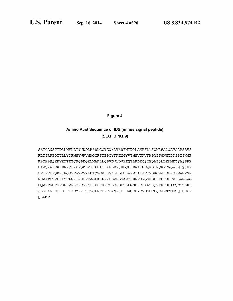
Figure 4
Amino Acid Sequence of IDS (minus signal peptide) (SEQID NO:9)
SETOANSTTDALNVLLITVDDLRPSLGCYGDKTVRSPNIDOLASHSLLFONAFAOOAVCAPSRVS
FLTGRRPDTTRLYDFNSYWRVHAGNFSTIPOYFKENGYVTMSVGKVFHPGISSNHTDDSPYSWSF
PPYHPSSEKYENTKTCRGPDGELHANLLCPVDVLDVPEGTLPDKOSTEOAIOLLEKMKTSASPFF
LAVGYHKPHIPFRYPKEFOKLYPLENITLAPDPEVPDGLPPVAYNPWMDIROREDVOALNISVPY
GPIPVDFORKIROSYFASVSYLDTQVGRLLSALDDLOLANSTIIAFTSDHGWALGEHGEWAKYSN
FDVATHVPLIFYVPGRTASLPEAGEKLFPYLDPFDSASOLMEPGROSMDLVELVSLFPTLAGLAG
LQVPPRCPVPSFHVELCREGKNLLKHFRFRDLEEDPYLPGNPRELLAYSOY PRPSDIPOWNSDKP
SLKDIKIMGYSIRTIDYRYTVWVGFNPDEFLANFSDIHAGELYFWDSDPLODHNMYNDSQGGDLF
OLLMP


U.S. Patent Sep. 16, 2014 Sheet 6 of 20 US 8,834,874 B2
Figure 6
HIRMAb
IDS
US 8,834,874 B2
very .
Brain Ce
Sheet 7 of 20
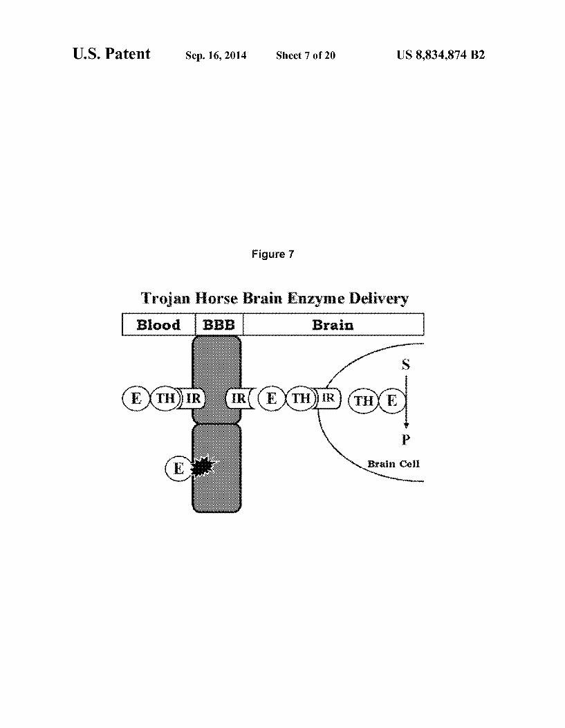
Figure 7
}:::::::::::::::::::::::::::::::::::::::::::::::::::::::::::::::::::: ; :?Ž? BBB
Sep. 16, 2014
Trojan Horse Brain Enzyme Del
U.S. Patent

U.S. Patent Sep. 16, 2014 Sheet 8 of 20 US 8,834,874 B2
Figure 8
pa:
pCD-HRMAb-HC ( or ) (amp) cars
iS pCR hipa product T4 ligase
pCD-HRMAb-IDS on
assa.

U.S. Patent Sep. 16, 2014 Sheet 9 of 20 US 8,834,874 B2
Figure 9
R HIR MAb
STD MAb DS
-35 kDa
-52 kDa
34 kDa
-28 kDa 26 kDa

U.S. Patent Sep. 16, 2014 Sheet 10 of 20 US 8,834,874 B2
Figure 10
A R B R VA- R VA. - R
DS DS MAb DS DS MAb ka
30 ka- & 3 ki3.
98 kDa. 95 kDa. 8: 72 k3. 72 kDa
35 k3 43 kDa
34 kar
26 ka- 8x. 8&
U.S. Patent Sep. 16, 2014 Sheet 11 of 20 US 8,834,874 B2
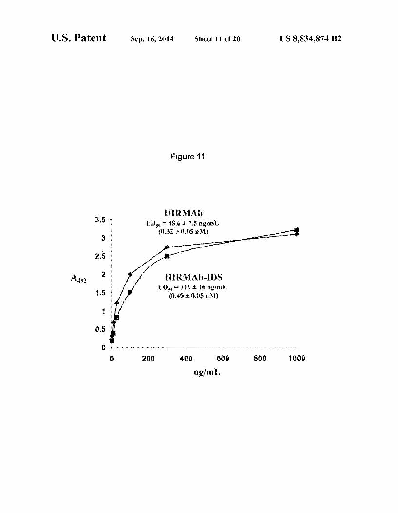
Figure 11
HKVA 3.5 EE) - 48.6 d. 7.5 agai.
3.32 : 0.05 axi
RVA-S Eids - 19 it ié agiari.
(8,43: ...5 avi)

U.S. Patent Sep. 16, 2014 Sheet 12 of 20 US 8,834,874 B2
Figure 12
A 4-methylustellifery a-i-idirositie-2-sulphate (4-8iS)
s
s / 4-methyluribatifery c--iduroaide (MUBI) is OS
- a . DuA O s s
4-methyium{eiliferones (4-8)
9.
8
B 600
F. s
51 t 7 innoithriag 3.
. .8 ,
HiRNAb-IDS (g)

U.S. Patent Sep. 16, 2014 Sheet 13 of 20 US 8,834,874 B2
Figure 13
160 140 120
intraceluiar . S enzyme 1. activity 80
(nmotihrimg) 60 40 20
2000 40 500
HiRMAb-DS (ngiri)

U.S. Patent Sep. 16, 2014 Sheet 14 of 20 US 8,834,874 B2
Figure 14
25
2000 1500
35S incorporation (CPig protein t
500 o daaaaaaaaaaaa.
hitter heality firesiasts firotiasts hts at
treated treated fier88sts
U.S. Patent Sep. 16, 2014 Sheet 15 of 20 US 8,834,874 B2
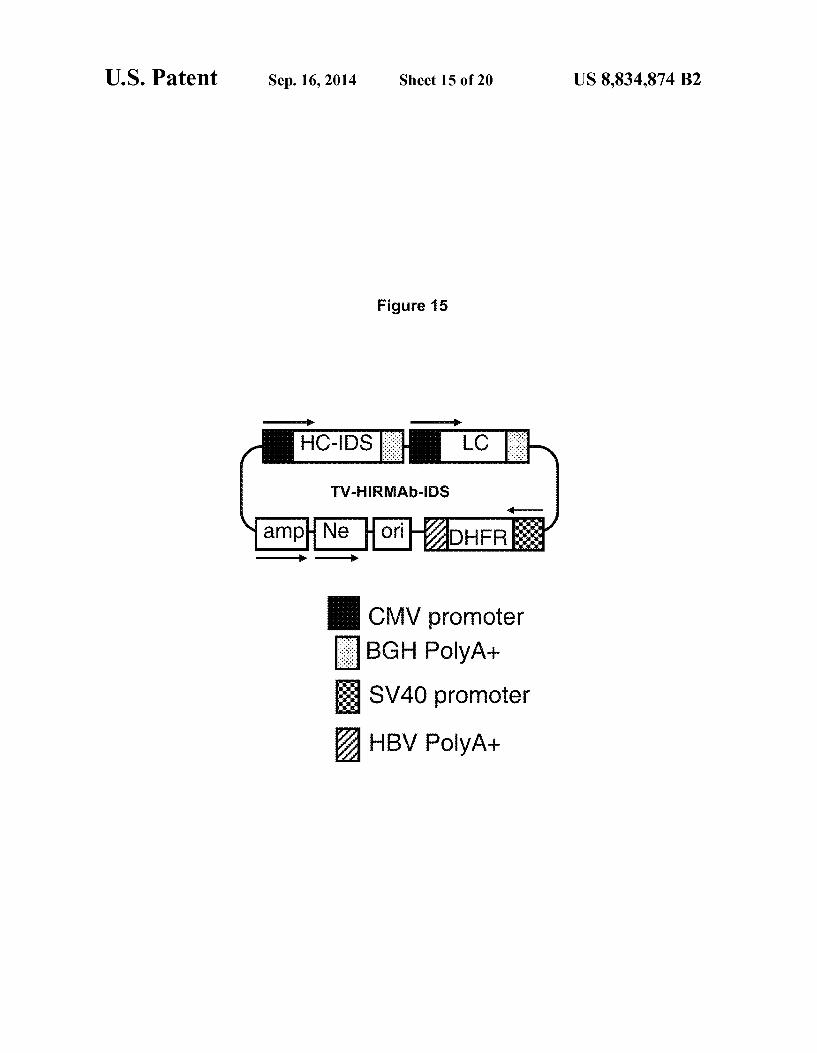
Figure 15
BGH PolyA+ & SV40 promoter HBV PolyA+

U.S. Patent Sep. 16, 2014 Sheet 16 of 20 US 8,834,874 B2
Figure 16
Nucleotide Sequence of HIR Ab-IDS LC (SEQ ID NO:13)
GCCGCCACCATGGAGACCCCCGCCCAGCTGCTGTTCCTGTTGCTGCTTTGGCTTCCAGATACTACCGGCG
ACATCCAGATGACCCAGTCTCCATCCTCCTTACTGCCTCTCTGGGAGAAAGAGTCAGTCT CACTTGTCG
GGCAAGTCAGGACATTGGTGGTAACTTATACTGGCTTCAGCAGGGACCAGATGGAACTATTAAACGCCTG
ATCTACGCCACATCCAGTTTAGATTCTGGTGTCCCCAAAAGGTTCAGTGGCAGTAGGTCTGGGTCAGATT
ATTCTCTCACCATCAGCAGCCTTGAGTCTGAAGATTTTGTAGACTATTACTGTCTACAGTATTCTAGTTC
TCCGTGGACGTTCGGTGGAGGCACAAAGCTGGAAATAAAACGAACTGTGGCTGCACCATCTGTCTTCATC
TTCCCGCCATCTGATGAGCAGTTGAAATCTGGAACTGCCTCTGTTGTGTGCCTGCTGAATAACTTCTATC
CCAGAGAGGCCAAAGTACAGTGGAAGGTGGATAACGCCCTCCAATCGGGTAACTCCCAGGAGAGTGTCAC
AGAGCAGGACAGCAAGGACAGCAC CTACAGCCTCAGCAGCACCCTGACGCTGAGCAAAGCAGACTACGAG
AAACACAAAGTCTACGCCTGCGAAGTCACCCACAGGGCCTGAGCTCGCCCGTCACAAAGAGCTTCA ACA
GGGGAGAGTGTTAG

U.S. Patent Sep. 16, 2014 Sheet 17 of 20 US 8,834,874 B2
Figure 17
Nucleotide Sequence of HIRAb-IDS HC (SEQID NO:14)
GCCGCCACCATGGACTGGACCTGGAGGGTGTTCTGCCTGCTTGCAGTGGCCCCCGGAGCCCACAGCCAGG TTCAGCTGCAGCAGTCGGACCGAGCTGGTGAAGCCTGGGGCTTAGTGAAGATATCCTGCAAGGCTTC
TCCTTACACCTTCACAAACTACCATATACACTCGCTGAACCACAGCCCTGCACAGGGACTTGACTCCATT
GGATGGATTTATCCTGGAGATGGTAGTACTAAGTACAATGAGAAATTCAAGGGCAAGGCCACACTGACTG CAGACAAATCCTCCAGCACAGCCTACATGCACCTCAGCAGCCTGACTTCTGAGAAATCTGCAGTCTATTT
CTGTGCAAGAGAGTGGGCTTACTGGGGCCAAGGGACTCTGGTCACTGTCTCTGCAGCTAGCACCAAGGGC
CCATCGGTCTTCCCCCTGGCACCCTCCTCCAAGAGCAC CTCTGGGGGCACAGCGGCCCTGGGCTGCCTGG
TCAAGGACTACTTCCCCGAACCGGTGACGGTGTCGTGGAACTCAGGCGCCCTGACCAGCGGCGTGCACAC
CTTCCCGGCTGICCACAGTCCCAGGACCACTCCCTCAGCAGCGTGGTGACCGTGCCCTCCAGCAGC
TTGGGCACCCAGACCTACATCTCCAACGTGAATCACAAGCCCAGCAACACCAAGGTGGACAAGAAAGTTG
AGCCCAAATCTTGTGACAAAACTCACACATGCCCACCGTGCCCAGCACCTGAACTCCTGGGGGGACCGTC
AGTCTTCCTCTCCCCCCAAAACCCAAGGACACCCTCATGATCTCCCGGACCCCTGAGGTCACATGCGTG
GTGGTGGACGTGAGCCACGAAGACCCTGAGGTCAAGTTCAACTGGTACGTGGACGGCGTGGAGGTGCATA
AGCCAAGACAAAGCCGCGGGAGGAGCAGTACAACAGCACGTACCGTGTGGTCAGCGTCCCACCGTCC
GCACCAGGACTGGCTGAATGGCAAGGAGTACAAGGCAAGGTCTCCAACAAAGCCCTCCCAGCCCCCATC GAGAAAACCACCCAAAGCCAAAGGGCAGCCCCGAGA ACCACAGGTGTACACCCTGCCCCCACCCGGG
ATGAGCTGACCAAGAACCAGGTCAGCCTGACCTGCCTGGTCAAAGGCTTCTATCCCAGCCACATCGCCGT
GGAGTGGGAGAGCAATGGGCAGCCGGAGAACAACTACAAGACCACGCCTCCCGTGCTGGACTCCGACGGC
TCCTTCTTCCTCTACAGCAAGCTCACCGTGGACAAGAGCAGGTGGCAGCAGGGGAACGTCTTCTCATGCT CCGIGATGCATGAGGCTCTGCACA ACCACTACACGCAGAAGAGCCTCTCCCTGTCTCC tAGTAGTTC
CTCCGAAACGCAGGCCAA CTCGACCACAGATGCTCTGAACGTTCTCTCATCATCGTGGATGACCTGCGC
CCCTCCCTGGGCTGTTATGGGGATAAGCTGGTGAGGTCCCCAAATATTGACCAACTGGCATCCCACAGCC TCCTCTCCAGAATGCCTTGCGCAGCAAGCAGTGTGCGCCCCGAGCCGCGTTTCTTTCCCACTGGCAG
GAGACCTGACACCACCCGCCTGTACGACTTCAACTCCTACTGGAGGGTGCACGCTGGAAACTTCTCCACC
ATCCCCCAGTACTTCAAGGAGAATGGCTATGTGACCATGTCGGTGGGAAAAGTCTTTCACCCTGGGATAT CTTCTAACCATACTGATGATTCTCCGTATAGCGGTCTTTTCCACCITATCATCCTTCCTCTGAGAAGTA
TGAAAACACTAAGACATGTCGAGGGCCAGATGGAGAACTCCATGCCAACCTGCTTTGCCCTGTGGATGTG
CTGGATGTTCCCGAGGGCACCTTGCCTGACAAACAGAGCACTGAGCAAGCCATACAGTTGTTGGAAAAGA
TGAAAACGTCAGCCAGTCCTTTCTTCCTGGCCGTTGGGTATCATAAGCCACACATCCCCTTCAGATACCC
CAAGGAATTTCAGAAGIGAICCCTTGGAGA ACATCACCCTGGCCCCCGATCCCGAGGTCCCTGATGGC
CTACCCCCTGTGGCCTACAACCCCTGGATGGACATCAGCCAACGGGAAGACGTCCAAGCCTTAAACATCA
GTGTGCCGTATGGTCCAATTCCTGTGGACTTTCAGCGGAAAATCCGCCAGAGCTACTTTGCCTCTGTGTC
ATATTGGATACACAGGTCGGCCGCCCTTGAGTGCTTGGACGATCTTCAGCTGGCCAACAGCACCATC
ATTGCATTTACCTCGGATCATGGGTGGGCTCTAGGTGAACATGGAGAATGGGCCAAATACAGCAATTTTG
ATGTTGCTACCCATGTTCCCCTGATATTCTATGTTCCTGGAAGGACGGCTTCACTTCCGGAGGCAGGCGA
GAAGCTTTTCCCTTACCTCGACCCTTTTGATTCCGCCT CACAGTTGATGGAGCCAGGCAGGCAATCCATG GACCTTGTGGAACTTGTGICCITTTTCCCACGCGGCTGGACTTGCAGGACTGCAGGTTCCACCTCGC
GCCCCGTTCCTTCATTTCACGTTGAGCTGTGCAGAGAAGGCAAGAACCTTCTGAAGCATTTTCGATTCCG
TGACTTGGAAGAGGATCCGTACCTCCCTGGTAATCCCCGTGAACTGATTGCCTATAGCCAGTATCCCCGG CCTTCAGACATCCCTCAGGGAATCGACAAGCCGAGTTTAAAAGATATAAAGATCATGGGCIATTCCA
TACGCACCATAGACTATAGGTATACTGTGTGGGTTGGCTTCAATCCTGATGAATTTCTAGCTAACTTTTC
GACATCCATGCAGGGGAACTGTATTTTGTGGATCTGACCCATTGCAGGACACAATAATGATAATGAT
CCCAAGGTGGAGATCTTTTCCAGTTGTTGAGCCTTGA

U.S. Patent Sep. 16, 2014 Sheet 18 of 20 US 8,834,874 B2
Figure 18
Nucleotide Sequence of HIR Ab-IDS DHFR (SEQ ID NO:15)
GCCGCCACCATGGTTCGACCATTGAACTGCATCGTCCCCGTGTCCCAAAATATCGGGATTGGCAAGAACG
GAGACCTACCCTGGCCTCCGCTCAGGAACGAGTTCAAGTACTTCCAAAGAATGACCACAACCTCTTCAGT
GGAAGGTAAACAGAATCTGGTGATTATGGGTAGGAAAACCTGGTTCTCCATTCCTGAGAAGAATCGACCT
TAAAGGACAGAATTAATAAGTCCAGTAGAGAACT CAAAGAACCACCACGAGGAGCCATTTCTG
CCAAAAGTTTGGAGAGCCAAGACTTATTGAACAACCGGAATTGGCAAGTAAAGTAGACATGGTTTG
GATAGTCGGAGGCAGTTCTGTTACCAGGAAGCCATGAATCA ACCAGGCCACCTCAGACTCTTTGTGACA
AGGATCATGCAGGAATTTGAAAGTGACACGTTTTTCCCAGAAATTGATTTGGGGAAATATAAACTTCTCC
CAGAATACCCAGGCGTCCTCTCTGAGCTCCAGGAGGAAAAAGGCATCAAGTATAAGTTTGAAGTCTACGA
GAAGAAAGACTAA
U.S. Patent Sep. 16, 2014
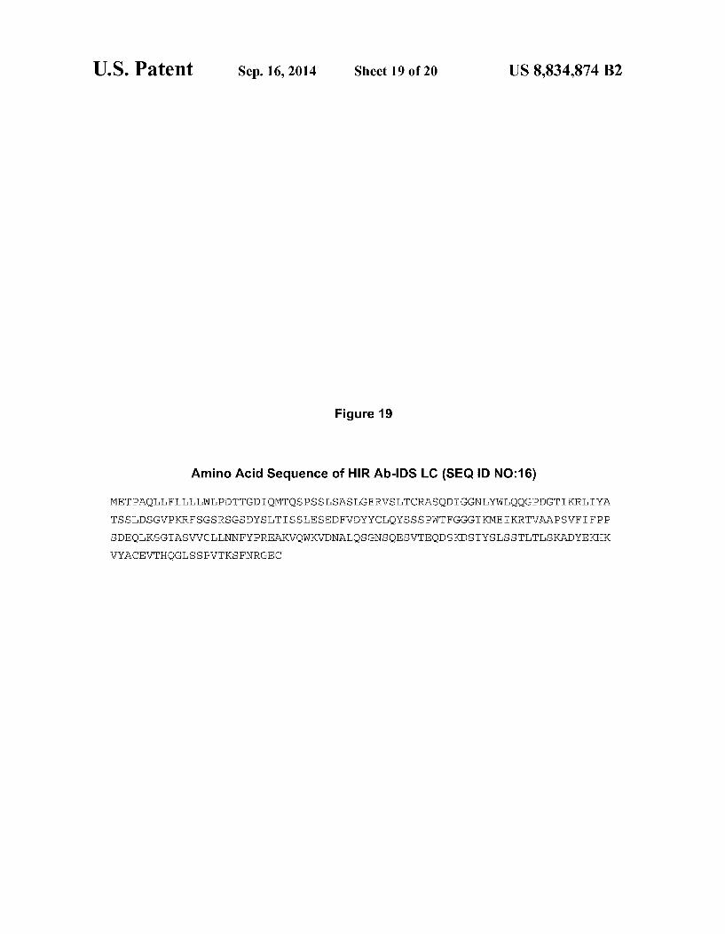
Figure 19
Sheet 19 of 20 US 8,834,874 B2
Amino Acid Sequence of HIRAb-IDS LC (SEQ ID NO:16)
METPAOLLFLLLLWLPDTTGDIOMTOSPSSLSASLGERVSLTCRASODIGGNLYWLOOGPDGTIKRLIYA
TSSLDSGVPKRFSGSRSGSDYSLTISSLESEDFVDYYCLOYSSSPWTFGGGTKMEIKRTVAAPSVFIFPP
SDEQTKSGTASVVCILNNFYPR
WYACEVTHOGLSSPVTKSFNRG
EAKVQWKVDNALQSGNSQESVT EC
EQDSKDSTYSLSSTLTLSKADYEKHK

U.S. Patent
MVRPLNCIVAVSQNMGTGKNGDLPWPPLRN
Sep. 16, 2014 Sheet 20 of 20 US 8,834,874 B2
Figure 20
Amino Acid Sequence of HIRAb-IDS DFHR (SEQID NO:17)
EFKY FORMTTTSSVEGKQNLVIMGRKTWFSIPEKNRPLKD
RINIVLSRELKEPPRGAHFLAKSLDDALRLIEOPELASKVDMVWIVGGSSVYOEAMNOPGHLRLFWTRIM
QEFES DTFFPEDIGKYKP EYPGVLSEVOE EKGKYKFEWYEKKD

US 8,834,874 B2 1.
METHODS AND COMPOSITIONS FOR INCREASING DURONATE 2-SULFATASE
ACTIVITY IN THE CNS
CROSS-REFERENCE TO RELATED APPLICATIONS
This application claims the benefit under 35 U.S.C. S 119 (e) of U.S. Provisional Application No. 61/250,378, filed Oct. 9, 2009, and U.S. Provisional Application No. 61/256,049, filed Oct. 29, 2009, both of which are incorporated herein by reference in their entirety.
SEQUENCE LISTING
The instant application contains a Sequence Listing which has been submitted in ASCII format via EFS-Web and is hereby incorporated by reference in its entirety. Said ASCII copy, created on Jan. 5, 2011, is named 28570201.txt and is 32,098 bytes in size.
BACKGROUND OF THE INVENTION
Type II mucopolysaccharidosis (MPS), also known as Hunter's syndrome, is an inherited metabolic disease caused by a defect in the enzyme iduronate 2-sulfatase (IDS), which functions to degrade mucopolysaccharides. An insufficient level of IDS causes a pathological buildup of heparan sulfate and dermatan Sulfate in, e.g., the heart, liver, and central nervous system (CNS). Symptoms including neurodegenera tion and mental retardation appear during childhood; and early death can occur due to organ damage in the brain. Typically, treatment includes intravenous enzyme replace ment therapy with recombinant IDS. However, systemically administered recombinant IDS does not cross the blood brain harrier (BBB), and therefore has little impact on the effects of the disease in the CNS.
SUMMARY OF THE INVENTION
Described herein are methods and compositions for treat ing a subject suffering from an iduronate 2-sulfatase (“IDS”) deficiency. The compositions provided herein comprise fusion antibodies comprising an IDS polypeptide fused to structure (e.g., antibody, immunoglobulin) capable of cross ing the blood-brain barrier (BBB). In some embodiments, the structure that is capable of crossing the BBB crosses the BBB on an endogenous BBB receptor. In some embodiments, the endogenous BBB receptor is the insulin receptor, transferrin receptor, leptin receptor, lipoprotein receptor, and the IGF receptor. In some embodiments, the endogenous BBB recep tor is the insulin receptor. In some embodiments, the methods allow delivery of IDS to the CNS by systemically adminis tering a therapeutically effective amount of a bifunctional human insulin receptor antibody (e.g., HIRAb)-IDS fusion antibody. In some embodiments, the HIRAb-IDS fusion anti body binds to the extracellular domain of the insulin receptor and is transported across the blood brain barrier (“BBB) into the CNS, while retaining iduronate 2-sulfatase activity. In some embodiments, the HIR Ab binds to the endogenous insulin receptor on the BBB, and acts as a molecular Trojan horse to ferry the IDS into the brain. A therapeutically effec tive systemic dose of a HIR Ab-IDS fusion antibody for systemic administration is based, in part, on the specific CNS uptake characteristics of the fusion antibody from peripheral blood as described herein.
10
15
25
30
35
40
45
50
55
60
65
2 In some embodiments, the invention provides composi
tions containing an IDS covalently linked to a structure (e.g., immunoglobulin, antibody) that is capable of crossing the blood brain barrier (BBB), where the structure and the IDS each retains at least about 10, 20, 30, 40, 50, 60, 70, 80,90, 95, 99, or 100% of its activity, compared to its activity as a separate entity. In some embodiments; the IDS retains at least about 10% of its activity compared to its activity as a separate entity. In some embodiments, the IDS retains at least 20% of its activity, compared to its activity as a separate entity. In some embodiments, the IDS retains at least 30% of its activ ity, compared to its activity as a separate entity. In some embodiments, the IDS retains at least 40% of its activity, compared to its activity as a separate entity. In some embodi ments, the IDS retains at least 50% of its activity, compared to its activity as a separate entity. In some embodiments, the IDS retains at least 60% of its activity, compared to its activity as a separate entity.
In some embodiments, a fusion antibody comprising IDS is post-translationally modified by a Sulfatase modifying factor type 1 (SUMF1). In some embodiments, the post-transla tional modification comprises a cysteine to formylglycine conversion. In some embodiments, a fusion antibody com prises a formylglycine residue.
In one aspect provided herein is a method for treating an IDS deficiency in the central nervous system of a subject in need thereof, comprising systemically administering to the subject a therapeutically effective dose of a fusion antibody having IDS activity. In some embodiments of this aspect: (i) the fusion antibody comprises the amino acid sequence of an immunoglobulin heavy chain, the amino acid sequence of an IDS, and the amino acid sequence of an immunoglobulin light chain; (ii) the fusion antibody binds to an extracellular domain of the human insulin receptor and catalyzes hydroly sis of the 2-sulfate groups of the L-iduronate 2-sulfate units of dermatan Sulfate, heparan Sulfate or heparin; and (iii) the amino acid sequence of the IDS is covalently linked to the carboxy terminus of the amino acid sequence of the immu noglobulin heavy chain. In some embodiments, the immuno globulin heavy chain is an immunoglobulin heavy chain of IgG. In some embodiments, the immunoglobulin heavy chain is an immunoglobulin heavy chain of kappa class.
In some embodiments at least about 250,000 units of IDS activity are delivered to the brain, where 1 unit=1 nmol/hr using a fluorometric assay. In some embodiments, the thera peutically effective dose of the fusion antibody comprises at least about 2.5x10 units of IDS activity or at least about 50,000 units/Kg of body weight. In some embodiments the IDS specific activity of the fusion antibody is at least 30,000 units/mg. In some embodiments, systemic administration is parenteral, intravenous, Subcutaneous, intra-muscular, trans nasal, intra-arterial, transdermal, or respiratory. In some embodiments, at least about 25,000, 30,000, 35,000, 40,000, 45,000, 50,000, 60,000, 70,000, 80,000, 90,000, 110,000, 120,000, 130,000, 140,000, 150,000, 160,000, 170,000, 180, 000, 190,000, 200,000, 210,000, 220,000, 230,000, 250,000 units of iduronate-2-sulfatase activity is delivered to the brain, normalized per 50 kg body weight. In some embodi ments, at least about 25,000 units of iduronate-2-sulfatase activity is delivered to the brain, normalized per 50 kg body weight.
In some embodiments, the fusion antibody is a chimeric antibody.
In Some embodiments, the immunoglobulin heavy chain of the fusion antibody comprises a CDR1 corresponding to the amino acid sequence of SEQ ID NO:1 with up to 4 single amino acid mutations, a CDR2 corresponding to the amino

US 8,834,874 B2 3
acid sequence of SEQID NO:2 with up to 6 singleamino acid mutations; or a CDR3 corresponding to the amino acid sequence of SEQ ID NO:3 with up to 3 single amino acid mutations, wherein the single amino acid mutations are Sub stitutions, deletions, or insertions. 5
In other embodiments, the immunoglobulin heavy chain of the fusion antibody comprises a CDR1 corresponding to the amino acid sequence of SEQ ID NO:1 with up to 3 single amino acid mutations, a CDR2 corresponding to the amino acid sequence of SEQID NO:2 with up to 6 singleamino acid 10 mutations, and a CDR3 corresponding to the amino acid sequence of SEQ ID NO:3 with up to 3 single amino acid mutations.
In other embodiments, the immunoglobulin heavy chain of is the fusion antibody comprises a CDR1 corresponding to the amino acid sequence of SEQID NO:1, a CDR2 correspond ing to the amino acid sequence of SEQID NO:2, or a CDR3 corresponding to the amino acid sequence of SEQID NO:3.
In further embodiments, the immunoglobulin heavy chain 20 of the fusion antibody comprises a CDR1 corresponding to the amino acid sequence of SEQ ID NO:1, a CDR2 corre sponding to the amino acid sequence of SEQID NO:2, and a CDR3 corresponding to the amino acid sequence of SEQID NO:3.
In some embodiments, the immunoglobulin light chain of the fusion antibody comprises a CDR1 corresponding to the amino acid sequence of SEQ ID NO:4 with up to 3 single amino acid mutations, a CDR2 corresponding to the amino acid sequence of SEQID NO:5 with up to 5 singleamino acid mutations, or a CDR3 corresponding to the amino acid sequence of SEQ ID NO:6 with up to 5 single amino acid mutations, wherein the single amino acid mutations are Sub stitutions, deletions, or insertions.
In other embodiments, the immunoglobulin light chain of the fusion antibody comprises a CDR1 corresponding to the amino acid sequence of SEQ ID NO:4 with up to 3 single amino acid mutations, a CDR2 corresponding to the amino acid sequence of SEQID NO:5 with up to 5 singleamino acid mutations, and a CDR3 corresponding to the amino acid sequence of SEQ ID NO:6 with up to 5 single amino acid mutations.
In other embodiments, the immunoglobulin light chain of the fusion antibody comprises a CDR1 corresponding to the 45 amino acid sequence of SEQID NO:4, a CDR2 correspond ing to the amino acid sequence of SEQID NO:5, or a CDR3 corresponding to the amino acid sequence of SEQID NO:6.
In further embodiments, the immunoglobulin light chain of the fusion antibody comprises a CDR1 corresponding to the 50 amino acid sequence of SEQID NO:4, a CDR2 correspond ing to the amino acid sequence of SEQID NO:5, and a CDR3 corresponding to the amino acid sequence of SEQID NO:6.
In some embodiments, the immunoglobulin heavy chain of the fusion antibody comprises a CDR1 corresponding to the amino acid sequence of SEQID NO:1, a CDR2 correspond ing to the amino acid sequence of SEQID NO:2, and a CDR3 corresponding to the amino acid sequence of SEQID NO:3: and the immunoglobulin light chain comprises a CDR1 cor responding to the amino acid sequence of SEQID NO:4, a CDR2 corresponding to the amino acid sequence of SEQID NO:5, and a CDR3 corresponding to the amino acid sequence of SEQID NO:6.
In some embodiments, the immunoglobulin heavy chain of the fusion antibody is at least 90% identical to SEQID NO:7 65 and the amino acid sequence of the light chain immunoglo bulin is at least 90% identical to SEQID NO:8.
25
30
35
40
55
60
4 In Some embodiments, the immunoglobulin heavy chain of
the fusion antibody comprises SEQID NO:7 and the amino acid sequence of the light chain immunoglobulin comprises SEQID NO: 8
In yet further embodiments, the IDS comprises an amino acid sequence at least 90% (e.g., 95%, or 100%) identical to SEQID NO:9.
In other embodiments, the amino acid sequence of the immunoglobulin heavy chain of the fusion antibody at least 90% identical to SEQID NO:7; the amino acid sequence of the light chain immunoglobulin is at least 90% identical to SEQID NO:8; and the amino acid sequence of the IDS is at least 95% identical to SEQ ID NO:9 or comprises SEQ ID NO:9.
In still other embodiments, the amino acid sequence of the immunoglobulin heavy chain of the fusion antibody com prises SEQID NO:8, the amino acid sequence of the immu noglobulin light chain comprises SEQ ID NO:8, and the amino acid sequence of the IDS comprises SEQID NO:9.
In some aspects, provided herein are pharmaceutical com positions comprising a therapeutically effective amount of a fusion antibody and a pharmaceutically acceptable excipient.
In some aspects, provided herein are isolated polynucle otides encoding a fusion antibody. In some embodiments, the isolated polynucleotide comprises the nucleic acid sequence of SEQID NO:14.
In some embodiments, provided herein are vectors com prising the isolated polynucleotides encoding a fusion anti body. In some embodiments, the vectors comprise the nucleic acid sequence of SEQID NO:14.
In some embodiments, provided herein are host cells com prising the vectors comprising the isolated polynucleotides encoding a fusion antibody. In Sone embodiments, the host cell is a Chinese Hamster Ovary (CHO).
In a further aspect provided herein is a method for treating an IDS deficiency in the central nervous system of a subject in need thereof, comprising systemically administering to the subject a therapeutically effective dose of a fusion antibody having IDS activity, wherein: (i) the fusion antibody com prises: (a) a fusion protein at least 95% identical to SEQID NO:10, and (b) an immunoglobulin light chain; and (ii) the fusion antibody binds to an extracellular domain of the human insulin receptor and catalyzes hydrolysis of linkages in der matan or heparan Sulfate.
In yet another aspect provided herein is a method for treat ing an IDS deficiency in the central nervous system of a Subject in need thereof, comprising systemically administer ing to the subject a therapeutically effective dose of a fusion antibody having IDS activity, wherein: (i) the fusion antibody comprises a fusion protein containing the amino acid sequence of an immunoglobulin heavy chain and an IDS or comprises a fusion protein containing the amino acid sequence of an immunoglobulin light chainandan IDS; the fusion antibody binds to the extracellular domain of the human insulin receptor, and the fusion antibody catalyzes hydrolysis of linkages in dermatan or heparan Sulfate; and (ii) the amino acid sequence of the IDS is covalently linked to the carboxy terminus of the amino acid sequence of the immunoglobulin heavy chain or the immunoglobulin light chain.
INCORPORATION BY REFERENCE
All publications, patents, and patent applications men tioned in this specification are herein incorporated by refer ence to the same extent as if each individual publication,

US 8,834,874 B2 5
patent, or patent application was specifically and individually indicated to be incorporated by reference.
BRIEF DESCRIPTION OF THE DRAWINGS
The novel features of the invention are set forth with par ticularity in the appended claims. A better understanding of the features and advantages of the present invention will be obtained by reference to the following detailed description that sets forth illustrative embodiments, in which the prin ciples of the invention are utilized, and the accompanying drawings, as follow:
FIG.1. Amino acid sequence of an immunoglobulin heavy chain variable region from an exemplary human insulin receptor antibody directed against the extracellular domain of the human insulin receptor. The underlined sequences are a signal peptide, CDR1, CDR2, and CDR3, respectively. The heavy chain constant region, taken from human IgG1, is shown in italics.
FIG. 2. Amino acid sequence of an immunoglobulin light chain variable region from an exemplary human insulin receptor antibody directed against the extracellular domain of the human insulin receptor. The underlined sequences are a signal peptide, CDR1, CDR2, and CDR3, respectively. The constant region, derived from human kappa light chain, is shown in italics.
FIG. 3. A table showing the CDR1, CDR2, and CDR3 amino acid sequences from a heavy and light chain of an exemplary human insulin receptor antibody directed against the extracellular domain of the human insulin receptor.
FIG. 4. Amino acid sequence of human iduronate 2-sulfa tase (IDS) (GenBank NP 000193), not including the initial 25 amino acid signal peptide (mature IDS).
FIG. 5. Amino acid sequence of a fusion of an exemplary human insulin receptor antibody heavy chain to mature human IDS. The underlined sequences are, in order, an IgG signal peptide, CDR1, CDR2, CDR3, and a peptide linker linking the carboxy terminus of the heavy chain to the amino terminus of the IDS. Sequence in italic corresponds to the heavy chain constant region, taken from human IgG1. The sequence in hold corresponds to human IDS.
FIG. 6. An exemplary HIR Ab-IDS fusion antibody is formed by fusion of the amino terminus of the mature IDS to the carboxyl terminus of the CH3 region of the heavy chain of the HIRAb
FIG. 7. Schematic depiction of a “molecular trojan horse' strategy in which the fusion antibody comprises an antibody to the extracellular domain of the human insulin receptor (IR), which acts as a molecular Trojan horse (TH), and IDS, a lysosomal enzyme (E).
FIG. 8. (A) Ethidium bromide stain of agarose gel of human IDS cDNA (lane 1), which was produced by PCR from human liver cDNA, and IDS-specific primers (Table 2). Lanes 2 and 3: PhiX174 Hael II digested DNA standard, and Lambda HindIII digested DNA standard. (B) The heavy chain of the HIRMAb-IDS fusion protein is expressed by the pCD HIRMAb-IDS plasmid, which is generated by subcloning the IDS cDNA into the HpaI site of the pCD-HIRMAb-HC plas mid.
FIG.9. SDS-PAGE of molecular weight standards (STD), the purified HIRMAb, and the purified HIRMAb-IDS fusion protein.
FIG. 10. Western blot with either anti-human (h) IgG pri mary antibody (A) or anti-human IDS primary antiserum (B). The immunoreactivity of the HIRMAb-IDS fusion protein is compared to the chimeric HIRMAb and recombinant human IDS. Both the HIRMAb-IDS fusion protein and the HIRMAb
10
15
25
30
35
40
45
50
55
60
65
6 have identical light chains on the anti-hIgG Western. The HIRMAb-IDS fusion heavy chain reacts with both the anti hIgG and the anti-human IDS antibody, whereas the HIRMAb heavy chain only reacts with the anti-hIgG anti body. The size of the HIRMAb-IDS fusion heavy chain, 130 kDa, is about 80 kDa larger than the size of the heavy chain of the HIRMAb, owing to the fusion of the 80 kDa IDS to the 50 kDa HIRMAb heavy chain.
FIG. 11. Binding of either the chimeric HIRMAb or the HIRMAb-IDS fusion protein to the HIR extracellular domain (ECD) is saturable. The EDs of HIRMAb-IDS binding to the HIR ECD is comparable to the EDs of the binding of the chimeric HIRMAb.
FIG. 12. (A) Substrate (4-MUS), intermediate (MUBI), and product (4-MU) of the 2-step enzymatic fluorometric assay of IDS enzyme activity. (B) The fluorometric units (FU) are proportional to the mass of purified HIRMAb-IDS fusion protein, and the average enzyme specific activity of the fusion protein is 51+7 nmol/hr/ug protein, which is equivalent to 51 units/ug protein, or 51,000 units/mg protein.
FIG. 13. Intracellular IDS enzyme activity is increased in Hunter fibroblasts in proportion to the concentration of medium HIRMAb-IDS fusion protein. Data are meant-SE (n=3 dishes/point). The horizontal bar is the IDS enzyme activity in healthy human fibroblasts (mean+SD).
FIG. 14. Reversal of glycosaminoglycan (GAG) accumu lation in Hunter fibroblasts with a single treatment of 0.3 ug/mL of HIRMAb-IDS fusion protein in the medium. There is an 84% reduction in GAG accumulation, as compared to the S incorporation in healthy human fibroblasts (p<0.0005). Data are mean+SE (n=4 dishes/point).
FIG. 15. Genetically engineered tandem vector (TV HIRMAb-IDS) encoding 4 separate and tandem expression cassettes encoding the heavy chain (HC) fusion gene, the light chain (LC) gene, the DH FR gene, and the neogene.
FIG. 16. Nucleotide sequence of the immunoglobulin light chain (LC) region of an exemplary HIRMAb-IDS fusion antibody.
FIG. 17. Nucleotide sequence of the immunoglobulin heavy chain (HC) region of an exemplary HIRMAb-IDS fusion antibody.
FIG. 18. Nucleotide sequence of the DHFR region of an exemplary HIRMAb-IDS fusion antibody.
FIG. 19. Amino acid sequence of the immunoglobulin light chain (LC) region of an exemplary HERMAb-IDS fusion antibody.
FIG. 20. Amino acid sequence of the DHFR region of an exemplary HIRMAb-IDS fusion antibody.
DETAILED DESCRIPTION OF THE INVENTION
Introduction
The blood brain barrier (BBB) is a severe impediment to the delivery of systemically administered IDS (e.g., recom binant IDS) to the central nervous system. The present dis closure provides methods and compositions for delivering therapeutically significant levels of IDS activity across the BBB to the CNS. In some embodiments, the IDS is modified to allow it to cross the BBB. In some embodiments, the amount and rate of uptake of systemically administered modified IDS into the CNS is provided. In some embodi ments, the LDS retains a certain activity after being modified or after crossing the BBB. The present disclosure provides, interalia: (1) IDS fusion antibodies comprising an IDS fused, with or without intervening sequence, to an immunoglobulin (heavy chain or light chain) capable of crossing the BBB, and

US 8,834,874 B2 7
related methods and compositions. (2) human insulin recep tor (HIR) antibody (Ab)-IDS fusion antibodies comprising an IDS fused, with or without intervening sequence, to an immu noglobulin (heavy chain or light chain) directed against the extracellular domain of a human insulin receptor, and related methods and compositions; (3) methods of treating an IDS deficiency; and (4) methods of establishing therapeutically effective systemic doses of the fusion antibodies based on a characterization of their uptake in the CNS and their specific activity. In some embodiments, the invention provides com positions and methods for treating a nDS deficiency in the central nervous system by systemically administering to a subject in need thereof a therapeutically effective dose of a bifunctional IDS fusion antibody (e.g., HIRAb-IDS) having IDS activity and selectively binding to the receptor-mediated BBB transport system (e.g., the extracellular domain of a human insulin receptor).
SOME DEFINITIONS
“Treatment” or “treating as used herein includes achiev ing a therapeutic benefit and/or a prophylactic benefit. By therapeutic benefit is meant eradication or amelioration of the underlying disorder or condition being treated. For example, in an individual with Hunter's syndrome, therapeutic benefit includes partial or complete halting of the progression of the disorder, or partial or complete reversal of the disorder. Also, a therapeutic benefit is achieved with the eradication or ame lioration of one or more of the physiological or psychological symptoms associated with the underlying condition such that an improvement is observed in the patient, notwithstanding the fact that the patient may still be affected by the condition. A prophylactic benefit of treatment includes prevention of a condition, retarding the progress of a condition (e.g., slowing the progression of a lysosomal storage disorder), or decreas ing the likelihood of occurrence of a condition. As used herein, “treating or “treatment” includes prophylaxis. As used herein, the term “effective amount' can be an
amount, which when administered systemically, is sufficient to effect beneficial or desired results in the CNS, such as beneficial or desired clinical results, or enhanced cognition, memory, mood, or other desired CNS results. An effective amount is also an amount that produces a prophylactic effect, e.g., an amount that delays, reduces, or eliminates the appear ance of a pathological or undesired condition. Such condi tions include, but are not limited to, mental retardation, hear ing loss, and neurodegeneration. An effective amount can be administered in one or more administrations. In terms of treatment, an “effective amount of a composition of the invention is an amount that is sufficient to palliate, ameliorate, stabilize, reverse or slow the progression of a disorder, e.g., a neurological disorder. An “effective amount” may be of any of the compositions of the invention used alone or in conjunc tion with one or more agents used to treat a disease or disor der. An "effective amount of a therapeutic agent within the meaning of the present invention is determined by a patients attending physician or veterinarian. Such amounts are readily ascertained by one of ordinary skill in the art and will a therapeutic effect when administered in accordance with the present invention. Factors which influence what a therapeu tically effective amount will be include, the IDS specific activity of the IDS fusion antibody (e.g., HIR Ab-IDS) administered, its absorption profile (e.g., its rate of uptake into the brain), time elapsed since the initiation of the disor der, and the age, physical condition, existence of other disease states, and nutritional status of the individual being treated. Additionally, other medication the patient may be receiving
5
10
15
25
30
35
40
45
50
55
60
65
8 will affect the determination of the therapeutically effective amount of the therapeutic agent to administer. A “subject' or an “individual.” as used herein, is an animal,
for example, a mammal. In some embodiments a “subject' or an “individual' is a human. In some embodiments, the subject suffers from Mucopolysaccharidosis Type II (“Hunter's Syn drome').
In some embodiments, a pharmacological composition comprising an IDS fusion antibody (e.g., HIRAb-IDS) fusion antibody is “administered peripherally” or “peripherally administered.” As used herein, these terms refer to any form of administration of an agent, e.g., a therapeutic agent, to an individual that is not direct administration to the CNS, i.e., that brings the agent in contact with the non-brain side of the blood-brain barrier. “Peripheral administration, as used herein, includes intravenous, intra-arterial, Subcutaneous, intramuscular, intraperitoneal, transdermal, by inhalation, transbuccal, intranasal, rectal, oral, parenteral, Sublingual, or trans-nasal. A “pharmaceutically acceptable carrier' or “pharmaceuti
cally acceptable excipient herein refers to any carrier that does not itself induce the production of antibodies harmful to the individual receiving the composition. Such carriers are well known to those of ordinary skill in the art. A thorough discussion of pharmaceutically acceptable carriers/excipi ents can be found in Remington's Pharmaceutical Sciences, Gennaro, AR, ed., 20th edition, 2000: Williams and Wilkins PA, USA. Exemplary pharmaceutically acceptable carriers can include salts, for example, mineral acid salts such as hydrochlorides, hydrobromides, phosphates, Sulfates, and the like; and the salts of organic acids such as acetates, propi onates, malonates, benzoates, and the like. For example, com positions of the invention may be provided in liquid form, and formulated in Saline based aqueous solution of varying pH (5-8), with or without detergents such polysorbate-80 at 0.01 1%, or carbohydrate additives, such mannitol, sorbitol, or trehalose. Commonly used buffers include histidine, acetate, phosphate, or citrate. A “recombinant host cell' or "host cell refers to a cell that
includes an exogenous polynucleotide, regardless of the method used for insertion, for example, direct uptake, trans duction, f-mating, or other methods known in the art to create recombinant host cells. The exogenous polynucleotide may be maintained as a nonintegrated vector, for example, a plas mid, or alternatively, may be integrated into the host genome. The terms “polypeptide.” “peptide' and “protein’ are used
interchangeably herein to refer to a polymer of amino acid residues. That is, a description directed to a polypeptide applies equally to a description of a peptide and a description of a protein, and vice versa. The terms apply to naturally occurring amino acid polymers as well as amino acid poly mers in which one or more amino acid residues is a non naturally occurring amino acid, e.g., an amino acid analog. As used herein, the terms encompass amino acid chains of any length, including full length proteins (i.e., antigens), wherein the amino acid residues are linked by covalent peptide bonds. The term “amino acid refers to naturally occurring and
non-naturally occurring amino acids, as well as amino acid analogs and amino acid mimetics that function in a mariner similar to the naturally occurring amino acids. Naturally encoded amino acids are the 20 common amino acids (ala nine, arginine, asparagine, aspartic acid, cysteine, glutamine, glutamic acid, glycine, histidine, isoleucine, leucine, lysine, methionine, phenylalanine, proline, serine, threonine, tryp tophan, tyrosine, and valine) and pyrolysine and selenocys teine. Amino acid analogs refers to compounds that have the same basic chemical structure as a naturally occurring amino

US 8,834,874 B2
acid, i.e., an a carbon that is bound to a hydrogen, a carboxyl group, an amino group, and an R group, Such as, homoserine, norleucine, methionine Sulfoxide, methionine methyl Sulfo nium. Such analogs have modified R groups (such as, norleu cine) or modified peptide backbones, but retain the same basic chemical structure as a naturally occurring amino acid. Amino acids may be referred to herein by either their
commonly known three letter symbols or by the one-letter symbols recommended by the IUPAC-IUB Biochemical Nomenclature Commission. Nucleotides, likewise, may be referred to by their commonly accepted single-letter codes. The term “nucleic acid refers to deoxyribonucleotides,
deoxyribonucleosides, ribonucleosides, or ribonucleotides and polymers thereof in either single- or double-stranded form. Unless specifically limited, the term encompasses nucleic acids containing known analogues of natural nucle otides which have similar binding properties as the reference nucleic acid and are metabolized in a manner similar to natu rally occurring nucleotides. Unless specifically limited oth erwise, the term also refers to oligonucleotide analogs includ ing PNA (peptidonucleic acid), analogs of DNA used in antisense technology (phosphorothioates, phosphoroami dates, and the like). Unless otherwise indicated, a particular nucleic acid sequence also implicitly encompasses conserva tively modified variants thereof (including but not limited to, degenerate codon Substitutions) and complementary sequences as well as the sequence explicitly indicated. Spe cifically, degenerate codon Substitutions may be achieved by generating sequences in which the third position of one or more selected (or all) codons is substituted with mixed-base and/or deoxyinosine residues (Batzer et al., Nucleic Acid Res. 19:5081 (1991); Ohtsuka et al., J. Biol. Chem. 260:2605-2608 (1985); and Cassol et al. (1992); Rossolini et al., Mol. Cell. Probes 8:91-98 (1994)). The terms "isolated and “purified’ refer to a material that
is substantially or essentially removed from or concentrated in its natural environment. For example, an isolated nucleic acid may be one that is separated from the nucleic acids that normally flank it or other nucleic acids or components (pro teins, lipids, etc. . . . ) in a sample. In another example, a polypeptide is purified if it is substantially removed from or concentrated in its natural environment. Methods for purifi cation and isolation of nucleic acids and proteins are well known in the art. The Blood Brain Barrier
In some embodiments, the invention provides composi tions and methods that utilize an IDS fusion antibody (e.g., HIR Ab-IDS) capable of crossing the blood brain barrier (BBB). The compositions and methods are useful in trans porting IDS from the peripheral blood and across the blood brain barrier into the CNS. As used herein, the “blood-brain barrier refers to the barrier between the peripheral circula tion and the brain and spinal cord which is formed by tight junctions within the brain capillary endothelial plasma mem branes and creates an extremely tight barrier that restricts the transport of molecules into the brain; the BBB is so tight that it is capable of restricting even molecules as Small as urea, molecular weight of 60 Da. The blood-brain barrier within the brain, the blood-spinal cord barrier within the spinal cord, and the blood-retinal barrier within the retina, are contiguous capillary barriers within the central nervous system (CNS), and are collectively referred to as the blood-brain barrier or BBB. The BBB limits the development of new neurotherapeu
tics, diagnostics, and research tools for the brain and CNS. Most large molecule therapeutics Such as recombinant pro teins, antisense drugs, gene medicines, purified antibodies, or
10
15
25
30
35
40
45
50
55
60
65
10 RNA interference (RNAi)-based drugs, do not cross the BBB in pharmacologically significant amounts. While it is gener ally assumed that Small molecule drugs can cross the BBB, in fact, <2% of all small molecule drugs are active in the brain owing to the lack transport across the BBB. A molecule must be lipid soluble and have a molecular weight less than 400 Daltons (Da) in order to cross the BBB in pharmacologically significant amounts, and the vast majority of small molecules do not have these dual molecular characteristics. Therefore, most potentially therapeutic, diagnostic, or research mol ecules do not cross the BBB in pharmacologically active amounts. So as to bypass the BBB, invasive transcranial drug delivery strategies are used, such as intracerebro-ventricular (ICV) infusion, intracerebral (IC) administration, and con vection enhanced diffusion (CED). Transcranial drug deliv ery to the brain is expensive, invasive, and largely ineffective. The ICV route delivers IDS only to the ependymal surface of the brain, not into brain parenchyma, which is typical for drugs given by the ICV route. The IC administration of an enzyme Such as IDS, only provides local delivery, owing to the very low efficiency of protein diffusion within the brain. The CED results in preferential fluid flow through the white matter tracts of brain, which causes demyelination, and astro gliosis. The methods described herein offer an alternative to these
highly invasive and generally unsatisfactory methods for bypassing the BBB, allowing a functional IDS to cross the BBB from the peripheral blood into the CNS following sys temic administration of an the IDS fusion antibody (e.g., HER Ab-IDS) fusion antibody composition described herein. The methods described herein exploit the expression of insulin receptors (e.g., human insulin receptors) or other receptor on the BBB to shuttle a desired bifunctional IDS fusion antibody (e.g., HIRAb-IDS) from peripheral blood into the CNS. Endogenous BBB Receptor-Mediated Transport Systems The BBB has been shown to have specific receptors that
allow the transport from the blood to the brain of several macromolecules; these transporters are suitable as transport ers for compositions of the invention. Endogenous BBB receptor-mediated transport systems useful in the invention include those that transport insulin, transferrin, insulin-like growth factors 1 and 2 (IGF1 and IGF2), leptin, and lipopro teins. In some embodiments, the invention utilizes a structure (e.g., immunoglobulin, antibody) that is capable of crossing the BBB via the endogenous insulin BBB receptor-mediated transport system, e.g., the human endogenous insulin BBB receptor-mediated transport system. In some embodiments, the structure (e.g., immunoglobulin, antibody) that is capable of crossing the BBB, crosses the BBB by binding a receptor for one or more of the following: insulin, transferrin, insulin like growth factors 1 and 2 (IGF1 and IGF2), leptin, and/or lipoproteins. The BBB has been shown to have specific receptors,
including insulin receptors, that allow the transport from the blood to the brain of several macromolecules. In particular, insulin receptors are suitable as transporters for the IDS fusion antibodies described herein (e.g., HIRAb-IDS). The HER-IDS fusion antibodies described herein bind to the extracellular domain (ECD) of the human insulin receptor.
Insulin receptors and their extracellular, insulin binding domain (ECD) have been extensively characterized in the art both structurally and functionally. See, e.g., Yip etal (2003), J Biol. Chem, 278(30):27329-27332; and Whittaker et al. (2005), J Biol Chem, 280(22):20932-20936. The amino acid and nucleotide sequences of the human insulin receptor can be found under GenBank accession No. NM 000208.

US 8,834,874 B2 11
Structures that Bind to a BBB Receptor-Mediated Transport System One noninvasive approach for the delivery of drugs to the
CNS is to attach the agent of interest to a structure, e.g., molecule that binds with receptors on the BBB. The structure 5 then serves as a vector for transport of the agent across the BBB. Such structures are referred to herein as "molecular Trojan horses (MTH). Typically, though not necessarily, a MTH is an exogenous peptide or peptidomimetic moiety (e.g., a MAb) capable of binding to an endogenous BBB 10 receptor mediated transport system that traverses the BBB on the endogenous BBB receptor-mediated transport system. In certain embodiments, the MTH can be an antibody to a recep tor of the transport system, e.g., the insulin receptor. In some embodiments, the antibody is a monoclonal antibody (MAb). 15 In some embodiments, the MAb is a chimeric MAb. Thus, despite the fact that Abs normally are excluded from the brain, they can be an effective vehicle for the delivery of molecules into the brain parenchyma if they have specificity for recep tors on the BBB. 2O
In some embodiments, the method comprises a method of transporting IDS across the BBB, by using a fusion antibody comprising IDS fused to an antibody capable of binding to a BBB receptor-mediated transport system. In some embodi ments, the method comprises a method of transporting IDS 25 across the BBB, by using a fusion antibody comprising MS fused to an antibody capable of selectively binding to the a BBB receptor-mediated transport system (e.g., a receptor for one or more of the following: insulin, transferrin, insulin-like growth factors 1 and 2 (IGF1 and IGF2), leptin, and/or lipo- 30 proteins).
In some embodiments, the method comprises a method of transporting IDS across the BBB, by using a fusion antibody comprising IDS fused to an antibody capable of selectively binding to the ECD of the insulin receptor. Insulin receptors 35 expressed on the BBB can thereby serve as a vector for transport of the IDS across the BBB. Certain ECD-specific antibodies may mimic the endogenous ligand and thereby traverse a plasma membrane barrier via transport on the spe cific receptor system. Such insulin receptor antibodies act as 40 molecular “Trojan horses,” or “TH' as depicted schemati cally in FIG. 7. By itself. IDS normally does not cross the blood-brain barrier (BBB). However, following fusion of the IDS to the TH, the enzyme is able to cross the BBB, and the brain cell membrane, by trafficking on the IR, which is 45 expressed at both membranes in the brain.
Thus, despite the fact that antibodies and other macromol ecules are normally excluded from the brain, they can be an effective vehicle for the delivery of molecules into the brain parenchyma if they have specificity for the extracellular 50 domain of a receptor expressed on the BBB, e.g., the insulin receptor. In certain embodiments, an HIR Ab-IDS fusion antibody binds an exofacial epitope on the human BBB HIR and this binding enables the fusion antibody to traverse the BBB via a transport reaction that is mediated by the human 55 BBB insulin receptor. The term “antibody” describes an immunoglobulin
whether natural or partly or wholly synthetically produced. The term also covers any polypeptide or protein having a binding domain which is, or is homologous to, an antigen- 60 binding domain. CDR grafted antibodies are also contem plated by this term.
“Native antibodies' and “native immunoglobulins' are usually heterotetrameric glycoproteins of about 150,000 dal tons, composed of two identical light (L) chains and two 65 identical heavy (H) chains. Each light chain is typically linked to a heavy chain by one covalent disulfide bond, while
12 the number of disulfide linkages varies among the heavy chains of different immunoglobulin isotypes. Each heavy and light chain also has regularly spaced intrachain disulfide bridges. Each heavy chain has at one end a variable domain (“VH') followed by a number of constant domains (“CH). Each light chain has a variable domain at one end (“VL) and a constant domain (“CL”) at its other end; the constant domain of the light chain is aligned with the first constant domain of the heavy chain, and the light-chain variable domain is aligned with the variable domain of the heavy chain. Particular amino acid residues are believed to form an interface between the light- and heavy-chain variable domains. The term “variable domain refers to protein domains that
differ extensively in sequence among family members (i.e., among different isoforms, or in different species). With respect to antibodies, the term “variable domain” refers to the variable domains of antibodies that are used in the binding and specificity of each particular antibody for its particular antigen. However, the variability is not evenly distributed throughout the variable domains of antibodies. It is concen trated in three segments called hyperVariable regions both in the light chain and the heavy chain variable domains. The more highly conserved portions of variable domains are called the “framework region' or “FR. The variable domains of unmodified heavy and light chains each comprise four FRS (FR1, FR2, FR3 and FR4, respectively), largely adopting a B-sheet configuration, connected by three hyperVariable regions, which form loops connecting, and in some cases forming part of the B-sheet structure. The hypervariable regions in each chain are held together in close proximity by the FRs and, with the hypervariable regions from the other chain, contribute to the formation of the antigen-binding site of antibodies (see Kabat et al., Sequences of Proteins of Immunological Interest, 5th Ed. Public Health Service, National Institutes of Health, Bethesda, Md. (1991), pages 647-669). The constant domains are not involved directly in binding an antibody to an antigen, but exhibit various effector functions, such as participation of the antibody in antibody dependent cellular toxicity. The term “hypervariable region' when used herein refers
to the amino acid residues of an antibody which are respon sible for antigen-binding. The hyperVariable region com prises amino acid residues from three “complementarity determining regions” or “CDRs', which directly bind, in a complementary manner, to an antigen and are known as CDR1, CDR2, and CDR3 respectively.
In the light chain variable domain, the CDRS typically correspond to approximately residues 24-34 (CDRL1), 50-56 (CDRL2) and 89-97 (CDRL3), and in the heavy chain vari able domain the CDRS typically correspond to approximately residues 31-35 (CDRH1), 50-65 (CDRH2) and 95-102 (CDRH3); Kabat et al., Sequences of Proteins of Immuno logical Interest, 5th Ed. Public Health Service, National Insti tutes of Health, Bethesda. Md. (1991)) and/or those residues from a “hypervariable loop' (i.e., residues 26-32 (L1), 50-52 (L2) and 91-96 (L3) in the light chain variable domain and 26-32 (H1), 53-55 (H2) and 96-101 (H3) in the heavy chain variable domain; Chothia and Lesk, J. Mol. Biol. 196:901917 (1987)). As used herein, “variable framework region' or “VFR’
refers to framework residues that form a part of the antigen binding pocket or groove and/or that may contact antigen. In Some embodiments, the framework residues form a loop that is a part of the antigen binding pocket or groove. The amino acids residues in the loop may or may not contact the antigen. In an embodiment, the loop amino acids of a VFR are deter

US 8,834,874 B2 13
mined by inspection of the three-dimensional structure of an antibody, antibody heavy chain, or antibody light chain. The three-dimensional structure can be analyzed for Solvent accessible amino acid positions as Such positions are likely to form a loop and/or provide antigen contact in an antibody variable domain. Some of the solvent accessible positions can tolerate amino acid sequence diversity and others (e.g. struc tural positions) can be less diversified. The three dimensional structure of the antibody variable domain can be derived from a crystal structure or protein modeling. In some embodi ments, the VFR comprises, consist essentially of, or consists of amino acid positions corresponding to amino acid posi tions 71 to 78 of the heavy chain variable domain, the posi tions defined according to Kabat et al., 1991. In some embodi ments, VFR forms a portion of Framework Region 3 located between CDRH2 and CDRH3. The VFR can form a loop that is well positioned to make contact with a target antigen or form a part of the antigen binding pocket.
Depending on the amino acid sequence of the constant domain of their heavy chains, immunoglobulins can be assigned to different classes. There are five major classes of immunoglobulins: IgA, Ig), IgE, IgG, and IgM, and several of these can be further divided into Subclasses (isotypes), e.g., IgG1, IgG2, IgG3, IgG4, IgA, and IgA2. The heavy-chain constant domains (Fe) that correspond to the different classes of immunoglobulins are called C, 6, e, Y, and L, respectively. The Subunit structures and three-dimensional configurations of different classes of immunoglobulins are well known. The “light chains of antibodies (immunoglobulins) from
any vertebrate species can be assigned to one of two clearly distinct types, called kappa or (“K”) and lambda or (“ ”), based on the amino acid sequences of their constant domains.
In referring to an antibody or fusion antibody described herein, the terms “selectively bind.” “selectively binding.” “specifically binds, or “specifically binding refer to binding to the antibody or fusion antibody to its target antigen for which the dissociation constant (Kd) is about 10 M or lower, i.e., 107, 10, 10, 10, 10'', or 10 M. The termantibody as used herein will also be understood to
mean one or more fragments of an antibody that retain the ability to specifically bind to an antigen, (see generally, Hol liger et al., Nature Biotech. 23 (9) 1126-1129 (2005)). Non limiting examples of Such antibodies include (i) a Fab frag ment, a monovalent fragment consisting of the VL, VH, CL and CH1 domains; (ii) a F(ab')2 fragment, a bivalent fragment comprising two Fab fragments linked by a disulfide bridge at the hinge region; (iii) a Fd fragment consisting of the VHand CH1 domains; (iv) a Fv fragment consisting of the VL and VH domains of a single arm of an antibody, (v) a dAb fragment (Ward et al., (1989) Nature 341:544546), which consists of a VH domain; and (vi) an isolated complementarity determin ing region (CDR). Furthermore, although the two domains of the Fv fragment, VL and VH, are coded for by separate genes, they can be joined, using recombinant methods, by a synthetic linker that enables them to be made as a single protein chain in which the VL and VH regions pair to form monovalent molecules (known as single chain FV (ScFV); see e.g., Bird et al. (1988) Science 242:423 426; and Huston et al. (1988) Proc. Natl. Acad. Sci. USA 85:5879 5883; and Osbournet al. (1998) Nat. Biotechnol. 16:778). Such single chain antibod ies are also intended to be encompassed within the term antibody. Any VH and VL sequences of specific schv can be linked to human immunoglobulin constant region cDNA or genomic sequences, in order to generate expression vectors encoding complete IgG molecules or other isotypes. VH and VL can also be used in the generation of Fab, Fv or other fragments of immunoglobulins using either protein chemistry
10
15
25
30
35
40
45
50
55
60
65
14 or recombinant DNA technology. Other forms of single chain antibodies, such as diabodies are also encompassed.
“F(ab')2 and “Fab' moieties can be produced by treating immunoglobulin (monoclonal antibody) with a protease Such as pepsin and papain, and includes an antibody fragment generated by digesting immunoglobulin near the disulfide bonds existing between the hinge regions in each of the two H chains. For example, papain cleaves IgG upstream of the disulfide bonds existing between the hinge regions in each of the two H chains to generate two homologous antibody frag ments in which an L chain composed of VL (L chain variable region) and CL (L chain constant region), and an H chain fragment composed of VH (H chain variable region) and CHY 1 (y1 region in the constant region of H chain) are con nected at their C terminal regions through a disulfide bond. Each of these two homologous antibody fragments is called Fab'. Pepsin also cleaves IgG downstream of the disulfide bonds existing between the hinge regions in each of the two H chains to generate an antibody fragment slightly larger than the fragment in which the two above-mentioned Fab' are connected at the hinge region. This antibody fragment is called F(ab')2. The Fab fragment also contains the constant domain of the
light chain and the first constant domain (CH1) of the heavy chain. Fab' fragments differ from Fab fragments by the addi tion of a few residues at the carboxyl terminus of the heavy chain CH1 domain including one or more cysteine(s) from the antibody hinge region. Fab'-SH is the designation herein for Fab' in which the cysteine residue(s) of the constant domains bear a free thiol group. F(ab')2 antibody fragments originally were produced as pairs of Fab' fragments which have hinge cysteines between them. Other chemical couplings of anti body fragments are also known.
“Fv' is the minimum antibody fragment which contains a complete antigen-recognition and antigen-binding site. This region consists of a dimer of one heavy chain and one light chain variable domain in tight, non-covalent association. It is in this configuration that the three hyperVariable regions of each variable domain interact to define an antigen-binding site on the surface of the VH-VL dimer. Collectively, the six hyperVariable regions confer antigen-binding specificity to the antibody. However, even a single variable domain (or half of an Fv comprising only three hyperVariable regions specific for an antigen) has the ability to recognize and bind antigen, although at a lower affinity than the entire binding site.
“Single-chain Fv' or “slfv' antibody fragments comprise a VH, a VL, or both a VH and VL domain of an antibody, wherein both domains are present in a single polypeptide chain. In some embodiments, the Fv polypeptide further com prises a polypeptide linker between the VH and VL domains which enables the sRv to form the desired structure for anti gen binding. For a review of sv see, e.g., Pluckthun in The Pharmacology of Monoclonal Antibodies, Vol. 113, Rosen burg and Moore eds. Springer-Verlag, New York, pp. 269315 (1994). A “chimeric' antibody includes an antibody derived from
a combination of different mammals. The mammal may be, for example, a rabbit, a mouse, a rat, a goat, or a human. The combination of different mammals includes combinations of fragments from human and mouse sources.
In Some embodiments, an antibody of the present invention is a monoclonal antibody (MAb), typically a chimeric human-mouse antibody derived by humanization of a mouse monoclonal antibody. Such antibodies are obtained from, e.g., transgenic mice that have been “engineered to produce specific human antibodies in response to antigenic challenge. In this technique, elements of the human heavy and light

US 8,834,874 B2 15
chain locus are introduced into strains of mice derived from embryonic stem cell lines that contain targeted disruptions of the endogenous heavy chain and light chain loci. The trans genic mice can synthesize human antibodies specific for human antigens, and the mice can be used to produce human antibody-secreting hybridomas.
For use in humans, a chimeric antibody (e.g., HIR Ab. other antibodies capable of crossing the BBB) is preferred that contains enough human sequence that it is not signifi cantly immunogenic when administered to humans, e.g., about 80% human and about 20% mouse, or about 85% human and about 15% mouse, or about 90% human and about 10% mouse, or about 95% human and 5% mouse, or greater thanabout 95% human andless thanabout 5% mouse. A more highly humanized form of the antibody (e.g., HIRAb, other antibodies capable of crossing the BBB) can also be engi neered, and the humanized antibody (e.g. HIRAb) has activ ity comparable to the murine HIR Ab and can be used in embodiments of the invention. See, e.g., U.S. Patent Appli cation Publication Nos. 200401 01904, filed Nov. 27, 2002 and 20050142141, filed Feb. 17, 2005. Humanized antibodies to the human BBB insulin receptor with sufficient human sequences for use in the invention are described in, e.g., Boado et al. (2007), Biotechnol Bioeng, 96(2):381-391.
In exemplary embodiments, the HIR antibodies or HTR IDS fusion antibodies derived therefrom contain an immuno globulin heavy chain comprising CDRS corresponding to the sequence of at least one of the HCCDRs listed in FIG.3 (SEQ ID NOs 1-3) or a variant thereof. For example, a HC CDR1 corresponding to the amino acid sequence of SEQID NO:1 with up to 1,2,3,4, 5, or 6 singleamino acid mutations, a FIC CDR2 corresponding to the amino acid sequence of SEQID NO:2 with up to 1,2,3,4,5,6,7,8,9, or 10 singleamino acid mutations, or a HC CDR3 corresponding to the amino acid sequence of SEQID NO:3 with up to 1, or 2 singleamino acid mutations, where the single amino acid mutations are Substi tutions, deletions, or insertions.
In other embodiments, the HIRAbs or HIRAb-IDS fusion Abs contain an immunoglobulin HC the amino acid sequence of which is at least 50% identical (i.e., at least, 55, 60, 65,70, 75, 80, 85,90, 95, or any other percent up to 100% identical) to SEQID NO:7 (shown in FIG. 1).
In some embodiments, the HIRAbs or HIRAb-IDS fusion Abs include an immunoglobulin light chain comprising CDRs corresponding to the sequence of at least one of the LC CDRs listed in FIG.3 (SEQIDNOs: 4-6) or a variant thereof. For example, a LC CDR1 corresponding to the amino acid sequence of SEQ ID NO:4 with up to 1, 2, 3, 4, or 5 single amino acid mutations, a LC CDR2 corresponding to the amino acid sequence of SEQID NO:5 with up to 1, 2, 3, or 4 singleamino acid mutations, or a LCCDR3 corresponding to the amino acid sequence of SEQID NO:6 with up to 1, 2, 3, 4, or 5 single amino acid mutations.
In other embodiments, the HIRAbs or HIRAb-IDS fusion Abs contain an immunoglobulin LC the amino acid sequence of which is at least 50% identical (i.e., at least, 55, 60, 65,70, 75, 80, 85,90, 95, or any other percent up to 100% identical) to SEQID NO:8 (shown in FIG. 2).
In yet other embodiments, the HIRAbs or HIRAb-IDS fusion Abs contain both a heavy chain and a light chain corresponding to any of the above-mentioned HIR heavy chains and HIR light chains. HIR antibodies used in the invention may be glycosylated
or non-glycosylated. If the antibody is glycosylated, any pat tern of glycosylation that does not significantly affect the function of the antibody may be used. Glycosylation can occur in the pattern typical of the cell in which the antibody is
5
10
15
25
30
35
40
45
50
55
60
65
16 made, and may vary from cell type to cell type. For example, the glycosylation pattern of a monoclonal antibody produced by a mouse myeloma cell can be different than the glycosy lation pattern of a monoclonal antibody produced by a trans fected Chinese hamster ovary (CHO) cell. In some embodi ments, the antibody is glycosylated in the pattern produced by a transfected Chinese hamster ovary (CHO) cell. One of ordinary skill in the art will appreciate that current
technologies permit a vast number of sequence variants of candidate antibodies (e.g., HIRAb, other antibodies capable of crossing the BBB) can be generated be (e.g., in vitro) and screened forbinding to a target antigen Such as the ECD of the human insulin receptor or an isolated epitope thereof. See, e.g., Fukuda et al. (2006) “In vitro evolution of single-chain antibodies using mRNA display. Nuc. Acid Res., 34(19) (published online) for an example of ultra high throughput screening of antibody sequence variants. See also, Chen et al. (1999), “In vitro scanning saturation mutagenesis of all the specificity determining residues in an antibody binding site.” Prot Eng, 12(4): 349-356. An insulin receptor ECD can be purified as described in, e.g., Coloma et al. (2000) Pharm Res, 17:266-274, and used to screen for HIRAbs and HIR Ab sequence variants of known HIRAbs.
Accordingly, in some embodiments, a genetically engi neered HIRAb, with the desired level of human sequences, is fused to an IDS, to produce a recombinant fusion antibody that is a bi-functional molecule. The HIR Ab-IDS fusion antibody: (i) binds to an extracellular domain of the human insulin receptor; (ii) catalyzes hydrolysis of linkages in der matan and/or heparan Sulfate; and (iii) is able to cross the BBB, via transport on the BBB HIR, and retain IDS activity once inside the brain, following peripheral administration. Iduronate 2-Sulfatase (IDS)
Systemic administration (e.g., by intravenous injection) of recombinant IDS (e.g., Elaprase(R) fails to rescue a deficiency of IDS in the CNS of patients suffering from Hunter's syn drome. IDS does not cross the BBB, and the lack of transport of the enzyme across the BBB prevents it from having a significant therapeutic effect in the CNS following peripheral administration. However, in some embodiments of the present invention, when the IDS is fused to an antibody capable of crossing the BBB (e.g., HIRAb), the IDS is able to enter the CNS from blood following a non-invasive periph eral route of administration Such as intravenous, intra-arte rial, intramuscular, Subcutaneous, intraperitoneal, or even oral administration, or other route described herein. Admin istration of a IDS fusion antibody (e.g., HIRAb-IDS) enables delivery of IDS activity into the brain from peripheral blood. Described herein is the determination of a systemic dose of a IDS fusion antibody (e.g., HER Ab-IDS) that is therapeuti cally effective for treating an IDS deficiency in the CNS. As described herein, appropriate systemic doses of an IDS fusion antibody (e.g., HIRAb-IDS) are established based on a quan titative determination of CNS uptake characteristics and enzymatic activity of an HERAb-enzyme fusion antibody. Dermatan Sulfate, heparan Sulfate and heparin are variably Sulfated glycosaminoglycans, which are long, unbranched polysaccharides made up of a repeating disaccharide unit. L-iduronate (or L-iduronic acid) is a major component of dermatan Sulfate and heparin. It is also present in heparan sulfate. As used herein, IDS (e.g., the human IDS sequence listed under GenBank Accession No. NP 000193) refers to any naturally occurring or artificial enzyme that can catalyze the hydrolysis or removal of 2-sulfate groups of the L-idur onate 2-sulfate units of dermatan Sulfate, heparan Sulfate and heparin.

US 8,834,874 B2 17
IDS is a member of a family of sulfatases that requires a specific post-translational modification for expression of IDS enzyme activity. The activity of the IDS enzyme is activated following the conversion of Cys-59 to a formylglycine resi due by a sulfatase modifying factor type 1 (SUMF1), which is also called the formylglycine generating enzyme (FGE). In Some embodiments, the fusion antibody comprising IDS is post-translationally modified by a Sulfatase modifying factor type 1 (SUMF1). In some embodiments, the post-transla tional modification comprises a cysteine to fonnylglycine conversion. In some embodiments, the fusion antibody com prises an IDS that comprises a formylglycine residue.
In some embodiments, the Subject composition (or method) comprises an IDS has an amino acid sequence that is at least 50% identical (i.e., at least, 55, 60, 65, 70, 75, 80, 85, 90.95, or any other percent up to 100% identical) to the amino acid sequence of human IDS, a 550 amino acid protein listed under GenBank Accession No. NP 000193, or a 525 amino acid Subsequence thereof, which lacks a 25 amino acid signal peptide, and corresponds to SEQ ID NO:9 (FIG. 4). The structure-function relationship of human IDS is well estab lished, as described in, e.g., Sukegawa-Hayasaka et al. (2006), “Effect of Hunter disease (mucopolysaccharidosis type II) mutations on molecular phenotypes of iduronate-2- Sulfatase: enzymatic activity, protein processing and struc tural analysis. J. Inherit. Metab. Dis., 29: 755-761. In par ticular, residues that are critical to the function of IDS include, e.g., Arg 48, Ala85, Pro 86, Ser 333, Trp 337, Ser 349, Arg 468, and Gln 531.
In some embodiments, IDS has an amino acid sequence at least 50% identical (i.e., at least, 55,60, 65,70, 75,80, 85,90, 95, or any other percent up to 100% identical) to SEQ ID NO:9 (shown in FIG. 4). Sequence variants of a canonical IDS sequence such as SEQID NO:9 can be generated, e.g., by random mutagenesis of the entire sequence or specific Sub sequences corresponding to particular domains. Alterna tively, site directed mutagenesis can be performed reitera tively while avoiding mutations to residues known to be critical to IDS function such as those given above. Further, in generating multiple variants of an IDS sequence, mutation tolerance prediction programs can be used to greatly reduce the number of non-functional sequence variants that would be generated by strictly random mutagenesis. Various programs) for predicting the effects of amino acid substitutions in a protein sequence on protein function (e.g., SIFT. PolyPhen, PANTHER PSEC, PMUT, and TopoSNP) are described in, e.g., Henikoffetal. (2006), “Predicting the Effects of Amino Acid Substitutions on Protein Function.” Annu. Rev. Genom ics Hum. Genet., 7:61-80. IDS sequence variants can be screened for of IDS activity/retention of IDS activity by, e.g., 4-methylumbelliferyl C-L-iduronide-2-sulphate (4-MUS) fluorometric IDS assays known in the art. See, e.g., Voznyi et al. (2001), J. Inherit. Metab. Dis. 24: 675-680. One unit of IDS activity is defined as the hydrolysis of 1 nmole substrate/ hour. Accordingly, one of ordinary skill in the art will appre ciate that a very large number of operable IDS sequence variants can be obtained by generating and screening extremely diverse “libraries' of IDS sequence variants by methods that are routine in the art, as described above.
Percent sequence identity is determined by conventional methods. See, for example, Altschul et al., Bull. Math. Bio. 48:603 (1986), and Henikoff and Henikoff, Proc. Natl. Acad. Sci. USA 89:10915 (1992). Briefly, two amino acid sequences are aligned to optimize the alignment scores using a gap opening penalty of 10, a gap extension penalty of 1, and the “BLOSUM62 scoring matrix of Henikoff and Henikoff (ibid.). The percent identity is then calculated as: (Total
10
15
25
30
35
40
45
50
55
60
65
18 number of identical matches/length of the longer sequence plus the number of gaps introduced into the longer sequence in order to align the two sequences)(100).
Those skilled in the art appreciate that there are many established algorithms available to align two amino acid sequences. The “FASTA similarity search algorithm of Pear son and Lipman is a suitable protein alignment method for examining the level of identity shared by an amino acid sequence disclosed herein and the amino acid sequence of another peptide. The FASTA algorithm is described by Pear son and Lipman, Proc. Nat'l Acad. Sci. USA 85:2444 (1988), and by Pearson, Meth. Enzymol. 183:63 (1990). Briefly, FASTA first characterizes sequence similarity by identifying regions shared by the query sequence (e.g., SEQID NO:9 or SEQ ID NO: 16) and a test sequence that have either the highest density of identities (if the ktup variable is 1) or pairs of identities (if ktup=2), without considering conservative amino acid substitutions, insertions, or deletions. The ten regions with the highest density of identities are then rescored by comparing the similarity of all paired amino acids using an amino acid Substitution matrix, and the ends of the regions are “trimmed to include only those residues that contribute to the highest score. If there are several regions with scores greater than the “cutoff value (calculated by a predetermined formula based upon the length of the sequence and the ktup value), then the trimmed initial regions are examined to deter mine whether the regions can be joined to form an approxi mate alignment with gaps. Finally, the highest scoring regions of the two amino acid sequences are aligned using a modifi cation of the Needleman-Wunsch-Sellers algorithm (Needle man and Wunsch, J. Mol. Biol. 48:444 (1970); Sellers, SIAM J. Appl. Math. 26:787 (1974)), which allows for amino acid insertions and deletions. Illustrative parameters for FASTA analysis are: ktup-1, gap opening penalty=10, gap extension penalty=1, and substitution matrix=BLOSUM62. These parameters can be introduced into a FASTA program by modifying the scoring matrix file (“SMATRIX”), as explained in Appendix 2 of Pearson, Meth. Enzymol. 183:63 (1990). The present invention also includes proteins having a con
servative amino acid change, compared with an amino acid sequence disclosed herein. Among the common amino acids, for example, a “conservative amino acid substitution' is illus trated by a substitution among amino acids within each of the following groups: (1) glycine, alanine, Valine, leucine, and isoleucine, (2) phenylalanine, tyrosine, and tryptophan, (3) serine and threonine, (4) aspartate and glutamate, (5) glutamine and asparagine, and (6) lysine, arginine and histi dine. The BLOSUM62 table is an amino acid substitution matrix derived from about 2,000 local multiple alignments of protein sequence segments, representing highly conserved regions of more than 500 groups of related proteins (Henikoff and Henikoff, Proc. Nat'l Acad. Sci. USA 89:10915 (1992)). Accordingly, the BLOSUM62 substitution frequencies can be used to define conservative amino acid substitutions that may be introduced into the amino acid sequences of the present invention. Although it is possible to designamino acid Substitutions based solely upon chemical properties (as dis cussed above), the language "conservative amino acid Substi tution' preferably refers to a substitution represented by a BLOSUM62 value of greater than -1. For example, an amino acid substitution is conservative if the substitution is charac terized by a BLOSUM62 value of 0, 1, 2, or 3. According to this system, preferred conservative amino acid substitutions are characterized by a BLOSUM62 value of at least 1 (e.g., 1,

US 8,834,874 B2 19
2 or 3), while more preferred conservative amino acid substi tutions are characterized by a BLOSUM62 value of at least 2 (e.g., 2 or 3).
It also will be understood that amino acid sequences may include additional residues, such as additional N- or C-termi nal amino acids, and yet still be essentially as set forth in one of the sequences disclosed herein, so long as the sequence retains sufficient biological protein activity to be functional in the compositions and methods of the invention. Compositions
Compositions of the invention are useful for multiple rea Sons including being useful for: transporting IDS across the BBB, delivering a therapeutic effective dose of IDS, and/or retaining activity of the IDS once transported across the BBB. or once fused to a targeting antibody. Compositions of the invention are also useful in that the IDS and/or the structure it is bound to (e.g., immunoglobulin, antibody) within the fusion antibody each retains a certain amount of its activity compared to its activity as a separate entity.
In some embodiments, the invention provides composi tions containing an IDS covalently linked to a structure (e.g., immunoglobulin, antibody) that is capable of crossing the blood brain barrier (BBB), where the IDS retains at least about 10, 20, 30, 40, 50, 60, 70, 80,90, 95, 99, or 100% of its activity, compared to its activity as a separate entity. In some embodiments, the invention provides compositions contain ing an IDS covalently linked to a structure (e.g., immunoglo bulin, antibody) that is capable of crossing the blood brain barrier (BBB), where the structure retains at least about 10, 20, 30, 40, 50, 60, 70, 80,90, 95, 99, or 100% of its activity, compared to its activity as a separate entity. In some embodi ments, the invention provides compositions containing an IDS covalently linked to a structure (e.g., immunoglobulin, antibody) that is capable of crossing the blood brain barrier (BBB), where the structure and the IDS each retains at least about 10, 20, 30, 40, 50, 60, 70, 80,90, 95, 99, or 100% of its activity, compared to its activity as a separate entity. In some embodiments, the IDS retains at least about 10% of its activity compared to its activity as a separate entity. In some embodi ments, the IDS retains at least 20% of its activity, compared to its activity as a separate entity. In some embodiments, the IDS retains at least 30% of its activity, compared to its activity as a separate entity. In some embodiments, the IDS retains at least 40% of its activity, compared to its activity as a separate entity. In some embodiments, the IDS retains at least 50% of its activity, compared to its activity as a separate entity. In some embodiments, the IDS retains at least 60% of its activ ity, compared to its activity as a separate entity. The invention also provides compositions containing an
IDS that is covalently linked to a chimeric MAb to the human BBB insulin receptor. The invention also provides pharma ceutical compositions that contain one or more compositions of the invention and a pharmaceutically acceptable excipient.
In some embodiments, the Subject composition comprises a IDS fusion antibody where at least about 0.3% (i.e., about 0.32%), 0.4%, 0.48%, 0.6%, 0.74%, 0.8%, 0.9%, 1.05, 1.1, 1.2, 1.3%, 1.5%, 2%, 2.5%. 5%, or any% from about 0.3% to about 12%) of the systemically administered IDS fusion anti body is capable of being delivered to the brain as a result of its uptake from peripheral blood across the BBB. In some embodiments, the composition comprises an IDS fusion anti body wherein at least 0.5%, (i.e., about 0.32%, 0.4%, 0.48%, 0.6%, 0.74%, 0.8%, 0.9%, 1.05, 1.1, 1.2, 1.3%, 1.5%, 2%, 2.5%. 5%, or any % from about 0.3% to about 12%) of the systemically administered dose of the IDS fusion antibody is delivered to the brain within two hours or less, i.e., 1.8, 1.7,
10
15
25
30
35
40
45
50
55
60
65
20 1.5, 1.4, 1.3, 1.2, 1.1, 0.9, 0.8, 0.6, 0.5 or any other period from about 0.5 to about two hours after systemic administra tion.
In some embodiments, the present IDS fusion antibodies can cross the BBB, and thereby provide at least 0.125 units of IDS activity/mg protein in the subjects brain, e.g., 0.14, 0.15, 0.16, 0.17, 0.18, 0.19, 0.2, 0.21, 0.22, 0.23, 0.24, 0.25, 0.3, 0.4,0.5,0.75, 1.0, 1.5, 2, or any other value from 0.125 to 2.5 of units of IDS activity/mg protein in the subjects brain. In some embodiments, the total number of units of IDS activity delivered to a subject’s brain is at least, 12,500 units, e.g., at least 25,000, 30,000, 35,000, 40,000, 45,000, 50,000, 60,000, 70,000, 80,000, 90,000, 110,000, 120,000, 130,000, 140,000, 150,000, 160,000, 170,000, 180,000, 190,000, 200,000, 210, 000, 220,000, 230,000, 250,000 or any other total number of IDS units from about 12,500 to 250,000 units of IDS activity. In some embodiments, the total number of units of IDS activ ity delivered to a subject’s brain is at least, 10,000 units, e.g., at least 10,000, 12,500, 25,000, 30,000, 35,000, 40,000, 45,000, 50,000, 60,000, 70,000, 80,000, 90,000, 110,000, 120,000, 130,000, 140,000, 150,000, 160,000, 170,000, 180, 000, 190,000, 200,000, 210,000, 220,000, 230,000, 250,000, 300,000, 5000,000 any other total number of IDS units from about 10,000 to 500,000 units of IDS activity. In some embodiments, at least about 25,000 units of iduronate-2- sulfatase activity is delivered to the brain, normalized per 50 kg body weight. In some embodiments, at least about 10,000, 15,000, 25,000, 30,000, 35,000, 40,000, 45,000, 50,000, 60,000, 70,000, 80,000, 90,000, 110,000, 120,000, 130,000, 140,000, 150,000, 160,000, 170,000, 180,000, 190,000, 200, 000, 210,000, 220,000, 230,000, 250,000 units of iduronate 2-sulfatase activity is delivered to the brain, normalized per 50 kg body weight. In some embodiments, at least about 25,000 000 units of iduronate-2-sulfatase activity is delivered to the brain, normalized per 50 kg body weight. In some embodiments, a therapeutically effective systemic dose com prises at least 5x10, 1x10, 2x10, 3x10, 4, 10, 5x10, 6x10, 7x10, 8x10, 9x10, 1x10", 1.1x10", 1.2x10", 1.5x 10⁷, 1.6x10", 1.7x10", 1.8x10", 1.9x10⁷, 2x10⁷, 2.1x10⁷, 3x10⁷, or any other systemic dose from about 5x10 to 3x10⁷ units of IDS activity. In other embodiments, a therapeutically effective systemic dose is at least about 20,000 units, or at least about 10,000 units of IDS activity/kg body weight, at least about 10,000, 15,000, 20,000, 22,000, 24,000, 25,000, 30,000, 40,000, 50,000, 60,000, 70,000, 80,000, 90,000, 100, 000, 125,000, 150,000, 200,000, or 500,000 units/kg body weight. One of ordinary skill in the art may appreciate that the mass
amount of a therapeutically effective systemic dose of an IDS fusion antibody (e.g., HIRAb-IDS) will depend, in part, on its IDS specific activity. In some embodiments, the IDS specific activity of the IDS fusion antibody is at least 10,000U/mg of protein, at least about 11,000, 12,000, 13,000, 14,000, 20,000, 21,000, 22,000, 23,000, 24,000, 25,000, 26,000, 27,000, 28,000, 30,000, 32,000, 34,000, 35,000, 36,000, 37,000, 40,000, 45,000, 50,000, or any other specific activity value from about 10,000 units/mg to about 50,000 units/mg.
In some embodiments, the structure that is capable of crossing the BBB utilizes an endogenous BBB receptor medi ated transport system, Such as a system that utilizes the insulin receptor, transferrin receptor, leptin receptor, LDL receptor, or IGF receptor. In some embodiments, the endogenous BBB receptor mediated transport system is the insulin BBB recep tor mediated transport system. In some embodiments, the structure that is capable of crossing the BBB is an antibody, e.g., a monoclonal antibody (MAb) Such as a chimeric MAb. The antibody can be a chimeric antibody with sufficient
US 8,834,874 B2 21
human sequence that it is suitable for administration to a human. In embodiments of the above fusion proteins, the structure capable of crossing the blood brain barrier crosses the BBB on an endogenous BBB receptor-mediated trans porter, such as a transporter selected from the group consist ing of the insulin transporter, the transferrin transporter, the leptin transporter, the LDL transporter, and the IGF receptor. In some embodiments, the endogenous BBB receptor-medi ated transporter is selected from the group consisting of the insulin transporter and the transferrin transporter. In some embodiments, the endogenous BBB receptor-mediated trans porter is the insulin transporter, e.g., the human insulin trans porter. The structure capable of crossing the BBB can be an antibody, e.g., a MAb such as a chimeric MAb. The antibody can be an antibody to an endogenous BBB receptor-mediated transporter, as described herein.
The antibody can be glycosylated or nonglycosylated; in Some embodiments, the antibody is glycosylated, e.g., in a glycosylation pattern produced by its synthesis in a CHO cell. In embodiments in which the structure is an antibody, the covalent linkage between the antibody and the IDS may be a linkage between any suitable portion of the antibody and the IDS, as long as it allows the IDS fusion antibody to cross the blood brain barrier and/or the IDS to retain a therapeutically useful portion of its activity within the CNS. In certain embodiments, the covalent link is between one or more light chains of the antibody and the IDS. The IDS can be covalently linked by its carboxy or amino terminus to the carboxy or amino terminus of the light chain (LC) or heavy chain (HC) of the antibody. Any suitable linkage may be used, e.g., carboxy terminus of light chain to amino terminus of peptide, carboxy terminus of heavy chain to amino terminus of peptide, amino terminus of light chain to amino terminus of peptide, amino terminus of heavy chain to amino terminus of peptide, car boxy terminus of light chain to carboxy terminus of peptide, carboxy terminus of heavy chain to carboxy terminus of peptide, amino terminus of light chain to carboxy terminus of peptide, or amino terminus of heavy chain to carboxy termi nus of peptide. In some embodiments, the linkage is from the carboxy terminus of the HC to the amino terminus of the peptide. It will be appreciated that a linkage between terminal amino acids is not required, and any linkage which meets the requirements of the invention may be used; Such linkages between non-terminal amino acids of peptides are readily accomplished by those of skill in the art.
In some embodiments, more than one molecule of the IDS is attached to the structure that crosses the BBB. In some embodiments, 2, 3, 4, 5, 6, 7, 8, 9, 10, or more than 10 IDS molecules (or fraction thereof) may be attached to the struc ture that is capable of crossing the blood brain barrier.
The bifunctional IDS fusion antibody (e.g., HIRAb-IDS) described herein, may retain a high proportion of the activity of their separate constituent proteins, e.g., binding of the HIR Ab to the IRECD, and the enzymatic activity of IDS. Con struction of cDNAS and expression vectors encoding any of the proteins described herein, as well as their expression and purification are well within those of ordinary skill in the art, and are described in detail herein in, e.g., Examples 1-3, and, in Boado et al (2007), Biotechnol Bioeng 96:381-391, U.S. patent application Ser. No. 1 1/061.956, and U.S. patent appli cation Ser. No. 1 1/245,710.
Described herein are bifunctional IDS fusion antibodies (e.g., HIRAb-IDS) containing a targeting antibody (e.g., HIR Ab), as described herein, capable of crossing the BBB fused to IDS, where the targeting antibody (e.g., HIRAb) capable of crossing the blood brain barrier and the IDS each retain an average of at least about 10, 20, 30, 40, 50, 60, 70, 80,90, 95,
10
15
25
30
35
40
45
50
55
60
65
22 99, or 100% of their activities, compared to their activities as separate entities. In some embodiments, the invention pro vides a HIRAb-IDS fusion antibody where the HIRAb and IDS each retain an average of at least about 50% of their activities, compared to their activities as separate entities. In some embodiments, the invention provides a HIR Ab-IDS fusion antibody where the HIRAb and IDS each retain an average of at least about 60% of their activities, compared to their activities as separate entities. In some embodiments, the invention provides a HIRAb-IDS fusion antibody where the HIRAb and IDS each retain an average of at least about 70% of their activities, compared to their activities as separate entities. In some embodiments, the invention provides a HIR Ab-IDS fusion antibody where the HIRAb and IDS each retain an average of at least about 80% of their activities, compared to their activities as separate entities. In some embodiments, the invention provides a fusion HIRAb-IDS fusion antibody where the HIRAb and IDS each retain an average of at least about 90% of their activities, compared to their activities as separate entities. In some embodiments, the HIRAb retains at least about 10, 20, 30, 40, 50, 60, 70, 80,90, 95, 99, or 100% of its activity, compared to its activity as a separate entity, and the IDS retains at least about 10, 20, 30, 40, 50,60, 70,80,90,95,99, or 100% of its activity, compared to its activity as a separate entity. Accordingly, described herein are compositions containing a bifunctional IDS fusion antibody (e.g., HIRAb-IDS) capable of crossing the BBB. where the constituent antibody (e.g., HIRAb) and IDS each retain, as part of the fusion antibody, an average of at least about 10, 20, 30, 40, 50, 60, 70, 80, 90, 95, 99, or 100% of their activities, e.g., HIR binding and IDS activity, respec tively, compared to their activities as separate proteins. An HIRAb IDS fusion antibody refers to a fusion protein com prising any of the HIRantibodies and IDSs described herein.
In some embodiments, the IDS fusion antibodies (e.g., HIR Ab-IDS) described herein, comprise a covalent linkage between the carboxy terminus of the antibody (e.g., HIRAb) and the amino terminus of the IDS (or, between the amino terminus of the antibody and the carboxy terminus of the IDS), wherein the IDS fusion antibody (e.g., HIRAb-IDS) binds to the receptor-mediated BBB transport system (e.g., to the ECD of the IR) and crosses the blood brain barrier. In some embodiments, the IDS retains a therapeutically useful portion of its activity. In some embodiments of the invention comprising an IDS fusion antibody (e.g., HIR Ab-EDS) described herein, the covalent linkage between the antibody and the IDS may be to the carboxy or amino terminal of the targeting antibody (e.g., HIR antibody) and the amino or carboxy terminal of the IDS and linkage allows the HIR Ab-IDS fusion antibody to bind to the ECD of the IR and cross the blood brain harrier, and allows the IDS to retain a therapeutically useful portion of its activity. In certain embodiments, the covalent link is between an HC of the antibody and the IDS or a LC of the antibody and the IDS. Any suitable linkage may be used, e.g., carboxy terminus of light chain to amino terminus of IDS, carboxy terminus of heavy chain to amino terminus of IDS, amino terminus of light chain to amino terminus of IDS, amino terminus of heavy chain to amino terminus of IDS, carboxy terminus of light chain to carboxy terminus of IDS, carboxy terminus of heavy chain to carboxy terminus of IDS, amino terminus of light chain to carboxy terminus of IDS, or amino terminus of heavy chain to carboxy terminus of IDS. In some embodi ments, the linkage is from the carboxy terminus of the HC to the amino terminus of the IDS. The IDS may be fused, or covalently linked, to the targeting
antibody (e.g., MAb, HIR-MAb) through a linker. A linkage
US 8,834,874 B2 23
between terminal amino acids can be accomplished by an intervening peptide linker sequence that forms part of the fused amino acid sequence. The peptide sequence linker may be 0, 1, 2, 3, 4, 5, 6, 7, 8, 9, 10, or more than 10 amino acids in length. In some embodiments, including some preferred embodiments, the peptide linker is less than 20, 15, 14, 13, 12, 11, 10,9,8,7,6, 5, 4, 3, 2, or 1 amino acids in length. In some embodiments, including some preferred embodiments, the peptide linker is at least 0, 1, 2, 3, 4, 5, 6, 7, 8, 9, 10 amino acids in length. In some embodiments, the IDS is directly linked to the targeting antibody, and is therefore 0 amino acids in length. In some embodiments, there is no linker linking the IDS to the targeting antibody. In some embodiments, the amino terminus of the IDS is fused directly to the carboxyl terminal of the targeting antibody (e.g., HIRMAb), and there fore there is no linker linking the IDS to the targeting antibody (e.g., HIR MAb). In some embodiments, the carboxy termi nus of the IDS is fused directly to the amino terminus of the targeting antibody (e.g., HIRMAb), and therefore there is no linker linking the IDS to the targeting antibody (e.g., HIR MAb). In some embodiments, the amino terminus of the IDS is fused directly to the carboxyl terminus of the HC of the to the targeting antibody (e.g., HIR MAb). In some embodi ments, the amino terminus of the IDS is fused to the carboxyl terminal of the targeting antibody (e.g., HIR MAb) through a linker (e.g., any linker described herein). In some embodi ments, the carboxy terminus of the IDS is fused to the amino terminus of the targeting antibody (e.g., HIRMAb) through a linker (e.g., any linker described herein). In some embodi ments, the amino terminus of the IDS is fused to the carboxyl terminus of the HC of the HIR through a linker (e.g., any linker described herein).
In some embodiments, the linker comprises glycine, serine, and/or alanine residues in any combination or order. In Some cases, the combined percentage of glycine, serine, and alanine residues in the linker is at least 5%, 10%, 15%, 20%, 25%, 30%, 40%, 50%, 60%, 70%, 75%, 80%, 90%, or 95% of the total number of residues in the linker. In some preferred embodiments, the combined percentage of glycine, serine, and alanine residues in the linker is at least 50%, 60%, 70%, 75%, 80%, 90%, or 95% of the total number of residues in the linker. In some embodiments, any number of combinations of amino acids (including natural or synthetic amino acids) can be used for the linker. In some embodiments, a two amino acid linker is used. In some embodiments, the linker has the sequence Ser-Ser. In some embodiments, a two amino acid linker comprises glycine, serine, and/or alanine residues in any combination or order (e.g., Gly-Gly, Ser-Gly, Gly-Ser, Ser-Ser, Ala-Ala, Ser-Ala, or Ala-Ser linker). In some embodiments, a two amino acid linker consists of one gly cine, serine, and/or alanine residue along with another amino acid (e.g., Ser-X, where X is any known amino acid). In still other embodiments, the two-amino acid linker consists of any two amino acids (e.g., X-X), except gly, ser, orala. As described herein, in Some embodiments, a linker for use
in the present disclosure, is greater than two amino acids in length. Such linker may also comprise glycine, serine, and/or alanine residues in any combination or order, as described further herein. In some embodiments, the linker consists of one glycine, serine, and/or alanine residue along with other amino acids (e.g., Ser-nX, where X is any known amino acid, and n is the number of amino acids). In still other embodi ments, the linker consists of any two amino acids (e.g., X-X). In some embodiments, said any two amino acids are Gly, Ser, or Ala, in any combination or order, and within a variable number of amino acids intervening between them. In an example of an embodiment, the linker consists of at least one
10
15
25
30
35
40
45
50
55
60
65
24 Gly. In an example of an embodiment, the linker consists of at least one Ser. In an example of an embodiment, the linker consists of at least one Ala. In some embodiments, the linker consists of at least 1,2,3,4,5,6,7,8,9, or 10 Gly, Ser, and/or Ala residues. In preferred embodiments, the linker comprises Gly and Ser in repeating sequences, in any combination or number, such as (Gly-Ser) (SEQID NO: 19), or other varia tions. A linker for use in the present invention may be designed
by using any method known in the art. For example, there are multiple publicly-available programs for determining opti mal amino acid linkers in the engineering of fusion proteins. Publicly-available computer programs (such as the LINKER program) that automatically generate the amino acid sequence of optimal linkers based on the users input of the sequence of the protein and the desired length of the linker may be used for the present methods and compositions. Often, such programs may use observed trends of naturally occurring linkers joining protein Subdomains to predict opti mal protein linkers for use in protein engineering. In some cases, such programs use other methods of predicting optimal linkers. Examples of some programs suitable for predicting a linker for the present invention are described in the art, see, e.g., Xue et al. (2004) Nucleic Acids Res. 32, W562-W565 (Web Server issue providing internet link to LINKER pro gram to assist the design of linker sequences for constructing functional fusion proteins); George and Heringa, (2003), Pro tein Engineering, 15(11):871-879 (providing an internet link to a linker program and describing the rational design of protein linkers); Argos, (1990), J. Mol. Biol. 211:943-958; Arai et al. (2001) Protein Engineering, 14(8):529-532; Crasto and Feng, (2000) Protein Engineering 13(5):309-312. The peptide linker sequence may include a protease cleav
age site, however this is not a requirement for activity of the IDS; indeed, an advantage of these embodiments of the present invention is that the bifunctional IDS fusion antibody (e.g., HIR Ab-IDS) described herein, without cleavage, is partially or fully active both for transport and for activity once across the BBB. FIG. 5 shows an exemplary embodiment of the amino acid sequence of a HIRAb-IDS fusion antibody (SEQID NO:10) in which the HC is fused through its carboxy terminus via a two amino acid “ser-ser' linker to the amino terminus of the IDS. In some embodiments, the fused IDS sequence is devoid of its 25 amino acid signal peptide, as shown in FIG. 4.
In some embodiments, an IDS fusion antibody (e.g., HIR Ab-IDS) described herein comprises both a HC and a LC. In some embodiments, an IDS fusion antibody (e.g., HIR Ab IDS) described herein is a monovalent antibody. In other embodiments, the IDS fusion antibody (e.g., HIR Ab-IDS) described herein is a divalent antibody, as described herein in the Example section. The targeting antibody (e.g., HIRAb) used as part of the
IDS fusion antibody can be glycosylated or nonglycosylated; in Some embodiments, the antibody is glycosylated, e.g., in a glycosylation pattern produced by its synthesis in a CHO cell. As used herein, “activity” includes physiological activity
(e.g., ability to cross the BBB and/or therapeutic activity), binding affinity (including binding affinity of the targeting antibody (e.g., HIR MAb) for the receptor-mediated BBB transport system), or the enzymatic activity of IDS.
Transport of the IDS fusion antibody (e.g., HIRAb-IDS) across the BBB may be compared to transport across the BBB of the targeting antibody (e.g., HIR Ab) alone by standard methods. For example, pharmacokinetics and brain uptake of the HIRAb-IDS fusion antibody by a model animal, e.g., a mammal Such as a primate, may be used. Similarly, standard

US 8,834,874 B2 25
models for determining IDS activity may also be used to compare the function of the IDS alone and as part of a HIR Ab-IDS fusion antibody. See, e.g., Example 3, which dem onstrates the enzymatic activity of IDS versus HIRAb-IDS fusion antibody. Binding affinity for the IR ECD can be compared for the HIRAb-IDS fusion antibody versus the HIR Ab alone. See, e.g., Example 3 herein.
Also included herein are pharmaceutical compositions that contain one or more IDS fusion antibodies (e.g., HIR Ab IDS) described herein and a pharmaceutically acceptable excipient. A thorough discussion of pharmaceutically accept able carriers/excipients can be found in Remington’s Phar maceutical Sciences, Gennaro, AR, ed., 20th edition, 2000: Williams and Wilkins PA, USA. Pharmaceutical composi tions of the invention include compositions suitable for administration via any peripheral route, including intrave nous, Subcutaneous, intramuscular, intraperitoneal injection; oral, rectal, transbuccal, pulmonary, transdermal, intranasal, or any other suitable route of peripheral administration. The compositions of the invention are particular suited for
injection, e.g., as a pharmaceutical composition for intrave nous, Subcutaneous, intramuscular, or intraperitoneal admin istration. Aqueous compositions of the present invention comprise an effective amount of a composition of the present invention, which may be dissolved or dispersed in a pharma ceutically acceptable carrier oraqueous medium. The phrases “pharmaceutically or pharmacologically acceptable' refer to molecular entities and compositions that do not produce an adverse, allergic or other untoward reaction when adminis tered to an animal, e.g., a human, as appropriate. As used herein, “pharmaceutically acceptable carrier includes any and all solvents, dispersion media, coatings, antibacterial and antifungal agents, isotonic and absorption delaying agents and the like. The use of Such media and agents for pharma ceutically active substances is well known in the art. Except insofar as any conventional media or agent is incompatible with the active ingredient, its use in the therapeutic compo sitions is contemplated. Supplementary active ingredients can also be incorporated into the compositions.
Exemplary pharmaceutically acceptable carriers for inject able compositions can include salts, for example, mineral acid salts such as hydrochlorides, hydrobromides, phos phates, Sulfates, and the like; and the salts of organic acids Such as acetates, propionates, malonates, benzoates, and the like. For example, compositions of the invention may be provided in liquid form, and formulated in Saline based aque ous solution of varying pH (5-8), with or without detergents such polysorbate-80 at 0.01-1%, or carbohydrate additives, such mannitol, sorbitol, or trehalose. Commonly used buffers include histidine, acetate, phosphate, or citrate. Under ordi nary conditions of storage and use, these preparations can contain a preservative to prevent the growth of microorgan isms. The prevention of the action of microorganisms can be brought about by various antibacterial and antifungal agents, for example, parabens, chlorobutanol; phenol, Sorbic acid, thimerosal, and the like. In many cases, it will be preferable to include isotonic agents, for example, Sugars or sodium chlo ride. Prolonged absorption of the injectable compositions can be brought about by the use in the compositions of agents delaying absorption, for example, aluminum monostearate, and gelatin.
For human administration, preparations meet sterility, pyrogenicity, general safety, and purity standards as required by FDA and other regulatory agency standards. The active compounds will generally be formulated for parenteral administration, e.g., formulated for injection via the intrave nous, intramuscular, Subcutaneous, intralesional, or intraperi
10
15
25
30
35
40
45
50
55
60
65
26 toneal routes. The preparation of an aqueous composition that contains an active component or ingredient will be known to those of skill in the art in light of the present disclosure. Typically, such compositions can be prepared as injectables, either as liquid solutions or Suspensions; solid forms suitable for use in preparing Solutions or Suspensions upon the addi tion of a liquid prior to injection can also be prepared; and the preparations can also be emulsified.
Sterile injectable solutions are prepared by incorporating the active compounds in the required amount in the appropri ate solvent with various of the other ingredients enumerated above, as required, followed by filtered sterilization. Gener ally, dispersions are prepared by incorporating the various sterilized active ingredients into a sterile vehicle which con tains the basic dispersion medium and the required other ingredients from those enumerated above. In the case of ster ile powders for the preparation of sterile injectable solutions, methods of preparation include vacuum-drying and freeze drying techniques which yield a powder of the active ingre dient plus any additional desired ingredient from a previously sterile-filtered solution thereof. Upon formulation, Solutions will be systemically adminis
tered in a manner compatible with the dosage formulation and in Such amount as is therapeutically effective based on the criteria described herein. The formulations are easily admin istered in a variety of dosage forms, such as the type of injectable solutions described above, but drug release cap Sules and the like can also be employed The appropriate quantity of a pharmaceutical composition
to be administered, the number of treatments, and unit dose will vary according to the CNS uptake characteristics of the IDS fusion antibody (e.g., HIRAb-IDS) as described herein, and according to the subject to be treated, the state of the subject and the effect desired. The person responsible for administration will, in any event, determine the appropriate dose for the individual subject.
In addition to the compounds formulated for parenteral administration, such as intravenous or intramuscular injec tion, other alternative methods of administration of the present invention may also be used, including but not limited to intradermal administration (See U.S. Pat. Nos. 5,997.501; 5,848,991; and 5,527.288), pulmonary-administration (See U.S. Pat. Nos. 6,361,760; 6,060,069; and 6,041,775), buccal administration (See U.S. Pat. Nos. 6,375,975; and 6.284, 262), transdermal administration (See U.S. Pat. Nos. 6,348, 210; and 6,322.808) and transmucosal administration (See U.S. Pat. No. 5,656.284). Such methods of administration are well known in the art. One may also use intranasal adminis tration of the present invention, such as with nasal Solutions or sprays, aerosols or inhalants. Nasal Solutions are usually aqueous solutions designed to be administered to the nasal passages in drops or sprayS. Nasal Solutions are prepared so that they are similar in many respects to nasal Secretions. Thus, the aqueous nasal Solutions usually are isotonic and slightly buffered to maintain a pH of 5.5 to 6.5. In addition, antimicrobial preservatives, similar to those used in oph thalmic preparations and appropriate drug stabilizers, if required, may be included in the formulation. Various com mercial nasal preparations are known and include, for example, antibiotics and antihistamines and are used for asthma prophylaxis.
Additional formulations, which are suitable for other modes of administration, include Suppositories and pessaries. A rectal pessary or Suppository may also be used. Supposi tories are solid dosage forms of various weights and shapes, usually medicated, for insertion into the rectum or the urethra. After insertion, Suppositories soften, melt or dissolve in the

US 8,834,874 B2 27
cavity fluids. For suppositories, traditional binders and carri ers generally include, for example, polyalkylene glycols or triglycerides; Such suppositories may be formed from mix tures containing the active ingredient in any suitable range, e.g., in the range of 0.5% to 10%, preferably 1%-2%.
Oral formulations include Such normally employed excipi ents as, for example, pharmaceutical grades of mannitol, lac tose, starch, magnesium Stearate, sodium saccharine, cellu lose, magnesium carbonate and the like. These compositions take the form of Solutions, Suspensions, tablets, pills, cap Sules, Sustained release formulations, or powders. In certain defined embodiments, oral pharmaceutical compositions will comprise an inert diluent or assimilable edible carrier, or they may be enclosed in a hard or soft shell gelatin capsule, or they may be compressed into tablets, or they may be incorporated directly with the food of the diet. For oral therapeutic admin istration, the active compounds may be incorporated with excipients and used in the form of ingestible tablets, buccal tables, troches, capsules, elixirs, Suspensions, syrups, wafers, and the like. Such compositions and preparations can contain at least 0.1% of active compound. The percentage of the compositions and preparations may, of course, be varied, and may conveniently be between about 2 to about 75% of the weight of the unit, or between about 25-60%. The amount of active compounds in Such therapeutically useful composi tions is such that a suitable dosage will be obtained.
The tablets, troches, pills, capsules and the like may also contain the following: a binder, such as gum tragacanth, aca cia, cornstarch, or gelatin; excipients, such as dicalcium phos phate; a disintegrating agent, Such as corn starch, potato starch, alginic acid and the like; a lubricant, Such as magne sium Stearate; and a Sweetening agent. Such as Sucrose, lac tose or saccharin may be added or a flavoring agent, Such as peppermint, oil of wintergreen, or cherry flavoring. When the dosage unit form is a capsule, it may contain, in addition to materials of the above type, a liquid carrier. Various other materials may be present as coatings or to otherwise modify the physical form of the dosage unit. For instance, tablets, pills, or capsules may be coated with shellac, Sugar or both. A syrup of elixir may contain the active compounds Sucrose as a Sweetening agent, methylene and propyl parabens as pre servatives, a dye and flavoring, such as cherry or orange flavor. In some embodiments, an oral pharmaceutical compo sition may be enterically coated to protect the active ingredi ents from the environment of the stomach; enteric coating methods and formulations are well-known in the art. Methods
Described herein are methods for delivering an effective dose of IDS to the CNS across the BBB by systemically administering a therapeutically effective amount of a HIR Ab-IDS fusion antibody, as described herein. Suitable sys temic doses for delivery of a IDS fusion antibody (e.g., HIR MAb) is based on its CNS uptake characteristics and IDS specific activity as described herein. Systemic administration of a IDS fusion antibody (e.g., HIRMAb) to a subject suffer ing from an IDS deficiency is an effective approach to the non-invasive delivery of IDS to the CNS. The amount of a IDS fusion antibody (e.g., HIR MAb)
fusion antibody that is a therapeutically effective systemic dose depends, in part, on the CNS uptake characteristics of the of a IDS fusion antibody (e.g., HIR MAb) to be adminis tered, as described herein, e.g., the percentage of the systemi cally administered dose to be taken up in the CNS.
In some embodiments, 0.3% (i.e., about 0.32%), 0.4%, 0.48%, 0.6%, 0.74%, 0.8%, 0.9%, 1.05, 1.1, 1.2, 1.3%, 1.5%, 2%, 2.5%. 5%, or any % from about 0.3% to about 12%) of the systemically administered of the IDS fusion antibody
10
15
25
30
35
40
45
50
55
60
65
28 (e.g., HIR MAb) is delivered to the brain as a result of its uptake from peripheral blood across the BBB. In some embodiments, at least 0.5%, (i.e., about 0.32%, 0.4%, 0.48%, 0.6%, 0.74%, 0.8%, 0.9%, 1.05, 1.1, 1.2, 1.3%, 1.5%, 2%, 2.5%. 5%, or any % from about 0.3% to about 12%) of the systemically administered dose of a IDS fusion antibody (e.g., HIRMAb) is delivered to the brain within two hours or less, i.e., 1.8, 1.7, 1.5, 1.4, 1.3, 1.2, 1.1, 0.9, 0.8, 0.6,0.5 or any other period from about 0.5 to about two hours after systemic administration.
Accordingly, in some embodiments the invention provides methods of administering a therapeutically effective amount of a IDS fusion antibody (e.g., HIRMAb) systemically, such that the amount of the IDS fusion antibody (e.g., HIR MAb) to cross the BBB provides at least 0.125 units of IDS activity/ mg protein in the subject’s brain, e.g., 0.14, 0.15, 0.16, 0.17. 0.18, 0.19, 0.2,0.21, 0.22, 0.23, 0.24, 0.25, 0.3, 0.4,0.5,0.75, 1.0, 1.5, 2, or any other value from 0.125 to 2.5 of units of IDS activity/mg protein in the Subject's brain.
In some embodiments, the total number of units of IDS activity delivered to a subjects brain is at least, 12,500 units, e.g., at least 12,500, 25,000, 30,000, 35,000, 40,000, 45,000, 50,000, 60,000, 70,000, 80,000, 90,000, 110,000, 120,000, 130,000, 140,000, 150,000, 160,000, 170,000, 180,000, 190, 000, 200,000, 210,000, 220,000, 230,000, 250,000 or any other total number of IDS units from about 12,500 to 250,000 units of IDS activity.
In some embodiments, a therapeutically effective systemic dose comprises at least 5x10, 1x10, 2x10, 3x10°, 4, 10, 5x10, 6x10°, 7x10, 8x10°, 9x10, 1x10⁷, 1.1x10⁷, 1.2x 10⁷, 1.5x10", 1.6x10", 1.7x10", 1.8x10", 1.9x10⁷, 2x10⁷, 2.1x107,3x107, or any other systemic dose from about 5x10 to 3x10⁷ units of IDS activity.
In other embodiments, a therapeutically effective systemic dose is at least about 20,000 units of IDS activity/kg body weight, at least about 22,000, 24,000, 25,000, 30,000, 40,000, 50,000, 60,000, 70,000, 80,000, 90,000, 100,000, 125,000, 150,000, 200,000 or any other number of IDS units from about 20,000 to 200,000 units of IDS activity/kg of body weight. In some embodiments, at least about 10,000, 15,000, 25,000, 30,000, 35,000, 40,000; 45,000, 50,000, 60,000, 70,000, 80,000, 90,000, 110,000, 120,000, 130,000, 140,000, 150,000, 160,000, 170,000, 180,000, 190,000, 200,000, 210, 000, 220,000, 230,000, 250,000 units of iduronate-2-sulfa tase activity is delivered to the brain, normalized per 50 kg body weight. In some embodiments, at least about 25,000 000 units of iduronate-2-sulfatase activity is delivered to the brain, normalized per 50 kg body weight. One of ordinary skill in the art will appreciate that the mass
amount of a therapeutically effective systemic dose of a IDS fusion antibody (e.g., HIRMAb) will depend, in part, on its IDS specific activity. In some embodiments, the IDS specific activity of a IDS fusion antibody (e.g., HIR MAb) is at least 10,000 U/mg of protein, at least about 11,000, 12,000, 13,000, 14,000, 20,000, 21,000, 22,000, 23,000, 24,000, 25,000, 26,000, 27,000, 28,000, 30,000, 32,000, 34,000, 35,000, 36,000, 37,000, 40,000, 45,000, 50,000, or any other specific activity value from about 10,000 units/mg to about 50,000 units/mg.
Thus, with due consideration of the specific activity of a IDS fusion antibody (e.g., MRMAb) and the body weight of a subject to be treated, a systemic dose of a IDS fusion antibody (e.g., HIRMAb) can be at least 2 mg, e.g., 5, 10, 15, 20, 25, 30,35, 40, 45,50, 55, 60, 65,70, 75, 100, or any other value from about 2 mg to about 100 mg of a IDS fusion antibody (e.g., HIR MAb). The term “systemic administration” or “peripheral admin
istration as used herein, includes any method of administra tion that is not direct administration into the CNS, i.e., that

US 8,834,874 B2 29
does not involve physical penetration or disruption of the BBB. “Systemic administration' includes, but is not limited to, intravenous, intra-arterial intramuscular, Subcutaneous, intraperitoneal, intranasal, transbuccal, transdermal, rectal, transalveolar (inhalation), or oral administration. Any Suit able IDS fusion antibody (e.g., HIR MAb), as described herein, may be used. An IDS deficiency as referred to herein includes, one or
more conditions known as Hunter's syndrome, Hunter's dis ease, and mucopolysaccharidosis type II. The IDS deficiency is characterized by the buildup of heparan sulfate and derma tan sulfate that occurs in the body (the heart, liver, brain etc.).
The compositions of the invention, e.g., an HIR Ab-IDS fusion antibody, may be administered as part of a combina tion therapy. The combination therapy involves the adminis tration of a composition of the invention in combination with another therapy for treatment or relief of symptoms typically found in a patient suffering from an IDS deficiency. If the composition of the invention is used in combination with another CNS disorder method or composition, any combina tion of the composition of the invention and the additional method or composition may be used. Thus, for example, if use of a composition of the invention is in combination with another CNS disorder treatmentagent, the two may be admin istered simultaneously, consecutively, in overlapping dura tions, in similar, the same, or different frequencies, etc. In Some cases a composition will be used that contains a com position of the invention in combination with one or more other CNS disorder treatment agents.
In some embodiments, the composition, e.g., an HIRAb IDS fusion antibody is co-administered to the patient with another medication, either within the same formulation or as a separate composition. For example, the HIRAb-IDS fusion antibody may be formulated with another fusion protein that is also designed to deliver across the human blood-brain barrier a recombinant protein other than IDS. Further, the fusion HIR Ab-IDS fusion antibody may be formulated in combination with other large or Small molecules.
TABLE 1.
GUSB enzyme activity in COS cells following transfection
Medium GUSB activity Experiment Treatment (nmol/hour mL)
A. Lipofectamine 2000 651 pCD-GUSB 6892 631
B Lipofectamine 2000 763 pCD-HC-GUSB, 723 pCD-LC
C Lipofectamine 2000 1627 pCD-HC-GUSB, 1SS 2 pCD-LC pCD-GUSB-HC, 111954 pCD-LC
Mean + SE (n = 3 dishes per point).
TABLE 2
10
15
25
30
35
40
45
50
30 EXAMPLES
The following specific examples are to be construed as merely illustrative, and not limitative of the remainder of the disclosure in any way whatsoever. Without further elabora tion, it is believed that one skilled in the art can, based on the description herein, utilize the present invention to its fullest extent. All publications cited herein are hereby incorporated by reference in their entirety. Where reference is made to a URL or other such identifier or address, it is understood that Such identifiers can change and particular information on the internet can come and go, but equivalent information can be found by searching the internet. Reference thereto evidences the availability and public dissemination of such information.
Example 1
Expression and Functional Analysis of HIR Ab-GUSB Fusion Protein
The lysosomal enzyme mutated in MPS-VII, also called Sly syndrome, is 3-glucuronidase (GUSB). MPS-VII results in accumulation of glycosoaminoglycans in the brain. Enzyme replacement therapy (ERT) of MPS-VII would not likely be effective for treatment of the brain because the GUSB enzyme does not cross the BBB. In an effort to re engineer human GUSB to cross the BBB, a HIRAb-GUSB fusion protein project was initiated. Human GUSB cDNA corresponding to amino acids Met
Thres of the human GUSB protein (NP 000172), including the 22 amino acid signal peptide, and the 18 amino acid carboxyl terminal propeptide, was cloned by reverse tran scription (RT) polymerase chain reaction (PCR) and custom oligodexoynucleotides (ODNs). PCR products were resolved in 1% agarose gel electrophoresis, and the expected major single band of -2.0 kb corresponding to the human GUSB cDNA was isolated. The cloned human GUSB was inserted into a eukaryotic expression plasmid, and this GUSB expres sion plasmid was designated pCD-GUSB. The entire expres sion cassette of the plasmid was confirmed by bi-directional DNA sequencing. Transfection of COS cells in a 6-well for mat with the pCD-GSUB resulted in high GUSB enzyme activity in the conditioned medium at 7 days (Table 1, Experi ment A), which validated the Successful engineering of a functional human GUSB cDNA. The GUSB enzyme activity was determined with a fluorometric assay using 4-methylum belliferyl beta-L-glucuronide (MUGlcU), which is commer cially available. This substrate is hydrolyzed to 4-methylum belliferone (4-MU) by GUSB, and the 4-MU is detected fluorometrically with a fluorometer using an emission wave length of 450 nm and an excitation wavelength of 365 nm. A standard curve was constructed with known amounts of 4-MU. The assay was performed at 37 C with 60 min incu bations at pH-4.8, and was terminated by the addition of glycine-carbonate buffer (pH=10.5).
Oligodeoxynucleotide primers used in the RT-PCR cloning of human iduronate 2-sulfatase (IDS) minus signal peptide and in the engineering of the HIRMAb-IDS expression vector.
IDS FWD: phosphate- CCTCCGAAACGCAGGCCAACTCG (SEQ ID NO. 11)
IDS REV: phosphate-TCAAGGCATCAACAACTGGAAAAGATC (SEQ ID NO. 12)
IDS FWD2 : phosphate-CCACAGATGCTCTGAACGTTCTTC (SEQ ID NO. 19)

US 8,834,874 B2 31
A new pCD-HC-GUSB plasmid expression plasmid vas engineered, which expresses the fusion protein wherein the carboxyl terminus of the heavy chain (HC) of the HIRAb is fused to the amino terminus of human GUSB, minus the 22 amino acid GUSB signal peptide, and minus the 18 amino acid carboxyl terminal GUSB propeptide. The GUSB cDNA was cloned by PCR using the pCD-GUSB as template. The forward PCR primer introduces “CA' nucleotides to main tain the open reading frame and to introduce a Ser-Serlinker between the carboxyl terminus of the CH3 region of the HIR Ab HC and the amino terminus of the GUSB minus the 22 amino acid signal peptide of the enzyme. The GUSB reverse PCR primer introduces a stop codon, “TGA, immediately after the terminal Thr of the mature human GUSB protein. DNA sequencing of the expression cassette of the pCD-FIC GUSB encompassed 4,321 nucleotides (nt), including a 714 nt cytomegalovirus (CMV) promoter, a 9 nt Kozak site (GC CGCCACC), a 3,228 nt HC-GUSB fusion protein open read ing frame, and a 370 nt bovine growth hormone (BGH) tran Scription termination sequence. The plasmid encoded for a 1,075 amino acid protein, comprised of a 19 amino acid IgG signal peptide, the 443 amino acid HIRMAb HC, a 2 amino acid linker (Ser-Ser), and the 611 amino acid human GUSB minus the enzyme signal peptide and carboxyl terminal propeptide. The GUSB sequence was 100% identical to Leu-Thr of human. GUSB (NP 000172). The predicted molecular weight of the heavy chain fusion protein, minus glycosylation, is 119,306 Da, with a predicted isoelectric point (pl) of 7.83. COS cells were plated in 6-well cluster dishes, and were
dual transfected with pCD-LC and pCD-HC-GUSB, where pCD-LC is the expression plasmid encoding the light chain (LC) of the chimeric HIRAb. Transfection was performed using Lipofectamine 2000, with a ratio of 1:2.5, ug DNA:ul Lipofectamine 2000, and conditioned serum free medium was collected at 3 and 7 days. However, there was no specific increase in GUSB enzyme activity following dual transfec tion of COS cells with the pCD-HC-GUSB and pCD-LC expression plasmids (Table 1, Experiment B). However, the low GUSB activity in the medium could be attributed to the low secretion of the HIRMAb-GUSB fusion protein, as the medium IgG was only 23+2 ng/mL, as determined by a human IgG-specific ELISA. Therefore, COS cell transfection was scaled up to 10xT500 plates, and the HIRMAb-GUSB fusion protein was purified by protein A affinity chromatog raphy. IgG Western blotting demonstrated the expected increase in size of the fusion protein heavy chain. However, the GUSB enzyme activity of the HIRMAb-GUSB fusion protein was low at 6.1-0.1 nmol/hr/ug protein. In contrast, the specific activity of human recombinant GUSB is 2,000 nmol/ hr/ug protein Sands et al (1994) Enzyme replacement therapy for murine mucopolysaccharidosis type VII. J Clin Invest 93,2324-2331. These results demonstrated the GUSB enzyme activity of the HIR Ab-GUSB fusion protein was >95% lost following fusion of the GUSB to the carboxyl terminus of the HC of the HIRAb. The affinity of HIRAb GUSB fusion protein binding to the extracellular domain (ECD) of the HIR was examined with an ELISA. CHO cells permanently transfected with the HIRECD were grown in serum free media (SFM), and the HIRECD was purified with a wheat germ agglutinin affinity column. The HIRECD was plated on 96-well dishes and the binding of the HIRAb, and the HIR Ab-GUSB fusion protein to the HIR ECD was detected with a biotinylated goat anti-human IgG (H+L) sec ondary antibody, followed by avidin and biotinylated peroxi dase. The concentration of protein that gave 50% maximal binding, EDso, was determined with a non-linear regression
10
15
25
30
35
40
45
50
55
60
65
32 analysis. The HIR receptor assay showed there was no decrease in affinity for the HIR following fusion of the 611 amino acid GUSB to the carboxyl terminus of the HIRMAb heavy chain. The ED50 of the HIRAb binding to the HIR ECD was 0.77+0.10 nM and the ED50 of binding of the HIR Ab-GUSB fusion protein was 0.81+0.04 nM.
In summary, fusion of the GUSB to the carboxyl terminus of the HIRAb HC resulted in no loss in affinity of binding of the fusion protein to the HIR. However, the GUSB enzyme activity of the fusion protein was decreased by >95%.
In an effort to successfully produce a fusion protein of the HIRAb and GUSB, a new approach was undertaken, in which the carboxyl terminus of the mature human GUSB, including the GUSB signal peptide, was fused to the amino terminus of the HC of the HIRAb. This fusion protein was designated GUSB-HIRAb. The first step was to engineer a new expres sion plasmid encoding this new fusion protein, and this plas mid was designated pCD-GUSB-HC. The pCD-GUSB-HC plasmid expresses the fusion protein wherein the amino ter minus of the heavy chain (HC) of the HIRMAb, minus its 19 amino acid signal peptide, is fused to the carboxyl terminus of human GUSB, including the 22 amino acid GUSB signal peptide, but minus the 18 amino acid carboxyl terminal GUSB propeptide. The pCD-GUSB vector was used as tem plate for PCR amplification of the GUSB cDNA expressing a GUSB protein that contained the 22 amino acid GUSB signal peptide, but lacking the 18 amino acid propeptide at the GUSB carboxyl terminus. The GUSB 18 amino acid carboxyl terminal propeptide in pCD-GUSB was deleted by site-di rected mutagenesis (SDM). The latter created an Mel site on the 3'-flanking region of the Thr' residue of GUSB, and it was designated pCD-GUSB-Afel. The carboxyl terminal propeptide was then deleted with Afel and HindIII (located on the 3'-non coding region of GUSB). The HIRMAb HC open reading frame, minus the 19 amino acid IgG signal peptide and including the HIRMAb HC stop codon, was generated by PCR using the HIRMAb HC cDNA as template. The PCR generated HIRMAb HC cDNA was inserted at the Afel HindIII sites of pCD-GUSB-Afel to form the pCD-GUSB HC. A Ser-Ser linker between the carboxyl terminus of GUSB and amino terminus of the HIRMAb HC was intro duced within the Afel site by the PCR primer used for the cloning of the HIRMAb HC clNA. DNA sequencing of the pCD-GUSB-HC expression cassette showed the plasmid expressed 1,078 amino acid protein, comprised of a 22 amino acid GUSB signal peptide, the 611 amino acid GUSB, a 2 amino acid linker (Ser-Ser), and the 443 amino acid HIRMAb HC. The GUSB sequence was 100% identical to Met-Thr of human GUSB (NP 000172).
Dual transfection of COS cells in a 6-well format with the pCD-LC and pCD-GUSB-HC expression plasmids resulted in higher GUSB enzyme activity in the conditioned medium at 7 days, as compared to dual transfection with the pCD-LC and pCD-HC-GUSB plasmids (Table 1, Experiment C). However, the GUSB-HIRMAb fusion protein was also secreted poorly by the COS cells, as the medium human IgG concentration in the 7 day conditioned medium was only 13+2 ng/mL, as determined by ELISA. COS cell transfection was scaled up to 10xT500 plates, and the GUSB-HIRMAb fusion protein was purified by protein A affinity chromatog raphy. SDS-PAGE demonstrated the expected increase in size of the fusion protein heavy chain. The GUSB enzyme activity of the purified GUSB-HIRMAb fusion protein was high at 226+8 nmol/hr/ug protein, which is 37-fold higher than the specific GUSB enzyme activity of the HIRMAb-GUSB fusion protein. However, the HIR receptor assay showed there was a marked decrease in affinity for the HIR following

US 8,834,874 B2 33
fusion of the GUSB to the amino terminus of the HIRMAb heavy chain, which resulted in a 95% reduction in receptor binding affinity. The ED50 of the HIRAb binding to the HIR ECD was 0.25+0.03 nM and The ED50 of binding of the HIR Ab-GUSB fusion protein was 4.8+0.4 nM.
In summary, fusion of the GUSB to the amino terminus of the HIRAb HC resulted in retention of GUSB enzyme activ ity of the fusion protein, but caused a 95% reduction in bind ing of the GUSB-HIRAb fusion protein to the HIR. In con trast, fusion of the GUSB to the carboxyl terminus of the HIR Ab HC resulted in no loss in affinity of binding of the HIR Ab-GUSB fusion protein to the HIR. However, the GUSB enzyme activity of this fusion protein was decreased by >95%.
Example 2
Construction of Human HIRAb Heavy Chain-IDS Fusion Protein Expression Vector
The lysosomal enzyme mutated in MPS-II, also called Hunter's syndrome, is iduronate 2-sulfatase (IDS). MPS-II results inaccumulation of glycosoaminoglycans in the brain. Enzyme replacement therapy of MPS-II would likely not be effective for treatment of the brain because the IDS enzyme does not cross the BBB. IDS was fused to the HIRAb in order to develop a bifunctional molecule capable of both crossing the BBB and exhibiting enzymatic activity.
It was unclear whether the enzymatic activity of the IDS would be retained when it was fused to the HIRAb. This is because IDS undergoes a post-translational modification within the endoplasmic reticulum, and it was not known whether that process would be compromised when IDS was fused to HIRAb. IDS is a member of a family of sulfatases, wherein the activity of the enzyme is activated following the conversion of Cys-59 to a formylglycine residue by a sulfa tase modifying factor in the endoplasmic reticulum Zito etal, Sulphatase activities are regulated by the interaction of sul phatase-modifying factor 1 with SUMF2. EMBO Rep 6 (2005) 655-660). Without this conversion of the internal cys teine into a formylglycine residue, the enzyme has no activity. If the IDS was fused to the carboxyl terminus of the HC of the HIRAb, e.g. in an effort to retain high affinity binding of the fusion protein to the HIR, then the IgG heavy chain would fold into the 3-dimensional structure following translation within the host cell, followed by folding of the IDS part of the fusion protein. It was uncertain as to whether the IDS part of the HIRAb HC-IDS fusion protein would fold into a 3-di mensional structure that would be recognized by, and acti vated by, the IDS-modifying factors in the endoplasmic reticulum, resulting in expression of full IDS enzyme activity in the HIRAb-IDS fusion protein. The human IDS cDNA, encoding Ser-Pro, minus the
25 amino acid signal peptide (Genbank NP 000193) was produced by reverse transcription and PCR, starting with human liver polyA+ RNA (Clontech). Human liver cDNA was prepared using the SuperScript first-strand synthesis kit (Invitrogen, San Diego, Calif.) and oligodeoxythymidine priming. The IDS cDNA was cloned using 2 ul liver cDNA reverse transcription reaction, 0.2 uM IDS forward and reverse ODN primers (Table 2), 0.2 mM deoxynucleotidet riphosphates and 2.5 U Pful Jltra DNA polymerase (Strat agene, San Diego, Calif.) in a 50 lul Pfu buffer (Stratag.cne). The amplification was performed in a Mastercycler tempera ture cycler (Eppendorf, Hamburg, Germany) with an initial denaturing step of 95°C. for 2 min followed by 30 cycles of denaturing at 95°C. for 30 sec, annealing at 55° C. for 30 sec
10
15
25
30
35
40
45
50
55
60
65
34 and amplification at 72° C. for 1 min. PCR products were resolved in 1% agarose gel electrophoresis, and the expected major single band of ~1.6 kb corresponding to the human IDS cDNA was isolated (FIG. 8A). The cloned human IDS was inserted into the pCD-HIRMAb HC eukaryotic expression plasmid at the HpaI site, and this expression plasmid was designated pCD-HIRMAb-IDS, as outlined in FIG. 8B; the pCD-HIRMAb-HC expression plasmid encodes the heavy chain (HC) of the chimeric HIRMAb. The entire expression cassette of the plasmid was confirmed by bi-directional DNA sequencing. The IDS forward PCR primer (SEQ ID NO:11) was a 23-mer coding for the 7 amino acids at the beginning of the mature IDS protein. This primer introduces “CC nucle otides to maintain the open reading frame and to introduce a Ser-Ser linker between the carboxyl terminus of the CH3 region of the HIRMAb HC and the amino terminus of the IDS minus the 25 amino acid signal peptide of the enzyme. The IDS reverse PCR primer (SEQ ID NO:12) was a 27-mer complementary to the end of the IDS cDNA including the stop codon, “TGA immediately after the terminal Pro of the mature human IDS protein. The fusion of the IDS monomerto the carboxyl terminus of each HC is depicted in FIG. 6. The entire expression cassette of the plasmid was confirmed by sequencing both Strands. DNA sequencing of the expression cassette of the pCD
HIRMAb-IDS plasmid encompassed 4,063 nucleotides (nt), including a 714 nt cytomegalovirus promoter, a 9 nt full Kozak site (GCCGCCACC), a 2,970 nt HIRMAb HC-IDS fusion protein open reading frame, and a 370 nt bovine growth hormone polyA sequence. The plasmid encoded for a 989 amino acid protein (SEQID NO:10), comprised of a 19 amino acid IgG signal peptide, the 443 amino acid HIRMAb HC, a 2 amino acid linker (Ser-Ser), and the 525 amino acid human IDS minus the enzyme signal peptide. The predicted molecular weight of the heavy chain fusion protein, minus glycosylation, is 108,029 Da, with a predicted isoelectric point (pl) of 6.03.
Example 3
Expression Analysis of HIRAb-IDS Fusion Protein in COS Cells
COS cells were plated in 6-well cluster dishes, and were dually transfected with the pCD-HC-IDS, and pCD-LC, where pCD-LC is the expression plasmid encoding the light chain (LC) of the chimeric HIRMAb, which is also used by the HIR Ab-IDS fusion protein. Expression of the fusion protein was screened with an ELISA specific for human IgG. For production of larger amounts of fusion protein, COS cells were transfected in 10xT500 flasks. The 3 day and 7 day medium was pooled, and the 2 L of serum free conditioned medium was concentrated to 400 mL with tangential flow filtration (Millipore) followed by purification with protein A affinity chromatography. The purity of protein A purified fusion protein produced by
COS cells was evaluated with 12% sodium dodecyl sulfate polyacrylamide gel electrophoresis (SDSPAGE) with 5% B-mercaptoethanol, and shown to have a homogeneity com parable to the purified HIR Ab (FIG. 9). Immunoreactivity was tested with a goat antibody to human IDS, or a primary goat antiserum against human IgG heavy and light chains. On Western blotting of the purified 1-HR Ab-IDS fusion
protein, the anti-human IgG antibody reacts with a 135 kDa HC for the fusion protein, and a 50 kDa HC for the chimeric HIRAb, and the difference in size, 85kDa, is due to the fusion of IDS (FIG. 10A). The anti-human IgG antibody reacts

US 8,834,874 B2 35
equally with the light chain of either the HIRAb-IDS fusion protein or the HIRAb, since both proteins are comprised of the same light chain. The anti-IDS antibody reacts with the 135 kDa HC of the fusion protein, but not with the HC of the chimeric HIRAb (FIG. 10B).
Example 4
Analysis of HIR Binding and IDS Activity
The affinity of the fusion protein for the HIR extracellular domain (ECD) was determined with an ELISA. CHO cells permanently transfected with the HIRECD were grown in serum free media (SFM), and the HIRECD was purified with a wheat germ agglutinin affinity column, as previously described in Coloma et al. (2000) Pharm Res, 17:266-274. The HIRECD was plated on Nunc-Maxisorb 96 well dishes and the binding of the HIR Ab, or the HIR Ab-IDS fusion protein, to the HIRECD was detected with a biotinylated goat anti-human IgG (H+L) secondary antibody, followed by avi din and biotinylated peroxidase (Vector Labs, Burlingame, Calif.). The concentration of either HIRAb or HIRAb-IDS fusion protein that gave 50% maximal binding was deter mined with a non-linear regression analysis. As shown in FIG. 11 there was comparable binding of either the chimeric HIRAb or the HIRAb-IDS fusion protein for the HIRECD with ED50 of 0.32+0.05 nM and 0.40+0.05 nM, respectively.
The IDS enzyme activity was determined with a fluoro metric assay using 4-methylumbelliferyl a-L-iduronide-2- sulphate (4-MUS), which was purchased from Moscerdam Substrates (Rotterdam, The Netherlands). This substrate is hydrolyzed by IDS to 4-methylumbelliferyl a-L-iduronide (MUBI), and the MUBI is hydrolyzed by iduronidase (IDUA, Aldurazyme, Genzyme, Boston, Mass.) to 4-methylumbellif erone (4-MU), which is detected fluorometrically with a Far rand filter fluorometer using an emission wavelength of 450 nm and an excitation wavelength of 365 nm. A standard curve was constructed with known amounts of 4-MU (Sigma-Ald rich, St. Louis, Mo.). The assay was performed by incubation at 37 Cat pH-4.5 for 4 hours in McIlvaine's buffer, followed by the addition of 12 ug of IDUA and an additional 24 incu bation at 37 C. The incubation was terminated by the addition of 0.2 mL of 0.5M sodium carbonate (pH=10.3). One unit=1 nmol/hr. The 2-step enzymatic fluorometric assay is outlined in FIG. 12A. The fluorometric units were proportional to the mass of purified HIRMAb-IDS fusion protein and the enzy matic activity of the fusion protein was 51+7 nmol/hr/ug protein (FIG. 12B), which is comparable to the IDS enzyme activity reported for human recombinant Idursulfase (G. Zareba, Idursulfase in Hunter syndrome treatment. Drugs Today (Barc) 43 (2007) 759-767).
Example 5
HIRAb-IDS Fusion Protein Uptake and Biological Activity in MPS Type II Fibroblasts
Type II MPS Hunter fibroblasts (GMO00298) and healthy human fibroblasts (GMO00497) were obtained from the Cori ell Institute for Medical Research (Camden, N.J.), and grown in 6-well cluster dishes to confluency. The medium was aspi rated, wells washed with phosphate buffered saline (PBS), and incubated with 1 mL of Dulbecco's modified Eagle medium without serum, along with a range of concentrations of the HIRMAb-IDS fusion protein, for 2 hr at 37 C. The medium was aspirated, and the wells were washed exten sively (1 mL/well, 5 washes) with PBS, and the monolayer
10
15
25
30
35
40
45
50
55
60
65
36 was extracted in 0.3 mL/well of lysis buffer (5 mM sodium formate, 0.2% TritonX-100, pH=4.0), followed by 4 freeze/ thaw cycles, and microfuged 10 min 4°C. The supernatant was removed for IDS enzyme activity and bicinchoninic acid protein (BCA) assay. The uptake of the fusion protein was expressed as nmol/hr of IDS enzyme activity per mg cell protein. The HIRMAb-IDS fusion protein was taken up by MPS
Type II fibroblasts (FIG. 13). The basal IDS activity in these cells without treatment is very low, <10 nmol/hr/mg. The intracellular IDS enzyme activity increases in proportion to the concentration of medium HIRMAb-IDS (FIG. 13). The normal IDS enzymatic activity inhealthy human fibroblasts is shown by the horizontal bar in FIG. 13. The effect of the HIRMAb-IDS fusion protein on cell
glycosoaminoglycan (GAG) accumulation was assessed with a S incorporation assay. Type II MPS or healthy human fibroblasts were plated in 6-well cluster dishes at 250,000 cells/well and grown for 4 days in DMEM with 10% fetal bovine serum. The medium was discarded, the wells were washed with PBS, and 1 mL/well of low sulfate F12 medium with 10% dialyzed fetal bovine serum was added, along with 5 mM CaCl2, the HIRMAb-IDS fusion protein (0.3 ug/mL), and 10 uCi/mL of S-sodium sulfate (PerkinElmer, Boston, Mass.). Following a 24hr incubation at 37 C, the medium was aspirated, the wells were washed with cold PBS (1 mL, 5 washes), and the cells were lysed with 0.4 mL/well of 1 N NaOH. The lysate was heated 60° C. 60 minto solubilize protein, an aliquot was removed for BCA protein assay, and the sample was counted for radioactivity with a PerkinElmer Tri-Carb 2100 liquid scintillation counter. The data were expressed as Scounts perminute (CPM) perug protein. The percent normalization of GAG accumulation was computed from (A-B/(A-C)x100, where A-the S radioactivity incorporated in untreated Hunterfibroblasts, B-the S radio activity incorporated in Hunter fibroblasts treated with the HIRMAb-IDS fusion protein, and C=the S radioactivity incorporated in healthy human fibroblasts. The Hunter fibroblasts, with or without treatment with 0.3
ug/mL HIRMAb-IDS fusion protein in the medium, and the healthy human fibroblasts, were incubated 24hrs in the pres ence of S-sodium sulfate, which is incorporated into intra cellular GAGs. Treatment with the HIRMAb-IDS fusion pro tein reduces GAG accumulation in Hunter fibroblasts by 84% as compared to healthy fibroblasts (p<0.0005) (FIG. 14). The prevention of GAG accumulation in Hunter fibroblasts (FIG. 14) indicates the HIRAb-IDS fusion antibody was directed to the lysosomal compartment of the cell, where GAG accumu lates.
Example 6
Expression Vectors for Permanent Transfection of Host Cell
The HIRAb-IDS fusion protein is a hetero-tetrameric pro tein comprised of 2 heavy chains (HC) and 2 light chains (LC) (FIG. 6), wherein the separate HC and LC proteins are pro duced from separate HC and LC genes. Therefore, in order to insure high production of the entire fusion protein by a per manently transfected host cell, it is necessary to achieve equally high expression of both the HC and the LC within the host cell. In addition, the host cell must be permanently trans fected with a marker gene that allows for selective amplifica tion of the host genome around the site of insertion of the transgene. For example, persistent exposure of host cells to a drug such as methotrexate (MTX) will select for clones with

US 8,834,874 B2 37
high gene expression of the target enzyme, which is dihydro folate reductase (DHFR). So as to insure equally high expres sion of the HC fusion gene, the LC gene, and the DHFR gene, the expression cassettes encoding these 3 genes were all placed on a single strand of DNA, called a tandem vector, which is outlined in FIG. 15. The HC fusion gene and the LC gene are 5'-flanked by a cytomegalovirus (CMV)-derived promoter and 5% flanked by the poly A+ sequence from the bovine growth hormone (BGH) gene. The DHFR gene was 5'-flanked by the SV40 promoter and 3'-flanked by the polyA sequence from the hepatitis B virus (HBV) genome. The TV-HIRMAb-IDS also included the expression cassette encoding neo, the neomycin resistance gene, to enable selec tion with G418 (FIG. 15)
The engineering of the TV was validated by (a) agarose gel electrophoresis, (b) IgG expression in COS cells, and (c) by bi-directional DNA sequencing.
The nucleotide (nt) sequence encoding the open reading frame of the LC, the HC fusion protein, and the DHFR is given in SEQID NO:13, SEQID NO:14, and SEQID NO:15, respectively (FIGS. 16, 17, 18 respectively). The amino acid (AA) sequences encoded by the HC fusion gene, the LC gene, and the DHFR gene on the tandem vector are given in SEQID NO:10, SEQ ID NO:16, and SEQ ID NO:17, respectively (FIGS. 5, 19, and 20, respectively).
Example 7
Permanent Transfection of Chinese Hamster Ovary Cells with TV-HIRMAB-IDS
Chinese hamster ovary (CHO) cells were grown in serum free HyOSFM4CHO utility medium (HyClone), containing 1xEHT supplement (hypoxanthine and thymidine). CHO cells (5x10 viable cells) were electroporated with 5 ug Pvul linearized TV-HIRMAb-IDS plasmid DNA. The cell-DNA suspension was then incubated for 10 min on ice. Cells were electroporated with BioRad pre-set protocol for CHO cells, i.e. square wave with pulse of 15 msec and 160 volts. After electroporation, cells were incubated for 10 min on ice. The cell suspension was transferred to 50 ml culture medium and plated at 125ul per well in 4x96-well plates (10,000 cells per well). A total of 10 electroporations and 4,000 wells were performed per study.
Following electroporation (EP), the CHO cells were placed in the incubator at 37C and 8% CO2. Owing to the presence of the neogene in the TV, transfected cell lines were initially Selected with G418. The TV-HIRMAb-IDS also contains the gene for DHFR (FIG. 15), so the transfected cells were also selected with 20 nM methotrexate (MTX) and HT deficient medium. Once visible colonies were detected at about 21 days after EP, the conditioned medium was sampled for human IgG by ELISA. Wells with high human IgG signals in the ELISA were transferred from the 96-well plate to a 24-well plate with 1 mL of HyO SFM4CHO-Utility. The 24-well plates were returned to the incubator at 37C and 8% CO2. The following week IgG ELISA was performed on the clones in the 24-well plates. This was repeated through the 6-well plates to T75 flasks and finally to 60 mL and 125 mL square plastic bottles on an orbital shaker. At this stage, the final MTX concentration was 80 nM, and the medium IgG concentration, which was a measure of HIRMAb-IDS fusion protein in the medium is >10 mg/L at a cell density of 10°/mL.
Clones selected for dilutional cloning (DC) were removed from the orbital shaker in the incubator and transferred to the Sterile hood. The cells were diluted to 500 mL in F-12K medium with 5% dialyzed fetal bovine serum (d-FBS) and
10
15
25
30
35
40
45
50
55
60
65
38 Penicillin/Streptomycin, and the final dilution is 8 cells per mL, so that 4,000 wells in 40x96-well plates can be plated at a cell density of 1 cell per well (CPW). Once the cell suspen sion was prepared, within the sterile hood, a 125 uL aliquot was dispensed into each well of a 96-well plate using an 8-channel pipettor or a precision pipettor system. The plates were returned to the incubator at 37 C and 8% CO2. The cells diluted to 1 cell/well cannot survive without scrum. On day 6 or 7, DC plates were removed from the incubator and trans ferred to the sterile hood where 125ul of F-12K medium with 5% dialyzed fetal bovine serum (d-FBS) was added to each well. This selection media now contained 5% d-FBS, 30 nM MTX and 0.25 mg/mL Geneticin. On day 21 after the initial 1 CPW plating, aliquots from each of the 4,000 wells were removed for human IgG ELISA, using robotics equipment. DC plates were removed from the incubator and transferred to the sterile hood, where 100 ul of media was removed per well of the 96-well plate and transferred into a new, sterile sample 96-well plate using an 8-channel pipettor or the precision pipettor system. On day 20 after the initial 1 CPW plating, 40x96-well
Immunoassay plates were plated with 100 u, of 1 lug/mL Solution of Primary antibody, a mouse anti-human IgG in 0.1M NaHCO3. Plates are incubated overnight in the 4 C refrigerator. The following day, the ELISA plates were washed with 1xTBST 5 times, and 100 uL of 1 ug/mL solu tion of secondary antibody and blocking buffer were added. Plates are washed with 1XTBST 5 times. 100 uL of 1 mg/mL of 4-nitrophenyl phosphate di(2-amino-2-ethyl-1,3-pro panediol) salt in 0.1M glycine buffer are added to the 96-well immunoassay plates. Plates were read on a microplate reader. The assay produced IgG output data for 4,000 wells/experi ment. The highest producing 24-48 wells were selected for further propagation. The highest producing 24-well plates from the 1 CPW DC
were transferred to the sterile hood and gradually subcloned through 6-well dishes, T75 flasks, and 125 mL square plastic bottles on an orbital shaker. During this process the serum was reduced to Zero, at the final stage of centrifugation of the cells and resuspension in SFM. The above procedures were repeated with a second round
of dilutional cloning, at 0.5 cells/well (CPW). At this stage, approximately 40% of the wells showed any cell growth, and all wells showing growth also secreted human IgG. These results confirmed that on average only 1 cell is plated per well with these procedures, and that the CHO cell line originates from a single cell. The HIR Ab-IDS fusion protein was secreted to the
medium by the stably transfected CHO cells in high amounts at medium concentrations of 10-20 mg/L at a cell density of 1-2 million cells/mL. The CHO-derived HIRMAb was puri fied by protein A affinity chromatography, and the patterns of migration of the fusion protein on SDS-PAGE and on IgG or IDS Western blotting was identical to that shown in FIGS. 9 and 10 for the HIRAb-IDS fusion protein produced by tran siently transfected COS cells. The CHO-derived fusion pro tein migrated as a single peak, without aggregation, on size exclusion HPLC. The CHO-derived fusion protein retained high affinity binding to the HIR. Using the same methods as performed for the study in FIG. 11, the CHO-derived fusion protein was shown to have a high affinity for binding to the HIR, with an EC50 of 0.36+0.04 nM, which was not signifi cantly different from the EC50, 0.41+0.09 nM, for the HIR Ab without the fused IDS. The CHO-derived HIR Ab-IDS fusion protein retained high IDS enzyme activity despite fusion of the IDS to the HIRAb. Using the IDS enzyme assay described in FIG. 12, the IDS enzyme specific activity of the

US 8,834,874 B2 39
CHO-derived HIRAb-IDS fusion protein is 115+7 nmol/ug protein/hour, which is even higher than the IDS specific activ ity of the COS-derived fusion protein (FIG. 12B).
The high IDS enzyme activity of the CHO-derived HIR Ab-IDS fusion protein is surprising, because IDS is a member of a family of Sulfatases that requires a specific post-transla tional modification for expression of IDS enzyme activity. The activity of the IDS enzyme is activated following the conversion of Cys-59 to a formylglycine residue by a sulfa tase modifying factor type 1 (SUMF1), which is also called the formylglycine generating enzyme (FGE). The retention of IDS enzyme activity in the HIRMAb-IDS fusion protein pro duced by the stably transfected CHO cells indicates the IDS enzyme is activated within the host cell despite fusion to the HIRMAb heavy chain.
Example 8
Removal of IDS Propeptide from Fusion Protein
The first 8 amino acids of IDS following the 25 amino acid signal peptide constitute a propeptide (Flomen et al. Deter mination of the organization of coding sequences within the iduronate sulphate sulphatase (IDS) gene, Hum. Mol. Genet. 2, 5-10, 1993), which may be subject to cleavage by endopro teases. Such cleavage could result in the separation of the IDS from the HIRAb, in which case the IDS could not be carried across the BBB by the HIRAb Trojan horse. In this event, the IDS cDNA would be re-amplified by PCR using the new forward ODN listed in Table 2 (SEQID NO: 18). PCR with the IDS FWD2 ODN and IDS REVODN listed in Table 2 will amplify an IDS cDNA that encodes for the IDS enzyme minus the 25 amino acid signal peptide, from Met-1 to Gly 25, and minus the 8 amino acid propeptide, from Ser-26 to Thr-33, and beginning with Thr-34 and ending in Pro-550 of the human IDS sequence (NP 000193). The IDS FWD2 ODN has CC on the 5'-end to maintain the open reading frame with the carboxyl terminus of the CH3 region of the HC of the HIR Ab, and the Ser-Ser linker placed between the carboxyl terminus of the HIRAb HC and the amino terminus of the IDS.
Example 9
Amino Acid Linker Joining the IDS and the Targeting Antibody
The mature human IDS is fused to the carboxyl terminus of the HC of the HIRAb with a 2-amino acid linker, Ser-Ser (underlined in FIG. 5). Any number of variations of linkers are used as substitutions for the Ser-Ser linker. The 2-amino acid linker may be retained, but the amino acid sequence is changed to alternative amino acids, such as Gly-Gly, or Ser-Gly, or Ala-Ser, or any number of combinations of the 20 natural amino acids. Or, the linker is reduced to a single amino acid, or Zero amino acids. In the case of a Zero amino acid linker, the amino terminus of the IDS is fused directly to the carboxyl terminus of the HC of the HIRAb. Alternatively, the length of the linker is expanded to 3, 4, 5, 6, 7, 8, 9, 10. 11, 12, 13, 14, 15 amino acids. Such linkers are well known in the art, as there are multiple publicly available programs for determining optimal amino acid linkers in the engineering of fusion proteins. A frequently used linker includes various combinations of Gly and Ser in repeating sequences, such as (Gly Ser) (SEQ ID NO: 19), or other variations.
10
15
25
30
35
40
45
50
55
60
65
40 Example 10 (Prophetic Example)
Receptor-Mediated Delivery of IDS to the Human Brain
Mucopolysaccharidosis (MPS) Type II (MPS-II), or Hunt er's syndrome, is a lysosomal storage disorder caused by defects in the gene encoding the lysosomal enzyme, idur onate-2-sulfatase (IDS). MPS-II is treated with recombinant human IDS in enzyme replacement therapy (ERT) Muenzer, et al. A phase II/III clinical study of enzyme replacement therapy with idurosulfase in mucopolysaccharidosis II (Hunter syndrome). Genet. Med. 8 (2006) 465-473). How ever, many cases of MPS-II affect the central nervous system Al Sawaf, et al. Neurological findings in Hunter disease: pathology and possible therapeutic effects reviewed. J. Inherit Metah Dis31 (2008) 473–480). ERT is not effective for the brain, because the IDS does not cross the BBB, and in MPS-II cases that do affect the brain, the use of ERT is considered optional Wraith, et al. Mucopolysaccharidosis type II (Hunter syndrome): a clinical review and recommendations for treatment in the era of enzyme replacement therapy. Eur J Pediatr 167 (2008) 267-77). It is currently not possible to treat the brain of subjects with MPS-II, and new treatments are needed to prevent the inexorable neurologic deterioration and death associated with MPS-II. IDS is made to cross the human BBB following the re
engineering of the enzyme as a fusion protein with a BBB molecular Trojan horse such as the HIR Ab (FIG. 6). The brain uptake of the HIRAb in the Rhesus monkey is about 1% of injected dose (ID) per 100 gram brain Boado et al. (2007), Biotechnol Bioeng, 96(2):381 -391... The size of the Rhesus monkey brain is approximately 100 grams; therefore, about 1% of the injected dose is distributed to the primate brain. Given a dose of intravenous recombinant HIR Ab-IDS in Hunter's syndrome of about 1.0 mg/kg, then 50 mg of fusion protein would be injected in a 50 kg patient, which is equiva lent to 5x10⁷ ng fusion protein. The uptake of the fusion protein by brain, expressed as a % of ID/gram, in the human is expected to be reduced, as compared to the primate, in proportion to body weight. Therefore, the expected brain uptake of the fusion protein in the human brain is about 1% of the injected dose per human brain, or about 1% of the ID per 1000 g human brain. One gram of brain contains about 100 mg brain protein. The brain uptake of the fusion protein is about 10°/human brain, or about 10/gram brain, or about 10.7/mg brain protein. Therefore, the brain concentration of the HIRAb-IDS fusion protein is about (10.7/mg protein), (5x10⁷ ng of fusion protein injected) or about 5 ng fusion protein per mg brain protein. Given an IDS enzyme specific activity of 115 units/ug fusion protein for the HIRAb-IDS fusion protein (FIG. 12B), which is 0.12 units/ng of fusion protein, then the IDS activity in brain is about 0.6 units/mg brain protein, where 1 unit=1 nmol/hr. Given 10 mg protein per human brain, the IDS activity delivered to the human brain is expected to be about 60,000 units. The normal IDS enzyme activity in brain is about 2.5 units/mg protein (Tomatsu et al. Murine model of MPS IVA with missense mutation at the active site cysteine conserved among Sulfatase proteins. Molec. Genet. Metab. 91, 251-258, 2007).
Therefore, the administration of the HIRAb-IDS fusion protein, at a dose of 1 mg/kg, and a body weight of 50 kg, is expected to produce a 20% replacement of the normal brain IDS enzyme activity. Therapeutic effects in lysosomal stor age disorders are achieved with the replacement of <5% of normal tissue enzyme activity Muenzer and Fisher, Advances in the treatment of mucopolysaccharidosis type I. N Engl J Med 350 (2004) 1932-1934). Higher degrees of replacement of IDS enzyme activity in the human brain would be possible by increasing the dosage of the HIR Ab IDS fusion protein.

US 8,834,874 B2 41
SEQUENCE LISTING
<16O is NUMBER OF SEO ID NOS : 19
<210s, SEQ ID NO 1 &211s LENGTH: 10 212. TYPE: PRT
<213> ORGANISM: Artificial Sequence 22 Os. FEATURE:
<223> OTHER INFORMATION: Description of Artificial Sequence: Synthetic peptide
<4 OOs, SEQUENCE: 1
Gly Tyr Thr Phe Thr Asn Tyr Asp Ile His 1. 5 1O
<210s, SEQ ID NO 2 &211s LENGTH: 17 212. TYPE: PRT
<213> ORGANISM: Artificial Sequence 22 Os. FEATURE:
<223> OTHER INFORMATION: Description of Artificial Sequence: Synthetic peptide
<4 OOs, SEQUENCE: 2
Trp Ile Tyr Pro Gly Asp Gly Ser Thr Lys Tyr Asn Glu Lys Phe Lys 1. 5 1O 15
Gly
<210s, SEQ ID NO 3 &211s LENGTH: 4 212. TYPE: PRT
<213> ORGANISM: Artificial Sequence 22 Os. FEATURE:
<223> OTHER INFORMATION: Description of Artificial Sequence: Synthetic peptide
<4 OOs, SEQUENCE: 3
Glu Trp Ala Tyr 1.
<210s, SEQ ID NO 4 &211s LENGTH: 11 212. TYPE: PRT
<213> ORGANISM: Artificial Sequence 22 Os. FEATURE:
<223> OTHER INFORMATION: Description of Artificial Sequence: Synthetic peptide
<4 OOs, SEQUENCE: 4
Arg Ala Ser Glin Asp Ile Gly Gly Asn Lieu. Tyr 1. 5 1O
<210s, SEQ ID NO 5 &211s LENGTH: 7 212. TYPE: PRT
<213> ORGANISM: Artificial Sequence 22 Os. FEATURE:
<223> OTHER INFORMATION: Description of Artificial Sequence: Synthetic peptide
<4 OOs, SEQUENCE: 5
Ala Thir Ser Ser Lieu. Asp Ser 1. 5
<210s, SEQ ID NO 6 &211s LENGTH: 9
212. TYPE: PRT
<213> ORGANISM: Artificial Sequence
42

US 8,834,874 B2 43
- Continued
22 Os. FEATURE:
<223> OTHER INFORMATION: Description of Artificial Sequence: Synthetic peptide
<4 OOs, SEQUENCE: 6
Lieu. Glin Tyr Ser Ser Ser Pro Trp Thr 1. 5
<210s, SEQ ID NO 7 &211s LENGTH: 462 212. TYPE: PRT
<213> ORGANISM: Artificial Sequence 22 Os. FEATURE:
<223> OTHER INFORMATION: Description of Artificial Sequence: Synthetic polypeptide
<4 OO > SEQUENCE: 7
Met Asp Trp Thir Trp Arg Val Phe Cys Lieu. Lieu Ala Val Ala Pro Gly 1. 5 1O 15
Ala His Ser Glin Val Glin Lieu. Glin Glin Ser Gly Pro Glu Lieu Val Lys 2O 25 3O
Pro Gly Ala Leu Val Lys Ile Ser Cys Lys Ala Ser Gly Tyr Thr Phe 35 4 O 45
Thir Asn Tyr Asp Ile His Trp Wall Lys Glin Arg Pro Gly Glin Gly Lieu. SO 55 6 O
Glu Trp Ile Gly Trp Ile Tyr Pro Gly Asp Gly Ser Thr Lys Tyr Asn 65 70 7s 8O
Glu Lys Phe Lys Gly Lys Ala Thr Lieu. Thir Ala Asp Llys Ser Ser Ser 85 90 95
Thr Ala Tyr Met His Leu Ser Ser Lieu. Thir Ser Glu Lys Ser Ala Val 1OO 105 11 O
Tyr Phe Cys Ala Arg Glu Trp Ala Tyr Trp Gly Glin Gly Thr Lieu Val 115 12 O 125
Thr Val Ser Ala Ala Ser Thr Lys Gly Pro Ser Val Phe Pro Leu Ala 13 O 135 14 O
Pro Ser Ser Lys Ser Thr Ser Gly Gly Thr Ala Ala Lieu. Gly Cys Lieu. 145 150 155 160
Val Lys Asp Tyr Phe Pro Glu Pro Val Thr Val Ser Trp Asn Ser Gly 1.65 17O 17s
Ala Lieu. Thir Ser Gly Val His Thr Phe Pro Ala Val Lieu. Glin Ser Ser 18O 185 19 O
Gly Lieu. Tyr Ser Leu Ser Ser Val Val Thr Val Pro Ser Ser Ser Lieu. 195 2OO 2O5
Gly Thr Glin Thr Tyr Ile Cys Asn Val Asn His Llys Pro Ser Asn Thr 21 O 215 22O
Llys Val Asp Llys Llys Val Glu Pro Llys Ser Cys Asp Llys Thir His Thr 225 23 O 235 24 O
Cys Pro Pro Cys Pro Ala Pro Glu Lieu. Leu Gly Gly Pro Ser Val Phe 245 250 255
Lieu. Phe Pro Pro Llys Pro Lys Asp Thr Lieu Met Ile Ser Arg Thr Pro 26 O 265 27 O
Glu Val Thr Cys Val Val Val Asp Val Ser His Glu Asp Pro Glu Val 27s 28O 285
Llys Phe Asn Trp Tyr Val Asp Gly Val Glu Val His Asn Ala Lys Thr 29 O 295 3 OO
Llys Pro Arg Glu Glu Gln Tyr Asn Ser Thr Tyr Arg Val Val Ser Val 3. OS 310 315 32O
44

US 8,834,874 B2 45
- Continued
Lieu. Thr Val Lieu. His Glin Asp Trp Lieu. Asn Gly Lys Glu Tyr Lys Cys 3.25 330 335
Llys Val Ser Asn Lys Ala Lieu Pro Ala Pro Ile Glu Lys Thir Ile Ser 34 O 345 35. O
Lys Ala Lys Gly Glin Pro Arg Glu Pro Glin Val Tyr Thr Lieu. Pro Pro 355 360 365
Ser Arg Asp Glu Lieu. Thir Lys Asn Glin Val Ser Lieu. Thir Cys Lieu Val 37 O 375 38O
Lys Gly Phe Tyr Pro Ser Asp Ile Ala Val Glu Trp Glu Ser Asn Gly 385 390 395 4 OO
Gln Pro Glu Asn Asn Tyr Lys Thr Thr Pro Pro Val Lieu. Asp Ser Asp 4 OS 41O 415
Gly Ser Phe Phe Lieu. Tyr Ser Lys Lieu. Thr Val Asp Llys Ser Arg Trp 42O 425 43 O
Gln Glin Gly Asn Val Phe Ser Cys Ser Val Met His Glu Ala Lieu. His 435 44 O 445
Asn His Tyr Thr Gln Lys Ser Leu Ser Leu Ser Pro Gly Ser 450 45.5 460
<210s, SEQ ID NO 8 &211s LENGTH: 234 212. TYPE: PRT
<213> ORGANISM: Artificial Sequence 22 Os. FEATURE:
<223> OTHER INFORMATION: Description of Artificial Sequence: Synthetic polypeptide
<4 OOs, SEQUENCE: 8
Met Glu Thr Pro Ala Glin Lieu. Lieu. Phe Lieu. Lieu Lleu Lleu Trp Lieu Pro 1. 5 1O 15
Asp Thir Thr Gly Asp Ile Gln Met Thr Glin Ser Pro Ser Ser Leu Ser 2O 25 3O
Ala Ser Lieu. Gly Glu Arg Val Ser Lieu. Thir Cys Arg Ala Ser Glin Asp 35 4 O 45
Ile Gly Gly Asn Lieu. Tyr Trp Lieu. Glin Glin Gly Pro Asp Gly. Thir Ile SO 55 6 O
Lys Arg Lieu. Ile Tyr Ala Thir Ser Ser Lieu. Asp Ser Gly Val Pro Llys 65 70 7s 8O
Arg Phe Ser Gly Ser Arg Ser Gly Ser Asp Tyr Ser Lieu. Thir Ile Ser 85 90 95
Ser Lieu. Glu Ser Glu Asp Phe Val Asp Tyr Tyr Cys Lieu. Glin Tyr Ser 1OO 105 11 O
Ser Ser Pro Trp Thr Phe Gly Gly Gly Thr Lys Met Glu Ile Lys Arg 115 12 O 125
Thr Val Ala Ala Pro Ser Val Phe Ile Phe Pro Pro Ser Asp Glu Gln 13 O 135 14 O
Lieu Lys Ser Gly Thr Ala Ser Val Val Cys Lieu. Lieu. Asn. Asn. Phe Tyr 145 150 155 160
Pro Arg Glu Ala Lys Val Glin Trp Llys Val Asp Asn Ala Lieu. Glin Ser 1.65 17O 17s
Gly Asn. Ser Glin Glu Ser Val Thr Glu Glin Asp Ser Lys Asp Ser Thr 18O 185 19 O
Tyr Ser Lieu. Ser Ser Thr Lieu. Thir Lieu. Ser Lys Ala Asp Tyr Glu Lys 195 2OO 2O5
46

47 US 8,834,874 B2
- Continued
His Llys Val Tyr Ala Cys Glu Val Thr His Glin Gly Lieu Ser Ser Pro 21 O 215
Val Thr Lys Ser Phe Asn Arg Gly Glu. Cys 225
PRT
SEO ID NO 9 LENGTH: TYPE :
ORGANISM: Artificial Sequence FEATURE:
OTHER INFORMATION: Description of
525
polypeptide
<4 OOs, SEQUENCE:
Ser 1.
Ile
Lell
Phe
Ser 65
Asn
His
Ser
Thir 145
Wall
Thir
Pro
Ala 225
Pro
Ser
Glin
Lell
Ala 3. OS
Ala
Glu Thir Glin
Ile
Wall
Glin SO
Phe
Ser
Phe
Pro
Phe 13 O
Asp
Glu
Phe
Pro 21 O
Pro
Trp
Wall
Ser
Luell 29 O
Phe
Wall
Arg 35
Asn
Luell
Gly 115
Pro
Arg
Wall
Glin
Phe 195
Asp
Met
Pro
Tyr 27s
Ser
Thir
Asp
Ser
Ala
Thir
Trp
Glu
Ile
Pro
Gly
Luell
Ala 18O
Luell
Glu
Pro
Asp
Tyr 26 O
Phe
Ala
Ser
Ser
9
Ala 5
Asp
Pro
Phe
Gly
Arg 85
Asn
Ser
Tyr
Pro
Asp 1.65
Ile
Ala
Phe
Glu
Ile 245
Gly
Ala
Lell
Asp
Asn 3.25
23 O
Asn
Lell
Asn
Ala
Arg 70
Wall
Gly
Ser
His
Asp 150
Wall
Glin
Wall
Glin
Wall 23 O
Arg
Pro
Ser
Asp
His 310
Phe
Ser
Arg
Ile
Glin 55
Arg
His
Asn
Pro 135
Gly
Pro
Lell
Gly
Lys 215
Pro
Glin
Ile
Wall
Asp 295
Gly
Asp
Thir
Pro
Asp 4 O
Glin
Pro
Ala
Wall
His 12 O
Ser
Glu
Glu
Luell
Tyr
Luell
Asp
Arg
Pro
Ser 28O
Luell
Trp
Wall
Thir
Ser 25
Glin
Ala
Asp
Gly
Thir 105
Thir
Ser
Luell
Gly
Glu 185
His
Gly
Glu
Wall 265
Glin
Ala
Ala
Asp
Luell
Luell
Wall
Thir
Asn 90
Met
Asp
Glu
His
Thir 17O
Pro
Luell
Asp 250
Asp
Luell
Luell
Luell
Thir 330
Artificial Sequence:
Ala
Gly
Ala
Thir 7s
Phe
Ser
Asp
Ala 155
Luell
Met
Pro
Luell
Pro 235
Wall
Phe
Asp
Ala
Gly 315
His
22O
Lell
Ser
Ala 6 O
Arg
Ser
Wall
Ser
Tyr 14 O
Asn
Pro
His
Glu
Pro
Glin
Glin
Thir
Asn 3 OO
Glu
Wall
Asn
His 45
Pro
Lell
Thir
Gly
Pro 125
Glu
Lell
Asp
Thir
Ile
Asn
Wall
Ala
Arg
Glin 285
Ser
His
Pro
Wall
Gly 3O
Ser
Ser
Ile
Lys 11 O
Asn
Luell
Ser 19 O
Pro
Ile
Ala
Luell
Lys 27 O
Wall
Thir
Gly
Luell
Luell 15
Asp
Luell
Arg
Asp
Pro 95
Wall
Ser
Thir
Glin 17s
Ala
Phe
Thir
Asn 255
Ile
Gly
Ile
Glu
Ile 335
Synthetic
Luell
Luell
Wall
Phe
Glin
Phe
Trp
Pro 160
Ser
Ser
Arg
Luell
Asn 24 O
Ile
Arg
Arg
Ile
Trp
Phe
48

Phe
Gly
Lell 385
Ser
Phe
Arg
Glin
Tyr 465
Asn
Lell
Asp
Wall
Pro
Arg 37 O
Ala
Phe
Arg
Glu
Trp 450
Ser
Pro
Tyr
Ser
Pro
Tyr 355
Glin
Gly
His
Phe
Luell 435
Asn
Ile
Asp
Phe
Glin 515
Gly 34 O
Luell
Ser
Luell
Wall
Arg
Ile
Ser
Arg
Glu
Wall SOO
Gly
PRT
Arg
Asp
Met
Ala
Glu 4 OS
Asp
Ala
Asp
Thir
Phe 485
Asp
Gly
SEQ ID NO 10 LENGTH: TYPE :
ORGANISM: Artificial Sequence FEATURE:
OTHER INFORMATION: Description of
989
polypeptide
<4 OOs, SEQUENCE:
Met Asp Trp Thr 1.
Ala
Pro
Thir
Glu 65
Glu
Thir
Thir
Pro 145
Wall
His
Gly
Asn SO
Trp
Ala
Phe
Wall 13 O
Ser
Ser
Ala 35
Tyr
Ile
Phe
Cys 115
Ser
Ser
Asp
Glin 2O
Luell
Asp
Gly
Met 1OO
Ala
Ala
10
Trp 5
Wall
Wall
Ile
Trp
Gly 85
His
Arg
Ala
Ser
Phe 1.65
Thir
Pro
Asp
Gly 390
Lell
Lell
Ile 470
Lell
Ser
Asp
Arg
Glin
His
Ile 70
Lell
Glu
Ser
Thir 150
Pro
49
Ala
Phe
Lell 375
Lell
Glu
Ser
Pro 45.5
Asp
Ala
Asp
Lell
Wall
Lell
Ile
Trp 55
Ala
Ser
Trp
Thir 135
Ser
Glu
Ser
Asp 360
Wall
Glin
Arg
Glu
Glin 44 O
Ser
Asn
Pro
Phe 52O
Phe
Glin
Ser 4 O
Wall
Pro
Thir
Ser
Ala 12 O
Gly
Pro
Luell 345
Ser
Glu
Wall
Glu
Asp 425
Tyr
Luell
Arg
Phe
Luell 505
Glin
Gly
Luell
Luell 105
Gly
Gly
Wall
Pro
Ala
Luell
Pro
Gly
Pro
Pro
Ser 490
Glin
Luell
Luell 1O
Ser
Glin
Asp
Thir 90
Thir
Trp
Pro
Thir
Thir 17O
Glu
Ser
Wall
Pro 395
Lys
Arg
Asp
Thir
Asp
Asp
Luell
Artificial Sequence:
Luell
Gly
Ala
Arg
Gly
Ala
Ser
Gly
Ser
Ala 155
Wall
US 8,834,874 B2
- Continued
Ala
Glin
Ser
Arg
Asn
Lell
Pro
Ile 460
Wall
Ile
His
Met
Ala
Pro
Ser
Pro 6 O
Ser
Asp
Glu
Glin
Wall 14 O
Ala
Ser
Gly Glu Lys
Lell 365
Lell
Lell
Pro
Ser 445
Trp
His
Asn
Pro 525
Wall
Glu
Gly 45
Gly
Thir
Gly 125
Phe
Lell
Trp
35. O
Met
Phe
Pro
Luell
Gly 43 O
Asp
Ile
Wall
Ala
Met 51O
Ala
Luell 3O
Tyr
Glin
Ser
Ser 11 O
Thir
Pro
Gly
Asn
Glu
Pro
Wall
Lys 415
Asn
Ile
Met
Gly
Gly 495
Pro 15
Wall
Thir
Gly
Ser 95
Ala
Luell
Luell
Ser 17s
Luell
Pro
Thir
Pro 4 OO
His
Pro
Pro
Gly
Phe
Glu
Asn
Synthetic
Gly
Phe
Luell
Asn 8O
Ser
Wall
Wall
Ala
Luell 160
Gly
50

Ala
Gly
Gly
Lys 225
Lell
Glu
Lys 3. OS
Lell
Ser
Lys 385
Glin
Gly
Glin
Asn
Ser 465
Ile
Lell
Phe
Ser
Asn 5.45
His
Luell
Luell
Thir 21 O
Wall
Pro
Phe
Wall
Phe 29 O
Pro
Thir
Wall
Ala
Arg 37 O
Gly
Pro
Ser
Glin
His 450
Glu
Ile
Wall
Glin
Phe 53 O
Ser
Phe
Pro
Thir
Tyr 195
Glin
Asp
Pro
Pro
Thir 27s
Asn
Arg
Wall
Ser
Lys 355
Asp
Phe
Glu
Phe
Gly 435
Thir
Wall
Arg
Asn 515
Luell
Gly
Ser 18O
Ser
Thir
Pro 26 O
Trp
Glu
Luell
Asn 34 O
Gly
Glu
Asn
Phe
Asn
Thir
Glin
Asp
Ser SOO
Ala
Thir
Trp
Glu
Ile 58O
Gly
Lell
Tyr
Lys
Pro 245
Wall
Tyr
Glu
His 3.25
Glin
Lieu
Pro
Asn 4 OS
Lell
Wall
Glin
Ala
Asp 485
Pro
Phe
Gly
Arg
Asn 565
Ser
Wall
Ser
Ile
Wall 23 O
Ala
Pro
Wall
Wall
Glin 310
Glin
Ala
Pro
Thir
Ser 390
Tyr
Tyr
Phe
Asn 470
Lell
Asn
Ala
Arg
Wall 550
Gly
Ser
51
His
Ser
Cys 215
Glu
Pro
Wall
Asp 295
Tyr
Asp
Lell
Arg
Lys 375
Asp
Ser
Ser
Ser 45.5
Ser
Arg
Ile
Glin
Arg 535
His
Tyr
Asn
Thir
Wall
Asn
Pro
Glu
Asp
Asp
Gly
Asn
Trp
Pro
Glu 360
Asn
Ile
Thir
Cys 44 O
Luell
Thir
Pro
Asp
Glin
Pro
Ala
Wall
His
Phe 185
Wall
Wall
Luell
Thir 265
Wall
Wall
Ser
Luell
Ala 345
Pro
Glin
Ala
Thir
Luell 425
Ser
Ser
Thir
Ser
Glin 505
Ala
Asp
Gly
Thir
Thir 585
Pro
Thir
Asn
Ser
Luell 250
Luell
Ser
Glu
Thir
Asn 330
Pro
Glin
Wall
Wall
Pro
Thir
Wall
Luell
Asp
Luell 490
Luell
Wall
Thir
Asn
Met st O
Asp
Ala
Wall
His
Cys 235
Gly
Met
His
Wall
Tyr 315
Gly
Ile
Wall
Ser
Glu 395
Pro
Wall
Met
Ser
Ala
Gly
Ala
Thir
Phe 555
Ser
Asp
US 8,834,874 B2
- Continued
Wall
Pro
Lys 22O
Asp
Gly
Ile
Glu
His 3 OO
Arg
Glu
Lieu
Trp
Wall
Asp
His
Pro 460
Lell
Ser
Ala
Arg 54 O
Ser
Wall
Ser
Lell
Ser 2O5
Pro
Pro
Ser
Asp 285
Asn
Wall
Glu
Thir 365
Thir
Glu
Lell
Glu 445
Gly
Asn
His
Pro 525
Lell
Thir
Gly
Pro
Glin 19 O
Ser
Ser
Thir
Ser
Arg 27 O
Pro
Ala
Wall
Tyr
Thir 35. O
Luell
Ser
Asp
Ser 43 O
Ala
Ser
Wall
Gly
Ser
Ser
Tyr
Ile
Tyr 59 O
Ser
Ser
Asn
His
Wall 255
Thir
Glu
Ser
Lys 335
Ile
Pro
Lieu
Asn
Ser 415
Arg
Luell
Ser
Luell
Asp 495
Luell
Arg
Asp
Pro
Wall sts
Ser
Ser
Luell
Thir
Thir 24 O
Phe
Pro
Wall
Thir
Wall
Ser
Pro
Wall
Gly 4 OO
Asp
Trp
His
Ser
Luell
Luell
Wall
Phe
Glin 560
Phe
Trp
52

Ser
Thir
Wall 625
Thir
Pro
Ala
Pro 7 Os
Ser
Glin
Lell
Ala
Ala
Phe
Gly
Lell
Ser 865
Phe
Arg
Glin
Asn
945
Lell
Asp
Phe
Cys 610
Asp
Glu
Phe
Pro
Pro 69 O.
Trp
Wall
Ser
Luell
Phe
Wall
Pro
Arg
Ala 850
Phe
Arg
Glu
Trp
Ser
93 O
Pro
Ser
Pro 595
Arg
Wall
Glin
Phe
Lys 675
Asp
Met
Pro
Ser 7ss
Thir
Pro
Glin 835
Gly
His
Phe
Luell
Asn
915
Ile
Asp
Phe
Glin
Pro
Gly
Luell
Ala
Luell 660
Glu
Pro
Asp
Phe 740
Ala
Ser
Ser
Gly
Luell
Ser
Luell
Wall
Arg
Ile
9 OO
Ser
Arg
Glu
Wall
Gly 98O
Tyr
Pro
Asp
Ile 645
Ala
Phe
Glu
Ile
Gly 72
Ala
Lell
Asp
Asn
Arg 805
Asp
Met
Ala
Glu
Asp 885
Ala
Asp
Thir
Phe
Asp 965
Gly
His
Asp
Wall 630
Glin
Wall
Glin
Wall
Arg
Pro
Ser
Asp
His
Phe 79 O
Thir
Pro
Asp
Gly
Lell
Lell
Ile
Lell
950
Ser
Asp
53
Pro
Gly 615
Pro
Lell
Gly
Pro 695
Glin
Ile
Wall
Asp
Gly 77
Asp
Ala
Phe
Lell
Lell 855
Glu
Ser
Pro
Asp 935
Ala
Asp
Lell
Ser
Glu
Glu
Luell
Luell
Asp
Arg
Pro
Ser
Luell 760
Trp
Wall
Ser
Asp
Wall 84 O
Glin
Arg
Glu
Glin
Ser
92 O
Asn
Pro
Phe
Ser
Luell
Gly
Glu
His 665
Gly
Glu
Wall
Tyr 74.
Glin
Ala
Ala
Luell
Ser 825
Glu
Wall
Glu
Asp
905
Luell
Arg
Phe
Luell
Glin 985
Glu
His
Thir
Lys 650
Pro
Luell
Asp
Asp 73 O
Luell
Luell
Luell
Thir
Pro 810
Ala
Luell
Pro
Gly
Pro
890
Pro
Ser
Glin
97O
Luell
Lys
Ala
Luell 635
Met
Pro
Luell
Pro
Wall
Phe
Asp
Ala
Gly
His 79.
Glu
Ser
Wall
Pro
Lys
Arg
Asp
Thir
Asp 955
Asp
Luell
US 8,834,874 B2
- Continued
Tyr
Asn
Pro
His
Glu
Pro 7 OO
Glin
Glin
Thir
Asn
Glu
Wall
Ala
Glin
Ser
Arg 860
Asn
Lell
Pro
Ile
Wall 94 O
Ile
His
Met
Glu 605
Lell
Asp
Thir
Ile
Asn 685
Wall
Ala
Arg
Glin
Ser 765
His
Pro
Gly
Lell
Lell 845
Lell
Pro
Ser
Lys 925
Trp
His
Asn
Pro
Asn
Luell
Ser
Pro 67 O
Ile
Ala
Luell
Wall 7 O
Thir
Gly
Luell
Glu
Met 83 O
Phe
Pro
Luell
Gly
Asp 91 O
Ile
Wall
Ala
Met
Thir
Cys
Glin
Ala 655
Phe
Thir
Asn
Pro
Wall
Asn
895
Ile
Met
Gly
Gly
97.
Pro
Ser 64 O
Ser
Arg
Luell
Asn
Ile
Arg
Arg
Ile
Trp
Phe
Luell
Pro
Thir
Pro
His
Pro
Pro
Gly
Phe
Glu
96.O
Asn
54
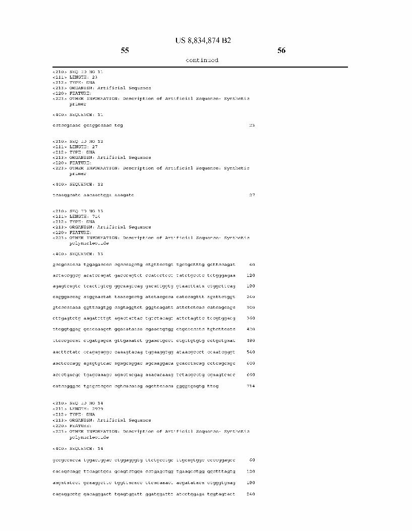
US 8,834,874 B2 55
- Continued
SEQ ID NO 11 LENGTH: 23 TYPE: DNA
ORGANISM: Artificial Sequence FEATURE:
OTHER INFORMATION: Description of Artificial Sequence: Synthetic primer
SEQUENCE: 11
Cctic.cgaaac gcaggccaac tog 23
SEQ ID NO 12 LENGTH: 27 TYPE: DNA
ORGANISM: Artificial Sequence FEATURE:
OTHER INFORMATION: Description of Artificial Sequence: Synthetic prlmer
SEQUENCE: 12
t caaggcatc aacaactgga aaagatc 27
SEQ ID NO 13 LENGTH: 714 TYPE: DNA
ORGANISM: Artificial Sequence FEATURE:
OTHER INFORMATION: Description of Artificial Sequence: Synthetic polynucleotide
<4 OOs, SEQUENCE: 13
gcc.gc.cacca tigaga CCCC CCCC agctg. CtgttcCtgt tictgctttg gct tccagat 60
act accoggcg a catccagat gacccagt ct c catcct cot tatctgcct c tictdggagaa 12 O
agagt cagtc. tcacttgtcg ggcaa.gtcag gacattggtg gta acttata Ctggctt cag 18O
cagggaccag atggaact at taaacgc.ctg atctacgc.ca catccagttt agattctggit 24 O
gtc.cccaaaa ggttcagtgg cagtaggit ct gggtcagatt attct ct cac cat cago agc 3OO
cittgagtctgaagattttgt agact attac tdtctacagt attctagttc. tcc.gtggacg 360
titcggtggag gcacaaagct ggaaataaaa Caactgtgg Ctgcaccatc tetctitcatC 42O
titc.ccgc.cat Ctgatgagca gttgaaatct ggaactgcct Ctgttgttgttg cctgctgaat 48O
aact tctatc ccagagaggc caaagtacag taaggtgg ataacgc.cct C caatcgggt 54 O
aact Cocagg agagtgtcac agagcaggac agcaaggaca gcacctacag cct cagcagc 6OO
accctgacgc tigagcaaagc agacitacgag aaacacaaag tictacgc.ctg. Caagtcacc 660
catcagggcc tagotcgcc cqt cacaaag agcttcaa.ca ggggaga.gtg ttag 714.
<210s, SEQ ID NO 14 &211s LENGTH: 2979 &212s. TYPE: DNA
<213> ORGANISM: Artificial Sequence 22 Os. FEATURE:
<223> OTHER INFORMATION: Description of Artificial Sequence: Synthetic polynucleotide
<4 OOs, SEQUENCE: 14
gcc.gc.cacca tactggac Ctggagggitg ttctgcctgc titgcagtggc ccc.cggagcc 6 O
CacagcCagg ttcagctgca gcagtctgga cct gagctgg taagcctgg ggctittagtg 12 O
aagatat cct gcaaggct to tdgttacacic titcacaaact acgatataca citgggtgaag 18O
cagaggcctg gaCagggact tagtggatt ggatggattt atcCtggaga tigg tagtact 24 O

59 US 8,834,874 B2
- Continued
ctgaag catt titcgatt.ccg tdacttggaa gaggat.ccgt acctic cctdg taatcc.ccgt. 27 OO
gaactgattg cctatagoca gitatic cccgg cct tcagaca tocct cagtg gaattctgac 276 O
aag.ccgagtt taaaagatat aaagat catg ggctatt coa tacgcac cat agactatagg 282O
tatact.gtgt giggttggctt caatcctgat gaatttctag ctaacttitt c tda catc cat 288O
gCaggggaac tt attttgt ggattctgac C cattgcagg at Cacaatat gtataatgat 294 O
t cccaaggtg gagatcttitt C cagttgttg atgccttga 2979
<210s, SEQ ID NO 15 &211s LENGTH: 573 &212s. TYPE: DNA
<213> ORGANISM: Artificial Sequence 22 Os. FEATURE:
<223> OTHER INFORMATION: Description of Artificial Sequence: Synthetic polynucleotide
<4 OOs, SEQUENCE: 15
gcc.gc.cacca tttcgacc attgaactgc atcgt.cgc.cg tdt CC caaaa tatggggatt 6 O
ggcaagaacg gagacct acc Ctggcctic cq Ctcaggaacg agttcaagta Ctt coaaaga 12 O
atgaccacaa cct Ctt cagt ggalaggtaaa Cagaatctgg tatt atggg taggaaaac C 18O
tggttct c catt cctdagaa gaatcgacct ttaaaggaca gaattaatat agttct cagt 24 O
agagaactica aagaaccacc acgaggagct catttitcttg C caaaagttt ggatgatgcc 3OO
ttalagacitta ttgaacaa.cc ggaattggca agtaaagtag acatggtttg gat agtc.gga 360
ggcagttctg tttaccagga agc catgaat caaccaggcc acct cagact citttgttgaca 42O
aggat catgc aggaatttga aagtgacacg tttitt Cocag aaattgattt ggggaaatat 48O
aaacttct co Cagaat accc aggcgt.cctic tictgaggtoc aggaggaaaa aggcatcaag 54 O
tataagtttg aagtictacga gaagaaagac taa sf3
<210s, SEQ ID NO 16 &211s LENGTH: 234 212. TYPE: PRT
<213> ORGANISM: Artificial Sequence 22 Os. FEATURE:
<223> OTHER INFORMATION: Description of Artificial Sequence: Synthetic polypeptide
<4 OOs, SEQUENCE: 16
Met Glu. Thr Pro Ala Glin Leu Lleu. 1.
Asp
Ala
Ile
Lys 65
Arg
Ser
Ser
Thir
Ser
Gly SO
Arg
Phe
Luell
Ser
5
Thr Gly Asp Ile Gln Met
Lieu. Gly Glu Arg Val Ser 35 4 O
Gly Asn Lieu. Tyr Trp Lieu. 55
Lieu. Ile Tyr Ala Thr Ser 70
Ser Gly Ser Arg Ser Gly 85
Glu Ser Glu Asp Phe Val
Pro Trp Thr Phe Gly Gly 115 12 O
Phe Lieu. Lieu 1O
Thr Glin Ser 25
Lieu. Thir Cys
Gln Glin Gly
Ser Lieu. Asp 7s
Ser Asp Tyr 90
Asp Tyr Tyr 105
Gly Thr Lys
Lell
Pro
Arg
Pro 6 O
Ser
Ser
Met
Lell
Ser
Ala 45
Asp
Gly
Lell
Lell
Glu 125
Trp Leu Pro 15
Ser Luel Ser 3O
Ser Glin Asp
Gly. Thir Ile
Val Pro Llys 8O
Thir Ile Ser 95
Gln Tyr Ser 11 O
Ile Lys Arg
60
Thir
Lell 145
Pro
Gly
His
Wall 225
Wall 13 O
Arg
Asn
Ser
Lys 21 O
Thir
Ala
Ser
Glu
Ser
Luell 195
Wall
Ala
Gly
Ala
Glin 18O
Ser
Ser
PRT
Pro
Thir
Lys 1.65
Glu
Ser
Ala
Phe
SEO ID NO 17 LENGTH: TYPE :
ORGANISM: Artificial Sequence FEATURE:
OTHER INFORMATION: Description of
187
polypeptide
<4 OOs, SEQUENCE:
Met Val Arg Pro 1.
Ile
Asn
Asn 65
Ala
Wall
Pro
Ser 145
Pro
Gly
Tyr
Luell SO
Arg
Glu
Luell
Trp
Gly 13 O
Asp
Glu
Lys
Phe 35
Wall
Pro
Pro
Arg
Ile 115
His
Thir
Asn
Glin
Ile
Luell
Pro
Luell
Wall
Luell
Phe
Pro
Phe 18O
24
17
Lell 5
Gly
Arg
Met
Lys
Arg 85
Ile
Gly
Arg
Phe
Gly 1.65
Glu
SEQ ID NO 18 LENGTH:
TYPE: DNA
ORGANISM: Artificial Sequence FEATURE:
OTHER INFORMATION: Description of Artificial Sequence: primer
Ser
Ala 150
Wall
Ser
Thir
Asn 23 O
Asn
Asp
Met
Gly
Asp 70
Gly
Glu
Gly
Lell
Pro 150
Wall
Wall
61
Wall 135
Ser
Glin
Wall
Lell
Glu 215
Arg
Lieu
Thir
Arg 55
Arg
Ala
Glin
Ser
Phe 135
Glu
Lell
Phe
Wall
Trp
Thir
Thir 2OO
Wall
Gly
Ile
Pro
Thir 4 O
Ile
His
Pro
Ser 12 O
Wall
Ile
Ser
Glu
Ile
Wall
Glu 185
Luell
Thir
Glu
Wall
Trp 25
Thir
Thir
Asn
Phe
Glu 105
Wall
Thir
Asp
Glu
Lys 185
Phe
Cys
Wall 17O
Glin
Ser
His
Cys
Ala
Pro
Ser
Trp
Ile
Luell 90
Luell
Arg
Luell
Wall 17O
Pro
Luell 155
Asp
Asp
Lys
Glin
Artificial Sequence:
Wall
Pro
Ser
Phe
Wall 7s
Asp
US 8,834,874 B2
- Continued
Pro 14 O
Lell
Asn
Ser
Ala
Gly 22O
Ser
Lieu
Wall
Ser 6 O
Lell
Ser
Glu
Met 14 O
Glu
Ser
Asn
Ala
Asp 2O5
Lell
Glin
Arg
Glu 45
Ile
Ser
Ser
Ala 125
Glin
Glu
Asp
Asn
Luell
Asp 19 O
Ser
Asn
Asn
Gly
Pro
Arg
Luell
Wall 11 O
Met
Glu
Glu
Phe
Glin 17s
Ser
Glu
Ser
Met 15
Glu
Glu
Glu
Asp 95
Asp
Asn
Phe
Luell
Gly 17s
Glin
Tyr 160
Ser
Thir
Pro
Synthetic
Gly
Phe
Glin
Luell
Asp
Met
Glin
Glu
Luell 160
Ile
Synthetic
62

US 8,834,874 B2 63 64
- Continued
<4 OOs, SEQUENCE: 18
ccacagatgc tictogaacgtt ctitc
SEQ ID NO 19 LENGTH: 15 TYPE PRT
ORGANISM: Artificial Sequence FEATURE:
peptide
SEQUENCE: 19
OTHER INFORMATION: Description of Artificial Sequence:
24
Synthetic
Gly Gly Gly Gly Ser Gly Gly Gly Gly Ser Gly Gly Gly Gly Ser 1. 5 1O
What is claimed: 1. A fusion antibody comprising: (a) an immunoglobulin that crosses a blood brain barrier
(BBB), wherein the immunoglobulin comprises an immunoglobulin heavy chain and an immunoglobulin light chain; and
(b) an iduronate-2-sulfatase, wherein the amino acid sequence of the iduronate-2-sulfatase is covalently linked to the carboxy terminus of the amino acid sequence of the immunoglobulin heavy chain, thereby forming a fusion protein;
wherein the fusion antibody crosses the blood brain barrier (BBB) and catalyzes hydrolysis of 2-sulfate groups of the L-iduronate 2-sulfate units of dermatan Sulfate, heparan Sul fate or heparin and wherein the iduronate-2-sulfatase retains at least 20% of its activity compared to its activity as a sepa rate entity.
2. The fusion antibody of claim 1, wherein the fusion antibody is post-translationally modified by a Sulfatase modi fying factor type 1 (SUMF1).
3. The fusion antibody of claim 1, wherein the fusion antibody comprises a formylglycine.
4. The fusion antibody of claim 1, wherein the fusion protein further comprises a linker between the amino acid sequence of the iduronate-2-sulfatase and the carboxy termi nus of the amino acid sequence of the immunoglobulin heavy chain.
5. The fusion antibody of claim 1, wherein the iduronate 2-sulfatase specific activity of the fusion antibody is at least 10,000 units/mg.
6. The fusion antibody of claim 1, wherein the immuno globulin retains at least 20% of its activity compared to its activity as a separate entity.
7. The fusion antibody of claim 1, wherein the immuno globulin heavy chain is an immunoglobulin heavy chain of IgG.
25
30
35
40
45
50
15
8. The fusion antibody of claim 1, wherein the immuno globulin heavy chain comprises a CDR1 corresponding to the amino acid sequence of SEQID NO:1, a CDR2 correspond ing to the amino acid sequence of SEQID NO:2, or a CDR3 corresponding to the amino acid sequence of SEQID NO:3.
9. The fusion antibody of claim 1, wherein the immuno globulin light chain is an immunoglobulin light chain of lambda class.
10. The fusion antibody of claim 1, wherein the immuno globulin light chain is an immunoglobulin light chain of kappa class.
11. The fusion antibody of claim 1, wherein the immuno globulin light chain comprises a CDR1 corresponding to the amino acid sequence of SEQID NO:4, a CDR2 correspond ing to the amino acid sequence of SEQID NO:5, or a CDR3 corresponding to the amino acid sequence of SEQID NO:6.
12. The fusion antibody of claim 1, wherein the fusion antibody crosses the BBB by binding an endogenous BBB receptor-mediated transport system.
13. The fusion antibody of claim 1, wherein the fusion antibody crosses the BBB via an endogenous BBB receptor selected from the group consisting of the insulin receptor, transferrin receptor, leptin receptor, lipoprotein receptor, and the IGF receptor.
14. The fusion antibody of claim 1, wherein the fusion antibody crosses the BBB by binding an insulin receptor.
15. A pharmaceutical composition comprising a therapeu tically effective amount of a fusion antibody of claim 1, and a pharmaceutically acceptable excipient.
16. The fusion antibody of claim 1, wherein the iduronate 2-sulfatase retains at least 50% of its activity compared to its activity as a separate entity.
k k k k k