1xuvh - nebula.wsimg.com
TRANSCRIPT

Forensic Examination of Gunshot Wounds: Best Practices for the Emergency NurseApril 2017
Gina Carbino, BSN, RN, CEN, CPEN, CCRN, PCCN, SANE-A @gcarbino Lecture Overview:Explain proper procedure for identifying, documenting, and caring for suspected or known gunshot wounds1. Identify critical components of ballistics
Types of firearms Types of ammunition Factors affecting mechanism of injuries
2. Identification and documentation of gunshot wounds Entrance versus exit Distance Documentation
3. Implementing best practices for caring for gunshot wounds in your emergencydepartment
1
Ammunition:
2. The caliper of the bullet is determined by the diameter of the bullets base
3. Take a trip back to nursing school, a bullet's weight is measured in grains
•
1. Parts of ammunition: (fig A.)• Bullet• Cartridge• Powder• Flash Hole• Primerl• Measured in hundredths of an inch or in millimeters
o a .45 caliber is 11 mm
4. Examples:• Hollow points: can increase up to 2.5x their size upon impact
and increase soft tissue damage up 6.5x bullet sizeBlackTalon: now illegal, hollow point; ask me how this bullet hashistorical significance in emergency medicine
• Full metal jackets: high velocity, typically exit body• Shotgun shells
(fig A.)
Firearms and Ballistics:
Firearms :1. Rifled Firearms
• Barrel contains helical grooves on internal surface; this helpsincrease velocity and distance
• Creates unique markings on the bullet that assists in identifying thefirearm it was fired from
• Examples:o Handguns (85%)o Rifles (8%)
2. Smooth Barrel• Shotguns (5%)• Higher rate of accidental injury

Forensic Examination of Gunshot Wounds: Best Practices for the Emergency Nurse April 2017 - @gcarbino
(fig B.)
1. Yaw and Tumble (fig B.)2. Locard's Principle3. Tissue destruction = KE = ½ m v2
• Increased velocity = more damage4. Cavities
• Primary Cavity (fig C.1)o Direct tissue contact by the bullet
• Secondary or Temporary (fig C.2)o Trauma caused from shockwaveo Tissue stretcheso High velocity bullets can cause a
shockwave 10x their size
(fig C)
Addressing the Wounds: 1. Entrance versus Exit :
• Entranceo Smaller, more circularo May contain products of combustiono Cherry red appearance =carboxyhemoglobin (fig D)
• Exito Edges typically everted, larger, and irregularo Does not contain products of combustion
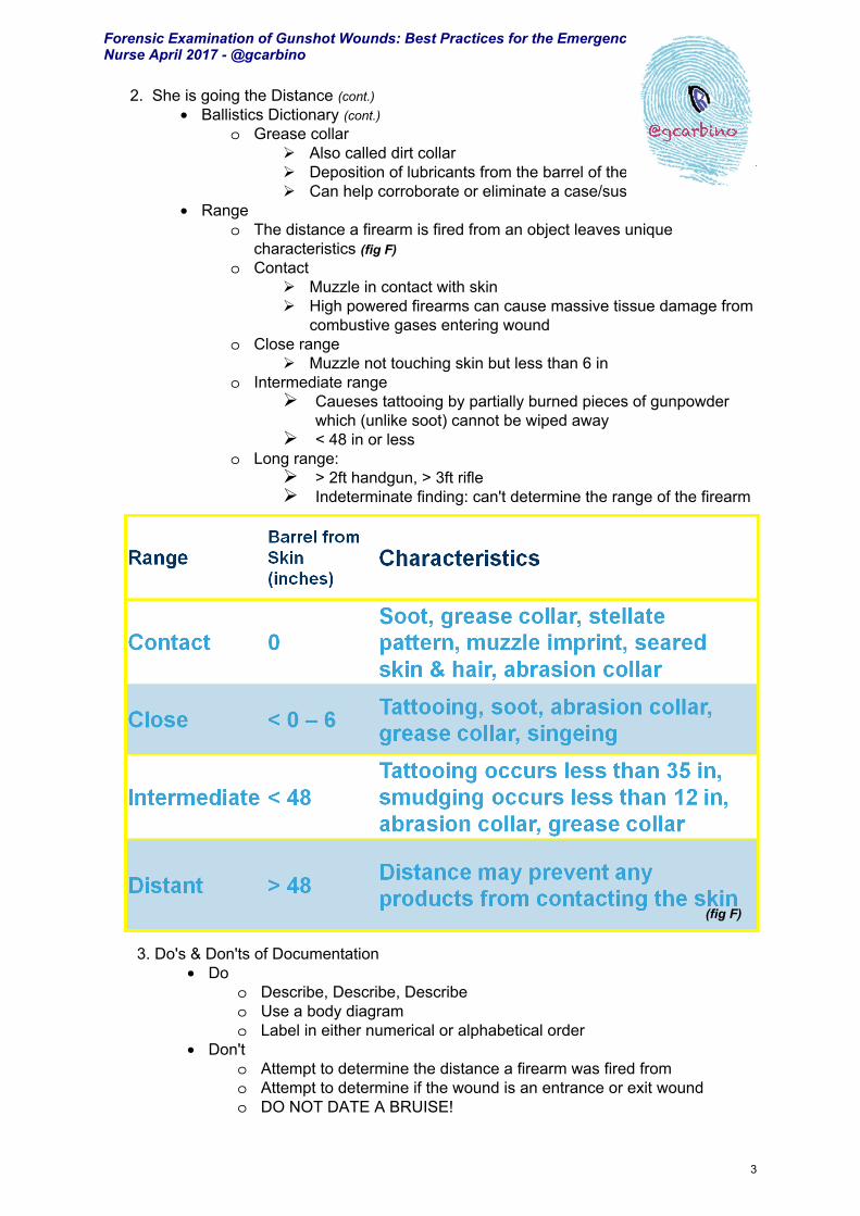
2. She is going the Distance• Ballistics Dictionary
o Tattooing (fig E) Also called peppering & stippling Can't be wiped/washed off in the trauma bay Small discrete, black specks caused by unburnt/partially
burnt gunpowder grains embedded into skino Smudging
Can be wiped/washed off Carbonaceaous deposition of smoke on the skin
o Lead ring Also called metal ring Deposition of lead at entry site
o Muzzle imprint (fig D) Occurs from gases of combustion
entering wound and pushing skin againstmuzzle of gun
2
(fig D)
(fig E)
Mechanism of Injury:
Forensic Examination of Gunshot Wounds: Best Practices for the Emergency Nurse April 2017 - @gcarbino
3
2. She is going the Distance (cont.)• Ballistics Dictionary (cont.)
o Grease collar Also called dirt collar Deposition of lubricants from the barrel of the firearm Can help corroborate or eliminate a case/suspect
• Rangeo The distance a firearm is fired from an object leaves unique
characteristics (fig F)o Contact
Muzzle in contact with skin High powered firearms can cause massive tissue damage from
combustive gases entering woundo Close range
Muzzle not touching skin but less than 6 ino Intermediate range
Caueses tattooing by partially burned pieces of gunpowderwhich (unlike soot) cannot be wiped away
< 48 in or lesso Long range:
> 2ft handgun, > 3ft rifle Indeterminate finding: can't determine the range of the firearm
(fig F)
3. Do's & Don'ts of Documentation• Do
o Describe, Describe, Describeo Use a body diagramo Label in either numerical or alphabetical order
• Don'to Attempt to determine the distance a firearm was fired fromo Attempt to determine if the wound is an entrance or exit woundo DO NOT DATE A BRUISE!

4
Forensic Examination of Gunshot Wounds: Best Practices for the Emergency Nurse April 2017 - @gcarbino
Shine at Your Shop: 1. Document, Document, and Document some more :
• This IS going to courto In most cases of violent crimes, your District Attorney ONLY has your
documentation and the victim's testimony to win the case• A picture is worth a thousand words
o Cameras are cheap!o Don't forgot a cheap photo printer if your shop isn't ready to store digital
photography• Draw injury on a body/anatomical diagram
2. Handle with CARE!• Clothing
o Do not cut through stains or holeso Do not use your trauma shears
Are you kidding me, they are filthy! Use sterile shears; suture removal kit works for lighter fabrics Find me at some point during the conference and we will use my
alternate light source and examine your shearso Wear gloves and change between each itemo Bag my items in paper and separately please
Make sure you document what is in the bag: left sock Sharpie markers work great for this!
• Other potentially probative materialso Remove shrapnel/bullets with suture booties or gauzed covered
hemostatso Place in plastic container: urine specimen cups are your best friends
here3. Chain of Custody
• Not following chain of custody can get all your hard work and potentially all theDNA evidence being declared inadmissable in court
• Use a chain of custody formo Document what, when, and whoo Use the same form for all evidence: KISSo Make a copy and keep with patient charto This is the only proof that YOU maintained chain of custody
4. Collaborate with your forensic nurse and local forensic team• Have a representative from your shop on your local SART, Fatality Review
Team, MDT, and/or DVCC• PRIOR to an event is the best time to build relationships and ask questions

5
• Abrasionso Disruption in top layer of epidermis, typically from shearing force
• Avulsionso Tearing away of skin, tissue, or bone
• Bite markso Curvillinear contusion or abrasion; should be swabbed for assailant DNA
• Bruisingo Blood infiltrating surrounding tissue from damaged vesselso Will follow the direction of gravity; typically does not occur with postmortem
traumao Caused by trauma
• Central sparing or clearingo Bruising occurring in "negative" imprint of causative object; seen in blunt
force trauma• Ecchymoses
o Large subcutaneous area of bleedingo Caused by physiologic responses (non-intentional)
• Hematomao Collection of blood under dermis; typically indurated
• Hesitation markso Superficial, parallel sharp force trauma that is consistent with self inflicted
wounds• Incision
o Caused by sharp force trauma, such as, a knife; typically length exceeds depth
o Can be wide when crossing Langer's (cleavage) lines• Laceration
o Tear in the skin from blunt force trauma Crushed skin/tissue
o May have tissue bridging; typically not self inflicted• Petechiae
o Small, pinpoint hemorrhageso Check palate and conjunctiva in strangulation cases
• Purpurao Larger than petechiae, irregular borders, may be purplish in color
• Stab woundo Sharp and blunt force trauma in etiology; typically depth exceeds length
Forensic Examination of Gunshot Wounds: Best Practices for the Emergency Nurse April 2017 - @gcarbino
Get Your Forensic Terminology On:
Anatomical Descriptions:• Lateral – towards a side (indicate left or right)• Distal – away from center of the body• Medial – toward middle• Posterior – back• Superior – Top, up, above• Inferior – below, down, below• Proximal – Towards the center of the body• Palmar – towards palm• Plantar – towards bottom of foot• Dorsal – opposite of plantar

Forensic Examination of Gunshot Wounds: Best Practices for the Emergency Nurse April 2017 - @gcarbino
Additional Resources:
The Wound Consultant: Wound Documentation Tips
The NursePath: How to Document Wound and Bruises
Basic Gunshot Wound Treatment: Daniel Ricciardi
Management of GSWs of the Limbs
Treating GSWs in the Lower Extremity
Shootings: What EMS Providers Need to Know
Gunshot Wounds: Management and Myths
Patterns of Tissue Injury
Link to Alternate Light Source and another example
Link to Inexpensive Photo Printer Example
Link to Chain of Custody Form
Link to Body diagram
6