2. example safety and quality manual (word 80 kb)
TRANSCRIPT

Appendix 2: Example - Safety and Quality Manual
Example
SAFETY AND QUALITY MANUAL
SunnySide Imaging
DIAGNOSTIC IMAGING ACCREDITATION SCHEME USER GUIDE – Appendix 2 1
Appendix 2: Example - Safety and Quality Manual
Contents
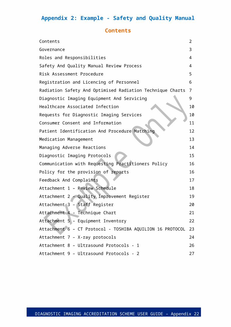
Contents 2
Governance 3
Roles and Responsibilities 4
Safety And Quality Manual Review Process 4
Risk Assessment Procedure 5
Registration and Licencing of Personnel 6
Radiation Safety And Optimised Radiation Technique Charts 7
Diagnostic Imaging Equipment And Servicing 9
Healthcare Associated Infection 10
Requests for Diagnostic Imaging Services 10
Consumer Consent and Information 11
Patient Identification And Procedure Matching 12
Medication Management 13
Managing Adverse Reactions 14
Diagnostic Imaging Protocols 15
Communication with Requesting Practitioners Policy 16
Policy for the provision of reports 16
Feedback And Complaints 17
Attachment 1 – Review Schedule 18
Attachment 2 – Quality Improvement Register 19
Attachment 3 – Staff Register 20
Attachment 4 – Technique Chart 21
Attachment 5 – Equipment Inventory 22
Attachment 6 – CT Protocol - TOSHIBA AQUILION 16 PROTOCOL 23
Attachment 7 – X-ray protocols 24
Attachment 8 – Ultrasound Protocols - 1 26
Attachment 9 – Ultrasound Protocols - 2 27
DIAGNOSTIC IMAGING ACCREDITATION SCHEME USER GUIDE – Appendix 2 2

Appendix 2: Example - Safety and Quality Manual
Governance
This Practice is owned and operated by Mr Brad Waugh.
Australian Public Company, Limited by Guarantee
Registered on 17 January 2004 in the state of Victoria.
ABN: 61 004 381 462
ACN: 004 381 462
Registered office and principal place of business:
14 Gilmore St
Sunnyside, Victoria 3000
Australia
LSPN 009999 registered with the Department of Human Services for Medicare purposes, and accredited with approved accreditor XYZ.
Key Contacts:
Managing Director: Mr Brad Waugh
Medical Director: Dr Jean Smith
Our insurance broker is Watford Insurance Brokers. Certificates of currency are held by the Medical Director including:
Workers Compensation to the amount required by law, $20,000,000.00 Public Liability, and $10,000,000.00 Professional Indemnity.
DIAGNOSTIC IMAGING ACCREDITATION SCHEME USER GUIDE – Appendix 2 3
Appendix 2: Example - Safety and Quality Manual
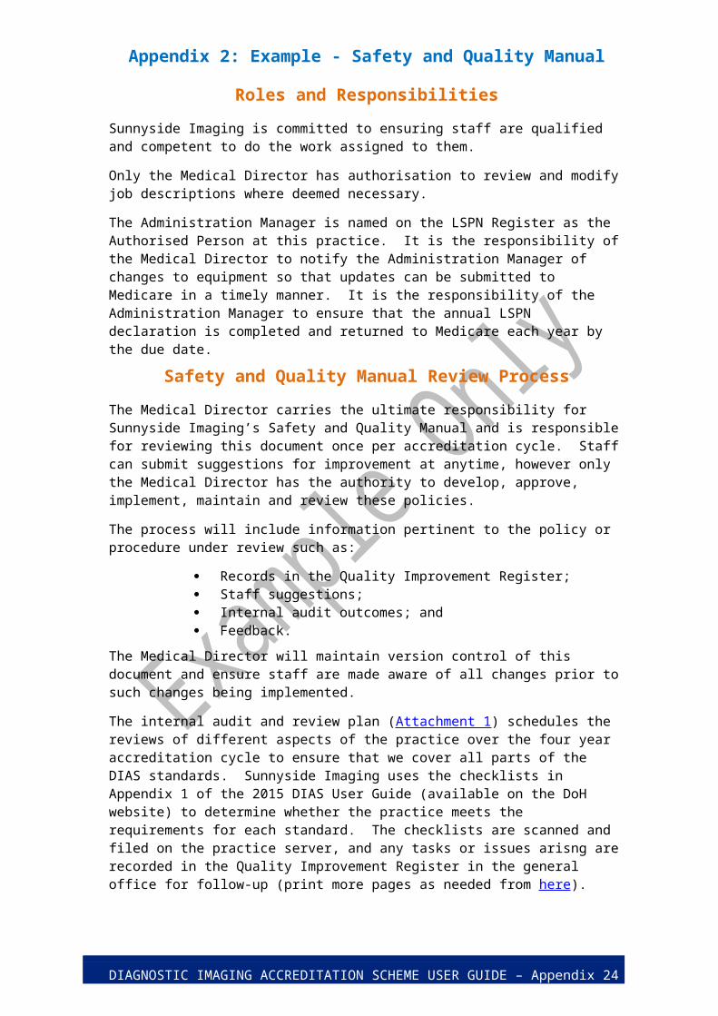
Roles and Responsibilities
Sunnyside Imaging is committed to ensuring staff are qualified and competent to do the work assigned to them.
Only the Medical Director has authorisation to review and modify job descriptions where deemed necessary.
The Administration Manager is named on the LSPN Register as the Authorised Person at this practice. It is the responsibility of the Medical Director to notify the Administration Manager of changes to equipment so that updates can be submitted to Medicare in a timely manner. It is the responsibility of the Administration Manager to ensure that the annual LSPN declaration is completed and returned to Medicare each year by the due date.
Safety and Quality Manual Review Process
The Medical Director carries the ultimate responsibility for Sunnyside Imaging’s Safety and Quality Manual and is responsible for reviewing this document once per accreditation cycle. Staff can submit suggestions for improvement at anytime, however only the Medical Director has the authority to develop, approve, implement, maintain and review these policies.
The process will include information pertinent to the policy or procedure under review such as:
Records in the Quality Improvement Register; Staff suggestions; Internal audit outcomes; and Feedback.
The Medical Director will maintain version control of this document and ensure staff are made aware of all changes prior to such changes being implemented.
The internal audit and review plan (Attachment 1) schedules the reviews of different aspects of the practice over the four year accreditation cycle to ensure that we cover all parts of the DIAS standards. Sunnyside Imaging uses the checklists in Appendix 1 of the 2015 DIAS User Guide (available on the DoH website) to determine whether the practice meets the requirements for each standard. The checklists are scanned and filed on the practice server, and any tasks or issues arisng are recorded in the Quality Improvement Register in the general office for follow-up (print more pages as needed from here).
DIAGNOSTIC IMAGING ACCREDITATION SCHEME USER GUIDE – Appendix 2 4
Appendix 2: Example - Safety and Quality Manual
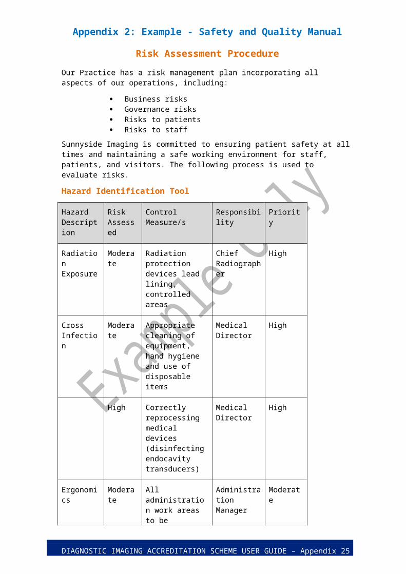
Risk Assessment Procedure
Our Practice has a risk management plan incorporating all aspects of our operations, including:
Business risks Governance risks Risks to patients Risks to staff
Sunnyside Imaging is committed to ensuring patient safety at all times and maintaining a safe working environment for staff, patients, and visitors. The following process is used to evaluate risks.
Hazard Identification Tool
Hazard Description
Risk Assessed
Control Measure/s Responsibility Priority
Radiation Exposure
Moderate Radiation protection devices lead lining, controlled areas
Chief Radiographer
High
Cross Infection
Moderate Appropriate cleaning of equipment, hand hygiene and use of disposable items
Medical Director
High
High Correctly reprocessing medical devices (disinfecting endocavity transducers)
Medical Director
High
Ergonomics Moderate All administration work areas to be ergonomically designed
Administration Manager
Moderate
DIAGNOSTIC IMAGING ACCREDITATION SCHEME USER GUIDE – Appendix 2 5

Appendix 2: Example - Safety and Quality Manual
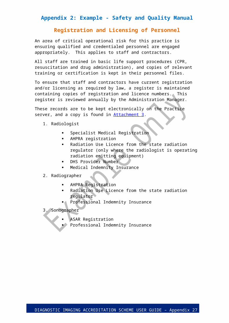
Registration and Licensing of Personnel
An area of critical operational risk for this practice is ensuring qualified and credentialed personnel are engaged appropriately. This applies to staff and contractors.
All staff are trained in basic life support procedures (CPR, resuscitation and drug administration), and copies of relevant training or certification is kept in their personnel files.
To ensure that staff and contractors have current registration and/or licensing as required by law, a register is maintained containing copies of registration and licence numbers. This register is reviewed annually by the Administration Manager.
These records are to be kept electronically on the Practice server, and a copy is found in Attachment 3.
1. Radiologist
Specialist Medical Registration AHPRA registration Radiation Use Licence from the state radiation regulator (only where the
radiologist is operating radiation emitting equipment) DHS Provider Number Medical Indemnity Insurance
2. Radiographer
AHPRA Registration Radiation Use Licence from the state radiation regulator Professional Indemnity Insurance
3. Sonographer
ASAR Registration Professional Indemnity Insurance
DIAGNOSTIC IMAGING ACCREDITATION SCHEME USER GUIDE – Appendix 2 6
Appendix 2: Example - Safety and Quality Manual
Radiation Safety and Optimised Radiation Technique Charts
Our approach to the safety of patients and staff is executed by:
1. Use of a Radiation Safety Plan;2. Maintaining technique charts that are available for each piece of equipment capable of
producing ionising radiation;3. Annual review and authorisation of both manual settings and settings embedded in the
imaging equipment software by the Medical Director. The Medical Director may delegate the review to the Chief Radiographer, or other qualified person.
4. Annual comparison in October each year of the Sunnyside Imaging facility reference levels (FRLs) to the published Australian Diagnostic Reference Levels (DRLs) jointly by the Medical Director and Chief Radiographer.
5. Four yearly review of the Radiation Safety Plan by the Chief Radiographer and Medical Director to ensure that the content is current, followed by personnel, and appropriate to the practice at that time.
Radiation Management Plan
Sunnyside Imaging provides a range of services that includes the use of ionising radiation in the delivery of x-ray services.
We are committed to using the minimum radiation doses to make appropriate diagnostic images.
Sunnyside Imaging’s Radiation Safety Plan is based on Schedule A of the Radiation Protection Series Guideline for Using Radiation in Medical Imaging – RPS 14. The Radiation Safety Plan is maintained electronically on the practice server, and a hardcopy is in the general office.
[Please note that an example Radiation Safety Plan (RSP) has not been provided with this example Safety and Quality Manual. RSPs are specific to the practice, and requirements vary between jurisdictions. Practices are strongly encouraged to base their own RSP on RPS 14 Schedule A and to consult their state or territory regulator to check additional requirements.]
Technique Charts
Separate technique charts are available for each piece of equipment capable of producing ionising radiation.
Technique charts can be found next to each piece of equipment for quick reference. As Sunnyside Imaging is a digital practice, the exposure index related to radiation dose is included to ensure that over exposure can be monitored. A copy of the Technique Chart is in Attachment 4.
Annual DRL comparisons
Each year in November, the Chief Radiographer is responsible for ensuring that all protocols for Adults and Children (Sunnyside Imaging does not image infants) are entered in the ARPANSA DRL online tool.
When 20 episodes have been entered for a protocol, the report is requested. If less than 20 episodes are run in a year, the report is requested on 30 September based those already entered.
DIAGNOSTIC IMAGING ACCREDITATION SCHEME USER GUIDE – Appendix 2 7
Appendix 2: Example - Safety and Quality Manual
The Chief Radiographer and Medical Director are responsible for reviewing the reports, and determining whether any optimisation of settings is required.
Any FRLs higher than published DRLs must be justified.
If any equipment is altered, repaired, upgraded or commissioned during the year, a DRL comparison must be performed and reviewed as soon as practicable.
Any actions arising from the comparison process must be recorded in the Quality Improvement Register.
DIAGNOSTIC IMAGING ACCREDITATION SCHEME USER GUIDE – Appendix 2 8

Appendix 2: Example - Safety and Quality Manual
Diagnostic Imaging Equipment and Servicing
Sunnyside Imaging is committed to quality patient outcomes and understands that equipment performance can have a significant impact on the quality and accuracy of diagnostic reports.
All equipment is registered on the Department of Human Service’s Diagnostic Imaging Equipment Register (LSPN Register). This information will be updated as required.
Diagnostic Imaging Equipment Inventory
An inventory is maintained on the practice server, with a copy at Attachment 5 that includes:
The name of the item with its serial number and manufacturer Date of the last service Due date for the next planned service
Equipment listed on LSPN 009999
The Administration Manager is responsible for maintaining the practice LSPN.
Updating DHS (using the LSPN Amendment form) whenever equipment is purchased or retired from use.
Forwarding copies of all DHS correspondence pertaining to changes to equipment are to be copied to the approved accreditor XYZ.
Returning annual Location Specific Practice Number Declarations to Medicare.
Periodically requesting an equipment list from DHS and ensuring that the practice inventory matches the equipment recorded for the LSPN.
Diagnostic Imaging Equipment Servicing
All equipment will be serviced in accordance with the manufacturer’s requirements.
All service contracts include the requirement that service agents hold appropriate qualifications and licences, and that copies of the service providers Use License and training records for the equipment being serviced will be provided for our records annually in September.
The date of next service for each item is recorded in the inventory.
DIAGNOSTIC IMAGING ACCREDITATION SCHEME USER GUIDE – Appendix 2 9

Appendix 2: Example - Safety and Quality Manual
Healthcare Associated Infection
Sunnyside Imaging is committed to maintaining the highest levels of safety for our staff and patients by complying with recognised infection control standards and procedures.
Sunnyside Imaging does not perform interventional procedures.
Our policies for healthcare associated infection are to:
Ensure the cleanliness of our hands, equipment and examination areas For ultrasound, meet the requirements of TGO 54 Standard for
Disinfectants & Sterilants as directed by the disinfectant manufacturer Use single-use items where appropriate, and Ensure infectious waste containers are available in each imaging room.
Information about the policies and procedures in practice to reduce the risk of the transmission of infectious agents at Sunnyside Imaging is available in the reception area to all patients.
We ensure that our procedures for hand hygiene and disinfection are followed by scheduling audits annually. Any issues raised in the audits are documented as incidents in the Quality Improvement Register for action.
[Please note that this manual does not include a procedure for disinfection as a practice’s procedure will depend on the disinfectant chosen for use.]
Requests for Diagnostic Imaging Services
Diagnostic imaging procedures are only undertaken at Sunnyside Imaging where there is an identified, documented, clinical need and:
a.) Upon receipt of a signed request from a medical practitioner or a practitioner specified in the Act for the purpose of requesting services of that kind and for which a Medicare benefit is payable; or
b.) Where the practitioner interpreting the image is permitted to self determine the service for which a Medicare benefit is payable under the Act.
Any request having inadequate clinical details must be discussed with the referrer and further information is to be requested as described below.
Inappropriate requests must be referred to the Medical Director or Chief Radiographer for advice prior to continuing. Inappropriate requests include:
High risk imaging on pregnant patients; Requests with insufficient identification (ie, fewer than 3 identifiers) Requests with incorrect identification (ie not the same patient) Requests for screening purposes Requests for examinations that will not answer the clinical need stated on
the request Requests without a clinical need recorded
Physicians at Sunnyside Imaging do not self-determine, but may substitute or add services where it is permissible and appropriate to the patient’s care.
DIAGNOSTIC IMAGING ACCREDITATION SCHEME USER GUIDE – Appendix 2 10

Appendix 2: Example - Safety and Quality Manual
Consumer Consent and Information
Information pamphlets are available for the full range of examinations performed at our Practice.
Insideradiologyis also a recommended source of information to our clients.
Sunnyside performs only a few examinations which are considered to be high-risk or invasive. These are trans-vaginal ultrasounds, CT with contrast, and joint injections under ultrasound guidance.
A pre-procedure information sheet is available for each high risk or invasive procedure which describes the procedure to be undertaken and the risks involved. Staff are to go through this information sheet with each patient prior to the examination. The relevant patient history is obtained by staff and recorded on this form, along with the patient’s consent to proceed. This form is then scanned into the practice radiology information system (RIS).
If a patient chooses not to proceed with all, or part, of a procedure, this is to be recorded on the request form and the Medical Director informed. The Medical Director will inform the referring practitioner of the patient’s decision.
Where procedures of low or no risk are performed, the procedure is to be explained to the patient and their verbal consent to the examination obtained. A “tick box” is selected on the RIS to indicate that verbal consent was received.
DIAGNOSTIC IMAGING ACCREDITATION SCHEME USER GUIDE – Appendix 2 11

Appendix 2: Example - Safety and Quality Manual
Patient Identification and Procedure Matching
Sunnyside Imaging ensures that all patients are correctly identified when performing a diagnostic imaging examination. A minimum of three (3) patient identifiers are used throughout the entire procedure and are present on all records, including reports, worksheets and images. Patients are also matched with their intended diagnostic imaging procedure, including the anatomical site and side.
All patient identifiers and the intended procedure are confirmed at reception, and then again when presenting to the imaging room.
The following identifiers are approved for use at Sunnyside Imaging
The patient’s full name Date of birth Home address Unique practice identifier
Prior to administration of iodinated contrast, a period of “time out” will be called. Two staff must be present for a “time out” period. They must check the patient’s identity, the intended procedure and record that the check was completed.
The “time out” procedure must include confirmation of the following:
The patient’s full name Date of birth Home address The procedure to be undertaken The body region/site and side (where relevant) That relevant patient history has been collected That patient consent has been recorded
The record of time out is to be signed by the two staff members present and scanned into the RIS.
All mismatching events – even those that cause no immediate harm to patients - are considered serious and must be reported immediately to the Medical Director, followed by entering the event in the Quality Improvement Register. Mismatching events can be of several different types, including:
Incorrect Patient Incorrect Examination Incorrect Side Incorrect information on images Near miss
The Medical Director is responsible for investigating the incident, and then designating and implementing corrective actions, which are to be recorded against the entry in the Quality Improvement Register.
DIAGNOSTIC IMAGING ACCREDITATION SCHEME USER GUIDE – Appendix 2 12
Appendix 2: Example - Safety and Quality Manual
Medication Management
Sunnyside Imaging administers contrast media to patients when required to assist with diagnostic procedures. Emergency drugs are kept for use in the event of any reaction to contrast agents.
Storage, preparation and disposal of medications follow the manufacturer’s guidelines:
IV contrast - stored away from radiation sources and in a low light area Barium products - no special requirements Buscopan - store between 15 – 30 degrees C Atropine - store in a cool dry place under 25 degrees C Adrenaline - store in cool dry place under 25 degrees C
Out of date medications are to be returned to the pharmacy next door. Partially used medications are disposed in the labelled waste container which will be collected and destroyed by our waste management contractor.
This Practice complies with the RANZCR guideline for the use of contrast agents.
Some patients may be at an increased risk of adverse contrast reactions. We assure the safety of our patients by asking for, and recording, relevant patient history prior to the administration of any medication.
The following is a list of indicators which are known to increase the risk of adverse contrast reactions:
Previous moderate or severe reaction to iodinated contrast Multiple allergies Asthma increased risk of contrast induced neuropathy,
Current drugs which may cause adverse reactions in association with iodinated contrast: − Metformin − Β-adrenergic blockers
Pregnant patients Hyperthyroidism
If a patient at increased risk is identified, the radiologist will re-assess the need for contrast to be given on a case-by-case basis.
Resuscitation equipment must be available at all times when injecting IV contrast.
A medical practitioner must be immediately available to attend to the patient in the event of an emergency or complication of contrast injection.
We closely monitor the patient post contrast administration for up to 30 minutes and longer if deemed necessary.
DIAGNOSTIC IMAGING ACCREDITATION SCHEME USER GUIDE – Appendix 2 13
Appendix 2: Example - Safety and Quality Manual
Managing Adverse Reactions
In the event of a severe reaction the diagnostic imaging staff are trained to provide a first line response. A second staff member is to call for emergency assistance from the hospital response team or ‘000’ immediately.
The resuscitation trolley is located in the CT scan room on the left hand side of the entry door.
The equipment includes:
Oxygen / Suction Stethoscope Sphygmomanometer Amby bag and intubation equipment Pulse oximeter
Emergency drugs include:
Adrenaline Phenergan Hydrocortisone Atropine Normal saline
All adverse reactions must be recorded in both the Quality Improvement Register and the patient record. Adverse events include, but are not limited to:
Anaphylaxis Extravasation Flushing Urticaria
Immediately following resolution of the event, an entry in the Quality Improvement Register is to be made by the senior staff member present.
DIAGNOSTIC IMAGING ACCREDITATION SCHEME USER GUIDE – Appendix 2 14
Appendix 2: Example - Safety and Quality Manual
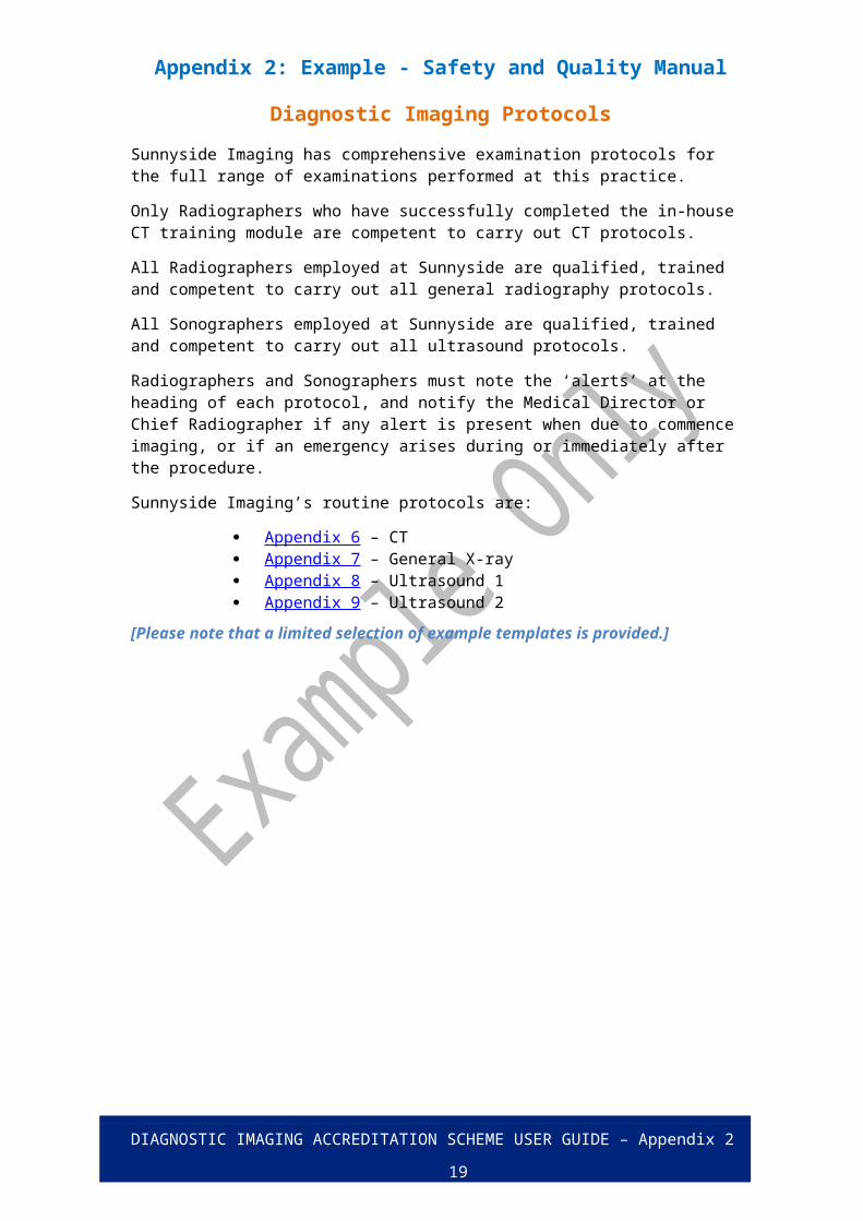
Diagnostic Imaging Protocols
Sunnyside Imaging has comprehensive examination protocols for the full range of examinations performed at this practice.
Only Radiographers who have successfully completed the in-house CT training module are competent to carry out CT protocols.
All Radiographers employed at Sunnyside are qualified, trained and competent to carry out all general radiography protocols.
All Sonographers employed at Sunnyside are qualified, trained and competent to carry out all ultrasound protocols.
Radiographers and Sonographers must note the ‘alerts’ at the heading of each protocol, and notify the Medical Director or Chief Radiographer if any alert is present when due to commence imaging, or if an emergency arises during or immediately after the procedure.
Sunnyside Imaging’s routine protocols are:
Appendix 6 – CT Appendix 7 – General X-ray Appendix 8 – Ultrasound 1 Appendix 9 – Ultrasound 2
[Please note that a limited selection of example templates is provided.]
DIAGNOSTIC IMAGING ACCREDITATION SCHEME USER GUIDE – Appendix 2 15

Appendix 2: Example - Safety and Quality Manual
Communication with Requesting Practitioners Policy
Optimal patient management depends on strong communication between Sunnyside Imaging and the referrer. Particular attention is given to urgent or unexpected findings.
Urgent and/or unexpected findings are communicated by the reporting Radiologist personally to the referring practitioner as a matter of urgency. Telephone contact is preferred. If the requesting practitioner is not available, the Radiologist will endeavour to discuss the matter with another practitioner at the practice who has authority to act on the findings. Any such calls and correspondence are documented in the report notes.
Policy for the provision of reports
The provision of appropriate and timely information to referrers is an important aspect to ensuring prompt patient management. The following is included in the final report:
Referrers name and address Date the report is issued Date the imaging procedure was performed Unique patient details – full name, date of birth, address, practice number Clinical details provided by the referrer Imaging procedure and modality Imaging procedure results including any measurements Comparison with any previous relevant images
It is the policy of Sunnyside Imaging policy that reports are not provided directly to the patient, but only to their referring practitioner.
Where feedback from referring practitioners is received regarding the content or provision of reports, the reporting radiologist will communicate directly with the referring practitioner. The feedback, and any resulting actions, is to be logged in the quality improvement register as soon as practicable.
DIAGNOSTIC IMAGING ACCREDITATION SCHEME USER GUIDE – Appendix 2 16

Appendix 2: Example - Safety and Quality Manual
Feedback and Complaints
Sunnyside Imaging is committed to ensuring the management of feedback and complaints is consistent with the principles of open disclosure and fairness, accessibility, responsiveness, efficiency and integration. Feedback can be:
Verbal feedback from patients to staff Written feedback via the Customer Feedback/Complaints Form, and Periodic patient and referrer surveys
All feedback received will be recorded in the Quality Improvement Register, and shall include the actions taken to resolve the issue.
If the complaint is about a matter which can be resolved immediately without reference to others, then the staff member is expected to take the necessary action, which must be recorded in the Quality Improvement Register.
Where the matter cannot be immediately resolved, it will be escalated to the Medical Director.
All staff receive training in managing and responding to feedback and complaints. Records of successful completion of training are retained by the Administration Manager.
Periodic referrer surveys are conducted annually in August to gauge referrer satisfaction with the practice.
Feedback is periodically reviewed and assessed and feeds into our continuous improvement processes.
DIAGNOSTIC IMAGING ACCREDITATION SCHEME USER GUIDE – Appendix 2 17

Appendix 2: Example - Safety and Quality Manual
Attachment 1 – Review Schedule
Audit Item Standard Mar 2016
Jul 2016
Nov 2016
Mar 2017
Jul 2017
Nov 2017
Mar 2018
Jul 2018
Nov 2018
Mar 2019
Jul 2019
Nov 2019
Governance 1.1
Personnel 1.2
Radiation Safety 1.3
Equipment 1.4; 1.5
Infection Control 1.6
Requests/Records 2.1
Informed Consent 2.2
Patient Identification 2.3
Medications 2.4
Protocols 3.1
Technique Charts 3.2
Reporting 4.1; 4.2
Feedback and Complaints 4.3
Quality Improvement All
Vertical Audit-Ultrasound All
Vertical Audit-MRI All
Vertical Audit- General X-ray All
Vertical Audit- CT All
Vertical Audit- Nuclear Medicine
All
DIAGNOSTIC IMAGING ACCREDITATION SCHEME USER GUIDE – Appendix 2 18
Appendix 2: Example - Safety and Quality Manual
Attachment 2 – Quality Improvement Register
Improvement Action Item
(include description of event/issue leading to the identification of improvement area)
Source
(eg. Patient, Referrer, etc)
Action Undertaken Person Responsible for Agreed Action
Completion Date
The current Quality Improvement Register is kept in the general office. Print more pages as required.
DIAGNOSTIC IMAGING ACCREDITATION SCHEME USER GUIDE – Appendix 2 19

Appendix 2: Example - Safety and Quality Manual
Attachment 3 – Staff Register
Name Position AHPRA No AHPRA expiry
Radiation Licence No
Radiation licence expiry
ASAR No ASAR expiry Review date
Next Review date
Dr Jean Smith
Medical Director
MED0058957210 06/11/2016
LX 3602/2005
30/06/2016
John Smith
Chief Radiographer
MRP0009876543 30/11/2016
LX 4321/2007
15/10/2016 NA NA Dec 2014 Dec 2015
Karen Banks
Radiographer MRP0007810592 19/09/2016
LX 3478/2005
25/09/2016 NA NA Dec 2014
Jane Wright
Sonographer NMW0014220872
12/05/2016
NA NA 7825 12/05/2016 Dec 2014
DIAGNOSTIC IMAGING ACCREDITATION SCHEME USER GUIDE – Appendix 2 20

Appendix 2: Example - Safety and Quality Manual
Attachment 4 – Technique Chart
EQUIPMENT TYPE: Shimadzu UD150, Fixed X-ray Room
Examination Projection kV mAs Distance BuckyExposure Index1
Chest PA 125 4 180 Yes 400
Lateral 125 10 180 Yes 400
Ribs Oblique 75 10 100 Yes 400
Shoulder AP 70 10 100 Yes 400
Lateral 80 20 100 Yes 400
Humerus AP 65 7 100 No 400
Lateral 65 7 100 No 400
Elbow AP 64 6 100 No 300
Lateral 66 6 100 No 300
Forearm AP 62 6 100 No 300
Lateral 66 6 100 No 300
Wrist
AP 62 3 100 No 250
Oblique 64 3 100 No 250
Lateral 66 4 100 No 250
Hand
AP 62 3 100 No 250
Oblique 64 3 100 No 250
Lateral 66 3 100 No 250
Finger AP/Lateral 50 3 100 No 250
1 For CR/DR systems the dose metric or exposure index will be presented and defined differently for each manufacturer. The common dose metric is the dose to the plate in units of microgray (uGy).For CT the dose metric should be the 'dose length product' (DLP in units of mGy.cm).For interventional fluoroscopy, where available, the dose metric should be the 'dose area product' (DAP in units of Gy.cm^2).
DIAGNOSTIC IMAGING ACCREDITATION SCHEME USER GUIDE – Appendix 2 21

Appendix 2: Example - Safety and Quality Manual
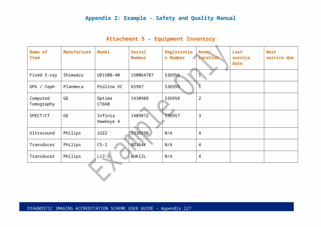
Attachment 5 – Equipment Inventory
Name of Item Manufacture Model Serial Number Registration Number
Room/Location Last service date Next service due
Fixed X-ray Shimadzu UD150B-40 150064787 536956 1
OPG / Ceph Planmeca Proline XC 65987 536959 1
Computed Tomography
GE Optima CT660 1430988 536958 2
SPECT/CT GE Infinia Hawkeye 4
1409872 536957 3
Ultrasound Philips iU22 3336598 N/A 4
Transducer Philips C5-2 BOJ54K N/A 4
Transducer Philips L17-5 BUKI2L N/A 4
DIAGNOSTIC IMAGING ACCREDITATION SCHEME USER GUIDE – Appendix 2 22

Appendix 2: Example - Safety and Quality Manual
Attachment 6 – CT Protocol - TOSHIBA AQUILION 16 PROTOCOL
Application: fracture, tumour, cellulitis, sinusitis
Alerts:
1. Request must be protocolled by a radiologist.2. If the patient is diabetic or over 60 years old renal function tests less than 3 months
old must be available for the radiologist to check to ensure it is safe to proceed.3. Ensure radiologist or a medical practitioner is onsite and available before
administering contrast.
Criteria Protocol InformationPosition/Landmark Head first or feet first-Supine
1cm superior to skull vertexTopogram Direction CraniocaudalScan Type HelicalRespiratory Phase AnyKV / mA / Rotation time (sec) 120kv / Sure Exp (100-450) / .75 secPitch / Speed (mm/rotation) 688:1 , 5.5mmSD Standard 6.0Detector width x Rows = Beam Collimation .5 x 16 = 8mmHelical Set
Slice Thickness/ SpacingKernelRecon Destination
body thickness/ reconrecon part spacing kernel destinationvolume thin face .5mm x .3mm FC30 very sharp for multiview
1 - facial bones 3mm x 3mm FC30 very sharp pacs2 - facial soft tissue 3mm x 3mm FC3 medium smooth pacs
Scan Start / End LocationsDFOV
1cm inferior to mandible1cm superior to frontal sinuses20 cmdecrease appropriately
IV Contrast Volume / Type / Rate 70cc omni 350 2cc/sec (if needed)Scan Delay 50 secondsArchiving to MOD Only thin data will be archived: volume recon2D/3D Technique Used Coronal face 3mm x 3mm reformats from volume
reconImages required in PACS Scouts, 3mm x 3mm sharp axial face/sinus, 3mm
x 3mm standard axial face/sinus, 3mm x 3mm sharp coronal face/sinus
Comments: This protocol is the routine for all face, sinus, and orbit studies. The volume is a very thin bone algorithm for reformats. Coronal reformats, 3mm x 3mm, average mode are routine for this protocol. The coronal plane is perpendicular to the hard palate. If the
DIAGNOSTIC IMAGING ACCREDITATION SCHEME USER GUIDE – Appendix 2 23

Appendix 2: Example - Safety and Quality Manual
mandible is the primary reason for the face CT scan, then also include sagittal oblique 3mm x 3mm reformats parallel with the mandibular rami
DIAGNOSTIC IMAGING ACCREDITATION SCHEME USER GUIDE – Appendix 2 24

Appendix 2: Example - Safety and Quality Manual
Attachment 7 – X-ray protocols
Alerts:
1. Unexpected findings – escalate priority reporting.
Chest X-ray (high kV technique):
Alerts:
1. Ensure pregnancy status is checked and recorded prior to imaging.
a.) Standard views;
PA and Lateral
b.) Pneumothorax
As above plus PA view on expiration
c.) Other views
Lordotic View as requested by radiologist
d.) For children up to the age of 12
PA or AP view is taken then referred to the radiologist for further views as necessary
Ribs (low kV technique):
PA Chest Oblique view of the affected side
Abdominal X-rays:
Alerts:
1. Ensure pregnancy status is checked and recorded prior to imaging.2. Gonad protection should be used.
a.) Obstruction
PA Chest to assess for gas under the diaphragm AP abdomen erect and supine (only perform erect view if fluid levels
seen in small bowel)
b.) Renal Colic
KUB plus oblique view of affected side
c.) Loaded Colon
AP full length supine
DIAGNOSTIC IMAGING ACCREDITATION SCHEME USER GUIDE – Appendix 2 25

Appendix 2: Example - Safety and Quality Manual
Ankle X-ray:
AP, oblique and lateral For trauma, please include the base of the 5th metatarsal to rule out
fracture here
Wrist X-ray:
PA, oblique and lateral Scaphoid view is also required if fracture suspected
DIAGNOSTIC IMAGING ACCREDITATION SCHEME USER GUIDE – Appendix 2 26
Appendix 2: Example - Safety and Quality Manual
Attachment 8 – Ultrasound Protocols - 1
Alerts:
1. Unexpected findings – escalate priority reporting.
Gall bladder and Common Bile duct
Assess:
Distension GB wall / thickness GB lumen GB tenderness Peri-cholecystic region Mobility of calculi when possible
Information to record:
Longitudinal view of gall bladder body / fundus in oblique or supine Longitudinal view of gall bladder neck Alternate position with view of gall bladder such as LLD or erect (when
possible) Bile duct view with intrahepatic measurement. Transverse view of body / neck / wall thickness Bile duct view with extrahepatic measurement
DIAGNOSTIC IMAGING ACCREDITATION SCHEME USER GUIDE – Appendix 2 27

Appendix 2: Example - Safety and Quality Manual
Attachment 9 – Ultrasound Protocols - 2
Alerts:
1. Unexpected findings – escalate priority reporting.
Liver and Biliary Tract
Assess:
Hepatic size Hepatic echotexture / echogenicity Hepatic contour Intrahepatic Biliary tree Hepatic vasculature Bile duct calibre Bile duct lumen Peri-hepatic collections / pleural space IVC phasicity
Information to record:
Longitudinal view of lateral segment of the left lobe Longitudinal view of the left lobe and caudate lobe and IVC Longitudinal view of the porta hepatis Longitudinal views of the right lobe and gall bladder Longitudinal view of the mid right hepatic lobe Longitudinal view of the right lobe and right kidney Transverse view of the hepatic veins / IVC Transverse view of the portal vein
DIAGNOSTIC IMAGING ACCREDITATION SCHEME USER GUIDE – Appendix 2 28