2010 medsolutions, inc medsolutions, inc. clinical ... imaging... · medsolutions, inc. clinical...
TRANSCRIPT

©2010 MedSolutions, Inc. PET Imaging Guidelines Page 1 of 25
PET IMAGING GUIDELINES
2010 MedSolutions, Inc
MedSolutions, Inc. Clinical Decision Support Tool for Advanced Diagnostic Imaging
Common symptoms and symptom complexes are addressed by this tool. Imaging requests for patients with atypical symptoms or clinical presentations that are not specifically addressed will require physician review. Consultation with the referring physician may provide additional insight.
This version incorporates MSI accepted revisions prior to 12/18/09
MedSolutions, Inc. This tool addresses common symptoms and symptom complexes. Imaging requests for patients with atypicalClinical Decision Support Tool symptoms or clinical presentations that are not specifically addressed will require physician review. Diagnostic Strategies Consultation with the referring physician, specialist and/or patient’s Primary Care Physician (PCP) may provide additional insight.
© 2010 MedSolutions, Inc. RETURN Page 2 of 25
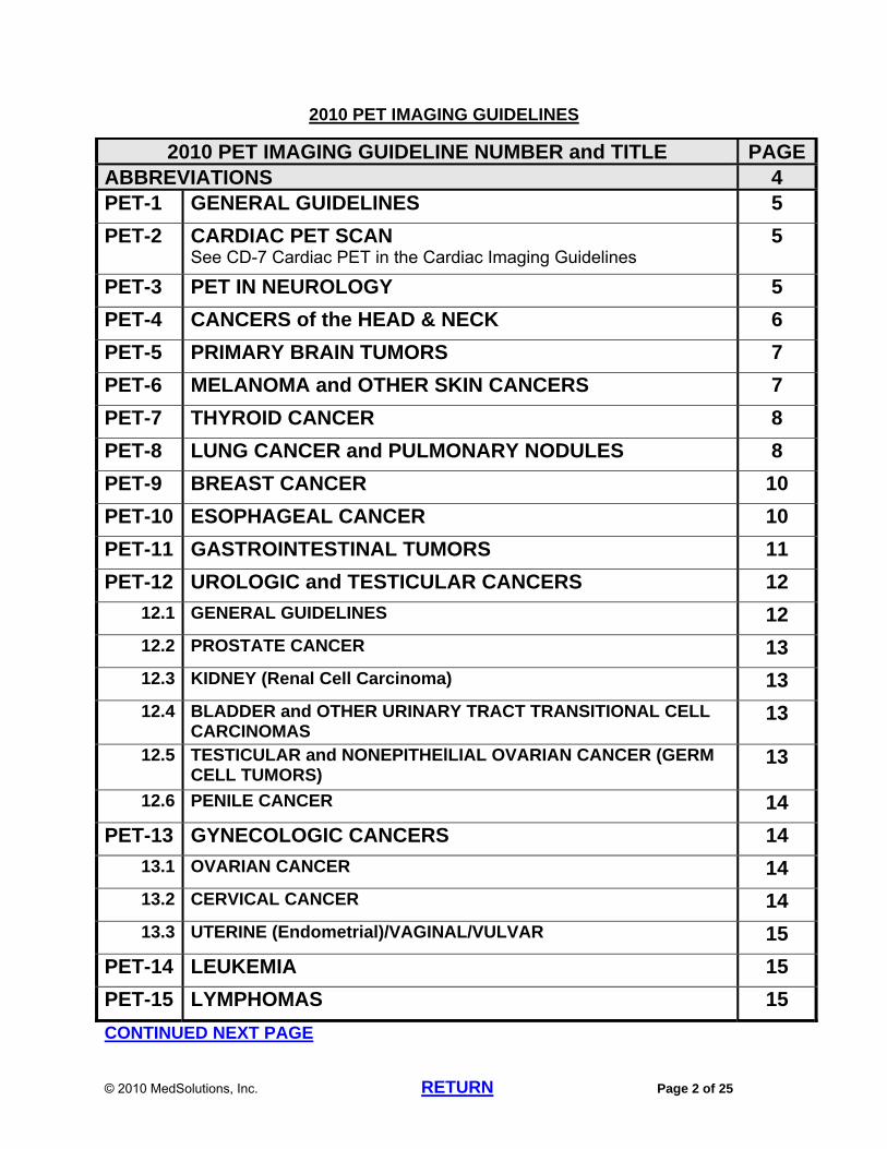
2010 PET IMAGING GUIDELINES
2010 PET IMAGING GUIDELINE NUMBER and TITLE PAGEABBREVIATIONS 4 PET-1 GENERAL GUIDELINES 5
PET-2 CARDIAC PET SCAN See CD-7 Cardiac PET in the Cardiac Imaging Guidelines
5
PET-3 PET IN NEUROLOGY 5
PET-4 CANCERS of the HEAD & NECK 6
PET-5 PRIMARY BRAIN TUMORS 7
PET-6 MELANOMA and OTHER SKIN CANCERS 7
PET-7 THYROID CANCER 8
PET-8 LUNG CANCER and PULMONARY NODULES 8
PET-9 BREAST CANCER 10
PET-10 ESOPHAGEAL CANCER 10
PET-11 GASTROINTESTINAL TUMORS 11
PET-12 UROLOGIC and TESTICULAR CANCERS 12
12.1 GENERAL GUIDELINES 12
12.2 PROSTATE CANCER 13
12.3 KIDNEY (Renal Cell Carcinoma) 13
12.4 BLADDER and OTHER URINARY TRACT TRANSITIONAL CELL CARCINOMAS
13
12.5 TESTICULAR and NONEPITHElLIAL OVARIAN CANCER (GERM CELL TUMORS)
13
12.6 PENILE CANCER 14
PET-13 GYNECOLOGIC CANCERS 14
13.1 OVARIAN CANCER 14
13.2 CERVICAL CANCER 14
13.3 UTERINE (Endometrial)/VAGINAL/VULVAR 15
PET-14 LEUKEMIA 15
PET-15 LYMPHOMAS 15
CONTINUED NEXT PAGE

© 2010 MedSolutions, Inc. RETURN Page 3 of 25
2010 PET IMAGING GUIDELINES
2010 PET IMAGING GUIDELINE NUMBER and TITLE PAGEPET-16 MULTIPLE MYELOMA, WALDENSTROM’S
MACROGLOBULINEMIA, and PLASMACYTOMAS 17
PET-17 MEDICARE COVERAGE POLICIES 17
PET-18 OTHER PET GUIDELINES (Not Elsewhere Covered) 21
18.1 Carcinomas of Unknown Primary Site 21
18.2 Soft Tissue Carcinoma 21
18.3 Generalized Lymphadenopathy and Mediastinal Abnormalities 21
18.4 Liver Lesions 22
18.5 Adrenal Lesions and Neuroendocrine Lesions 22
PET GUIDELINE REFERENCES 23

© 2010 MedSolutions, Inc. RETURN Page 4 of 25
ABBREVIATIONS for PET IMAGING GUIDELINES
CA-125 cancer antigen 125 test
CEA carcinoembryonic antigen
CNS central nervous system
CT computed tomography
ESR erythrocyte sedimentation rate
FDG fluorodeoxyglucose
GI gastrointestinal
GIST gastrointestinal stromal tumor
LFT liver function tests
MALT Mucosa-Associated Lymphoid Tissue (rare form of Non-Hodgkin’s lymphoma)
MGUS Monoclonal Gammopathy of Unknown Significance
MIBG I-123 metaiodobenzylguanidine scintigraphy
MM multiple myeloma
NHL Non-Hodgkin’s lymphoma
NSCLC Non-Small Cell Lung Cancer
NSGCT Non Seminomatous Germ Cell Tumor
PET positron emission tomography
PSA prostate specific antigen
RCC renal cell carcinoma

© 2010 MedSolutions, Inc. RETURN Page 5 of 25
PET IMAGING GUIDELINES
PET-1~GENERAL GUIDELINES The usefulness of PET is now well established in many cardiac, neurological, and
oncologic situations. All of the indications for PET also apply to PET/CT fusion scan.
o In general, the anatomic detail acquired in PET/CT is reasonable for the evaluation of many oncologic conditions; however, the diagnostic quality may be inconsistent.
o For initial diagnosis or staging, a diagnostic CT may be appropriate in addition to a PET/CT.
o For restaging, therapy monitoring, and evaluation of recurrence, either PET/CT or diagnostic CT, but not both, should be chosen by the clinician as the initial imaging modality.
For Oncologic applications, the skull base to mid-femur (“eyes-to thighs”) procedure code for PET (CPT®78812 or CPT®78815) is usually the most appropriate procedure to order. o Exceptions (use CPT®78813 or CPT®78816).include the following: Malignant melanoma Some unusual presentations of sarcomas and lymphomas Pediatric malignancies in pre-adolescent children
PET is a poor choice for imaging metastatic disease in the central nervous system (CNS).
PET is unreliable for imaging lesions less than 7 mm in size. PET is inappropriate for use as a surveillance test in the absence of clinical or other
imaging evidence to suggest possible recurrence. PET guidelines for cancers should be applied in conjunction with the corresponding
Oncology guidelines (ONC-1 through ONC 28).
PET-2~CARDIAC PET SCAN See CD-7 Cardiac PET Scan in the Cardiac Imaging Guidelines.
PET-3~PET IN NEUROLOGY See in the Head Imaging Guidelines:
o HD-1 General Guidelines o HD-13 Dementia o HD-14 Adult Epilepsy/Seizure o HD-21 Movement Disorders o HD-24.13 PET in brain tumor
See in the Pediatric and Congenital Head Imaging Guidelines: o PACHD-5 Pediatric Epilepsy/Seizure
© 2010 MedSolutions, Inc. RETURN Page 6 of 25
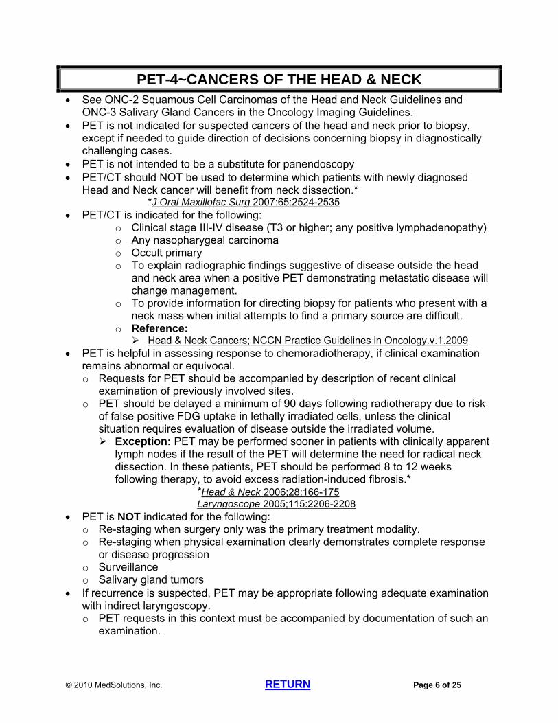
PET-4~CANCERS OF THE HEAD & NECK See ONC-2 Squamous Cell Carcinomas of the Head and Neck Guidelines and
ONC-3 Salivary Gland Cancers in the Oncology Imaging Guidelines. PET is not indicated for suspected cancers of the head and neck prior to biopsy,
except if needed to guide direction of decisions concerning biopsy in diagnostically challenging cases.
PET is not intended to be a substitute for panendoscopy PET/CT should NOT be used to determine which patients with newly diagnosed
Head and Neck cancer will benefit from neck dissection.* *J Oral Maxillofac Surg 2007:65:2524-2535
PET/CT is indicated for the following: o Clinical stage III-IV disease (T3 or higher; any positive lymphadenopathy) o Any nasopharygeal carcinoma o Occult primary o To explain radiographic findings suggestive of disease outside the head
and neck area when a positive PET demonstrating metastatic disease will change management.
o To provide information for directing biopsy for patients who present with a neck mass when initial attempts to find a primary source are difficult.
o Reference: Head & Neck Cancers; NCCN Practice Guidelines in Oncology.v.1.2009
PET is helpful in assessing response to chemoradiotherapy, if clinical examination remains abnormal or equivocal. o Requests for PET should be accompanied by description of recent clinical
examination of previously involved sites. o PET should be delayed a minimum of 90 days following radiotherapy due to risk
of false positive FDG uptake in lethally irradiated cells, unless the clinical situation requires evaluation of disease outside the irradiated volume. Exception: PET may be performed sooner in patients with clinically apparent
lymph nodes if the result of the PET will determine the need for radical neck dissection. In these patients, PET should be performed 8 to 12 weeks following therapy, to avoid excess radiation-induced fibrosis.*
*Head & Neck 2006;28:166-175 Laryngoscope 2005;115:2206-2208
PET is NOT indicated for the following: o Re-staging when surgery only was the primary treatment modality. o Re-staging when physical examination clearly demonstrates complete response
or disease progression o Surveillance o Salivary gland tumors
If recurrence is suspected, PET may be appropriate following adequate examination with indirect laryngoscopy. o PET requests in this context must be accompanied by documentation of such an
examination.

© 2010 MedSolutions, Inc. RETURN Page 7 of 25
o Otherwise, recurrence must be confirmed by biopsy prior to consideration of advanced imaging
PET-5~PRIMARY BRAIN TUMORS PET, Brain Metabolic (CPT®78608) is not indicated in the detection or initial work-up
of primary brain tumors. o A rare exception to this is in distinguishing high-grade from low-grade gliomas,
either due to indeterminate histology by biopsy, or because the lesion is in a surgically inaccessible location of the brain, such as the brain stem.
PET Brain Metabolic (CPT®78608) may be helpful in distinguishing tumor from radiation necrosis when recurrent disease is suspected and other imaging modalities are indeterminate (see HD-24.13 PET in brain tumor in the Head Guidelines). These cases should be sent for Medical Director review.
PET/CT (CPT®78815) can be considered if findings of a brain biopsy or resection suggest a lesion is a metastasis from an unknown primary. o See ONC-4 Central Nervous System Cancers and ONC-27.7 Carcinoma of
Unknown Primary Site in the Oncology Imaging Guidelines
PET-6~MELANOMA and OTHER SKIN CANCERS See ONC-5 Melanomas and Skin Cancers in the Oncology Imaging Guidelines. MELANOMA
o PET is indicated in the initial evaluation of Stage II or greater (lesions greater than 1 mm thick with ulceration, lesions greater than 2 mm thick with no ulceration, obvious clinical lymphadenopathy, or lymphatic disease confirmed histologically) and in patients with recurrent disease, except when widespread metastatic disease has already been documented on other imaging modalities. PET is indicated when disease is initially found in a lymph node or
distant organ and the original site of the disease remains occult PET (CPT®78813 or CPT®78816) may be considered to address
specific signs and symptoms not explained by conventional imaging PET (CPT®78813 or CPT®78816) can be performed for initial staging
for orbital/ocular melanoma. o In patients with known Stage IV (metastatic) disease, PET is appropriate only
when needed to rule in or rule out involvement of a specific organ, if information from the study will change clinical management decisions.
o PET is not indicated for Stage I disease (lesions less than 2 mm thick if no ulceration and without lymphadenopathy or any lesion less than or equal to 1 mm without lymphadenopathy).*
*Current Op in Oncol 2005;17:154-159 *Clin Nuc Med 2003;28:961-965
o PET is not indicated for surveillance. OTHER SKIN CANCERS
o Merkel Cell Carcinoma
© 2010 MedSolutions, Inc. RETURN Page 8 of 25
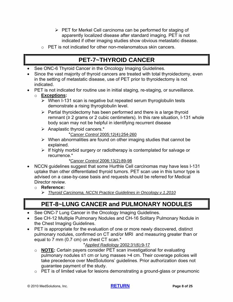
PET for Merkel Cell carcinoma can be performed for staging of apparently localized disease after standard imaging. PET is not indicated if other imaging studies show obvious metastatic disease.
o PET is not indicated for other non-melanomatous skin cancers.
PET-7~THYROID CANCER See ONC-6 Thyroid Cancer in the Oncology Imaging Guidelines. Since the vast majority of thyroid cancers are treated with total thyroidectomy, even
in the setting of metastatic disease, use of PET prior to thyroidectomy is not indicated.
PET is not indicated for routine use in initial staging, re-staging, or surveillance. o Exceptions: When I-131 scan is negative but repeated serum thyroglobulin tests
demonstrate a rising thyroglobulin level. Partial thyroidectomy has been performed and there is a large thyroid
remnant (≥ 2 grams or 2 cubic centimeters). In this rare situation, I-131 whole body scan may not be helpful in identifying recurrent disease
Anaplastic thyroid cancers.* *Cancer Control 2005;12(4):254-260
When abnormalities are found on other imaging studies that cannot be explained.
If highly morbid surgery or radiotherapy is contemplated for salvage or recurrence.*
*Cancer Control 2006;13(2):89-98 NCCN guidelines suggest that some Hurthle Cell carcinomas may have less I-131
uptake than other differentiated thyroid tumors. PET scan use in this tumor type is advised on a case-by-case basis and requests should be referred for Medical Director review. o Reference: Thyroid Carcinoma, NCCN Practice Guidelines in Oncology.v.1.2010
PET-8~LUNG CANCER and PULMONARY NODULES See ONC-7 Lung Cancer in the Oncology Imaging Guidelines. See CH-12 Multiple Pulmonary Nodules and CH-16 Solitary Pulmonary Nodule in
the Chest Imaging Guidelines. PET is appropriate for the evaluation of one or more newly discovered, distinct
pulmonary nodules, confirmed on CT and/or MRI and measuring greater than or equal to 7 mm (0.7 cm) on chest CT scan.*
*Applied Radiology 2002;31(6):9-17 o NOTE: Certain payers consider PET scan investigational for evaluating
pulmonary nodules ≤1 cm or lung masses >4 cm. Their coverage policies will take precedence over MedSolutions’ guidelines. Prior authorization does not guarantee payment of the study.
o PET is of limited value for lesions demonstrating a ground-glass or pneumonic

© 2010 MedSolutions, Inc. RETURN Page 9 of 25
(pneumonia-like) appearance. o If PET scan is negative, chest CT should be performed at 3, 6, 12, and 24
months. Serial PET scans to evaluate lung nodules are not appropriate.
PET can be obtained for the initial staging of patients with histologically proven lung cancer in the absence of evidence for Stage IV disease. PET is especially helpful in evaluating mediastinal lymphadenopathy.*
*Lung Cancer, 2008 April;60:62-68 o Exceptions are bronchioalveolar carcinoma, bronchial carcinoid, and mucinous
carcinoma, as these histologies may not take up sufficient FDG to warrant the use of PET.
o PET for small cell carcinoma can be performed for initial staging of apparently limited stage disease after standard imaging. PET is not indicated if other imaging studies show extensive stage
disease. PET is not indicated following chemotherapy
PET is not indicated for re-staging of lung cancer, except as noted below. o PET following radiotherapy should be delayed a minimum of 12 weeks, due to
risk of false positive FDG uptake in lethally irradiated cells and in radiation pneumonitis, unless the clinical situation requires evaluation of disease outside the irradiated volume.
o PET can differentiate persistent or recurrent tumor from necrotic or fibrotic tissue following chemotherapy or radiotherapy.
o PET is useful for evaluation of abnormalities that are newly discovered by CT or other imaging modalities used for re-staging.
o PET may help differentiate persistent or recurrent tumor from radiation- induced fibrosis or pleural thickening.
PET is inappropriate for re-staging if surgery was the primary treatment modality and all known tumor was resected, or for tumors initially treated with radiation therapy as the only treatment modality.
PET is inappropriate for re-staging small cell carcinomas. PET may be considered if LFTs or tumor markers become elevated and CT scans
are negative or equivocal. PET is inappropriate for routine surveillance.

© 2010 MedSolutions, Inc. RETURN Page 10 of 25
PET-9~BREAST CANCER See ONC-10 Breast Cancer in the Oncology Imaging Guidelines. PET is indicated for the following:
o Staging evaluation of unresectable clinical Stage III, and Stage IV disease (locally advanced, or limited metastatic disease). This includes patients with inflammatory breast cancer and patients with breast cancers greater than 5 cm.
o When lymph node disease is found in four or more axillary lymph nodes o When axillary lymph nodes are fixed to one another or to other structures o When disease is found in lymph node sites other than the axilla. o Prior to neoadjuvant chemotherapy for locally advanced disease. o To document response to therapy in Stage IV disease when identification of
responders vs non-responders will influence future therapy decisions. Repeated use of PET in Stage IV disease is of unproven benefit.
o Evaluation of a patient with recurrence documented by other imaging, elevations of laboratory tests, or histologic confirmation.
PET following radiotherapy is usually not indicated. In select cases when PET is performed following radiotherapy, the PET scan should be delayed a minimum of 12 weeks due to risk of false positive FDG uptake in lethally irradiated cells, unless the clinical situation requires evaluation of disease outside the irradiated volume.
PET is NOT indicated for the following: o Non-invasive breast cancers o Prior to lymph node sampling in a patient with clinical Stage I, II, or operable
IIIa disease.* *Invasive Breast Cancer. NCCN Practice Guidelines in Oncology.v.1.2010
o Obvious multi-organ metastatic disease is present o Preoperative assessment of response after neoadjuvant chemotherapy
Bone scan is the initial study of choice for breast cancer patients with bone pain. o In some circumstances, PET may be indicated if additional information about
organ systems other than skeletal is clinically relevant.* *AJR 2005;184:1266-1273
PET-10~ESOPHAGEAL CANCER See ONC-8 Esophageal Cancer in the Oncology Imaging Guidelines. PET is usually contraindicated prior to biopsy; however, when other imaging
modalities yield conflicting information, PET may be considered to evaluate the feasibility of an approach to biopsy.
PET is indicated for staging, but may be omitted if conventional imaging finds clear evidence of metastatic disease*
*Esophageal Cancer, NCCN Practice Guidelines in Oncology. v.1 PET is appropriate for the evaluation of response to radiation or chemotherapy:

© 2010 MedSolutions, Inc. RETURN Page 11 of 25
o Upper endoscopy or endoscopic ultrasound (EUS) should be performed prior to PET, since evidence of obvious progression or complete response negates the need for PET
o PET should be delayed as much as feasible to allow time for tumor response to be assessed but not so late as to unduly delay required surgery.
o If surgery is not an option, PET following radiotherapy should be delayed a minimum of 12 weeks, due to risk of false positive FDG uptake in lethally irradiated cells, unless the clinical situation requires evaluation of disease outside the irradiated volume.
PET is not appropriate for patients who are receiving supportive care only References:
Lancet Oncology 2007;8:797-805 N Engl J Med 2006;355:11-20
PET-11~GASTROINTESTINAL TUMORS See ONC-13 Upper GI Cancers, ONC-14 Other GI Neuroendocrine Cancers, and
ONC-15 Colorectal Cancer in the Oncology Imaging Guidelines. Histologic confirmation of malignancy should be obtained prior to considering PET
scan. PET is inappropriate for non-invasive carcinomas, carcinomas contained within a
polyp, or for any completely resected, lymph node negative colon cancer.
PET is indicated for the initial staging of: o Adenocarcinoma of the stomach*
*Gastric Cancer. NCCN Practice Guidelines in Oncology v.1.2010 o Pancreatic cancers under consideration for resection by Whipple procedure, or
subtotal pancreatectomy of pancreatic tail tumors* *Annals of Surgery 2005;242(2):235-243
o Any GI tumor where a solitary metastatic lesion is detected in a site where aggressive local therapy of the primary site and of the solitary metastasis can confer a survival advantage.
o Any GI tumor with an elevated CEA preoperatively, and the CEA fails to normalize after apparently curative resection.
o Any GI tumor where pathologic diagnosis suggests a primary site other than the GI system, and a search for a second primary site is reasonable.
o Malignant and non-adrenal pheochromocytomas, and any symptomatic neuroendocrine tumor when an apparently complete resection fails to resolve secretion of pathologic levels of hormones or neurotransmitter compounds, if functional nuclear imaging (MIBG or Octreoscan) is negative.*
*Ann NY Acad Sci 2004;1018:495-504 Current Opinions Gastroenterology 2007;23(1):74-78
o Initial staging of Anal Canal Squamous Cell Carcinomas o Restaging of Anal Canal Carcinomas, if the initial staging study was PET avid
and not easily evaluated on other imaging tests or by physical examination. PET is not indicated for Anal Margin Carcinomas.
© 2010 MedSolutions, Inc. RETURN Page 12 of 25
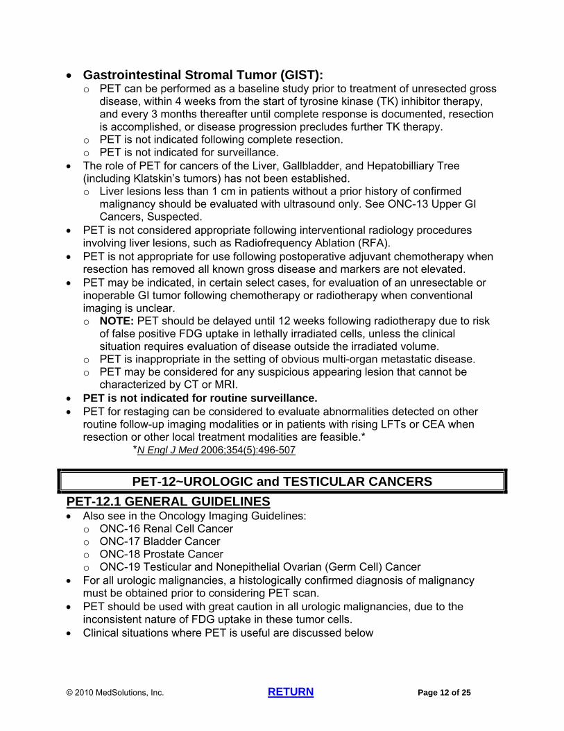
Gastrointestinal Stromal Tumor (GIST): o PET can be performed as a baseline study prior to treatment of unresected gross
disease, within 4 weeks from the start of tyrosine kinase (TK) inhibitor therapy, and every 3 months thereafter until complete response is documented, resection is accomplished, or disease progression precludes further TK therapy.
o PET is not indicated following complete resection. o PET is not indicated for surveillance.
The role of PET for cancers of the Liver, Gallbladder, and Hepatobilliary Tree (including Klatskin’s tumors) has not been established. o Liver lesions less than 1 cm in patients without a prior history of confirmed
malignancy should be evaluated with ultrasound only. See ONC-13 Upper GI Cancers, Suspected.
PET is not considered appropriate following interventional radiology procedures involving liver lesions, such as Radiofrequency Ablation (RFA).
PET is not appropriate for use following postoperative adjuvant chemotherapy when resection has removed all known gross disease and markers are not elevated.
PET may be indicated, in certain select cases, for evaluation of an unresectable or inoperable GI tumor following chemotherapy or radiotherapy when conventional imaging is unclear. o NOTE: PET should be delayed until 12 weeks following radiotherapy due to risk
of false positive FDG uptake in lethally irradiated cells, unless the clinical situation requires evaluation of disease outside the irradiated volume.
o PET is inappropriate in the setting of obvious multi-organ metastatic disease. o PET may be considered for any suspicious appearing lesion that cannot be
characterized by CT or MRI. PET is not indicated for routine surveillance. PET for restaging can be considered to evaluate abnormalities detected on other
routine follow-up imaging modalities or in patients with rising LFTs or CEA when resection or other local treatment modalities are feasible.*
*N Engl J Med 2006;354(5):496-507
PET-12~UROLOGIC and TESTICULAR CANCERS
PET-12.1 GENERAL GUIDELINES Also see in the Oncology Imaging Guidelines:
o ONC-16 Renal Cell Cancer o ONC-17 Bladder Cancer o ONC-18 Prostate Cancer o ONC-19 Testicular and Nonepithelial Ovarian (Germ Cell) Cancer
For all urologic malignancies, a histologically confirmed diagnosis of malignancy must be obtained prior to considering PET scan.
PET should be used with great caution in all urologic malignancies, due to the inconsistent nature of FDG uptake in these tumor cells.
Clinical situations where PET is useful are discussed below

© 2010 MedSolutions, Inc. RETURN Page 13 of 25
PET-12.2 PROSTATE CANCER: See ONC-18 Prostate Cancer in the Oncology Imaging Guidelines PET has not been found to be helpful in prostate cancer except in patients with
Hormone Refractory disease, a rising PSA, or a suspicious finding on another imaging modality following initially successful local therapy.
Bone Scan is the imaging modality of choice for detecting osseous metastases.* *Semin Nucl Med 2005:35(2):135-142
PET is unable to differentiate neoplastic disease from benign prostatic hypertrophy. Radiopharmaceuticals other than FDG seem to offer exciting potential in clinical
trials, but their use is considered investigational at this time.* *J. Urology 2006;176:954-960
PET-12.3 KIDNEY (Renal Cell Carcinoma): See ONC-16 Renal Cell Cancer in the Oncology Imaging Guidelines In general, PET is of limited value because of inconsistent uptake of FDG. PET is not indicated for initial diagnosis or staging of renal cell carcinoma.
o Exception: if there is suspicion of a metastatic lesion and biopsy of that lesion is considered potentially less invasive than biopsy of the kidney or performing nephrectomy, then PET may be appropriate.
PET is helpful for clarifying findings on other imaging modalities that are suspicious for, but not diagnostic of, metastatic disease, bilateral disease, or recurrence, particularly in settings where a positive PET will avoid an invasive biopsy. o PET, in conjunction with conventional imaging, is helpful in differentiating
possible osseous metastasis from benign bone lesions. o PET is helpful in differentiating local tumor recurrence from postoperative and/or
post-radiation changes in patients who have undergone complete nephrectomy. PET is not appropriate for routine surveillance in renal cell carcinoma.*
*J Urol 2004;171:1806-1809 PET-12.4 BLADDER AND OTHER URINARY TRACT TRANSITIONAL CELL CARCINOMAS: See ONC-17 Bladder Cancer in the Oncology Imaging Guidelines There is currently insufficient evidence to support the routine use of PET in
evaluating patients with transitional cell carcinomas of the bladder or other urinary tract sites.
PET-12.5 TESTICULAR AND NONEPITHELIAL OVARIAN CANCER (GERM CELL TUMORS): See ONC-19 Testicular and Nonepithelial Ovarian (Germ Cell) Cancer in the
Oncology Imaging Guidelines The following comments apply to testicular cancers, extragonadal germ cell tumors,
and germ cell tumors of the ovary. There is insufficient data to support the use of PET in the diagnosis, initial staging, or
routine surveillance of Testicular Cancers. PET may be considered in the evaluation of a germ cell tumor found in a
retroperitoneal and/or mediastinal mass, when testicular examination is negative.

© 2010 MedSolutions, Inc. RETURN Page 14 of 25
PET has been shown to be non-contributory for routine use in Non Seminomatous Germ Cell Tumors (NSGCT)*
*J Clin Oncol 2008;26:5930-5935 * Annals of Oncology Advance Access published May 16, 2008, doi:10.1093/annonc/mdn170
PET is indicated in a patient with advanced seminoma and a CT-documented residual mass after chemotherapy, to differentiate viable tumor from fibrosis or necrosis.*
*Eur J Radiol 2005;54(2):284-288 o PET is not appropriate for re-staging if CT is negative. o If used following radiation therapy, PET should be delayed until 12 weeks after
completion of therapy due to risk of false positive FDG uptake in lethally irradiated cells, unless the clinical situation requires evaluation of disease outside the irradiated volume.
Most testicular cancers are very chemo and radio-sensitive, therefore, conventional CT is sufficient for evaluating response to therapy.
PET can be helpful, in conjunction with conventional imaging, in evaluating patients with germ cell tumors who are found to have rising tumor markers following potentially curative therapies.
PET-12.6 PENILE CANCER See ONC-23 Cancers of the External Genitalia in the Oncology Imaging Guidelines. PET is not indicated in this disease.
PET-13~GYNECOLOGIC CANCERS PET-13.1 OVARIAN CANCER: See ONC-20 Ovarian Cancer in the Oncology Imaging Guidelines PET is not indicated for initial work-up of Ovarian Cancers except for cases of
primary peritoneal disease where the pelvic structures are normal. PET is not indicated for surveillance of Ovarian Cancers. Following complete resection, PET may be considered for the following:
o Patients with elevated CA-125 or other relevant tumor markers, and/or changes in physical examination, with normal conventional imaging.
o Evaluation of radiographic abnormalities suspicious for recurrence, or for elevation of LFTs when abdominal imaging is negative.
Repeat PET scans can be considered if prior conventional imaging failed to demonstrate tumor, or if a persistent radiographic mass is seen, in order to document response to therapy.
PET-13.2 CERVICAL CANCER: See ONC-22 Cervix Cancer in the Oncology Imaging Guidelines PET is indicated for the evaluation of newly diagnosed cervical cancers that are
clinical Stage IB2 or higher (>4 cm or invasion beyond uterus, positive lymph nodes, or distant metastasis).*
*Radiology 2006;238(1):272-279 o PET is not indicated in patients with non-invasive cervical cancer.

© 2010 MedSolutions, Inc. RETURN Page 15 of 25
o PET can be considered to evaluate abnormalities seen on other imaging modalities in lower stage invasive cancers, and for cancers incidentally discovered during hysterectomy.
PET is indicated following radiation therapy for advanced disease, but should be delayed for 12 weeks from completion of therapy, due to risk of false positive FDG uptake in lethally irradiated cells, unless the clinical situation requires evaluation of disease outside the irradiated volume.*
*JAMA 2007;298(19):2289-2295 PET is indicated for defining and staging recurrent disease when suspected by
physical examination or other imaging. PET-13.3 UTERINE (Endometrial)/VAGINAL/VULVAR: See ONC-21 Uterine Cancer and ONC-23 Anal Cancer, Vaginal Cancer, and
Cancers of the External Genitalia in the Oncology Imaging Guidelines PET is currently not indicated for these cancers.
PET-14~LEUKEMIA See ONC-25 Leukemia in the Oncology Imaging Guidelines There is currently no routine indication for PET in the evaluation of leukemia. Richter’s transformation is a rare and aggressive type of leukemia that results from a
transformation of Chronic Lymphocytic Leukemia (CLL) into diffuse large cell lymphoma.
o Diagnosis is made based on microscopic examination of blood cells and by bone marrow biopsy.
o PET/CT may be considered in selected cases. These requests should be sent for Medical Director Review
PET-15~LYMPHOMAS See ONC-26 Lymphomas in the Oncology Imaging Guidelines All imaging requests must clearly document the diagnosis with the cell subtype of
lymphoma which is being evaluated. PET is inappropriate prior to histologic confirmation of cell type, except as discussed
under Generalized Lymphadenopathy in PET-18~Other PET Guidelines (Not Elsewhere Covered).
PET is helpful in the setting of most Lymphomas for initial staging, except for indolent Non-Hodgkin’s lymphomas, including Chronic Lymphocytic Leukemia/Small Lymphocytic Lymphoma (CLL/SLL), Gastric MALT lymphomas, and Low-grade Follicular
o If the lymphoma is an appropriate cell type for PET, both PET and CT scans with contrast as outlined in ONC-26 Lymphomas in the Oncology Imaging Guidelines can be performed for initial staging.
For the initial chemotherapy regimens for Hodgkin’s and many Non-Hodgkin’s Lymphomas (NHL), an “interim” re-staging PET study is suggested to evaluate

© 2010 MedSolutions, Inc. RETURN Page 16 of 25
response after 4 cycles, and a “final” re-staging PET is suggested when all therapies are complete. o For interim re-staging, either CT scans with contrast of body areas previously
positive or PET scan (but not both) can be performed. See Re-staging/Recurrence section of ONC-26 Lymphomas in the
Oncology guidelines for full guidelines regarding interim re-staging o For final re-staging, either CT scans with contrast of body areas previously
positive or PET scan (but not both) can be performed. Both PET and CT scans with contrast of areas previously involved can be
performed for final re-staging after therapy is completed if one of the following circumstances applies: Radiation therapy is planned in a patient who initially presented with
bulky mediastinal adenopathy. PET only was used for interim re-staging and variable FDG uptake by
the tumor occurred, and/or FDG activity cleared but an enlarged abnormality remains.
See Re-staging/Recurrence section of ONC-26 Lymphomas in the Oncology Imaging Guidelines for full guidelines regarding final re-staging
o Most patients will not require any more than these two restaging studies. o Interim PET is not indicated when there is no stated change in management that
will be impacted by the results of the scans. o Once PET becomes negative, additional PET scanning is discouraged.
PET is useful for detection of recurrence: o When recurrence is known or suspected based upon physical examination,
laboratory studies, or conventional imaging in a patient with a history of Hodgkin’s lymphoma or history of an aggressive or intermediate sub-type of Non-Hodgkin’s lymphoma. Both PET and CT chest/abdomen/pelvis with contrast
(CPT®71260/74160/72193) can be performed for evaluation of a recurrence of lymphoma that was previously in remission, especially if transformation to a more aggressive type is suspected.
o To confirm absence of lymphoma in a radiographically persistent CT abnormality. o When repeated lab tests such as LFT’s or ESR become elevated after previously
being normalized, and CT scans are negative. PET is not indicated for routine surveillance.
o CT scans only should be used for surveillance once an acceptable response to therapy has been documented. This includes patients under “maintenance” therapies such as Rituxan

© 2010 MedSolutions, Inc. RETURN Page 17 of 25
PET-16~MULTIPLE MYELOMA, WALDENSTROM’S MACROGLOBULINEMIA, and PLASMACYTOMAS
See ONC-24 Multiple Myeloma, Waldenstrom’s Macroglobulinemia, and Plasmacytomas in the Oncology Imaging Guidelines
As a rule, PET is not indicated in patients who clearly have MGUS or clearly have Stage III Myeloma.
PET (CPT®78812 or CPT®78815) may be used to ensure that an apparently solitary plasmacytoma is truly solitary.
PET may be indicated if lab studies suggest that an MGUS patient has possible progression to a more malignant form of disease, or for unusual signs, symptoms, or unexplained radiographic abnormalities.
PET is useful to ensure that a patient with less than “full-blown” MM, (Stage I or II, or so-called “smoldering” myeloma) may be safely observed.
PET is useful to evaluate for extraosseous plasmacytomas, if clinically suspected. PET may be helpful in certain cases of refractory disease, to aid in determining
additional therapies. PET is not indicated for Full Stage (Stage III) multiple myeloma, when standard
imaging and lab tests can define extent of disease and response to therapy.* *Multiple Myeloma. NCCN Practice Guidelines in Oncology. v.2.2010 *Radiology 2004; 231:11-23
PET-17~MEDICARE COVERAGE POLICIES
For Medicare and Medicare Advantage patients, the coverage policies of CMS (Centers for Medicare and Medicaid Services) will take precedence over MedSolutions’ guidelines. Prior authorization does not guarantee payment of the study.
For PET requests that are not covered by CMS, MedSolutions will review the request based upon the payer’s coverage policy and MedSolutions imaging guidelines.
The NATIONAL ONCOLOGIC PET REGISTRY (NOPR) o In 2009, CMS expanded coverage for PET to many cancer types.
MedSolutions will authorize PET imaging for Medicare and Medicare Advantage patients per CMS’ coverage policy (see list below from the NOPR website http://www.cancerpetregistry.org/index.htm )
o MedSolutions may elect to authorize certain requests as medically necessary; however, most NOPR eligible studies will not meet MedSolutions’ appropriateness criteria. If the patient is enrolled and a PET scan is performed as part of a
NOPR approved study, the PET scan can be retrospectively authorized, pending receipt by MedSolutions of a copy of the appropriate Case Completion Notification (CCN).
See the NOPR website http://www.cancerpetregistry.org/index.htm for additional information

© 2010 MedSolutions, Inc. RETURN Page 18 of 25
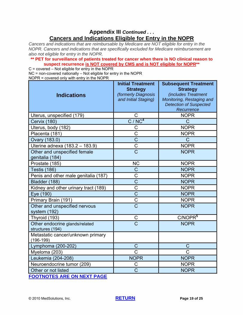
Appendix III Cancers and Indications Eligible for Entry in the NOPR
Cancers and indications that are reimbursable by Medicare are NOT eligible for entry in the NOPR. Cancers and indications that are specifically excluded for Medicare reimbursement are also not eligible for entry in the NOPR.
** PET for surveillance of patients treated for cancer when there is NO clinical reason to suspect recurrence is NOT covered by CMS and is NOT eligible for NOPR**
C = covered – Not eligible for entry in the NOPR NC = non-covered nationally – Not eligible for entry in the NOPR NOPR = covered only with entry in the NOPR
Indications
Initial Treatment Strategy
(formerly Diagnosis and Initial Staging)
Subsequent Treatment Strategy
(includes Treatment Monitoring, Restaging and
Detection of Suspected Recurrence
Lip, Oral Cavity, and Pharynx (140-149)
C C
Esophagus (150) C C Stomach (151) C NOPR Small Intestine (152) C NOPR Colon (153) and Rectum (154) C C Anus (154) C NOPR1
Liver and intrahepatic bile ducts (156) C NOPR Gallbladder & extrahepatic bile ducts (156)
C NOPR
Pancreas (157) C NOPR Retroperitoneum and peritoneum (158)
C NOPR
Nasal Cavity, ear, and sinuses (160) C C Larynx (161) C C Lung, non-small cell (162) C C Lung, small cell (162) C NOPR Pleura (163) C NOPR Thymus, heart, mediastinum (164) C NOPR Bone/cartilage (170) C NOPR Connective/ other soft tissue (171) C NOPR Melanoma (172) C / NC2 C Non-melanoma skin (173) C NOPR Female breast (174) C / NC2,3 C Male breast (175) C / NC2,3 C Kaposi’s sarcoma (176) C NOPR
APPENDIX III CONTINUED ON NEXT PAGE
© 2010 MedSolutions, Inc. RETURN Page 19 of 25
Appendix III Continued . . . Cancers and Indications Eligible for Entry in the NOPR
Cancers and indications that are reimbursable by Medicare are NOT eligible for entry in the NOPR. Cancers and indications that are specifically excluded for Medicare reimbursement are also not eligible for entry in the NOPR. ** PET for surveillance of patients treated for cancer when there is NO clinical reason to
suspect recurrence is NOT covered by CMS and is NOT eligible for NOPR** C = covered – Not eligible for entry in the NOPR NC = non-covered nationally – Not eligible for entry in the NOPR NOPR = covered only with entry in the NOPR
Indications
Initial Treatment Strategy
(formerly Diagnosis and Initial Staging)
Subsequent Treatment Strategy
(includes Treatment Monitoring, Restaging and
Detection of Suspected Recurrence
Uterus, unspecified (179) C NOPR Cervix (180) C / NC4 C Uterus, body (182) C NOPR Placenta (181) C NOPR Ovary (183.0) C C Uterine adnexa (183.2 – 183.9) C NOPR Other and unspecified female genitalia (184)
C NOPR
Prostate (185) NC NOPR Testis (186) C NOPR Penis and other male genitalia (187) C NOPR Bladder (188) C NOPR Kidney and other urinary tract (189) C NOPR Eye (190) C NOPR Primary Brain (191) C NOPR Other and unspecified nervous system (192)
C NOPR
Thyroid (193) C C/NOPR5
Other endocrine glands/related structures (194)
C NOPR
Metastatic cancer/unknown primary (196-199)
Lymphoma (200-202) C C Myeloma (203) C C Leukemia (204-208) NOPR NOPR Neuroendocrine tumor (209) C NOPR Other or not listed C NOPR
FOOTNOTES ARE ON NEXT PAGE

© 2010 MedSolutions, Inc. RETURN Page 20 of 25
NOTES 1 Some Medicare contractors include anal cancer in their local coverage of “colorectal cancer”; for PET facilities served by those carriers, PET for subsequent treatment evaluation of anal cancer would be a covered indication. 2 PET is non-covered for initial staging of axillary lymph nodes in patients with breast cancer and of regional lymph nodes in patients with melanoma, but is covered for detection of distant metastatic disease in high-risk patients with breast cancer or melanoma. 3 PET is non-covered for “diagnosis” of breast cancer to evaluate a suspicious breast mass. However, PET is covered for initial treatment strategy evaluation of a patient with axillary nodal metastasis of unknown primary origin or in a patient with a paraneoplastic syndrome potentially caused by an occult breast cancer. 4 PET is non-covered for “diagnosis” of cervical cancer. PET is covered for initial staging of biopsy proven cervical cancer. 5 To qualify as a covered indication for subsequent treatment strategy evaluation, thyroid cancer must be of follicular cell origin and been previously treated by thyroidectomy and radioiodine ablation and the patient must have a serum thyroglobulin > 10 ng/mL and a negative whole-body I-131 scan. Patients who do not qualify for this covered indication (e.g., because the tumor is of other than follicular cell origin, the thyroglobulin is not elevated, or I-131 whole-body imaging was not performed or is positive) can be entered on NOPR.
© 2010 MedSolutions, Inc. RETURN Page 21 of 25
PET-18~OTHER PET GUIDELINES (NOT ELSEWHERE COVERED)
PET-18.1 Carcinomas of Unknown Primary Site See ONC-27.7 Carcinoma of Unknown Primary Site in the Oncology Guidelines
o PET/CT (CPT®78815, or CPT®78816 if melanoma) is appropriate if CT scans of chest/abdomen/pelvis with contrast have failed to demonstrate the site of the primary
o If PET demonstrates the primary site, then subsequent imaging decisions should be made according to guidelines for that site.
*ACR Practice Guideline for Performing FDG-PET/CT in Oncology 2007 *Occult Primary. NCCN Practice Guidelines in Oncology v.1.2010
PET-18.2 Soft Tissue Sarcoma See ONC-11 Sarcoma in the Oncology Imaging Guidelines PET is generally not indicated for most soft tissue sarcomas.
o Exceptions: For patients with high grade sarcomas who have abnormalities found on other
imaging studies or on physical examination, PET can be used to solve specific clinical questions or problems.*
*ACR Appropriateness Criteria: Follow-up of Malignant or Aggressive Musculoskeletal Tumors, 2008 *Clinical Nuclear Medicine 2006 Dec;31(12):754-760
To assist in determining the grade of an unresectable lesion, such as in the retroperitoneum, when the grade of the pathologic specimen is in doubt.
To assist in re-staging if needed to differentiate tumor from radiation or surgical fibrosis, or to determine response to therapy
Prior to resection of an apparent solitary metastasis. PET-18.3 Generalized Lymphadenopathy and Mediastinal Abnormalities: Also see CH-21 Mediastinal Lymphadenopathy in the Chest Imaging Guidelines Lymphadenopathy from neoplasms as well as benign sources of inflammation can
result in a positive PET scan. Therefore, the use of PET may not be helpful prior to histologic diagnosis. o Delaying biopsy of an accessible pathologic lymph node while awaiting the
results of imaging tests is usually ill-advised. Biopsy should proceed as quickly as feasible.
PET may be helpful when biopsy of a relatively inaccessible body region is contemplated, to confirm the likelihood of yielding a pathologic diagnosis and to determine if a more favorable site for biopsy exists.
PET may be helpful in characterizing anterior mediastinal abnormalities, especially since the thymus gland has a characteristic uptake pattern on most PET scans, and the study may differentiate normal or benign hypertrophic thymus tissue from pathologic mediastinal lesions.
© 2010 MedSolutions, Inc. RETURN Page 22 of 25
PET-18.4 Liver Lesions PET is indicated when a patient with a history of extrahepatic malignancy, with a cell
type known to be FDG-avid, has a new finding of a liver lesion greater than 1 cm, judged to be suspicious for malignancy.*
*ACR Appropriateness Criteria, Suspected Liver Metastases, 2008
PET-18.5 Adrenal Lesions and Neuroendocrine Lesions PET is considered inappropriate for the evaluation of adrenal masses prior to
histologic confirmation and for most applications for neuroendocrine tumors. PET can be useful for evaluation of an adrenal mass found on other imaging in a
patient with a prior diagnosis of a cancer that is predisposed to metastasize to the adrenal gland.
Reference: o AJR 2009;192:956-962
© 2010 MedSolutions, Inc. RETURN Page 23 of 25
PET GUIDELINE REFERENCES PET-1~General Guidelines
Neyman E, Kamel IR, Georgiades CS. Use of combined PET/CT imaging in evaluation of the solitary pulmonary nodule: principles, techniques, and pitfalls. Appl Radiol 2006;35(4):24-43.
PET-4~Cancers of the Head & Neck Nahmias C, Carlson ER, Duncan LD, et al. Positron emission
tomography/computerized tomography (PET/CT) scanning for preoperative staging of patients with oral/head and neck cancer. J Oral Maxillofac Surg 2007;65:2524-2535.
Head & Neck Cancers; NCCN Practice Guidelines in Oncology. v.1.2009. Pellitteri PK, Ferlito A, Rinaldo A, et al. Planned neck dissection following
chemoradiotherapy for advanced head and neck cancer: is it necessary for all? Head & Neck 2006 Feb;28:166-175.
Canning CA, Gubbels S, Chinn C, et al. Positron emission tomography scan to determine the need for neck dissection after chemoradiation for head and neck cancer: Timing is everything. Laryngoscope 2005;115:2206-2208.
PET-6~Melanoma Kumar R, Alavi A. Clinical applications of fluorodeoxyglucose-positron emission
tomography in the management of malignant melanoma. Current Op in Oncol 2005;17:154-159.
Gulec SA, Faries MB, Lee CC, et al. The role of fluorine-18 deoxyglucose positron emission tomography in the management of patients with metastatic melanoma: impact on surgical decision making. Clin Nuc Med 2003;28:961-965.
PET-7~Thyroid Cancer Khan N, Oriuchi N, Higuchi T, Endo K. Review of fluorine-18-2-fluoro-2-deoxy-D-
glucose positron emission tomography (FDG-PET) in the follow-up of medullary and anaplastic thyroid carcinomas. Cancer Control 2005 Oct;12(4):254-260.
Lansford CD, Teknos TN. Evaluation of the thyroid nodule. Cancer Control 2006 April;13(2):89-98.
Thyroid Carcinoma, NCCN Practice Guidelines in Oncology, v.1.2010. PET-8~Lung Cancer and Pulmonary Nodules
Line BR, Maragh MR, Ahamed T. Positron emission tomography imaging of lung and esophageal cancer. Applied Radiology 2002 June;31(6):9-17.
Al-Sarraf N, Gately K, Lucey J, et al. Lymph node staging by means of positron emission tomography is less accurate in non-small cell lung cancer patients with enlarged lymph nodes: Analysis of 1145 lymph nodes. Lung Cancer 2008 April;60:62-68.
PET-9~Breast Cancer Invasive Breast Cancer. NCCN Practice Guidelines in Oncology v.1.2010. Uematsu T, Yuen S, Yukisawa S, et al. Comparison of FDG PET and SPECT for
detection of bone metastases in breast cancer. AJR 2005;184:1266-1273.
PET-10~Esophageal Cancer Esophageal Cancer, NCCN Practice Guidelines in Oncology. v. 2010. Lordick F, Ott K, Krause B-J, et al. PET to assess early metabolic response and to
guide treatment of adenocarcinoma of the oesophagogastric junction: The MUNICON phase II trial. Lancet Oncology 2007;8:797-805.
© 2010 MedSolutions, Inc. RETURN Page 24 of 25
Cunningham D, Allum W, Stenning S, et al. Perioperative chemotherapy versus surgery alone for resectable gastroesophageal cancer. N Engl J Med 2006;355:11-20.
PET-11~Gastrointestinal Tumors Gastric Cancer. NCCN Practice Guidelines in Oncology v.1.2010. Heinrich S, Goerres GW, Schafer M, et al. Positron emission tomography/computed
tomography influences on the management of resectable pancreatic cancer and its cost-effectiveness. Annals of Surgery 2005;242(2):235-243.
Ilias I, Pacak K. Anatomical and functional imaging of metastatic pheochromocytoma. Ann NY Acad Sci 2004;1018:495-504.
Juweid ME, Cheson BD. Positron emission tomography and assessment of cancer therapy. N Engl J Med 2006;354(5):496-507.
PET-12~Urologic and Testicular Cancers Fogelman I, Cook G, Israel O, Van der Wall H. Positron emission tomography and
bone metastases. Semin Nucl Med 2005:35(2):135-142. Martorana G, Schiavina R, Corti B, et al. 11C-Choline positron emission
tomography/computed tomography for tumor localization of primary prostate cancer in comparison with 12-core biopsy. J. Urology 2006;176:954-960.
Kang DE, White RL Jr., Zuger JH, et al. Clinical use of fluorodeoxyglucose F18 positron emission tomography for detection of renal cell carcinoma. J Urol 2004;171:1806-1809.
Oechsle K, Hartmann M, Brenner W, et al. [18F] Fluorodeoxyglucose positron emission tomography in nonseminomatous germ cell tumors after chemotherapy: The German multicenter positron emission tomography study group. J Clin Oncol 2008;26:5930-5935
de Wit M, Brenner W, Hartmann M, et al. [18F]-FDG PET in clinical stage I/II non-seminomatous germ cell tumours: Results of the German multicentre trial. Annals of Oncology Advance Access published May 16, 2008, doi:10.1093/annonc/mdn170
Becherer A, De Santis M, Karanikas G, et al. FDG PET is superior to CT in the prediction of viable tumour in post-chemotherapy seminoma residuals. Eur J Radiol 2005;54(2):284-288.
PET-13~Gynecologic Cancers Sironi S, Buda A, Picchio M, et al. Lymph node metastasis in patients with clinical
early stage cervical cancer: detection with integrated FDG PET/CT. Radiology 2006 Jan;238(1):272-279.
Schwarz JK, Siegel BA, Dehdashti F, et al. Association of posttherapy positron emission tomography with tumor response and survival in cervical carcinoma. JAMA 2007;298(19):2289-2295.
PET-15~Lymphomas Kazama T, Faria SC, Varavithya V, et al. FDG PET in the evaluation of treatment for
lymphoma: clinical usefulness and pitfalls. RadioGraphics 2005;25:191-207. Blake MA, Slattery J, Sahani D, Kaira MK. Practical issues in abdominal PET/CT.
Applied Radiology 2005;34(11):8-18. PET-16~Multiple Myeloma, Waldenstrom’s Macroglobulinemia, and Plasmacytomas
Multiple Myeloma. NCCN Practice Guidelines in Oncology. v.2.2009. Angtuaco EJ, Fassas AB, Walker R, et al. Multiple myeloma: clinical review and
diagnostic imaging. Radiology 2004 April;231(1):11-23.
© 2010 MedSolutions, Inc. RETURN Page 25 of 25
PET-17~Medicare Coverage Policies http://www.cancerpetregistry.org/index.htm.
PET-18~Other PET Guidelines (Not Elsewhere Covered) ACR Practice Guideline for Performing FDG-PET/CT in Oncology 2007. Occult Primary. NCCN Practice Guidelines in Oncology v.1.2010. ACR Appropriateness Criteria: Follow-up of Malignant or Aggressive Musculoskeletal
Tumors, 2008. Iagaru, A, Quon A, McDougall IR, et al. F-18 FDG PET/CT evaluation of osseous
and soft tissue sarcomas. Clinical Nuclear Medicine 2006 Dec;31(12):754-760. ACR Appropriateness Criteria, Suspected Liver Metastases, 2008. Boland GWL, Blake MA, Holalkere NS, and Hahn PF. PET/CT for the
characterization of adrenal masses in patients with cancer: Qualitative versus quantitative accuracy in 150 consecutive patients. AJR 2009;192:956-962.