chest imaging guidelines 2011 - welcome to tmhp imagi… · © 2011 medsolutions, inc. chest...
TRANSCRIPT
© 2011 MedSolutions, Inc. Chest Imaging Guidelines Page 1 of 62
CHEST IMAGING GUIDELINES 2011 MedSolutions, Inc
MedSolutions, Inc. Clinical Decision Support Tool for Advanced Diagnostic Imaging
Common symptoms and symptom complexes are addressed by this tool. Imaging requests for patients with atypical symptoms or clinical presentations that are not specifically addressed will require physician review. Consultation with the referring physician may provide additional insight.
This version incorporates MSI accepted revisions prior to 7/22/11
MedSolutions, Inc. This tool addresses common symptoms and symptom complexes. Imaging requests for patients with atypicalClinical Decision Support Tool symptoms or clinical presentations that are not specifically addressed will require physician review. Diagnostic Strategies Consultation with the referring physician, specialist and/or patient’s Primary Care Physician (PCP) may provide additional insight.
© 2011 MedSolutions, Inc. RETURN Page 2 of 62
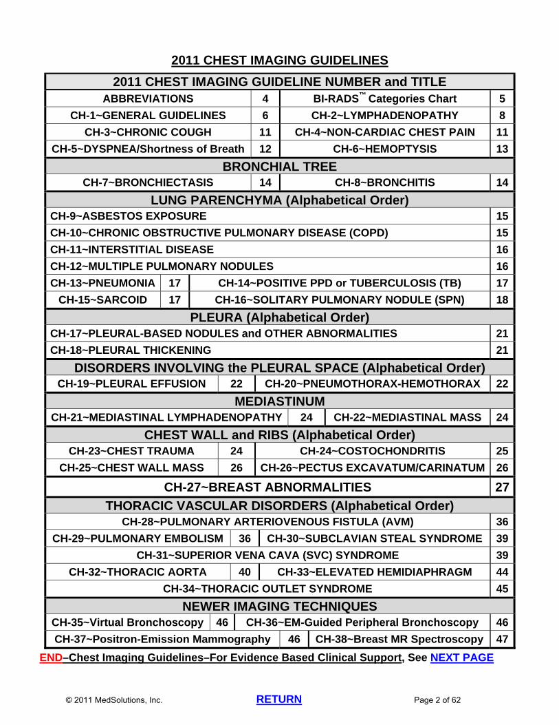
2011 CHEST IMAGING GUIDELINES
2011 CHEST IMAGING GUIDELINE NUMBER and TITLE ABBREVIATIONS 4 BI-RADS™ Categories Chart 5
CH-1~GENERAL GUIDELINES 6 CH-2~LYMPHADENOPATHY 8
CH-3~CHRONIC COUGH 11 CH-4~NON-CARDIAC CHEST PAIN 11
CH-5~DYSPNEA/Shortness of Breath 12 CH-6~HEMOPTYSIS 13
BRONCHIAL TREE CH-7~BRONCHIECTASIS 14 CH-8~BRONCHITIS 14
LUNG PARENCHYMA (Alphabetical Order) CH-9~ASBESTOS EXPOSURE 15
CH-10~CHRONIC OBSTRUCTIVE PULMONARY DISEASE (COPD) 15
CH-11~INTERSTITIAL DISEASE 16
CH-12~MULTIPLE PULMONARY NODULES 16
CH-13~PNEUMONIA 17 CH-14~POSITIVE PPD or TUBERCULOSIS (TB) 17
CH-15~SARCOID 17 CH-16~SOLITARY PULMONARY NODULE (SPN) 18
PLEURA (Alphabetical Order) CH-17~PLEURAL-BASED NODULES and OTHER ABNORMALITIES 21
CH-18~PLEURAL THICKENING 21
DISORDERS INVOLVING the PLEURAL SPACE (Alphabetical Order) CH-19~PLEURAL EFFUSION 22 CH-20~PNEUMOTHORAX-HEMOTHORAX 22
MEDIASTINUM CH-21~MEDIASTINAL LYMPHADENOPATHY 24 CH-22~MEDIASTINAL MASS 24
CHEST WALL and RIBS (Alphabetical Order) CH-23~CHEST TRAUMA 24 CH-24~COSTOCHONDRITIS 25
CH-25~CHEST WALL MASS 26 CH-26~PECTUS EXCAVATUM/CARINATUM 26
CH-27~BREAST ABNORMALITIES 27
THORACIC VASCULAR DISORDERS (Alphabetical Order) CH-28~PULMONARY ARTERIOVENOUS FISTULA (AVM) 36
CH-29~PULMONARY EMBOLISM 36 CH-30~SUBCLAVIAN STEAL SYNDROME 39
CH-31~SUPERIOR VENA CAVA (SVC) SYNDROME 39
CH-32~THORACIC AORTA 40 CH-33~ELEVATED HEMIDIAPHRAGM 44
CH-34~THORACIC OUTLET SYNDROME 45
NEWER IMAGING TECHNIQUES CH-35~Virtual Bronchoscopy 46 CH-36~EM-Guided Peripheral Bronchoscopy 46
CH-37~Positron-Emission Mammography 46 CH-38~Breast MR Spectroscopy 47
END–Chest Imaging Guidelines–For Evidence Based Clinical Support, See NEXT PAGE
© 2011 MedSolutions, Inc. RETURN Page 3 of 62
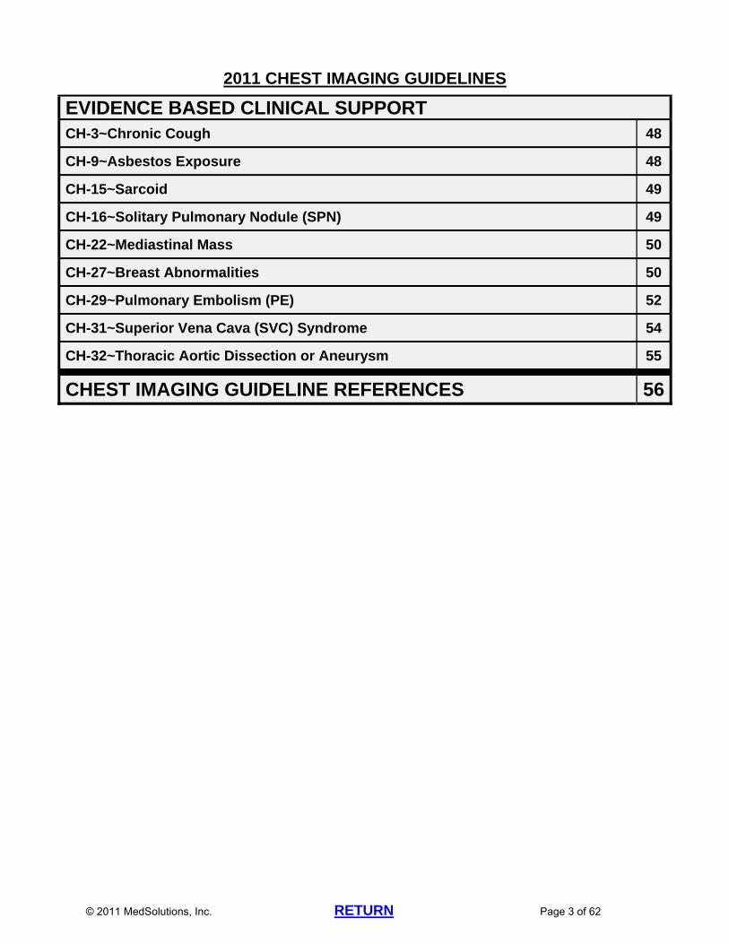
2011 CHEST IMAGING GUIDELINES
EVIDENCE BASED CLINICAL SUPPORT CH-3~Chronic Cough 48
CH-9~Asbestos Exposure 48
CH-15~Sarcoid 49
CH-16~Solitary Pulmonary Nodule (SPN) 49
CH-22~Mediastinal Mass 50
CH-27~Breast Abnormalities 50
CH-29~Pulmonary Embolism (PE) 52
CH-31~Superior Vena Cava (SVC) Syndrome 54
CH-32~Thoracic Aortic Dissection or Aneurysm 55
CHEST IMAGING GUIDELINE REFERENCES 56
© 2011 MedSolutions, Inc. RETURN Page 4 of 62
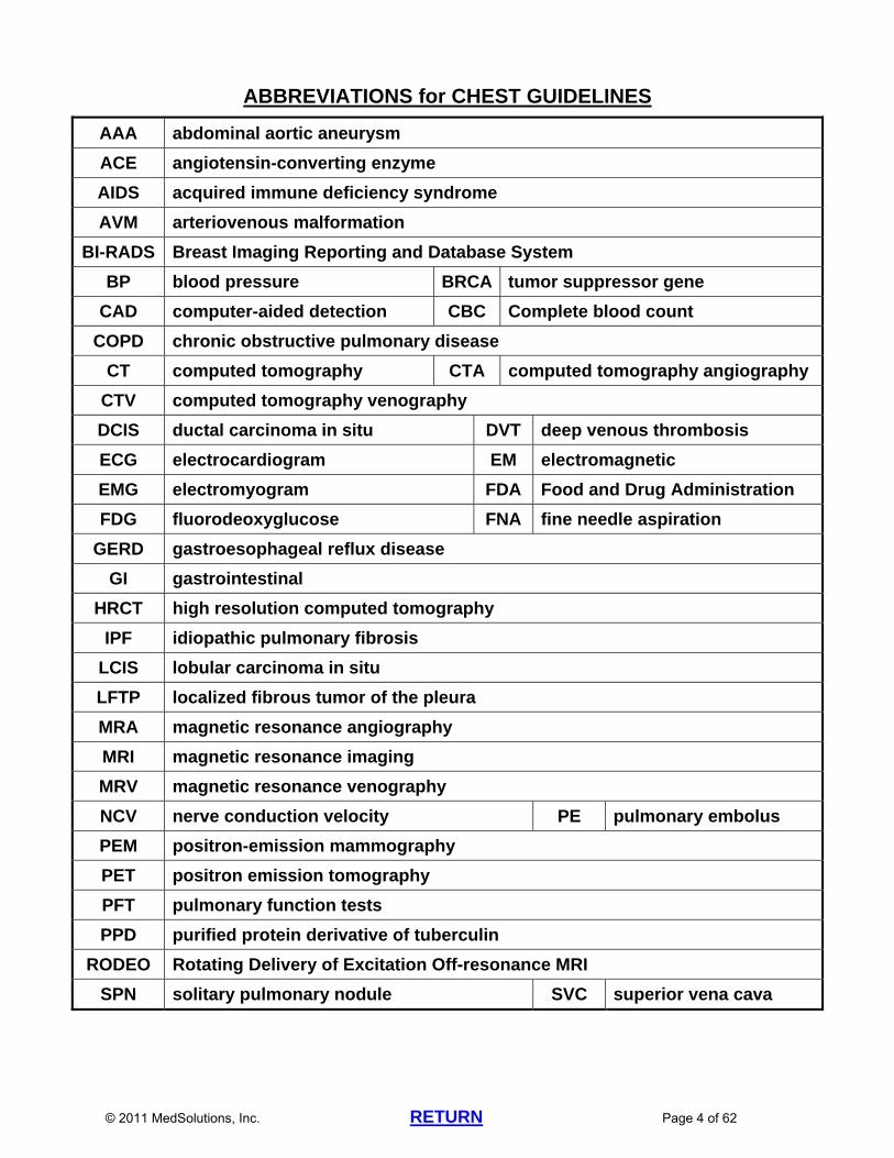
ABBREVIATIONS for CHEST GUIDELINES
AAA abdominal aortic aneurysm
ACE angiotensin-converting enzyme
AIDS acquired immune deficiency syndrome
AVM arteriovenous malformation
BI-RADS Breast Imaging Reporting and Database System
BP blood pressure BRCA tumor suppressor gene
CAD computer-aided detection CBC Complete blood count
COPD chronic obstructive pulmonary disease
CT computed tomography CTA computed tomography angiography
CTV computed tomography venography
DCIS ductal carcinoma in situ DVT deep venous thrombosis
ECG electrocardiogram EM electromagnetic
EMG electromyogram FDA Food and Drug Administration
FDG fluorodeoxyglucose FNA fine needle aspiration
GERD gastroesophageal reflux disease
GI gastrointestinal
HRCT high resolution computed tomography
IPF idiopathic pulmonary fibrosis
LCIS lobular carcinoma in situ
LFTP localized fibrous tumor of the pleura
MRA magnetic resonance angiography
MRI magnetic resonance imaging
MRV magnetic resonance venography
NCV nerve conduction velocity PE pulmonary embolus
PEM positron-emission mammography
PET positron emission tomography
PFT pulmonary function tests
PPD purified protein derivative of tuberculin
RODEO Rotating Delivery of Excitation Off-resonance MRI
SPN solitary pulmonary nodule SVC superior vena cava
© 2011 MedSolutions, Inc. RETURN Page 5 of 62
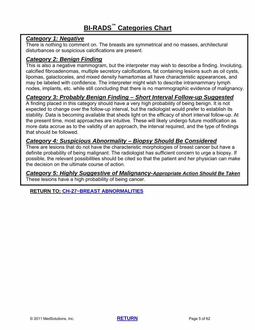
BI-RADS™ Categories Chart
Category 1: Negative There is nothing to comment on. The breasts are symmetrical and no masses, architectural disturbances or suspicious calcifications are present.
Category 2: Benign Finding This is also a negative mammogram, but the interpreter may wish to describe a finding. Involuting, calcified fibroadenomas, multiple secretory calcifications, fat containing lesions such as oil cysts, lipomas, galactoceles, and mixed density hamartomas all have characteristic appearances, and may be labeled with confidence. The interpreter might wish to describe intramammary lymph nodes, implants, etc. while still concluding that there is no mammographic evidence of malignancy.
Category 3: Probably Benign Finding – Short Interval Follow-up Suggested A finding placed in this category should have a very high probability of being benign. It is not expected to change over the follow-up interval, but the radiologist would prefer to establish its stability. Data is becoming available that sheds light on the efficacy of short interval follow-up. At the present time, most approaches are intuitive. These will likely undergo future modification as more data accrue as to the validity of an approach, the interval required, and the type of findings that should be followed.
Category 4: Suspicious Abnormality – Biopsy Should Be Considered There are lesions that do not have the characteristic morphologies of breast cancer but have a definite probability of being malignant. The radiologist has sufficient concern to urge a biopsy. If possible, the relevant possibilities should be cited so that the patient and her physician can make the decision on the ultimate course of action.
Category 5: Highly Suggestive of Malignancy-Appropriate Action Should Be Taken These lesions have a high probability of being cancer.
RETURN TO: CH-27~BREAST ABNORMALITIES

© 2011 MedSolutions, Inc. RETURN Page 6 of 62
2011 CHEST IMAGING GUIDELINES
CH-1~GENERAL GUIDELINES A recent complete history and physical examination should be performed prior to
considering advanced imaging of the chest. Chest X-ray
o A recent chest x-ray (generally within the last 30 days) that has been overread by a radiologist should be performed prior to considering advanced imaging.
Chest Ultrasound o Chest ultrasound (CPT®76604) includes transverse, longitudinal, and oblique
images of the chest wall with measurements of chest wall thickness, and also includes imaging of the mediastinum.
o Chest x-ray should be performed prior to chest ultrasound o Indications for chest ultrasound (CPT®76604) include: Evaluate presence of fluid within the pleural spaces Evaluate mediastinal masses Measure the distance between the anterior surface to chest wall prior to
radiation therapy Evaluate other masses suspected within the chest or chest wall
o Coding Notes Chest ultrasound--CPT®76604 Breast ultrasound--CPT®76645 (unilateral or bilateral) Axillary ultrasound--CPT®76882 (unilateral) If bilateral axillary ultrasounds are being performed, this should be
coded as CPT®76882 x 2. Chest CT
o Intrathoracic abnormalities found on chest x-ray, fluoroscopy, abdominal CT scan, or other imaging modalities can be further evaluated with chest CT with contrast (CPT®71260).
o Non-contrast chest CT (CPT®71250) can be used for the following: Patient has contraindication to contrast Follow-up of pulmonary nodule(s) High Resolution CT (HRCT) Noncontrast CT is specifically requested by pulmonary specialist Other circumstances as specified in the guidelines
o Chest CT without and with contrast (CPT®71270) does not add significant diagnostic information above and beyond that provided by chest CT with contrast, unless a question regarding calcification needs to be resolved.
o Coding notes: High resolution chest CT should be reported only with an appropriate
CPT® code from the set 71250-71270. No additional CPT® codes should be used for the “high resolution” portion of the scan. The “high resolution” involves additional slices which are not separately billable.
Chest CTA o Pre-op evaluation for minimally invasive or robotic surgery:

© 2011 MedSolutions, Inc. RETURN Page 7 of 62
There is insufficient data to support the routine use of CTA for the routine evaluation of peripheral arteries, iliac arteries, and/or aorta prior to minimally invasive or robotic surgery.
Chest MRI o Chest CT is indicated in the majority of cases to evaluate pathology in the
chest when advanced imaging is appropriate. Indications for chest MRI are much less common.
o MRI chest is appropriate when there are concerns about CT contrast such as renal insufficiency or contrast allergy.
o MRI chest may be appropriate in order to clarify equivocal findings on previous imaging studies. Appropriateness will need to be determined on a case by case basis.
© 2011 MedSolutions, Inc. RETURN Page 8 of 62
2011 CHEST IMAGING GUIDELINES
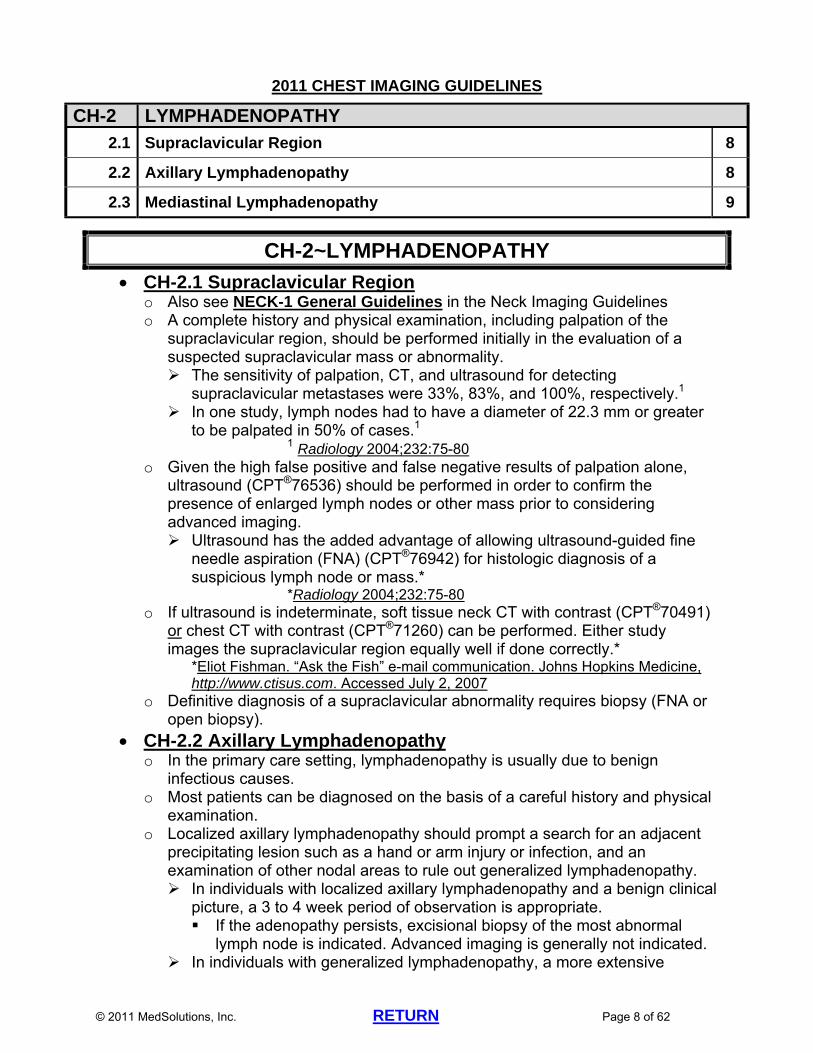
CH-2 LYMPHADENOPATHY
2.1 Supraclavicular Region 8
2.2 Axillary Lymphadenopathy 8
2.3 Mediastinal Lymphadenopathy 9
CH-2~LYMPHADENOPATHY
CH-2.1 Supraclavicular Region o Also see NECK-1 General Guidelines in the Neck Imaging Guidelines o A complete history and physical examination, including palpation of the
supraclavicular region, should be performed initially in the evaluation of a suspected supraclavicular mass or abnormality. The sensitivity of palpation, CT, and ultrasound for detecting
supraclavicular metastases were 33%, 83%, and 100%, respectively.1 In one study, lymph nodes had to have a diameter of 22.3 mm or greater
to be palpated in 50% of cases.1 1 Radiology 2004;232:75-80
o Given the high false positive and false negative results of palpation alone, ultrasound (CPT®76536) should be performed in order to confirm the presence of enlarged lymph nodes or other mass prior to considering advanced imaging. Ultrasound has the added advantage of allowing ultrasound-guided fine
needle aspiration (FNA) (CPT®76942) for histologic diagnosis of a suspicious lymph node or mass.*
*Radiology 2004;232:75-80 o If ultrasound is indeterminate, soft tissue neck CT with contrast (CPT®70491)
or chest CT with contrast (CPT®71260) can be performed. Either study images the supraclavicular region equally well if done correctly.*
*Eliot Fishman. “Ask the Fish” e-mail communication. Johns Hopkins Medicine, http://www.ctisus.com. Accessed July 2, 2007
o Definitive diagnosis of a supraclavicular abnormality requires biopsy (FNA or open biopsy).
CH-2.2 Axillary Lymphadenopathy o In the primary care setting, lymphadenopathy is usually due to benign
infectious causes. o Most patients can be diagnosed on the basis of a careful history and physical
examination. o Localized axillary lymphadenopathy should prompt a search for an adjacent
precipitating lesion such as a hand or arm injury or infection, and an examination of other nodal areas to rule out generalized lymphadenopathy. In individuals with localized axillary lymphadenopathy and a benign clinical
picture, a 3 to 4 week period of observation is appropriate. If the adenopathy persists, excisional biopsy of the most abnormal
lymph node is indicated. Advanced imaging is generally not indicated. In individuals with generalized lymphadenopathy, a more extensive

© 2011 MedSolutions, Inc. RETURN Page 9 of 62
diagnostic work-up including serological tests to rule out systemic infectious diseases, and lymph node biopsy is indicated.
o Reference: American Family Physician 1998 Oct.
http://www.aafp.org/afp/981015ap/ferrer.html. Accessed June 6, 2011 o Axillary Lymphadenopathy from an Occult Primary Cancer Axillary lymph node metastasis, without identification of a primary cancer,
is an uncommon finding. Adenocarcinoma is the most common histology, with breast cancer being the most common cancer (although non-palpable breast cancer presenting as axillary metastases accounts for less than 0.5% of all breast cancers). Breast MRI (CPT®77059) can be performed if breast cancer is
suspected and physical exam and mammography are negative. Carcinomas of the lung, thyroid, stomach, colon, rectum, and pancreas
have the potential to spread to axillary lymph nodes, but these metastases are rarely the first manifestations of disease. Symptomatology, risk factors, and clinical suspicion should lead to
imaging of these possible primary sites. Also see ONC-29 Metastatic Cancer and Carcinomas of
Unknown Primary Site in the Oncology and PET Imaging Guidelines
Immunohistochemical markers have proven useful for differentiating metastatic breast carcinoma from adenocarcinoma arising in other primary sites.
References: J Natl Compr Canc Netw 2009;7(2):193-201 The Breast 2006;15:259-262
CH-2.3 Mediastinal Lymphadenopathy o Mediastinal abnormalities detected on chest x-ray (overread by a radiologist)
can be further evaluated by chest CT with contrast (CPT®71260). o Mediastinal masses identified on screening chest CT scans should be
approached conservatively. In the I-ELCAP study which involved almost 30,000 individuals who
received screening chest CT scans, 123 (1%) had a mediastinal lesion, but only 4 were cancers.*
*Imaging Economics 2005 Feb, p.37 o If chest CT shows enlarged lymph nodes in the mediastinum with no other
abnormalities in a patient at low risk for malignancy and with no clinical suspicion for malignancy, one follow-up chest CT (CPT®71260) at 4 to 8 weeks can be performed. Requests for additional CT scans or for PET should be sent for Medical
Director review. Lymph node biopsy should be considered in cases of persistent
lymphadenopathy in order to obtain a histologic diagnosis. o Lymphadenopathy from neoplasms as well as from benign sources of
inflammation can result in a positive PET scan. Therefore, the use of PET may not be helpful prior to histologic diagnosis.
o If biopsy can only be accomplished by mediastinoscopy or thoracoscopy/thoracotomy (i.e. percutaneous biopsy, transbronchial biopsy,
© 2011 MedSolutions, Inc. RETURN Page 10 of 62
transbronchial biopsy using endobronchial ultrasound, and endoscopic ultrasound-guided FNA cannot be performed), and a negative PET scan will allow the patient to be observed, then PET can be considered to confirm the likelihood of yielding a pathologic diagnosis and to determine if a more favorable site for biopsy exists.
o PET may be helpful in characterizing anterior mediastinal abnormalities, especially since the thymus gland has a characteristic uptake pattern on most PET scans, and the study may differentiate normal or benign hypertrophic thymus tissue from pathologic mediastinal lesions.
© 2011 MedSolutions, Inc. RETURN Page 11 of 62
SYMPTOM-BASED GUIDELINES (ALPHABETICAL ORDER)
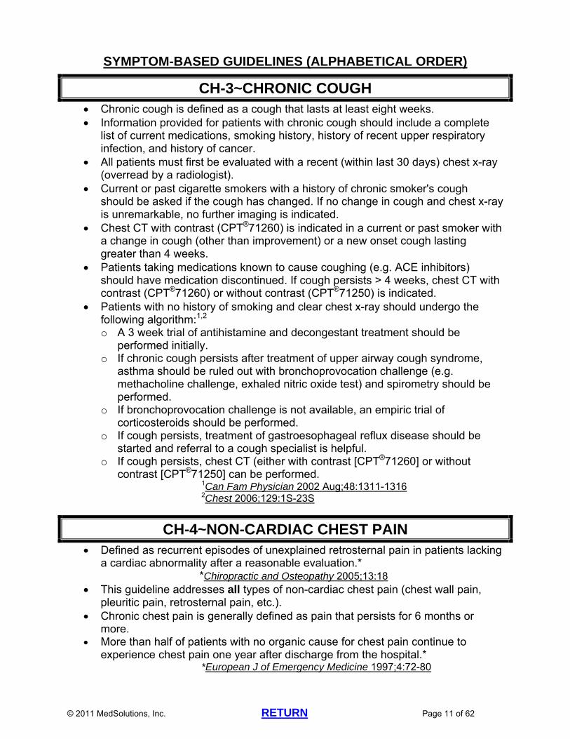
CH-3~CHRONIC COUGH Chronic cough is defined as a cough that lasts at least eight weeks. Information provided for patients with chronic cough should include a complete
list of current medications, smoking history, history of recent upper respiratory infection, and history of cancer.
All patients must first be evaluated with a recent (within last 30 days) chest x-ray (overread by a radiologist).
Current or past cigarette smokers with a history of chronic smoker's cough should be asked if the cough has changed. If no change in cough and chest x-ray is unremarkable, no further imaging is indicated.
Chest CT with contrast (CPT®71260) is indicated in a current or past smoker with a change in cough (other than improvement) or a new onset cough lasting greater than 4 weeks.
Patients taking medications known to cause coughing (e.g. ACE inhibitors) should have medication discontinued. If cough persists > 4 weeks, chest CT with contrast (CPT®71260) or without contrast (CPT®71250) is indicated.
Patients with no history of smoking and clear chest x-ray should undergo the following algorithm:1,2 o A 3 week trial of antihistamine and decongestant treatment should be
performed initially. o If chronic cough persists after treatment of upper airway cough syndrome,
asthma should be ruled out with bronchoprovocation challenge (e.g. methacholine challenge, exhaled nitric oxide test) and spirometry should be performed.
o If bronchoprovocation challenge is not available, an empiric trial of corticosteroids should be performed.
o If cough persists, treatment of gastroesophageal reflux disease should be started and referral to a cough specialist is helpful.
o If cough persists, chest CT (either with contrast [CPT®71260] or without contrast [CPT®71250] can be performed.
1Can Fam Physician 2002 Aug;48:1311-1316 2Chest 2006;129:1S-23S
CH-4~NON-CARDIAC CHEST PAIN Defined as recurrent episodes of unexplained retrosternal pain in patients lacking
a cardiac abnormality after a reasonable evaluation.* *Chiropractic and Osteopathy 2005;13:18
This guideline addresses all types of non-cardiac chest pain (chest wall pain, pleuritic pain, retrosternal pain, etc.).
Chronic chest pain is generally defined as pain that persists for 6 months or more.
More than half of patients with no organic cause for chest pain continue to experience chest pain one year after discharge from the hospital.*
*European J of Emergency Medicine 1997;4:72-80

© 2011 MedSolutions, Inc. RETURN Page 12 of 62
Etiology: o Studies have found that the most common etiologies include idiopathic (60%),
musculoskeletal chest pain (esp. costochondritis) (36%), and reflux disease (GERD) (13%).*
*J Fam Pract 1994;38(4):345-352 o Esophageal angina: Approximately 10%-20% of patients with GERD present
with symptoms that are clinically indistinguishable from angina pectoris. Clinical features that may suggest the esophagus as the source of the
atypical pain include: posturally aggravated symptoms, history of dysphagia, substernal pain limited to the midline and radiating to the interscapular area.
Reference: Hiebert CA. Clinical Features.In Pearson FG, Deslauriers J, Ginsberg
RJ, et al. (Eds.). Thoracic Surgery. New York, Churchill Livingstone, Inc., 1995, pp.68-69
25%-50% of chest pain presentations in ambulatory settings may be musculoskeletal. o Musculoskeletal pain is a diagnosis of exclusion. o Some patients with Thoracic Outlet Syndrome can present with anterior chest
wall or parascapular pain.* o Also see CH-34 Thoracic Outlet Syndrome
*Mackinnon S, Patterson GA, Urschel HC, Jr. Thoracic Outlet Syndromes. Pearson FG, Deslauriers J, Ginsberg RJ, et. al. (Eds.). Thoracic Surgery. New York, Churchill Livingstone, Inc.,1995, p.1221
Chest x-ray should be performed initially and overread by a radiologist. Abnormalities present on chest x-ray that were not present on previous imaging
studies (if available) can be further evaluated with chest CT with contrast (CPT®71260).
If chest x-ray is unremarkable, a thorough cardiac (ECG, echocardiogram, stress test), GI (trial of anti-reflux medication, possible upper endoscopy, pH probe, esophageal manometry), and pulmonary (PFT’s) evaluation should be performed at least once.
If the above evaluations have not yielded an explanation for the chest pain, symptoms have not improved after a 6 to 8 week trial of rest, analgesics, and anti-inflammatory treatment under the direction of a physician, and a recent chest x-ray (within 2 to 4 weeks) has been performed, then chest CT with contrast (CPT®71260) can be performed.
There is no evidence to support MRI for the evaluation of chest pain. Repeat advanced imaging of the chest in patients with unchanged or improving
symptoms is not appropriate.
CH-5~DYSPNEA/SHORTNESS OF BREATH Dyspnea is the subjective experience of breathing discomfort Evaluation of dyspnea/shortness of breath is aimed at determining whether the
cause is cardiac, pulmonary, mixture of cardiac and pulmonary, or noncardiac/nonpulmonary o Most cases are due to cardiac or pulmonary disease
© 2011 MedSolutions, Inc. RETURN Page 13 of 62
If pulmonary embolus (PE) is suspected, see CH-29 Pulmonary Embolism Prior to considering advanced imaging of the chest, work up should include the
following: o Thorough history and physical examination o Recent (within past 30 days) chest x-ray that has been overread by a
radiologist o ECG o Pulse oximetry o Pulmonary function studies (PFT’s) o Blood work including CBC and thyroid function tests Intrathoracic abnormalities found on chest x-ray that were not present on
previous imaging studies and do not have benign features such as a benign calcification pattern typical of granuloma or hamartoma can be evaluated with chest CT without (CPT®71250) or with (CPT®71260) contrast.
High resolution chest CT scan (HRCT) without contrast (CPT® 71250) can be performed if PFT’s are consistent with interstitial lung disease such as idiopathic pulmonary fibrosis.
If chest x-ray, ECG, and PFT’s do not yield a diagnosis, then arterial blood gas measurement, echocardiography, and cardiac stress testing should be performed prior to considering advanced imaging of the chest. o See the following in the Cardiac Imaging Guidelines for the appropriate
cardiac stress test: CD-1.3 Stress Testing
CD-2.4 Stress Echocardiography
CD-3.2 Indications for MPI
CD-6.3 Indications for Stress MRI
Chest CT without (CPT®72150) or with (CPT®71260) contrast can be performed if the above work up has been completed and dyspnea/shortness of breath that is not cardiac in origin persists for greater than 6 to 8 weeks.
CH-6~HEMOPTYSIS A careful history should help determine the amount of blood and differentiate
between hemoptysis, pseudohemoptysis, and hematemesis. Most common etiologies for hemoptysis:
o Adults: Bronchitis, bronchogenic carcinoma, pneumonia Work up:
o Careful history and physical examination and chest x-ray. o Low risk patient with normal chest x-ray: treat on an outpatient basis with
close monitoring and antibiotics if indicated. o Patients with risk factors for malignancy (e.g. male sex, age >40, smoking,
duration of hemoptysis >1 week): chest CT with contrast (CPT®71260) should be performed even if chest x-ray is normal.
Reference: o Am Fam Physician 2005;72(7):1253-1260
In the non-trauma patient with a history of clinically documented hemoptysis, chest CT (either with contrast [CPT®71260] or without contrast [CPT®71250]

© 2011 MedSolutions, Inc. RETURN Page 14 of 62
depending on physician preference) is indicated prior to bronchoscopy.* *AJR 2002;179:1217-1224
BRONCHIAL TREE
CH-7~BRONCHIECTASIS Bronchiectasis is defined as localized, irreversible dilatation of bronchi >2 mm in
diameter. Patients have excessive mucus production. Bronchiectasis is associated with a wide range of disorders, including cystic
fibrosis, AIDS, alpha1-antitrypsin deficiency, rheumatoid arthritis, obstruction of the bronchi, and necrotizing bacterial infections.
Chest x-ray and PFT’s should be performed initially in patients with known or suspected bronchiectasis, but may be normal.
High resolution chest CT scan (HRCT) without contrast (CPT®71250) is the advanced imaging study of choice to confirm the diagnosis of bronchiectasis and/or evaluate patients with known bronchiectasis who have worsening symptoms or worsening PFT’s.
There is no published data to support performing routine follow-up advanced imaging of the chest in the absence of new or worsening symptoms or worsening lung function studies in patients with known bronchiectasis.
MRI is not used to evaluate patients with bronchiectasis. Patients with bronchiectasis who present with hemoptysis should undergo chest
CTA (CPT®71275) or chest MRA (CPT®71555). Reference:
o Emmons EE. Bronchiectasis. eMedicine, May 13, 2011, http://emedicine.medscape.com/article/354167-overview#a24I, Accessed May 23, 2011
CH-8~BRONCHITIS
Acute inflammation of trachea and/or large and small bronchi due to infection or other causes. The majority of cases of bronchitis are due to viral infections.
Symptoms can include coughing, wheezing, shortness of breath, fever o Acute bronchitis usually improves within a few days but the cough may
continue for weeks. Imaging Studies:
o Chest x-ray to rule out pneumonia if symptoms do not improve after a trial of conservative therapy (rest, analgesics, fluids, humidifier, etc.)
© 2011 MedSolutions, Inc. RETURN Page 15 of 62
LUNG PARENCHYMA (ALPHABETICAL ORDER)
CH-9~ASBESTOS EXPOSURE Chest x-ray must be performed initially in patients with suspected asbestos-
related lung disease. In patients with stable calcified pleural plaques seen on chest x-ray, no advanced
imaging of the chest is indicated. If a change is seen on chest x-ray, high resolution chest CT (HRCT)
(CPT®71250) can be performed. Patients with progressive pleural and parenchymal changes are at particularly
high risk of developing malignant mesothelioma and should have HRCT (CPT®71250) every 3 to 6 months.
CH-10~CHRONIC OBSTRUCTIVE PULMONARY DISEASE (COPD)
COPD includes a spectrum of diseases: asthmatic bronchitis, chronic bronchitis, and emphysema.
Typical presenting symptoms include cough, excess mucus, dyspnea on exertion, and/or wheezing.
Diagnosis is best made by performing spirometry (PFT’s).1 o In addition, chest x-ray and arterial blood gas measurement should be
performed.1 1Swierzewski SJ. COPD diagnosis.Healthcommunities.com,, June 1, 2000, updated May 3, 2011, http://www.healthcommunities.com/copd/diagnosis.shtml. Accessed May 24, 2011
o Chest CT without contrast (CPT®71250), high resolution chest CT without contrast (CPT®71250), or chest CT with contrast (CPT®71260) can be performed if emphysema is suspected and the above initial studies are indeterminate.
o Chest MRI is generally not indicated in the evaluation of COPD. o Patients with a family history of emphysema or chronic bronchitis should have
a spirometry test as part of their initial evaluation.* *Making the Diagnosis of COPD. National Lung Health Education Program, http://www.nlhep.org. Accessed October 31, 2007
An exacerbation of COPD is characterized by a change in baseline dyspnea, cough, and/or sputum that is acute in onset and beyond normal day-to-day variations. o Advanced imaging of the chest is not typically indicated in the evaluation of
COPD exacerbation. o Evaluation of COPD exacerbation should include arterial blood gas
measurement, chest x-ray, ECG, sputum culture, and blood work to measure electrolytes and complete blood count.*
*Buist AS, Rodriguez-Roisin R, Anzueto A, et al. Global Initiative for Chronic Obstructive Lung Disease: Pocket guide to COPD diagnosis, management, and prevention. National Institutes of Health, National Heart, Lung, and Blood Institute; April 2001 (updated December 2010 ),
© 2011 MedSolutions, Inc. RETURN Page 16 of 62
http://www.goldcopd.com. Accessed May 24, 2011 There is no published data to support performing routine follow-up advanced
imaging of the chest in patients with COPD. Lung Volume Reduction Surgery
o Chest CT either without contrast (CPT®71250) or with contrast (CPT®71260) can be performed for preoperative evaluation in patients who are being considered for lung volume reduction surgery.*
*Radiology 1999;212:1-3 o There is insufficient data to support obtaining routine follow-up advanced
imaging of the chest in patients who have had lung volume reduction surgery. o New or worsening signs/symptoms in patients who have had lung volume
reduction surgery should be evaluated with chest x-ray prior to considering advanced imaging of the chest
CH-11~INTERSTITIAL DISEASE High resolution chest CT scan (HRCT) without contrast (CPT®71250) is the
diagnostic modality of choice to evaluate for interstitial changes in patients with pulmonary symptoms and abnormal pulmonary function studies (PFT’S). Chest x-ray may be normal in some cases of interstitial lung disease and PFT’s are the best indicator of the need for HRCT.
Evaluation by a Pulmonologist is helpful in determining the need for advanced imaging.
HRCT can be performed in patients with known interstitial pneumonia, idiopathic pulmonary fibrosis, or other interstitial lung disease if there are new or worsening pulmonary symptoms or worsening PFT’s.
HRCT can be performed once a year in patients with known idiopathic pulmonary fibrosis (IPF) who are asymptomatic or have stable symptoms and stable PFT’s, if imaging results showing progression or regression of disease will change patient management.*
*Proceedings of the American Thoracic Society 2006;3:307-314
CH-12~MULTIPLE PULMONARY NODULES More than 6 nodules usually indicates inflammatory lung disease, and this has
been confirmed after years of follow-up.* *Chest 2004;125:1522-1529
Clustering of multiple nodules in a single location in the lung tends to favor an infectious process, although a dominant nodule with adjacent small satellite nodules can be seen in primary lung cancer.*
*Radiology 2005:237:395-400 In patients with multiple pulmonary nodules, the largest nodule should be imaged
based on CH-16 Solitary Pulmonary Nodule Imaging Guidelines. If infection is highly suspected in a patient with multiple pulmonary nodules, the
first follow-up chest CT (CPT®71250 or 71260) can be performed sooner than 3 months.
© 2011 MedSolutions, Inc. RETURN Page 17 of 62
CH-13~PNEUMONIA Chest x-ray (overread by a radiologist) must be performed initially in all patients
with suspected pneumonia prior to considering advanced imaging. Chest CT with contrast (CPT®71260) may be helpful in evaluating a patient with
pneumonia that has shown no improvement by chest x-ray after two weeks or has not cleared by chest x-ray after four weeks.
Chest CT with contrast (CPT®71260) is indicated when chest x-ray shows a possible complication of pneumonia (e.g. abscess, effusion) or possible lung mass associated with the infiltrate.
CH-14~POSITIVE PPD or TUBERCULOSIS (TB) Chest CT with contrast (CPT®71260) can be performed in patients with positive
PPD skin test or other positive tuberculin skin tests and normal chest x-ray who have not had a previous normal chest CT.
Chest CT can show evidence of tuberculosis (e.g. primary complexes, mediastinal or hilar lymphadenopathy) in up to 20% of patients with unremarkable chest x-rays.*
*AJR 1997 Apr;168(4):1005-1009 *Eur J Radiol 2003 Dec;48(3):258-262
o Evidence of tuberculosis on chest CT will alter clinical management and result in full multi-drug treatment for these patients rather than single drug treatment for positive PPD.
If chest CT is unremarkable, there is insufficient data to support performing subsequent chest CT scans unless symptoms develop or chest x-ray shows a new abnormality.
Follow-up chest CT with contrast (CPT®71260) can be used to re-evaluate patients undergoing active treatment for tuberculosis who had abnormalities seen only on chest CT. o The frequency of the follow-up chest CT scans should be at the discretion of
the pulmonary specialist following the patient, as there are no published guidelines or evidence-based data addressing this issue.
Patients with suspected complications or progression of tuberculosis (e.g. pleural tuberculosis, empyema, mediastinitis) can be evaluated with chest CT with contrast (CPT®71260).
CH-15~SARCOID Also see ONC-30.5 Sarcoidosis in the Oncology Imaging Guidelines and HD-
32.3 Sarcoidosis in the Head Imaging Guidelines. CT of the chest either with contrast (CPT®71260) or without contrast
(CPT®71250) is superior to chest x-ray in establishing the diagnosis of sarcoid. CT scan helps differentiate sarcoid from other granulomatous disorders, especially tuberculosis, and allows follow-up for the detection of complications, especially fibrosis.*
*Rev Mal Respir 2003;20 (2 pt 1):207-213
© 2011 MedSolutions, Inc. RETURN Page 18 of 62
Patients with suspected sarcoid should have chest CT either with contrast (CPT®71260) or without contrast (CPT® 71250) to establish or rule out the diagnosis.
Bronchoscopy with biopsy is indicated to make a definitive diagnosis; if positive for sarcoidosis, no further imaging is necessary.
PET can also be useful in making the diagnosis of sarcoid, as sarcoid has a distinctive appearance on PET. However, definitive diagnosis can only be made by biopsy. o There is currently no evidence-based data to support performing serial PET
scans to monitor disease activity while tapering steroid therapy. Cardiac PET (CPT®78459) is useful for identifying and monitoring response to
therapy for cardiac sarcoid. The diagnosis should be established or strongly suspected prior to imaging.*
*J Nucl Med 2004;45(12):1989-1998 Chest CT (either with or without contrast) is indicated in patients with worsening
symptoms, new symptoms after a period of being asymptomatic, or if a treatment change is being considered.
CH-16~SOLITARY PULMONARY NODULE (SPN) A nodule is any pulmonary or pleural lesion represented in a radiograph by a
sharply defined, discrete, nearly circular opacity 2-30 mm in diameter which is surrounded by normal lung tissue. o A linear or essentially two-dimensional opacity that does not have an
approximately spherical component is not a nodule. o Purely linear or sheet like lung opacities are unlikely to represent neoplasms
and do not require follow-up, even when the maximum dimension exceeds 8 mm (0.8 cm).*
*Radiology 2005;237:395-400 o Nodular opacities and/or thickening that are typical of scarring do not require
follow-up advanced imaging and do not require imaging with contrast for further delineation.*
*Radiology 2005;237:395-400 o Malignant features can include spiculation, abnormal calcification, size
greater than 7-10 mm, ground glass opacity, interval growth, history of a cancer that tends to metastasize to the lung or mediastinum, and/or smoking history.
o Benign features can include benign calcification (granuloma, hamartoma), multiple areas of calcification, small size, multiple nodules, linear/streaking/sheet-like opacities, negative PET, and stability of size over 2 years.
A pulmonary nodule seen on an imaging study other than a dedicated chest CT (e.g. nodule seen on abdominal CT, spine MRI, chest or coronary artery CTA, etc.) can be further evaluated with one chest CT without contrast (CPT®71250) or with contrast (CPT®71260).
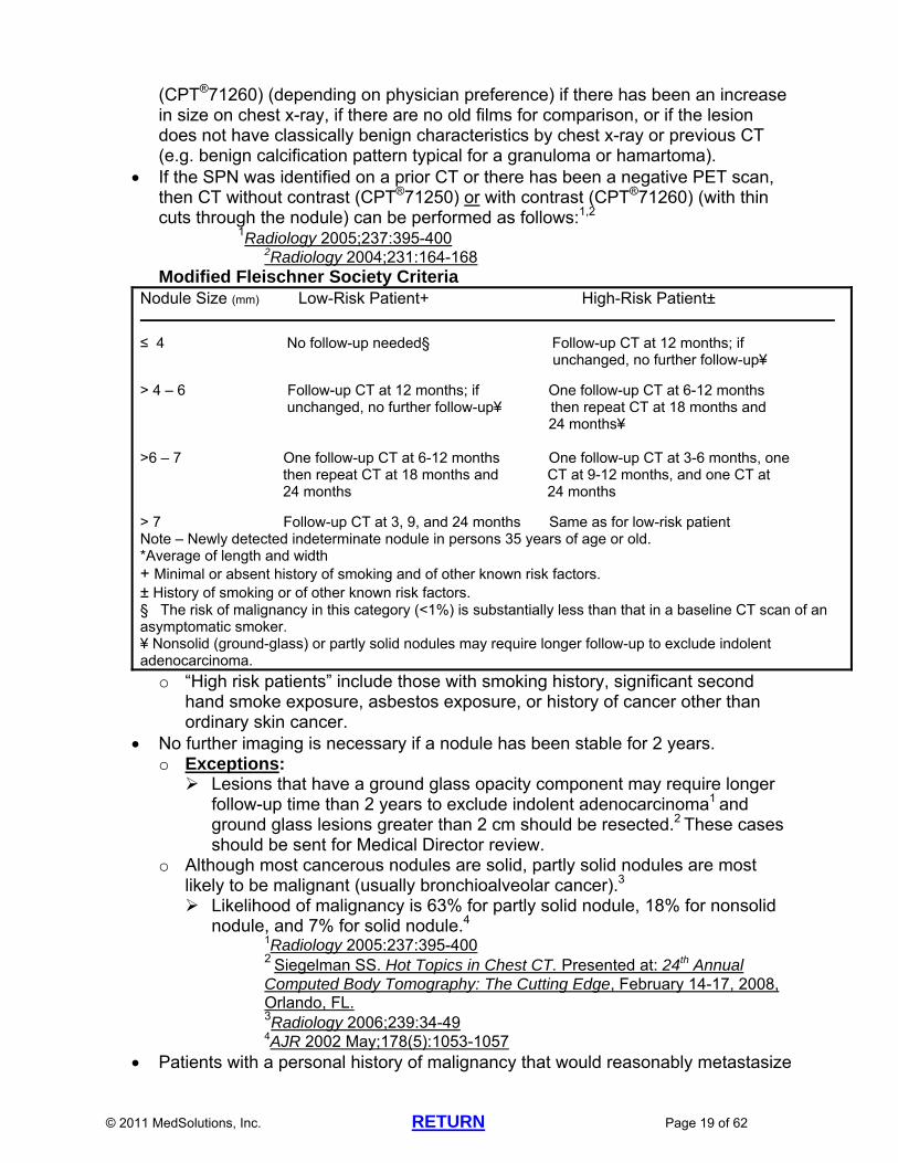
Follow-up imaging should proceed based upon the Modified Fleischner Society Criteria (next page) and other guidelines below. A solitary pulmonary nodule (SPN) can be imaged by chest CT without contrast (CPT®71250) or with contrast
© 2011 MedSolutions, Inc. RETURN Page 19 of 62
(CPT®71260) (depending on physician preference) if there has been an increase in size on chest x-ray, if there are no old films for comparison, or if the lesion does not have classically benign characteristics by chest x-ray or previous CT (e.g. benign calcification pattern typical for a granuloma or hamartoma).
If the SPN was identified on a prior CT or there has been a negative PET scan, then CT without contrast (CPT®71250) or with contrast (CPT®71260) (with thin cuts through the nodule) can be performed as follows:1,2
1Radiology 2005;237:395-400 2Radiology 2004;231:164-168
Modified Fleischner Society Criteria Nodule Size (mm) Low-Risk Patient+ High-Risk Patient± _________________________________________________________________________________
≤ 4 No follow-up needed§ Follow-up CT at 12 months; if unchanged, no further follow-up¥ > 4 – 6 Follow-up CT at 12 months; if One follow-up CT at 6-12 months unchanged, no further follow-up¥ then repeat CT at 18 months and 24 months¥ >6 – 7 One follow-up CT at 6-12 months One follow-up CT at 3-6 months, one then repeat CT at 18 months and CT at 9-12 months, and one CT at 24 months 24 months > 7 Follow-up CT at 3, 9, and 24 months Same as for low-risk patient Note – Newly detected indeterminate nodule in persons 35 years of age or old. *Average of length and width + Minimal or absent history of smoking and of other known risk factors. ± History of smoking or of other known risk factors. § The risk of malignancy in this category (<1%) is substantially less than that in a baseline CT scan of an asymptomatic smoker. ¥ Nonsolid (ground-glass) or partly solid nodules may require longer follow-up to exclude indolent adenocarcinoma.
o “High risk patients” include those with smoking history, significant second hand smoke exposure, asbestos exposure, or history of cancer other than ordinary skin cancer.
No further imaging is necessary if a nodule has been stable for 2 years. o Exceptions: Lesions that have a ground glass opacity component may require longer
follow-up time than 2 years to exclude indolent adenocarcinoma1 and ground glass lesions greater than 2 cm should be resected.2 These cases should be sent for Medical Director review.
o Although most cancerous nodules are solid, partly solid nodules are most likely to be malignant (usually bronchioalveolar cancer).3 Likelihood of malignancy is 63% for partly solid nodule, 18% for nonsolid
nodule, and 7% for solid nodule.4 1Radiology 2005:237:395-400 2 Siegelman SS. Hot Topics in Chest CT. Presented at: 24th Annual Computed Body Tomography: The Cutting Edge, February 14-17, 2008, Orlando, FL. 3Radiology 2006;239:34-49 4AJR 2002 May;178(5):1053-1057
Patients with a personal history of malignancy that would reasonably metastasize
© 2011 MedSolutions, Inc. RETURN Page 20 of 62
to the lungs or mediastinum who are found to have pulmonary nodules of any size can have repeat chest imaging at 3, 6, 12, and 24 months.
A nodule that grows at a rate consistent with cancer (doubling time 30 to 360 days) should be sampled for biopsy or resected.*
*Chest 2004;125:1522-1529 PET scan (CPT®78812 or CPT®78815) is appropriate for the characterization of
an SPN if the lesion is a distinct parenchymal lung nodule (not an infiltrate, ground glass opacity, or hilar enlargement) measuring greater than or equal to 7 mm (0.7 cm) on chest CT scan. o NOTE: Certain payers consider PET scan investigational for evaluating
pulmonary nodules ≤1 cm or lung masses >4 cm. Their coverage policies will take precedence over MedSolutions’ guidelines.
o Reference: J Nucl Med 2008;49:179-185
If PET scan is negative, chest CT should be performed at 3, 9, and 24 months.* *Radiology 2005; 237:395-400.
Serial PET scans to evaluate lung nodules are not appropriate: if the original PET is positive, biopsy should be performed. If the original PET is negative but subsequent chest CT shows increase in size of the nodule, biopsy should be performed. *
*Radiology 2006; 239:34-49 MRI of the chest is the least preferred modality for evaluation of lung nodules*
*ACR Appropriateness Criteria, Solitary pulmonary nodule, 2008
© 2011 MedSolutions, Inc. RETURN Page 21 of 62
PLEURA (ALPHABETICAL ORDER)
CH-17~PLEURAL-BASED NODULES and OTHER ABNORMALITIES
An indeterminate pleural-based nodule or lesion seen on an imaging study other than a dedicated chest CT (e.g. nodule or lesion seen on chest x-ray overread by a radiologist, abdominal CT, spine MRI, chest or coronary artery CTA, etc.) can be further evaluated with one chest CT without contrast (CPT®71250) or with contrast (CPT®71260). o If CT scan shows findings consistent with a benign process (round
atelectasis, scarring, apical thickening, etc.), no follow-up advanced imaging is indicated. “Round atelectasis”: twisting or folding of the lung which becomes
adherent to the adjacent pleura. o If the abnormality cannot be read as benign, repeat chest CT (CPT®71250 or
CPT®71260) can be performed using the Modified Fleischner Society Criteria and other guidelines in CH-16 Solitary Pulmonary Nodule (SPN)
Nodular opacities and/or thickening that are typical of scarring do not require follow-up advanced imaging and do not require imaging with contrast for further delineation.*
*Radiology 2005;237:395-400 There is no evidence-based data to support performing PET scan in patients with
pleural-based nodules or lesions.
CH-18~PLEURAL THICKENING Pleural thickening may be the residual effect of inflammatory processes,
including pneumonia with parapneumonic effusion, empyema, hemothorax, asbestos exposure, talc exposure, rheumatoid lung disease, radiation therapy, and drugs.
May occur due to infiltration of the pleura by malignant tumors such as mesothelioma or metastatic adenocarcinoma.
May occur due to localized fibrous tumor of the pleura (LFTP) o LFTP’s exist in benign and malignant forms with the benign form occurring
seven times more frequently than the malignant form. o Etiology of LFTP’s is unknown. o If LFTP is suspected due to a chest x-ray abnormality, chest CT with contrast
(CPT®71260) or chest MRI without and with contrast (CPT®71552) can be performed.
o Histologic examination is needed for a definitive diagnosis. o Treatment is resection. o Reference: Meziane MA and Lababede O. Imaging in localized fibrous tumor of the pleura.
eMedicine, February 21, 2010 , http://emedicine.medscape.com/article/359358-overview. Accessed May 24, 2011
Localized pleural thickening often occurs at the lung apices with increasing age, forming a pleural cap. Unless the patient is at high risk for malignancy or tuberculosis, no advanced imaging is indicated.
© 2011 MedSolutions, Inc. RETURN Page 22 of 62
Patients with suspected pleural thickening seen on chest x-ray (overread by a radiologist) can have chest CT with contrast (CPT®71260) or high resolution chest CT without contrast (CPT®71250) for further evaluation. o If the chest CT shows pleural plaques or findings consistent with asbestosis,
follow-up imaging guidelines described in CH-9 Asbestos Exposure should be followed.
o If CT scan shows findings consistent with a benign process (round atelectasis, scarring, apical thickening, etc.), no follow-up advanced imaging is indicated.
o If there is concern for malignancy or a definitive diagnosis is desired, then pleural biopsy should be performed by thoracoscopy or open biopsy.
o Serial advanced imaging of pleural thickening is not indicated unless patients have a known diagnosis such as asbestos-related disease, silicosis, or tuberculosis that is causing progressive pleural changes.
DISORDERS INVOLVING THE PLEURAL SPACE (ALPHABETICAL ORDER)
CH-19~PLEURAL EFFUSION Chest x-ray (including lateral decubitus films) should be performed initially in
patients with suspected pleural effusion. Chest ultrasound (CPT®76604) can be used to evaluate for the presence of fluid
within the pleural spaces. In patients with large pleural effusions, thoracentesis and analysis of the pleural
effusion (cytology, culture, cell count, biochemical studies) to distinguish transudative vs exudative should be performed prior to considering advanced imaging.
The most common causes of pleural effusions in the United States are congestive heart failure, bacterial pneumonia, malignancy (esp. lung cancer, breast cancer, and lymphoma), and pulmonary emboli.
If the pleural effusion is transudative and the etiology has been established (e.g. congestive heart failure, cirrhosis, nephrotic syndrome, peritoneal dialysis), advanced imaging of the chest is rarely indicated.
If the pleural effusion is exudative, chest CT with contrast (CPT®71260) can be performed after as much fluid as possible has been removed by thoracentesis. o There is little utility to obtaining chest CT in a patient with a large effusion
prior to thoracentesis, since the fluid will obscure the underlying lung parenchyma.
o Pleural biopsy is indicated for unexplained exudative effusions, most of which are found to result from malignancy or tuberculosis.
References: o Rubins J. Pleural effusion. eMedicine, April 25, 2011 ,
http://emedicine.medscape.com/article/299959-overview. Accessed May 24, 2011 o Sriram PS, Antony VB, Holm KA. Pleural Effusions: How to confirm the cause and
manage effectively. Consultant, Sept 15, 2007, http://www.consultantlive.com. Accessed May 24, 2011

© 2011 MedSolutions, Inc. RETURN Page 23 of 62
o Rusch VW. Pleural Effusion: benign and malignant.In Pearson FG, Deslauriers J, Ginsberg RJ, et. al. (Eds.). Thoracic Surgery. New York, Churchill Livingstone, Inc., 1995, pp.1003-1008
CH-20~PNEUMOTHORAX/HEMOTHORAX Advanced imaging of the chest is rarely indicated in the diagnosis or
management of pneumothorax. If the diagnosis of a small pneumothorax is in doubt, and the presence of a
pneumothorax will affect patient treatment decisions, then noncontrast chest CT (CPT®71250) can be performed.
There is no data supporting the use of serial chest CT scans to follow patients with known pneumothorax who are asymptomatic or have stable symptoms.
Patients with trauma significant enough to raise suspicion for hemothorax should be evaluated in the Emergency Department.
Chest CT with contrast (CPT®71260) can be performed as a preoperative study in patients in whom pleurodesis or other invasive procedure for pneumothorax is being considered.
There is no data supporting the use of serial chest CT scans to follow patients with known hemothorax who are asymptomatic or have stable symptoms.
Chest CT with contrast (CPT®71260) can be performed in patients with suspected complications from hemothorax (e.g. empyema).
Chest CT with contrast (CPT®71260) can be performed as a preoperative study in patients undergoing surgical evacuation for hemothorax.
© 2011 MedSolutions, Inc. RETURN Page 24 of 62
MEDIASTINUM
CH-21~MEDIASTINAL LYMPHADENOPATHY
See CH-2.3 Mediastinal Lymphadenopathy
CH-22~MEDIASTINAL MASS
Chest ultrasound (CPT®76604) can be used to evaluate a mediastinal mass if requested. However, chest CT with contrast (CPT®71260) is the imaging study of choice to evaluate mediastinal abnormalities.
Chest CT with contrast (CPT®71260) is indicated to evaluate a widened mediastinum on a chest x-ray (overread by a radiologist).
Chest CT (either with contrast [CPT®71260] or without contrast [CPT®71250]) is indicated in patients diagnosed with myasthenia gravis in order to rule out a thymoma. Note: iodinated contrast has been reported to provoke myasthenia crisis. o Also see PN-6.1 Neuromuscular Disease in the Peripheral Nerve Disorders
Imaging Guidelines and Thymoma in ONC-11 Other Thoracic Tumors in the Oncology Imaging Guidelines
Patients with a suspected substernal goiter should have a neck ultrasound (CPT®76536) or radionuclide study first to confirm extension of the thyroid to the sternum.
In patients who present with dysphagia and no history of prior malignancy, barium swallow should be performed initially (see NECK-3 Dysphagia in the Neck Imaging Guidelines).
CHEST WALL AND RIBS (ALPHABETICAL ORDER)
CH-23~CHEST TRAUMA Rib Fracture
o A complete history and physical examination, including palpation of the chest, should be performed initially in patients with chest trauma and suspected rib fracture.
o A recent chest x-ray, including erect posteroanterior (PA) and oblique views should be performed prior to considering advanced imaging.
o Suspicion of an occult rib fracture is not an indication for chest CT. o If the patient remains symptomatic, repeat plain x-rays of the ribs should be
obtained. These may show signs of early healing of a rib fracture. o If the diagnosis is still uncertain, bone scan is indicated. A delay of several
days should be allowed after an acute trauma to increase the sensitivity of bone scan to detect rib fracture(s).
o Patients with multiple new rib fractures can undergo chest CT without contrast (CPT®71250) or with contrast (CPT®71260) to rule out any associated intrathoracic pathology.
o Routine follow-up advanced imaging of rib fractures is not indicated. Fracture of the Sternum
o Injury to the sternum or suspected fracture of the sternum should be
© 2011 MedSolutions, Inc. RETURN Page 25 of 62
evaluated initially with lateral and oblique x-rays centered on the sternum. o If the diagnosis is still uncertain, chest CT without (CPT®71250) or with
contrast (CPT®71260) can be performed. o If a new sternal fracture is found, cardiac evaluation with ECG, cardiac
enzymes, and rhythm monitoring should be performed to rule out significant arrhythmia due to blunt cardiac trauma.
o Routine follow-up advanced imaging of sternal fractures is not indicated. References:
o Mahoney LK and Doty CI. Rib fracture. eMedicine, June 28, 2010, http://emedicine.medscape.com/article/825981-overview. Accessed May 24, 2010
Imaging of the Abdomen and Pelvis in Patients with Chest Trauma o If there was no significant trauma involving the abdomen or pelvis, and a
careful physical examination, laboratory studies, and urinalysis do not raise suspicion for abdominal or pelvic pathology, no advanced imaging of the abdomen or pelvis is indicated.
CH-24~COSTOCHONDRITIS Inflammatory process of the costochondral or costosternal joints that causes
localized pain and tenderness. More than one site is affected in 90% of cases. The 2nd to 5th costochondral junctions are most commonly involved. Pain is usually described as follows:
o Worse with movement of the trunk, deep breath, and/or exertion o Decreases with change of position o Sharp, nagging, aching, or pressure-like o Usually localized but may radiate extensively o May wax and wane
Physical examination with palpation of the chest should be performed initially. o Pain with palpation of the affected costochondral joints is a constant finding in
costochondritis. o The diagnosis should be reconsidered if there is absence of local tenderness
to palpation. Chest x-ray should be the initial imaging study to rule out other pathology. Bone scan is sometimes performed to confirm the diagnosis of costochondritis or
if infection of the costochondral joint is suspected. If signs, symptoms, and physical examination are consistent with costochondritis,
advanced imaging is not indicated. If pain persists despite treatment with rest and anti-inflammatory medication,
work-up should proceed as described in: CH-4 Non-Cardiac Chest Pain.
Reference: o Flowers LK. Costochondritis. eMedicine, August 25, 2009,
http://emedicine.medscape.com/article/808554-overview. Accessed May 24, 2011
© 2011 MedSolutions, Inc. RETURN Page 26 of 62
CH-25~CHEST WALL MASS Chest ultrasound (CPT®76604) can be used to evaluate a chest wall mass if
requested. Chest x-ray should be performed initially to rule out intrathoracic pathology,
evaluate the presence of calcification in the mass and rule out bony destruction of the chest wall.
If chest x-ray shows a suspicious intrathoracic abnormality, chest CT with contrast (CPT®71260) can be performed.
If chest x-ray does not show a suspicious intrathoracic abnormality, but there is a palpable chest lesion that is not clinically consistent with a lipoma or simple skin lesion, then chest MRI without and with contrast (CPT®71552) is the advanced imaging modality of choice. Chest CT with contrast (CPT®71260) is acceptable if MRI cannot be performed.
If lipoma or simple skin lesion is high on the differential diagnosis list, then evaluation by a surgeon or dermatologist is helpful in determining the need for advanced imaging. o Lipomas are one of the most common chest wall lesions. o The preferred imaging technique for evaluating lipomas depends on the
clinical question. If the study is being performed to diagnose a mass as a lipoma,
noncontrast chest CT (CPT®71250) is sufficient and enables specific recognition of fat and is faster than MRI.
If surgical removal of the lesion is planned and the lesion is large, infiltrating, or near important neurovascular structures, chest MRI (contrast as requested) can be performed.
o Reference: RadioGraphics 1994 May;14(3):571-595
CH-26~PECTUS EXCAVATUM and PECTUS CARINATUM See also PACCH-12 Pectus Excavatum and Pectus Carinatum in the
Pediatric Chest Imaging Guidelines Initial evaluation of patients with suspected or known pectus excavatum (ribs and
sternum grow abnormally producing a concave or caved-in appearance in the anterior chest wall), pectus carinatum (anterior protrusion of the chest wall), or other deformities of the chest wall or sternum should include a complete history and physical examination and plain chest x-rays.
Chest CT without contrast (CPT®71250) can be performed in selected cases of asymmetric pectus excavatum if significant cardiac displacement and rotation is suspected, or for preoperative planning.
ECG and echocardiography should be performed initially in patients with cardiac symptoms or evidence of abnormalities of cardiac function.
Chest x-ray and PFT’s should be performed initially in patients with known pectus who present with increasing shortness of breath.
Reference: o Hebra A. Pectus excavatum. eMedicine, July 22, 2010,
http://emedicine.medscape.com/article/1004953-overview. Accessed May 24, 2011
© 2011 MedSolutions, Inc. RETURN Page 27 of 62
BREAST
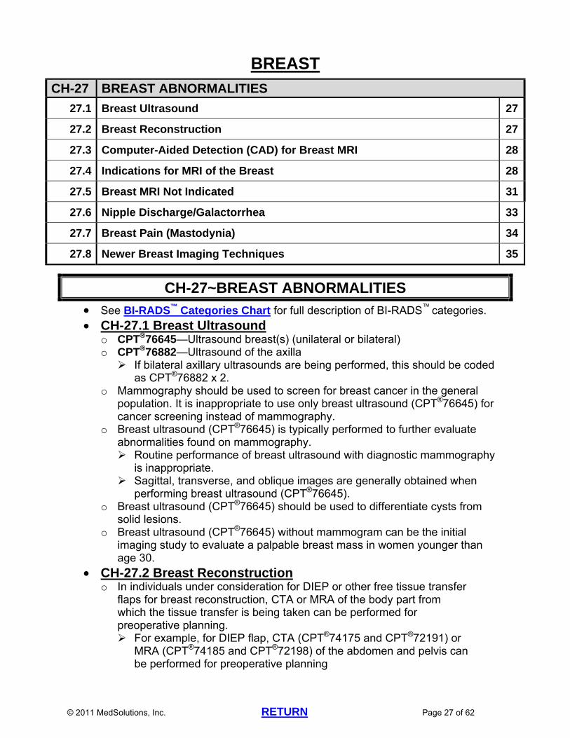
CH-27 BREAST ABNORMALITIES
27.1 Breast Ultrasound 27
27.2 Breast Reconstruction 27
27.3 Computer-Aided Detection (CAD) for Breast MRI 28
27.4 Indications for MRI of the Breast 28
27.5 Breast MRI Not Indicated 31
27.6 Nipple Discharge/Galactorrhea 33
27.7 Breast Pain (Mastodynia) 34
27.8 Newer Breast Imaging Techniques 35
CH-27~BREAST ABNORMALITIES
See BI-RADS™ Categories Chart for full description of BI-RADS™ categories. CH-27.1 Breast Ultrasound
o CPT®76645—Ultrasound breast(s) (unilateral or bilateral) o CPT®76882—Ultrasound of the axilla If bilateral axillary ultrasounds are being performed, this should be coded
as CPT®76882 x 2. o Mammography should be used to screen for breast cancer in the general
population. It is inappropriate to use only breast ultrasound (CPT®76645) for cancer screening instead of mammography.
o Breast ultrasound (CPT®76645) is typically performed to further evaluate abnormalities found on mammography. Routine performance of breast ultrasound with diagnostic mammography
is inappropriate. Sagittal, transverse, and oblique images are generally obtained when
performing breast ultrasound (CPT®76645). o Breast ultrasound (CPT®76645) should be used to differentiate cysts from
solid lesions. o Breast ultrasound (CPT®76645) without mammogram can be the initial
imaging study to evaluate a palpable breast mass in women younger than age 30.
CH-27.2 Breast Reconstruction o In individuals under consideration for DIEP or other free tissue transfer
flaps for breast reconstruction, CTA or MRA of the body part from which the tissue transfer is being taken can be performed for preoperative planning. For example, for DIEP flap, CTA (CPT®74175 and CPT®72191) or
MRA (CPT®74185 and CPT®72198) of the abdomen and pelvis can be performed for preoperative planning
© 2011 MedSolutions, Inc. RETURN Page 28 of 62
There is currently insufficient evidence-based data to support the need for routine advanced imaging for TRAM flaps or other flaps performed on a vascular pedicle.
CH-27.3 Computer-Aided Detection (CAD) for Breast MRI o Certain payers consider computer-aided detection (CAD) for breast MRI
investigational, and their coverage policies will take precedence over MedSolutions' guidelines.
o There have been no large, prospective studies showing that CAD definitively improves the sensitivity, specificity, and recall rates of breast MRI.
o Therefore, the use of CAD with breast MRI should be considered investigational at this time.
o It is not appropriate to report 3D rendering (CPT®76376 or CPT®76377) when requesting breast MRI with CAD, since 3D rendering is included in the Category III code for CAD (CPT®0159T). A dictated report of either the breast MRI study for which 3D rendering is
being requested (if the 3D rendering request is a retro request), or a previous breast MRI report if one was done, should be sent to MSI prior to approving a 3D rendering request for breast MRI.
CH-27.4 Indications for MRI of the Breast o Breast MRI: either unilateral [CPT®77058] or bilateral [CPT®77059] per
physician request. o Mammography, ultrasound (CPT®76645), and percutaneous biopsy should be
used to screen for breast cancer in the general population. o Breast ultrasound (CPT®76645), should be used to differentiate cysts from
solid lesions. o Indications for Breast MRI: Breast Augmentation, Breast Implants (saline or silicone), Breast
Reconstruction, Free Injection, Capsular Contracture Breast MRI can be performed if mammography or ultrasound
(CPT®76645) is uninterpretable Findings on the mammogram or ultrasound report should be
obtained or documented prior to starting breast MRI surveillance studies.
Evaluate or confirm breast implant rupture Patients with silicone breast implants can have a surveillance
breast MRI three years after implantation and every two years thereafter to screen for rupture per the current FDA recommendations.
If leakage is detected on MRI, the implant(s) should be removed. Once the implant(s) have been removed, no further surveillance
MRI of the affected breast(s) is indicated. NOTE: Certain payers do not include breast implants in their
coverage policies if the breast implants were placed as part of purely cosmetic surgery. Thus, surveillance MRI scans in these patients would also not be included in the coverage policy. Their coverage policies will take precedence over MedSolutions’ guidelines.
© 2011 MedSolutions, Inc. RETURN Page 29 of 62
Annual Screening Breast MRI Study for High Risk Patients: Starting at age 25 for patients with BRCA 1 or BRCA 2 mutation. Patients with a first degree relative with BRCA mutation (if patient has
not been tested). Screening studies for the patient should start 10 years before the
relative with BRCA was diagnosed with cancer. If the relative with BRCA was not diagnosed with cancer, then
screening studies for the patient should start at age 25. Starting at Age 25 for High Risk Patients, Defined as 20%-25% or
Greater Lifetime Risk of Developing Breast Cancer as Determined By: Clinical lifetime risk estimated at greater than 20% using clinical risk
estimator such as the Gail, Claus, Tyrer-Cuzick or BRCAPRO models
Two or more first degree relatives (parent, sibling, child) with breast or ovarian cancer
One first degree relative with breast cancer or ovarian cancer diagnosed before age 50
One first degree relative with bilateral breast cancer or both breast and ovarian cancer
History of breast cancer in a male relative (not a distant relative) Ashkenazi Jewish women from families with onset of breast cancer
before age 40 Li-Fraumeni Syndrome and first degree relatives Cowdan and Bannayan-Riley-Ruvalcaba Syndromes and first
degree relatives Women with history of radiation to the chest between ages 10 and
30. (If history of Hodgkin’s Disease, breast screening should start 8 to 10 years post-therapy, or at age 40, whichever comes first)
The American Cancer Society, the Society of Breast Imaging, and the National Comprehensive Cancer Network (NCCN) Clinical Guidelines in Oncology recommend that breast MRI be performed in facilities that have the capability to perform MRI-guided breast biopsies.
References: CA Cancer J Clin 2007;57:75-89 Institute for Clinical Systems Improvement (ICSI), Diagnosis of Breast
Disease. Jan 2010, , http://www.icsi.org/breast_disease_diagnosis/diagnosis_of_breast_disease_2.html. Accessed May 24, 2011
Equivocal or Occult Findings Patients in whom mammography, ultrasound, and clinical findings are
inconclusive or conflicting.* *J Am Coll Surg 2005 Oct;201(4):586-597
“MRI should not be used to determine biopsy recommendations for indeterminate lesions that can be readily biopsied, such as palpable masses and microcalcifications.”*
*AJR 2009;193:986-993 Although breast MRI has superior sensitivity in identifying new
© 2011 MedSolutions, Inc. RETURN Page 30 of 62
unknown malignancies, it carries a significant false positive risk when compared to mammogram and ultrasound. Incidental lesions are seen on 15% of breast MRI’s. The percentage of incidental lesions that turn out to be malignant varies from 3% to 20% depending on the patient population. Cancer is identified by breast MRI in only 0.7% of those with “inconclusive mammographic lesions.”*
*AJR 2009;193:986-993 Evaluate patients who present with axillary metastases suspicious for
primary breast cancer with negative physical exam and negative mammogram (MRI detects breast cancer in 90%-100% of cases if tumor is indeed present)
Guide biopsy of lesions seen only on MRI If diagnostic breast MRI has previously been performed, and the
currently requested breast MRI is being performed solely to guide a breast biopsy, then the breast MRI portion of the procedure is included in the CPT code for the MRI-guided procedure (CPT®77021) and requests for CPT®77058 or CPT®77059 are inappropriate.
Exception: If the previous diagnostic breast MRI was of poor quality or an unanswered clinical question remains, a repeat diagnostic breast MRI (CPT®77058 or CPT®77059) can be performed prior to MRI guided breast biopsy, especially if the biopsy is being performed at a different facility than the original breast MRI.
Newly Diagnosed Breast Cancer Breast MRI (including contralateral breast) can be performed to
evaluate patients with newly-diagnosed, biopsy-proven breast cancer
A recent study found that breast MRI detected cancers in the contralateral breast that were not detected by clinical exam or mammogram in 30 of 969 women* *N Engl J Med 2007 March;356(13):1295-1303
Tumor Response Assess response to neoadjuvant chemotherapy for locally advanced
breast cancer NOTE: If no pre-chemotherapy breast MRI was performed, then a
post-chemotherapy MRI should not be performed, as the findings will be too confusing.* *Huff JG. Clinical Applications of Breast MRI: Current Indications and Examples. Presented at: Identification and Management of Breast Cancer, October 6, 2007; Nashville,TN
Residual or Recurrent Malignancy Assessment of residual tumor load in patients who have undergone
lumpectomy and have close or positive margins, when the findings may indicate a significant change in surgical management.
Evaluate clinical suspicion of recurrence, following evaluations with mammography and/or ultrasound, if those evaluations are inconclusive

© 2011 MedSolutions, Inc. RETURN Page 31 of 62
or conflict with physical examination or other clinical indicators. This applies to intact breasts, reconstructed breasts, and possible chest wall recurrences following mastectomy.
NOTE: Breast MRI in the first 12 months after surgery has poor sensitivity
and specificity due to post surgical edema, hemorrhage, inflammation, scarring, and fat necrosis.*
It is advisable to wait 6 to 12 months after radiation before performing breast MRI. MRI prior to this time may give misleading results due to edema, architectural distortion, and cancers may exhibit benign appearing kinetics.*
*Huff JG. Clinical Applications of Breast MRI: Current Indications and Examples. Presented at: Identification and Management of Breast Cancer, October 6, 2007; Nashville,TN
Reference: ACR Practice Guideline for the Performance of Contrast-Enhanced Magnetic
Resonance Imaging (MRI) of the Breast. Revised 2008
CH-27.5 Breast MRI Not Indicated o See BI-RADS™ Categories Chart for full description of BI-RADS™ categories. o Routine Surveillance: Breast MRI should not be used for routine surveillance in patients with a
history of breast cancer (including DCIS, premenopausal women with a history of breast cancer, and women with a history of breast cancer that was only found on MRI), unless the patient meets criteria for Annual Screening Breast MRI Study for High Risk Patients in CH-27.4 Indications for MRI of the Breast. NCCN guidelines state that “a diagnosis of LCIS is associated with
estimated risks of 10%-20% for the subsequent development of cancer in either breast over the next 15 years…the panel also recommends consideration of MRI annually for women with LCIS.”*
Therefore, LCIS by itself does not confer a 20%-25% lifetime risk of breast cancer and the patient must still have additional risk factors that meet criteria for Annual Screening Breast MRI Study for High Risk Patients in CH-27.4 Indications for MRI of the Breast.
Dense Breasts Evidence-based data does not support routine breast MRI screening
for patients with dense breasts by mammogram (density 4 or 5). The patient must still have additional risk factors that meet criteria for Annual Screening Breast MRI Study for High Risk Patients in CH-27.4 Indications for MRI of the Breast. There are currently no data to show reduction in mortality from breast cancer from any screening technique other than mammography.*
The findings on the most recent mammogram should be obtained or documented. If the mammogram has been given a BI-RADS™ designation of BI-RADS™ 1, 2, 3, 4, or 5, then the radiologist has been able to interpret the mammogram and breast MRI is not indicated even if the mammogram report states the presence of dense breasts.
If the mammogram has been given a BI-RADS™ 0 then further
© 2011 MedSolutions, Inc. RETURN Page 32 of 62
imaging is needed and MRI can be considered. *AJR 2009;192:390-399
Currently, there is insufficient data to support the use of breast MRI for breast cancer screening in women with atypical hyperplasia or mutations other than BRCA unless the patient satisfies the criteria for annual screening with breast MRI outlined under Annual Screening Breast MRI Study for High Risk Patients in CH-27.2 Indications for MRI of the Breast .* References: CA Cancer J Clin 2007;57:75-89 *American Society of Breast Disease Policy Statement, The Use of
Magnetic Resonance Imaging of the Breast for Screening of Women at High Risk of Breast Cancer, June 28, 2004. http://www.asbd.org. Accessed November 27, 2006
o Low Risk, Probably Benign (BI-RADS™3) Solid Lesion on Mammogram and/or Ultrasound A solid lesion found on mammogram/ultrasound (CPT®76645), can be
observed and followed with repeat mammogram/ultrasound in 6 months if the lesion is a low-risk, probably benign lesion (includes the following: <15 mm, three or fewer lobulations, more than 50% of the lesion margin appears well-circumscribed in any view). Lesions not fitting all of the above criteria should be considered
indeterminate and the patient should be referred for surgical evaluation for biopsy.*
* Institute for Clinical Systems Improvement (ICSI), Diagnosis of Breast Disease. Jan 2010, http://www.icsi.org/breast_disease_diagnosis/diagnosis_of_breast_disease_2.html. Accessed May 24, 2011
If a probably benign lesion is clearly seen on mammogram and/or ultrasound (CPT®76645), then repeat mammography and/or ultrasound should be performed at 6 months. Follow-up breast MRI is not indicated.*
* Institute for Clinical Systems Improvement (ICSI), Diagnosis of Breast Disease. Jan 2010, http://www.icsi.org/breast_disease_diagnosis/diagnosis_of_breast_disease_2.html. Accessed May 24, 2011
In the evaluation of BI-RADSTM category 3 lesions, MRI did not provide additional information (low positive predictive value [33%]) and was similar to that of short interval (6 month) mammography follow-up.*
*Eur J Radiol 2006 Mar;57(3):436-444
o Suspicious (BI-RADS™4 or 5) Solid Lesion on Mammogram and/or Ultrasound Bilateral total breast ultrasound (CPT®76645), and bilateral axillary
ultrasound (CPT®76882) are recommended for patients who have BI-RADSTM 4 or 5 abnormalities. If additional suspicious breast lesions or more extensive malignant breast disease is detected by ultrasound, the extent of disease can be mapped with ultrasound-guided biopsies (CPT®76942).*
*J Am Coll Surg 2005 Oct;201(4):586-597

© 2011 MedSolutions, Inc. RETURN Page 33 of 62
A breast mass categorized as BI-RADS 4 or 5 should be biopsied.* *ACR Appropriateness Criteria, Nonpalpable mammographic findings (excluding
calcifications), Updated 2010 Biopsy of microcalcifications seen on mammogram in order to rule out
malignancy (including DCIS) should be performed rather than breast MRI. The sensitivity of MRI in evaluating mammographically detected
suspicious microcalcifications was only 87% with specificity 68%. The sensitivity of MRI for DCIS was 79%.* Therefore, biopsy of these lesions is warranted rather than MRI.
Some DCIS is only seen on MRI; therefore MRI-guided biopsy (CPT®77021) is appropriate in these cases.
*AJR 2006 Jun;186(6):1723-1732
o Serial Follow-up on Breast MRI Scans There is insufficient data to support using serial breast MRI studies to
follow patients with mammographic abnormalities or abnormalities seen on other imaging studies.
Breast MRI should be able to characterize a lesion as benign, probably benign, or as suspicious.
If breast MRI was obtained because a mammogram or ultrasound was unclear, then a probably benign lesion on MRI (MRI BI-RADS 3) should undergo repeat mammography and one repeat breast MRI in 6 months.
A report from The Agency for Healthcare Research and Quality (AHRQ) Effective Health Care Program concluded that none of the four commonly used noninvasive tests for breast abnormalities (MRI, ultrasound, PET, scintimammography) is sufficiently accurate to preclude breast biopsy in average risk women with nonpalpable breast lesions. The data were insufficient to estimate the accuracy of these tests in women with only palpable lesions.*
*Barclay L and Vega C. Noninvasive Tests May Be Insufficiently Accurate to Preclude Breast Biopsy. MedScape Medical News, Feb.10, 2006. http://www.medscape.com.Accessed November 27, 2006
Also see ONC-12 Breast Cancer in the Oncology guidelines
CH-27.6 Nipple Discharge/Galactorrhea o Mammogram should be obtained. Ultrasound (CPT®76645), may be helpful to
locate a duct papilloma, an intraductal nodule, or dilated duct. o The appearance of the fluid generally correlates with the etiology: Yellow, brown, green, or gray fluid is associated with fibrocystic change in
most patients. Purulent discharge can result from duct ectasia or partial duct obstruction. Pathologic discharges are usually bloody, blood-containing, or sometimes
watery and usually are unilateral and involve a single duct. Patients with bloody or unilateral discharge or palpable abnormality
should have a mammogram, with or without an ultrasound (CPT®76645) and referral to a surgeon for open biopsy is recommended.*
*Institute for Clinical Systems Improvement (ICSI), Diagnosis of Breast Disease. Jan 2010, http://www.icsi.org/breast_disease_diagnosis/diagnosis_of_breast_disease_2.html. Accessed May 24, 2011

© 2011 MedSolutions, Inc. RETURN Page 34 of 62
Physiologic discharges are usually bilateral, involve multiple ducts, are multicolored or milky, sticky, and are stimulated rather than occurring spontaneously.
o Prolactin and TSH levels should be obtained. A prolactinoma typically causes a milky or clear discharge bilaterally. Imaging of the pituitary is not necessary in patients with galactorrhea and
normal prolactin levels. See HD-27.1 Prolactinomas in the Head Imaging Guidelines
o Bloody or, less commonly, watery discharge raises the possibility of cancer (cancer accounts for 8%-15% of bloody nipple discharges), although most hemoccult-positive discharges are due to a benign etiology such as intraductal papilloma (45%), duct ectasia (36%), and infection and other causes (5%-10%). Ductogram and duct excision can be considered. A papilloma should be
resected by lumpectomy or vacuum-assisted lumpectomy. Breast MRI is not indicated* *Lawson LL. State of the Art Diagnosis of Breast Abnormalities: From Clinical Exam to MRI. Presented at: Identification and Management of Breast Cancer, October 6, 2007; Nashville,TN
o If mammography and endocrine studies are normal, observation and clinical re-evaluation should be performed. If clinical evaluation at the time of follow- up does not reveal any palpable or visible abnormalities, the patient should return to routine screening interval studies with mammogram or clinical exam.
o Reference: Institute for Clinical Systems Improvement (ICSI), Diagnosis of Breast Disease.
Jan 2010, http://www.icsi.org/breast_disease_diagnosis/diagnosis_of_breast_disease_2.html. Accessed May 24, 2011
CH-27.7 Breast Pain (Mastodynia) o Three classifications: Cyclic mastalgia: occurs in premenopausal women and is clearly related
to the menstrual cycle. Non-cyclic mastalgia: intermittent or continuous pain that is not related to
the menstrual cycle. Usually occurs in older women. Non-mammary pain: may present with the symptom of breast pain.
History and physical exam should help differentiate breast pain from pain radiating from the chest wall or another site.
o Evaluation of breast pain: Careful history and physical exam Pregnancy test is generally the only laboratory study that is needed Mammogram/ultrasound (CPT®76645)
o Advanced imaging is not routinely indicated in patients with breast pain and negative evaluation as outlined above. The risk of malignancy following a negative examination has been
estimated to be only 0.5%. o Reference: Institute for Clinical Systems Improvement (ICSI), Diagnosis of Breast Disease.
Jan 2010,

© 2011 MedSolutions, Inc. RETURN Page 35 of 62
http://www.icsi.org/breast_disease_diagnosis/diagnosis_of_breast_disease_2.html. Accessed May 24, 2011
CH-27.8 Newer Breast Imaging Techniques o RODEO MRI: Rotating Delivery of Excitation Off-Resonance MRI High resolution 1.5T MRI system designed specifically for the breast. This
system utilizes a unique fat suppression technology which provides greater detail about a lesion including distance, length/width, area, surface area, and volume without the distraction of fat tissue in the image. This reduces signal from normal ductal tissue and avoids false positive enhancement from benign lesions and dense fibroglandular tissue.
RODEO MRI is a particular software sequence for breast MRI. With current MRI techniques on a non-RODEO MRI, multiple MRI sequences give similar results to those obtained on a RODEO MRI.
There is no unique CPT® code or different reimbursement for breast MRI scans performed using the RODEO system, and the indications for breast MRI are no different (see CH-27.2 Indications for MRI of the Breast).
o Positron-Emission Mammography (PEM) or Naviscan: See CH-37 o Breast MR Spectroscopy: See CH-38 o Breast Tomosynthesis: Uses conventional mammographic x-ray tubes, but the x-ray source is
movable and swings over the breast in a 50-degree arc, creating 11 discrete images that can be combined or analyzed in many different ways to provide a three-dimensional data set or to create cross-sectional images.
There is insufficient data currently to generate appropriateness criteria for the use of breast tomosynthesis, and this procedure should be considered investigational at this time.
o Scintimammography Nuclear medicine study that uses a radioisotope such as Tc-99
tetrofosmin to image the breast. Breast cancer typically shows increased uptake of the radioisotope compared to benign lesions.
Acts as a “poor man’s MRI”. Scintimammography does not require preauthorization by MedSolutions at
this time.
© 2011 MedSolutions, Inc. RETURN Page 36 of 62
THORACIC VASCULAR DISORDERS (ALPHABETICAL ORDER)
CH-28~PULMONARY ARTERIOVENOUS FISTULA (AVM) Definition: abnormal connection between pulmonary arteries and veins. Etiology:
o Acquired: penetrating or blunt trauma to the chest; bronchiectasis Pulmonary AVM’s are most commonly found in the lower lobes. Chest x-rays are abnormal in approximately 98% of patients with pulmonary
AVM. o Chest x-ray usually shows a peripheral, circumscribed, non-calcified lesion
connected by blood vessels to the hilum of the lung. Chest CT (contrast as requested) and chest MRA (CPT®71555) or chest CTA
(CPT®71275) can be obtained for evaluation of possible pulmonary AVM. First degree relatives of a patient with a pulmonary AVM (not due to trauma or
bronchiectasis) can undergo screening with chest CT (CPT®71260). Treatment of pulmonary AVM is by surgery (usually lobectomy) or embolization
of the feeding artery using platinum coils or detachable balloons. References:
o Australasian Radiology 2005;49:242-245 o Current Pharmaceutical Design 2006;12:1243-1248
CH-29~PULMONARY EMBOLISM (PE) Patients who present with severe findings and dyspnea (including heart rate
>100 beats/minute along with systolic BP<90, syncope, new onset right heart failure) should be referred to the Emergency Department for immediate evaluation and treatment.
The clinical probability of PE is important in making an accurate diagnosis. An often cited point system (Wells criteria) includes the following:*
*Thromb Haemost 2000;83:416-420 o Clinical signs/symptoms of DVT (at minimum: leg swelling and pain with
palpation of the deep veins) (3 points) o No alternate diagnosis likely or more likely than pulmonary emboli (3 points) o Heart rate >100 beats/minute (1.5 points) o Immobilization (>3 days) or surgery in last 4 weeks (1.5 points) o Previous history of DVT or PE (1.5 points) o Hemoptysis (1 point) o Cancer actively treated in last 6 months or receiving palliative treatment (1
point) o Low probability <2 points; moderate 2 to 6 points; high>6 points o Using the above criteria, only 3% of patients with a low pretest probability had
PE versus 63% of those with a high pretest probability. Signs/Symptoms of PE include (in decreasing order):
o Dyspnea (73%) o Pleuritic chest pain (70%) PE is found in 5%-20% of patients who present to the Emergency
Department with pleuritic pain.* *Am Fam Physician 2007 May;75(9):1357-1364
© 2011 MedSolutions, Inc. RETURN Page 37 of 62
o Tachypnea (70%) o Leg swelling or pain (54%) o Cough (37%) o Tachycardia (30%) o Hemoptysis (13%) o Palpitations (10%) o Reference: Chest 1991;100(3):598-603
Other risk factors for deep venous thrombosis (DVT)/PE not listed above: o Recent history of a long airplane flight o Use of birth control pills o Trauma o Advanced age o Congestive heart failure
Evaluation of outpatients with suspected pulmonary embolism should include a consideration for clinical probability of PE using the Wells criteria point chart above as well as the urgent nature of the request, and results of a quantitative D-dimer study.
CT or CTA is not indicated for the following: o Wells criteria score is ≤4 and D-dimer is negative*
*JAMA 2006; 295:172-179 Negative D-dimer in combination with low or moderate PE risk classification
do not require CTA since the negative predictive value approaches 100% Chest CT with contrast (CPT®71260) or chest CTA (CPT®71275) is indicated
for the following: o Abnormal D-dimer test NOTE: recent surgery, injury, malignancy, sepsis, diabetes, pregnancy, or
other conditions where fibrin products are likely to be present can give a false positive D-dimer result.
o Wells criteria score higher than 4 points* *JAMA 2006; 295:172-179
o High clinical probability of PE In general, this includes individuals with one or more risk factors for PE who
present with recent onset (within past two weeks) of shortness of breath (dyspnea) and one other sign or symptom that cannot readily be attributed to a cardiac etiology (ischemia, congestive heart failure, valve problem, etc.) or primary pulmonary etiology (pneumonia, pleural effusion, pneumothorax, etc.).
In non-urgent cases, chest x-ray should be obtained prior to considering advanced imaging of the chest.
Pregnant patients with suspected PE should have D-dimer performed. o If there is low clinical pre-test probability for PE and D-dimer is negative, no
further work-up is needed. o If D-dimer is positive (D-dimer is normally increased in pregnancy and the
levels tend to increase as pregnancy goes to term), or if clinical pre-test probability is intermediate or high, lower extremity Doppler study should be performed. If Doppler study is positive, the patient should be treated for PE.
© 2011 MedSolutions, Inc. RETURN Page 38 of 62
If Doppler study is negative, ventilation/perfusion (V/Q) scan is preferred due to the much lower dose of radiation absorbed by the maternal breast than with CTA.
Chest CTA (CPT®71275) or chest MRA (CPT®71555) can be performed if V/Q scan is not available.
If CTA is performed, neonates need to have thyroid functions tested in the first week of life to rule out contrast-induced hypothyroidism.
o Reference: Radiology 2007 Jan;242(1):15-21
CTA combined with CTV o Although the use of CTA combined with venous phase imaging (CTA-CTV)
for diagnosing PE was found to have a higher sensitivity (90%) than CTA alone (sensitivity 83%),* there is insufficient data at this time to justify routinely performing CTA-CTV in patients with suspected PE. If routine diagnostic testing (including CTA) is inconclusive, and clinical
suspicion remains high, then CTA-CTV can be considered. *N Eng J Med 2006;354:2317-2327
Follow-up Imaging in Patients with Known PE: o The duration of treatment with anticoagulation in patients with known PE is
based upon the patient’s history and risk factors and is NOT based upon advanced imaging studies (see CH-29 Pulmonary Embolism Evidence Based Clinical Support section).1
o D-dimer levels can help guide continued treatment decisions since persistent elevation of the D-dimer is associated with increased recurrence.2
o There is insufficient evidence-based data to support routine follow-up advanced imaging in asymptomatic patients or patients with stable symptoms who have known PE, including advanced imaging prior to discontinuing anticoagulation therapy.
o PE and persistent pulmonary hypertension Pulmonary hypertension occurs when more than 40% of the pulmonary
vasculature is obstructed due to either recurrent thromboembolism or in situ pulmonary artery thrombosis.3
More than 50% of patients will have repeat CTA evidence of pulmonary thromboembolism at 6 months after diagnosis and undergoing anticoagulation treatment. Some patients will have complete resolution while others will have
evidence of chronic embolism. Pulmonary hypertension develops in 8% of patients, primarily from the persistent thromboembolism group.4
Chest CTA is not able to distinguish between intravascular chronic and recurrent thromboembolism and pulmonary hypertension has a limited correlation with repeat CTA findings.3,5 Therefore, transthoracic echo (CPT®93306) is a better screening test
for pulmonary hypertension in patients treated for PE. Right heart catheterization is the best study to evaluate suspected pulmonary hypertension.5
o References: 1Am Fam Physician 2004 June;69(12):2841-2848 2N Engl J Med 2006;355:1780-1789
© 2011 MedSolutions, Inc. RETURN Page 39 of 62
3N Engl J Med 2001;345:1465-1472 4Chest 2006;129:192-197 5Chest 2009;136:1202-1210
PULMONARY HYPERTENSION—See PVD-5~Pulmonary Artery Hypertension in the Peripheral Vascular Disease Imaging Guidelines
CH-30~SUBCLAVIAN STEAL SYNDROME Definition: reversal of flow in the ipsilateral vertebral artery distal to a stenosis or
occlusion of the proximal subclavian or innominate artery. Blood flows up the contralateral vertebral artery to the basilar artery and retrograde down the ipsilateral vertebral artery to supply collateral circulation to the arm on the side of the subclavian lesion.
Symptoms include vertebral basilar artery insufficiency, vertigo, limb paresis, and paresthesias. Bilateral cortical visual disturbances, ataxia, syncope, and dysarthria occur less frequently. o Also see HD-30.1 Vertebrobasilar Ischemia in the Head Imaging
Guidelines. Patients have a difference in the brachial systolic blood pressure of at least 30
mmHg between the two arms associated with a bruit in the supraclavicular area on the affected side.
Symptoms of cerebral ischemia may be produced by exercise of the affected arm.
Carotid duplex study (CPT®93882) should be the initial imaging study in patients with suspected Subclavian Steal Syndrome. o Duplex study will show reversal of flow in the ipsilateral vertebral artery. o If there are no symptoms of vertebrobasilar ischemia, then no further imaging
is generally needed. Neck and chest MRA (CPT®70548 and CPT®71555) or CTA (CPT®70498 and
71275) can be performed for diagnosis in patients with symptoms of vertebrobasilar ischemia if the clinical exam and duplex study are indeterminate, or as preoperative studies if they will substitute for invasive angiography.
Upper extremity MRA (CPT®73225) or CTA (CPT®73206) can be performed in symptomatic patients if needed to exclude pathology distal to the subclavian artery and if they will substitute for invasive angiography.
Treatment options include ligation of the ipsilateral vertebral artery, aorta-subclavian artery bypass graft, or subclavian endarterectomy.
o Reference: Whittemore AD and Mannick JA. Subclavian Steal Syndrome.In Sabiston
DC and Lyerly HK. Textbook of Surgery. 15th Ed. Philadelphia, WB Saunders, 1997, pp.1685-1688
CH-31~SUPERIOR VENA CAVA (SVC) SYNDROME Chest x-ray and CT of the chest with contrast (CPT®71260) are the initial imaging
studies of choice for the evaluation of suspected SVC syndrome. MRV (CPT®71555) or CTV (CPT®71275) of the chest may be indicated when
stenting of the SVC is being considered.
© 2011 MedSolutions, Inc. RETURN Page 40 of 62
THORACIC VASCULAR DISORDERS (ALPHABETICAL ORDER)
CH-32 THORACIC AORTA
32.1 Aortic Dissection 40
32.2 Thoracic Aortic Aneurysm (TAA) 41
32.3 Imaging the Thoracic Aorta in Indiviuals with Bicuspid Aortic Valve 42
32.4 Screening Guidelines for familial Syndromes and Genetic Disorders 43
CH-32~THORACIC AORTA See also AB-22.1 Abdominal Aortic Aneurysm in the Abdomen Imaging
Guidelines Pre-op evaluation for minimally invasive or robotic surgery
o There is insufficient data to support the routine use of CTA for the routine evaluation of peripheral arteries, iliac arteries, and/or aorta prior to minimally invasive or robotic surgery.
Type A Aortic Intramural Hematoma o A variant form of aortic dissection in which there is no intimal tear and no
false lumen o May lead to aortic dissection or actually be a precursor of aortic dissection o Evaluation should follow that of aortic dissection
(See CH-32.1 Aortic Dissection) o References: Circulation 2009;120:2029-2032 Circulation 2009;120;2046-2052
CH-32.1 Aortic Dissection o Suspicion of acute dissection should be handled as a medical emergency.
Patients typically present with sharp, severe retrosternal or interscapular chest pain with subsequent migration down the back (ripping or tearing sensation). This occurs in 90% of patients with aortic dissections and usually causes patients to seek medical attention within minutes or hours of onset.
o In patients with aortic dissection, CTA or MRA of the entire aorta (including arch branches) and extending through the femoral arteries is indicated.
o Patients with Type A dissection (involving the ascending aorta) require urgent surgical intervention with placement of an aortic graft or endovascular stent graft. Follow-up imaging after repair of Type A dissection: In the small number of patients with pure, truly isolated ascending aortic
dissection that has been completely repaired, there is no evidence-based data to support routine follow-up imaging.
Many patients with Type A dissection also have an aortic arch and descending aortic component to the dissection. In these patients, follow-up imaging should proceed as in patients with Type B dissection.
o Patients with Type B dissection (involves the descending aorta; usually originates distal to the origin of the left subclavian artery) can usually be treated medically with careful blood pressure control. Surgery is reserved for

© 2011 MedSolutions, Inc. RETURN Page 41 of 62
distal dissections that are leaking, ruptured, or compromising blood flow to a vital organ, or if there is inability to control the blood pressure Follow-up imaging in patients with Type B dissection who are being
treated medically: If false lumen is <4.5 cm, routine imaging can be performed every 12
months If false lumen is ≥4.5 cm, routine imaging can be performed every 6
months Follow-up imaging in patients with Type B dissection who underwent
surgical procedure: Routine imaging can be performed at 1 month, 3 months, 6 months, 12
months, and then every 12 months for patients who have had stent grafts placed or other types of surgical procedures for dissection
Imaging can be one of the following: CT of chest, abdomen and pelvis (contrast as requested), or MRI of chest, abdomen and pelvis (contrast as requested), or CTA of chest, abdomen and pelvis (CPT®71275, CPT®74175, and
CPT®72191), or MRA of chest, abdomen and pelvis (CPT®71555, CPT®74185, and
CPT®72198) The purpose of routine follow-up imaging is due to the fact that 30%-40%
of chronic dissections will become aneurysmal in 5 years and will require intervention. Patency of the false lumen is an independent risk-factor for secondary dilatation of the aorta.*
*European Journal of Cardio-thoracic Surgery 2005;27:654-657
CH-32.2 Thoracic Aortic Aneurysm (TAA) o The normal size of the aortic arch and descending thoracic aorta is 3 cm. The
aortic root is normally 3.5 cm. o Suspicion for TAA Chest x-ray that has been overread by a radiologist should be performed
initially, especially in patients with low clinical risk of TAA. Widened mediastinum or other abnormalities on chest x-ray (overread by
a radiologist) that raise the suspicion for TAA can be further evaluated with chest CT with contrast (CPT®71260)
Suspicion for TAA based on other previous imaging studies such as fluoroscopy, spine MRI, abdominal CT, etc. can be further evaluated with chest CT (contrast as requested), chest MRA (CPT®71555), or chest CTA (CPT®71275).
o Follow-up imaging of known TAA Thoracic aortic aneurysms (TAA) measuring greater than 3 cm can be
followed every year by chest CT (contrast as requested), chest MRI (contrast as requested), chest MRA (CPT®71555), or chest CTA (CPT®71275).*
*Circulation 2009;119:880-890 Consultation with a thoracic surgeon is helpful in determining the
frequency of imaging. o Patients with TAA should be screened for AAA using the Abdominal
Guidelines (see AB-22.1 Abdominal Aortic Aneurysm).

© 2011 MedSolutions, Inc. RETURN Page 42 of 62
There is insufficient evidence-based data to support using advanced imaging to screen for thoracic aortic aneurysm (TAA) in patients with known abdominal aortic aneurysm.
o Patients with known TAA who present with chest pain or back pain should have chest CT (contrast as requested), chest MRI (contrast as requested), chest MRA (CPT®71555), or chest CTA (CPT®71275).
o Preoperative imaging if endovascular or open repair of TAA is being considered: CT chest, abdomen, pelvis (contrast as requested) or CTA (CPT®71275,
CPT®74175, and CPT®72191) or MRA (CPT®71555, CPT®74185, and CPT®72198).
o Follow-up imaging in patients who have had open repair or endovascular stent graft repair of TAA: If open repair was performed, routine chest imaging can be performed
every 3 to 5 years. If endovascular graft was placed, routine chest imaging can be performed
at 1 month, 3 months, 6 months, 12 months, and then every 12 months. Chest imaging can be chest CT (contrast as requested), chest MRI
(contrast as requested), chest MRA (CPT®71555), or chest CTA (CPT®71275).
CH-32.3 Imaging the Thoracic Aorta in Individuals with Bicuspid Aortic Valve o Since 20% of individuals who underwent bicuspid aortic valve surgery had
concurrent ascending aortic aneurysms that needed repair1,2, all patients with bicuspid aortic valve should have both the aortic root and ascending thoracic aorta evaluated for evidence of aortic dilatation3 Evaluation of the aortic root and ascending thoracic aorta can be by one
of the following (based on the preference of the requesting provider): Echocardiogram (CPT®93306, CPT®93307, or CPT®93308), or Chest CT (CPT®71250 or CPT®71260), or Chest CTA (CPT®71275), or Chest MRI (CPT®71550 or CPT®71551), or Chest MRA (CPT®71555)
Additional imaging such as cardiac MRI, cardiac CT, or CCTA is not generally indicated.
If dilatation of the aortic root and/or ascending thoracic aorta is found on the initial imaging study, follow-up imaging (echo, chest CT, chest CTA, chest MRI, or chest MRA) can be performed yearly.
If no dilatation of the aortic root or ascending thoracic aorta is found on the initial imaging study, there is no evidence-based data to support continued surveillance imaging.
There is no evidence-based data to support imaging of the aortic root and ascending thoracic aorta for screening purposes in relatives of patients with bicuspid aortic valve.
o References: 1Curr Probl Cardiol 2003;28:417-480 2J Cardiovasc Surg 2008;49:297-303 3J Am Coll Cardiol 2010;55:e27-e129
© 2011 MedSolutions, Inc. RETURN Page 43 of 62
CH-32.4 Screening Guidelines for Familial Syndromes and Genetic Syndromes o Familial Thoracic Aortic Aneurysm: In one study of 520 patients with TAA, an inherited pattern for TAA was
present in 21.5% of non-Marfan syndrome patients. The predominant inheritance pattern was autosomal dominant (76.9%) with varying degrees of penetrance and expressivity. Familial TAA’s have a relatively early age of onset. Aortic growth rate was highest for the familial group (0.21 cm/year).1
First-degree relatives (parents, siblings, children) of patients with thoracic aortic aneurysm and/or dissection should have a screening study.2 Screening studies should include echocardiogram and chest x-ray
initially. If these studies are equivocal or do not visualize the ascending aorta adequately, chest CT with contrast (CPT®71260) can be performed.
If one or more first-degree relatives of a patient with known thoracic aortic aneurysm and/or dissection is found to have thoracic aortic dilatation, aneurysm, or dissection, then screening studies of second-degree relatives can be performed.2
1Annals of Thoracic Surgery 2006 Oct;82(4):1400-1405 2J Am Coll Cardiol 2010;55:e27-e129
o Marfan Syndrome Echocardiogram (CPT®93306, 93307, or 93308) should be performed at
the time of diagnosis of Marfan syndrome. Annual echocardiogram (CPT®93306, 93307, or 93308), chest CT
(contrast as requested), or chest MRI (contrast as requested) can be performed if aortic diameter is stable.
If maximal aortic diameter is 4.5 cm or greater, or if there is significant growth of the aorta, more frequent imaging can be considered at the discretion of the specialist following the patient.
Reference: J Am Coll Cardiol 2010;55:e27-e129
o Ehlers-Danlos Syndrome, Vascular form or Type IV Imaging as per Marfan Syndrome
© 2011 MedSolutions, Inc. RETURN Page 44 of 62
2011 CHEST IMAGING GUIDELINES
CH-33~ELEVATED HEMIDIAPHRAGM The right hemidiaphragm usually sits 1-2.5 cm higher than the left. The most common cause of a significant discrepancy between the two
hemidiaphragms is focal or diffuse eventration of the higher diaphragm, which occurs more commonly on the left.
Eventration occurs when the muscular sheet of the diaphragm is replaced by a thin membranous sheet causing elevation of the diaphragm due to upward pressure from the adjacent abdominal viscera.
Work-up of an elevated hemidiaphragm includes the following: o Comparison with previous chest x-rays should be performed initially. If the elevation is an old finding, further evaluation is not indicated. If the elevation is a new finding, work-up to rule out phrenic nerve
pathology is indicated. o Work-up to rule out phrenic nerve pathology: Interruption of the phrenic nerve anywhere between the neck and the
diaphragm results in paralysis of the ipsilateral hemidiaphragm. Fluoroscopic examination (“sniff test”) should be performed initially to
evaluate whether there is true diaphragmatic paralysis versus diaphragmatic weakness.
Common causes of diaphragmatic paralysis include: Phrenic nerve injury Malignancy involving the phrenic nerve such as lung carcinoma
(usually involving the mediastinum or mediastinal lymph nodes) or other mediastinal tumors such as thymoma, lymphoma, or germ cell tumors.
Other thoracic causes of elevated hemidiaphragm include lobar pneumonia, tuberculosis, empyema, substernal thyroid, pulmonary infarction, rib fracture, atelectasis, aortic aneurysm, and radiation treatment
Elevated hemidiaphragm can also be idiopathic Chest CT with contrast (CPT®71260) and neck CT with contrast
(CPT®7049) (if requested) can be performed in patients with new diaphragmatic paralysis
If chest CT does not reveal the etiology of the elevated hemidiaphram, CT abdomen with contrast (CPT®74160) can be performed to rule out liver pathology, subphrenic abscess, or intraabdominal mass
Repeat advanced imaging studies in the absence of new signs or symptoms are not indicated.
Reference: o Khan AN, Ghanem SA, Irion KL, et al. Imaging in diaphragm injury and paresis.
Medscape, December 1, 2008, http://emedicine.medscape.com/article/355284-overview#a20. Accessed May 25, 2011
© 2011 MedSolutions, Inc. RETURN Page 45 of 62
2011 CHEST IMAGING GUIDELINES
CH-34~THORACIC OUTLET SYNDROME (TOS) Refers to compression of the subclavian vessels and/or brachial plexus at the
thoracic outlet of the chest (the area bounded by the two scalene muscles and the first rib).
Three types of TOS: o Neurogenic TOS (80% of TOS): Etiologies include an injury causing tearing and spasm in the scalene
muscles which then irritate the adjacent nerves. Symptoms include pain and paresthesias (95% of patients) as well as
motor weakness and sometimes atrophy of the hypothenar and interosseous muscles (10% of patients).
Generally occurs in patients < age 45. Almost never occurs bilaterally. EMG/NCV studies should be performed initially in the evaluation of
patients with suspected neurogenic TOS in order to exclude carpal tunnel syndrome.
Also see PN-4 Brachial Plexus in the Peripheral Nerve Disorders guideline.
o Arterial TOS (5% of TOS cases): Symptoms include coldness, weakness, easy fatigability of the arm and
hand. Emboli from thrombosis in the proximal subclavian artery may travel to the
hand, causing distal ischemia. Generally occurs in patients < age 25.
o Venous TOS (also called “effort thrombosis”) (15% of TOS cases): Due to compression of the subclavian vein between the rib and clavicle. Symptoms include arm edema, discoloration of the arm, distention of the
superficial veins of the limb and shoulder, and arm pain. Competitive athletes and individuals who repeatedly use their arms
overhead are susceptible. Generally occurs in patients < age 25.
Neck and chest MRA (CPT®70548 and CPT®71555) or CTA (CPT®70498 and CPT®71275) can be performed to evaluate for arterial or venous TOS.
Since true TOS is a rare entity and diagnosis is difficult, specialist evaluation by a vascular surgeon or thoracic surgeon is helpful in determining the appropriate imaging pathway.
Reference: o MacKinnon S, Patterson GA, Urschel HC. Thoracic Outlet Syndromes. In Pearson
FG, Deslauriers J, Ginsberg RJ, et al. (Eds.). Thoracic Surgery. New York, Churchill Livingstone, Inc., 1995, pp.1211-1235

© 2011 MedSolutions, Inc. RETURN Page 46 of 62
NEWER IMAGING TECHNIQUES
CH-35~VIRTUAL BRONCHOSCOPY Virtual bronchoscopy uses multidetector CT with 3D rendering (CPT®71260 and
CPT®76377) to generate an image of the tracheobronchial tree down to the level of the sixth- to seventh-generation bronchi, and can visualize areas inaccessible to the flexible bronchoscope.
There is insufficient data currently to generate appropriateness criteria for the use of virtual bronchoscopy, and this procedure should be considered investigational at this time.
CH-36~EM-GUIDED PERIPHERAL BRONCHOSCOPY Peripheral bronchoscopy using electromagnetic (EM) guidance on a CT road
map is a technology for performing biopsies of peripheral lesions of the lungs. A 3D image of the lungs is generated using CT and transferred to the peripheral bronchoscopy system. The target nodule position is marked by the physician. The next day, the patient is placed on a location board in the procedure room. The location board detects an EM sensor inserted through the working channel of the bronchoscope. When the bronchoscope reaches the target, the working channel is locked in place and the location sensors removed, allowing the physician to biopsy the suspect tissue using the system’s steerable flexible catheter.
EM-guided bronchoscopy enables biopsies to be performed on regions of the lungs that were formerly very difficult or dangerous to reach.
Clinical trials are currently underway to evaluate this technique for mediastinal lymph node biopsies.
Currently this procedure should fall under the classification of CT Guidance (CPT®77012).
CH-37~POSITRON-EMISSION MAMMOGRAPHY (PEM) OR NAVISCAN
High-resolution positron-emission mammography (PEM) by Naviscan PET Systems, also referred to as Naviscan or PET mammography, performs high-resolution metabolic imaging of breast cancer using FDG tracer. The PEM detectors are integrated into a conventional mammography system, allowing acquisition of the emission images immediately after the mammogram.
Requesting providers often code requests for PEM as CPT®78811 or “PET scan of the breast.”
The spatial resolution of this technique is at the individual duct level (1.5 mm) and allows visualization of intraductal as well as invasive breast cancers. This technique is especially adept at detecting ductal carcinoma in situ.
Early clinical trials have shown high clinical accuracy in characterizing lesions identified as suspicious on conventional imaging or physical examination, as well as detecting incidental breast cancers not seen on other imaging modalities.
A prospective multi-center clinical trial for women with newly diagnosed breast cancer anticipating breast-conservation surgery was performed. These women

© 2011 MedSolutions, Inc. RETURN Page 47 of 62
underwent both high-resolution PEM imaging and breast MRI. Results showed that PEM and MRI had comparable breast-level sensitivity, although MRI had greater lesion-level sensitivity and more accurately depicted the need for mastectomy. PEM had greater specificity at the breast and lesion levels. 3.6% of the women had tumors seen only at PEM. .*
*Berg WA, Madsen KS, Schilling K, et al. Breast cancer: comparative effectiveness of positron emission mammography and MR imaging in presurgical planning for the ipsilateral breast. Radiology, November 12, 2010, http://radiology.rsna.org/content/early/2010/11/12/radiol.10100454.full. Accessed May 25, 2011
The radiation exposure from a PEM study is 23 times higher than for digital mammography.*
*Hendrick RE. Radiation doses and cancer risks from breast imaging studies. Radiology. August 24, 2010, http://radiology.rsna.org/content/early/2010/08/09/radiol.10100570.full. Accessed May 25, 2011
There is currently insufficient data to generate appropriateness criteria for this modality, and this procedure should be considered investigational at this time.
References: o Technol Cancer Res Treat 2005 Feb;4(1):55-60 o Am J Surg 2005 Oct;190(4):628-632 o J Nucl Med 2000 Nov;41(11):1851-1858
CH-38~BREAST MR SPECTROSCOPY Breast MR Spectroscopy identifies the presence of choline, which is a strong
indicator of malignancy. Preliminary studies show that breast MR Spectroscopy can help reduce breast
MRI false positives. A clinical study from Memorial Sloan-Kettering Cancer Center in New York
showed that imaging suspicious breast lesions with both MRI and MR spectroscopy reduced the need for biopsy by 58% without missing any of the resultant cancers. However, only 56 patients were included in the study.*
*Radiology 2006 June;239(3):686-692 There is currently insufficient data to generate appropriateness criteria for breast
MR Spectroscopy, and this procedure should be considered investigational at this time.
© 2011 MedSolutions, Inc. RETURN Page 48 of 62
Evidence Based Clinical Support
Evidence Based Clinical Support CH-3~CHRONIC COUGH
The American College of Chest Physicians has updated their evidence-based guidelines on cough*
*Chest 2006;129:1-25 Chronic cough is defined as a cough that lasts at least eight weeks. One percent
of the population is affected by chronic cough, and it is the fifth most common reason for consultation with a primary care physician.
The most common cause of chronic cough is upper airway cough syndrome which usually follows a viral infection of the upper respiratory tract and usually resolves by 8 weeks.
In 95% of immunocompetent persons, chronic cough is caused by one of the following: cough variant asthma, upper airway cough syndrome, eosinophilic bronchitis, reflux disease, chronic bronchitis from cigarette smoking, bronchiectasis, or medication side effect (especially ACE inhibitors).
In the remaining 5%, cough is caused by lung cancer, carcinomatosis, sarcoidosis, left ventricular failure, or aspiration.
A normal chest x-ray in an immunocompetent patient rules out carcinoma, tuberculosis, sarcoidosis, or bronchiectasis in the majority of patients.
The cause of chronic cough can be determined in 88%-100% of cases with treatment for specific causes yielding a success rate from 84%-98%.*
*New Eng J Med 2000 Dec;343(23):1715-1721 Cough variant asthma occurs in almost 50% of all asthma cases, and chronic
cough is the only symptom. Methacholine challenge test has a positive predictive value of 88% and negative predictive value of 100%. Cough resolves in 6 to 8 weeks after treatment with beta agonists and steroids.
Resolution of cough after smoking cessation or stopping medications with cough as a known side effect may take 4 weeks.
The character of the cough (productive vs dry), timing (night, with meals, etc.) has not been shown to be diagnostically useful.
Evidence Based Clinical Support CH-9~ASBESTOS EXPOSURE
Asbestos-related benign and neoplastic diseases of the lung and pleura include pleural effusion, pleural plaques, lung cancer, and malignant mesothelioma.
Asbestosis is a well-recognized risk factor for developing lung cancer. The risk of developing pleural disease (mesothelioma) increases with increasing
intensity and duration of exposure. Rales and low diffusion capacity on PFT’s support the diagnosis of asbestosis. The sensitivity and specificity of chest x-ray and high resolution chest CT (HRCT)
in detecting pleural lesions are 64.9% and 98.5%, respectively.* However, out of 2,080 patients exposed to asbestos without chest x-ray signs of asbestosis or pleural changes,13 (0.6%) developed malignant mesothelioma.*

© 2011 MedSolutions, Inc. RETURN Page 49 of 62
*Croat Med J 2003;44(5):618-625 *Scand J Work Environ Health 2003;29(5):388-395
Evidence Based Clinical Support CH-15~SARCOID
Sarcoidosis is a systemic disease of unknown etiology that commonly affects young and middle aged patients with a higher prevalence in women, African Americans, Swedes and Danes.
Symptoms commonly include dyspnea and dry cough. Half of patients are asymptomatic.
Clinical signs include fatigue, weight loss, general malaise, and fever. Treatment can include steroids, Methotrexate, and/or cyclophosphamide.
Ninety percent of patients with sarcoidosis have pulmonary involvement (usually asymptomatic mediastinal lymphadenopathy). 50% of patients present with lymphadenopathy only.
Bilateral hilar lymphadenopathy is the most common radiologic finding and there is frequently an associated pulmonary infiltrate. Mediastinal adenopathy without hilar involvement is rare and sometimes seen in older patients.
Lung involvement occurs in 20% of patients and can include multiple small perivascular nodules, miliary nodules, bronchial wall thickening, or ground glass attenuation.
Sarcoidosis can have spontaneous resolution or progress to fibrosis of lung or other organs.
Sarcoid can involve the heart, eyes (uveitis or lacrimal glands), parotid glands, liver, spleen, kidney and paraaortic lymph nodes (rare).
Evidence Based Clinical Support CH-16~SOLITARY PULMONARY NODULE
A solitary pulmonary nodule (SPN) is a lesion less than 3 cm in diameter that is completely surrounded by pulmonary parenchyma. Lesions larger than 3 cm are called lung masses and are often malignant.
An estimated 150,000 SPN’s are identified on chest imaging each year. The malignancy rate in nodules 1 cm or smaller in the Early Lung Cancer Action
Project study was 8%. This study did not include patients with known primary malignancies.*
*Lancet 1999;354:99-105 In a Mayo Clinic study, three year follow-up detected lung cancer in 1.4% of all
lung nodules found. In nodules less than 7 mm in size, less than 1% were malignant.*
*Radiology 2005;235:259-265 In ELCAP II which screened 2,897 subjects, there were zero cancers among 378
subjects whose largest detected nodule was <5mm at baseline. There were 14 cancers among 238 subjects (5.9%) whose largest nodule was 5 mm to 9 mm.*
*Radiology 2004;231:164-168
© 2011 MedSolutions, Inc. RETURN Page 50 of 62
Patients with known primary malignancies have a higher rate of malignancy of SPN’s (between 12% and 58% depending on the study).
New nodules discovered on a 1 year repeat CT more frequently contain cancer and at smaller size than on the baseline CT scan.
Infectious granulomas constitute about 80% of the benign lesions, and hamartomas 10%.
A lung nodule that doubles in volume in less than 1 month is uncharacteristic of lung cancer.
Nodules are considered benign if they resolve, decrease in size, or demonstrate no perceptible growth over 2 years. However, only biopsy with pathological diagnosis can give a definitive diagnosis.
If a nodule does not grow in volume in 6 months, the risk of malignancy is <10%.* *Chest 2004;125:1522-1529
Malignant nodules have a doubling time of 40 to 360 days. Therefore, CT scan will detect nodule growth in virtually all patients with malignant lesions within 12 months.*
*Radiology 2003;226:489-493 The National Lung Screening Trial is an ongoing trial to determine whether there
is a mortality benefit from x-ray or CT lung screening. o Protocol: If lung nodule <4 mm, annual screening; Lung nodules 4-10 mm,
follow up scan at 6, 12, 24 months. A false-negative PET scan occurred in 27% of cancers that were 1cm or smaller,
in 10% of cancers between 1 to 2 cm, and in 12% of cancers >2cm.* *AJR 2005;185:126-131
Current PET technology is likely inaccurate in discriminating nodules smaller than 7 mm.*
*AJR 2005;185:126-131
Evidence Based Clinical Support CH-22~MEDIASTINAL MASS
The most common primary mediastinal tumors are lymphoma, thymus gland neoplasia, thymus cysts/hyperplasia, and endocrine tumors (mainly goiters).
Other tumors include germ cell tumors such as mature teratomas, seminomas, and nonseminomatous germ cell tumors. Overall, 43% of mediastinal tumors are malignant and 57% benign.
Evidence Based Clinical Support CH-27~BREAST ABNORMALITIES
MRI has an 80%-100% sensitivity for detecting breast cancers, but positive predictive value is only 26%-75%. Therefore, MRI has a high false-positive rate (i.e. low specificity).
This makes MRI a poor screening device for the general population. Dense breasts lower the sensitivity of mammography to detect breast cancer
because the cancers are obscured. There is also an independent increased risk (1.8 to 6 times higher) of malignancy in dense breasts.
© 2011 MedSolutions, Inc. RETURN Page 51 of 62
In a study of 11,130 women undergoing 27,825 screening exams for breast cancer, mammography was shown to have 98% sensitivity in detecting breast cancer in patients with fatty breasts. The sensitivity decreased to 48% in grade 4 breasts (defined as having tissue that can obscure cancer in >75% of the breast).*
*Radiology 2002;224:165-175 In the same study cited above, the sensitivity of ultrasound in detecting breast
cancer was 75% in patients with dense breasts. The combined sensitivity of mammography and ultrasound in patients with minimally (grade 2) to extremely dense breasts (grade 4) was 97%. (grade 2: having at least one area of tissue that could obscure cancer; grade 3: having tissue that can obscure cancer in 50% to 75% of the breast).
In several studies, MRI showed no additional lesions in patients with fatty breasts, but showed additional true positive lesions in 28% of grade 2, 57% of grade 3, and 14% of grade 4 breasts.
Numerous studies have shown the usefulness of breast MRI in the preoperative staging of breast cancer: o MRI detects intraductal spread more accurately than mammography or
ultrasound. Intraductal spread is a principal risk factor for local recurrence. o In women with biopsy-proven unilateral breast cancer who were considered
candidates for breast conservation surgery and had MRI of the ipsilateral breast preoperatively, MRI identified mammographically and clinically occult cancer other than the index lesion in 27% of women.*
*AJR 2003 April;180(4):901-910 o Screening MRI of both breasts in patients with newly diagnosed breast cancer
demonstrated that 15 out of 182 patients (8.2%) had suspicious lesions in the contralateral breast. 7 patients (3.8%) had malignant results on biopsy (7 true positives, 8 false positives).*
*Radiology 2003 March;226(3):773-778 o Another study found that breast MRI detected cancers in the contralateral
breast that were not detected by clinical exam or mammogram in 30 of 969 women with newly diagnosed breast cancer.*
*N Engl J Med 2007 March;356(13):1295-1303 o In 26%-30% of cases, preoperative breast MRI resulted in a change from the
planned surgical procedure (e.g. re-excision of the lumpectomy site or planned conservation therapy) to mastectomy, neoadjuvant chemotherapy, biopsy of an additional lesion in the ipsilateral breast or contralateral breast.*
*AJR 2004 Feb;182:473-480 *Cancer 2003 Aug;98(3):468-473
o MRI is especially useful in predicting the extent of disease in patients with invasive lobular cancer (ILC) which accounts for 15% of all breast cancers and is more likely to occur in multiple sites and in both breasts.
MRI is more sensitive in detecting residual cancer in patients who have undergone lumpectomy. Sensitivity 61.2%, specificity 69.7%, positive predictive value 75%, negative predictive value 54.8%.*
*AJR 2004 Feb;182:473-480 Tumor recurrence in the lumpectomy site occurs at a rate of 1%-2% per year. In
one study, MRI had 100% sensitivity and 88.8% specificity in detecting recurrent
© 2011 MedSolutions, Inc. RETURN Page 52 of 62
breast cancer in patients who had undergone breast conservation surgery and had completed at least one year of radiation therapy. Dynamic MRI is accurate in differentiating post-treatment changes from recurrent carcinoma.*
*J Am Coll Surg 2004 Feb;198(2):190-197 MRI can assess the response to neoadjuvant chemotherapy better than physical
exam and mammography. The American Society of Breast Disease statement June 2004 (found at
http://www.guideline.gov): “At this time there are no data on the use of MRI for breast cancer screening of women at high risk based on personal history of breast cancer, previous chest irradiation, lobular carcinoma in situ, atypical hyperplasia, or mutations other than BRCA”. However, the latest recommendations from the American Cancer Society do recommend screening breast MRI in women who have had chest radiation between the ages of 10 and 30 years old.*
*CA Cancer J Clin 2007;57:75-89
Evidence Based Clinical Support CH-29~PULMONARY EMBOLISM (PE)
It is estimated that 600,000 episodes of pulmonary embolism (PE) occur each year in the U.S. resulting in 100,000 to 200,000 deaths.
The most common signs/symptoms of PE include unexplained dyspnea (>80% of patients with PE), unexplained tachycardia, and pleuritic chest pain either with or without dyspnea. Also, SaO2 <95% in a nonsmoker with no asthma or COPD.
25%-65% of patients with suspected PE have a low clinical probability of embolism.
In patients with a low pretest probability and a negative D-dimer study, the 3 month follow-up rate of PE was 0%.*
*Arch Intern Med 2002;162:1631-1635 D-dimer level has a high sensitivity and low specificity for diagnosing PE. A number of conditions in which fibrin products are likely to be present often lead
to false positive D-dimer exams including: patients with recent surgery or trauma, malignancy, sepsis, diabetes, GI problems, certain liver and blood disorders, pregnancy, and Alzheimer care givers. The exam specificity in these situations is approx. 50%.
Highly sensitive D-dimer assays based on ELISA safely rule out PE in outpatients presenting with low clinical probability. However, low sensitivity assays based on latex agglutination or whole blood agglutination cannot be used in isolation to rule out PE. There is a lack of standardization among assays, which makes them less useful. However, newer automated ELISA assays and quantitative latex-agglutination assays compare favorably with the manual ELISA. The whole blood agglutination assay is a qualitative study.
90% of CT angiograms obtained in one hospital were negative for PE. A study was then performed with 419 patients evaluated by both quantitative D-dimer and pulmonary CTA. Conclusion: If the D-dimer was <1.0 micrograms/ml, no CTA should be performed unless there is a high clinical suspicion. A 3 month follow up of all patients with D-dimer <1.0 micrograms/ml showed that none of the 247 patients
© 2011 MedSolutions, Inc. RETURN Page 53 of 62
had a subsequent acute PE. Therefore, if a D-dimer <1.0 micrograms/ml had been used, 60% of the CTA’s could have been avoided. If a positive D-dimer threshold is defined as 1.0 micrograms/ml, the sensitivity and negative predictive value are 100%, specificity is 62% and positive predictive value is 17%.*
*AJR 2004;182:1377-1381 CT pulmonary angiography (which is largely equivalent to contrasted chest CT
scan with PE protocol—120cc of I.V. contrast and slice thicknesses of 1.25 mm) has a sensitivity of 60%-100% and specificity of 78%-100% in diagnosing PE.*
*AJR 2004;182:499-504 CT scan also showed additional potentially significant findings in 30%-78% of
patients that provided alternative diagnoses to PE. 47% of these findings were not suspected on chest x-ray.*
*AJR 2004;182:499-504 Most clinical studies predict patients with a high probability of PE based on
physical exam, chest x-ray, EKG, and ABG. However, these studies are not readily available in physicians’ offices.
The most cost-effective strategies for PE diagnosis are D-dimer level (provided there are no risk factors for a false positive exam) followed by spiral CT if the D-dimer is positive, or leg ultrasound followed by spiral CT if the ultrasound is negative.* However, this study estimated that as the sensitivity of CT scan approached 100% and the specificity approached 96%, the most cost-effective strategy became the spiral CT alone. With the advent of multidetector CT scanners, these high sensitivity and specificity levels are being realized.
*Chest 2001;119:1791-1800 Although V/Q scan is an accurate study in patients in whom there is a clinical
suspicion for PE, a normal chest x-ray, and no known chronic pulmonary disease, this study is difficult to obtain quickly, does not provide a substantial cost savings, and does not diagnose other pulmonary pathology if the diagnosis of PE is negative. Thus, many of the patients with low probability V/Q scan would potentially go on to have a chest CT to rule out other pathology.
The Prospective Investigation of Pulmonary Embolism Diagnosis II trial (PIOPED II) was a prospective, multicenter investigation of the accuracy of CTA alone combined with venous-phase imaging (CTA-CTV) for the diagnosis of acute PE.
o Results: Among 824 patients, sensitivity of CTA was 83%, specificity was 96%. Positive predictive values were 96% with a concordantly high or low probability on clinical assessment, and 92% with an intermediate probability on clinical assessment. Sensitivity of CTA-CTV for PE was 90%, specificity 95%. Both CTA and CTA-CTV were nondiagnostic with a discordant clinical probability.*
*N Eng J Med 2006;354:2317-2327
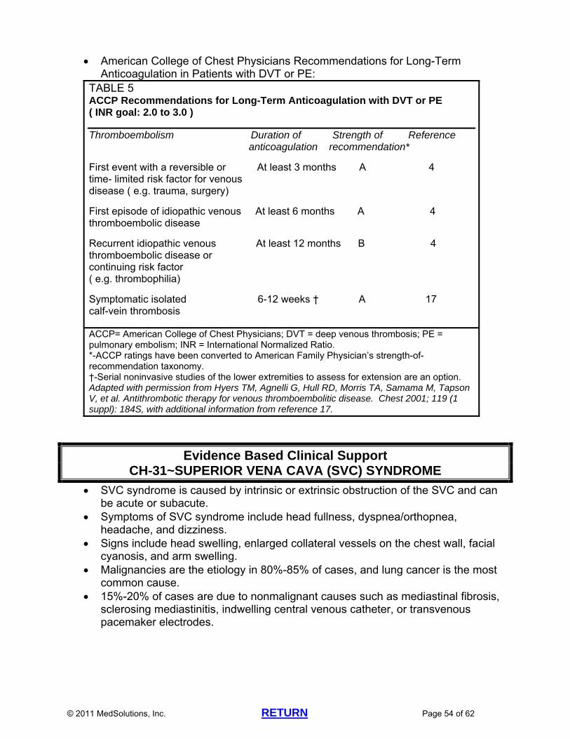
© 2011 MedSolutions, Inc. RETURN Page 54 of 62
American College of Chest Physicians Recommendations for Long-Term Anticoagulation in Patients with DVT or PE:
TABLE 5 ACCP Recommendations for Long-Term Anticoagulation with DVT or PE ( INR goal: 2.0 to 3.0 )
Thromboembolism Duration of Strength of Reference anticoagulation recommendation*
First event with a reversible or At least 3 months A 4 time- limited risk factor for venous disease ( e.g. trauma, surgery) First episode of idiopathic venous At least 6 months A 4 thromboembolic disease
Recurrent idiopathic venous At least 12 months B 4 thromboembolic disease or continuing risk factor ( e.g. thrombophilia)
Symptomatic isolated 6-12 weeks † A 17 calf-vein thrombosis
ACCP= American College of Chest Physicians; DVT = deep venous thrombosis; PE = pulmonary embolism; INR = International Normalized Ratio. *-ACCP ratings have been converted to American Family Physician’s strength-of-recommendation taxonomy. †-Serial noninvasive studies of the lower extremities to assess for extension are an option. Adapted with permission from Hyers TM, Agnelli G, Hull RD, Morris TA, Samama M, Tapson V, et al. Antithrombotic therapy for venous thromboembolitic disease. Chest 2001; 119 (1 suppl): 184S, with additional information from reference 17.
Evidence Based Clinical Support CH-31~SUPERIOR VENA CAVA (SVC) SYNDROME
SVC syndrome is caused by intrinsic or extrinsic obstruction of the SVC and can be acute or subacute.
Symptoms of SVC syndrome include head fullness, dyspnea/orthopnea, headache, and dizziness.
Signs include head swelling, enlarged collateral vessels on the chest wall, facial cyanosis, and arm swelling.
Malignancies are the etiology in 80%-85% of cases, and lung cancer is the most common cause.
15%-20% of cases are due to nonmalignant causes such as mediastinal fibrosis, sclerosing mediastinitis, indwelling central venous catheter, or transvenous pacemaker electrodes.
© 2011 MedSolutions, Inc. RETURN Page 55 of 62
Evidence Based Clinical Support CH-32~THORACIC AORTIC DISSECTION OR ANEURYSM
For confirming or ruling out thoracic aortic dissection, transesophageal echo (TEE), CT, and MRI have equally reliable diagnostic values.*
*Arch Intern Med 2006;166(13):1350-1356 Thoracic aortic aneurysms (TAA) occur in the ascending aorta (25%), aortic arch
(25%) or descending aorta (50%). Risk factors include connective tissue disorders (e.g. Marfan’s or Ehlers-Danlos),
atherosclerosis, previous aortic dissection, prolonged hypertension, and trauma. Mean age is 65 years old. Most patients are asymptomatic until the aneurysm begins to leak or expand.
Chest or back pain may indicate acute expansion or leakage. 25% of patients with TAA also have AAA. The normal diameter of the aorta is 2.5 cm to 3 cm. The normal diameter of the aortic root is 3.5 cm. The usual size of a TAA is 4 to 5 cm. Risk of rupture at 5 years is 0% for TAA less than 4 cm, 16% for diameter 4-5.9
cm, and 31% for aneurysms greater than 6 cm. The critical point for rupture or dissection of an ascending TAA is 6 cm (31% risk)
and for a descending TAA, 7 cm (43% risk). Surgery is usually recommended if the aneurysm is 5.5 cm in the ascending
aorta or 6.5 cm in the descending aorta. Ann Thorac Surg 2002;74:S1877-S1880 Surgery is recommended earlier (when aneurysm is 5 cm) in Marfan’s patients. The median size of an ascending aortic or arch aneurysm at rupture or dissection
is 5.9 cm. All symptomatic TAA’s require surgery or intervention regardless of size.
© 2011 MedSolutions, Inc. RETURN Page 56 of 62
2011 CHEST IMAGING GUIDELINE REFERENCES
CHEST GUIDELINE REFERENCES CH-2~Lymphadenopathy
van Overhagen H, Brakel K, Heijenbrok MW, et al. Metastases in supraclavicular lymph nodes in lung cancer: assessment with palpation, US, and CT. Radiology 2004;232:75-80
Eliot Fishman. “Ask the Fish” e-mail communication. Johns Hopkins Medicine, http://www.ctisus.com. Accessed July 2, 2007.
Ferrer R. Lymphadenopathy: differential diagnosis and evaluation. American Family Physician 1998 Oct. http://www.aafp.org/afp/981015ap/ferrer.html. Accessed June 6, 2011.
Lehman CD, DeMartini W, Anderson BO, et al. Indications for breast MRI in the patient with newly diagnosed breast cancer. J Natl Compr Canc Netw 2009;7(2):193-201.
Yamaguchi H, Ishikawa M, Hatanaka K, et al. Occult breast cancer presenting as axillary metastases. The Breast 2006;15:259-262.
CH-3~Chronic Cough D’Urzo A, Jugovic P. Chronic cough: three most common causes.
Can Fam Physician 2002 August;48:1311-1316. Irwin RS, Baumann MH, Boulet LP, et al. Diagnosis and management of cough—
Executive Summary. Chest 2006;129:1S-23S. CH-4~Non-Cardiac Chest Pain
Smith M, Lawrence DJ, Rowell RM. Management of chest pain: exploring the views and experiences of chiropractors and medical practitioners in a focus group interview. Chiropractic & Osteopathy 2005;13:18.
Karlson BW, Sjoland H, Wahrborg P, Lindquist J, Herlitz J. Patients discharged from emergency care after acute myocardial infarction was ruled out: early follow-up in relation to gender. Eur J Emerg Med 1997 Jun;4(2):72-80.
Klinkman MS, Stevens D, Gorenflo DW. Episodes of care for chest pain: a preliminary report from MIRNET. Michigan Research Network. J Fam Pract 1994 Apr;38(4):345-352.
Hiebert CA. Clinical Features.In Pearson FG, Deslauriers J, Ginsberg RJ, et al. (Eds.). Thoracic Surgery. New York, Churchill Livingstone, Inc., 1995, pp.68-69.
Mackinnon S, Patterson GA, Urschel HC, Jr. Thoracic Outlet Syndromes.In Pearson FG, Deslauriers J, Ginsberg RJ, et. al. (Eds.). Thoracic Surgery. New York, Churchill Livingstone, Inc.1995, p.1221.
CH-6~Hemoptysis Bidwell JL, Pachner RW. Hemoptysis: diagnosis and management. Am Fam Physician 2005;72:1253-1260. Revel MP, Fournier LS, Hennebicque AS, et al. Can CT replace bronchoscopy in the
detection of the site and cause of bleeding in patients with large or massive hemoptysis? AJR 2002 Nov;179(5):1217-1224.
CH-7~Bronchiectasis Emmons EE. Bronchiectasis. eMedicine, May 13, 2011,
http://emedicine.medscape.com/article/354167-overview#a24. Accessed May 23, 2011.
CH-10~Chronic Obstructive Pulmonary Disease Swierzewski SJ. COPD diagnosis. Healthcommunities.com,, June 1, 2000, updated
May 3, 2011, http://www.healthcommunities.com/copd/diagnosis.shtml. Accessed May 24, 2011.
© 2011 MedSolutions, Inc. RETURN Page 57 of 62
Making the Diagnosis of COPD. National Lung Health Education Program, http://www.nlhep.org. Accessed October 31, 2007.
Buist AS, Rodriguez-Roisin R, Anzueto A, et al. Global Initiative for Chronic Obstructive Lung Disease: Pocket guide to COPD diagnosis, management, and prevention. National Institutes of Health, National Heart, Lung, and Blood Institute; April 2001 (updated December 2010 ). Accessed May 24, 2011.
Austin JHM. Pulmonary emphysema: Imaging assessment of lung volume reduction surgery. Radiology 1999;212:1-3.
CH-11~Interstitial Disease Misumi S and Lynch DA. Idiopathic pulmonary fibrosis/Usual interstitial pneumonia.
Imaging diagnosis, spectrum of abnormalities, and temporal progression. Proceedings of the American Thoracic Society 2006;3:307-314.
CH-12~Multiple Pulmonary Nodules Libby DM, Smith JP, Altorki NK, et al. Managing the small pulmonary nodule
discovered by CT. Chest 2004 April;125(4):1522-1529. MacMahon H, Austin JHM, Gamsu G, et al. Guidelines for management of small
pulmonary nodules detected on CT scans: a statement from the Fleischner Society. Radiology 2005;237:395-400.
CH-14~Positive PPD or Tuberculosis (TB) Kim WS, Moon WK, Kim IO, et al. Pulmonary tuberculosis in children: evaluation with
CT. AJR 1997 April;168(4):1005-1009. Uzum K, Karahan OI, Dogan S, et al. Chest radiography and thoracic computed
tomography findings in children who have family members with active pulmonary tuberculosis. Eur J Radiol 2003 Dec;48(3):258-262.
CH-15~Sarcoid Hantous-Zannad S, Charrada L, Zidi A, et al. Value of CT scanning in the
investigation of thoracic sarcoidosis. Rev Mal Respir 2003 April;20(2 pt 1):207-213. Okumura W, Iwasaki T, Toyama T, et al. Usefulness of fasting 18F-FDG PET in
identification of cardiac sarcoidosis. J Nucl Med 2004;45(12):1989-1998. CH-16~Solitary Pulmonary Nodule (SPN)
MacMahon H, Austin JHM, Gamsu G, et al. Guidelines for management of small pulmonary nodules detected on CT scans: a statement from the Fleischner Society. Radiology 2005;237:395-400.
Henschke CI, Yankelevitz DF, Naidich DP, et al. CT screening for lung cancer: suspiciousness of nodules according to size on baseline scans. Radiology 2004;231:164-168.
McCarville MB, Lederman HM, Santana VM. Distinguishing benign from malignant pulmonary nodules with helical chest CT in children with malignant solid tumors. Radiology 2006 May;239(2):514-520.
Libby DM, Smith JP, Altorki NK, et al. Managing the small pulmonary nodule discovered by CT. Chest 2004 April;125(4):1522-1529.
Fletcher JW, Kymes SM, Gould M, et al. A comparison of the diagnostic accuracy of 18F-FDG PET and CT in the characterization of solitary pulmonary nodules. J Nucl Med 2008;49:179-185.
Winer-Muram HT. The solitary pulmonary nodule. Radiology 2006 April;239(1):34-49.
Henschke CI, Yankelevitz DF, Mirtcheva R, et al. CT screening for lung cancer: frequency and significance of part-solid and nonsolid nodules. AJR 2002 May:178(5):1053-1057.
ACR Appropriateness Criteria, Solitary pulmonary nodule, 2008. CH-17~Pleural-Based Nodules and Other Abnormalities
MacMahon H, Austin JHM, Gamsu G, et al. Guidelines for management of small
© 2011 MedSolutions, Inc. RETURN Page 58 of 62
pulmonary nodules detected on CT scans: a statement from the Fleischner Society. Radiology 2005;237:395-400.
CH-18~Pleural Thickening Meziane MA and Lababede O. Imaging in localized fibrous tumor of the pleura.
eMedicine, February 21, 2010, http://emedicine.medscape.com/article/359358-overview. Accessed May 24, 2011.
CH-19~Pleural Effusion Rubins J. Pleural effusion. eMedicine, April 25, 2011,
http://emedicine.medscape.com/article/299959-overview. Accessed May 24, 2011. Sriram PS, Antony VB, Holm KA. Pleural Effusions: How to confirm the cause and
manage effectively. Consultant, Sept 15, 2007, http://www.consultantlive.com. Accessed May 24, 2011.
Rusch VW. Pleural Effusion: benign and malignant. In Pearson FG, Deslauriers J, Gingsberg RJ, et al. (Eds.). Thoracic Surgery. New York, Churchill Livingstone, Inc., 1995, pp.1003-1008.
CH-21~Mediastinal Lymphadenopathy Bronson JG. Looking at the lungs: 2005. Imaging Economics 2005 Feb,p.37.
CH-23~Chest Trauma Mahoney LK and Doty CI. Rib fracture. eMedicine, June 28, 2010,
http://emedicine.medscape.com/article/825981-overview. Accessed May 24, 2010. CH-24~Costochondritis
Flowers LK. Costochondritis. eMedicine, August 25, 2009, http://emedicine.medscape.com/article/808554-overview. Accessed May 24, 2011.
CH-25~Chest Wall Mass Kuhlman JE, Bouchardy L, Fishman EK, Zerhouni EA. CT and MR imaging
evaluation of chest wall disorders. RadioGraphics 1994 May;14(3):571-595. CH-26~Pectus Excavatum and Pectus Carinatum
Hebra A. Pectus excavatum. eMedicine, July 22, 2010, http://emedicine.medscape.com/article/1004953-overview. Accessed May 24, 2011.
CH-27~Breast Abnormalities Saslow D, Boetes C, Burke W, et al. American Cancer Society guidelines for breast
screening with MRI as an adjunct to mammography. CA Cancer J Clin 2007;57:75-89.
Institute for Clinical Systems Improvement (ICSI), Diagnosis of Breast Disease. Jan 2010.http://www.icsi.org/breast_disease_diagnosis/diagnosis_of_breast_disease_2.html. Accessed May 24, 2011.
Silverstein MJ, Lagios MD, Recht A, et al. Image-detected breast cancer: state of the art diagnosis and treatment. J Am Coll Surg 2005 Oct;201(4):586-597.
Moy L, Elias K, Patel V, et al. Is breast MRI helpful in the evaluation of inconclusive mammographic findings? AJR 2009;193:986-993.
Lehman CD, Gatsonis C, Kuhl CK, et al. MRI evaluation of the contralateral breast in women with recently diagnosed breast cancer. N Engl J Med 2007 March;356(13):1295-1303.
Huff JG. Clinical Applications of Breast MRI: Current Indications and Examples. Presented at: Identification and Management of Breast Cancer, October 6, 2007; Nashville, TN.
ACR Practice Guideline for the Performance of Contrast-Enhanced Magnetic Resonance Imaging (MRI) of the Breast. Revised 2008.
Berg WA. Tailored supplemental screening for breast cancer: what now and what next? AJR 2009 Feb;192:390-399.
© 2011 MedSolutions, Inc. RETURN Page 59 of 62
American Society of Breast Disease Policy Statement, The Use of Magnetic Resonance Imaging of the Breast for Screening of Women at High Risk of Breast Cancer, June 28, 2004. http://www.asbd.org. Accessed November 27, 2006.
Gokalp G, Topal U. MR imaging in probably benign lesions (BI-RADS category 3) of the breast. Eur J Radiol 2006 Mar;57(3):436-444.
ACR Appropriateness Criteria, Nonpalpable mammographic findings (excluding calcifications), Updated 2010.
Bazzocchi M, Zuiani C, Panizza P, et al. Contrast-enhanced breast MRI in patients with suspicious microcalcifications on mammography: results of a multicenter trial. AJR 2006 Jun;186(6):1723-1732.
Barclay L and Vega C. Noninvasive Tests May Be Insufficiently Accurate to Preclude Breast Biopsy. Medscape Medical News, Feb.10, 2006. http://www.medscape.com. Accessed November 27, 2006.
Institute for Clinical Systems Improvement (ICSI), Diagnosis of Breast Disease. Updated 2007. http://www.icsi.org. Accessed November 28, 2007.
Lawson LL. State of the Art Diagnosis of Breast Abnormalities: From Clinical Exam to MRI. Presented at: Identification and Management of Breast Cancer, October 6, 2007; Nashville, TN.
CH-28~Pulmonary Arteriovenous Fistula (AVM) Halefoglu AM. Rendu-Osler-Weber syndrome presenting with pulmonary
arteriovenous fistula. Australas Radiol 2005 Jun;49(3):242-245. De Cillis E, Burdi N, Bortone AS, et al. Endovascular treatment of pulmonary and
cerebral arteriovenous malformations in patients affected by hereditary haemorrhagic teleangiectasia. Current Pharmaceutical Design 2006;12(10):1243-1248.
CH-29~Pulmonary Embolism (PE) Wells PS, Anderson DR, Rodger M, et al. Derivation of a simple clinical model to
categorize patient’s probability of pulmonary embolism: increasing the models utility with the SimpliRED D-dimer. Thromb Haemost 2000 Mar;83(3):416-420.
Kass SM, Williams PM, Reamy BV. Pleurisy. Am Fam Physician 2007 May;75(9):1357-1364.
Stein PD, Terrin ML, Hales C, et al. Clinical, laboratory, roentgenographic, and electrocardiographic findings in patients with acute pulmonary embolism and no pre-existing cardiac or pulmonary disease. Chest 1991;100(3):598-603.
Writing Group for the Christopher Study Investigators. Effectiveness of managing suspected pulmonary embolism using an algorithm combining clinical probability, D-dimer testing, and computed tomography. JAMA 2006 Jan;295(2):172-179.
Stein PD, Woodard PK, Weg JG, et al. Diagnostic pathways in acute pulmonary embolism: Recommendations of the PIOPED II investigators. Radiology 2007 Jan;242(1):15-21.
Stein PD, Fowler SE, Goodman LR, et al. Multidetector computed tomography for acute pulmonary embolism. N Engl J Med 2006 June;354(22):2317-2327.
Ramzi DW and Leeper KV. DVT and pulmonary embolism: part II. Treatment and prevention. Am Fam Physician 2004 June;69(12):2841-2848.
Palareti G, Cosmi B, Legnani C, et al. d-Dimer testing to determine the duration of anticoagulation therapy. N Engl J Med 2006;355:1780-1789.
Fedullo PF, Auger WR, Kerr KM, et al. Chronic thromboembolic pulmonary hypertension. N Engl J Med 2001;345:1465-1472.
Nijkeuter M, Hovens MMC, Davidson BL, et al. Resolution of thromboemboli in patients with acute pulmonary embolism. A systematic review. Chest 2006;129:192-197.
© 2011 MedSolutions, Inc. RETURN Page 60 of 62
Kline JA, Steuerwald MT, Marchick MR, et al. Prospective evaluation of right ventricular function and functional status 6 months after acute submassive pulmonary embolism: frequency of persistent or subsequent elevation in estimated pulmonary artery pressure. Chest 2009;136:1202-1210.
CH-30~Subclavian Steal Syndrome Whittemore AD and Mannick JA. Subclavian Steal Syndrome. In Sabiston DC and
Lyerly HK. Textbook of Surgery. 15th Ed. Philadelphia, WB Saunders, 1997, pp.1685-1688.
CH-32~Thoracic Aortic Dissection or Aneurysm Evangelista A and Eagle KA. Is the optimal management of acute type a aortic
intramural hematoma evolving? Circulation 2009;120:2029-2032. Song JK, Yim JH, Ahn JM, et al. Outcomes of patients with acute type A aortic
intramural hematoma. Circulation 2009;120;2046-2052. Immer FF, Hagen U, Berdat PA, et al. Risk factors for secondary dilatation of the
aorta after acute type A aortic dissection. European Journal of Cardio-thoracic Surgery 2005;27:654-657.
Tadros TM, Klein MD, and Shapira OM. Ascending aortic dilatation associated with bicuspid aortic valve. Circulation 2009;119:880-890.
Svensson LG, Blackstone EH, Cosgrove DM, III. Surgical options in young adults with aortic valve disease. Curr Probl Cardiol 2003;28:417-480.
Svensson LG. Aortic valve stenosis and regurgitation: an overview of management. J Cardiovasc Surg 2008;49:297-303.
Hiratzka LF, Bakris GL, Beckman JA, et al. 2010 ACCF/AHA/AATS/ACR/ASA/SCA/SCAI/SIR/STS/SVM guidelines for the diagnosis and management of patients with thoracic aortic disease. J Am Coll Cardiol 2010;55:e27-e129.
Albornoz G, Coady MA, Roberts M, et. al. Familial thoracic aortic aneurysms and dissections—incidence, modes of inheritance, and phenotypic patterns. Annals of Thoracic Surgery 2006 Oct;82(4):1400-1405.
CH-33~Elevated Hemidiaphragm Khan AN, Ghanem SA, Irion KL, et al. Imaging in diaphragm injury and paresis.
Medscape, December 1, 2008, http://emedicine.medscape.com/article/355284-overview#a20. Accessed May 25, 2011.
CH-34~Thoracic Outlet Syndrome (TOS) MacKinnon S, Patterson GA, Urschel HC. Thoracic Outlet Syndromes. In Pearson
FG, Deslauriers J, Ginsberg RJ, et al. (Eds.). Thoracic Surgery. New York, Churchill Livingstone, Inc., 1995, pp.1211-1235.
CH-37~Positron-Emission Mammography (PEM) or Naviscan Berg WA, Madsen KS, Schilling K, et al. Breast cancer: comparative effectiveness of
positron emission mammography and MR imaging in presurgical planning for the ipsilateral breast. Radiology, November 12, 2010, http://radiology.rsna.org/content/early/2010/11/12/radiol.10100454.full. Accessed May 25, 2011.
Hendrick RE. Radiation doses and cancer risks from breast imaging studies. Radiology. August 24, 2010, http://radiology.rsna.org/content/early/2010/08/09/radiol.10100570.full. Accessed May 25, 2011.
Weinberg IN, Beylin D, Zavarzin V, et al. Positron emission mammography: high-resolution biochemical breast imaging. Technol Cancer Res Treat 2005 Feb;4(1):55-60.

© 2011 MedSolutions, Inc. RETURN Page 61 of 62
Tafra L, Cheng Z, Uddo J, et al. Pilot clinical trial of 18F-fluorodeoxyglucose positron-emission mammography in the surgical management of breast cancer. Am J Surg 2005 Oct;190(4):628-632.
Murthy K, Aznar M, Thompson CJ. Results of preliminary clinical trials of the positron emission mammography system PEM-I: a dedicated breast imaging system producing glucose metabolic images using FDG. J Nucl Med 2000 Nov;41(11):1851-1858.
CH-38~Breast MR Spectroscopy Bartella L, Morris EA, Dershaw DD, et al. Proton MR spectroscopy with choline peak
as malignancy marker improves positive predictive value for breast cancer diagnosis: preliminary study. Radiology 2006 June;239(3):686-692.
Chest Evidence Based Clinical Support References CH-3~Chronic Cough-Evidence Based Clinical Support
Irwin RS, Baumann MH, Boulet LP, et al. Diagnosis and management of cough—Executive Summary. Chest 2006;129:1S-23S.
Irwin RS, Madison JM. The diagnosis and treatment of cough. N Engl J Med 2000 Dec;343(23):1715-1721.
CH-9~Asbestos Exposure-Evidence Based Clinical Support Cvitanovic S, Znaor L, Konsa T, et al. Malignant and non-malignant asbestos-related
pleural and lung disease: 10-year follow-up study. Croat Med J 2003 Oct;44(5):618-625.
Lebedova J, Dlouha B, Rychla L, et al. Lung function impairment in relation to asbestos-induced pleural lesions with reference to the extent of the lesions and the initial parenchymal fibrosis. Scand J Work Environ Health 2003 Oct;29(5):388-395.
CH-16~Solitary Pulmonary Nodule-Evidence Based Clinical Support Henschke CI, McCauley DI, Yankelevitz DF, et al. Early Lung Cancer Action Project:
overall design and findings from baseline screening. Lancet 1999 July;354(9173):99-105.
Swensen SJ, Jett JR, Hartman TE, et al. CT screening for lung cancer: five-year prospective experience. Radiology 2005;235:259-265.
Henschke CI, Yankelevitz DF, Naidich DP, et al. CT screening for lung cancer: suspiciousness of nodules according to size on baseline scans. Radiology 2004;231:164-168.
Libby DM, Smith JP, Altorki NK, et al. Managing the small pulmonary nodule discovered by CT. Chest 2004 April;125(4):1522-1529.
Benjamin MS, Drucker EA, McLoud TC, Shepard JAO. Small pulmonary nodules: detection at chest CT and outcome. Radiology 2003;226:489-493.
Lindell RM, Hartman TE, Swensen SJ, et al. Lung cancer screening experience: a retrospective review of PET in 22 non-small cell lung carcinomas detected on screening chest CT in a high-risk population. AJR 2005;185:126-131.
CH-27~Breast Abnormalities-Evidence Based Clinical Support Kolb TM, Lichy J, Newhouse JH. Comparison of the performance of screening
mammography, physical examination, and breast US and evaluation of factors that influence them: an analysis of 27,825 patient evaluations. Radiology 2002 Oct;225(1):165-175.
Liberman L, Morris EA, Dershaw DD, et al. MR imaging of the ipsilateral breast in women with percutaneous proven breast cancer. AJR 2003 April;180(4):901-910.
Lee SG, Orel SG, Woo IJ, et al. MR imaging screening of the contralateral breast in patients with newly diagnosed breast cancer: preliminary results. Radiology 2003 March;226(3):773-778.
Lehman CD, Gatsonis C, Kuhl CK, et al. MRI evaluation of the contralateral breast in

© 2011 MedSolutions, Inc. RETURN Page 62 of 62
women with recently diagnosed breast cancer N Engl J Med 2007 March;356(13):1295-1303.
Lee JM, Orel SG, Czerniecki BJ, et al. MRI before reexcision surgery in patients with breast cancer. AJR 2004;182:473-480.
Bedrosian I, Mick R, Orel SG, et al. Changes in the surgical management of patients with breast carcinoma based on preoperative magnetic resonance imaging. Cancer 2003 August;98(3):468-473.
Hata T, Takahashi H, Watanabe K, et al. Magnetic resonance imaging for preoperative evaluation of breast cancer: a comparative study with mammography and ultrasonography. J Am Coll Surg 2004 Feb;198(2):190-197.
Saslow D, Boetes C, Burke W, et al. American Cancer Society guidelines for breast screening with MRI as an adjunct to mammography. CA Cancer J Clin 2007;57:75-89.
CH-29~Pulmonary Embolism-Evidence Based Clinical Support Kruip MJ, Slob MJ, Schijen JH, et al. Use of a clinical decision rule in combination
with D-dimer concentration in diagnostic workup of patients with suspected pulmonary embolism: a prospective management study. Arch Intern Med 2002 July;162(14):1631-1635.
Abcarian PW, Sweet JD, Watabe JT, Yoon HC. Role of a quantitative D-dimer assay in determining the need for CT angiography of acute pulmonary embolism. AJR 2004;182:1377-1381.
Kavanagh EC, O’Hare A, Hargaden G, Murray JG. Risk of pulmonary embolism after negative MDCT pulmonary angiography findings. AJR 2004;182:499-504.
Paterson DI, Schwartzman K. Strategies incorporating spiral CT for the diagnosis of acute pulmonary embolism: a cost-effectiveness analysis. Chest 2001 June;119(6):1791-1800.
Stein PD, Fowler SE, Goodman LR, et al. Multidetector computed tomography for acute pulmonary embolism. N Engl J Med 2006 June;354(22):2317-2327.
Hyers TM, Agnelli G, Hull RD, et. al. Antithrombotic therapy for venous thromboembolic disease. Chest 2001;119(1 suppl):184S.
CH-32 ~Thoracic Aortic Dissection or Aneurysm-Evidence Based Clinical Support Shiga T, Wajima Z, Apfel CC, et al. Diagnostic accuracy of transesophageal
echocardiography, helical computed tomography, and magnetic resonance imaging for suspected thoracic aortic dissection: systematic review and meta-analysis. Arch Intern Med 2006;166(13):1350-1356.
Elefteriades JA. Natural history of thoracic aortic aneurysms: indications for surgery, and surgical versus nonsurgical risks. Ann Thorac Surg 2002;74:S1877-S1880.