36 - biologyscbiologysc.weebly.com/uploads/8/5/8/5/8585406/transport.pdf · apago pdf enhancer a...
TRANSCRIPT

Apago PDF Enhancer
A Part
VI P
lant
For
m a
nd F
unct
ion
Chapter 36 Plant Form
Chapter Outline
36.1 Organization of the Plant Body: An Overview
36.2 Plant Tissues
36.3 Roots: Anchoring and Absorption Structures
36.4 Stems: Support for Above-Ground Organs
36.5 Leaves: Photosynthetic Organs
Introduction
Although the similarities among a cactus, an orchid, and a hardwood tree might not be obvious at first sight, most plants have a basic unity of structure. This unity is reflected in how the plants are constructed; in how they grow, manufacture, and transport their food; and in how their development is regulated. This chapter addresses the question of how a vascular plant is “built.” We will focus on the cells, tissues, and organs that compose the adult plant body. The roots and shoots that give the adult plant its distinct above- and below-ground architecture are the final product of a basic body plan first established during embryogenesis, a process we will explore in detail in this chapter.
CHAPTER
rav32223_ch36_729-752.indd 729rav32223_ch36_729-752.indd 729 11/16/09 11:40:32 AM11/16/09 11:40:32 AM

Apago PDF Enhancer
Shoot apex
Flower
Stipule
Axillary bud
Internode
Node
Vascular system
Primary root
Lateral root
Root apex
Root
Shoot
Petiole
Vein
Blade
Leaflet Leaf
Tendril
Figure 36.1 Diagram of a plant body. Branching root and shoot systems create the plant’s architecture. Each root and shoot has an apex that extends growth. Leaves are initiated at the nodes of the shoot, which also contain axillary buds that can remain dormant, grow to form lateral branches, or make fl owers. A leaf can be a simple blade or consist of multiple parts as shown here. Roots, shoots, and leaves are all connected with vascular (conducting) tissue.
Roots and shoots are composed of three types of tissuesRoots, shoots, and leaves all contain three basic types of tissues: dermal, ground, and vascular tissue. Because each of these tis-sues extend through the root and shoot systems, they are called tissue systems. Plant cell types can be distinguished by the size of their vacuoles, whether they are living or not at maturity, and by the thickness of secretions found in their cellulose cell walls, a dis-tinguishing feature of plant cells (see chapter 4 to review cell structure). Some cells have only a primary cell wall of cellulose, synthesized by the protoplast near the cell membrane. Micro-tubules align within the cell and determine the orientation of the cellulose fibers (figure 36.2a) . Cells that support the plant
36.1 Organization of the Plant Body: An Overview
Learning OutcomesDistinguish between the functions of roots 1. and shoots.Name the three types of tissues in a plant.2. Compare primary growth and secondary 3. growth.
As you learned in chapter 30, the plant kingdom has great di-versity, not only among its many phyla but even within species. The earliest vascular plants, many of which are extinct, did not have a clear differentiation of the plant body into specialized organs such as roots and leaves. Among modern vascular plants, the presence of these or-gans reflects increasing specialization, particularly in relation to the demands of a terrestrial existence. Obtaining water, for example, is a major challenge, and roots are adapted for water absorption from the soil. Leaves, roots, branches, and flowers all exhibit variations in size and number from plant to plant. Development of the form and structure of these parts may be precisely controlled, but some aspects of leaf, stem, and root development are quite flexible. This chapter emphasizes the unifying aspects of plant form, using the flowering plants as a model.
Vascular plants have roots and shootsA vascular plant consists of a root system and a shoot system (figure 36.1) . Roots and shoots grow at their tips, which are called apices (singular, apex). The root system anchors the plant and penetrates the soil, from which it absorbs water and ions crucial for the plant’s nutrition. Root systems are often extensive, and growing roots can exert great force to move matter as they elongate and ex-pand. Roots developed later than the shoot system as an adapta-tion to living on land. The shoot system consists of the stems and their leaves. Stems serve as a scaffold for positioning the leaves, the principal sites of photosynthesis. The arrangement, size, and other features of the leaves are critically important in the plant’s production of food. Flowers, other reproductive or-gans, and ultimately, fruits and seeds are also formed on the shoot (flower morphology and plant reproduction is covered in chapter 42). The iterative (repeating) unit of the vegetative shoot consists of the internode, node, leaf, and axillary bud, but not reproductive structures. An axillary bud is a lateral shoot apex that allows the plant to branch or replace the main shoot if it is eaten by an herbivore. A vegetative axillary bud has the capacity to reiterate the development of the primary shoot. When the plant has shifted to the reproductive phase of development, these axillary buds may produce flowers or floral shoots.
730 part VI Plant Form and Function
rav32223_ch36_729-752.indd 730rav32223_ch36_729-752.indd 730 11/16/09 11:40:39 AM11/16/09 11:40:39 AM

Apago PDF Enhancer
Meristem cell
Meristem cell
Differentiated cell
Differentiated cell
Cell division
Meristem cell
Cell division
Meristem cell Differentiated cell
Cell division
a. b.
Cellulose fiber Cell membrane
Cytosol
Microtubule
Cellulose- forming rosettes
Cytosol
Cell membrane
Parallel cellulose fibers
Primary cell wall
Time
Primary cell wall remains outside as inner layers are laid down
Secondary cell wall 1
Secondary cell wall 2
Figure 36.2 Synthesis of a plant cell wall. a. Cellulose is a glucose polymer that is produced at the cellulose-forming rosettes in the cell membrane to form the cell wall. Cellulose fi bers are laid down parallel to microtubules inside the cell membrane. Additional substances that strengthen and waterproof the cell wall are added to the cell wall in some cell types. b. Some cells extrude additional layers of cellulose, increasing the mechanical strength of the wall. Because new cellulose is produced at the cell, the oldest layers of cellulose are on the outside of the cell wall. All cells have a primary cell wall. Additional layers of cellulose and lignin contribute to the secondary cell wall.
Figure 36.3 Meristem cell division. Plant meristems consist of cells that divide to give rise to a differentiating daughter cell and a cell that persists as a meristem cell.
body have more heavily reinforced cell walls with multiple layers of cellulose. Cellulose layers are laid down at angles to adjacent layers like plywood; this enhances the strength of the cell wall (figure 36.2b). Plant cells contribute to three tissue systems. Dermal tissue, primarily epidermis, is one cell layer thick in most plants, and it forms an outer protective covering for the plant. Ground tissue cells function in storage, photosynthesis, and secretion, in addition to forming fibers that support and protect plants. Vascular tissue conducts fluids and dissolved substances throughout the plant body. Each of these tissues and their many functions are described in more detail in later sections.
Meristems elaborate the body plan throughout the plant’s lifeWhen a seed sprouts, only a tiny portion of the adult plant ex-ists. Although embryo cells can undergo division and differen-tiation to form many cell types, the fate of most adult cells is more restricted. Further development of the plant body de-pends on the activities of meristems, specialized cells found in shoot and root apices, as well as other parts of the plant.
Overview of meristemsMeristems are clumps of small cells with dense cytoplasm and proportionately large nuclei that act as stem cells do in ani-mals. That is, one cell divides to give rise to two cells, of which one remains meristematic, while the other undergoes differen-tiation and contributes to the plant body (figure 36.3). In this way, the population of meristem cells is continually renewed. Molecular genetic evidence supports the hypothesis that ani-mal stem cells and plant meristem cells may also share some common pathways of gene expression. Extension of both root
and shoot takes place as a result of repeated cell divisions and subsequent elongation of the cells produced by the apical meristems. In some vascular plants, including shrubs and most trees, lateral meristems produce an increase in root and shoot diameter.
chapter 36 Plant Form 731www.ravenbiology.com
rav32223_ch36_729-752.indd 731rav32223_ch36_729-752.indd 731 11/16/09 11:40:41 AM11/16/09 11:40:41 AM

Apago PDF Enhancer
Young leaf primordium
Shoot apical meristem
Older leaf primordium
Lateral bud primordium
dermal tissue ground tissue vascular tissue
Root apical meristem
Root cap
400 μm
100 μm
Figure 36.4 Apical meristems. Shoot and root apical meristems extend the plant body above and below ground. Leaf primordia protect the fragile shoot meristem, while the root meristem produces a protective root cap in addition to new root tissue.
moves through the soil. In contrast, leaf primordia shelter the growing shoot apical meristem, which is particularly suscepti-ble to desiccation because of its exposure to air and sun. The apical meristem gives rise to the three tissue systems by first initiating primary meristems. The three primary mer-istems are the protoderm, which forms the epidermis; the procambium, which produces primary vascular tissues (pri-mary xylem for water transport and primary phloem for nutri-ent transport); and the ground meristem, which differentiates further into ground tissue. In some plants, such as horsetails and corn, intercalary meristems arise in stem internodes (spaces between leaf attachments), adding to the internode lengths. If you walk through a cornfield on a quiet summer night when the corn is about knee high, you may hear a soft popping sound. This sound is caused by the rapid growth of the intercalary meristems. The amount of stem elongation that oc-curs in a very short time is quite surprising.
Lateral meristemsMany herbaceous plants (that is, plants with fleshy, not woody stems) exhibit only primary growth, but others also exhibit secondary growth, which may result in a substantial increase of diameter. Secondary growth is accomplished by the lateral meristems—peripheral cylinders of meristematic tissue within the stems and roots that increase the girth (diameter) of gym-nosperms and most angiosperms. Lateral meristems form from ground tissue that is derived from apical meristems. Monocots are the major exception (figure 36.5) . Although secondary growth increases girth in many nonwoody plants, its effects are most dramatic in woody
plants, which have two lateral meristems. Within the bark of a woody stem is the cork cambium—a lat-eral meristem that contributes to the outer bark of the tree. Just beneath the bark is the vascular cambium—a lateral meristem that produces secondary vascular tissue. The vascular cambium
forms between the xylem and phloem in vascular bundles, adding secondary vascular tissue to both
of its sides. Secondary xylem is the main component of wood. Sec-
ondary phloem is very close to the outer surface of a woody stem. Removing the bark of a tree damages the phloem and may eventually kill the tree. Tissues formed from lateral mer-istems, which comprise most of the trunk, branches, and older roots of trees and shrubs, are known as secondary tissues and are collectively called the secondary plant body.
Learning Outcomes Review 36.1The root system anchors plants and absorbs water and nutrients, whereas the shoot system, consisting of stems, leaves, and fl owers carries out photosynthesis and sexual reproduction. The three general types of tissue in both roots and shoots are dermal, ground, and vascular tissue. Primary growth is produced by apical meristems at the tips of roots and shoots; secondary growth is produced by lateral meristems that are peripheral and increase girth.
■ Why are both primary and secondary growth necessary in a woody plant?
Apical meristemsApical meristems are located at the tips of stems and roots (figure 36.4) . During periods of growth, the cells of apical mer-istems divide and continually add more cells at the tips. Tissues derived from apical meristems are called primary tissues, and the extension of the root and stem forms what is known as the primary plant body. The primary plant body comprises the young, soft shoots and roots of a tree or shrub, or the entire plant body in some plants. Both root and shoot apical meristems are composed of delicate cells that need protection (see figure 36.4). The root apical meristem is protected by the root cap, the anatomy of which is described later on. Root cap cells are produced by the root meristem and are sloughed off and replaced as the root
732 part VI Plant Form and Function
rav32223_ch36_729-752.indd 732rav32223_ch36_729-752.indd 732 11/16/09 11:40:44 AM11/16/09 11:40:44 AM

Apago PDF Enhancer
Pri
mar
y S
tem
S
eco
nd
ary
Ste
m
Pri
mar
y R
oo
t S
eco
nd
ary
Ro
ot
Apical growth
Ground meristem
Procambium
Primary xylem
Primary phloem
Lateral growth
Secondary xylem
Secondary phloem
Primary phloem
Primary phloem
Primary xylem
Primary xylem
Vascular cambium Cork cambium
Lateral growth
Vascular cambium
Secondary xylem
Secondary phloem
Apical growth
Primary xylem
Primary phloem
Ground meristem
Procambium
Figure 36.5 Apical and lateral meristems. Apical meristems produce the primary plant body. In some plants, the lateral meristems produce an increase in the girth of a plant. This type of growth is secondary because the lateral meristems were not directly produced by apical meristems. Woody plants have two types of lateral meristems: a vascular cambium that produces xylem and phloem tissues, and a cork cambium that contributes to the bark of a tree.
36.2 Plant Tissues
Learning OutcomesDescribe the functions of dermal, ground, and 1. vascular tissues.Name the three cell types found in ground tissue and 2. their functions.Distinguish between xylem and phloem. 3.
Three main categories of tissue can be distinguished in the plant body. These are (1) dermal tissue on external sur faces that serves a protective function; (2) ground tissue that forms several different internal tissue types and that can participate in photo-synthesis, serve a storage function, or provide structural sup-port; and (3) vascular tissue that conducts water and nutrients.
Dermal tissue forms a protective interface with the environmentDermal tissue derived from an embryo or apical meristem forms epidermis. This tissue is one cell layer thick in most
chapter 36 Plant Form 733www.ravenbiology.com
rav32223_ch36_729-752.indd 733rav32223_ch36_729-752.indd 733 11/16/09 11:40:50 AM11/16/09 11:40:50 AM

Apago PDF Enhancer
Guard cells
Stoma
272 µm
a.
b.
c.
Epidermal cell
Stoma
Epidermal cell
Stomata
Guard cells
Guard cells
200 µm
71 µm
4 µm
plants and forms the outer protective covering of the plant. In young, exposed parts of the plant, the epidermis is covered with a fatty cutin layer constituting the cuticle; in plants such as desert succulents, several layers of wax may be added to the cuticle to limit water loss and protect against ultraviolet dam-age. In some cases, the dermal tissue forms the bark of trees. Epidermal cells, which originate from the protoderm, cover all parts of the primary plant body. A number of types of specialized cells occur in the epidermis, including guard cells, trichomes, and root hairs.
Guard cellsGuard cells are paired, sausage-shaped cells flanking a stoma (plural, stomata), a mouth-shaped epidermal opening. Guard cells, unlike other epidermal cells, contain chloroplasts. Stomata occur in the epidermis of leaves (figure 36.6a) and sometimes on other parts of the plant, such as stems or fruits. The passage of oxygen and carbon dioxide, as well as the diffusion of water in vapor form, takes place almost exclusively through the stomata. There are from 1000 to more than 1 million stomata per square centimeter of leaf surface. In many plants, stomata are more numerous on the lower epidermis of the leaf than on the upper—a factor that helps minimize water loss. Some plants have stomata only on the lower epidermis, and a few, such as water lilies, have them only on the upper epidermis to maximize gas exchange. Guard cell formation is the result of an asymmetrical cell division producing a guard cell and a subsidiary cell that aids in the opening and closing of the stoma. The patterning of these asymmetrical divisions that results in stomatal distribution has intrigued developmental biologists (figure 36.6b, c). Research on mutants that get “confused” about where to position stomata is providing information on the timing of sto-matal initiation and the kind of intercellular communication that triggers guard cell formation. For example, the too many mouths (tmm) mutation that occurs in Arabidopsis disrupts the normal pattern of cell division that spatially separates stomata
Figure 36.6 Stomata. a. A stoma is the space between two guard cells that regulate the size of the opening. Stomata are evenly distributed within the epidermis of monocots and eudicots, but the patterning is quite different. b. A pea (eudicot) leaf with a random arrangement of stomata. c. A maize (corn, a monocot) leaf with stomata evenly spaced in rows. These photomicrographs also show the variety of cell shapes in plants. Some plant cells are boxlike, as seen in maize (c), and others are irregularly shaped, as seen in the jigsaw puzzle shapes of the pea epidermal cells, (b).
Figure 36.7 The too many mouths stomatal mutant. This Arabidopsis mutant plant lacks an essential signal for spacing stomata. Usually a differentiating guard cell pair inhibits differentiation of a nearby cell into a guard cell.
(figure 36.7) . Investigations of this and other stomatal pattern-ing genes revealed a coordinated network of cell–cell commu-nication (see chapter 9) that informs cells of their position relative to other cells and determines cell fate. The TMM gene encodes a membrane-bound receptor that is part of a signaling pathway controlling asymmetrical cell division.
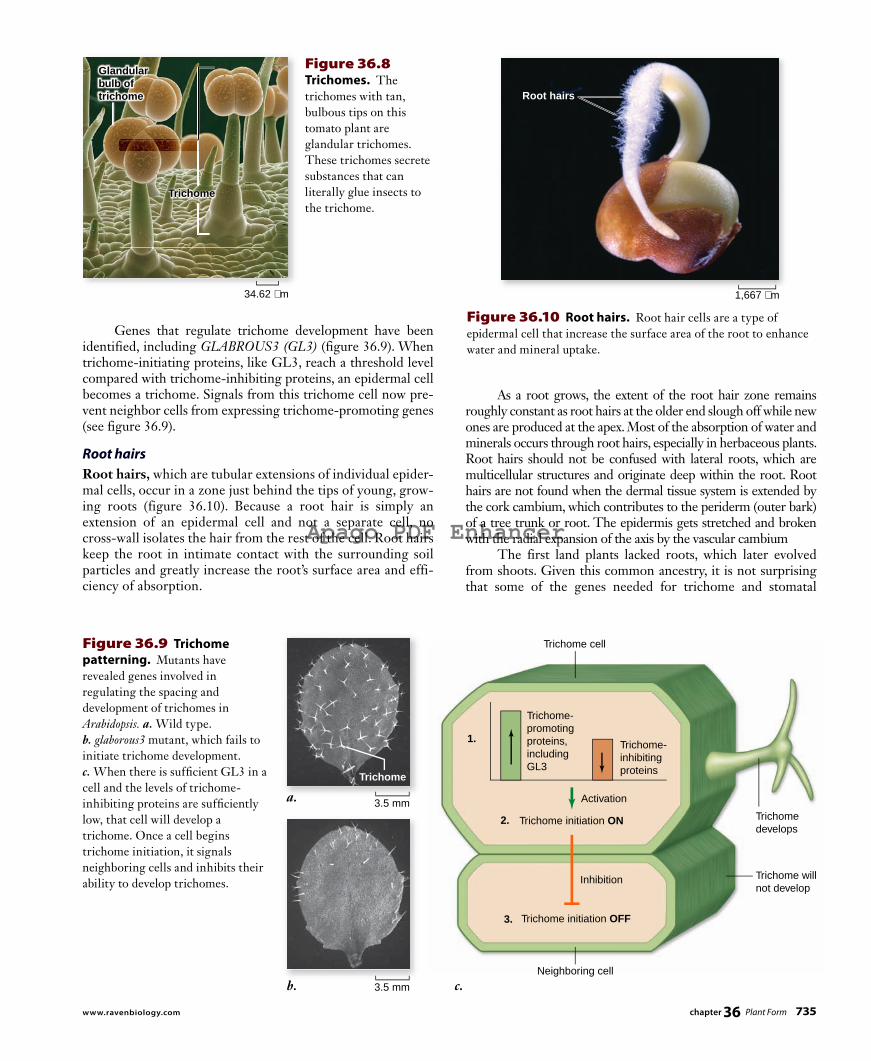
TrichomesTrichomes are cellular or multicellular hairlike outgrowths of the epidermis (figure 36.8) . They occur frequently on stems, leaves, and reproductive organs. A “fuzzy” or “woolly” leaf is covered with trichomes that can be seen clearly with a micro-scope under low magnification. Trichomes keep leaf surfaces cool and reduce evaporation by covering stomatal openings. They also protect leaves from high light intensities and ultra-violet radiation and can buffer against temperature fluctuations. Trichomes can vary greatly in form; some consist of a single cell; others are multicellular. Some are glandular, often secret-ing sticky or toxic substances to deter herbivory.
734 part VI Plant Form and Function
rav32223_ch36_729-752.indd 734rav32223_ch36_729-752.indd 734 11/16/09 11:40:53 AM11/16/09 11:40:53 AM
Apago PDF Enhancer
34.62 µm
Trichome
Glandularbulb oftrichome
a. 3.5 mm
3.5 mm b. c.
Trichome cell
Neighboring cell
Trichome develops
Trichome will not develop
1.
Trichome- promoting proteins, including GL3
Trichome- inhibiting proteins
Trichome initiation ON
Trichome initiation OFF
2.
3.
Inhibition
Activation
Trichome
Root hairs
1,667 µm
Figure 36.8 Trichomes. The trichomes with tan, bulbous tips on this tomato plant are glandular trichomes. These trichomes secrete substances that can literally glue insects to the trichome.
As a root grows, the extent of the root hair zone remains roughly constant as root hairs at the older end slough off while new ones are produced at the apex. Most of the absorption of water and minerals occurs through root hairs, especially in herbaceous plants. Root hairs should not be confused with lateral roots, which are multicellular structures and originate deep within the root. Root hairs are not found when the dermal tissue system is extended by the cork cambium, which contributes to the peri derm (outer bark) of a tree trunk or root. The epidermis gets stretched and broken with the radial expansion of the axis by the vascular cambium The first land plants lacked roots, which later evolved from shoots. Given this common ancestry, it is not surprising that some of the genes needed for trichome and stomatal
Genes that regulate trichome development have been identified, including GLABROUS3 (GL3) (figure 36.9) . When trichome-initiating proteins, like GL3, reach a threshold level compared with trichome-inhibiting proteins, an epidermal cell becomes a trichome. Signals from this trichome cell now pre-vent neighbor cells from expressing trichome-promoting genes (see figure 36.9).
Root hairsRoot hairs, which are tubular extensions of individual epider-mal cells, occur in a zone just behind the tips of young, grow-ing roots (figure 36.10 ). Because a root hair is simply an extension of an epidermal cell and not a separate cell, no cross-wall isolates the hair from the rest of the cell. Root hairs keep the root in intimate contact with the surrounding soil particles and greatly increase the root’s surface area and effi-ciency of absorption.
Figure 36.9 Trichome patterning. Mutants have revealed genes involved in regulating the spacing and development of trichomes in Arabidopsis. a. Wild type. b. glaborous3 mutant, which fails to initiate trichome development. c. When there is suffi cient GL3 in a cell and the levels of trichome-inhibiting proteins are suffi ciently low, that cell will develop a trichome. Once a cell begins trichome initiation, it signals neighboring cells and inhibits their ability to develop trichomes.
Figure 36.10 Root hairs. Root hair cells are a type of epidermal cell that increase the surface area of the root to enhance water and mineral uptake.
chapter 36 Plant Form 735www.ravenbiology.com
rav32223_ch36_729-752.indd 735rav32223_ch36_729-752.indd 735 11/17/09 12:16:44 PM11/17/09 12:16:44 PM

Apago PDF Enhancer
a. b. c. 5.8 µm 120 µm 22 µm
Parenchyma cells have functional nuclei and are capable of dividing, and they usually remain alive after they mature; in some plants (for example, cacti), they may live to be over 100 years old. The majority of cells in fruits such as apples are parenchyma. Some parenchyma contain chloroplasts, especially in leaves and in the outer parts of herbaceous stems. Such photo synthetic parenchyma tissue is called chlorenchyma.
CollenchymaIf celery “strings” have ever been caught between your teeth, you are familiar with tough, flexible collenchyma cells. Like paren-chyma cells, collenchyma cells have living protoplasts and may live for many years. These cells, which are usually a little longer than wide, have walls that vary in thickness (figure 36.11b). Flexible collenchyma cells provide support for plant or-gans, allowing them to bend without breaking. They often form strands or continuous cylinders beneath the epidermis of stems or leaf petioles (stalks) and along the veins in leaves. Strands of collenchyma provide much of the support for stems in the pri-mary plant body.
SclerenchymaSclerenchyma cells have tough, thick walls. Unlike collenchyma and parenchyma, they usually lack living protoplasts at matu-rity. Their secondary cell walls are often impregnated with lignin, a highly branched polymer that makes cell walls more rigid; for example, lignin is an important component in wood. Cell walls containing lignin are said to be lignified. Lignin is common in the walls of plant cells that have a structural or mechanical function. Some kinds of cells have lignin deposited in primary as well as secondary cell walls. Sclerenchyma is present in two general types: fibers and sclereids. Fibers are long, slender cells that are usually grouped
differentiation in shoot epidermal cells also play a role in root hair development.
Inquiry question
? Identify three dermal tissue traits that are adaptive for a terrestrial lifestyle and explain why these traits are advantageous.
Ground tissue cells perform many functions, including storage, photosynthesis, and supportGround tissue consists primarily of thin-walled parenchyma cellsthat function in storage, photosynthesis, and secretion. Other ground tissue, composed of collenchyma cells and sclerenchyma cells, provide support and protection.
ParenchymaParenchyma cells are the most common type of plant cell. They have large vacuoles, thin walls, and are initially (but briefly) more or less spherical. These cells, which have living protoplasts, push up against each other shortly after they are produced, however, and assume other shapes, often ending up with 11 to 17 sides. Parenchyma cells may live for many years; they function in storage of food and water, photosynthesis, and secretion. They are the most abundant cells of primary tissues and may also oc-cur, to a much lesser extent, in secondary tissues (figure 36.11a) . Most parenchyma cells have only primary walls, which are walls laid down while the cells are still maturing. Parenchyma are less specialized than other plant cells, although many variations oc-cur with special functions, such as nectar and resin secretion or storage of latex, proteins, and metabolic wastes.
Figure 36.11 The three types of ground tissue. a. Parenchyma cells. Only primary cell walls are seen in this cross section of parenchyma cells from grass. b. Collenchyma cells. Thickened side walls are seen in this cross section of collenchyma cells from a young branch of elderberry (Sambucus). In other kinds of collenchyma cells, the thickened areas may occur at the corners of the cells or in other kinds of strips. c. Sclereids. Clusters of sclereids (“stone cells”), stained red in this preparation. The surrounding thin-walled cells, stained green, are parenchyma. Sclereids are one type of sclerenchyma tissue, which also contains fi bers.
736 part VI Plant Form and Function
rav32223_ch36_729-752.indd 736rav32223_ch36_729-752.indd 736 11/16/09 11:41:04 AM11/16/09 11:41:04 AM

Apago PDF Enhancer
100 µm
45 µm
Tracheid Vessel
Pits
Water flow
Perforation plate
Vessel member
Pits
broken stream through the xylem from the roots up through the shoot and into the leaves. When the water reaches the leaves, much of it diffuses in the form of water vapor into the intercellular spaces and out of the leaves into the surrounding air, mainly through the stomata. This diffusion of water vapor from a plant is known as transpiration (see chapter 38). In ad-dition to conducting water, dissolved minerals, and inorganic ions such as nitrates and phosphates throughout the plant, xy-lem supplies support for the plant body. Vessel members tend to be shorter and wider than tra-cheids. When viewed with a microscope, they resemble bever-age cans with both ends removed. Both vessel members and tra cheids have thick, lignified secondary walls and no living protoplasts at maturity. Lignin is produced by the cell and se-creted to strengthen the cellulose cell walls before the proto-plast dies, leaving only the cell wall. Tracheids contain pits, which are small, mostly rounded-to-elliptical areas where no secondary wall material has been depos-ited. The pits of adjacent cells occur opposite one another; the continuous stream of water flows through these pits from tracheid to tracheid. In contrast, vessel members, which are joined end to end, may be almost completely open or may have bars or strips of wall material across the open ends (see figure 36.12). Vessels ap-pear to conduct water more efficiently than do the overlapping strands of tracheids. We know this partly because vessel members have evolved from tracheids independently in several groups of plants, suggesting that they are favored by natural selection. In addition to conducting cells, xylem typically includes fibers and parenchyma cells (ground tissue cells). It is probable that some types of fibers have evolved from tracheids, becom-ing specialized for strengthening rather than conducting. The parenchyma cells, which are usually produced in horizontal
together in strands. Linen, for example, is woven from strands of sclerenchyma fibers that occur in the phloem of flax (Linum spp.) plants. Sclereids are variable in shape but often branched. They may occur singly or in groups; they are not elongated, but may have many different forms, including that of a star. The gritty texture of a pear is caused by groups of sclereids that occur throughout the soft flesh of the fruit (figure 36.11c). Sclereids are also found in hard seed coats. Both of these tough, thick-walled cell types serve to strengthen the tissues in which they occur.
Vascular tissue conducts water and nutrients throughout the plantVascular tissue, as mentioned earlier, includes two kinds of conducting tissues: (1) xylem, which conducts water and dis-solved minerals, and (2) phloem, which conducts a solution of carbohydrates —mainly sucrose—used by plants for food. The phloem also transports hormones, amino acids, and other sub-stances that are necessary for plant growth. Xylem and phloem differ in structure as well as in function.
XylemXylem, the principal water-conducting tissue of plants, usually contains a combination of vessels, which are continuous tubes formed from dead, hollow, cylindrical cells arranged end-to-end, and tracheids, which are dead cells that taper at the ends and overlap one another (figure 36.12) . Primary xylem is de-rived from the procambium produced by the apical meristem. Secondary xylem is formed by the vascular cambium, a lateral meristem. Wood consists of accumulated secondary xylem. In some plants (but not flowering plants), tracheids are the only water-conducting cells present; water passes in an un-
Figure 36.12 Comparison between tracheids and vessel members. In tracheids, the water passes from cell to cell by means of pits. In vessel members, water moves by way of perforation plates (as seen in the photomicrograph in this fi gure). In gymnosperm wood, tracheids both conduct water and provide support; in most kinds of angiosperms, vessels are present in addition to tracheids. These two types of cells conduct water, and fi bers provide additional support. The wood of red maple, Acer rubrum, contains both tracheids and vessels as seen in the electron micrographs in this fi gure.
chapter 36 Plant Form 737www.ravenbiology.com
rav32223_ch36_729-752.indd 737rav32223_ch36_729-752.indd 737 11/16/09 11:41:08 AM11/16/09 11:41:08 AM

Apago PDF Enhancer
2 µm
Sieve tube
Sieve-tubemember
Plasmodesma
Nucleus
Companion cell
Sieve plate
Water andnutrient flow
Cell membrane
a. b.
are living, but most sieve cells and all sieve-tube members lack a nucleus at maturity. In sieve-tube members, some sieve areas have larger pores and are called sieve plates (figure 36.13) . Sieve-tube members occur end to end, forming longitudinal series called sieve tubes. Sieve cells are less specialized than sieve-tube members, and the pores in all of their sieve areas are roughly of the same di-ameter. Sieve-tube members are more specialized, and presum-ably, more efficient than sieve cells. Each sieve-tube member is associated with an adjacent, specialized parenchyma cell known as a companion cell. Com-panion cells apparently carry out some of the metabolic func-tions needed to maintain the associated sieve-tube member. In angiosperms, a common initial cell divides asymmetrically to produce a sieve-tube member cell and its companion cell. Com-panion cells have all the components of normal parenchyma cells, including nuclei, and numerous plasmodesmata (cytoplas-mic connections between adjacent cells) connect their cyto-plasm with that of the associated sieve-tube members. Sieve cells in nonflowering plants have albuminous cells that function as companion cells. Unlike a companion cell, an albuminous cell is not necessarily derived from the same mother cell as its associated sieve cell. Fibers and parenchyma cells are often abundant in phloem.
Learning Outcomes Review 36.2Dermal tissue protects a plant from its environment and contains specialized cells such as guard cells, trichomes, and root hairs. Ground tissue serves several functions, including storage (parenchyma cells), photosynthesis (specialized parenchyma called chlorenchyma), and structural support (collenchyma and sclerenchyma). Vascular tissue carries water through the xylem (primarily vessels) and nutrients through the phloem (primarily sieve-tube members).
■ Contrast the structure and function of mature vessels and sieve-tube members.
rows called rays by special ray initials of the vascular cambium, function in lateral conduction and food storage. (An initial is another term for a meristematic cell. It divides to produce an-other initial and a cell that differentiates.) In cross sections of woody stems and roots, the rays can be seen radiating out from the center of the xylem like the spokes of a wheel. Fibers are abundant in some kinds of wood, such as oak (Quercus spp.), and the wood is correspondingly dense and heavy. The arrangements of these and other kinds of cells in the xylem make it possible to identify most plant genera and many species from their wood alone. Over 2000 years ago paper as we recognize it today was made in China by mashing herbaceous plants in water and sepa-rating out a thin layer of phloem fibers on a screen. Not until the third century of the common era did the secret of making paper make its way out of China. Today the ever-growing de-mand for paper is met by extracting xylem fibers from wood, including spruce, that is relatively soft, having fewer ray fibers than oak. The lignin-rich cell walls yield brown paper that is often bleached. In addition, tissues from many other plants have been developed as sources of paper, including kenaf and hemp. United States paper currency is 75% cotton and 25% flax.
PhloemPhloem, which is located toward the outer part of roots and stems, is the principal food-conducting tissue in vascular plants. If a plant is girdled (by removing a substantial strip of bark down to the vascular cambium around the entire circumference), the plant eventually dies from starvation of the roots. Food conduction in phloem is carried out through two kinds of elongated cells: sieve cells and sieve-tube members. Gymnosperms, ferns, and horsetails have only sieve cells; most angiosperms have sieve-tube members. Both types of cells have clusters of pores known as sieve areas because the cell walls re-semble sieves. Sieve areas are more abundant on the overlap-ping ends of the cells and connect the protoplasts of adjoining sieve cells and sieve-tube members. Both of these types of cells
Figure 36.13 A sieve-tube member. a. Sieve-tube member cells are stacked, with sieve plates forming the connection. The narrow cell with the nucleus at the right of the sieve-tube member is a companion cell. This cell nourishes the sieve-tube members, which have plasma membranes, but no nuclei. b. Looking down into sieve plates in squash phloem reveals the perforations through which sucrose and hormones move.
© Dr. Richard Kessel & Dr. Gene Shih/Visuals Unlimited
738 part VI Plant Form and Function
rav32223_ch36_729-752.indd 738rav32223_ch36_729-752.indd 738 11/16/09 11:41:12 AM11/16/09 11:41:12 AM

Apago PDF Enhancer
Zone of maturation
Zone of elongation
Zone of cell division
Epidermis
Ground meristem
Procambium
Columella cells
Protoderm
Endodermis
Ground tissue
Vascular tissue
Quiescent center
Root in cross-section
Apical meristem Root cap
dermal tissue ground tissue vascular tissue
Root hair
400 µm
The root capThe root cap has no equivalent in stems. It is composed of two types of cells: the inner columella cells (they look like columns), and the outer, lateral root cap cells, which are continuously re-plenished by the root apical meristem. In some plants with larger roots, the root cap is quite obvious. Its main function is to protect the delicate tissues behind it as growth extends the root through mostly abrasive soil particles. Golgi bodies in the outer root cap cells secrete and re-lease a slimy substance that passes through the cell walls to the outside. The root cap cells, which have an average life of less than a week, are constantly being replaced from the inside, forming a mucilaginous lubricant that eases the root through the soil. The slimy mass also provides a medium for the growth of beneficial nitrogen-fixing bacteria in the roots of plants such as legumes. A new root cap is produced when an existing one is artificially or accidentally removed from a root. The root cap also functions in the perception of gravity. The columella cells are highly specialized, with the endoplas-mic reticulum in the periphery and the nucleus located at either the middle or the top of the cell. They contain no large vacu-oles. Columella cells contain amyloplasts (plastids with starch grains) that collect on the sides of cells facing the pull of gravity. When a potted plant is placed on its side, the amyloplasts drift or tumble down to the side nearest the source of gravity, and the root bends in that direction. Lasers have been used to ablate (kill) individual columella cells in Arabidopsis. It turns out that only two columella cells are sufficient for gravity sensing! The precise nature of the gravita-tional response is unknown, but some evidence indicates that calcium ions in the amyloplasts influence the distribution of growth hormones (auxin in this case) in the cells. Multiple sig-naling mechanisms may exist, because bending has been observed in the absence of auxin. A current hypothesis is that an electrical signal moves from the columella cell to cells in the elongation zone (the region closest to the zone of cell division).
The zone of cell divisionThe apical meristem is located in the center of the root tip in the area protected by the root cap. Most of the activity in this zone of cell division takes place toward the edges of the meri-stem, where the cells divide every 12 to 36 hours, often coordi-nately, reaching a peak of division once or twice a day. Most of the cells are essentially cuboidal, with small vacu-oles and proportionately large, centrally located nuclei. These rapidly dividing cells are daughter cells of the apical meristem. A group of cells in the center of the root apical meristem, termed the quiescent center, divide only very infrequently. The presence of the quiescent center makes sense if you think about a solid ball expanding—the outer surface would have to in-crease far more rapidly than the very center. The apical meristem daughter cells soon subdivide into the three primary tissues previously discussed: protoderm, procambi-um, and ground meristem. Genes have been identified in the rela-tively simple root of Arabidopsis that regulate the patterning of these tissue systems. The patterning of these cells begins in this zone, but the anatomical and morphological expression of this patterning is not fully revealed until the cells reach the zone of maturation.
36.3 Roots: Anchoring and Absorption Structures
Learning OutcomesDescribe the four regions of a typical root.1. Explain the function of root hairs.2. Describe functions of modified roots.3.
Roots have a simpler pattern of organization and development than stems, and we will consider them first. Keep in mind, how-ever, that roots evolved after shoots and are a major innovation for terrestrial living.
Roots are adapted for growing underground and absorbing water and solutesFour regions are commonly recognized in developing roots: the root cap, the zone of cell division, the zone of elongation, and the zone of maturation (figure 36.14 ). In these last three zones, the boundaries are not clearly defined. When apical initials divide, daughter cells that end up on the tip end of the root become root cap cells. Cells that divide in the opposite direction pass through the three other zones before they finish differentiating. As you consider the different zones, visualize the tip of the root moving deeper into the soil, actively growing. This counters the static image of a root that diagrams and photos convey.
Figure 36.14 Root structure. A root tip in corn, Zea mays.
chapter 36 Plant Form 739www.ravenbiology.com
rav32223_ch36_729-752.indd 739rav32223_ch36_729-752.indd 739 11/16/09 11:41:15 AM11/16/09 11:41:15 AM

Apago PDF Enhancer
Hair cell (no WER)
Nonhair cell (WER expressed)
b. a.
WER (wild type) wer (mutant)
Root tip Epidermal cell
WER
No WER
No WER
No WER
Nonhair
Hair will develop in zone of maturation
Hair will develop in zone of maturation
50 µm
SCR is expressed onlyin endodermal cells
b.a.
SCR (wild type) scr (mutant)
Root tip
Endodermalcell
SCR
Cell withground andendodermaltraitsAsymmetrical
division
Groundcell
Groundmeristem
cell
2 layers of cells
Root tip
Groundmeristem
cell
SCR
400 μm
Figure 36.16 Scarecrow regulates asymmetrical cell division. a. SCR is needed for an asymmetrical cell division leading to the differentiation of daughter cells into endodermal and ground cells. b. The SCR promoter was attached to a gene coding for a green fl uorescent protein to fi nd out exactly where in the wild type root SCR is expressed. SCR is only expressed in the endodermal cells, not the ground cells.
Figure 36.15 Tissue-specifi c gene expression. a. The WEREWOLF gene of Arabidopsis is expressed in some, but not all, epidermal cells and suppresses root hair development. The wer mutant is covered with root hairs. b. The WER promoter was attached to a gene coding for a green fl uorescent protein and used to make a transgenic plant. The green fl uorescence shows the nonhair epidermal cells where the gene is expressed. The red visually indicates cell boundaries because cell walls autofl uoresce.
The zone of elongationIn the zone of elongation, roots lengthen because the cells produced by the primary meristems become several times lon-ger than wide, and their width also increases slightly. The small vacuoles present merge and grow until they occupy 90% or more of the volume of each cell. No further increase in cell size occurs above the zone of elongation. The mature parts of the root, except for increasing in girth, remain stationary for the life of the plant.
The zone of maturationThe cells that have elongated in the zone of elongation become differentiated into specific cell types in the zone of maturation (see figure 36.14). The cells of the root surface cylinder mature into epidermal cells, which have a very thin cuticle, and include both root hair and nonhair cells. Although the root hairs are not visible until this stage of development, their fate was established much earlier, as you saw with the expression patterns of WER (see figure 36.15).
For example, the WEREWOLF (WER) gene is required for the patterning of the two root epidermal cell types, those with and those without root hairs (figure 36.15). Plants with the wer mutation have an excess of root hairs because WER is needed to prevent root hair development in nonhair epidermal cells. Simi-larly, the SCARECROW (SCR) gene is necessary in ground cell differentiation (figure 36.16). A ground meristem cell undergoes an asymmetrical cell division that gives rise to two nested cylin-ders of cells from one if SCR is present. The outer cell layer be-comes ground tissue and serves a storage function. The inner cell layer forms the endodermis, which regulates the intercellular flow of water and solutes into the vascular core of the root (see figure 36.5). The scr mutant, in contrast, forms a single layer of cells that have both endodermal and ground cell traits. SCR illustrates the importance of the orientation of cell division. If a cell’s relative position changes because of a mistake in cell division or the ablation of another cell, the cell develops according to its new position. The fate of most plant cells is determined by their position relative to other cells.
740 part VI Plant Form and Function
rav32223_ch36_729-752.indd 740rav32223_ch36_729-752.indd 740 11/16/09 11:41:17 AM11/16/09 11:41:17 AM
Apago PDF Enhancer
1250 µm
Epidermis
Cortex
Endodermis Location of Casparian strip
Pericycle
Primary phloem
Primary xylem
Pith
Mo
no
cot
Eu
dic
ot
Casparian strip
Endodermal cell H2O
H2O
Endodermis
Cortex
Epidermis
Endodermis Location of Casparian strip
Primary xylem
Pericycle
Primary phloem
Xylem
Phloem
Cortex
Pericycle
48 µm
385 µm
8 µm
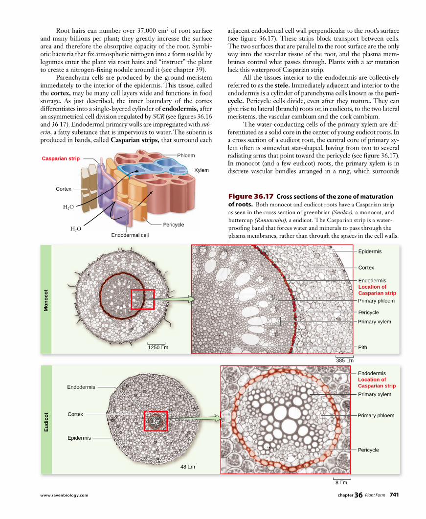
adjacent endodermal cell wall perpendicular to the root’s surface (see figure 36.17). These strips block transport between cells. The two surfaces that are parallel to the root surface are the only way into the vascular tissue of the root, and the plasma mem-branes control what passes through. Plants with a scr mutation lack this waterproof Casparian strip. All the tissues interior to the endodermis are collectively referred to as the stele. Immediately adjacent and interior to the endodermis is a cylinder of parenchyma cells known as the peri-cycle. Pericycle cells divide, even after they mature. They can give rise to lateral (branch) roots or, in eudicots, to the two lateral meristems, the vascular cambium and the cork cambium. The water-conducting cells of the primary xylem are dif-ferentiated as a solid core in the center of young eudicot roots. In a cross section of a eudicot root, the central core of primary xy-lem often is somewhat star-shaped, having from two to several radiating arms that point toward the pericycle (see figure 36.17). In monocot (and a few eudicot) roots, the primary xylem is in discrete vascular bundles arranged in a ring, which surrounds
Root hairs can number over 37,000 cm2 of root surface and many billions per plant; they greatly increase the surface area and therefore the absorptive capacity of the root. Symbi-otic bacteria that fix atmospheric nitrogen into a form usable by legumes enter the plant via root hairs and “instruct” the plant to create a nitrogen-fixing nodule around it (see chapter 39). Parenchyma cells are produced by the ground meristem immediately to the interior of the epidermis. This tissue, called the cortex, may be many cell layers wide and functions in food storage. As just described, the inner boundary of the cortex differentiates into a single-layered cylinder of endodermis, after an asymmetrical cell division regulated by SCR (see figures 36.16 and 36.17). Endodermal primary walls are impregnated with sub-erin, a fatty substance that is impervious to water. The suberin is produced in bands, called Casparian strips, that surround each
Figure 36.17 Cross sections of the zone of maturation of roots. Both monocot and eudicot roots have a Casparian strip as seen in the cross section of greenbriar (Smilax), a monocot, and buttercup (Ranunculus), a eudicot. The Casparian strip is a water-proofi ng band that forces water and minerals to pass through the plasma membranes, rather than through the spaces in the cell walls.
chapter 36 Plant Form 741www.ravenbiology.com
rav32223_ch36_729-752.indd 741rav32223_ch36_729-752.indd 741 11/16/09 11:41:20 AM11/16/09 11:41:20 AM

Apago PDF Enhancer
Zygote Embryo
Shoot apical meristem
Root apical meristem
Cork cambium
Vascular cambium
Leaf primordia
Bud primordia
Shoot elongation
Outer bark
Phloem
Xylem
Inner bark
Wood
Bark
Bark
Leaves
Lateral shoots
Cork cambium
Vascular cambium
Pericycle
Phloem
Xylem
Lateral roots
Root elongation
Outer bark
Inner bark
Wood
Figure 36.18 Stages in the diff erentiation of plant tissues.
parenchyma cells, called pith, at the very center of the root (see figure 36.17). Primary phloem, composed of cells involved in food conduction, is differentiated in discrete groups of cells adja-cent to the xylem in both eudicot and monocot roots. In eudicots and other plants with secondary growth, part of the pericycle and the parenchyma cells between the phloem patches and the xylem become the root vascular cambium, which starts producing secondary xylem to the inside and sec-ondary phloem to the outside. Eventually, the secondary tissues acquire the form of concentric cylinders. The primary phloem, cortex, and epidermis become crushed and are sloughed off as more secondary tissues are added. In the pericycle of woody plants, the cork cambium con-tributes to the outer bark, which will be discussed in more de-tail when we look at stems. In the case of secondary growth in eudicot roots, everything outside the stele is lost and replaced with bark. Figure 36.18 summarizes the process of differentia-tion that occurs in plant tissue.
Modifi ed roots accomplish specialized functionsMost plants produce either a taproot system, characterized by a single large root with smaller branch roots, or a fibrous root sys-tem, composed of many smaller roots of similar diameter. Some plants, however, have intriguing root modifications with specific functions in addition to those of anchorage and absorption. Not all roots are produced by preexisting roots. Any root that arises along a stem or in some place other than the root of the plant is called an adventitious root. For example, climbing plants such as ivy produce roots from their stems; these can anchor the stems to tree trunks or to a brick wall. Adventitious root formation in ivy depends on the developmental stage of the shoot. When the shoot enters the adult phase of develop-ment, it is no longer capable of initiating these roots. Below we investigate functions of modified roots.
Prop roots. Some monocots, such as corn, produce thick adventitious roots from the lower parts of the stem. These so-called prop roots grow down to the ground and brace
the plants against wind (fi gure 36.19a) . Adventious roots are common in wetland plants, allowing them to tolerate wet conditions.
Aerial roots. Plants such as epiphytic orchids, which are attached to tree branches and grow unconnected to the ground (but are not parasites), have roots that extend into the air (fi gure 36.19b). Some aerial roots have an epidermis that is several cell layers thick, an adaptation to reduce water loss. These aerial roots may also be green and photosynthetic, as in the vanilla orchid (Vanilla planifolia).
Pneumatophores. Some plants that grow in swamps and other wet places may produce spongy outgrowths called pneumatophores from their underwater roots (fi gure 36.19c). The pneumatophores commonly extend several centimeters above water, facilitating oxygen uptake in the roots beneath (fi gure 36.19c).
Contractile roots. The roots from the bulbs of lilies and from several other plants, such as dandelions, contract by spiraling to pull the plant a little deeper into the soil each year, until they reach an area of relatively stable temperature. The roots may contract to one-third their original length as they spiral like a corkscrew due to cellular thickening and constricting.
Parasitic roots. The stems of certain plants that lack chlorophyll, such as dodder (Cuscuta spp.) , produce peglike roots called haustoria that penetrate the host plants around which they are twined. The haustoria establish contact with the conducting tissues of the host and effectively parasitize their host. Dodder not only weakens plants but can also spread disease when it grows and attaches to several plants.
Food storage roots. The xylem of branch roots of sweet potatoes and similar plants produce at intervals many extra parenchyma cells that store large quantities of carbohydrates. Carrots, beets, parsnips, radishes, and turnips have combinations of stem and root that also function in food storage. Cross sections of these roots reveal multiple rings of secondary growth.
742 part VI Plant Form and Function
rav32223_ch36_729-752.indd 742rav32223_ch36_729-752.indd 742 11/16/09 11:41:23 AM11/16/09 11:41:23 AM

Apago PDF Enhancer
a. b.
c. d. e.
Shootapicalmeristem
Young leafprimordium
Older leafprimordium
67 µm
Figure 36.19 Five types of modifi ed roots. a. Maize (corn) prop roots originate from the stem and keep the plant upright. b. Epiphytic orchids attach to trees far above the tropical soil. Their roots are adapted to obtain water from the air rather than the soil. c. Pneumatophores ( foreground) are spongy outgrowths from the roots below . d. A water storage root weighing over 25 kg (60 pounds). e. Buttress roots of a tropical fi g tree .
Water storage roots. Some members of the pumpkin family (Cucurbitaceae), especially those that grow in arid regions, may produce water storage roots weighing 50 kg or more (fi gure 36.19d).
Buttress roots. Certain species of fi g and other tropical trees produce huge buttress roots toward the base of the trunk, which provide considerable stability (fi gure 36.19e).
Learning Outcomes Review 36.3The root cap protects the root apical meristem and helps to sense gravity. New cells formed in the zone of cell division grow in length in the zone of elongation. Cells diff erentiate in the zone of maturation, and root hairs appear here. Root hairs greatly increase the absorptive surface area of roots. Modifi ed roots allow plants to carry out many additional functions, including bracing, aeration, and storage of nutrients and water.
■ Why do you suppose root hairs are not formed in the region of elongation?
36.4 Stems: Support for Above-Ground Organs
Learning OutcomesList the potential products of an axillary bud.1. Differentiate between cross sections of a monocot stem 2. and a eudicot stem.Describe three functions of modified stems.3.
The supporting structure of a vascular plant’s shoot system is the mass of stems that extend from the root system below ground into the air, often reaching great height. Stiff stems ca-pable of rising upward against gravity are an ancient adaptation that allowed plants to move into terrestrial ecosystems.
Stems carry leaves and fl owers and support the plant’s weightLike roots, stems contain the three types of plant tissue. Stems also undergo growth from cell division in apical and lateral meristems. The stem may be thought of as an axis from which other stems or organs grow. The shoot apical meristems are capable of producing these new stems and organs.
External stem structureThe shoot apical meristem initiates stem tissue and intermit-tently produces bulges (primordia ) that are capable of develop-ing into leaves, other shoots, or even flowers (figure 36.20) .
Figure 36.20 A shoot apex. Scanning electron micrograph of the apical meristem of wheat (Triticum).
chapter 36 Plant Form 743www.ravenbiology.com
rav32223_ch36_729-752.indd 743rav32223_ch36_729-752.indd 743 11/16/09 11:41:23 AM11/16/09 11:41:23 AM
Apago PDF Enhancer
Alternate: Ivy
Opposite: Periwinkle
Whorled: Sweet woodruff
a. b.
Bundle scar
Terminal bud
Leaf scar
Petiole
Blade
Node
Axillary bud
Terminal bud scale
scars
Internode
Epidermis(outer layer)
Pith
Vascular bundle
XylemPhloem
Cortex
Collenchyma(layers below epidermis)
a.
b.
Xylem
Ground tissue
Phloem
Vascular bundle
1176 µm
2353 µm
Figure 36.21 Types of leaf arrangements. The three common types of leaf arrangements are alternate, opposite, and whorled.
Figure 36.22 A woody twig. a. In summer. b. In winter.
Leaves may be arranged in a spiral around the stem, or they may be in pairs opposite or alternate to one another; they also may occur in whorls (circles) of three or more (figure 36.21). The spiral arrangement is the most common, and for reasons still not understood, sequential leaves tend to be placed 137.5° apart. This angle relates to the golden mean, a mathematical ratio found in nature. The angle of coiling in shells of some gastropods is the same. The golden mean has been used in classi cal architecture (the Greek Parthenon wall dimensions), and even in modern art (for example, in paintings by Mondrian). In plants, this pattern of leaf arrangement, called phyllotaxy, may optimize the exposure of leaves to the sun. The region or area of leaf attachment to the stem is called a node; the area of stem between two nodes is called an inter-node. A leaf usually has a flattened blade and sometimes a peti-ole (stalk). The angle between a leaf’s petiole (or blade) and the stem is called an axil. An axillary bud is produced in each axil. This bud is a product of the primary shoot apical meristem, and it is itself a shoot apical meristem. Axillary buds frequently de-velop into branches with leaves or may form flowers. Neither monocots nor herbaceous eudicot stems produce a cork cambium. The stems in these plants are usually green and photosynthetic, with at least the outer cells of the cortex containing chloroplasts. Herbaceous stems commonly have stomata, and may have various types of trichomes (hairs). Woody stems can persist over a number of years and de-velop distinctive markings in addition to the original organs that form (figure 36.22). Terminal buds usually extend the length of the shoot system during the growing season. Some buds, such as those of geraniums, are unprotected, but most buds of woody plants have protective winter bud scales that drop off, leaving tiny bud scale scars as the buds expand. Some twigs have tiny scars of a different origin. A pair of butterfly-like appendages called stipules (part of the leaf) develop at the base of some leaves. The stipules can fall off and leave stipule scars. When the leaves of deciduous trees drop in the fall, they leave leaf scars with tiny bundle scars, marking where vas-cular connections were. The shapes, sizes, and other features of leaf scars can be distinctive enough to identify deciduous plants in winter, when they lack leaves (see figure 36.22).
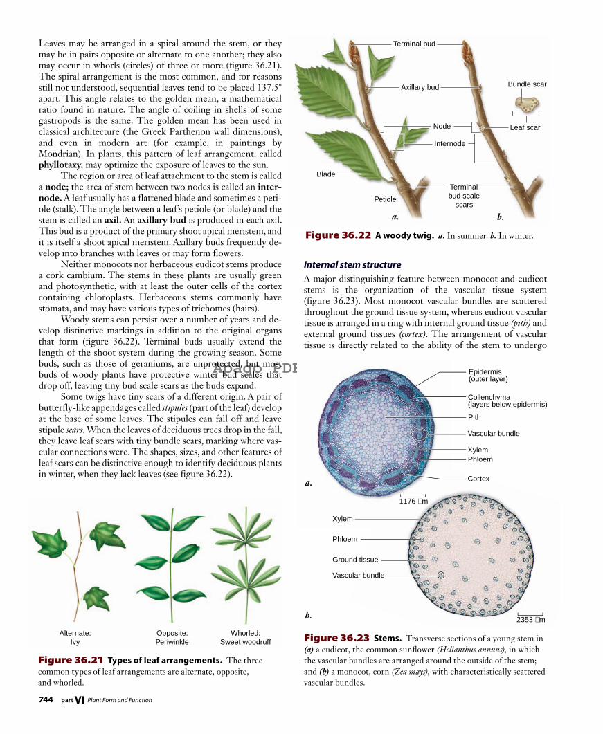
Figure 36.23 Stems. Transverse sections of a young stem in (a) a eudicot, the common sunfl ower (Helianthus annuus), in which the vascular bundles are arranged around the outside of the stem; and (b) a monocot, corn (Zea mays), with characteristically scattered vascular bundles.
Internal stem structureA major distinguishing feature between monocot and eudicot stems is the organization of the vascular tissue system (figure 36.23) . Most monocot vascular bundles are scattered throughout the ground tissue system, whereas eudicot vascular tissue is arranged in a ring with internal ground tissue (pith) and external ground tissues (cortex). The arrangement of vascular tissue is directly related to the ability of the stem to undergo
744 part VI Plant Form and Function
rav32223_ch36_729-752.indd 744rav32223_ch36_729-752.indd 744 11/16/09 11:41:33 AM11/16/09 11:41:33 AM

Apago PDF Enhancer
a.
b.
c.
Primary xylem
Secondary xylem
Primary phloem
Secondary phloem
Vascular cambium(lateral meristem)
Vascular cambium(lateral meristem)
Primary xylem
Secondary xylem
Primary phloem
Secondary phloem
Annualgrowth layers
EpidermisPrimary xylem Primary phloem
Heartwood
Sapwood
Vascular cambium
Phloem
Outer bark
Cork cambium
Xylem
Cork
Cork cambium
Phelloderm
Parenchyma
Periderm
52 µm
Figure 36.24 Secondary growth. a. Before secondary growth begins in eudicot stems, primary tissues continue to elongate as the apical meristems produce primary growth. b. As secondary growth begins, the vascular cambium produces secondary tissues, and the stem’s diameter increases. c. In this four-year-old stem, the secondary tissues continue to widen, and the trunk has become thick and woody. Note that the vascular cambium forms a cylinder that runs axially (up and down) in the roots and shoots that have them.
secondary growth. In eudicots, a vascular cambium may develop between the primary xylem and primary phloem (figure 36.24). In many ways, this is a connect-the-dots game in which the vascular cambium connects the ring of primary vas-cular bundles. There is no logical way to connect primary monocot vascular tissue that would allow a uniform increase in girth. Lacking a vascular cambium, therefore, monocots do not have secondary growth. Rings in the stump of a tree reveal annual patterns of vas-cular cambium growth; cell size varies, depending on growth
conditions (figure 36.25). Large cells form under favorable conditions such as abundant rainfalls. Rings of smaller cells mark the seasons where growth is limited. In woody eudicots and gymnosperms, a second cambium, the cork cambium, arises in the outer cortex (occasionally in the epidermis or phloem); it produces boxlike cork cells to the outside and also may produce parenchyma -like phelloderm cells to the inside (figure 36.26). The cork cambium, cork, and phelloderm are collectively referred to as the periderm (see figure 36.26). Cork tissues, the cells of which become impregnated with water-repellent su ber in shortly after they are formed and which then die, constitute the outer bark. The cork tissue cuts off water and food to the epidermis, which dies and sloughs off. In young stems, gas exchange between stem tissues and the air takes place through stomata, but as the cork cambium produces cork, it also produces patches of unsuberized cells beneath the stomata. These unsuberized cells, which permit gas ex-change to continue, are called lenticels (figure 36.27).
Modifi ed stems carry out vegetative propagation and store nutrientsAlthough most stems grow erect, some have modifications that serve special purposes, including natural vegetative propagation. In fact, the widespread artificial vegetative propagation of plants,
Figure 36.25 Tree stump. The vascular cambium produces rings of xylem (sapwood and nonconducting heartwood) and phloem, and the cork cambium produces the cork.
Figure 36.26 Section of periderm. An early stage in the development of periderm in cottonwood, Populus sp.
chapter 36 Plant Form 745www.ravenbiology.com
rav32223_ch36_729-752.indd 745rav32223_ch36_729-752.indd 745 11/16/09 11:41:35 AM11/16/09 11:41:35 AM

Apago PDF Enhancer
a. b.
Lenticel
Periderm
Lenticel
Gas exchange
833 µm
a. b. c.
d. e. f.
Fleshyleavesof bulb
Tuber (swollentip of stolon)
Tendril
Cladophyll
Leaves
Stolon
Knoblikestem
Adventitiousroots Adventitious roots
Runner
Photosyntheticleaf
Rhizome
Figure 36.27 Lenticels. a. Lenticels, the numerous small, pale, raised areas shown here on cherry tree bark (Prunus cerasifera), allow gas exchange between the external atmosphere and the living tissues immediately beneath the bark of woody plants. b. Transverse section through a lenticel in a stem of elderberry, Sambucus canadensis.
next year’s foliage comes from the tip of the shoot apex, protected by storage leaves from the previous year
Corms. Crocuses, gladioluses, and other popular garden plants produce corms that superfi cially resemble bulbs. Cutting a corm in half, however, reveals no fl eshy leaves. Instead, almost all of a corm consists of stem, with a few papery, brown nonfunctional leaves on the outside, and adventitious roots below.
Rhizomes. Perennial grasses, ferns, bearded iris, and many other plants produce rhizomes, which typically are horizontal stems that grow underground, often close to the surface (fi gure 36.28b). Each node has an inconspicuous scalelike leaf with an axillary bud; much larger photosynthetic leaves may be produced at the rhizome tip. Adventitious roots are produced throughout the length of the rhizome, mainly on the lower surface.
Runners and stolons. Strawberry plants produce horizontal stems with long internodes that unlike rhizomes, usually grow along the surface of the ground. Several runners may radiate out from a single plant (fi gure 36.28c). Some biologists use the term stolon synonymously with runner; others reserve the term stolon for a stem with long internodes (but no roots) that grows underground, as seen in potato plants (Solanum sp.). A potato itself, however, is another type of modifi ed stem—a tuber.
Tubers. In potato plants, carbohydrates may accumulate at the tips of rhizomes, which swell, becoming tubers; the rhizomes die after the tubers mature (fi gure 36.28d). The “eyes” of a potato are axillary buds formed in the axils of scalelike leaves. These leaves, which are present when the potato is starting to form, soon drop off; the tiny ridge adjacent to each “eye” of a mature potato is a leaf scar.
both commercial and private, frequently involves cutting modi-fied stems into segments, which are then planted, producing new plants. As you become acquainted with the following modi-fied stems, keep in mind that stems have leaves at nodes, with internodes between the nodes, and buds in the axils of the leaves, whereas roots have no leaves, nodes, or axillary buds.
Bulbs. Onions, lilies, and tulips have swollen underground stems that are really large buds with adventitious roots at the base (fi gure 36.28a) . Most of a bulb consists of fl eshy leaves attached to a small, knoblike stem. For most bulbs,
Figure 36.28 Types of modifi ed stems. a. Bulb. b. Adventitious roots. c. Runner. d. Stolon. e. Tendril. f. Cladophyll.
746 part VI Plant Form and Function
rav32223_ch36_729-752.indd 746rav32223_ch36_729-752.indd 746 11/16/09 11:41:39 AM11/16/09 11:41:39 AM
Apago PDF Enhancer
Shoot apical meristem
Top of developing leaf
YAB
KAN Ac
tivat
ion
PHB
and
PHV
Bottom of developingleaf
kan
adi
Mu
tan
t
Wild
-typ
eIn
hibitio
n
36.5 Leaves: Photosynthetic Organs
Learning OutcomesDistinguish between a simple and a compound leaf.1. Compare the mesophyll of a monocot leaf with that of a 2. eudicot leaf.
Leaves, which are initiated as primordia by the apical meristems (see figure 36.20), are vital to life as we know it because they are the principal sites of photosynthesis on land, providing the base of the food chain. Leaves expand by cell enlargement and cell division. Like arms and legs in humans, they are determinate structures, which means their growth stops at maturity. Because leaves are crucial to a plant, features such as their arrangement, form, size, and internal structure are highly significant and can differ greatly. Different patterns have adaptive value in differ-ent environments. Leaves are an extension of the shoot apical meristem and stem development. When they first emerge as primordia, they are not committed to being leaves. Experiments in which very young leaf primordia are isolated from fern and coleus plants and grown in culture have demonstrated this feature: If the pri-mordia are young enough, they will form an entire shoot rather than a leaf. The positioning of leaf primordia and the initial cell divisions occur before those cells are committed to the leaf de-velopmental pathway.
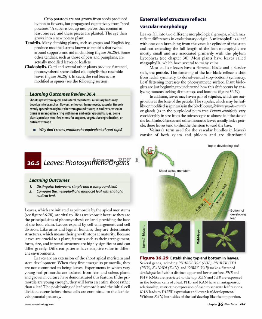
External leaf structure refl ects vascular morphologyLeaves fall into two different morphological groups, which may reflect differences in evolutionary origin. A microphyll is a leaf with one vein branching from the vascular cylinder of the stem and not extending the full length of the leaf; microphylls are mostly small and are associated primarily with the phylum Lycophyta (see chapter 30). Most plants have leaves called megaphylls, which have several to many veins. Most eudicot leaves have a flattened blade and a slender stalk, the petiole . The flattening of the leaf blade reflects a shift from radial symmetry to dorsal –ventral (top–bottom) symmetry. Leaf flattening increases the photosynthetic surface. Plant biolo-gists are just beginning to understand how this shift occurs by ana-lyzing mutants lacking distinct tops and bottoms (figure 36.29). In addition, leaves may have a pair of stipules , which are out-growths at the base of the petiole. The stipules, which may be leaf-like or modified as spines (as in the black locust, Robinia pseudo-acacia) or glands (as in the purple-leaf plum tree Prunus cerasifera), vary considerably in size from the microscopic to almost half the size of the leaf blade. Grasses and other monocot leaves usually lack a peti-ole; these leaves tend to sheathe the stem toward the base. Veins (a term used for the vascular bundles in leaves) consist of both xylem and phloem and are distributed
Crop potatoes are not grown from seeds produced by potato fl owers, but propagated vegetatively from “seed potatoes.” A tuber is cut up into pieces that contain at least one eye, and these pieces are planted. The eye then grows into a new potato plant.
Tendrils. Many climbing plants, such as grapes and English ivy, produce modifi ed stems known as tendrils that twine around supports and aid in climbing (fi gure 36.28e). Some other tendrils, such as those of peas and pumpkins, are actually modifi ed leaves or leafl ets.
Cladophylls. Cacti and several other plants produce fl attened, photosynthetic stems called cladophylls that resemble leaves (fi gure 36.28f ). In cacti, the real leaves are modifi ed as spines (see the following section).
Learning Outcomes Review 36.4Shoots grow from apical and lateral meristems. Auxilliary buds may develop into branches, fl owers, or leaves. In monocots, vascular tissue is evenly spaced throughout the stem ground tissue; in eudicots, vascular tissue is arranged in a ring with inner and outer ground tissues. Some plants produce modifi ed stems for support, vegetative reproduction, or nutrient storage.
■ Why don’t stems produce the equivalent of root caps?
Figure 36.29 Establishing top and bottom in leaves. Several genes, including PHABULOSA (PHB), PHAVOLUTA
(PHV), KANADI (KAN), and YABBY (YAB) make a fl attened Arabidopsis leaf with a distinct upper and lower surface. PHB and PHV RNAs are restricted to the top; KAN and YAB are expressed in the bottom cells of a leaf. PHB and KAN have an antagonistic relationship, restricting expression of each to separate leaf regions. KAN leads to YABBY expression and lower leaf development. Without KAN, both sides of the leaf develop like the top portion.
chapter 36 Plant Form 747www.ravenbiology.com
rav32223_ch36_729-752.indd 747rav32223_ch36_729-752.indd 747 11/16/09 11:41:53 AM11/16/09 11:41:53 AM

Apago PDF Enhancer
a.
b.
a. b.
Hypothesis: The gene UNIFOLIATA (UNI) is necessary for compound
leaf development in garden pea, Pisum sativum.
Prediction: Developing leaf primordia of a wild-type pea plant will
express the UNI gene while uni mutant plants will not express the gene.
Test: Cut thin sections of wild-type and mutant pea shoots and place
them on a microscope slide. Test for the presence of UNI RNA using a
color-labeled, single-stranded DNA probe that will hybridize (bind) only
the UNI RNA. View the labeled sections under a microscope.
Result: UNI RNA was found in the wild-type and also in mutant leaves,
but at lower levels.
Conclusion: The mutant uni gene is transcribed but at lower levels than
the wild-type gene. Thus the prediction was incorrect.
Further Experiments: Although the uni gene is expressed, compound
leaves do not develop. Refine the hypothesis and propose an experiment
to test the revised hypothesis.
S C I E N T I F I C T H I N K I N G
mutant plants will not express the gene.
Test: Cut
them on a
color-labe
the UNI RN
Result: U
but at low
Conclusio
the wild-ty
Further E
e
ly
es,
han
p g gene is expressed, compound
et thin sections of wild-type and mutant pea shoots and place
a microscope slide. Test for the presence of UNI RNA using a
eled, single-stranded DNA probe that will hybridize (bind) onl
NA. View the labeled sections under a microscope.
UNI RNA was found in the wild-type and also in mutant leave
wer levels.
on: The mutant uni gene is transcribed but at lower levels th
ype gene. Thus the prediction was incorrect.
Experiments: Although the uni gene is expressed, compoun
ype and mutant pea shoots and place
Wild-type Shoot uni Mutant Shoot
SM SM
P1P1
P2
P3P4
P5
P6
P2P3
P4
P5
P6
SM shoot meristemP1–P6 leaf primordia
Figure 36.30 Eudicot and monocot leaves. a. The leaves of eudicots, such as this African violet relative from Sri Lanka, have netted, or reticulate, veins. b. Those of monocots, such as this cabbage palmetto, have parallel veins. The eudicot leaf has been cleared with chemicals and stained with a red dye to make the veins show more clearly.
throughout the leaf blades. The main veins are parallel in most monocot leaves; the veins of eudicots, on the other hand, form an often intricate network (figure 36.30) . Leaf blades come in a variety of forms, from oval to deeply lobed to having separate leaflets. In simple leaves (figure 36.31a), such as those of lilacs or birch trees, the blades are undivided, but simple leaves may have teeth, indentations, or lobes of various sizes, as in the leaves of maples and oaks. In compound leaves (figure 36.31b), such as those of ashes, box elders, and walnuts, the blade is divided into leaflets. The relationship between the development of compound and simple leaves is an open question. Two explanations are being debated: (1) A compound leaf is a highly lobed simple leaf, or (2) a compound leaf utilizes a shoot development program, and each leaflet was once a leaf. To address this question, research-ers are using single mutations that are known to convert com-pound leaves to simple leaves (figure 36.32).
Figure 36.31 Simple versus compound leaves. a. A simple leaf, its margin deeply lobed, from the oak tree (Quercus
robur). b. A pinnately compound leaf, from a black walnut (Juglans
nigra). A compound leaf is associated with a single lateral bud, located where the petiole is attached to the stem.
Figure 36.32 Genetic regulation of leaf development.
Internal leaf structure regulates gas exchange and evaporationThe entire surface of a leaf is covered by a transparent epider-mis, and most of these epidermal cells have no chloroplasts. As described earlier, the epidermis has a waxy cuticle, and different types of glands and trichomes may be present. Also, the lower epidermis (and occasionally the upper epidermis) of most leaves contains numerous slitlike or mouth-shaped stomata flanked by guard cells (figure 36.33) . The tissue between the upper and lower epidermis is called mesophyll. Mesophyll is interspersed with veins of vari-ous sizes.
748 part VI Plant Form and Function
rav32223_ch36_729-752.indd 748rav32223_ch36_729-752.indd 748 11/16/09 11:41:58 AM11/16/09 11:41:58 AM

Apago PDF Enhancer
Epidermal cell
Guard cell
Stoma
Thickened inner wall of guard cell
Stoma
Epidermal cell
Nucleus
Chloroplast
Guard cell
a. b.
Upper epidermis
Palisade mesophyll
Spongy mesophyll
Lower epidermis
Cuticle
Guard cell
Stoma
Guard cell
Stoma
Vein
Vein
Xylem
Phloem
200 µm
water loss, gas exchange, and transport of photosynthetic prod-ucts to the rest of the plant.
Modifi ed leaves are highly versatile organsAs plants colonized a wide variety of environments, from deserts to lakes to tropical rain forests, plant organ modifications arose that would adapt the plants to their specific habitats. Leaves, in particular, have evolved some remarkable adaptations. A brief discussion of a few of these modifications follows:
Floral leaves (bracts). Poinsettias and dogwoods have relatively inconspicuous, small, greenish yellow fl owers. However, both plants produce large modifi ed leaves called bracts (mostly colored red in poinsettias and white or pink in dogwoods). These bracts surround the true fl owers and perform the same function as showy petals. In other plants, however, bracts can be quite small and inconspicuous.
Spines. The leaves of many cacti and other plants are modifi ed as spines (see fi gure 36.28f ). In cacti, having less leaf surface reduces water loss, and the sharp spines also may deter predators. Spines should not be confused with thorns, such as those on the honey locust (Gleditsia triacanthos), which are modifi ed stems, or with the prickles on raspberries, which are simply outgrowths from the epidermis or the cortex just beneath it.
Reproductive leaves. Several plants, notably Kalanchoë, produce tiny but complete plantlets along their margins. Each plantlet, when separated from the leaf, is capable of growing independently into a full-sized plant. The walking fern (Asplenium rhizophyllum) produces new plantlets at the tips of its fronds. Although many species can regenerate a whole plant from isolated leaf tissue, this in vivo regeneration is found among just a few species.
Window leaves. Several genera of plants growing in arid regions produce succulent, cone-shaped leaves with transparent tips. The leaves often become mostly buried
Figure 36.33 A stoma. a. Surface view. b. View in cross section.
Most eudicot leaves have two distinct types of mesophyll. Closest to the upper epidermis are one to several (usually two) rows of tightly packed, barrel-shaped to cylindrical chloren-chyma cells (parenchyma with chloroplasts) that constitute the palisade mesophyll (figure 36.34) . Some plants, including spe-cies of Eucalyptus, have leaves that hang down, rather than ex-tend horizontally. They have palisade mesophyll on both sides of the leaf. Nearly all eudicot leaves have loosely arranged spongy mesophyll cells between the palisade mesophyll and the lower epidermis, with many air spaces throughout the tissue. The inter-connected intercellular spaces, along with the stomata, function in gas exchange and the passage of water vapor from the leaves. The mesophyll of monocot leaves often is not differenti-ated into palisade and spongy layers, and there is often little distinction between the upper and lower epidermis. Instead, cells surrounding the vascular tissue are distinctive and are the site of carbon fixation. This anatomical difference often corre-lates with a modified photosynthetic pathway, C4 photosynthesis, that maximizes the amount of CO2 relative to O2 to reduce energy loss through photorespiration (see chapter 9). The anat-omy of a leaf directly relates to its juggling act of balancing
Figure 36.34 A leaf in cross section. Transection of a leaf showing the arrangement of palisade and spongy mesophyll, a vascular bundle or vein, and the epidermis with paired guard cells fl anking the stoma.
chapter 36 Plant Form 749www.ravenbiology.com
rav32223_ch36_729-752.indd 749rav32223_ch36_729-752.indd 749 11/16/09 11:42:13 AM11/16/09 11:42:13 AM

Apago PDF Enhancer
glands that secrete sticky mucilage that traps insects, which are then digested by enzymes.
The Venus fl ytrap (Dionaea muscipula) produces leaves that look hinged at the midrib. When tiny trigger hairs on the leaf blade are stimulated by a moving insect, the two halves of the leaf snap shut, and digestive enzymes break down the soft parts of the trapped insect into nutrients that can be absorbed through the leaf surface. Nitrogen is the most common nutrient needed. Curiously, the Venus fl ytrap cannot survive in a nitrogen-rich environment, perhaps as a result of a biochemical trade-off during the intricate evolutionary process that developed its ability to capture and digest insects.
Learning Outcomes Review 36.5Leaves come in a range of forms. A simple leaf is undivided, whereas a compound leaf has a number of separate leafl ets. Pinnate leaves have a central rib like a feather; palmate leaves have several ribs radiating from a central point, like the palm of the hand. Monocots typically produce leaves with parallel veins, while those of eudicots are netted. Mesophyll cells carry out photosynthesis; in monocots, mesophyll is undiff erentiated, whereas in eudicots it is divided into palisade and spongy mesophyll. Leaves may be modifi ed for reproduction, protection, water conservation, uptake of nutrients, and even as traps for insects.
■ Why would a plant with vertically oriented leaves produce palisade, but not spongy mesophyll cells?
in sand blown by the wind, but the transparent tips, which have a thick epidermis and cuticle, admit light to the hollow interiors. This strategy allows photosynthesis to take place beneath the surface of the ground.
Shade leaves. Leaves produced in the shade, where they receive little sunlight, tend to be larger in surface area, but thinner and with less mesophyll than leaves on the same tree receiving more direct light. This plasticity in development is remarkable. Environmental signals can have a major effect on development.
Insectivorous leaves. Almost 200 species of fl owering plants are known to have leaves that trap insects; some plants digest the insects’ soft parts. Plants with insectivorous leaves often grow in acid swamps that are defi cient in needed elements or contain elements in forms not readily available to the plants; this inhibits the plants’ capacities to maintain metabolic processes needed for their growth and reproduction. Their needs are met, however, by the supplementary absorption of nutrients from the animal kingdom.
Pitcher plants (for example, Sarracenia, Darlingtonia, or Nepenthes spp.) have cone-shaped leaves in which rainwater can accumulate. The insides of the leaves are very smooth, but stiff, downward-pointing hairs line the rim. An insect falling into such a leaf fi nds it very diffi cult to escape and eventually drowns. The leaf absorbs the nutrients released when bacteria, and in most species the plant’s own digestive enzymes, decompose the insect bodies. Other plants, such as sundews (Drosera), have
Ground tissue cells perform many functions, including storage, photosynthesis, and support.Ground tissue is mainly composed of parenchyma cells, which function in storage, photosynthesis, and secretion. Collenchyma cells provide fl exible support, and sclerenchyma cells provide rigid support.
Vascular tissue conducts water and nutrients throughout the plant.Xylem tissue conducts water through dead cells called tracheids and vessel elements.Phloem tissue conducts nutrients such as dissolved sucrose through living cells called sieve-tube members and sieve cells.
36.3 Roots: Anchoring and Absorption StructuresRoots evolved after shoots and are a major innovation for terrestrial living.
Roots are adapted for growing underground and absorbing water and solutes.Developing roots exhibit four regions: (1) the root cap, which protects the root; (2) the zone of cell division, which contains the apical meristem; (3) the zone of elongation, which extends the root through the soil; and (4) the zone of maturation, in which cells become differentiated.
36.1 Organization of the Plant Body: An Overview Vascular plants have roots and shoots.The root system is primarily below ground; roots anchor the plant and take up water and minerals. The shoot system is above ground and provides support for leaves and fl owers.
Roots and shoots are composed of three types of tissues.The three types of tissues are dermal tissue, ground tissue, and vascular tissue.
Meristems elaborate the body plan throughout the plant’s life.Apical meristems are located on the tips of stems and near the tips of roots. Lateral meristems are found in plants that exhibit secondary growth. They add to the diameter of a stem or root.
36.2 Plant TissuesDermal tissue forms a protective interface with the environment.Dermal tissue is primarily the epidermis, which is usually one cell thick and is covered with a fatty or waxy cuticle to retard water loss.Guard cells in the epidermis control water loss through stomata. Root hairs are epidermal cell structures that help increase the absorptive area of roots.
Chapter Review
750 part VI Plant Form and Function
rav32223_ch36_729-752.indd 750rav32223_ch36_729-752.indd 750 11/16/09 11:42:17 AM11/16/09 11:42:17 AM
Apago PDF Enhancer
Modified roots accomplish specialized functions.Most plants produce either a taproot system containing a single large root with smaller branch roots, or a fi brous root system composed of many small roots.Adventitious roots may be modified for support, stability, acquisition of oxygen, storage of water and food, or parasitism of a host plant.
36.4 Stems: Support for Above-Ground OrgansStems carry leaves and flowers and support the plant’s weight.Leaves are attached to stems at nodes. The axil is the area between the leaf and stem, and an axillary bud develops in axils of eudicots.The vascular bundles in stems of monocots are randomly scattered, whereas in eudicots the bundles are arranged in a ring.Vascular cambium develops between the inner xylem and the outer phloem, allowing for secondary growth.
Modified stems carry out vegetative propagation and store nutrients.Bulbs, corms, rhizomes, runners and stolons, tubers, tendrils, and cladophylls are examples of modifi ed stems. The tubers of potatoes are both a food source and a means of propagating new plants.
36.5 Leaves: Photosynthetic OrgansLeaves are the principle sites of photosynthesis. Leaf features such as their arrangement, form, size, and internal structure can be highly variable across environments.
External leaf structure reflects vascular morphology.Vascular bundles are parallel in monocots, but form a network in eudicots. The leaves of most eudicots have a flattened blade and a slender petiole; monocots usually do not have a petiole.Leaf blades may be simple or compound (divided into leaflets). Leaves may also be pinnate (with a central rib, like a feather) or palmate (with ribs radiating from a central point).
Internal leaf structure regulates gas exchange and evaporation.The tissues of the leaf include the epidermis with guard cells, vascular tissue, and mesophyll in which photosynthesis takes place.In eudicot leaves with a horizontal orientation, the mesophyll is partitioned into palisade cells near the upper surface and spongy cells near the lower surface.The mesophyll of monocot leaves is often not differentiated.
Modified leaves are highly versatile organs.Leaves are highly variable in form and are adapted to serve many different functions. Leaves may be modifi ed for reproduction, protection, storage, mineral uptake, or even as insect traps in carnivorous plants.
8. Palisade and spongy parenchyma are typically found in the mesophyll of
a. monocots. c. monocots and eudicots . b. eudicots. d. neither monocots or eudicots 9. In vascular plants, one difference between root and shoot
systems is that:a. root systems cannot undergo secondary growth.b. root systems undergo secondary growth, but do not
form bark.c. root systems contain pronounced zones of cell elongation,
whereas shoot systems do not.d. root systems can store food reserves, whereas stem
structures do not. 10. Which of the following statements is not true of the stems
of vascular plants?a. Stems are composed of repeating segments, including
nodes and internodes.b. Primary growth only occurs at the shoot apical meristem.c. Vascular tissues may be arranged on the outside of the stem
or scattered throughout the stem.d. Stems can contain stomata.
11. Which of the following plant cell type is mismatched to its function?a. Xylem—conducts mineral nutrientsb. Phloem—serves as part of the barkc. Trichomes—reduces evaporationd. Collenchyma—performs photosynthesis
U N D E R S T A N D 1. Which cells lack living protoplasts at maturity? a. Parenchyma c. Collenchyma b. Companion d. Sclerenchyma 2. The food-conducting cells in an oak tree are called a. tracheids. c. companion cells. b. vessels. d. sieve-tube members. 3. Root hairs form in the zone of a. cell division. c. maturation. b. elongation. d. more than one of the above 4. Roots differ from stems because roots lack a. vessel elements. c. an epidermis. b. nodes. d. ground tissue. 5. A plant that produces two axillary buds at a node is said to have
what type of leaf arrangement? a. Opposite c. Whorled b. Alternate d. Palmate 6. Unlike eudicot stems, monocot stems lack a. vascular bundles. c. pith. b. parenchyma. d. epidermis. 7. The function of guard cells is to
a. allow carbon dioxide uptake.b. repel insects and other herbivores.c. support leaf tissue.d. allow water uptake.
Review Questions
chapter 36 Plant Form 751www.ravenbiology.com
rav32223_ch36_729-752.indd 751rav32223_ch36_729-752.indd 751 11/16/09 11:42:18 AM11/16/09 11:42:18 AM