3d slicer - queen's universityperk.cs.queensu.ca/sites/perk.cs.queensu.ca/files/... · 3d...
TRANSCRIPT
!
Surgical Planning Laboratory Brigham and Women’s HospitalBoston, Massachusetts USA
a teaching affiliate of Harvard Medical School
3D Slicer
Ron Kikinis, M.D. !Robert Greenes Distinguished Director of Biomedical Informatics, Dept. of Radiology, BWH Professor of Radiology, Harvard Medical School Professor of Medical Image Computing, FB 3, Uni Bremen
• Founding Director, Surgical Planning Laboratory, Brigham and Women’s Hospital • Institutsleiter, Fraunhofer MEVIS • Principal Investigator, National Alliance for Medical Image Computing, Quantitative Image Informatics for Cancer Research,
and Neuroimage Analysis Center, • Research Director, National Center for Image Guided Therapy

©2013 Surgical Planning Laboratory, ARR
AcknowledgmentsNational Alliance for Medical Image
Computing www.na-mic.org !Neuroimage Analysis Center nac.spl.harvard.edu !Surgical Planning Laboratory,
Brigham and Women’s Hospital spl.harvard.edu !National Center For Image Guided Therapy www.ncigt.org
2
• Ferenc Jolesz, MD, my mentor
• Collaborators and colleagues
©2013 Surgical Planning Laboratory, ARR
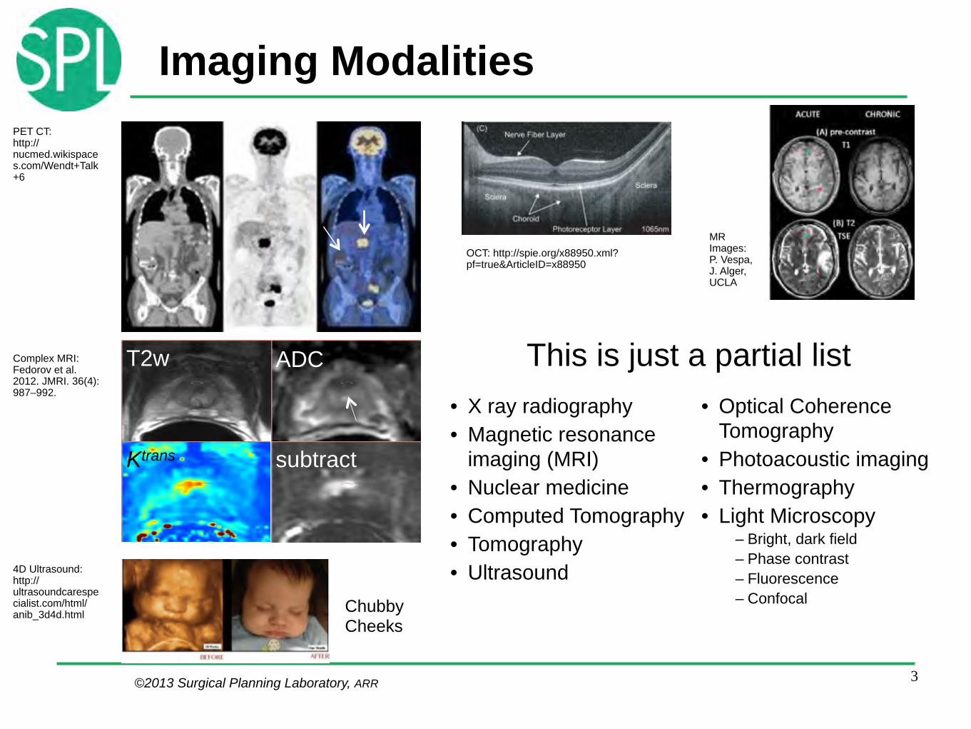
Imaging Modalities
3
OCT: http://spie.org/x88950.xml?pf=true&ArticleID=x88950
PET CT: http://nucmed.wikispaces.com/Wendt+Talk+6
T2w ADC
Ktrans subtract
Complex MRI: Fedorov et al. 2012. JMRI. 36(4):987–992.
4D Ultrasound: http://ultrasoundcarespecialist.com/html/anib_3d4d.html Chubby
Cheeks
• X ray radiography • Magnetic resonance
imaging (MRI) • Nuclear medicine • Computed Tomography • Tomography • Ultrasound
• Optical Coherence Tomography
• Photoacoustic imaging • Thermography • Light Microscopy
– Bright, dark field – Phase contrast – Fluorescence – Confocal
This is just a partial list
MR Images: P. Vespa, J. Alger, UCLA

©2013 Surgical Planning Laboratory, ARR
Examples of Complexity
4
DTI processing: http://www.loni.ucla.edu/~ophillip/DTIPipelines.html
DTI streamlines: http://wiki.slicer.org/slicerWiki/index.php/Slicer4:VisualBlog
http://www.frontiersin.org/human_neuroscience/10.3389/fnhum.2010.00181/full
fMRI dMRI
©2013 Surgical Planning Laboratory, ARR
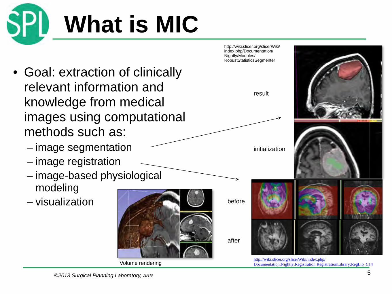
What is MIC
• Goal: extraction of clinically relevant information and knowledge from medical images using computational methods such as: – image segmentation – image registration – image-based physiological
modeling – visualization
5
http://wiki.slicer.org/slicerWiki/index.php/Documentation/Nightly/Modules/RobustStatisticsSegmenter
http://wiki.slicer.org/slicerWiki/index.php/Documentation:Nightly:Registration:RegistrationLibrary:RegLib_C14
before
after
initialization
result
Volume rendering
©2013 Surgical Planning Laboratory, ARR
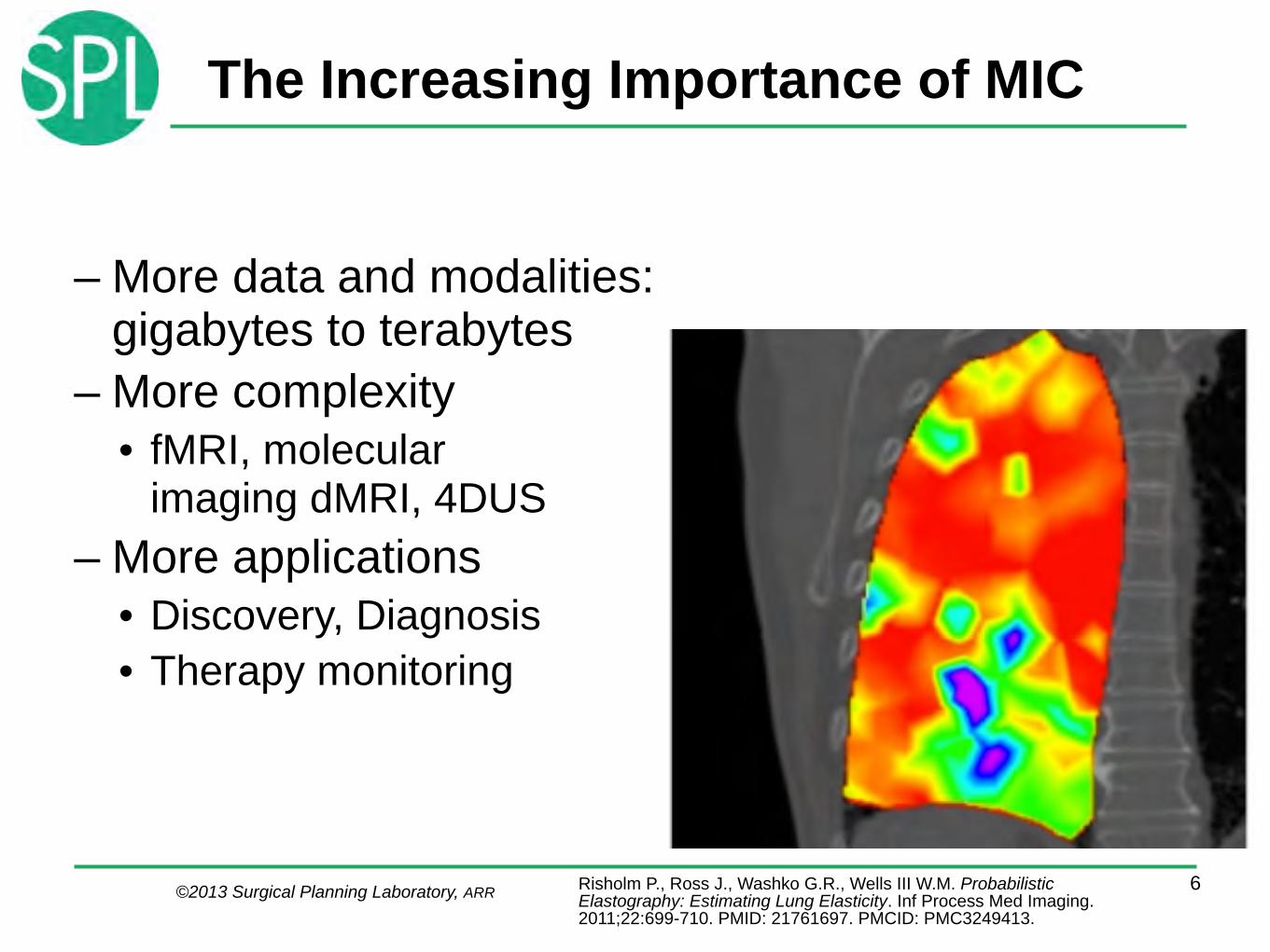
The Increasing Importance of MIC
– More data and modalities: gigabytes to terabytes
– More complexity • fMRI, molecular
imaging dMRI, 4DUS – More applications
• Discovery, Diagnosis • Therapy monitoring
6Risholm P., Ross J., Washko G.R., Wells III W.M. Probabilistic Elastography: Estimating Lung Elasticity. Inf Process Med Imaging. 2011;22:699-710. PMID: 21761697. PMCID: PMC3249413.
©2013 Surgical Planning Laboratory, ARR
Different Styles of Research
• Group Comparisons • Subject Specific Analysis (SSA)
!
• Technologies are often developed for group comparisons
• Additional scientific research is necessary to use such technologies for SSA
7

©2013 Surgical Planning Laboratory, ARR
Group Comparisons• Often used in basic imaging research • Targets normal appearing structures. Questions: What is
the…. – Typical appearance – Normal variability
• Extensive resources are deployed: personnel, computational • Most of our research is of this type, it’s the easiest way to
get results suitable for publication
8Golland, et al - MIT
Young OldMiddle
M.R. Sabuncu, S.K. Balci, M.E. Shenton, and P. Golland. Image-Driven Population Analysis Through Mixture Modeling. IEEE Transactions on Medical Imaging, 28(9):1473 - 1487, 2009
3 Templates

©2013 Surgical Planning Laboratory, ARR
Subject Specific Analysis• Targets focal pathology:
• Where is the pathology? • What are important
surrounding structures • Limited resources:
– Time – Personnel – Computational
• Interactive work is the norm
9Golby A.J., Kindlmann G., Norton I., Yarmarkovich A., Pieper S., Kikinis R. Interactive Diffusion Tensor Tractography Visualization for Neurosurgical Planning. Neurosurgery. 2011 Feb; 68(2):496-505. PMID: 21135713
Lack of quality in the processing pipeline can NOT be compensated by adding subjects (you have only one subject)

©2013 Surgical Planning Laboratory, ARR
Subject Specific Analysis (SSA)
• Quick and good enough is better than slow and perfect!
• Image processing problems cannot be compensated by adding subjects (you have only one)
• Interactive work is the norm
10
"Ron's rules for tools" is an informal set of rules that developers should keep in mind when working on interactive tools for translational clinical research. If you follow them, you will create tools that many people will use.
• You make it, I break it. • Your tool does not exist, until it works on my laptop with my data. • I am lazy. I do not like to move the mouse or to type. • No more than one simple parameter. • I have Attention deficit disorder: Make your algorithm fast.

©2013 Surgical Planning Laboratory, ARR
SSA Challenges• Many patients have visible pathology. Most
MIC technology was developed for analysis of healthy looking subjects
• Tools need to be robust, easy to learn, and quick
• Due to the “valley of death”, very little technology has made it from research into clinical devices
11

©2013 Surgical Planning Laboratory, ARR
3D Slicer• Platform for subject
specific analysis • An end-user application • A platform for delivering
software tools !
– Free open source software • Enables scientific collaboration • License allows painless translation to proprietary
clinical tools – Well-engineered high-performance core
• Software engineering methodology, multi-platform – Many options for extensions and for sharing
them – Cross-platform
12Picture courtesy Wendy Plesniak

©2013 Surgical Planning Laboratory, ARR
Easy to Use, Easy to Extend
What does a developer need ? • Easily Deployable • Extensible and
Reconfigurable • Rich Utility Libraries • Stable Base
13Courtesy S. Pujol, S. Pieper
3D Slicer: a cross platform system for translating innovative algorithms into clinical research applications
What does a user expect ? • Easy Install and Upgrade • “Standard” Clinical Behavior • Advanced Functionality • Consistent Interface • Easily Deployable • Extensible and Reconfigurable • Rich Utility Libraries
©2013 Surgical Planning Laboratory, ARR
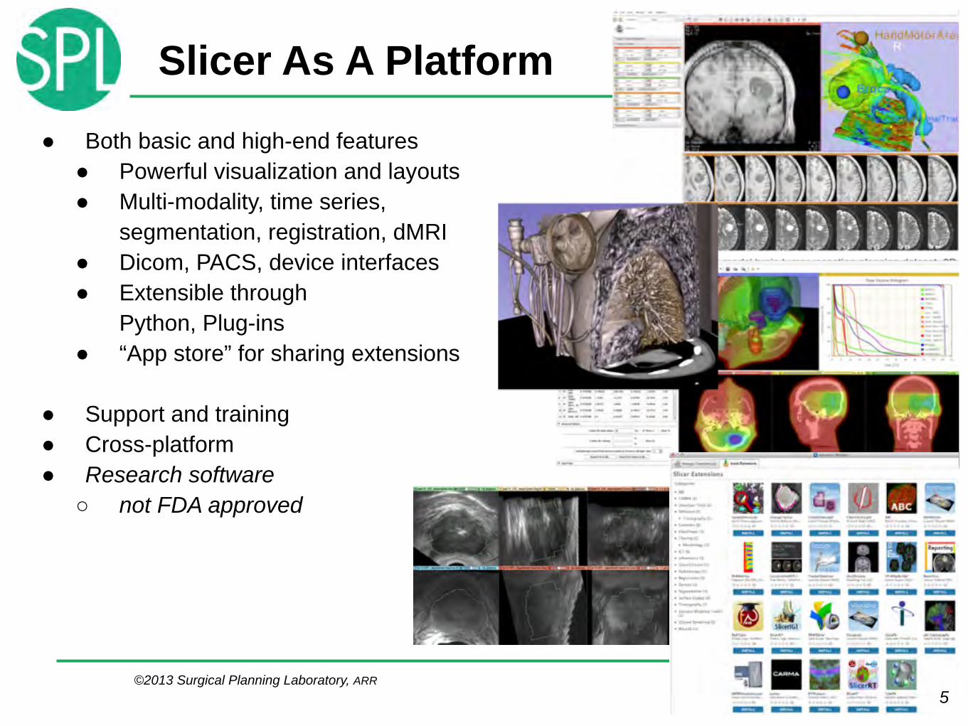
Slicer As A Platform
● Both basic and high-end features ● Powerful visualization and layouts ● Multi-modality, time series,
segmentation, registration, dMRI ● Dicom, PACS, device interfaces ● Extensible through
Python, Plug-ins ● “App store” for sharing extensions !
● Support and training ● Cross-platform ● Research software
○ not FDA approved
145

©2013 Surgical Planning Laboratory, ARR
3D Slicer History1997: Slicer started as the masters thesis of David Gering, in a collaboration between the Surgical Planning Lab (Harvard) and CSAIL (MIT)
15Images Courtesy of the CSAIL, MIT

©2013 Surgical Planning Laboratory, ARR
3D Slicer Today
• Community Effort
16

©2013 Surgical Planning Laboratory, ARR
Slicer Community At A Glance
17
Total number of 3D Slicer downloads in 2013: 61463, (160 per day)
http://download.slicer.org/stats http://massmail.spl.harvard.edu/public-archives/slicer-users/ http://massmail.spl.harvard.edu/public-archives/slicer-devel/
3D Slicer project analysis from Ohloh.net http://www.ohloh.net/p/3376
!Slicer version 4 re-
architecture and cleanup
http://commontk.org
Mailing list rosters: ● slicer-users: >1000 ● slicer-devel: >600
©2013 Surgical Planning Laboratory, ARR
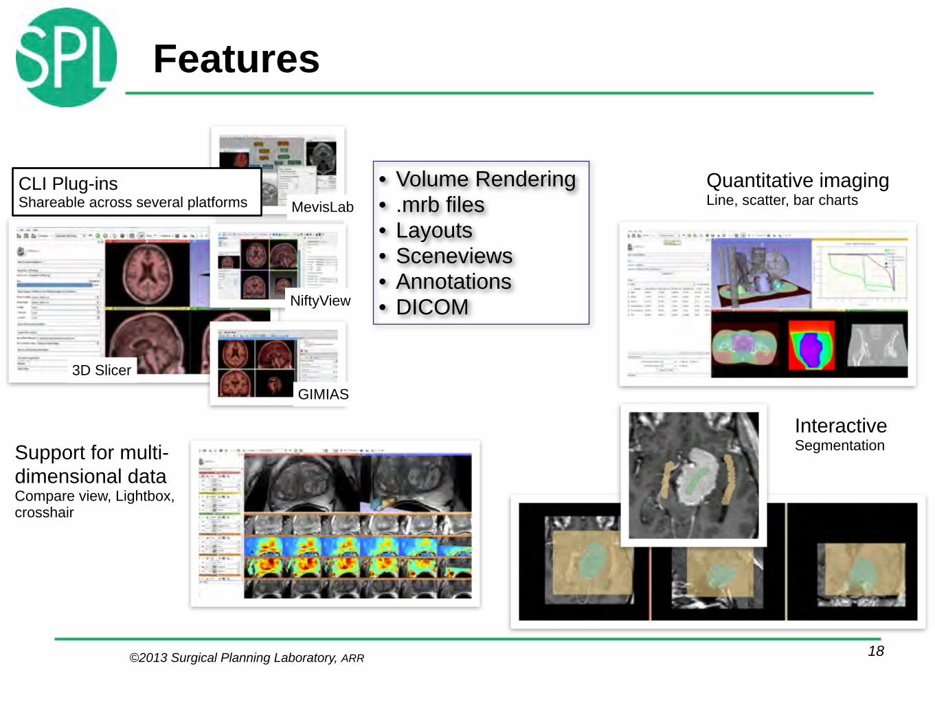
Features
18
Interactive Segmentation
Quantitative imaging Line, scatter, bar charts
Support for multi-dimensional data Compare view, Lightbox, crosshair
NiftyView
GIMIAS
MevisLabCLI Plug-ins Shareable across several platforms
• Volume Rendering • .mrb files • Layouts • Sceneviews • Annotations • DICOM
3D Slicer

©2013 Surgical Planning Laboratory, ARR
Slicer and Devices
©2013 Surgical Planning Laboratory, ARR
• Two-way communication – Imaging devices – Optical tracking devices – Robotic devices – More
19
Robot
Scanner
Navigation software
Tracking devices
Image
Transform
Status Command
Slide courtesy J. Tokuda
OpenIGTLink

©2013 Surgical Planning Laboratory, ARR
OpenIGTLink: API for Devices
20
Smartphone
WiFi Router
Sca
nner
Roo
mC
onso
le R
oom
OpenIGTLink Proxy
MRI Host Computer MRI Hardware
MRI Scanner
Com
man
dC
omm
and
Imag
eIm
age
Com
man
d
Imag
e
Tokuda J., et al. CARS 2012, June 27-30, Pisa Italy
Use an iPhone to control scan plane acquisition

©2013 Surgical Planning Laboratory, ARR
US Tracking: 2011: Bench
21Movie courtesy Ungi, Lasso, Fichtinger
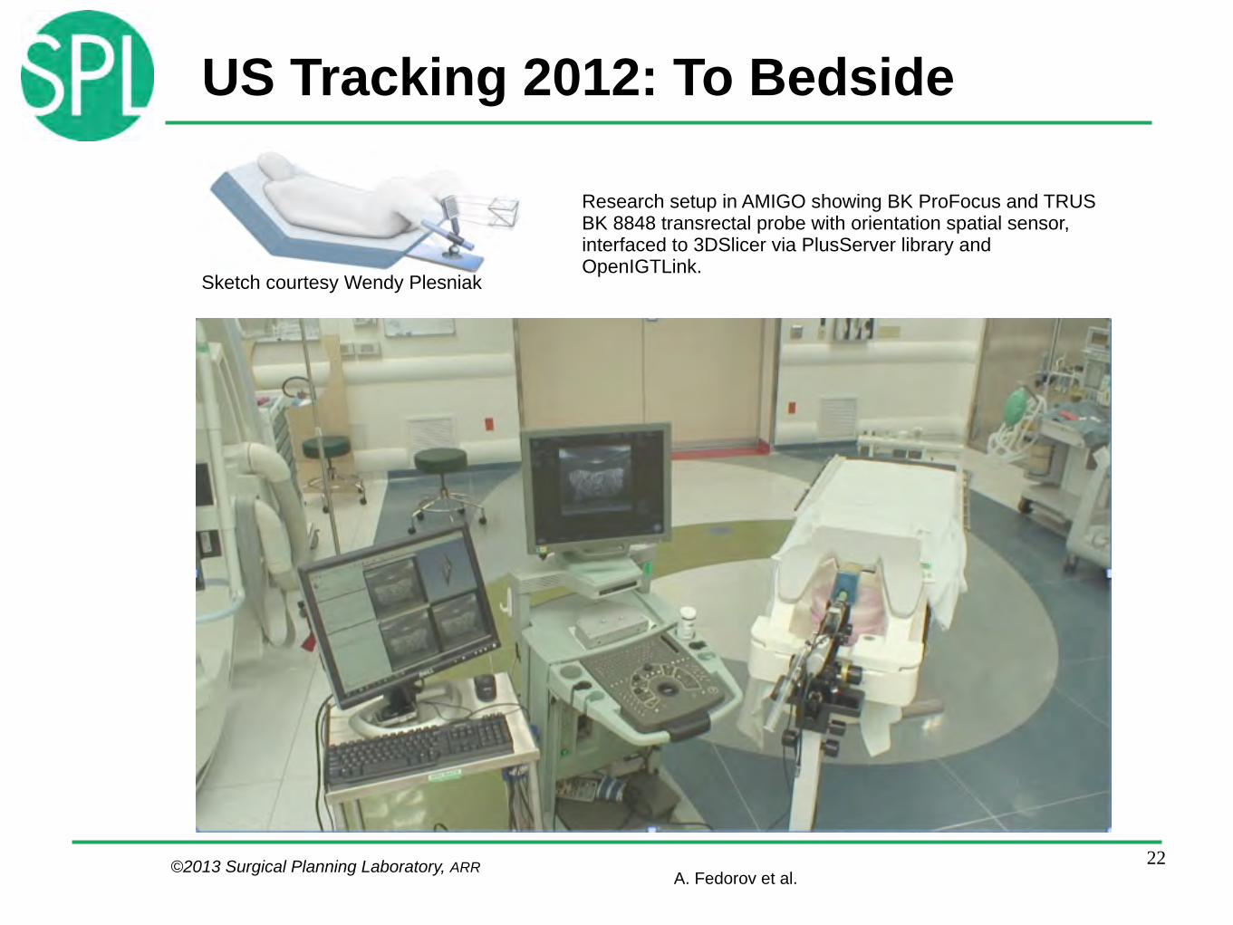
©2013 Surgical Planning Laboratory, ARR
Research setup in AMIGO showing BK ProFocus and TRUS BK 8848 transrectal probe with orientation spatial sensor, interfaced to 3DSlicer via PlusServer library and OpenIGTLink.
US Tracking 2012: To Bedside
22A. Fedorov et al.
Sketch courtesy Wendy Plesniak
©2013 Surgical Planning Laboratory, ARR
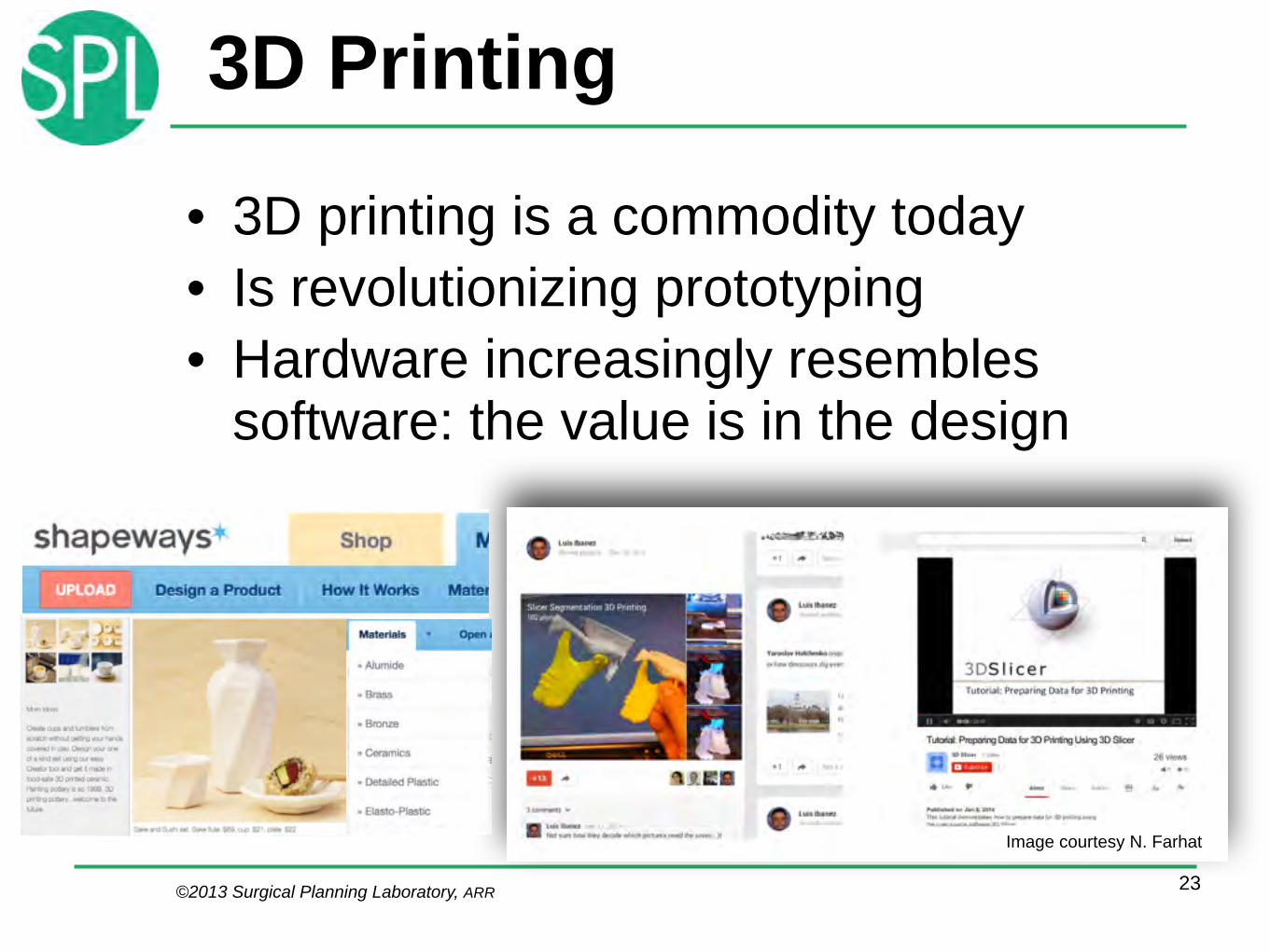
3D Printing
• 3D printing is a commodity today • Is revolutionizing prototyping • Hardware increasingly resembles
software: the value is in the design
23
Image courtesy N. Farhat

©2013 Surgical Planning Laboratory, ARR
Web Capabilities
QtWebKit enables Web services
24
Extension Manager and catalog • Share plug-ins with users • Easy Installation
Data Store • Web-based public repository of .mrb files allows sharing • Sceneviews are exposed in the web interface
©2013 Surgical Planning Laboratory, ARR
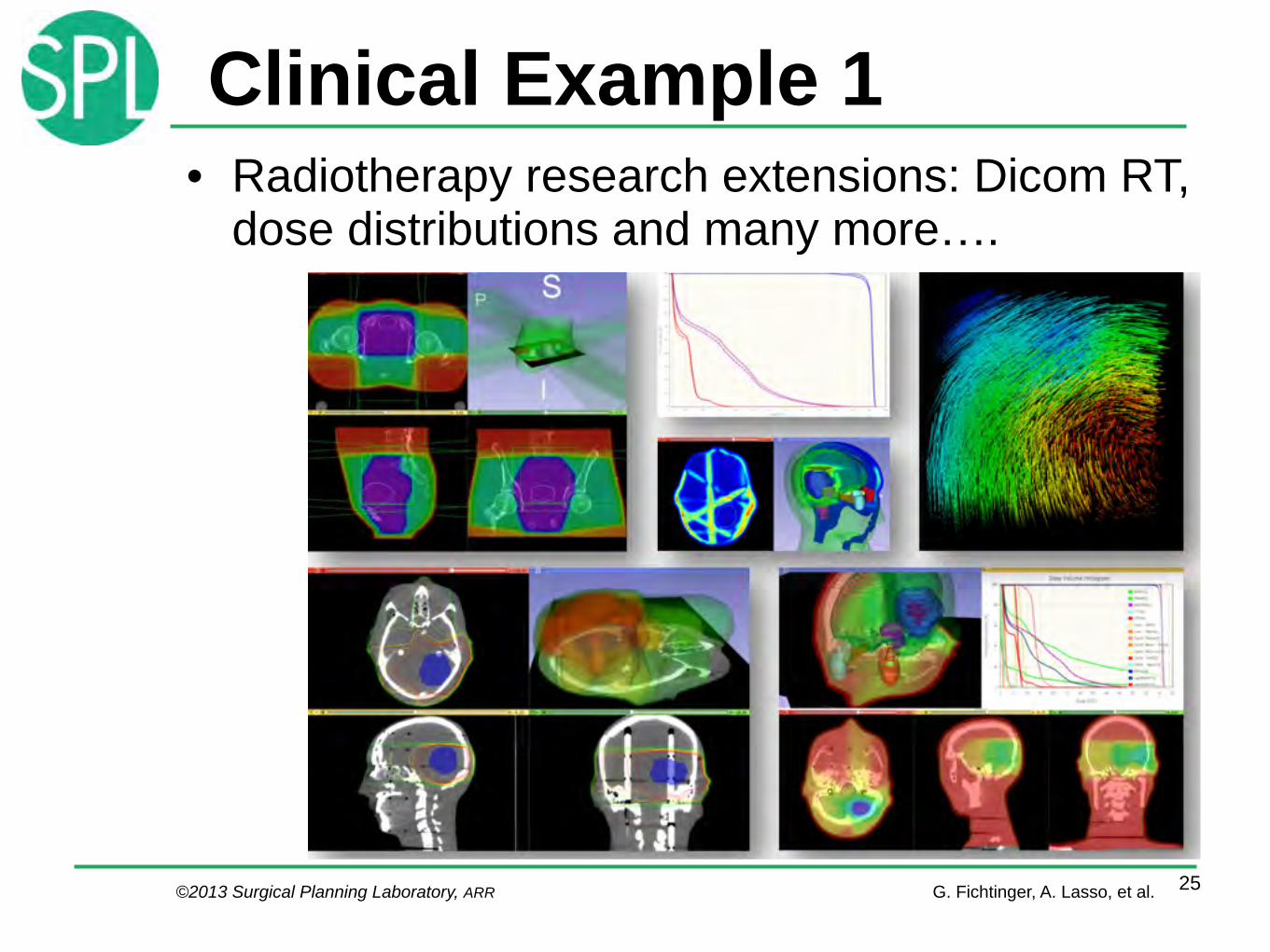
Clinical Example 1• Radiotherapy research extensions: Dicom RT,
dose distributions and many more….
25G. Fichtinger, A. Lasso, et al.
©2013 Surgical Planning Laboratory, ARR
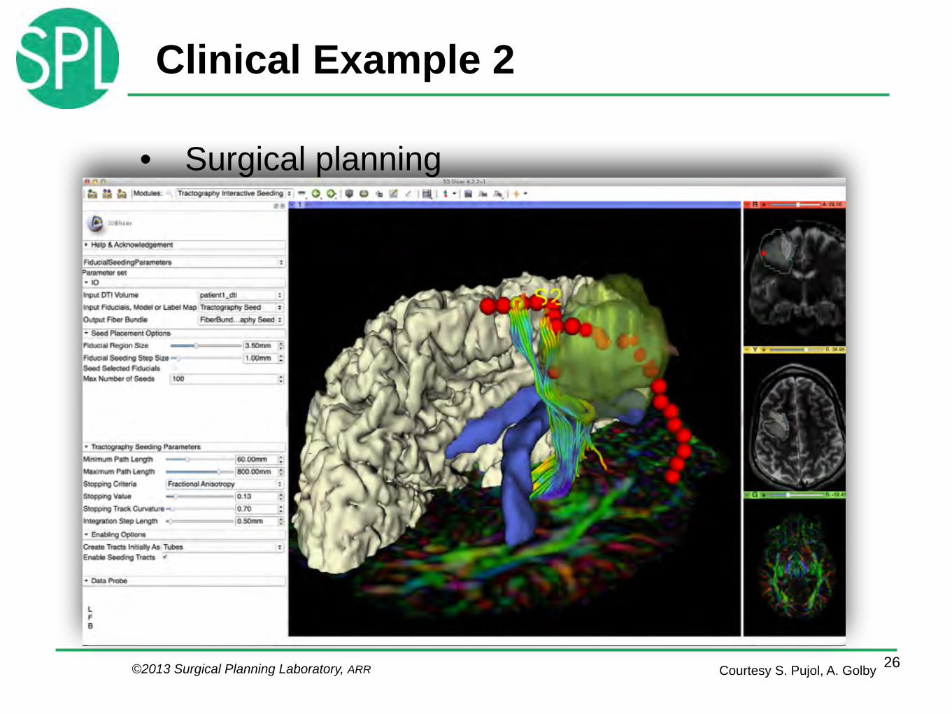
Clinical Example 2
• Surgical planning
26Courtesy S. Pujol, A. Golby

©2013 Surgical Planning Laboratory, ARR©2013 Surgical Planning Laboratory, ARR 27Images courtesy A. Golby
• Intraoperative Fiber Tracking
• Relies on pre-op data • Slicer+Brainlab
Image Guided Therapy Interfacing to clinical devices
Clinical Example 3
©2013 Surgical Planning Laboratory, ARR
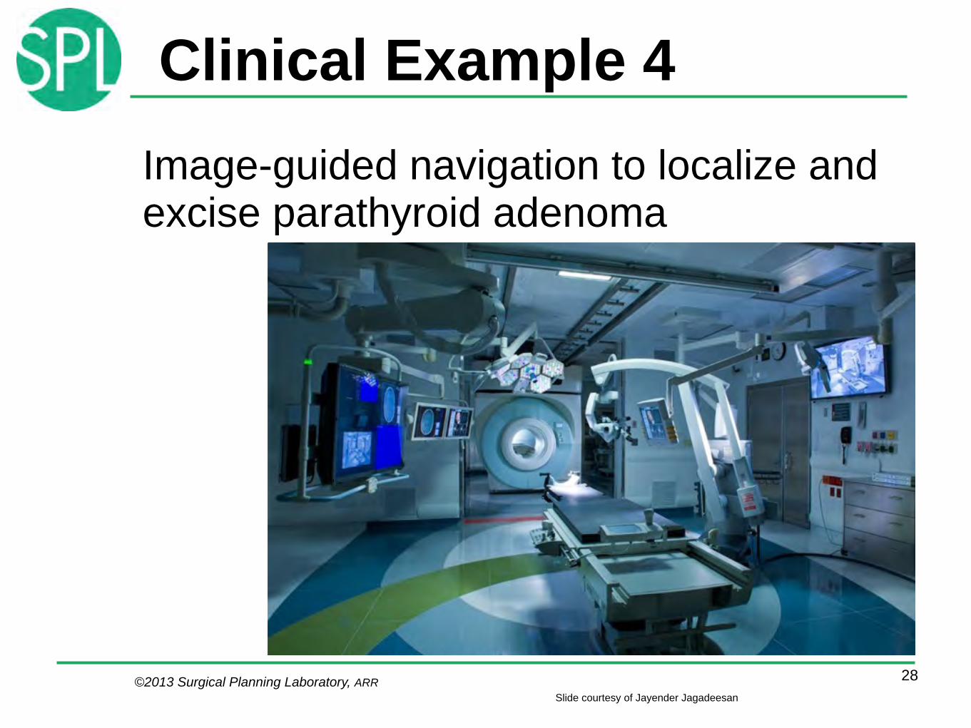
Clinical Example 4Image-guided navigation to localize and excise parathyroid adenoma
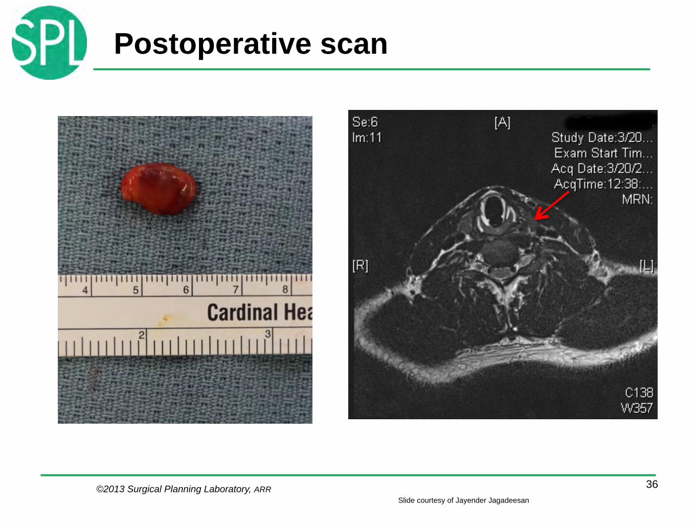
28Slide courtesy of Jayender Jagadeesan
©2013 Surgical Planning Laboratory, ARR
AMIGO Parathyroid Team
• Surgeon: Daniel Ruan, MD • Radiologist: Thomas Lee, MD • Navigation Scientist: Jayender Jagadeesan,
PhD !
AMIGO Support Team • Techs/Nurses: Dan Kacher, Janice Fairhurst,
Angela Kanan, Shivon Cesar, Sue Sheehan, Sandra Lawson, Julia Bousquet, Sean Jackson, Nikita Aristarkhov
29
©2013 Surgical Planning Laboratory, ARR
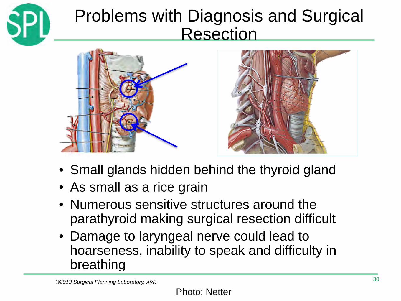
Problems with Diagnosis and Surgical Resection
• Small glands hidden behind the thyroid gland • As small as a rice grain • Numerous sensitive structures around the
parathyroid making surgical resection difficult
• Damage to laryngeal nerve could lead to hoarseness, inability to speak and difficulty in breathing
30
Photo: Netter
©2013 Surgical Planning Laboratory, ARR
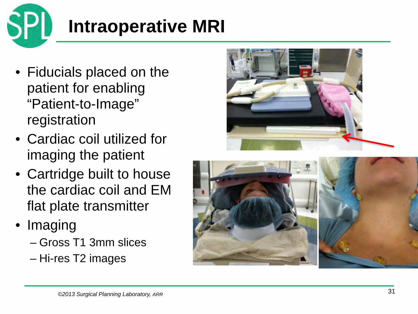
Intraoperative MRI
• Fiducials placed on the patient for enabling “Patient-to-Image” registration
• Cardiac coil utilized for imaging the patient
• Cartridge built to house the cardiac coil and EM flat plate transmitter
• Imaging – Gross T1 3mm slices – Hi-res T2 images
31

©2013 Surgical Planning Laboratory, ARR
Intraoperative Guidance
• Module developed in 3D Slicer • Software and ATC hardware
decoupled – OpenIGTLink communication
• Wizard Workflow !Preop Planning Setup instruments Calibration !!
32
Register Patient
Setup Displays
Refine Registration (Optional)Log Data

©2013 Surgical Planning Laboratory, ARR
Jayender et. al, Segmentation of parathyroid tumors from DCE-MRI using Linear Dynamic System analysis, ISBI 2013
Diagnostic Imaging
33
DCE MRI !!!!CT !!!Sestamibi scans
©2013 Surgical Planning Laboratory, ARR
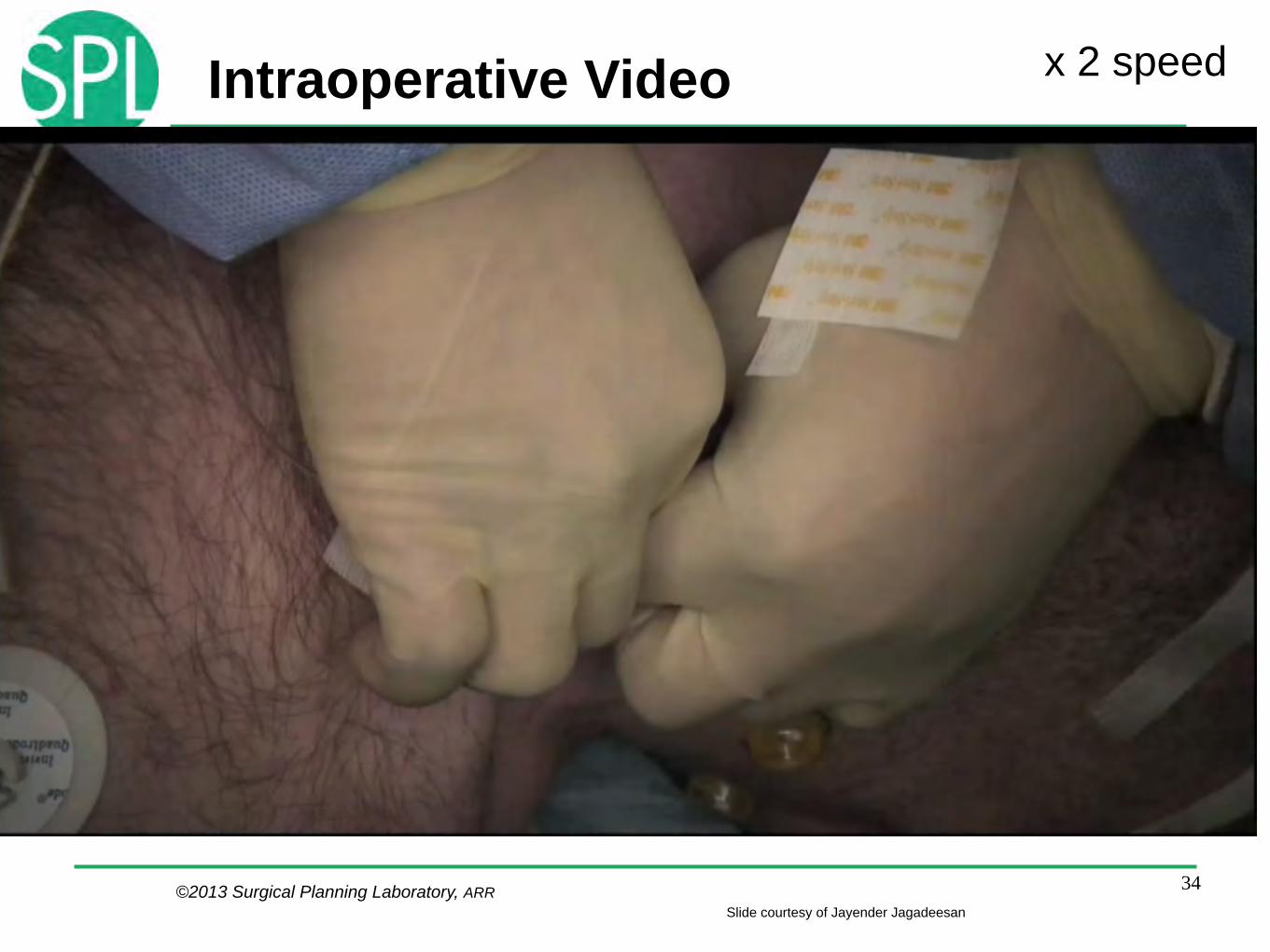
x 2 speed
Slide courtesy of Jayender Jagadeesan
Intraoperative Video
34

©2013 Surgical Planning Laboratory, ARR
Result of Navigation System
35Slide courtesy of Jayender Jagadeesan
©2013 Surgical Planning Laboratory, ARR
Postoperative scan
36Slide courtesy of Jayender Jagadeesan

©2013 Surgical Planning Laboratory, ARR
Clinical Research Example 5
37
Only 20% of smokers develop COPD Genetic factors
Multi-center study funded by the National Heart, Lung and Blood Institute (NHLBI). Co-PIs: Drs. James Crapo, Edwin Silverman.
21 clinical sites
3 image analysis centers: • Denver • Boston • Iowa
2 imaging platforms: • VIDA • Slicer
Slide courtesy R. San Jose

©2013 Surgical Planning Laboratory, ARR
Normal
Moderate CLE
Mild CLE
Emphysema Classification for Gene Discovery
38
Severe CLE
PLE
Paraseptal
• Centrilobular (CLE) and panacinar (PLE) emphysema
• GWAS in 9000 smokers • New genetic markers for emphysema were found
near the CHRNA3/5 locus on 15q25 and near MMP12 and MMP3 on 11q22
• Identification of emphysema patterns based on local histogram classification !
Castaldi PJ, San Jose Estepar R, Sanchez Mendoza C, Crapo JD, Lynch D, Beaty TH, Washko GR, Silverman EK, Proc. ATS, 2012, p.A3808.
Slide courtesy R. San Jose

©2013 Surgical Planning Laboratory, ARR
Phenotype Extraction In The Lung
39
Airw
ays
Vess
els
Kindlmann G, San José Estépar R, Smith SM, Westin CF. Sampling and visualizing creases with scale-space particles. IEEE Transactions on Visualization and Computer Graphics 2009;15(6):1415-1424. PMID: 19834216.
Severe Disease
Extraction Sizing Phenotype
Airway Wall corresponding to a 10 mm internal Perimeter
Smoker control Severe disease
Shift in blood volume per cross sectional area indicating distal pruning and proximal remodeling with disease progression
Population Study
Smoker Controls
San Jose Estepar R et al, Automatic Airway Analysis for Genome-Wide Association Studies in COPD, ISBI 2012
San Jose Estepar R et al, Computational Vascular Morphometry for the Assessment of Pulmonary Vascular Disease based on Scale-Space Particles, ISBI 2012

©2013 Surgical Planning Laboratory, ARR
NA-MIC
• The National Alliance for Medical Image Computing (NA-MIC), is a distributed community of researchers
• Focus on – Subject specific image analysis – NA-MIC kit, including 3D Slicer as a platform for dissemination
• Funded by NIH through the NCBC program since 2004
40

©2013 Surgical Planning Laboratory, ARR
NA-MIC in Numbers• 3D Slicer software used worldwide as platform for development and
sharing • Large impact on NIH grantees: 31 funded collaborations across
schizophrenia, lupus, autism, lung disease, cardiac disease, brain cancer, liver, colon, prostate, musculoskeletal disorders.
• International funding: Canada, Germany, Spain, Italy, Japan, Australia. • “Common Toolkit”: joint transatlantic effort • Trained 55 engineers, 35 grad students, 20 post-docs. • 2000+ investigators trained in 63 workshops • 500+ full size papers, including awards • 15 “Project-weeks”, weeklong working events
twice a year: over 650 participants
41
Image courtesy M. Halle

©2013 Surgical Planning Laboratory, ARR
NA-MIC Community
42Picture 2011, courtesy Kapur, Jakab, Kikinis
©2013 Surgical Planning Laboratory, ARR
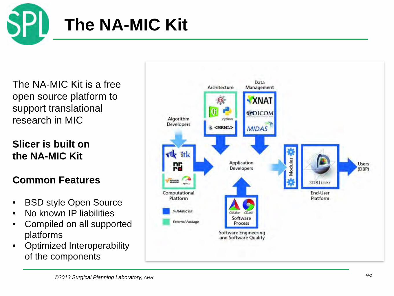
The NA-MIC Kit
43
The NA-MIC Kit is a free open source platform to support translational research in MIC !Slicer is built on the NA-MIC Kit !Common Features !
• BSD style Open Source • No known IP liabilities • Compiled on all supported
platforms • Optimized Interoperability
of the components

©2013 Surgical Planning Laboratory, ARR
Principled Software Process• Documented workflows • Github is used as repository
– distributed – allows offline work – sharing with
granularity • Slim trunk, most
functionality is in plug-ins
44
©2013 Surgical Planning Laboratory, ARR
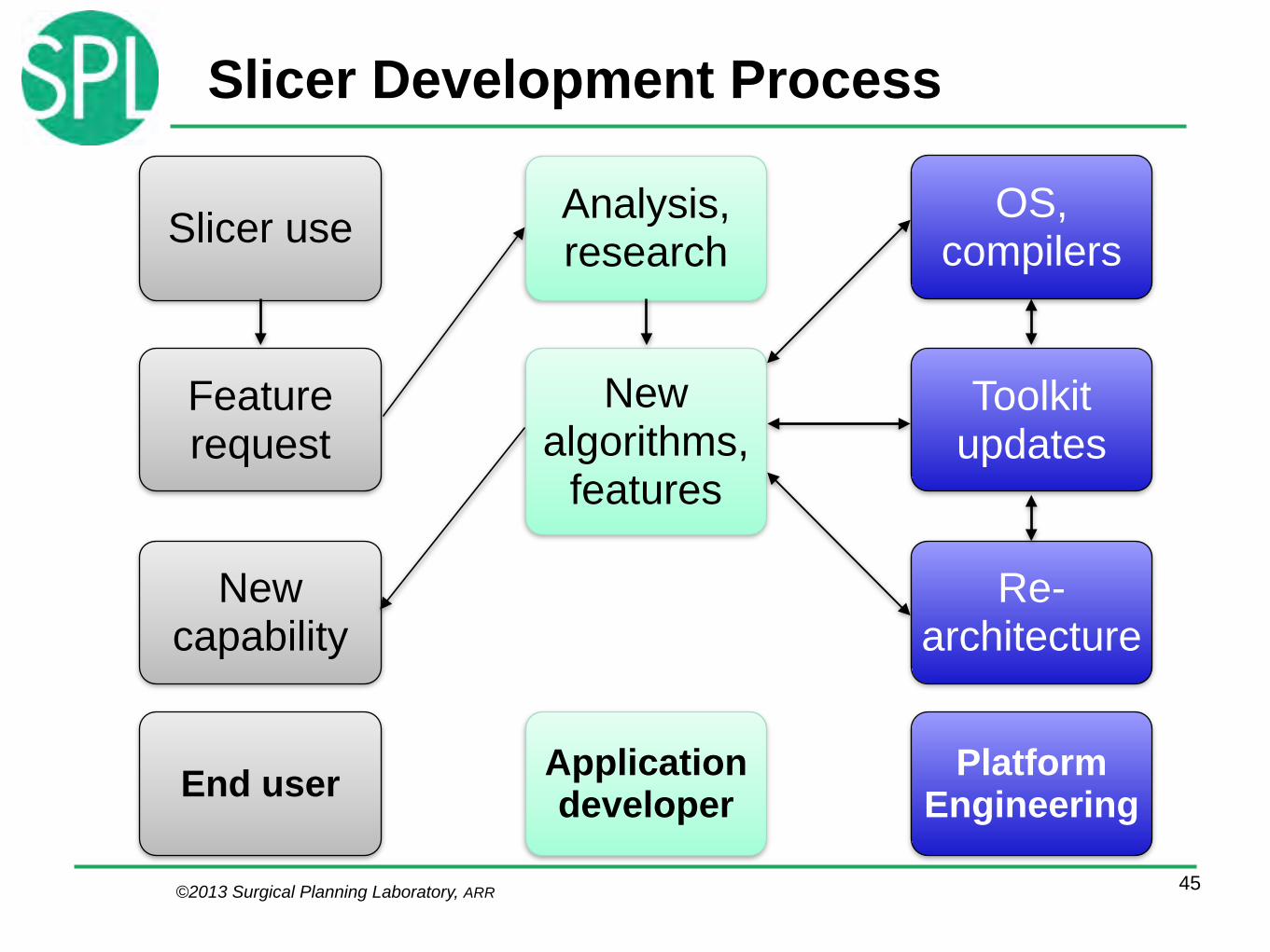
Slicer Development Process
45
Slicer use
Feature request
New capability
Analysis, research
New algorithms,
features
Toolkit updates
OS, compilers
Re-architecture
End user Application developer
Platform Engineering
©2013 Surgical Planning Laboratory, ARR
Application Development
• Algorithm research comes first • Implementation workflow once the
algorithms are known: – Create individual modules as plug-
ins – Create workflows based on the
modules – Use the extension manager for
distribution
46
©2013 Surgical Planning Laboratory, ARR
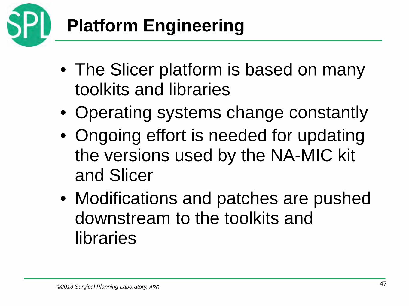
Platform Engineering
• The Slicer platform is based on many toolkits and libraries
• Operating systems change constantly • Ongoing effort is needed for updating
the versions used by the NA-MIC kit and Slicer
• Modifications and patches are pushed downstream to the toolkits and libraries
47

©2013 Surgical Planning Laboratory, ARR
What it takes
• Money, money, money • Time, time, time
!
– Platform engineering for translational MIC is expensive and difficult to find funding for
– It takes time to bring together an interdisciplinary community
48

©2013 Surgical Planning Laboratory, ARR
Work• 1 Ph.D. thesis is one to two person-years
of actual work • Slicer represents over 100 person years in
direct effort
49http://www.ohloh.net/p/slicer
work moved to plug-ins
©2013 Surgical Planning Laboratory, ARR
NA-MIC Kit Engineering Team
clisle davisb demian domibel dpace fedorov finetjul francois_budin freudling gcasey haehn harveerar hayes hjohnson
50
FeiZhao Michael.jeulinl Yong alexy andy atriveg awiles aylward barre benjamin.long bess blezek casey.goodlett christopher.mullins !
hliu hong hyang ibanez ilknur.kabul inorton jcfr jcross186 joe.snyder johan.andruejol jvs karthik kedar_p kerstin !
kquintus lantiga lassoan lauren lorensen maddah malaterre matthew.bowman mccormic mike millerjv mscully naucoin nicky !
nobyhata padfield partyd pieper pinter pkarasev pkarasev3 pohl rjosest rsierra samset sankhesh sylvain taox
taylor tgl tokuda tringo ungi vmagnotta vrnova wjp ygao yumin zach.mullen zack.galbreath
Special thanks to Jean-‐Christophe Fillion-‐Robin, Julien Finet, Steve Pieper, Nicole Aucoin, Andrey Fedorov, Jim
Miller, Andas Lasso

©2013 Surgical Planning Laboratory, ARR
Open Source (OS)
• Collaborate and move freely –Good match for the migratory lifestyle of scientist
–Advantageous for collaborations –Neutral territory in multi-vendor settings
• Extensible
51
©2013 Surgical Planning Laboratory, ARR
Upsides for Industry• Potential advantages:
– Compared to closed systems more people track changes and detect problems
– Easy access to world class algorithms and architectures.
– Community can be engaged • Open Source approaches are practical
– Costs are potentially lower – They permit the organization to focus on
its key product skills, not on commodity capabilities
52K. Vosburgh, GE
©2013 Surgical Planning Laboratory, ARR
Challenges for Industry
• Avoid leakage of proprietary information through clear rules and strategies
• Monitor the open source community for shifting focus and direction
• Internal development is likely to be needed for key features.
• Summary: Risks are manageable, but need to be managed
53KG Vosburgh, GE

©2013 Surgical Planning Laboratory, ARR©2013 Surgical Planning Laboratory, ARR
From Tools to Medical Product• Open Source facilitates scientific
exchange BTW: Open Source means no restriction on use (i.e. no restriction on commercial use)
• All Medical Products are closed source due to significant regulatory requirements !
• How to bridge?
54

©2013 Surgical Planning Laboratory, ARR
From Open to Closed Source
©2013 Surgical Planning Laboratory, ARR
Open source !
!
!
!
!
!
!
Closed source55
Plug-ins
3D Slicer MITK !
MedINRIA !
Mevis Lab !
syngo.via , Advantage Windows, Vitrea
NiftyView
GIMIAS
MevisCLI Plug-ins
Slicer

©2013 Surgical Planning Laboratory, ARR
CTK: An example of OPM• Common infrastructure elements • International and transatlantic group of
contributors • Free Open Source Software under a BSD license • Dicom, application hosting, CLI, Widgets and
more
56
©2013 Surgical Planning Laboratory, ARR©2013 Surgical Planning Laboratory, ARR
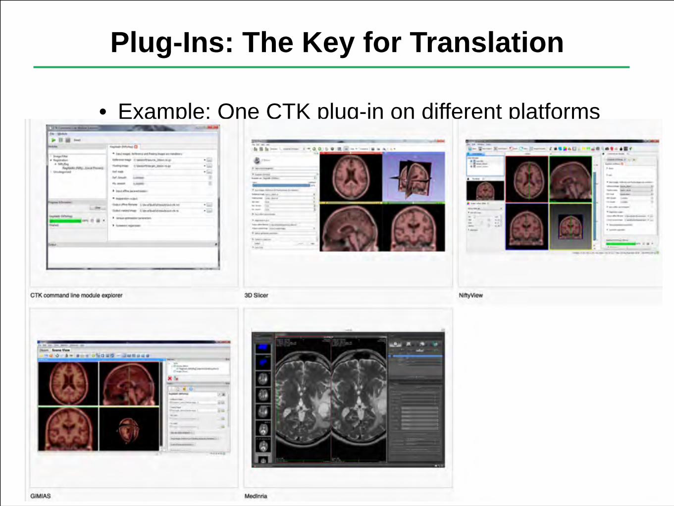
Plug-Ins: The Key for Translation
• Example: One CTK plug-in on different platforms
57
©2013 Surgical Planning Laboratory, ARR
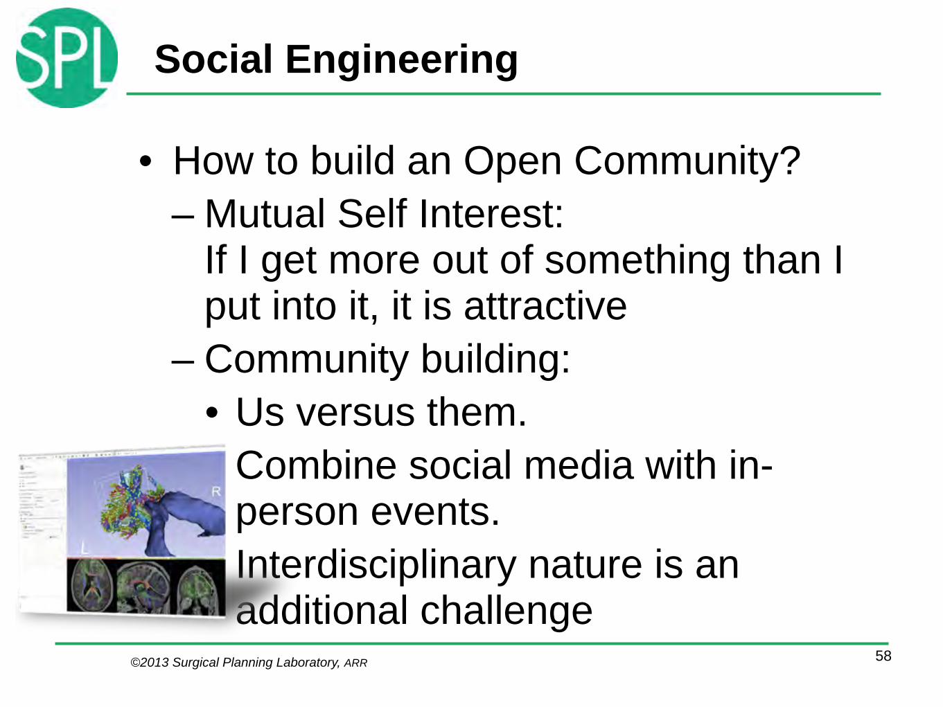
Social Engineering
• How to build an Open Community? – Mutual Self Interest:
If I get more out of something than I put into it, it is attractive
– Community building: • Us versus them. • Combine social media with in-
person events. • Interdisciplinary nature is an
additional challenge 58

©2013 Surgical Planning Laboratory, ARR
User Training Events
• Hands-on training workshops at national and international venues
• More than 2,700 clinicians, clinical researchers and scientists trained since 2005
59
©2013 Surgical Planning Laboratory, ARR
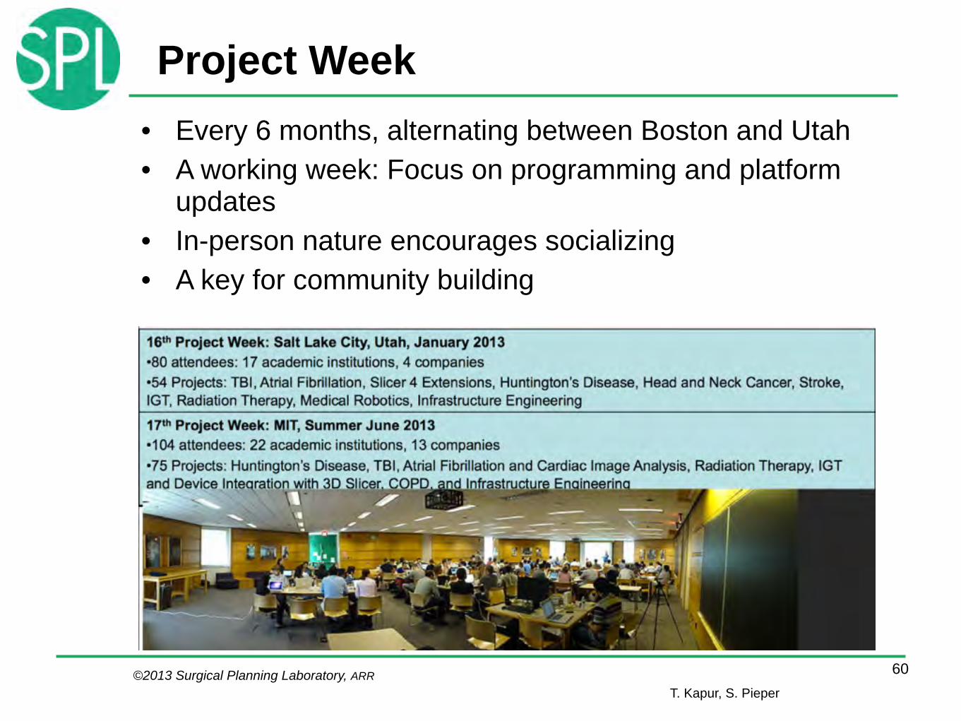
Project Week• Every 6 months, alternating between Boston and Utah • A working week: Focus on programming and platform
updates • In-person nature encourages socializing • A key for community building
60T. Kapur, S. Pieper

©2013 Surgical Planning Laboratory, ARR
Conclusions
• Free Open Source Software – Facilitates translation: bridging the
valley of death – Is a win-win proposition: the OPM
principle – Requires proper policies and
governance • Slicer and the NA-MIC kit are a good
example of FOSS for translational work
61

©2013 Surgical Planning Laboratory, ARR
AcknowledgmentsNational Alliance for Medical Image
Computing www.na-mic.org !Neuroimage Analysis Center nac.spl.harvard.edu !Surgical Planning Laboratory,
Brigham and Women’s Hospital spl.harvard.edu !National Center For Image Guided Therapy www.ncigt.org
62
• Ferenc Jolesz, MD, my mentor
• Collaborators and colleagues

©2013 Surgical Planning Laboratory, ARR
Traumatic Brain Injury Facts
• There are approximately 1.5 million new cases of non-fatal traumatic brain injury (TBI) in the US every year.
• The worldwide incidence of this condition has been estimated to amount to at least 6.8 million TBI cases every year.
• The financial burden of this condition in the USA alone amounts to over $56 billion annually
• More than half of the cases are classified as moderate or severe
!• NA-MIC collaboration:
– UCLA: Jack vanHorn, Andrey Imiria, Paul Vespa – UTAH: Guido Gerig, Marcel Prastawa, Bo Wang – Kitware: Stephen Aylward, Danielle Pace
63http://en.wikipedia.org/wiki/Traumatic_brain_injury

©2013 Surgical Planning Laboratory, ARR
Traumatic Brain Injury
Brain images of patients with traumatic brain injury undergo dramatic changes
64
©2013 Surgical Planning Laboratory, ARR
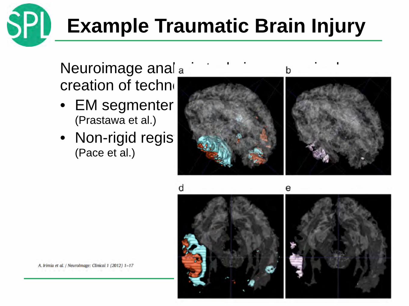
Example Traumatic Brain Injury
Neuroimage analysis techniques required creation of technology • EM segmenter
(Prastawa et al.)
• Non-rigid registration (Pace et al.)
65

©2013 Surgical Planning Laboratory, ARR
acute
chronic
SSA 2
66
Longitudinal change: acute vs. chronic
Wang et al., Univ. Utah
Wang B., Prastawa M., Irimia A., Chambers M.C., Vespa P.M., Van Horn J.D., Gerig G. A Patient-specific Segmentation Framework for Longitudinal MR Images of Traumatic Brain Injury. Proceedings of SPIE 2012;8314, 831402.

©2013 Surgical Planning Laboratory, ARR
DTI• The UCLA group had acquired good
quality DWI data in both acute and chronic patients
• Once segmentation and registration work on TBI subjects, parcellation of the grey matter and analysis of the white matter are possible
67

©2013 Surgical Planning Laboratory, ARR
Connectograms
Connectograms use parcellated gray matter regions to analyse the white matter
68

©2013 Surgical Planning Laboratory, ARR
Personalized Connectomics
Streamlines, which are reduced by more than 20% as a result of brain trauma
69

©2013 Surgical Planning Laboratory, ARR
SSA: The Effects of Pathology
• Focal pathology introduces focal changes, which make it difficult to define general rules upon which algorithms are based
• Example: Effect of brain tumors on fractional anisotropy of adjacent white matter.
70
©2013 Surgical Planning Laboratory, ARR
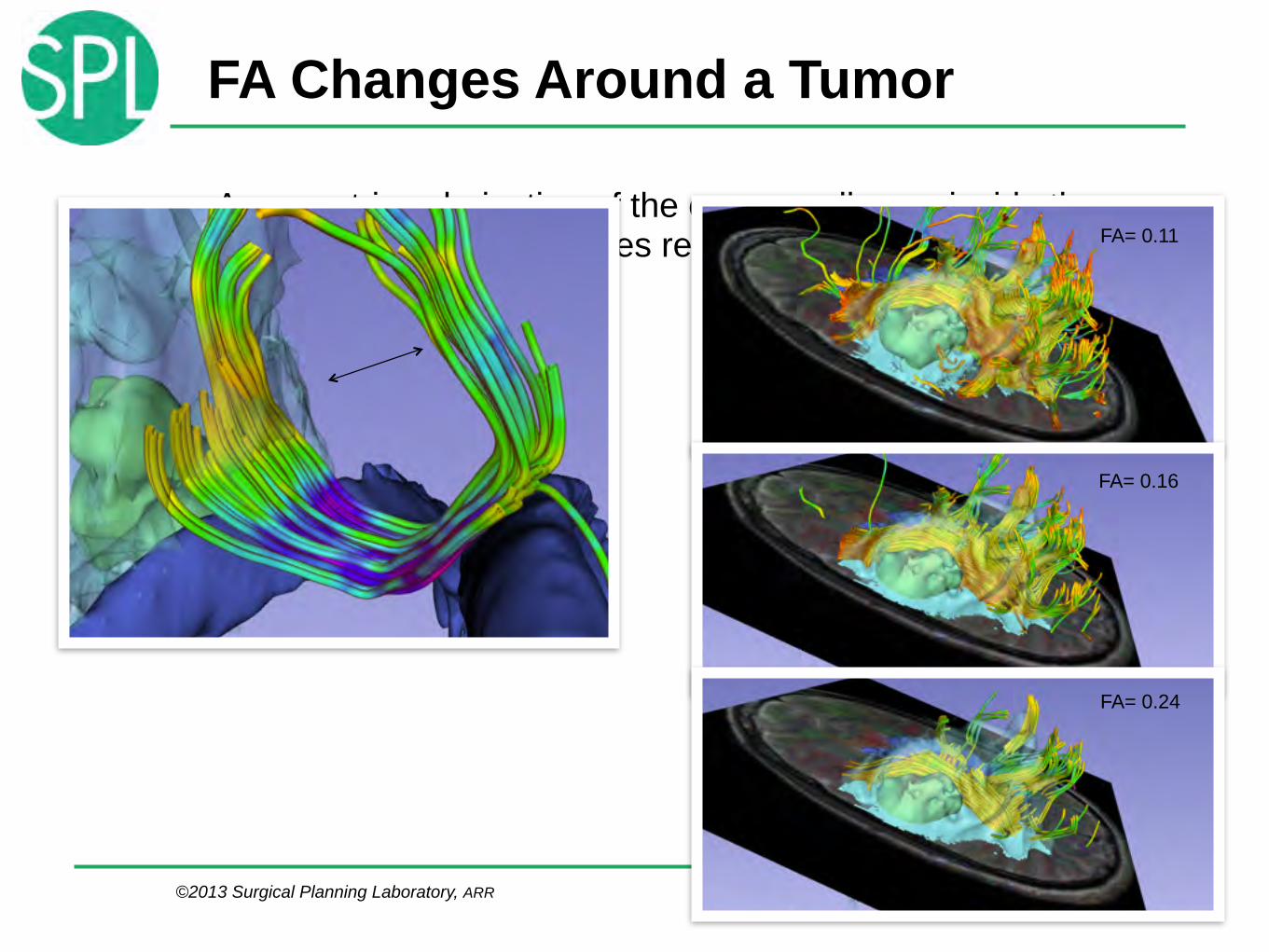
FA Changes Around a Tumor
• Asymmetric colorization of the corpus callosum inside the peritumoral edema indicates reduction in FA
71
FA= 0.11
FA= 0.16
FA= 0.24

©2013 Surgical Planning Laboratory, ARR
Dislocation of Normal Anatomy
The cortico-spinal tract is moved backwards, not toward the midline
72