48 year old man recurrent tachy episodes normal echo inputs from chandrashekhar
TRANSCRIPT

48 year old manRecurrent tachy episodes
Normal echo
Inputs from Chandrashekhar
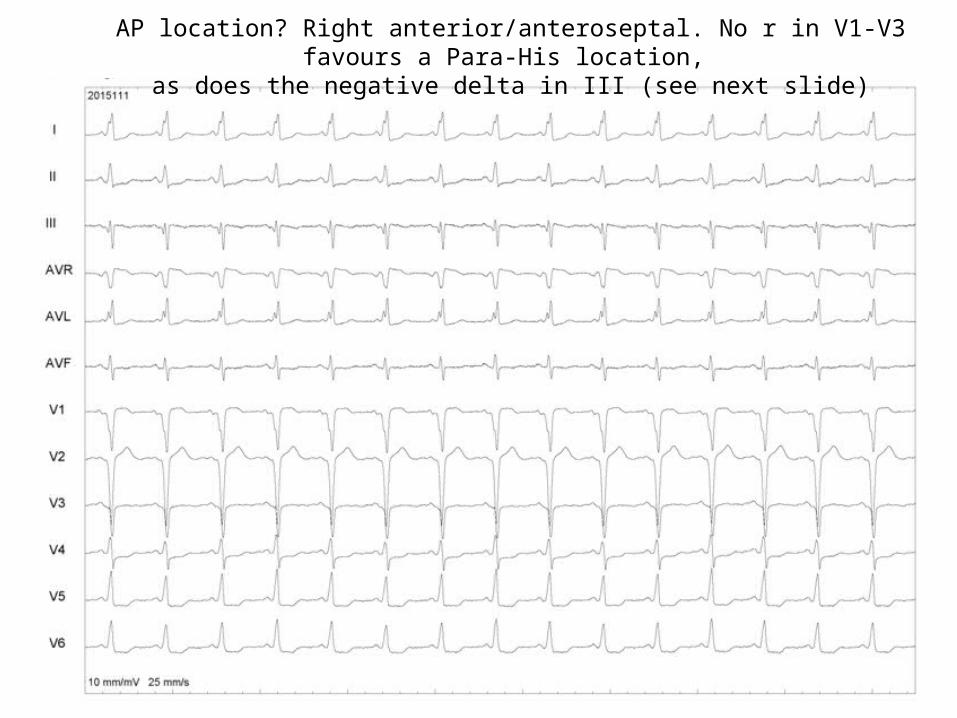
AP location? Right anterior/anteroseptal. No r in V1-V3 favours a Para-His location, as does the negative delta in III (see next slide)

Electrocardiographic Characteristics and Catheter Ablation of Parahissian Accessory Pathways Michel Haissaguerre, Frank Marcus, Franck Poquet, Laurent Gencel,
Philippe Le Metayer, Jacques Clementy(Circulation. 1994;90:1124-1128.)
During maximal preexcitation, the ECG showed a positive delta wave in leads I, II, and aVF in all patients: six had a negative delta wave in leads V, and V2 instead of the positivity usually observed in anteroseptal accessory pathways. This pattern had a sensitivity of 75%, a specificity of 96%, a positive predictive value of 86%, and a negative predictive value of 93% for a parahissian location in comparison with a group of 28 patients with anteroseptal accessory pathways.
The delta wave in lead III was isoelectric or negative in six (75%) parahissian APs and seven (25%) anteroseptal APs (P=.01).

Any further insight @ AP location? Not really. The V in HIS channels is not early (Delta-V=0).However, the His catheter location is not clear
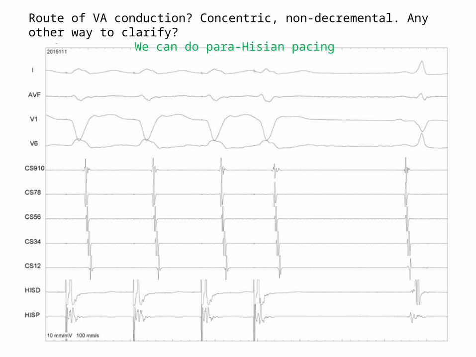
Route of VA conduction? Concentric, non-decremental. Any other way to clarify?We can do para-Hisian pacing

D/D? P soon after QRS, best seen in V1 – ORT/atypical AVNRT/AT

Analyse - His D A is earlier than all other, VA interval is more than 70 ms

Diagnosis clinched? Almost. His refractory VPD preexcites the A.However, no tachy reset (His refractory VPD preexcites the A are equal)

Any more confirmation now? Yes. Reset seen here.
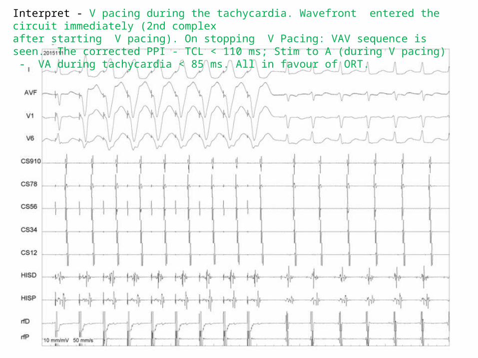
Interpret - V pacing during the tachycardia. Wavefront entered the circuit immediately (2nd complex after starting V pacing). On stopping V Pacing: VAV sequence is seen. The corrected PPI - TCL < 110 ms; Stim to A (during V pacing) - VA during tachycardia < 85 ms. All in favour of ORT.
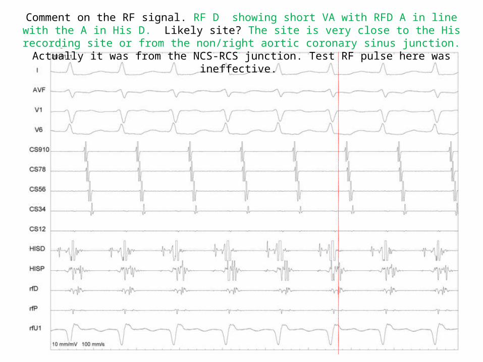
Comment on the RF signal. RF D showing short VA with RFD A in line with the A in His D. Likely site? The site is very close to the His recording site or from the non/right aortic coronary sinus junction. Actually it
was from the NCS-RCS junction. Test RF pulse here was ineffective.

Good RF signal? Better than the previous location. RFD showing almost continuous electrical activity, A earlier than in HISD (see the unipolar for A onset). Miniscule His signal in RFD. Likely site? May be
from the NCS-RCS or very close to the His recording site

RF site- RAO 30. SR0 sheath used for stability

RF site- LAO 40

RF energy…. clean termination of the tachycardia in the retrograde direction (AP)

After ablation - V pacing showing VA block

Adenosine after ablation – atrial pacing showing AV block