5/23/2015phonocardiography15/23/20151 saumya mohan kumar t.e. biomed-roll no.55
TRANSCRIPT

04/18/23Phonocardiography 104/18/23 1
Saumya Mohan Kumar
T.E. Biomed-Roll No.55

04/18/23Phonocardiography 2
Pioneers in auscultation Development of Stethoscope Development of Phonocardiograph Heart sounds Heart Murmurs Basic Block Diagram and Instrumentation Acquisition of phonocardiographic signals Writing methods for phonocardiography Pros and Cons Scope Echocardiography vs. Phonocardiography Case Study
04/18/23 2

04/18/23Phonocardiography 3
Hippocrates laid the foundation for auscultation Robert Hook realized diagnostic use of cardiac
auscultation Biggest breakthrough in auscultation:
Rene Laennec invented stethoscope Dr. Jean Bennett Maguire devised a method of
real-time spectral phonocardiography for the detection and classification of heart murmurs.

04/18/23Phonocardiography 4
Early monaural stethoscope Modern binaural stethoscope Modern electronic stethoscope
Development of Stethoscopes

04/18/23Phonocardiography 5
Acoustic stethoscopes transmit sound mechanically from a chest-piece via air filled hollow tubes to the listener's ears.
The diaphragm and the bell work as two filters, transmitting higher frequency sounds and lower frequency sounds,
respectively. Electronic stethoscopes function in a similar way, but the sound
is converted to an electronic signal which is transmitted to the listener by wire.
Functionalities often included in electronic stethoscopes are amplification of the signal, filters imitating the function of the diaphragm and the bell and in some cases recording abilities to allow storage of data.

04/18/23Phonocardiography 6
Allow volume control of heart and lung sounds heard more easily without amplifying other sounds.
Even subtle changes in breath sounds can be picked up and magnified
Aid health-care professionals in hearing heart murmurs Electronic stethoscopes also allow the user to distinguish
between body sounds of high and low frequency. They now have wireless capabilities, which allow data to be
transferred to a computer or handheld device for storage and retrieval at a later time.

04/18/23Phonocardiography 7
Patients undergoing surgery have the sterile field invaded thereby risking infection
Patients are frequently awakened and disturbed Serious developmental abnormalities in newborn
infants who are frequently disturbed In the absence of airtight seal between stethoscope and
skin, which determines the quality of sound wave transmission, background noise is detected and physiologic sound transmission is impaired.
They are not capable of generating constructive interference of physiologic sound waves.

04/18/23Phonocardiography 8
Bioacoustic research Establish a relationship between mechanical event-
conduction of heart- within the body and the sounds these events give rise to.
The medical use of this knowledge is to link sounds that diverge from normality to certain pathological conditions.

04/18/23Phonocardiography 9
Phonocardiograph: Instrument used for recording sounds connected with the pumping action of heart

04/18/23Phonocardiography 10
Phonocardiogram: A high fedility recording representing the rhythmicity and heart rate
04/18/23 10
04/18/23Phonocardiography 11
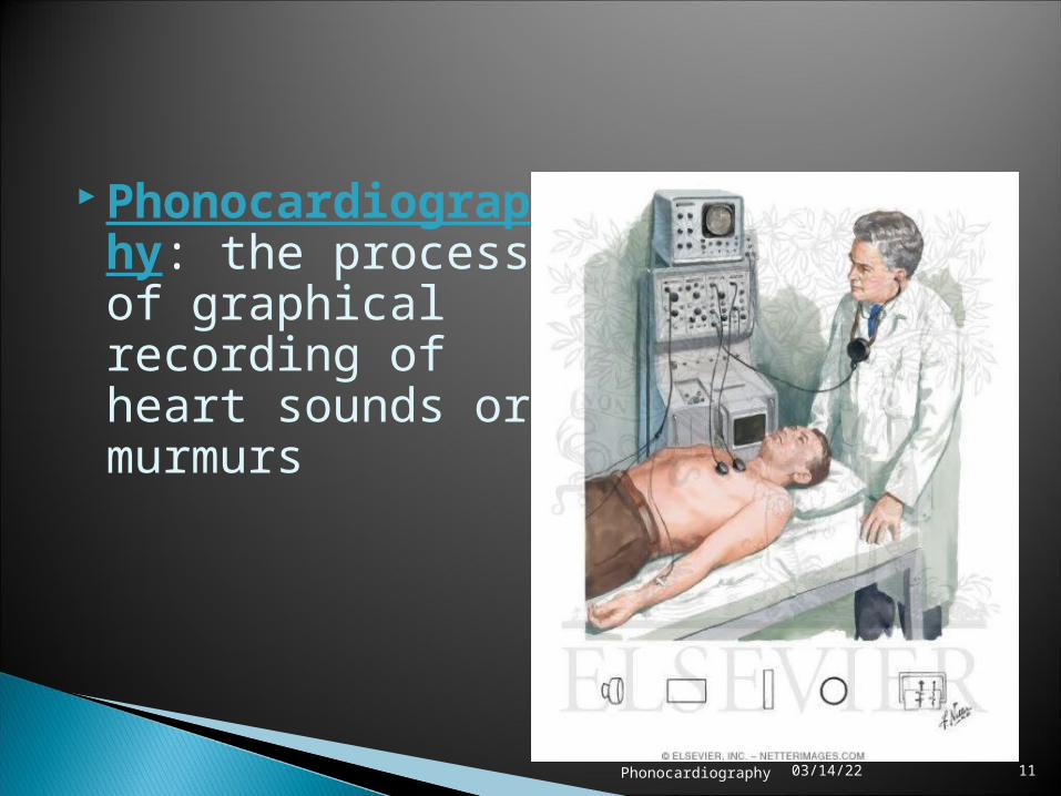
Phonocardiography: the process of graphical recording of heart sounds or murmurs

04/18/23Phonocardiography 12
Mechanical working processes of the heart produce sound which indicate health status of the individual.
The relationship between blood volumes, pressures and flows within the heart determines the opening and closing of the heart valves.
Normal heart sounds- lub and dub- occur during the closure of the valves.
The valvular theory states that heart sounds emanate from a point sources located near the valves.
In the cardiohemic theory the heart and the blood represent an interdependent system that vibrates as a whole and propagates sound as waves of alternate pressure.
04/18/23 12

04/18/23Phonocardiography 13
First heart sound:
- occurs when the atrioventricular (AV) valves close at the beginning of ventricular contraction.
- generated by the vibration of the blood and the ventricular wall
- is louder, longer, more resonant than the second heart sound.

04/18/23Phonocardiography 14
First Heart Sound
Initial vibrations occur when firstcontraction of ventricle move blood towardsatria, closingAV valves
Abrupt tensionof closed AV
valves,decelerating
the blood
Oscillation of blood between
root of aortaand
ventricular walls
Vibrations caused by turbulencein ejected
blood flowinginto aorta

04/18/23Phonocardiography 15
- occurs when aortic and pulmonary semilunar valves close at the beginning of ventricular dilation
- generated by the vibration of the blood and the aorta
- Aortic valve closes slightly before pulmonary valve.
Second heart sound

04/18/23Phonocardiography 16
The second sound (S2) signals the end of systole and the beginning of diastole
It is heard at the time of the closing of the aortic and pulmonary valves
S2 is probably the result of oscillations in the cardiohemic system caused by deceleration and reversal of flow into the aorta and the pulmonary artery

04/18/23Phonocardiography 17
A third heart sound (S3) connected with the diastolic filling period. The rapid
filling phase starts with the opening of the semilunar valves.
attributes energy released with the sudden deceleration of blood that enters the ventricle throughout this period
A fourth heart sound (S4) connected with the late diastolic filling period occur during atrial systole where blood is forced into
the ventricles.

04/18/23Phonocardiography 18

04/18/23Phonocardiography 19
Murmurs are extra heart sounds that are produced as a result of turbulent blood flow which is sufficient to produce audible noise.
Innocent murmurs are present in normal hearts without any heart disease.
Pathologic Murmurs are as a result of various problems, such as narrowing or leaking of valves, or the presence of abnormal passages through which blood flows in or near the heart.
Heart murmurs occur when the blood flow is accelerated above the Reynolds number, which induces non-stationary random vibrations, that are transmitted through the cardiac and thoracic tissues up to the surface of the thorax
They are graded by intensity from I to VI. Grade I is very faint and heard only with special effort Grade VI is extremely loud and accompanied by a palpable thrill

04/18/23Phonocardiography 20
Heart Murmurs
High rate of flow through
valves
Flow throughconstricted
valves(stenosis)
Backward flow through
incompetentvalve
Septal defects
Decreased viscosity,
which causes increased turbulence

04/18/23Phonocardiography 21
Name
Eve
nts
Pres
sure
(kP
a)
0
5
10
15
20
PCG
ECG
Time 0 (sec)
0.1 0.2 0.3 0.4 0.5 0.6 0.7 0.8

04/18/23Phonocardiography 2204/18/23 22

04/18/23Phonocardiography 23
Basic transducer
Amplifier
Filter
• Piezoelectric sensor to convert sound or vibrations to electricity
• Crystal or moving coil microphone having frequency response between 5Hz and 1000Hz
• Similar response characteristics• Offer selective high pass filter to allow
frequency cutoff• Bandwidth : 20- 2000Hz• Amplify signal
• Permit selection of suitable frequency bands
• Avoid aliasing• Separate louder low frequency signals
from lower intensity, much informative high frequency murmurs.
04/18/23 23

04/18/23Phonocardiography 24
Integrator
Power Amplifier
DAC and Readout or high frequency chart recorder or oscilloscope or headphones
• Recording envelope of higher frequency over 80Hz along with actual signals below 80Hz.
• Increase the power of incoming signal
• Efficiency is more• Effect of noise is lowered
• Signal is converted to digital form and stored permanently
• For faithful recording of heart sounds
04/18/23Phonocardiography 25
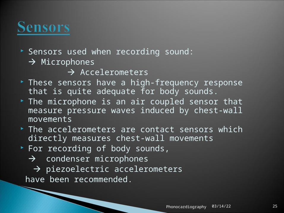
Sensors used when recording sound: Microphones
Accelerometers These sensors have a high-frequency response that is quite adequate for
body sounds. The microphone is an air coupled sensor that measure pressure waves
induced by chest-wall movements The accelerometers are contact sensors which directly measures chest-
wall movements For recording of body sounds,
condenser microphones piezoelectric accelerometers
have been recommended.
04/18/23Phonocardiography 26
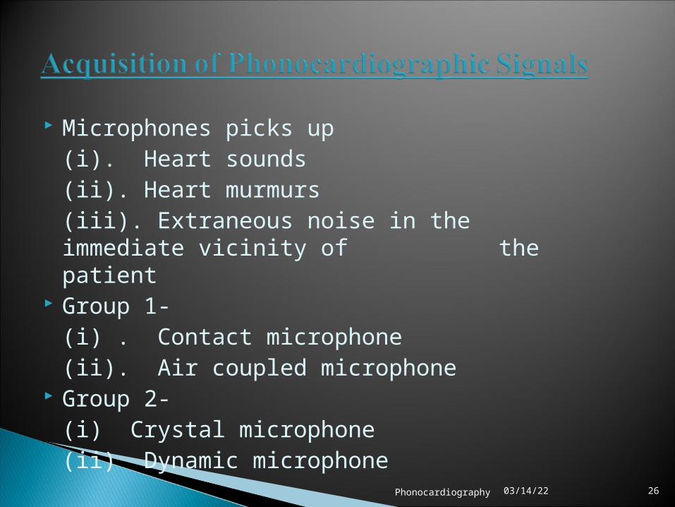
Microphones picks up(i). Heart sounds(ii). Heart murmurs(iii). Extraneous noise in the immediate vicinity
of the patient Group 1-
(i) . Contact microphone(ii). Air coupled microphone
Group 2-(i) Crystal microphone(ii) Dynamic microphone

04/18/23Phonocardiography 27
Contact Microphone also known as a pickup or a piezo
microphone made of a thin piezoelectric
ceramic round disc (+ve) glued to a thin brass or alloy metal disc (-ve)
designed to transmit audio vibrations through solid objects.
contact mics act as transducers which pick up vibrations and convert them into a voltage which can then be made audible.

04/18/23Phonocardiography 28
Air coupled Microphones shows a low-pass frequency response because of its air-
chamber compliance. In the pass band, it is considered that the microphone
has a flat response, where the mechanical impedance of air chamber is much higher than that of chest wall, the vibration of the measured chest-wall surface is stopped by both the air chamber and the coupler surface in contact with the chest wall.
The sound pressure, or normal stress exerted on the chamber should be constant to keep a flat response.

04/18/23Phonocardiography 29
Crystal Microphones uses the piezoelectric effect of Rochelle salt, quartz, or
other crystalline materials. This means that when mechanical stress, due to heart
sounds, is placed upon the material, a voltage electromagnetic force is generated.
Since Rochelle salt has the largest voltage output for a given mechanical stress, it is the most commonly used crystal in microphones.
smaller in size, more sensitive than dynamic ones

04/18/23Phonocardiography 30
a crystal is mounted so that the sound waves strike
it directly
a diaphragm that is mechanically linked to the crystal so that the sound waves are indirectly coupled to the
crystal.

04/18/23Phonocardiography 31
Dynamic Microphones consists of a moving coil with
fixed magnetic core inside. This moving coil moves with
heart sounds, and produces voltage because of its interaction with magnetic flux

04/18/23Phonocardiography 32
It does not transform acoustic oscillations into electrical potentials uniformly for all frequencies.
Hence heart sound recording done with microphone is valid for a particular type of frequency only..
Hence microphones of various types cannot be interchanged.

04/18/23Phonocardiography 33
Requires a writing system capable of responding to 2000 Hz.
Types of writing methods:(i). Mechanical Recorders(ii). Optical Galvanometric Recorders(iii). Envelope detection(iv). Direct recording using Ink Jet Recorders(v). Electrostatic Recorder(vi). Thermal Recorder

04/18/23Phonocardiography 34
Merits very little loss of diagnostically important
information eliminates the effort and delay of
photographic processing immediacy of the results affords a means
for continuously monitoring the records for quality and special content at the time of registration.
Demerits writing recorders with an upper frequency
response of 150 Hz cannot be used to write frequencies that lie beyond their working range.
can only record heart sound intensity picked up every 10 msec.

04/18/23Phonocardiography 35
Uses artificial frequency of about 100 Hz in heart sound amplifier
Employed to oscillate stylus so that high frequency sounds are modulated by 100Hz
04/18/23Phonocardiography 36
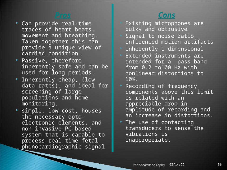
Pros Can provide real-time traces of
heart beats, movement and breathing. Taken together this can provide a unique view of cardiac condition.
Passive, therefore inherently safe and can be used for long periods.
Inherently cheap, (low data rates), and ideal for screening of large populations and home monitoring.
simple, low cost, houses the necessary opto-electronic elements. and non-invasive PC-based system that is capable to process real time fetal phonocardiographic signal
Cons Existing microphones are bulky
and obtrusive Signal to noise ratio influenced
motion artifacts Inherently 1 dimensional Extended instruments are
intended for a pass band from 0.2 to100 Hz with nonlinear distortions to 10%.
Recording of frequency components above this limit is related with an appreciable drop in amplitude of recording and an increase in distortions.
The use of contacting transducers to sense the vibrations is inappropriate.

04/18/23Phonocardiography 37
Further Work
1. Design of clinical prototype2. Improvements to signal conditioning and control electronics3. Investigate wireless links for cordless monitoring4. Remote measurement of small displacements at compliant surfaces
Suggested Applications
Remote sensing of sub 50 micron displacements Adult and fetal phonocardiography and phonography Remote measurements of compliant materials in wind-tunnels Infrasound intensity measurement Biomedical instrumentation Low-cost and low power confocal microscopy Cell culture measurement

04/18/23Phonocardiography 38
Echocardiography
better diagnosis of mitral valve defects ,evaluating the degree of its stenosis and characterizing the morphological changes of the valve.
more informative about tricuspid valve defects
echocardiographic data on the changes in the left ventricular outflow tract help to explain the origin of the spindle-form systolic murmur.
Phonocardiography
better diagnosing of mitral insufficiency, diagnosis of aortic valve defects
more informative about state of aortic valves
interpretation of systolic murmur was rather complicated, although they are often seen on phonocardiographic data of normal individuals and patients with heart diseases.
The two not alternative, and the less contradictory, but mutually supplementing methods.

04/18/23Phonocardiography 39

04/18/23Phonocardiography 40