5aminolevulinic acid induced apoptosis via
TRANSCRIPT

Original Article
J. Clin. Biochem. Nutr. | September 2019 | vol. 65 | no. 2 | 83–90doi: 10.3164/jcbn.18�46©2019 JCBN
JCBNJournal of Clinical Biochemistry and Nutrition0912-00091880-5086the Society for Free Radical Research JapanKyoto, Japanjcbn18-4610.3164/jcbn.18-46Original Article5�Aminolevulinic acid induced apoptosis via oxidative stress in normal gastric epithelial cellsHiromu Ito,1 Hiromi Kurokawa,2 Hideo Suzuki,2 Hiroko P. Indo,1 Hideyuki J. Majima1 and Hirofumi Matsui2,*
1Graduate School of Medical and Dental Sciences, Kagoshima University, 8�35�1 Sakuragaoka, Kagoshima, Kagoshima 890�8544, Japan2Department of Gastroenterology, Faculty of Medicine, University of Tsukuba, 1�1�1 Tennoh�dai, Tsukuba, Ibaraki 305�8575, Japan
*To whom correspondence should be addressed. E�mail: [email protected]
??(Received 10 April, 2018; Accepted 4 January, 2019; Published online 9 August, 2019)
Copyright © 2019 JCBN2019This is an open access article distributed under the terms of theCreative Commons Attribution License, which permits unre-stricted use, distribution, and reproduction in any medium, pro-vided the original work is properly cited.5�Aminolevulinic acid, a precursor of heme, is utilized in a variety
of applications including cancer treatment, surgery, and plant
nutrition. However, 5�aminolevulinic acid itself induces oxidative
stress and subsequent lipid peroxidation. Reactive oxygen species
are factors in oxidative stress, not only causing cellular injury but
also inducing several signal transduction pathways. Especially in
cancer cells, a significant amount of signalling activation and sub�
sequent activation of protein is caused by the enhancement of
reactive oxygen species production. Reactive oxygen species
levels in normal cells are low and an oxidative condition is harmful;
hence, administration of 5�aminolevulinic acid to normal cells may
induce oxidative stress, resulting in cell death. In this study, we
investigated the effect of 5�aminolevulinic acid on normal and
cancer cells with regard to oxidative stress. We used the rat
normal gastric cell line RGM and its cancer�like mutant cell line
RGK. 5�Aminolevulinic acid treatment of RGM cells enhanced reac�
tive oxygen species generation and induced apoptosis associated
with p53, whereas RGK cells were unaffected. In addition, RGM
cell viability was recovered by application of N�acetyl�L�cysteine
or p53 inhibitor. These results suggest that 5�aminolevulinic acid
causes oxidative stress in normal gastric cells and induces apoptosis
via the p53�dependent pathway.
Key Words: 5�aminolevulinic acid, reactive oxygen species,
gastric epithelial cell, apoptosis
IntroductionHeme is an essential substance for many kinds of animals tometabolize oxygen and generate energy. 5-Aminolevulinic
acid (5-ALA) is a precursor of heme and has some positive effectson health, such as promoting hair growth and boosting the immunesystem.(1–3) 5-ALA is also utilized in photodynamic therapy(PDT). ALA-PDT is a combination therapy utilizing appropriateirradiation and a photosensitizer (protoporphyrin IX: PpIX) thatis produced from 5-ALA.(4) In recent years, this photochemicalproperty has been used to diagnose brain tumours and determinethe area of resection.(5) 5-ALA is also an important nutrient forplants. It is a basic ingredient of chlorophyll where is the photo-synthetic reaction centre and accelerates plant growth.(6) In addi-tion, 5-ALA improves salt and cold resistance in plants.(7,8) How-ever, at high concentrations, 5-ALA causes photosensitivity andcould act as an herbicide.(6,9) It has been reported that 5-ALAinduces formation of reactive oxygen species (ROS) that causelipid peroxidation, particularly cardiolipin, a major phospholipidin the mitochondrial inner membrane.(10–12)
ROS have wide influence over organisms, affecting signalling,transcription factor activation, and the immune system.(13) Over-production of ROS results in oxidative stress and is associatedwith various intractable disease like cancer, Alzheimer’s disease,and inflammatory bowel disease.(14–16) ROS can be generated inthe endoplasmic reticulum and through the action of NADPH
oxidase in the cell membrane.(17,18) However, mitochondria arethe primary source of ROS because they consume oxygen in theelectron transport chain (ETC), which produces ROS as a by-product of energy production.(19,20) Mitochondrial DNA (mtDNA)mutations can decrease the efficiency of ETC and enhance theproduction of ROS.(21) In general, larger amounts of ROS areproduced in cancer cells than in normal cells because of mtDNAmutations.(22,23) Over-production of ROS activates signallingpathways that induce protein expression. ROS derived frommitochondria have been implicated in metastasis and cancerinvasiveness.(24,25) Thus, ROS play an important role in mainte-nance of morphology of cancer cells and are essential factors forsurvival of cancer. On the other hand, oxidative stress is harmfulto normal cells.(26) Taken together, ROS produced by administra-tion of 5-ALA may both elevate cancer activity and damagenormal cells. Especially in ALA-PDT, normal tissue is alsoexposed by 5-ALA and to examine the effect is important forpatients because the effect of photosensitizers for normal tissuewhich is not irradiated has not been discussed. In this study, weexamined and compared the effect of 5-ALA on ROS productionin normal and cancerous gastric cells. The rat normal gastricepithelial cell line, RGM and cancer-like cell line, RGK were usedfor comparison. RGM is derived from gastric mucosa of Wistarrats, and RGK is a chemically mutated strain of RGM.(27,28) Weused 1-methyl-3-nitro-1-nitrosoguanidine (MNNG) to transformRGM to RGK. Since these cell lines have the same genetic back-ground, they are suitable for comparing the behaviour of normaland cancer cells.
Materials and Methods
Cell culture. RGM, a normal gastric epithelial cell linederived from rat, and RGK, a cancer-like mutant cell line of RGM,were established previously.(27,28) RGM and RGK cells werecultured in Dulbecco’s modified Eagle medium/Nutrient MixtureF-12 (DMEM/F12) with (Thermo Fisher Scientific, Waltham,MA) and without (Sigma-Aldrich Co. LLC., St. Louis, MO) L-glutamine, respectively. Each medium was supplemented with10% foetal bovine serum (Equitech-Bio Inc., Kerrville, TX) and1% penicillin/streptomycin (Thermo Fisher Scientific). Cells weremaintained at 37°C in moist air with 5% CO2. All cell incubationwith 5-ALA was performed in the dark condition.
Cell viability test. Cell viability, which means cellular pro-liferation ability, after 5-ALA exposure was calculated using theCell Counting Kit-8 (CCK-8) colorimetric assay (DOJINDOLABORATORIES, Kumamoto, Japan), which is an alternativemethod of MTT assay, according to the manufacturer’s protocol.
H

doi: 10.3164/jcbn.18�46©2019 JCBN
84
Briefly, RGM and RGK cells were seeded on a 96-well cell cultureplate at a density of 5,000 cells/well and incubated overnight at37°C in 5% CO2. The medium of each well was replaced withfresh media containing 1, 5, 10, 50, 100, 500 and 1,000 mM 5-ALA (Cosmo Bio Co., Ltd., Tokyo, Japan). 5-ALA is cleared fromtissue and body in 48 h.(29) Thus, cells were incubated for 48 hbefore the 5-ALA containing medium was removed and cells werewashed with PBS twice. Cells were transferred to fresh mediumcontaining 10% (v/v) CCK-8 solution and additionally incubatedto develop colour. The absorbance of each well at 450 nm wasmeasured with a DTX880 multi-mode micro plate reader (BeckmanCoulter Inc., Brea, CA).
Annexin V assay. Flow cytometric analysis with annexin Vwas used to determine the cellular state after 5-ALA exposure. Inapoptotic phase, phosphatidylserine (PS), a constituent of theinner cell membrane, is exposed to the cell exterior. Annexin Vbinds to PS in a calcium dependent manner. This characteristic ofannexin V is often utilized to study cellular apoptosis. We usedMuse Annexin V & Dead Cell Kit (EMD Millipore) according tothe manufacturer’s instructions to determine the ratio of pro-grammed cell death after 5-ALA treatment. RGM and RGK cellscultured in 10 cm dishes were exposed to 50 mM 5-ALA andincubated for 48 h at 37°C. Cells were washed with PBS andharvested with trypsin. Cell concentration was adjusted to 3 ´ 105
cells/ml, and 100 ml of cell suspension was mixed thoroughlywith an equal volume of Muse Annexin V & Dead Cell. The cellswere incubated for 20 min in the dark at 25°C. After incubation,the cell suspension was transferred to a 1.5 ml tube and analysedby the Muse Cell Analyzer in Annexin V & Dead Cell mode.
Caspase assay. Flow cytometric analysis focused on caspasewas used to examine apoptosis by a separate method. Caspases arecysteine-aspartic proteases that play essential roles in programmedcell death via complex signal cascades. Their expression is anindicator of apoptosis.(30) We used the Muse Caspase-3/7 Kit(EMD Millipore) to distinguish caspase-3 and caspase-7 positivefrom negative cells to produce the apoptotic ratio after 5-ALAtreatment. Briefly, RGM and RGK cells cultured in 10 cm disheswere treated with 50 mM 5-ALA for 48 h in 5% CO2 at 37°C. Cellswere washed with PBS and collected with trypsin. Cell concentra-tion was adjusted to 3 ´ 105 cells/ml in 50 ml of 1X Assay BufferBA from the kit. Next, 5 ml of Muse Caspase-3/7 Reagent workingsolution was added to the cells and incubated in 5% CO2 at 37°Cfor 30 min. After incubation, 150 ml of Muse Caspase 7-AADworking solution was added and mixed thoroughly. The suspen-sion was incubated with light shielding at 25°C for 5 min. Cells wereanalysed by the Muse Cell Analyzer in Caspase-3/7 assay mode.
Western blotting. The levels of caspase-9 and p53 after 5-ALA treatment were investigated by western blotting using apreviously published method.(31) RGM and RGK cells post-5-ALAtreatment were collected and treated with NuPAGE LDS SampleBuffer (Thermo Fisher Scientific). The cell samples were heatedat 95°C for 5 min and 10 ml was added to the wells of NuPAGE12% Bis-Tris Protein Gels (Thermo Fisher Scientific). Proteinswere separated by polyacrylamide gel electrophoresis (PAGE) andtransferred to polyvinylidene difluoride (PVDF) membrane (EMDMillipore). The membrane was washed with PBS containing 0.1%Tween 20 (PBS-T) for 5 min and treated with PVDF BlockingReagent from Can Get Signal (TOYOBO Co., Ltd., Osaka, Japan)for 60 min at 25°C. After blocking, the membrane was washedthree times with PBS-T for 10 min followed by overnight incuba-tion with primary antibodies diluted 1:1,000 in Can Get SignalImmunoreaction Enhancer Solution 1: caspase-9 (Cell SignalingTechnology, Danvers, MA), phospho-p53 (Cell Signaling Tech-nology) or p53 (Cell Signaling Technology). After primaryantibody incubation, the membrane was washed three times withPBS-T and incubated with horseradish peroxidase-conjugatedanti-rabbit IgG antibody (Cell Signaling Technology) diluted1:1,000 in Can Get Signal Immunoreaction Enhancer Solution 2
for 2 h. The secondary antibody solution was removed and themembrane was washed three times with PBS-T. The membranewas immersed in Lumina forte western HRP substrate (EMDMillipore) and luminescence was detected with a LAS4000instrument (GE Health Care Japan, Tokyo, Japan). As a loadingcontrol, b-actin was detected with b-actin primary antibody (CellSignaling Technology).
ROS detection by electron spin resonance. ROS genera-tion in living cells after 5-ALA treatment was examined by elec-tron spin resonance (ESR) according to a previously publishedmethod.(25) RGM and RGK cells were cultured on glass coverslides (49 ´ 5 ´ 0.2 mm) and treated with 50 mM 5-ALA for 48 h.A cell-bearing slide was set in tissue glass and 100 ml of respiratorybuffer [5 mM succinate, 5 mM malate, 5 mM glutamate, 5 mMnicotinamide adenine dinucleotide (NADH), and 5% 5,5-dimethyl-1-pyrroline-N-oxide (DMPO)] was added. The tissue glass wasinserted in an ESR device, a JEOL-TE X-band spectrometer(JEOL Ltd., Tokyo, Japan). Spectra were obtained under 10 mWincident microwave power, 9.42 GHz frequency, and 0.1 mT fieldmodulation amplitude.
N�acetyl�L�cysteine treatment. RGM cells were exposed toN-acetyl-L-cysteine (NAC), which is an antioxidant, and 5-ALA,followed by cell viability measurement using CCK-8 accordingto the method described in section 2.2, supplementing with 50 mM5-ALA and increasing concentrations (0, 1, 5, 10, 50, 100 and500 mM) of NAC (Wako Pure Chemical Industries, Ltd., Osaka,Japan).
p53 inhibitor treatment. Cells were treated with p53 inhib-itor and 5-ALA, then cell viability was measured to examineinduction of the apoptotic pathway by 5-ALA. RGM cells werecultured in 96-well plates at 5,000 cells/well and incubated over-night. The medium was removed and replaced with fresh mediumcontaining increasing concentrations (0, 1, 2, 5, 10, 20, 50 and100 mM) of an inhibitor of p53-dependent apoptosis and transcrip-tion, Cyclic Pifithrin-a hydrobromide (Tokyo Chemical IndustryCo., Ltd., Tokyo, Japan). After 24 h incubation, the p53 inhibitorwas removed and cells were washed with PBS. Cells were addi-tionally incubated in medium containing 50 mM 5-ALA for 48 hand the CCK-8 assay was performed as described in section 2.2.
Statistical analysis. Statistical analysis was carried out usingSPSS statistics 21 software (IBM Corporation, NY). Tukey’s testwas used to compare more than two data sets and Student’s t testwas used for two data sets. P<0.05 and p<0.01 were considered asstatistically significant differences. All data are represented asmean ± SD.
Results
Cytotoxic effect of 5�ALA on gastric normal and cancercells. Rat gastric normal cell line RGM and cancer cell line RGKwere treated with 5-ALA for 48 h and cytotoxicity was calculatedby the CCK-8 method. RGM cells showed a gradual decrease inviability dependent on the 5-ALA dose, up to 50 mM, and theviability gradually recovered (Fig. 1). On the other hand, RGKcancer cells proliferated in a 5-ALA-dose dependent manner up to5 mM, and their numbers decreased from 5 mM to 100 mM. After1, 5, 10, 50, 500 and 1,000 mM 5-ALA treatment; viability ofRGM cells decreased significantly compared to that of RGK cells.These results indicate that 5-ALA is toxic to normal gastric cells,especially at 50 mM, but promotes the growth of cancer cells atconcentrations up to 50 mM (Fig. 1 and Supplemental Fig. 1*).As described above, 5-ALA also has characteristics to promotecellular growth such as hair and plants. Cancer cells may havehigher sensitivity to 5-ALA.
Apoptosis measurement by annexin V and caspase�3/7assay. Cellular apoptosis was studied using Muse Annexin V &Dead Cell Kit after exposure to 50 mM 5-ALA for 48 h. Fig. 2shows the clear change of histogram in RGM cells after 5-ALA
*See online. https://doi.org/10.3164/jcbn.18�46

J. Clin. Biochem. Nutr. | September 2019 | vol. 65 | no. 2 | 85
©2019 JCBNH. Ito et al.
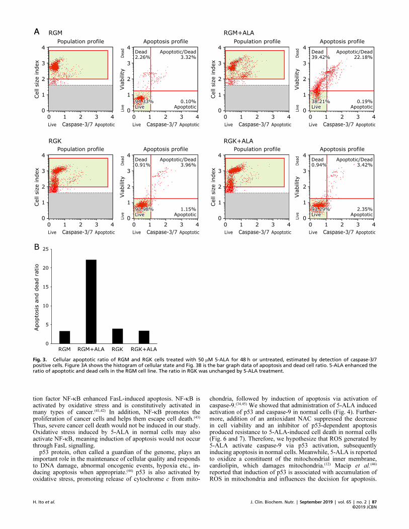
treatment. The late apoptotic or dead cell ratio of RGM cellsincreased significantly after 5-ALA treatment. In RGK cells, theratio did not change much and the early apoptotic ratio increasedslightly. The cellular apoptotic ratio was also measured andcalculated by a caspase-3/7 assay. Figure 3 shows most RGM cellswithout treatment were living and the apoptotic/dead ratio was3.32%. Exposure to 50 mM 5-ALA increased the ratio to 22.18%.On the other hand, the apoptotic/dead ratio in RGK cells wasnot changed by 5-ALA (from 3.96 to 3.42%) and over 90% ofcells were living. These results indicate that 50 mM 5-ALA easilyinfluences the viability of gastric normal cells compared to cancercells and induces an apoptotic state. In comparison between Fig. 2and 3, there was a little difference in the value of living andapoptotic ratio. These results may be from the difference of way tomeasure. However, ALA treatment obviously increased apoptoticindication in RGM cells in both results. Thus, 5-ALA inducesapoptotic signal transduction in RGM normal cells easier thanRGK cancer cells.
Protein expression analysis by western blotting. Caspase proteins are involved in many signalling cascades and are deeplyinvolved with apoptosis. One of the caspase proteins, caspase-9,especially takes part in the mitochondrial apoptotic pathway viaoxidative stress.(32,33) Normal p53 protein also induces cell deathin response to high oxidative stress.(34) We examined the changesin expression levels of these proteins after 5-ALA treatment toclarify the signalling pathway and mechanism of apoptosisderived from 5-ALA treatment. Figure 4A shows that the level ofcleaved caspase-9 increased after 5-ALA treatment in RGM cells.In RGK cells, the caspase-9 level did not change after 5-ALAtreatment. However, the expression of caspase-9 in RGK cells washigher than RGM normal cells when 5-ALA was not treated.This may be because the production level of ROS is higher andmany signal transductions are activated in cancer cells. Mean-while, the expression level of cleaved caspase-9 increased slightlyin RGM at 24 h treatment (Supplemental Fig. 2*). Figure 4B shows
that 5-ALA treatment increases the expression of phosphorylatedp53 protein in RGM cells. However, the expression tended todecrease after 5-ALA treatment in RGK cells. In addition, phos-phorylated p53 expression increased in RGM treated for 24 h(Supplemental Fig. 2*). These results indicate that oxidativestress-dependent apoptosis is induced by 5-ALA in RGM cells.
ROS measurement in living cells. We examined ROS pro-duction in living gastric normal and cancer cells after 5-ALAtreatment by ESR. ESR is a device which detects ROS easily withhigh-sensitivity. Figure 5 shows weak peaks were observed inRGM control cells. Treatment with 50 mM 5-ALA enhanced signalintensity. Compared to RGM cells, signal peaks in RGK cells werea little stronger in the control, and they were almost the sameintensity after 5-ALA treatment. In general, ROS productionlevel in cancer cells is higher than in normal cells because cancercellular mitochondria have mutation and electron leakage occursin electron transport chain. These results indicate that gastricnormal cells are more easily damaged and produce ROS in greaterlevels than cancer cells when treated with 5-ALA although RGKcells generated ROS more than RGM cells without treatment. Inaddition, generated ROS were likely to hydroxyl radical becauseof characteristical four signal peaks.
Cell viability measurement after co�treatment with NACand 5�ALA. 5-ALA treatment of gastric normal cells inducedapoptosis and ROS generation. To examine the relationshipbetween 5-ALA treatment, ROS production, and cell death indetail, an antioxidant NAC was used to co-treat with 5-ALA,measuring cell viability. The 50 mM 5-ALA caused cell damageand viability decreased (Fig. 6). NAC treatment recovered theviability in a dose-dependent manner up to 50 mM, and over50 mM of NAC induced cytotoxicity followed by decrease ofviability again in a dose-dependent. These results indicate thatROS induced by 5-ALA treatment damaged cells but scavengingthe ROS with NAC suppressed cellular apoptosis in a gastricnormal cell line.
p53 inhibitor treatment and cell viability. In order toinvestigate the role of p53 in 5-ALA-induced apoptosis, weexamined cell viability of RGM after co-treatment with aninhibitor of p53-dependent apoptosis and transcription and 5-ALA. Figure 7 shows that only 50 mM 5-ALA treatment decreasedcell viability and the decrease in cell viability caused by 5-ALArecovered in a dose-dependent manner with p53 inhibitor up to5 mM. Greater than 5 mM inhibitor caused dose-dependent cellularinjury by the toxicity of the reagent itself. Although 5-ALA itselfshowed cytotoxicity in this study, cell viability was increased by ap53-dependent apoptosis inhibitor. These results indicate thatapoptotic cell death of RGM caused by 5-ALA was dependent onthe p53 signalling process. Therefore, inhibition of p53 was likelyto induce the recovery of viability.
Discussion
In this study, we investigated the effect of 5-aminolevulinicacid on oxidative stress in normal and cancerous gastric cells andelucidated the involved cellular apoptotic pathway. 5-ALA is aprecursor of heme and is thought to be an important nutrient forliving organisms including plants. However, a certain concentra-tion of 5-ALA reduced normal cellular viability whereas cancercells proliferated in the same condition (Fig. 1 and SupplementalFig. 1*). 5-ALA was also shown to be an oxidative stress inducerin normal cells (Fig. 5). On the other hand, 5-ALA has a role topromote cell growth, as described above. Therefore, low concen-tration of 5-ALA may gradually increase the cell viability, espe-cially in cancer cell line.
ROS have a bifunctional property dependent on their concentra-tion; they function as a signalling mediator at low concentrationsand cause protein dysfunction or cell death at higher concentra-tions.(35) In general, ROS production is enhanced in cancer cells,
Fig. 1. Cell viability of RGM normal gastric epithelial cells and RGKcancer�like gastric cells after treatment with 5�ALA for 48 h, measuredby the CCK�8 colorimetric method. Viability of RGK cells increased withtreatment of 5�ALA whereas viability of RGM cells decreased in a dose�dependent manner up to 50 mM. Statistical significance was tested byStudent’s t test. n = 4, Error bar; SD. *p<0.05, **p<0.01.
*See online. https://doi.org/10.3164/jcbn.18�46

doi: 10.3164/jcbn.18�46©2019 JCBN
86
and excess amounts of ROS mediate various signal transductionsand the activation of various proteins.(36) In addition, cancer cellsdevelop protective systems against ROS such as superoxide dis-mutase and glutathione. These antioxidative systems enablecancer cells to survive with high levels of ROS. Such high concen-trations of ROS cause harmful effects to normal cells, and canresult in apoptosis. Namely, it is inferred that resistance for ROSin cancer cell is greater than in normal cell. Therefore, RGK cellswould be dead in higher concentration of 5-ALA than in RGMcells. In fact, RGK cells were dead at 100 mM and higher ROSgeneration was confirmed (Supplemental Fig. 3*). However, thecellular damage was not so serious and the viability was recovered
with 1 mM of NAC, and caspase-9 expression level was notchanged (Supplemental Fig. 4* and 5*). In addition, we confirmedelevated production of ROS, positive annexin V detection andenhanced expression of caspase-3/7 in gastric normal cells, but notin cancer cells, after treatment with 50 mM of 5-ALA (Fig. 2, 3and 5). These results show that an increase in ROS generationcaused by 5-ALA induces apoptotic cell death in normal cells.
Apoptosis is induced by various complicated signalling path-ways associated with Fas ligand (FasL), p53 protein, and endo-plasmic reticulum stress.(37–39) Lu et al.(40) reported that FasL, amember of tumour necrosis factor super family, induced apoptosisof macrophage RAW 264.7 cells, and that inhibition of transcrip-
Fig. 2. Annexin V apoptosis assay to estimate the ratio of cellular apoptosis in RGM and RGK cells using a Muse Annexin V & Dead Cell Kit aftertreatment with 50 mM 5�ALA for 48 h. Figure 2A shows the histogram of cellular state and Fig. 2B is the bar graph data of late apoptosis and deadcell ratio. Cellular apoptosis and dead cell ratio of RGM cells increased remarkably with 5�ALA treatment, whereas only a small increase of earlyapoptotic RGK cells was observed.
*See online. https://doi.org/10.3164/jcbn.18�46
J. Clin. Biochem. Nutr. | September 2019 | vol. 65 | no. 2 | 87
©2019 JCBNH. Ito et al.
tion factor NF-kB enhanced FasL-induced apoptosis. NF-kB isactivated by oxidative stress and is constitutively activated inmany types of cancer.(41,42) In addition, NF-kB promotes theproliferation of cancer cells and helps them escape cell death.(43)
Thus, severe cancer cell death would not be induced in our study.Oxidative stress induced by 5-ALA in normal cells may alsoactivate NF-kB, meaning induction of apoptosis would not occurthrough FasL signalling.
p53 protein, often called a guardian of the genome, plays animportant role in the maintenance of cellular quality and respondsto DNA damage, abnormal oncogenic events, hypoxia etc., in-ducing apoptosis when appropriate.(44) p53 is also activated byoxidative stress, promoting release of cytochrome c from mito-
chondria, followed by induction of apoptosis via activation ofcaspase-9.(34,45) We showed that administration of 5-ALA inducedactivation of p53 and caspase-9 in normal cells (Fig. 4). Further-more, addition of an antioxidant NAC suppressed the decreasein cell viability and an inhibitor of p53-dependent apoptosisproduced resistance to 5-ALA-induced cell death in normal cells(Fig. 6 and 7). Therefore, we hypothesize that ROS generated by5-ALA activate caspase-9 via p53 activation, subsequentlyinducing apoptosis in normal cells. Meanwhile, 5-ALA is reportedto oxidize a constituent of the mitochondrial inner membrane,cardiolipin, which damages mitochondria.(12) Macip et al.(46)
reported that induction of p53 is associated with accumulation ofROS in mitochondria and influences the decision for apoptosis.
Fig. 3. Cellular apoptotic ratio of RGM and RGK cells treated with 50 mM 5�ALA for 48 h or untreated, estimated by detection of caspase�3/7positive cells. Figure 3A shows the histogram of cellular state and Fig. 3B is the bar graph data of apoptosis and dead cell ratio. 5�ALA enhanced theratio of apoptotic and dead cells in the RGM cell line. The ratio in RGK was unchanged by 5�ALA treatment.

doi: 10.3164/jcbn.18�46©2019 JCBN
88
Accordingly, 5-ALA may enhance mitochondrial ROS generationand induce apoptosis. On the other hand, Schuler et al.(47) reportedthat a caspase inhibitor suppressed cell death in p53 cDNA-transduced cells, whereas NAC did not. In this study, we suggestthat neutralization of 5-ALA-induced intracellular ROS by NACprevented activation of p53, resulting in suppression of cell death.p53 gene mutations have been reported in many types of cancers,and expression of mutant p53 grants cells the ability to evadeapoptosis.(48) Consequently, apoptosis in RGK cancer cells was notobserved because p53 may have mutated.
In conclusion, 5-aminolevulinic acid promotes generation of
ROS and induction of apoptosis via activation of p53 and caspasesin gastric normal cells but increases viability in gastric cancercells. However, 5-ALA has already been utilized as a prodrug forPDT to treat cancer in clinical site. Due to its cytotoxic effect onnormal cells, long-term dosing may be harmful to patients. Asmentioned above, 5-ALA is reported to be excreted from tissueand body in 48 h. However, we showed the role of 5-ALA as anoxidative stressor in this study and we also have reported that5-ALA has a tendency to accumulate in cancer cells.(31) Therefore,5-ALA may have a risk to damage normal cells and reinforcecancer cells whereas PDT is a superior cancer treatment.
Fig. 4. Protein expression transitions after treatment with 50 mM 5�ALA for 48 h analysed by western blotting. (A) The expression level of cleavedcaspase�9. 5�ALA enhanced expression in RGM cells. While the basal level was higher in RGK cells, 5�ALA did not markedly alter the level. (B) Thelevel of phosphorylated p53 protein. Elevated level of phosphorylated p53 was observed in RGM cells after 5�ALA exposure. Expression was prone todecrease in RGK cells after 5�ALA treatment.
Fig. 5. Detection of ROS in living cells by electron spin resonance (ESR). ROS generation levels are represented as ESR signal intensities. CellularROS production in RGM cells increased but did not increase in RGK cells after incubation with 50 mM 5�ALA for 48 h.

J. Clin. Biochem. Nutr. | September 2019 | vol. 65 | no. 2 | 89
©2019 JCBNH. Ito et al.
Acknowledgments
The authors gratefully thank Kenichi Iwasaki, Ken Nakayamaand Nobuhiro Ohkohchi, who belong to the Department ofGastroenterological and Hepatobiliary Surgery and Organ Trans-plantation, Faculty of Medicine, University of Tsukuba for useof the MUSE Cell Analyzer. They also thank Aki Hirayama, whobelongs to Center for Integrative Medicine, Tsukuba University ofTechnology for use of the ESR system. This study was partiallysupported by JSPS KAKENHI Grant Number JP17K15007.
Conflict of Interest
No potential conflicts of interest were disclosed.
References
1 Morokuma Y, Yamazaki M, Maeda T, et al. Hair growth stimulatory effect
by a combination of 5-aminolevulinic acid and iron ion. Int J Dermatol 2008;
47: 1298–1303.
2 Sato K, Matsushita K, Takahashi K, et al. Dietary supplementation with 5-
aminolevulinic acid modulates growth performance and inflammatory re-
sponses in broiler chickens. Poult Sci 2012; 91: 1582–1589.
3 Chen YJ, Kim IH, Cho JH, Min BJ, Yoo JS, Wang Q. Effect of d-aminolevu-
linic acid on growth performance, nutrient digestibility, blood parameters and
the immune response of weanling pigs challenged with Escherichia coli lipo-
polysaccharide. Livest Sci 2008; 114: 108–116.
4 Robey RW, Steadman K, Polgar O, Bates SE. ABCG2-mediated transport of
photosensitizers: potential impact on photodynamic therapy. Cancer Biol
Ther 2005; 4: 187–194.
5 Marbacher S, Klinger E, Schwyzer L, et al. Use of fluorescence to guide re-
section or biopsy of primary brain tumors and brain metastases. Neurosurg
Focus 2014; 36: E10.
6 Hotta Y, Tanaka T, Takaoka H, Takeuchi Y, Konnai M. Promotive effects of
5-aminolevulinic acid on the yield of several crops. Plant Growth Regul
1997; 22: 109–114.
7 Watanabe K, Tanaka T, Hotta , Y , Kuramochi H, Takeuchi Y. Improving
salt tolerance of cotton seedlings with 5-aminolevulinic acid. Plant Growth
Regul 2000; 32: 97–101.
8 Hotta Y, Tanaka T, Bingshan L, Takeuchi Y, Konnai M. Improvement of
cold resistance in rice seedlings by 5-aminolevulinic acid. J Pestic Sci 1998;
23: 29–33.
9 Rebeiz CA, Montazer-Zouhoor A, Hopen HJ, Wu SM. Photodynamic herbi-
cides: 1. Concept and phenomenology. Enzyme Microb Tech 1984; 6: 390–
396.
10 Ahamed M, Siddiqui MK. Low level lead exposure and oxidative stress: cur-
rent opinions. Clin Chim Acta 2007; 383: 57–64.
11 Ito S, Miyoshi N, Degraff WG, Nagashima K, Kirschenbaum LJ, Riesz
P. Enhancement of 5-aminolevulinic acid-induced oxidative stress on two
cancer cell lines by gold nanoparticles. Free Radic Res 2009; 43: 1214–1224.
12 Oteiza PI, Bechara EJ. 5-Aminolevulinic acid induces lipid peroxidation in
cardiolipin-rich liposomes. Arch Biochem Biophys 1993; 305: 282–287.
13 Schieber M, Chandel NS. ROS function in redox signaling and oxidative
stress. Curr Biol 2014; 24: R453–R462.
14 Ray PD, Huang BW, Tsuji Y. Reactive oxygen species (ROS) homeostasis
and redox regulation in cellular signaling. Cell Signal 2012; 24: 981–990.
15 Klaunig JE, Kamendulis LM, Hocevar BA. Oxidative stress and oxidative
damage in carcinogenesis. Toxicol Pathol 2010; 38: 96–109.
16 Piechota-Polanczyk A, Fichna J. Review article: the role of oxidative stress
in pathogenesis and treatment of inflammatory bowel diseases. Naunyn
Schmiedebergs Arch Pharmacol 2014; 387: 605–620.
17 Bedard K, Krause KH. The NOX family of ROS-generating NADPH oxidas-
es: physiology and pathophysiology. Physiol Rev 2007; 87: 245–313.
18 Cao SS, Kaufman RJ. Endoplasmic reticulum stress and oxidative stress in
cell fate decision and human disease. Antioxid Redox Signal 2014; 21: 396–
413.
19 Marchi S, Giorgi C, Suski JM, et al. Mitochondria-ros crosstalk in the control
of cell death and aging. J Signal Transduct 2012; 2012: 329635.
20 Bratic I, Trifunovic A. Mitochondrial energy metabolism and ageing. Bio-
chim Biophys Acta 2010; 1797: 961–967.
21 van Gisbergen MW, Voets AM, Starmans MH, et al. How do changes in the
mtDNA and mitochondrial dysfunction influence cancer and cancer therapy?
Challenges, opportunities and models. Mutat Res Rev Mutat Res 2015; 764:
16–30.
22 Sullivan LB, Chandel NS. Mitochondrial reactive oxygen species and cancer.
Cancer Metab 2014; 2: 17.
23 Chatterjee A, Mambo E, Sidransky D. Mitochondrial DNA mutations in hu-
man cancer. Oncogene 2006; 25: 4663–4674.
24 Chen EI. Mitochondrial dysfunction and cancer metastasis. J Bioenerg Bio-
membr 2015; 44: 619–622.
25 Tamura M, Matsui H, Tomita T, et al. Mitochondrial reactive oxygen species
accelerate gastric cancer cell invasion. J Clin Biochem Nutr 2014; 54: 12–17.
26 Panieri E, Santoro MM. ROS homeostasis and metabolism: a dangerous lia-
son in cancer cells. Cell Death Dis 2016; 7: e2253.
27 Kobayashi I, Kawano S, Tsuji S, et al. RGM1, a cell line derived from nor-
mal gastric mucosa of rat. In Vitro Cell Dev Biol Anim 1996; 32: 259–261.
Fig. 6. N�acetyl�L�cysteine (NAC) was applied with 50 mM 5�ALA,measuring viability of RGM cells. The decrease in viability caused by5�ALA was ameliorated by NAC in a dose dependent manner up to50 mM. n = 6, Error bar; SD.
Fig. 7. RGM cell viability after treatment with p53�dependent apoptosisand transcription inhibitor with 50 mM 5�ALA. p53 inhibitor improvedthe viability in a dose dependent manner up to 5 mM. Statistical signifi�cance was tested by Tukey’s test. n = 6, Error bar; SD. *p<0.01 vs #.

doi: 10.3164/jcbn.18�46©2019 JCBN
90
28 Shimokawa O, Matsui H, Nagano Y, et al. Neoplastic transformation and in-
duction of H+,K+-adenosine triphosphatase by N-methyl-N'-nitro-N-nitroso-
guanidine in the gastric epithelial RGM-1 cell line. In Vitro Cell Dev Biol
Anim 2008; 44: 26–30.
29 Chen HM, Yu CH, Lin HP, Cheng SJ, Chiang CP. 5-Aminolevulinic acid-
mediated photodynamic therapy for oral cancers and precancers. J Dent Sci
2012; 7: 307–315.
30 Elmore S. Apoptosis: a review of programmed cell death. Toxicol Pathol
2007; 35: 495–516.
31 Ito H, Tamura M, Matsui H, Majima HJ, Indo HP, Hyodo I. Reactive oxygen
species involved cancer cellular specific 5-aminolevulinic acid uptake in gas-
tric epithelial cells. J Clin Biochem Nutr 2014; 54: 81–85.
32 Zuo Y, Xiang B, Yang J, et al. Oxidative modification of caspase-9 facili-
tates its activation via disulfide-mediated interaction with Apaf-1. Cell Res
2009; 19: 449–457.
33 Andoh T, Chock PB, Chiueh CC. The roles of thioredoxin in protection
against oxidative stress-induced apoptosis in SH-SY5Y Cells. J Biol Chem
2002; 277: 9655–9660.
34 Liu D, Xu Y. p53, oxidative stress, and aging. Antioxid Redox Signal 2011;
15: 1669–1678.
35 Circu ML, Aw TY. Reactive oxygen species, cellular redox systems and
apoptosis. Free Radic Biol Med 2010; 48: 749–762.
36 Liou GY, Storz P. Reactive oxygen species in cancer. Free Radic Res 2010;
44. DOI: 10.3109/10715761003667554
37 Waring P, Müllbacher A. Cell death induced by the Fas/Fas ligand pathway
and its role in pathology. Immunol Cell Biol 1999; 77: 312–317.
38 Haupt S, Berger M, Goldberg Z, Haupt Y. Apoptosis - the p53 network. J
Cell Sci 2003; 116: 4077–4085.
39 Szegezdi E, Logue SE, Gorman AM, Samali A. Mediators of endoplasmic
reticulum stress-induced apoptosis. EMBO Rep 2006; 7: 880–885.
40 Lu B, Wang L, Medan D, et al. Regulation of Fas (CD95)-induced apoptosis
by nuclear factor-kB and tumor necrosis factor-a in macrophages. Am J
Physiol Cell Physiol 2002; 283: C831–C838.
41 van den Berg R, Haenen GR, van den Berg H, Bast A. Transcription factor
NF-kB as a potential biomarker for oxidative stress. Brit J Nutr 2001; 86
Suppl 1: S121–S127.
42 Hoesel B, Schmid JA. The complexity of NF-kB signaling in inflammation
and cancer. Mol Cancer 2013; 12: 86.
43 Hassanzadeh P. Colorectal cancer and NF-kB signaling pathway. Gastroen-
terol Hepatol Bed Bench 2011; 4: 127–132.
44 Dai C, Gu W. p53 post-translational modification: deregulated in tumorigen-
esis. Trends Mol Med 2010; 16: 528–536.
45 Wu GS, Ding Z. Caspase 9 is required for p53-dependent apoptosis and che-
mosensitivity in a human ovarian cancer cell line. Oncogene 2002; 21: 1–8.
46 Macip S, Igarashi M, Berggren P, Yu J, Lee SW, Aaronson SA. Influence of
induced reactive oxygen species in p53-mediated cell fate decisions. Mol Cell
Biol 2003; 23: 8576–8585.
47 Schuler M, Bossy-Wetzel EB, Goldstein JC, Fitzgerald P, Green DR. p53 in-
duces apoptosis by caspase activation through mitochondrial cytochrome c re-
lease. J Biol Chem 2000; 275: 7337–7342.
48 Rivlin N, Brosh R, Oren M, Rotter V. Mutations in the p53 tumor suppressor
gene: important milestones at the various steps of tumorigenesis. Genes Can-
cer 2011; 2: 466–474.