6-vol.-6-issue-2-feb-2015-ijpsr-re-1434-paper-6
TRANSCRIPT
892019 6-Vol-6-Issue-2-Feb-2015-IJPSR-RE-1434-Paper-6
httpslidepdfcomreaderfull6-vol-6-issue-2-feb-2015-ijpsr-re-1434-paper-6 121
Deepshikha Gupta IJPSR 2015 Vol 6(2) 546-566 E-ISSN 0975-8232 P-ISSN 2320-5148
International Journal of Pharmaceutical Sciences and Research 546
IJPSR (2015) Vol 6 Issue 2 (Review Article)
Received on 26 May 2014 received in revised form 10 August 2014 accepted 13 October 2014 published 01 February 2015
METHODS FOR DETERMINATION OF ANTIOXIDANT CAPACITY A REVIEW
Deepshikha Gupta
Department of Chemistry Amity Institute of Applied Sciences Amity University Uttar Pradesh Sector125 Noida-20130 India
ABSTRACT Antioxidants have become a vital part of our livestoday Antioxidants help neutralize or destroy ldquoReactive Oxygen
Speciesrdquo (ROS) or free radicals before they can damage cells This
paper focuses on types of damaging free radicals generated inmetabolic processes and also gives an insight of mechanistic aspect ofvarious in-vitro methods for evaluation of antioxidant capacity of plantmetabolites and dietary supplements The various HAT based ET based assays and cellular antioxidant capacity assay (CAA) arediscussed here The oxidation induced by Reactive oxygen species(ROS) may result in cell membrane disintegration membrane proteindamage and DNA mutations which play an important role in aging andcan further initiate or propagate the development of many diseasessuch as arteriosclerosis cancer diabetes mellitus liver injuryinflammation skin damages coronary heart diseases and arthritis
INTRODUCTION The chemical compounds
which can delay the start or slow the rate of lipidoxidation reaction in food systems are calledAntioxidants By definition a substance thatopposes oxidation or inhibits reactions promoted byoxygen or peroxides many of these substances being used as preservatives in various products areantioxidants A more biologically relevantdefinition of antioxidant is ldquosynthetic or naturalsubstances added to products to prevent or delaytheir deterioration by action of oxygen in air In biochemistry and medicine antioxidants are
enzymes or other organic substances such asvitamin E or β-carotene that are capable ofcounteracting the damaging effects of oxidation inanimal tissuesrdquo
QUICK RESPONSE CODE
DOI
1013040IJPSR0975-82326(2)546-66
Article can be accessed online on
wwwijpsrcom
DOI link httpdxdoiorg1013040IJPSR0975-82326(2)546-66
In a chemical industry antioxidants often refer to
compounds that retard autoxidation of chemical products such as rubber and plastics Theautoxidation is caused primarily by radical chainreactions between oxygen and the substratesEffective antioxidants like sterically hindered phenols and amines are radical scavengers that break down radical chain reactions
In food chemistry antioxidants include componentsthat prevent rancidity of fat a substance thatsignificantly decreases the adverse effects of
reactive oxygen species on the normal physiological function of human being A dietaryantioxidant can (sacrificially) scavenge reactionoxygen nitrogen species (ROSRNS) to stopradical chain reactions or it can inhibit the reactiveoxidants from being formed in the first place(preventive) Biological antioxidants includeenzymatic antioxidants (eg Superoxidedismutase catalase and glutathione peroxidase) andnonenzymatic antioxidants such as oxidative
Keywords
Free radicals HAT ET ORACDPPH ABTS FRAP CUPRAC
CERAC Flavonoid
Correspondence to AuthorDr Deepshikha Gupta
Assistant ProfessorDepartment of Chemistry AmityInstitute of Applied SciencesE1 Block 4th Floor AmityUniversity Sector-125 Noida-201301 India
E-maildeep_shikha_1999yahoocom
892019 6-Vol-6-Issue-2-Feb-2015-IJPSR-RE-1434-Paper-6
httpslidepdfcomreaderfull6-vol-6-issue-2-feb-2015-ijpsr-re-1434-paper-6 221
Deepshikha Gupta IJPSR 2015 Vol 6(2) 546-566 E-ISSN 0975-8232 P-ISSN 2320-5148
International Journal of Pharmaceutical Sciences and Research 547
enzyme (eg cycloxygenase) inhibitorsantioxidant enzyme cofactors (Se Coenzyme Q10)ROSRNS scavengers (Vitamin C and E) andtransition metal chelators 1 The oxidation induced by Reactive oxygen species (ROS) may result incell membrane disintegration membrane proteindamage and DNA mutations which play an
important role in aging and can further initiate or propagate the development of many diseases suchas arteriosclerosis cancer diabetes mellitus liverinjury inflammation skin damages coronary heartdiseases and arthritis 2
Types of Free RadicalsThe damaging free radicals are broadly divided intotwo types Reactive Oxygen Species (ROS) andReactive Nitrogen Species (RNS) ROS includes both oxygen radicals and certain radicals that are
oxidizing agents or can easily converted intoradicals RNS is also a collective term includingnitric oxide and nitrogen dioxide radicals as well asnon radicals like nitrous acid N2O3 ONOO- arealso included
Superoxide anion (O2-) An oxygen molecule
with an extra electron that can damagemitochondria DNA and other moleculesSuperoxide generated both in vivo and in foods canundergo several reactions including dismutation to
give H2O2
O2-
+ O2- + 2H
+ H2O2
+ O2
Fe2+
+ H2O2 intermediate species OH +
OH- + Fe
3+
Hydroxyl radical (OH) A highly reactive
molecule formed by the reduction of an oxygenmolecule capable of damaging almost any organicmolecule in its vicinity including carbohydrateslipids proteins and DNA OH cannot be
eliminated by an enzymatic reaction
Singlet oxygen Formed by our immune systemsinglet oxygen causes oxidation of LDL
Hydrogen peroxide (H2O2) Not a free radicalitself but easily converts to free radicals like OHwhich then do the damage Hydrogen peroxide isneutralized by peroxidase (an enzymaticantioxidant)
Peroxyl radical (ROO) Formation of peroxyl
radicals (RO2middot) is the major chain-propagating stepin lipid peroxidation and in nonlipid systems suchas proteins(3) Decomposition of both lipid and protein peroxides on heating or by addition oftransition metal ions can generate peroxyl andalkoxyl (ROmiddot) radicals Peroxyl radicals can easily
be generated by allowing O2 to add to carbon-centered radicals
gtCmiddot + O2 gt C ndash OOmiddot
Peroxyl radical is very important in biologicalsystems including lipid peroxidation DNAcleavage protein backbone modification and alsoinvolved in food spoilage
Alkoxy radical (RO) The oxidative deterioration
of lipids or lipid peroxidation produces alkoxyl
radicals non enzymatically via a Fenton reaction aone electron reduction or the combination betweentwo peroxyl radicals Alkoxyl radicals are highlyoxidizing and can cause DNA mutations andapoptosis
Reactive Nitrogen Species (RNS) Nitrogen is present in foods as nitrates amines nitrites peptides proteins and amino acids and itsmetabolites in vivo include nitric oxide higheroxides of nitrogen and peroxynitrite 3 4 These
reactive nitrogen species may cause risk for cancerdevelopment in hepatitis or other chronicinflammatory processes 4 5
Dinitrogen trioxide (N2O3) nitrous acid (HNO2)and peroxynitrite (ONOO-) can lead to deaminationand nitration of DNA Peroxynitrite anion(ONOOminus) is stable at highly alkaline pH butundergoes reaction with CO2 protonationisomerization and decomposition at physiological pH to give noxious products that deplete
antioxidants and oxidize and nitrate lipids proteinsDNA and have a potential to cause changes incatalytic activity of enzymes altered cytoskeletalorganization and impaired cell signal transduction 4
6 These noxious products may include NO2middot NO2+and OHmiddot Peroxynitrite is a cytotoxic species thatcan be generated in several ways most usually bythe rapid addition of superoxide and nitric oxideradicals 3
NO + O2
- ONOO
-
892019 6-Vol-6-Issue-2-Feb-2015-IJPSR-RE-1434-Paper-6
httpslidepdfcomreaderfull6-vol-6-issue-2-feb-2015-ijpsr-re-1434-paper-6 321
892019 6-Vol-6-Issue-2-Feb-2015-IJPSR-RE-1434-Paper-6
httpslidepdfcomreaderfull6-vol-6-issue-2-feb-2015-ijpsr-re-1434-paper-6 421
Deepshikha Gupta IJPSR 2015 Vol 6(2) 546-566 E-ISSN 0975-8232 P-ISSN 2320-5148
International Journal of Pharmaceutical Sciences and Research 549
Catalase (CAT) Converts hydrogen peroxide intowater and oxygen (using iron and manganesecofactors) hence finishing up the detoxification process that SOD started
Glutathione peroxidase (GSHpx) and
glutathione reductase These selenium-containing
enzymes help break down hydrogen peroxide andorganic peroxides into alcohols and are particularly abundant in your liver Selenium is anessential trace element having fundamentalimportance to human health as it is a constituent ofthe small group of selenocysteine containingselenoproteins (over 25 different proteins) which isimportant for structural and enzymatic functionsSelenoproteins include several forms of theenzymes glutathione peroxidase (GPx) thioredoxinreductase and iodothyronine deiodinase Selenium
glutathione peroxidases catalyze the elimination ofhydrogen peroxide as well as organic peroxides (R-O-OH) by the oxidation of GSH 10
Water-Soluble (Hydrophilic) and Lipid-Soluble
(Lipophilic) AntioxidantsAnother categorization of antioxidants is based onwhether they are soluble in water (hydrophilic) orin lipids (hydrophobic) The interior of our cellsand the fluid between them are composed mainly ofwater but cell membranes are made largely made of
lipids
The lipid-soluble antioxidants (such as vitamins Eand A carotenoids and lipoic acid) are primarilylocated in the cell membranes whereas the water-soluble antioxidants (such as vitamin C polyphenols and glutathione) are present inaqueous body fluids such as blood and the fluidswithin and around the cells (the cytosol orcytoplasmic matrix) Free radicals can strike thewatery cell contents or the fatty cellular membrane
so the cell needs defenses for both The lipid-soluble antioxidants are the ones that protect thecell membranes from lipid peroxidation 3
Natural and Artificial Antioxidants Antioxidants are divided into two groups accordingto their origin as lsquonatural antioxidantsrsquo andlsquosynthetic antioxidantsrsquo Most of the synthetic
antioxidants are of the phenolic type Thedifferences in their antioxidant activities are relatedto their chemical structures which also influence
their physical properties such as volatilitysolubility and thermal stability 11 Thecommercially available and currently usedsynthetic antioxidants are butylated hydroxyanisole(BHA) butylated hydroxytoluene (BHT) and tert- butyl hydroquinone (TBHQ) as shown in Figure 1
FIG1 SYNTHETIC ANTIOXIDANTS
In recent years there is an increasing interest in
natural antioxidants and subsequently lookingthrough the literature it is recognized that thereplacement of synthetic antioxidants by naturalones may have several benefits and much of theresearch on natural antioxidants has focused on phenolic compounds in particular flavonoids as potential sources of natural antioxidants 12 13 14
Numbers of naturally existing antioxidantcompounds present in fruits vegetables and dietarysupplements are ascorbic acid α-tocopherol
phenolic acids (Benzoic acid trans-cinnamic acidand hydroxycinnamic acid) coumarins lignansstilbenes (in glycosylated form) flavonoidsisoflavonoids and phenolic polymers (tannins) 15
FLAVONOIDS AS ANTIOXIDANTSFlavonoids are secondary plant productsrecognized as the characteristic red blue and purpleanthocyanin pigments of plant tissues Apart fromtheir physiological roles in the plants flavonoids asimportant components in human diet but never
considered as nutrient 16 The basic structure offlavonoid is a phenylated benzopyrone consists of 3rings A B and C as shown in Figure 2a
The various classes of flavonoids differ in the levelof oxidation and pattern of substitution of the Cring Among the various classes of flavonoids theimportant ones are flavones flavanonesisoflavones flavonols flavanol (catechin)flavanonols flavan-3-ols and anthocyanidins
892019 6-Vol-6-Issue-2-Feb-2015-IJPSR-RE-1434-Paper-6
httpslidepdfcomreaderfull6-vol-6-issue-2-feb-2015-ijpsr-re-1434-paper-6 521
892019 6-Vol-6-Issue-2-Feb-2015-IJPSR-RE-1434-Paper-6
httpslidepdfcomreaderfull6-vol-6-issue-2-feb-2015-ijpsr-re-1434-paper-6 621
Deepshikha Gupta IJPSR 2015 Vol 6(2) 546-566 E-ISSN 0975-8232 P-ISSN 2320-5148
International Journal of Pharmaceutical Sciences and Research 551
important role in the antioxidant activity of certainflavonoids
A flavonoid chalcone (chalconaringenin) and aflavanone (naringenin) with no prenyl groups act as pro-oxidants ie they promote rather than limit theoxidation of LDL by copper Genistein an
isoflavone in soy also has high antioxidant potential 18
Antioxidant CapacityThe total antioxidant capacity or antioxidantactivity is a meaningless term without the contextof specific reaction conditions such as temperature pressure reaction medium reference pointschemical reactivity etc We must refer to an oxidantspecific terms like ldquoperoxy radical scavengingcapacityrdquo ldquosuperoxide scavenging capacityrdquo
ldquoferric ion reducing capacityrdquo etc Antioxidants have been traditionally divided intotwo classes primary or chain-breakingantioxidants and secondary or preventativeantioxidants 11
The chain breaking mechanisms are represented by
L+ AH LH + A
LO+ AH LOH +A
LOO+ AH LOOH + A
Here L
stands for lipid radical and AH stands foran antioxidant
The secondary or preventive antioxidants retard therate of oxidation Redox active metals like iron(Fe) copper (Cu) chromium (Cr) cobalt (Co) andother metals undergo redox cycling reactions and possess the ability to produce reactive radicals suchas superoxide anion radical and nitric oxide in biological systems 19
These metal ions are essential for many physiological functions as the constituents ofhemoproteins and cofactors of different enzymes(like Fe for catalase Cu for ceruloplasmin and CuZn-superoxide dismutase) in the antioxidantdefense Typical Fenton type reaction generatingfree radicals involves the oxidation of ferrous ionsto ferric ions by hydrogen peroxide to generate ahydroxyl radical and a hydroxyl anion Iron (III) isthen reduced back to iron (II) a superoxide radicaland a proton by the same hydrogen peroxide The
free radicals generated by this process get involvedin number of secondary reactions
Fe2+
(Cu+) + H2O2 Fe
3+ (Cu
2+) +
OH + OH
-
Fe3+
(Cu2+
) + H2O2 Fe2+
(Cu+) +HOO
+ H
+
The formation of free radicals may be inhibited byreducing the hydroperoxides and hydrogen peroxide and by sequestering metal ions throughcomplexationchelation reactions 20 A number offlavonoids (polyphenolic compounds) efficientlychelate trace metals like copper and iron which are potential enhancers of ROS Copper can alsooxidation of low density lipoproteins (LDL)represented as LH
Cu2+
LH L
LOO
The prooxidative effect of phenolic antioxidants(ArOH) generally induced by transition metal ionslike Cu(II) in the presence of dissolved oxygengives rise to oxidative damage to lipids as shown by the reactions below The prooxidant activity offlavonoids generally depends on concentration aswell as number and position of ndash OH substituents inits back bone structure 21
Cu(II) + ArOH rarr Cu(I) + ArO + H
+
ArO + LH rarr ArOH + L
L + O2 rarr LOO
LOO
+ LH rarr LOOH + L
Cu(I) + LOOH rarr Cu(II) + LO + OH
-
On the basis of the chemical reactions involvedmajor antioxidant capacity assays are divided intohydrogen atom transfer (HAT) reactions basedassays and single electron transfer (ET) reactions based assays The ET based assays involve oneredox reaction with the oxidant as indicator of thereaction endpoint Most HAT-based assays monitorcompetitive reaction kinetics and the quantificationis derived from the kinetic curves HAT-based
methods generally are composed of a synthetic freeradical generator an oxidizable molecular probeand an antioxidant HAT- and ET-based assays areintended to measure the radical (or oxidant)scavenging capacity instead of the preventiveantioxidant capacity of a sample 1
HATminusbased assays measure the capability of anantioxidant to quench free radicals (generally peroxyl radicals) by H-atom donation The HAT
892019 6-Vol-6-Issue-2-Feb-2015-IJPSR-RE-1434-Paper-6
httpslidepdfcomreaderfull6-vol-6-issue-2-feb-2015-ijpsr-re-1434-paper-6 721
892019 6-Vol-6-Issue-2-Feb-2015-IJPSR-RE-1434-Paper-6
httpslidepdfcomreaderfull6-vol-6-issue-2-feb-2015-ijpsr-re-1434-paper-6 821
Deepshikha Gupta IJPSR 2015 Vol 6(2) 546-566 E-ISSN 0975-8232 P-ISSN 2320-5148
International Journal of Pharmaceutical Sciences and Research 553
Total PhenolicContent
Mo +(yellow)Mo +(blue) 765 nm pH 10 Absorbancemeasurement
ET based assay
Ferric ion ReducingAntioxidant Powerassay (FRAP)
Chelated Fe3+ ions 595 nm pH 36 Absorbancemeasurement
ET based assay
DPPH DPPH 515 nm pH 70-74 Absorbancemeasurement
ET based assay
Trolox equivalent
Antioxidant capacity(TEAC)
ABTS+ 734 nm pH 74
(using PBS)
Absorbance
measurement
ET based assay
CUPRAC Cu2+Cu+ (complexed
with neocuproine)450 nm Acidic
Neutralalkaline
Absorbancemeasurement
ET based assay
CERAC Ce +Ce + λ ex=256 nm and
λ em=360 nm
Acidic (03M H2SO4)
Fluorescencedecaymeasurement
ET based assay
Lipid PeroxidationInhibition Assay
N-methyl-2-phenylindole 586 nm pH 74 Absorbancemeasurement
HAT based assay
Hydroxyl radicalaverting Capacity(HORAC assay)
HO (p-hydroxybenzoicacid) fluorescein
λ ex=488 nm andλ em=515 nm
Phosphate buffer
Fluorescencedecaymeasurement
HAT based assay
Fe2+ ions chelatingAssay
Ferrozine-Fe2+ complex 562 nm pH 4-10 Absorbancemeasurement
ET based assay
Nitric oxide freeradical scavengingactivity
Griess reagent 546 nm pH72 Absorbancemeasurement
ET based assay
PotassiumFerricyanideReducing Power
Fe3+Fe2+ 700 nm pH 66 Absorbance
measurementET based assay
Thiobarbituric acidreactive substances(TBARS)
MDA-TBA Adduct 532 nm pH 2 Absorbancemeasurement
ET based assay
NN-dimethyl-p- phenylenediamineDMPD
DMPD+(Purple) 505 nm pH 525 Absorbancemeasurement
Fenton type ET based reaction
Photochemiluminescence Assay
O2- (Using Luminol) 360 nm (blueluminescence)
pH 105 Chemiluminescence
HAT reaction
ORAC Assay (Oxygen radical absorbance
capacity) One of the standardized methods fordetermining antioxidant capacity is ORAC assay 25It is based upon the inhibition of peroxyl radicalinduced oxidation initiated by thermaldecomposition of azo compounds such as AAPH 26
The assay measures the loss of fluorescein (Fig 3a givesthe structure of fluorescein) fluorescence over
time due to peroxyl-radical formation by the breakdown of a bis azide initiator AAPH (2 2rsquo-azobis ndash 2 ndash methyl - propanimidamidedihydrochloride) at 37 degC The reduction influorescence is followed optically and antioxidantactivity is determined by slowing of loss influorescence in presence of antioxidantTrolox [6-Hydroxy-2578-tetramethylchroman-2-carboxylic acid] a water soluble vitamin E analogserves as a positive control inhibiting fluoresceindecay in a dose dependent manner
The peroxyl radical can oxidize fluorescein (3rsquo 6rsquo-dihydroxy-spiro [isobenzofuran-1[3H] 9rsquo [9H]-xanthen]-3-one) to generate a product withoutfluorescence Antioxidants suppress this reaction by a hydrogen atom transfer mechanism inhibitingthe oxidative degradation of the fluorescein signalThe fluorescence signal is measured over 30minutes by excitation at 485 nm emission at 538nm and cutoff 530 nm The concentration of
antioxidant in the test sample is proportional to thefluorescence intensity through the course of theassay and is assessed by comparing the net areaunder the curve to that of a known antioxidanttrolox 27
892019 6-Vol-6-Issue-2-Feb-2015-IJPSR-RE-1434-Paper-6
httpslidepdfcomreaderfull6-vol-6-issue-2-feb-2015-ijpsr-re-1434-paper-6 921
892019 6-Vol-6-Issue-2-Feb-2015-IJPSR-RE-1434-Paper-6
httpslidepdfcomreaderfull6-vol-6-issue-2-feb-2015-ijpsr-re-1434-paper-6 1021
892019 6-Vol-6-Issue-2-Feb-2015-IJPSR-RE-1434-Paper-6
httpslidepdfcomreaderfull6-vol-6-issue-2-feb-2015-ijpsr-re-1434-paper-6 1121
Deepshikha Gupta IJPSR 2015 Vol 6(2) 546-566 E-ISSN 0975-8232 P-ISSN 2320-5148
International Journal of Pharmaceutical Sciences and Research 556
phosphomolybdicphosphotungstic acid 38 The FCchromophore which is a multivalent charged phospho-tungsto- molybdate (V) having a greataffinity for water was found to be incapable ofmeasuring lipophilic antioxidants but the reagentwas modified and standardized to enablesimultaneous measurements of lipophilic and
hydrophilic antioxidants in NaOH addedisobutanol-water medium by R Apak et al 39
The modified procedure was successfully appliedto the total antioxidant capacity assay of troloxquercetin ascorbic acid gallic acid catechincaffeic acid ferulic acid rosmarinic acidglutathione and cysteine aswell as of lipophilicantioxidants such as α-tocopherol (Vitamin E) butylated hydroxyanisole butylatedhydroxytoluene tertiary butylhydroquinone lauryl
gallate and β-carotenePhenol
Heteropolyphosphotungstate-molybdateReduced forms
(Tungstate series P2W18O62-7 H4P2W18O62
-8)(Molybdate series H2P2Mo18O62
-6 H6P2Mo18O62
-6)
FRAP Assay (Ferric reducing antioxidant potential) Total antioxidant activity is measured byferric reducing antioxidant power (FRAP) assaygiven Benzie and Strain 40 FRAP assay uses
antioxidants as reductant in a redox-linkedcolorimetric method employing an easily reducedoxidant system present in stoichiometric excess Atlow pH (36) reduction of ferric tripyridyl triazine(Fe III TPTZ) complex to ferrous form (which hasan intense blue colour) can be monitored bymeasuring the change in absorption at 593nm asshown in Figure 4
The reaction is non specific in that any halfreaction that has lower redox potential under
reaction conditions than that of ferric ferrous halfreaction will drive the ferrous (Fe3+ to Fe2+) ionformation (redox potential 077V) The change inabsorbance is therefore directly related to thecombined or ldquototalrdquo reducing power of the electrondonating antioxidants present in the reactionmixture Standard ferrous sulphate solution is usedas reference solutionFe (TPTZ)2
3+ + ArOH Fe(TPTZ)2
2++ ArO
+ H
+
TPTZ= 2 4 6-tripyridyl-s-triazine ligand (λ max=593
nm)
FIG4 REDUCTION OF Fe3+
-TPTZ to Fe2+
-TPTZ
The FRAP assay involves the FRAP reagent prepared by mixing TPTZ (25 ml 10 mM in 40mM HCl) 25 ml acetate buffer and 25 mlFeCl3H2O (20 mM) The final solution contains167 mM Fe3+ and 083mM TPTZ To measureFRAP value 300microL of freshly prepared FRAPreagent is warmed to 37degC and a reagent blank
reading is taken at 593 nm then 10 microL of sampleand 30 microL of water are added Absorbance readingsare taken after 05 s and every 15 s until 4 min
The change in absorbance (ΔA=A4 min-A0min) iscalculated and related to ΔA of Fe
2+ standardsolution ΔA is linearly proportional to theconcentration of antioxidant One FRAP unit isarbitrarily defined as the reduction of 1 mol of Fe3+ to Fe2+ Pulido et al 41 measured the FRAP valuesof several polyphenols in water and methanol The
absorption at λ 593 does not stop at 4 min andslowly increased even after several hours for polyphenols like caffeic acid tannic acid ferulicacid ascorbic acid and quercetin Reducing powerappeared to be related to the extent of conjugationin phenols as well as the number of hydroxylconstituents 42
DPPH Assay DPPH (2 2-diphenyl-1- picrylhydrazyl) (Figure 5a shows the structure of
892019 6-Vol-6-Issue-2-Feb-2015-IJPSR-RE-1434-Paper-6
httpslidepdfcomreaderfull6-vol-6-issue-2-feb-2015-ijpsr-re-1434-paper-6 1221
Deepshikha Gupta IJPSR 2015 Vol 6(2) 546-566 E-ISSN 0975-8232 P-ISSN 2320-5148
International Journal of Pharmaceutical Sciences and Research 557
the violet chromophore) is a stable radical owing tostabilization by delocalization on to aromatic ringsDPPH can trap other radicals easily but does notdimerize Because a strong absorption band iscentered at about 515 nm the solution of DPPHradical form in deep violet in colour and it becomescolorless to pale yellow when reduced upon
reaction with hydrogen donor The decrease inabsorbance depends linearly on antioxidantconcentration Trolox is used as standardantioxidant 43 44
DPPH (λ max=520 nm) + ArOH DPPH +ArO + H+
FIG 5 STRUCTURES OF VARIOUS CHROMOPHORES USED IN DPPH ABTS AND FRAP ASSAYS
ABTS Method This assay requires 2 2rsquo-azino- bis(3-ethylbenzthiazoline-6-sulphonic acid) which
on treatment with sodium potassium persulphate45 or MnO2 46
give a bluish-green radical cation(ABTS+) The radical cation was obtained byreacting 7mM ABTS stock with 245 mM potassium persulphate and allowing the mixture tostand in the dark for 12-16 h before use Theradical cation is reduced in presence of hydrogendonating antioxidants (both lypophilic andhydrophilic compounds and food extracts includingflavonoids hydroxycinnamates and carotenoids)
The radical cation shows absorption maxima atwavelengths 415 nm 645 nm 734 nm and 815 nm
Trolox can be used as standard antioxidant Theinfluences of both the concentration of antioxidantand duration of the reaction on the inhibition of theradical cation absorption can be taken into accountwhen determining the antioxidant activity Thismethod is better than original Trolox equivalentantioxidant capacity assay TEAC-I (which utilizedmetmyoglobin-H2O2 to generate HO which thenreacted with ABTS to generate the radical cation)assay in number of ways 4748 Firstly direct
892019 6-Vol-6-Issue-2-Feb-2015-IJPSR-RE-1434-Paper-6
httpslidepdfcomreaderfull6-vol-6-issue-2-feb-2015-ijpsr-re-1434-paper-6 1321
Deepshikha Gupta IJPSR 2015 Vol 6(2) 546-566 E-ISSN 0975-8232 P-ISSN 2320-5148
International Journal of Pharmaceutical Sciences and Research 558
generation of ABTS radical cation with noinvolvement of an intermediary radical is donehence the stock solution of ABTS+ is stable formany months in refrigerator Secondly the radicalcation is pre-formed prior to addition of antioxidanttest systems Thirdly it is applicable to bothlypophilic and hydrophilic systems 45
ABTS + K 2S2O8 ABTS+
(λ max=734 nm)
ABTS+
+ ArOH ABTS +ArO + H
+
The ABTS+ radical cation is blue in color andabsorbs light at 734 nm Figure b indicates theelectron transfer reaction in ABTS+
When foodsubstances are being tested using this assay kinetic profiles should first be examined before end pointsare determined 49
The ABTS radical cation is reactive towards mostantioxidants including phenolics thiols andVitamin C 49 During this reaction the blue ABTSradical cation is converted back to its colorlessneutral form The reaction may be monitoredspectrophotometrically The reaction may completein four minutes to several hours This assay is oftenreferred to as the Trolox equivalent antioxidantcapacity (TEAC-II) assay The reactivity of thevarious antioxidants tested is compared to thatof Trolox which is a water-soluble analog
of vitamin E50
A drawback of ABTS based assayis the promiscuity of reactions of ABTS which is anon physiological free radical51
CUPRAC Assay Copper ion reducing antioxidantcapacity assay utilizes the copper (II) neocuproinereagent as the chromogenic oxidizing agent Thismethod is more advantageous over other ET basedassays as the working pH range for this assay is the physiological pH (7) in contrast to alkaline pH usedin Folin method or acidic pH used in FRAP
method It is applicable to both hydrophilic andlipophilic antioxidants (unlike folin and DPPHassays) has a selective action on antioxidantcompounds without affecting sugars and citric acidcommonly present in foodstuffs and has a capacityto assay ndash SH bearing antioxidants (unlike FRAP)24 The CUPRAC assay method describes thedevelopment of a simple and widely applicableantioxidant capacity assay for flavonoids phenolicacids hydroxycinnamic acids thiols syntheticantioxidants and vitamin C and E 52
n Cu(Nc)22+
+ Ar(OH)n n Cu(Nc)2+
+ Ar(=O)n
+ nH+
Figure 5c shows the reduction of Cu(II)neocuproine reagent The polyphenol is oxidized tothe corresponding quinone the reduction product is bis(neocuproine) copper(I) chelate shows
absorption maximum at 450 nm Neocuproine is(29-dimethyl-110-phenanthroline) The colourchange observed is from light blue (due to Cu(II)- Nc cation) to orange-yellow (due to reduced Cu(I)- Nc cation) Liberated protons are buffered inammonium acetate medium
CUPRAC reactions complete in 30 min for mostthe samples Slow reacting antioxidants may needelevated temperature incubation to complete theirreaction with CUPRAC reagent Although the
concentration of Cu2+ ions is in stoichiometricexcess of that of Nc in the CUPRAC reagent fordriving the above redox reaction to the right theactual oxidant is Cu(Nc)2
2+ species and not the soleCu2+ because the standard redox potential of theCu (III)-Nc is 06 V which is much higher thanthat of Cu2+Cu+ couple (017 V) Therefore the polyphenols are more easily oxidized with Cu(II)- Nc than with Cu2+ 53
The CUPRAC reagent does not involve any radical
reagent hence is insensitive to number of physical parameters like temperature sunlight pHhumidity etc The reagent can be adsorbed on acation-exchanger membrane to build a low costlinear response antioxidant sensor 54CUPRAC methodology can be best described as aself-sufficient and integrated train of measurements providing a useful ldquoantioxidant and antiradicalassay packagerdquo 55
CERAC Assay Cerric (IV) ion reducing
antioxidant capacity assay was developed by DOzyurt et al 56 57 CERAC assay selectivelyoxidized antioxidant compounds such as troloxquercetin gallic acid ascorbic acid catechinnaringin caffeic acid ferulic acid glutathione andcysteine but not citric acid and reducing sugars
This method is based on the electron transfer (ET)reaction between Ce (IV) ion and antioxidants inoptimized acidic sulphate medium (ie 03 MH2SO4 and 07 M Na2SO4 and subsequent
892019 6-Vol-6-Issue-2-Feb-2015-IJPSR-RE-1434-Paper-6
httpslidepdfcomreaderfull6-vol-6-issue-2-feb-2015-ijpsr-re-1434-paper-6 1421
Deepshikha Gupta IJPSR 2015 Vol 6(2) 546-566 E-ISSN 0975-8232 P-ISSN 2320-5148
International Journal of Pharmaceutical Sciences and Research 559
determination of the produced Ce (III) ions by afluorometric method In this method increasingamounts of antioxidant were added to a fixedamount of Ce (IV) For this purpose 1 ml of10times10minus3 M Ce (IV) solution 7 ml of 1 M Na2SO4 solution x ml (increasing variable) antioxidantsolution were placed in a 20-mL test tube
sufficient sulphuric acid was added to yield a finalacid concentration of 03 M H2SO4 and diluted to10 ml with distilled water
After standing for 30 min at room temperature thefluorescent product Ce (III) exhibited strongfluorescence at 360 nm with an excitationwavelength of 256 nm the fluorescence intensity being correlated to antioxidant power of theoriginal sample The titration curve wasconstructed as signal intensity against antioxidant
concentration The method is reproducible andshows a good correlation with those of referencemethods like ABTS and CUPRAC assay
Lipid peroxidation inhibition assay (LPO) Lipid peroxidation a well-established mechanism ofcellular injury in plants and animals is used as anindicator of oxidative stress in cells and tissuesLipid peroxides are unstable and decompose toform a complex series of compounds includingreactive carbonyl compounds Polyunsaturated
fatty acid peroxides generate malondialdehyde(MDA) and 4-hydroxyalkenals (HAE) upondecomposition and the measurement of MDA andHAE has been used as an indicator of lipid peroxidation
The assay is based on the reaction of achromogenic reagent N-methyl-2-phenylindolewith MDA and 4-hydroxyalkenals at 45degC Onemolecule of either MDA or 4-hydroxyalkenalreacts with 2 molecules of N-methyl-2-
phenylindole to yield a stable chromophore(carbocyanine dye) with maximal absorbance at586 nm as indicated in Figure 6a 58 59
FIG 6 REACTIONS INVOLVED IN TBARS ASSAY AND LIPID PEROXIDATION ASSAY
Thiobarbituric acid reactive substances
(TBARS) assay The measurement ofthiobarbituric acid reactive substances is a wellestablished method for screening and monitoringlipid peroxidation 60 The assay measures theinhibition of production of thiobarbituric acidreactive substances (TBARS) from sodium benzoate under the influence of the free oxygenradicals derived from Fentonrsquos reaction A solution
of 1mmolL uric acid was used as standard Astandardized solution of Fe-EDTA complex reactswith hydrogen peroxide by a Fenton type reactionleading to formation of hydroxyl radicals
The reactive oxygen species degrade benzoateresulting in the release of TBARS Antioxidantsfrom the added sample (human and animal tissuesfluids drugs and food) cause suppression of the
892019 6-Vol-6-Issue-2-Feb-2015-IJPSR-RE-1434-Paper-6
httpslidepdfcomreaderfull6-vol-6-issue-2-feb-2015-ijpsr-re-1434-paper-6 1521
Deepshikha Gupta IJPSR 2015 Vol 6(2) 546-566 E-ISSN 0975-8232 P-ISSN 2320-5148
International Journal of Pharmaceutical Sciences and Research 560
production of TBARS At low pH and elavatedtemperature (90-100˚C) MDA readily participatesin nucleophilic addition reaction with 2-thiobarbituric acid (TBA) generating a redfluorescent 12 MDA TBA adduct (Fig6b) 58 This reaction can be measured colorimetrically at530-540 nm or fluorometrically at an excitation
wavelength of 530 nm and emission wavelength of550 nm 61 62
Hydroxyl radical averting capacity (HORAC)The hydroxyl radical is generated by a Co2+ mediated Fenton-like reaction and the hydroxylradical formation under the experimental conditionis indirectly confirmed by the hydroxylation of p-hydroxybenzoic acid The fluorescence decay curveis monitored in the absence and presence ofantioxidant which is the index of the hydroxyl
radical prevention capacity Gallic acid is chosen asa reference standard and activity is measured interms of Gallic acid equivalents (GAE) Thehydroxyl radical prevention capacity is mainly dueto the metal-chelating capability of the compounds63
Ferrous ions chelating assay One of themechanism of antioxidative action is chelation oftransition metals preventing catalysis ofhydroperoxide decomposition and Fenton type
reactions64 65
In the presence of chelating agentsthe complex formation is disrupted leading tocolour reduction Measurement of colour reductionallows the estimation of the chelating activity of thecoexisting chelator The transition metal ion Fe2+ possess the ability to move single electron by virtueof which it can allow the formation and propagation of many radical reactions evenstarting with relatively non reactive radicals
A volume of 16 ml of 70 ethanol and 50microL of
FeCl2 (2mM) were treated with solution containingantioxidant compounds or plant extracts Themixture was mixed thoroughly and incubated for 5min Then 100microL of ferrozine (5mM in 70 EtOH)was added mixed and left to incubate in the dark atroom temperature for 5 min
Ferrozine react with diavalent iron to form a stablered or purple complex species that is very solublein water The absorbance of Fe2+- Ferrozinecomplex was measured at 562 nm Both EDTA and
BHA were used as positive controls and reactionmixture without extract was used as negativecontrol The metal ion chelating activity of eachextract for Fe2+ was calculated as
Chelating effect () = [(A0-Ae) A0] x 100
Where A0 is absorbance reading of the negativecontrol (blank without extract standard) and Ae isabsorbance reading in the presence of sampleEDTA is usually added to check the stability ofmetal complex 66
Nitric oxide free radical scavenging activitySodium nitroprusside in aqueous solution at physiological pH generates nitric oxide whichinteracts with oxygen to produce nitric ions that can be estimated by use of Griess reagent 67 68 69
Scavenger of nitric oxide competes with oxygenleading to reduced production of nitric oxideSodium nitroprusside (5 mM) in phosphate- buffered saline (PBS) was mixed with plantextracts and incubated at 25deg C for 150 min Theabove solution was reacted with Griess reagent (1sulphanilamide 2 phosphoric acid and 01napthylethylenediamine dihydrochloride NED)
The absorbance of chromophore formed during thediazotization of nitrite with sulphanilamide and
subsequent coupling with naphylethylenediaminewas read at 546 nm Colour of the solution changedfrom colourless to light pink to deep purple The percentage scavenging of nitric oxide of plantextract and standard solution of potassium nitrite iscalculated using the following formula
NO Scavenged () = (Ac-Ae)Ac x 100
Where Ac = Absorbance of control reaction and Ae = Absorbance in presence of sample
Potassium ferricyanide reducing power assay
(PFRAP) This method is based on the reductionof ferric (Fe3+) to ferrous (Fe2+) which isaccomplished in the presence of antioxidantsSubstances having a reduction potential react with potassium ferricyanide forming potassiumferrocyanide which further reacts with FeCl3 toform an intense prussian blue complex havingmaximum absorbance at 700 nm The amount of
892019 6-Vol-6-Issue-2-Feb-2015-IJPSR-RE-1434-Paper-6
httpslidepdfcomreaderfull6-vol-6-issue-2-feb-2015-ijpsr-re-1434-paper-6 1621
Deepshikha Gupta IJPSR 2015 Vol 6(2) 546-566 E-ISSN 0975-8232 P-ISSN 2320-5148
International Journal of Pharmaceutical Sciences and Research 561
complex formed is directly proportional to thereducing power of test sample 70
PCL Assay In the PCL assay(Photochemiluminescence) the photochemicalgeneration of free radicals is combined with thesensitive detection by using chemiluminescence 71
72 The reaction is induced by optical excitation of a photosensitiser S which results in the generation ofthe superoxide radical O2
-
S + hυ + O2 [SO2] S
++ O2
-
The free radicals are visualized with achemiluminescent detection reagent Luminol (5-amino-2 3-dihydro-14-phthalazinedione) acts as photosensitiser and oxygen radical detectionreagent Luminol upon excitation gives L an
intermediate and triplet oxygen 3O2 Once the O2- and luminol radicals are generated they proceedthrough a series of reactions resulting in the production of blue luminescence In presence ofany exogenous antioxidant species the O2
- radicalout compete the luminal radical via a HAT reactionleading to halt in luminescence until theconcentration of antioxidant is exhausted Theresultant laglog relationships of antioxidantcompounds are compared with effectiveness ofstandards
For the analysis 15 ml of buffer solution of pH105 1ml of distilled water 25microl of photo sensitiserand 10 microl of standard solution were mixed andmeasured the antioxidant potential was assayed bymeans of the lag phase at different concentrations
N N-dimethyl-p-phenylenediamine (DMPD)
assay An improved decolourization method formeasuring the antioxidant activity of samples usingDMPD has been developed by Asghar et al in 2007
for measurement of antioxidant activity in foodsamples
The purple coloured long lived DMPD radicalcation (DMPD +) is generated through a reaction between DMPD and potassium persulphate and issubsequently reduced in presence of H donatingantioxidants 73 The determination is done at pH525 using 01 M acetate buffer The DMPD radicalcation is stable upto 12 h One microL of DMPD + solution and 50 microL antioxidant solution were mixed
continuously for 10 min at 25degC after which theabsorbance of solution was taken at 5174 nm
This method has advantage over method usedearlier in which Fe (II) ions were involved ingeneration of radical cation which through FentonrsquosReaction could cause negative deviation in the
antioxidant activity of food extracts This assay canequally be applied to both lipophilic andhydrophilic antioxidants This method is rapid andinexpensive and reproducible It has a promisingaspect of use in screening large number of fruitsamples 74
Cellular Antioxidant Activity (CAA) Thecellular antioxidant activity model better representsthe complexity of biological systems and is animportant tool for screening foods phytochemicals
and dietary supplements for potential biologicalactivity Many of the chemical assays are performed at non physiological pH and temperatureand may therefore be unreliable indicators of true biological antioxidant levels
The technique accounts for some aspects of uptakemetabolism and distribution of antioxidantcompounds within cells so it provides a clearer picture of how the antioxidants act within a livingcell (in vivo) and by extension a living cell culture
rather than in a test tube (in vitro) Although CAAhelps in determining actual efficacy of antioxidantswithin the body of animal as it takes into accountaspects of cellular uptake distribution andmetabolism of antioxidant compounds but themethod used is very expensive and is not suitablefor initial antioxidant screening of foods anddietary supplements
Since liver is the major place for xenobioticmetabolism therefore liver cells can be used as
model cells for determination of oxidative stress incultured cells for evaluation of chemoprotectiveeffect of dietary compounds Human HepG2 adifferentiated cell line of hepatic origin is used asreliable model for such assays 75 The CAAmethod is a cell based assay that loads a cell withthe diacetate precursor of an indicator compound2rsquo 7rsquo-dichlorofluorescin (DCFH) ie DCFH-DAwhich is oxidized to DCF when reactive oxygenspecies such as peroxyl radicals are present Theconcentration of DCF a fluorescent compound can
892019 6-Vol-6-Issue-2-Feb-2015-IJPSR-RE-1434-Paper-6
httpslidepdfcomreaderfull6-vol-6-issue-2-feb-2015-ijpsr-re-1434-paper-6 1721
Deepshikha Gupta IJPSR 2015 Vol 6(2) 546-566 E-ISSN 0975-8232 P-ISSN 2320-5148
International Journal of Pharmaceutical Sciences and Research 562
be measured using a fluorescent plate reader Theassay involves the use of peroxyl radicals generatedfrom azobis (amidinopropane) dihydrochloride(ABAP)
When a sample of phytochemical origin such asfruit or vegetable extract or dietary supplements
containing antioxidants is added to the assay theantioxidants react with the peroxyl radicals preventing the peroxyl radicals from oxidizing theDCFH and thereby preventing the formation ofDCF Consequently the fluorescence decreases dueto the scavenging effects of the antioxidantsTherepresentative examples of antioxidants includevitamins carotenoids phenolics and flavonoids76
In -vitro Cellular Antioxidant Activity can also beassessed using a Light-Scattering Properties
(turbidity) of Human Erythrocytes It relies ondifferences in scattering properties between lysedand intact human erythrocytes AAPH a peroxylradical generator is used to enhance lipid peroxidation The consequent hemolysis triggered aloss of the light-scattering ability in the lysederythrocytes When an antioxidant is added thearea under the absorbance decay curve (AUC) waslinearly proportional to the concentration ofantioxidant compound
This erythrocyte cellular antioxidant activity(ERYCA) method is found to be relatively fastsensitive accurate and repeatable even whenusing erythrocytes from different donors and fordifferent storage times 77 The ERYCA assay hasthe advantage of assessing different mechanisms ofantioxidant protection including direct scavengingof free radicals in the surrounding medium and cellmediated antioxidant protection (Cell-MAP) in onestep
Cell-MAP addresses the following the physiochemical properties of antioxidants such astheir lipo-solubility the ability of both lipid andwater soluble compounds to diffuse effectively intolipoproteins and cell membranes and eventuallyenhance from there the erythrocytes defensesthrough mediation of both plasma membraneredox system (PMRS) and the anti oxidativedefense enzyme system 78
CONCLUSIONS This review compiles themethods for determination of antioxidant potentialsof food and dietary supplements 99 of thediseases caused to humans are stress related andonly 1 is genetic disorders Obesity is also one ofthe key reasons of developing diseases likecoronary disease blood pressure cancer diabetes
etc Many of these diseases can be reversed throughnutritional excellence Low nutrient eating (andtoxic eating) leads to increased cellular toxicitywith undesirable levels of free radicals andadvanced glycation end products (AGErsquos)
ldquoThe amount of antioxidants in your body isdirectly proportional to how long you will liverdquo
says Dr Richard Cutler former Director of the National Institute of Aging Washington Choosingright food in correct proportion can decrease the
risk of life threatening disorders and increase lifelongevity The antioxidant potential measurementassays can help in choosing naturally occurringantioxidant rich food containing ascorbic acidvitamin E carotenoids and natural polyphenols for preservation of food and decreasing the ROS in body
This review is a showcase of types of radicalspecies encountered generated in our body invarious metabolic processes and in vitro methods
used to quantify them The outcome of theantioxidant action for different assays (regardlessof mechanism) is similar in the sense ofquenchingreducing reactive radical species but thereaction conditions kinetics and potential sidereactions may vary
HAT and ET mechanisms occur parallel in mostsystems although one may dominate dependingupon the antioxidant structure and properties Nosingle assay can be used as universally accepted
method for determination of total antioxidant potential It may be concluded that raw and mildly processed fruits vegetables nuts dry fruitscereals spices and condiments have significantlyhigher level of bioactive compounds richer inantioxidants 52 79 Fruits like blueberry cranberry blackberry prunes raspberry strawberry redapples cherry plums peaches grapefruits lemonsoranges kiwi pomegranate are richer inantioxidant content 80 81 82 Among vegetablesgarlic red yellow and white onions red and green
892019 6-Vol-6-Issue-2-Feb-2015-IJPSR-RE-1434-Paper-6
httpslidepdfcomreaderfull6-vol-6-issue-2-feb-2015-ijpsr-re-1434-paper-6 1821
892019 6-Vol-6-Issue-2-Feb-2015-IJPSR-RE-1434-Paper-6
httpslidepdfcomreaderfull6-vol-6-issue-2-feb-2015-ijpsr-re-1434-paper-6 1921
892019 6-Vol-6-Issue-2-Feb-2015-IJPSR-RE-1434-Paper-6
httpslidepdfcomreaderfull6-vol-6-issue-2-feb-2015-ijpsr-re-1434-paper-6 2021
Deepshikha Gupta IJPSR 2015 Vol 6(2) 546-566 E-ISSN 0975-8232 P-ISSN 2320-5148
International Journal of Pharmaceutical Sciences and Research 565
53 Tuumltem E Apak R and Baykut F Spectrophotometricdetermination of trace amounts of copper(I) and reducingagents with neocuproine in the presence of copper(II)Analyst 1991 11689-94
54 Bener M et al Development of a low-cost optical sensorfor cupric reducing antioxidant capacity measurement offood extracts Anal Chem 2010 824252-8
55 Oumlzyuumlrek M et al A comprehensive review of CUPRACmethodology Critical Review Anal Methods 2011 3
2439-245356 Ozyurt D Demirata B and Apak R Modified cerium(IV)- based antioxidant capacity (CERAC) assay with selectivityover citric acid and simple sugars Journal of FoodComposition and Analysis 2010 23 282-288
57 Ozyurt D Demirata B and Apak R Determination of totalantioxidant capacity by a new spectrofluorometric method
based on Ce(IV) reduction Ce(III) fluorescence probe forCERAC assay Journal of Fluorescence 2011 21(6)2069-76
58 Janero R D Malondialdehyde and thiobarbituric acid-reactivity as diagnostic indices of lipid peroxidation and
peroxidative tissue injury Free Radic Biol Med 19909(6) 515-40
59 Esterbaur H and Cheeseman KH Determination of
aldehydic lipid peroxidation products malonaldehyde and4-hydroxynonenal Methods Enzymol 1990 186407-2160 Yagi K Simple Assay for the Level of Total Lipid
Peroxides in Serum or Plasma Free Radical andAntioxidant Protocols Methods in Molecular Biology1998 108101-106
61 Koracevic D et al Method for the measurement ofantioxidant activity in human fluids J Clin Pathol 200154 356-361
62 Benzie IF Lipid peroxidation a review of causesconsequences measurement and dietary influencesInternational Journal of Food Science and Nutrition 199647233-61
63 Ou B et al Novel Fluorometric Assay for HydroxylRadical Prevention Capacity Using Fluorescein as the
Probe J Agric Food Chem 2002 50 (10)2772-277764 Decker EA and Welch B Role of ferritin as a lipid
oxidation catalyst in muscle food J Agric Food Chem1990 38(3)674-677
65 Gordon MH The Mechanism of Antioxidant Action inVitro Food Antioxidants Elsevier 1990 1-18
66 Jimenez-Alvarez D et al High-throughput methods toassess lipophilic and hydrophilic antioxidant capacity offood extracts in vitro J Agric Food Chem 200856(10)3470-7
67 Marcocci L et al The nitric oxide-scavenging propertiesof Ginkgo biloba extract EGB 761 Biochem Biophys ResCommun 1994 201748-55
68 Olabinri BMet al In vitro Evaluation of Hydroxyl and Nitric Oxide Radical Scavenging Activities of ArtemetherResearch Journal of Biological Sciences 2010 5(1)102-105
69 Sumanont Y et al Evaluation of the nitric oxide radicalscavenging activity of manganese complexes Biol PharmBull 2004 27170-3
70 Chidan Kumar CS et al Structural Correlation of SomeHeterocyclic Chalcone Analogues and Evaluation of TheirAntioxidant Potential Molecules 2013 1811996-12011
71 Popov I and Lewin G Antioxidative homeostasischaracterization by means of chemiluminescent techniqueMethods Enzymol 1999 300437-56
72 Jimenez AM Navas MJ Chemiluminescence Methods(Present and Future) Grasas y Aceites 2002 5364-75
73 Verde V et al Use of NN-dimethyl-p-phenylenediamineto evaluate the oxidative status of human plasma FreeRadic Res 2002 36869-73
74 Asghar and Nedeem Evaluation of antioxidant activityusing an improved DMPD radical cation decolourizationassay Acta Chimica Slovenica 2007 54295
75 Liu RH Finley J Potential cell culture models forantioxidant research JAgric Food Chem 2005 534311-4314
76
Wolfe KL and Liu RH Cellular antioxidant activity(CAA) assay for assessing antioxidants foods and dietarysupplements J Agric Food Chem 2007 558896-907
77 Gonzalez E et al Novel Semiautomated Method forAssessing in Vitro Cellular Antioxidant Activity Using theLight Scattering Properties of Human ErythrocytesJournal of Agricultural and Food Chemistry 2010531455-1461
78 Gonzalez E et al In vitro Cell-Mediated AntioxidantProtection of Human Erythrocytes by Some CommonTropical Fruits J Nutrition amp Food Sciences 2012 2139
79 Brewer MS Natural Antioxidants SourcesCompounds Mechanisms of Action and PotentialApplications Comprehensive Reviews in Food Scienceand Food Safety 2011 10221-247
80
Proteggente AR et al The antioxidant activity ofregularly consumed fruit and vegetables reflects their phenolic and vitamin C composition Free Radic Res2002 36(2)217-33
81 Arshiya S The antioxidant effect of certain fruits- Areview Journal of Pharmaceutical Sciences and Research2013 5(12)265-268
82 Sun J et al Antioxidant and antiproliferative activities ofcommon fruits Journal of Agricultural and FoodChemistry 2002 50 (25)7449-7454
83 Gorinsteina S et al Comparative control of the bioactivityof some frequently consumed vegetables subjected todifferent processing conditions Food Control 2009 20(4)407-413
84 Zenon Jastrzebskia Hanna Leontowiczb Maria
Leontowiczb Jacek Namiesnikc Zofia ZachwiejadHenryk Bartond Elke Pawelzike Patricia Arancibia-Avilaf Fernando Toledof Gorinstein S The bioactivity of
processed garlic (Allium sativum L) as shown in vitro andin vivo studies on rats Food and Chemical Toxicology2007 45(9)1626 ndash 1633
85 Wootton-Beard PC and Ryan L Combined use ofMultiple Methodologies for the Measurement of TotalAntioxidant Capacity in UK Commercially AvailableVegetable Juices Plant Foods for Human Nutrition 201267 142-147
86
Weisburger JH Mechanisms of action of antioxidants asexemplified in vegetables tomatoes and tea Food ChemToxicol 1999 37943-8
87 Drzewiecki J Delgado-Licon E Haruenkit R Pawelzik EMartin-Belloso O Park YS Jung ST Trakhtenberg SGorinstein S Use of scanning electron microscopy toindicate the similarities and differences in pseudocerealand cereal proteins J Agric Food Chem 2003 517798
88 Gorinstein S Pawelzik E Yamamoto K Kobayashi STaniguchi H Haruenkit R Park YS Jung ST DrzewieckiJ Trakhten S Use of scanning electron microscopy toindicate the similarities and differences in pseudocerealand cereal proteins Int J Food Sci Technol 200439183-189
89 Gorinstein S Vargas M Jaramillo OJ Arnao Salas IMartinez-Ayala AL Arancibia-Avila P Toledo F KatrichE Trakhtenberg S The total polyphenols and theantioxidant potentials of some selected cereals and
892019 6-Vol-6-Issue-2-Feb-2015-IJPSR-RE-1434-Paper-6
httpslidepdfcomreaderfull6-vol-6-issue-2-feb-2015-ijpsr-re-1434-paper-6 2121
Deepshikha Gupta IJPSR 2015 Vol 6(2) 546-566 E-ISSN 0975-8232 P-ISSN 2320-5148
pseudocereals Eur Food Res Technol 2007 225321-328
90 Thanapornpoonpong S Vearasilp S Pawelzik EGorinstein S Influence of Various Nitrogen Applicationson Protein and Amino Acid Profiles of Amaranth andQuinoa J Agric Food Chem 2008 56(23)11464-11470
91 Pasko P Barton H Zagrodzki P Gorinstein S Fołta MZachwieja Z Anthocyanins total polyphenols andantioxidant activity in amaranth and quinoa seeds and
sprouts during their growth Food Chemistry 2009115994-99892 Milder IEJ Arts ICW van de Putte B Venema DP
Hollman PCH Lignan contents of Dutch plant foods adatabase including lariciresinol pinoresinol
secoisolariciresinol and matairesinol British Journal of Nutrition 2005 93393 ndash 402
93 Christodouleas D Fotakis C Papadopoulos K DimotikaliD Calokerinos AC Luminescent Methods in the Analysisof Untreated Edible Oils A Review Analytical Letters2012 45(5-6) 625-641
94 Gupta D Comparative analysis of spices for their phenoliccontent flavonoid content and antioxidant capacityAmerican International Journal of Research in Formal
Applied amp Natural Sciences 2013 438-4295 Paganga G Miller N and Rice-Evans CA The polyphenolic content of fruit and vegetables and theirantioxidant activities What does a serving constitute FreeRadical Research 1999 30153-62
All copy 2013 are reserved by International Journal o f Pharmaceutical Sciences and Research This Journal licensed under a Creative Commons Att ribution-NonCommercial-ShareAlike 30 Unported License
This article can be downloaded to ANDROID OS based mobile Scan QR Code using CodeBar Scanner from your mobile (Scanners areavailable on Google Playstore)
How to cite this articleGupta D Methods for Determination of Antioxidant Capacity A Review Int J Pharm Sci Res 2015 6(2) 546-66doi1013040IJPSR0975-82326 (2)546-66

892019 6-Vol-6-Issue-2-Feb-2015-IJPSR-RE-1434-Paper-6
httpslidepdfcomreaderfull6-vol-6-issue-2-feb-2015-ijpsr-re-1434-paper-6 1221
Deepshikha Gupta IJPSR 2015 Vol 6(2) 546-566 E-ISSN 0975-8232 P-ISSN 2320-5148
International Journal of Pharmaceutical Sciences and Research 557
the violet chromophore) is a stable radical owing tostabilization by delocalization on to aromatic ringsDPPH can trap other radicals easily but does notdimerize Because a strong absorption band iscentered at about 515 nm the solution of DPPHradical form in deep violet in colour and it becomescolorless to pale yellow when reduced upon
reaction with hydrogen donor The decrease inabsorbance depends linearly on antioxidantconcentration Trolox is used as standardantioxidant 43 44
DPPH (λ max=520 nm) + ArOH DPPH +ArO + H+
FIG 5 STRUCTURES OF VARIOUS CHROMOPHORES USED IN DPPH ABTS AND FRAP ASSAYS
ABTS Method This assay requires 2 2rsquo-azino- bis(3-ethylbenzthiazoline-6-sulphonic acid) which
on treatment with sodium potassium persulphate45 or MnO2 46
give a bluish-green radical cation(ABTS+) The radical cation was obtained byreacting 7mM ABTS stock with 245 mM potassium persulphate and allowing the mixture tostand in the dark for 12-16 h before use Theradical cation is reduced in presence of hydrogendonating antioxidants (both lypophilic andhydrophilic compounds and food extracts includingflavonoids hydroxycinnamates and carotenoids)
The radical cation shows absorption maxima atwavelengths 415 nm 645 nm 734 nm and 815 nm
Trolox can be used as standard antioxidant Theinfluences of both the concentration of antioxidantand duration of the reaction on the inhibition of theradical cation absorption can be taken into accountwhen determining the antioxidant activity Thismethod is better than original Trolox equivalentantioxidant capacity assay TEAC-I (which utilizedmetmyoglobin-H2O2 to generate HO which thenreacted with ABTS to generate the radical cation)assay in number of ways 4748 Firstly direct
892019 6-Vol-6-Issue-2-Feb-2015-IJPSR-RE-1434-Paper-6
httpslidepdfcomreaderfull6-vol-6-issue-2-feb-2015-ijpsr-re-1434-paper-6 1321
Deepshikha Gupta IJPSR 2015 Vol 6(2) 546-566 E-ISSN 0975-8232 P-ISSN 2320-5148
International Journal of Pharmaceutical Sciences and Research 558
generation of ABTS radical cation with noinvolvement of an intermediary radical is donehence the stock solution of ABTS+ is stable formany months in refrigerator Secondly the radicalcation is pre-formed prior to addition of antioxidanttest systems Thirdly it is applicable to bothlypophilic and hydrophilic systems 45
ABTS + K 2S2O8 ABTS+
(λ max=734 nm)
ABTS+
+ ArOH ABTS +ArO + H
+
The ABTS+ radical cation is blue in color andabsorbs light at 734 nm Figure b indicates theelectron transfer reaction in ABTS+
When foodsubstances are being tested using this assay kinetic profiles should first be examined before end pointsare determined 49
The ABTS radical cation is reactive towards mostantioxidants including phenolics thiols andVitamin C 49 During this reaction the blue ABTSradical cation is converted back to its colorlessneutral form The reaction may be monitoredspectrophotometrically The reaction may completein four minutes to several hours This assay is oftenreferred to as the Trolox equivalent antioxidantcapacity (TEAC-II) assay The reactivity of thevarious antioxidants tested is compared to thatof Trolox which is a water-soluble analog
of vitamin E50
A drawback of ABTS based assayis the promiscuity of reactions of ABTS which is anon physiological free radical51
CUPRAC Assay Copper ion reducing antioxidantcapacity assay utilizes the copper (II) neocuproinereagent as the chromogenic oxidizing agent Thismethod is more advantageous over other ET basedassays as the working pH range for this assay is the physiological pH (7) in contrast to alkaline pH usedin Folin method or acidic pH used in FRAP
method It is applicable to both hydrophilic andlipophilic antioxidants (unlike folin and DPPHassays) has a selective action on antioxidantcompounds without affecting sugars and citric acidcommonly present in foodstuffs and has a capacityto assay ndash SH bearing antioxidants (unlike FRAP)24 The CUPRAC assay method describes thedevelopment of a simple and widely applicableantioxidant capacity assay for flavonoids phenolicacids hydroxycinnamic acids thiols syntheticantioxidants and vitamin C and E 52
n Cu(Nc)22+
+ Ar(OH)n n Cu(Nc)2+
+ Ar(=O)n
+ nH+
Figure 5c shows the reduction of Cu(II)neocuproine reagent The polyphenol is oxidized tothe corresponding quinone the reduction product is bis(neocuproine) copper(I) chelate shows
absorption maximum at 450 nm Neocuproine is(29-dimethyl-110-phenanthroline) The colourchange observed is from light blue (due to Cu(II)- Nc cation) to orange-yellow (due to reduced Cu(I)- Nc cation) Liberated protons are buffered inammonium acetate medium
CUPRAC reactions complete in 30 min for mostthe samples Slow reacting antioxidants may needelevated temperature incubation to complete theirreaction with CUPRAC reagent Although the
concentration of Cu2+ ions is in stoichiometricexcess of that of Nc in the CUPRAC reagent fordriving the above redox reaction to the right theactual oxidant is Cu(Nc)2
2+ species and not the soleCu2+ because the standard redox potential of theCu (III)-Nc is 06 V which is much higher thanthat of Cu2+Cu+ couple (017 V) Therefore the polyphenols are more easily oxidized with Cu(II)- Nc than with Cu2+ 53
The CUPRAC reagent does not involve any radical
reagent hence is insensitive to number of physical parameters like temperature sunlight pHhumidity etc The reagent can be adsorbed on acation-exchanger membrane to build a low costlinear response antioxidant sensor 54CUPRAC methodology can be best described as aself-sufficient and integrated train of measurements providing a useful ldquoantioxidant and antiradicalassay packagerdquo 55
CERAC Assay Cerric (IV) ion reducing
antioxidant capacity assay was developed by DOzyurt et al 56 57 CERAC assay selectivelyoxidized antioxidant compounds such as troloxquercetin gallic acid ascorbic acid catechinnaringin caffeic acid ferulic acid glutathione andcysteine but not citric acid and reducing sugars
This method is based on the electron transfer (ET)reaction between Ce (IV) ion and antioxidants inoptimized acidic sulphate medium (ie 03 MH2SO4 and 07 M Na2SO4 and subsequent
892019 6-Vol-6-Issue-2-Feb-2015-IJPSR-RE-1434-Paper-6
httpslidepdfcomreaderfull6-vol-6-issue-2-feb-2015-ijpsr-re-1434-paper-6 1421
Deepshikha Gupta IJPSR 2015 Vol 6(2) 546-566 E-ISSN 0975-8232 P-ISSN 2320-5148
International Journal of Pharmaceutical Sciences and Research 559
determination of the produced Ce (III) ions by afluorometric method In this method increasingamounts of antioxidant were added to a fixedamount of Ce (IV) For this purpose 1 ml of10times10minus3 M Ce (IV) solution 7 ml of 1 M Na2SO4 solution x ml (increasing variable) antioxidantsolution were placed in a 20-mL test tube
sufficient sulphuric acid was added to yield a finalacid concentration of 03 M H2SO4 and diluted to10 ml with distilled water
After standing for 30 min at room temperature thefluorescent product Ce (III) exhibited strongfluorescence at 360 nm with an excitationwavelength of 256 nm the fluorescence intensity being correlated to antioxidant power of theoriginal sample The titration curve wasconstructed as signal intensity against antioxidant
concentration The method is reproducible andshows a good correlation with those of referencemethods like ABTS and CUPRAC assay
Lipid peroxidation inhibition assay (LPO) Lipid peroxidation a well-established mechanism ofcellular injury in plants and animals is used as anindicator of oxidative stress in cells and tissuesLipid peroxides are unstable and decompose toform a complex series of compounds includingreactive carbonyl compounds Polyunsaturated
fatty acid peroxides generate malondialdehyde(MDA) and 4-hydroxyalkenals (HAE) upondecomposition and the measurement of MDA andHAE has been used as an indicator of lipid peroxidation
The assay is based on the reaction of achromogenic reagent N-methyl-2-phenylindolewith MDA and 4-hydroxyalkenals at 45degC Onemolecule of either MDA or 4-hydroxyalkenalreacts with 2 molecules of N-methyl-2-
phenylindole to yield a stable chromophore(carbocyanine dye) with maximal absorbance at586 nm as indicated in Figure 6a 58 59
FIG 6 REACTIONS INVOLVED IN TBARS ASSAY AND LIPID PEROXIDATION ASSAY
Thiobarbituric acid reactive substances
(TBARS) assay The measurement ofthiobarbituric acid reactive substances is a wellestablished method for screening and monitoringlipid peroxidation 60 The assay measures theinhibition of production of thiobarbituric acidreactive substances (TBARS) from sodium benzoate under the influence of the free oxygenradicals derived from Fentonrsquos reaction A solution
of 1mmolL uric acid was used as standard Astandardized solution of Fe-EDTA complex reactswith hydrogen peroxide by a Fenton type reactionleading to formation of hydroxyl radicals
The reactive oxygen species degrade benzoateresulting in the release of TBARS Antioxidantsfrom the added sample (human and animal tissuesfluids drugs and food) cause suppression of the
892019 6-Vol-6-Issue-2-Feb-2015-IJPSR-RE-1434-Paper-6
httpslidepdfcomreaderfull6-vol-6-issue-2-feb-2015-ijpsr-re-1434-paper-6 1521
Deepshikha Gupta IJPSR 2015 Vol 6(2) 546-566 E-ISSN 0975-8232 P-ISSN 2320-5148
International Journal of Pharmaceutical Sciences and Research 560
production of TBARS At low pH and elavatedtemperature (90-100˚C) MDA readily participatesin nucleophilic addition reaction with 2-thiobarbituric acid (TBA) generating a redfluorescent 12 MDA TBA adduct (Fig6b) 58 This reaction can be measured colorimetrically at530-540 nm or fluorometrically at an excitation
wavelength of 530 nm and emission wavelength of550 nm 61 62
Hydroxyl radical averting capacity (HORAC)The hydroxyl radical is generated by a Co2+ mediated Fenton-like reaction and the hydroxylradical formation under the experimental conditionis indirectly confirmed by the hydroxylation of p-hydroxybenzoic acid The fluorescence decay curveis monitored in the absence and presence ofantioxidant which is the index of the hydroxyl
radical prevention capacity Gallic acid is chosen asa reference standard and activity is measured interms of Gallic acid equivalents (GAE) Thehydroxyl radical prevention capacity is mainly dueto the metal-chelating capability of the compounds63
Ferrous ions chelating assay One of themechanism of antioxidative action is chelation oftransition metals preventing catalysis ofhydroperoxide decomposition and Fenton type
reactions64 65
In the presence of chelating agentsthe complex formation is disrupted leading tocolour reduction Measurement of colour reductionallows the estimation of the chelating activity of thecoexisting chelator The transition metal ion Fe2+ possess the ability to move single electron by virtueof which it can allow the formation and propagation of many radical reactions evenstarting with relatively non reactive radicals
A volume of 16 ml of 70 ethanol and 50microL of
FeCl2 (2mM) were treated with solution containingantioxidant compounds or plant extracts Themixture was mixed thoroughly and incubated for 5min Then 100microL of ferrozine (5mM in 70 EtOH)was added mixed and left to incubate in the dark atroom temperature for 5 min
Ferrozine react with diavalent iron to form a stablered or purple complex species that is very solublein water The absorbance of Fe2+- Ferrozinecomplex was measured at 562 nm Both EDTA and
BHA were used as positive controls and reactionmixture without extract was used as negativecontrol The metal ion chelating activity of eachextract for Fe2+ was calculated as
Chelating effect () = [(A0-Ae) A0] x 100
Where A0 is absorbance reading of the negativecontrol (blank without extract standard) and Ae isabsorbance reading in the presence of sampleEDTA is usually added to check the stability ofmetal complex 66
Nitric oxide free radical scavenging activitySodium nitroprusside in aqueous solution at physiological pH generates nitric oxide whichinteracts with oxygen to produce nitric ions that can be estimated by use of Griess reagent 67 68 69
Scavenger of nitric oxide competes with oxygenleading to reduced production of nitric oxideSodium nitroprusside (5 mM) in phosphate- buffered saline (PBS) was mixed with plantextracts and incubated at 25deg C for 150 min Theabove solution was reacted with Griess reagent (1sulphanilamide 2 phosphoric acid and 01napthylethylenediamine dihydrochloride NED)
The absorbance of chromophore formed during thediazotization of nitrite with sulphanilamide and
subsequent coupling with naphylethylenediaminewas read at 546 nm Colour of the solution changedfrom colourless to light pink to deep purple The percentage scavenging of nitric oxide of plantextract and standard solution of potassium nitrite iscalculated using the following formula
NO Scavenged () = (Ac-Ae)Ac x 100
Where Ac = Absorbance of control reaction and Ae = Absorbance in presence of sample
Potassium ferricyanide reducing power assay
(PFRAP) This method is based on the reductionof ferric (Fe3+) to ferrous (Fe2+) which isaccomplished in the presence of antioxidantsSubstances having a reduction potential react with potassium ferricyanide forming potassiumferrocyanide which further reacts with FeCl3 toform an intense prussian blue complex havingmaximum absorbance at 700 nm The amount of
892019 6-Vol-6-Issue-2-Feb-2015-IJPSR-RE-1434-Paper-6
httpslidepdfcomreaderfull6-vol-6-issue-2-feb-2015-ijpsr-re-1434-paper-6 1621
Deepshikha Gupta IJPSR 2015 Vol 6(2) 546-566 E-ISSN 0975-8232 P-ISSN 2320-5148
International Journal of Pharmaceutical Sciences and Research 561
complex formed is directly proportional to thereducing power of test sample 70
PCL Assay In the PCL assay(Photochemiluminescence) the photochemicalgeneration of free radicals is combined with thesensitive detection by using chemiluminescence 71
72 The reaction is induced by optical excitation of a photosensitiser S which results in the generation ofthe superoxide radical O2
-
S + hυ + O2 [SO2] S
++ O2
-
The free radicals are visualized with achemiluminescent detection reagent Luminol (5-amino-2 3-dihydro-14-phthalazinedione) acts as photosensitiser and oxygen radical detectionreagent Luminol upon excitation gives L an
intermediate and triplet oxygen 3O2 Once the O2- and luminol radicals are generated they proceedthrough a series of reactions resulting in the production of blue luminescence In presence ofany exogenous antioxidant species the O2
- radicalout compete the luminal radical via a HAT reactionleading to halt in luminescence until theconcentration of antioxidant is exhausted Theresultant laglog relationships of antioxidantcompounds are compared with effectiveness ofstandards
For the analysis 15 ml of buffer solution of pH105 1ml of distilled water 25microl of photo sensitiserand 10 microl of standard solution were mixed andmeasured the antioxidant potential was assayed bymeans of the lag phase at different concentrations
N N-dimethyl-p-phenylenediamine (DMPD)
assay An improved decolourization method formeasuring the antioxidant activity of samples usingDMPD has been developed by Asghar et al in 2007
for measurement of antioxidant activity in foodsamples
The purple coloured long lived DMPD radicalcation (DMPD +) is generated through a reaction between DMPD and potassium persulphate and issubsequently reduced in presence of H donatingantioxidants 73 The determination is done at pH525 using 01 M acetate buffer The DMPD radicalcation is stable upto 12 h One microL of DMPD + solution and 50 microL antioxidant solution were mixed
continuously for 10 min at 25degC after which theabsorbance of solution was taken at 5174 nm
This method has advantage over method usedearlier in which Fe (II) ions were involved ingeneration of radical cation which through FentonrsquosReaction could cause negative deviation in the
antioxidant activity of food extracts This assay canequally be applied to both lipophilic andhydrophilic antioxidants This method is rapid andinexpensive and reproducible It has a promisingaspect of use in screening large number of fruitsamples 74
Cellular Antioxidant Activity (CAA) Thecellular antioxidant activity model better representsthe complexity of biological systems and is animportant tool for screening foods phytochemicals
and dietary supplements for potential biologicalactivity Many of the chemical assays are performed at non physiological pH and temperatureand may therefore be unreliable indicators of true biological antioxidant levels
The technique accounts for some aspects of uptakemetabolism and distribution of antioxidantcompounds within cells so it provides a clearer picture of how the antioxidants act within a livingcell (in vivo) and by extension a living cell culture
rather than in a test tube (in vitro) Although CAAhelps in determining actual efficacy of antioxidantswithin the body of animal as it takes into accountaspects of cellular uptake distribution andmetabolism of antioxidant compounds but themethod used is very expensive and is not suitablefor initial antioxidant screening of foods anddietary supplements
Since liver is the major place for xenobioticmetabolism therefore liver cells can be used as
model cells for determination of oxidative stress incultured cells for evaluation of chemoprotectiveeffect of dietary compounds Human HepG2 adifferentiated cell line of hepatic origin is used asreliable model for such assays 75 The CAAmethod is a cell based assay that loads a cell withthe diacetate precursor of an indicator compound2rsquo 7rsquo-dichlorofluorescin (DCFH) ie DCFH-DAwhich is oxidized to DCF when reactive oxygenspecies such as peroxyl radicals are present Theconcentration of DCF a fluorescent compound can
892019 6-Vol-6-Issue-2-Feb-2015-IJPSR-RE-1434-Paper-6
httpslidepdfcomreaderfull6-vol-6-issue-2-feb-2015-ijpsr-re-1434-paper-6 1721
Deepshikha Gupta IJPSR 2015 Vol 6(2) 546-566 E-ISSN 0975-8232 P-ISSN 2320-5148
International Journal of Pharmaceutical Sciences and Research 562
be measured using a fluorescent plate reader Theassay involves the use of peroxyl radicals generatedfrom azobis (amidinopropane) dihydrochloride(ABAP)
When a sample of phytochemical origin such asfruit or vegetable extract or dietary supplements
containing antioxidants is added to the assay theantioxidants react with the peroxyl radicals preventing the peroxyl radicals from oxidizing theDCFH and thereby preventing the formation ofDCF Consequently the fluorescence decreases dueto the scavenging effects of the antioxidantsTherepresentative examples of antioxidants includevitamins carotenoids phenolics and flavonoids76
In -vitro Cellular Antioxidant Activity can also beassessed using a Light-Scattering Properties
(turbidity) of Human Erythrocytes It relies ondifferences in scattering properties between lysedand intact human erythrocytes AAPH a peroxylradical generator is used to enhance lipid peroxidation The consequent hemolysis triggered aloss of the light-scattering ability in the lysederythrocytes When an antioxidant is added thearea under the absorbance decay curve (AUC) waslinearly proportional to the concentration ofantioxidant compound
This erythrocyte cellular antioxidant activity(ERYCA) method is found to be relatively fastsensitive accurate and repeatable even whenusing erythrocytes from different donors and fordifferent storage times 77 The ERYCA assay hasthe advantage of assessing different mechanisms ofantioxidant protection including direct scavengingof free radicals in the surrounding medium and cellmediated antioxidant protection (Cell-MAP) in onestep
Cell-MAP addresses the following the physiochemical properties of antioxidants such astheir lipo-solubility the ability of both lipid andwater soluble compounds to diffuse effectively intolipoproteins and cell membranes and eventuallyenhance from there the erythrocytes defensesthrough mediation of both plasma membraneredox system (PMRS) and the anti oxidativedefense enzyme system 78
CONCLUSIONS This review compiles themethods for determination of antioxidant potentialsof food and dietary supplements 99 of thediseases caused to humans are stress related andonly 1 is genetic disorders Obesity is also one ofthe key reasons of developing diseases likecoronary disease blood pressure cancer diabetes
etc Many of these diseases can be reversed throughnutritional excellence Low nutrient eating (andtoxic eating) leads to increased cellular toxicitywith undesirable levels of free radicals andadvanced glycation end products (AGErsquos)
ldquoThe amount of antioxidants in your body isdirectly proportional to how long you will liverdquo
says Dr Richard Cutler former Director of the National Institute of Aging Washington Choosingright food in correct proportion can decrease the
risk of life threatening disorders and increase lifelongevity The antioxidant potential measurementassays can help in choosing naturally occurringantioxidant rich food containing ascorbic acidvitamin E carotenoids and natural polyphenols for preservation of food and decreasing the ROS in body
This review is a showcase of types of radicalspecies encountered generated in our body invarious metabolic processes and in vitro methods
used to quantify them The outcome of theantioxidant action for different assays (regardlessof mechanism) is similar in the sense ofquenchingreducing reactive radical species but thereaction conditions kinetics and potential sidereactions may vary
HAT and ET mechanisms occur parallel in mostsystems although one may dominate dependingupon the antioxidant structure and properties Nosingle assay can be used as universally accepted
method for determination of total antioxidant potential It may be concluded that raw and mildly processed fruits vegetables nuts dry fruitscereals spices and condiments have significantlyhigher level of bioactive compounds richer inantioxidants 52 79 Fruits like blueberry cranberry blackberry prunes raspberry strawberry redapples cherry plums peaches grapefruits lemonsoranges kiwi pomegranate are richer inantioxidant content 80 81 82 Among vegetablesgarlic red yellow and white onions red and green
892019 6-Vol-6-Issue-2-Feb-2015-IJPSR-RE-1434-Paper-6
httpslidepdfcomreaderfull6-vol-6-issue-2-feb-2015-ijpsr-re-1434-paper-6 1821
892019 6-Vol-6-Issue-2-Feb-2015-IJPSR-RE-1434-Paper-6
httpslidepdfcomreaderfull6-vol-6-issue-2-feb-2015-ijpsr-re-1434-paper-6 1921
892019 6-Vol-6-Issue-2-Feb-2015-IJPSR-RE-1434-Paper-6
httpslidepdfcomreaderfull6-vol-6-issue-2-feb-2015-ijpsr-re-1434-paper-6 2021
Deepshikha Gupta IJPSR 2015 Vol 6(2) 546-566 E-ISSN 0975-8232 P-ISSN 2320-5148
International Journal of Pharmaceutical Sciences and Research 565
53 Tuumltem E Apak R and Baykut F Spectrophotometricdetermination of trace amounts of copper(I) and reducingagents with neocuproine in the presence of copper(II)Analyst 1991 11689-94
54 Bener M et al Development of a low-cost optical sensorfor cupric reducing antioxidant capacity measurement offood extracts Anal Chem 2010 824252-8
55 Oumlzyuumlrek M et al A comprehensive review of CUPRACmethodology Critical Review Anal Methods 2011 3
2439-245356 Ozyurt D Demirata B and Apak R Modified cerium(IV)- based antioxidant capacity (CERAC) assay with selectivityover citric acid and simple sugars Journal of FoodComposition and Analysis 2010 23 282-288
57 Ozyurt D Demirata B and Apak R Determination of totalantioxidant capacity by a new spectrofluorometric method
based on Ce(IV) reduction Ce(III) fluorescence probe forCERAC assay Journal of Fluorescence 2011 21(6)2069-76
58 Janero R D Malondialdehyde and thiobarbituric acid-reactivity as diagnostic indices of lipid peroxidation and
peroxidative tissue injury Free Radic Biol Med 19909(6) 515-40
59 Esterbaur H and Cheeseman KH Determination of
aldehydic lipid peroxidation products malonaldehyde and4-hydroxynonenal Methods Enzymol 1990 186407-2160 Yagi K Simple Assay for the Level of Total Lipid
Peroxides in Serum or Plasma Free Radical andAntioxidant Protocols Methods in Molecular Biology1998 108101-106
61 Koracevic D et al Method for the measurement ofantioxidant activity in human fluids J Clin Pathol 200154 356-361
62 Benzie IF Lipid peroxidation a review of causesconsequences measurement and dietary influencesInternational Journal of Food Science and Nutrition 199647233-61
63 Ou B et al Novel Fluorometric Assay for HydroxylRadical Prevention Capacity Using Fluorescein as the
Probe J Agric Food Chem 2002 50 (10)2772-277764 Decker EA and Welch B Role of ferritin as a lipid
oxidation catalyst in muscle food J Agric Food Chem1990 38(3)674-677
65 Gordon MH The Mechanism of Antioxidant Action inVitro Food Antioxidants Elsevier 1990 1-18
66 Jimenez-Alvarez D et al High-throughput methods toassess lipophilic and hydrophilic antioxidant capacity offood extracts in vitro J Agric Food Chem 200856(10)3470-7
67 Marcocci L et al The nitric oxide-scavenging propertiesof Ginkgo biloba extract EGB 761 Biochem Biophys ResCommun 1994 201748-55
68 Olabinri BMet al In vitro Evaluation of Hydroxyl and Nitric Oxide Radical Scavenging Activities of ArtemetherResearch Journal of Biological Sciences 2010 5(1)102-105
69 Sumanont Y et al Evaluation of the nitric oxide radicalscavenging activity of manganese complexes Biol PharmBull 2004 27170-3
70 Chidan Kumar CS et al Structural Correlation of SomeHeterocyclic Chalcone Analogues and Evaluation of TheirAntioxidant Potential Molecules 2013 1811996-12011
71 Popov I and Lewin G Antioxidative homeostasischaracterization by means of chemiluminescent techniqueMethods Enzymol 1999 300437-56
72 Jimenez AM Navas MJ Chemiluminescence Methods(Present and Future) Grasas y Aceites 2002 5364-75
73 Verde V et al Use of NN-dimethyl-p-phenylenediamineto evaluate the oxidative status of human plasma FreeRadic Res 2002 36869-73
74 Asghar and Nedeem Evaluation of antioxidant activityusing an improved DMPD radical cation decolourizationassay Acta Chimica Slovenica 2007 54295
75 Liu RH Finley J Potential cell culture models forantioxidant research JAgric Food Chem 2005 534311-4314
76
Wolfe KL and Liu RH Cellular antioxidant activity(CAA) assay for assessing antioxidants foods and dietarysupplements J Agric Food Chem 2007 558896-907
77 Gonzalez E et al Novel Semiautomated Method forAssessing in Vitro Cellular Antioxidant Activity Using theLight Scattering Properties of Human ErythrocytesJournal of Agricultural and Food Chemistry 2010531455-1461
78 Gonzalez E et al In vitro Cell-Mediated AntioxidantProtection of Human Erythrocytes by Some CommonTropical Fruits J Nutrition amp Food Sciences 2012 2139
79 Brewer MS Natural Antioxidants SourcesCompounds Mechanisms of Action and PotentialApplications Comprehensive Reviews in Food Scienceand Food Safety 2011 10221-247
80
Proteggente AR et al The antioxidant activity ofregularly consumed fruit and vegetables reflects their phenolic and vitamin C composition Free Radic Res2002 36(2)217-33
81 Arshiya S The antioxidant effect of certain fruits- Areview Journal of Pharmaceutical Sciences and Research2013 5(12)265-268
82 Sun J et al Antioxidant and antiproliferative activities ofcommon fruits Journal of Agricultural and FoodChemistry 2002 50 (25)7449-7454
83 Gorinsteina S et al Comparative control of the bioactivityof some frequently consumed vegetables subjected todifferent processing conditions Food Control 2009 20(4)407-413
84 Zenon Jastrzebskia Hanna Leontowiczb Maria
Leontowiczb Jacek Namiesnikc Zofia ZachwiejadHenryk Bartond Elke Pawelzike Patricia Arancibia-Avilaf Fernando Toledof Gorinstein S The bioactivity of
processed garlic (Allium sativum L) as shown in vitro andin vivo studies on rats Food and Chemical Toxicology2007 45(9)1626 ndash 1633
85 Wootton-Beard PC and Ryan L Combined use ofMultiple Methodologies for the Measurement of TotalAntioxidant Capacity in UK Commercially AvailableVegetable Juices Plant Foods for Human Nutrition 201267 142-147
86
Weisburger JH Mechanisms of action of antioxidants asexemplified in vegetables tomatoes and tea Food ChemToxicol 1999 37943-8
87 Drzewiecki J Delgado-Licon E Haruenkit R Pawelzik EMartin-Belloso O Park YS Jung ST Trakhtenberg SGorinstein S Use of scanning electron microscopy toindicate the similarities and differences in pseudocerealand cereal proteins J Agric Food Chem 2003 517798
88 Gorinstein S Pawelzik E Yamamoto K Kobayashi STaniguchi H Haruenkit R Park YS Jung ST DrzewieckiJ Trakhten S Use of scanning electron microscopy toindicate the similarities and differences in pseudocerealand cereal proteins Int J Food Sci Technol 200439183-189
89 Gorinstein S Vargas M Jaramillo OJ Arnao Salas IMartinez-Ayala AL Arancibia-Avila P Toledo F KatrichE Trakhtenberg S The total polyphenols and theantioxidant potentials of some selected cereals and
892019 6-Vol-6-Issue-2-Feb-2015-IJPSR-RE-1434-Paper-6
httpslidepdfcomreaderfull6-vol-6-issue-2-feb-2015-ijpsr-re-1434-paper-6 2121
Deepshikha Gupta IJPSR 2015 Vol 6(2) 546-566 E-ISSN 0975-8232 P-ISSN 2320-5148
pseudocereals Eur Food Res Technol 2007 225321-328
90 Thanapornpoonpong S Vearasilp S Pawelzik EGorinstein S Influence of Various Nitrogen Applicationson Protein and Amino Acid Profiles of Amaranth andQuinoa J Agric Food Chem 2008 56(23)11464-11470
91 Pasko P Barton H Zagrodzki P Gorinstein S Fołta MZachwieja Z Anthocyanins total polyphenols andantioxidant activity in amaranth and quinoa seeds and
sprouts during their growth Food Chemistry 2009115994-99892 Milder IEJ Arts ICW van de Putte B Venema DP
Hollman PCH Lignan contents of Dutch plant foods adatabase including lariciresinol pinoresinol
secoisolariciresinol and matairesinol British Journal of Nutrition 2005 93393 ndash 402
93 Christodouleas D Fotakis C Papadopoulos K DimotikaliD Calokerinos AC Luminescent Methods in the Analysisof Untreated Edible Oils A Review Analytical Letters2012 45(5-6) 625-641
94 Gupta D Comparative analysis of spices for their phenoliccontent flavonoid content and antioxidant capacityAmerican International Journal of Research in Formal
Applied amp Natural Sciences 2013 438-4295 Paganga G Miller N and Rice-Evans CA The polyphenolic content of fruit and vegetables and theirantioxidant activities What does a serving constitute FreeRadical Research 1999 30153-62
All copy 2013 are reserved by International Journal o f Pharmaceutical Sciences and Research This Journal licensed under a Creative Commons Att ribution-NonCommercial-ShareAlike 30 Unported License
This article can be downloaded to ANDROID OS based mobile Scan QR Code using CodeBar Scanner from your mobile (Scanners areavailable on Google Playstore)
How to cite this articleGupta D Methods for Determination of Antioxidant Capacity A Review Int J Pharm Sci Res 2015 6(2) 546-66doi1013040IJPSR0975-82326 (2)546-66
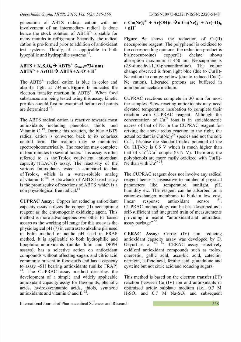
892019 6-Vol-6-Issue-2-Feb-2015-IJPSR-RE-1434-Paper-6
httpslidepdfcomreaderfull6-vol-6-issue-2-feb-2015-ijpsr-re-1434-paper-6 1721
Deepshikha Gupta IJPSR 2015 Vol 6(2) 546-566 E-ISSN 0975-8232 P-ISSN 2320-5148
International Journal of Pharmaceutical Sciences and Research 562
be measured using a fluorescent plate reader Theassay involves the use of peroxyl radicals generatedfrom azobis (amidinopropane) dihydrochloride(ABAP)
When a sample of phytochemical origin such asfruit or vegetable extract or dietary supplements
containing antioxidants is added to the assay theantioxidants react with the peroxyl radicals preventing the peroxyl radicals from oxidizing theDCFH and thereby preventing the formation ofDCF Consequently the fluorescence decreases dueto the scavenging effects of the antioxidantsTherepresentative examples of antioxidants includevitamins carotenoids phenolics and flavonoids76
In -vitro Cellular Antioxidant Activity can also beassessed using a Light-Scattering Properties
(turbidity) of Human Erythrocytes It relies ondifferences in scattering properties between lysedand intact human erythrocytes AAPH a peroxylradical generator is used to enhance lipid peroxidation The consequent hemolysis triggered aloss of the light-scattering ability in the lysederythrocytes When an antioxidant is added thearea under the absorbance decay curve (AUC) waslinearly proportional to the concentration ofantioxidant compound
This erythrocyte cellular antioxidant activity(ERYCA) method is found to be relatively fastsensitive accurate and repeatable even whenusing erythrocytes from different donors and fordifferent storage times 77 The ERYCA assay hasthe advantage of assessing different mechanisms ofantioxidant protection including direct scavengingof free radicals in the surrounding medium and cellmediated antioxidant protection (Cell-MAP) in onestep
Cell-MAP addresses the following the physiochemical properties of antioxidants such astheir lipo-solubility the ability of both lipid andwater soluble compounds to diffuse effectively intolipoproteins and cell membranes and eventuallyenhance from there the erythrocytes defensesthrough mediation of both plasma membraneredox system (PMRS) and the anti oxidativedefense enzyme system 78
CONCLUSIONS This review compiles themethods for determination of antioxidant potentialsof food and dietary supplements 99 of thediseases caused to humans are stress related andonly 1 is genetic disorders Obesity is also one ofthe key reasons of developing diseases likecoronary disease blood pressure cancer diabetes
etc Many of these diseases can be reversed throughnutritional excellence Low nutrient eating (andtoxic eating) leads to increased cellular toxicitywith undesirable levels of free radicals andadvanced glycation end products (AGErsquos)
ldquoThe amount of antioxidants in your body isdirectly proportional to how long you will liverdquo
says Dr Richard Cutler former Director of the National Institute of Aging Washington Choosingright food in correct proportion can decrease the
risk of life threatening disorders and increase lifelongevity The antioxidant potential measurementassays can help in choosing naturally occurringantioxidant rich food containing ascorbic acidvitamin E carotenoids and natural polyphenols for preservation of food and decreasing the ROS in body
This review is a showcase of types of radicalspecies encountered generated in our body invarious metabolic processes and in vitro methods
used to quantify them The outcome of theantioxidant action for different assays (regardlessof mechanism) is similar in the sense ofquenchingreducing reactive radical species but thereaction conditions kinetics and potential sidereactions may vary
HAT and ET mechanisms occur parallel in mostsystems although one may dominate dependingupon the antioxidant structure and properties Nosingle assay can be used as universally accepted
method for determination of total antioxidant potential It may be concluded that raw and mildly processed fruits vegetables nuts dry fruitscereals spices and condiments have significantlyhigher level of bioactive compounds richer inantioxidants 52 79 Fruits like blueberry cranberry blackberry prunes raspberry strawberry redapples cherry plums peaches grapefruits lemonsoranges kiwi pomegranate are richer inantioxidant content 80 81 82 Among vegetablesgarlic red yellow and white onions red and green
892019 6-Vol-6-Issue-2-Feb-2015-IJPSR-RE-1434-Paper-6
httpslidepdfcomreaderfull6-vol-6-issue-2-feb-2015-ijpsr-re-1434-paper-6 1821
892019 6-Vol-6-Issue-2-Feb-2015-IJPSR-RE-1434-Paper-6
httpslidepdfcomreaderfull6-vol-6-issue-2-feb-2015-ijpsr-re-1434-paper-6 1921
892019 6-Vol-6-Issue-2-Feb-2015-IJPSR-RE-1434-Paper-6
httpslidepdfcomreaderfull6-vol-6-issue-2-feb-2015-ijpsr-re-1434-paper-6 2021
Deepshikha Gupta IJPSR 2015 Vol 6(2) 546-566 E-ISSN 0975-8232 P-ISSN 2320-5148
International Journal of Pharmaceutical Sciences and Research 565
53 Tuumltem E Apak R and Baykut F Spectrophotometricdetermination of trace amounts of copper(I) and reducingagents with neocuproine in the presence of copper(II)Analyst 1991 11689-94
54 Bener M et al Development of a low-cost optical sensorfor cupric reducing antioxidant capacity measurement offood extracts Anal Chem 2010 824252-8
55 Oumlzyuumlrek M et al A comprehensive review of CUPRACmethodology Critical Review Anal Methods 2011 3
2439-245356 Ozyurt D Demirata B and Apak R Modified cerium(IV)- based antioxidant capacity (CERAC) assay with selectivityover citric acid and simple sugars Journal of FoodComposition and Analysis 2010 23 282-288
57 Ozyurt D Demirata B and Apak R Determination of totalantioxidant capacity by a new spectrofluorometric method
based on Ce(IV) reduction Ce(III) fluorescence probe forCERAC assay Journal of Fluorescence 2011 21(6)2069-76
58 Janero R D Malondialdehyde and thiobarbituric acid-reactivity as diagnostic indices of lipid peroxidation and
peroxidative tissue injury Free Radic Biol Med 19909(6) 515-40
59 Esterbaur H and Cheeseman KH Determination of
aldehydic lipid peroxidation products malonaldehyde and4-hydroxynonenal Methods Enzymol 1990 186407-2160 Yagi K Simple Assay for the Level of Total Lipid
Peroxides in Serum or Plasma Free Radical andAntioxidant Protocols Methods in Molecular Biology1998 108101-106
61 Koracevic D et al Method for the measurement ofantioxidant activity in human fluids J Clin Pathol 200154 356-361
62 Benzie IF Lipid peroxidation a review of causesconsequences measurement and dietary influencesInternational Journal of Food Science and Nutrition 199647233-61
63 Ou B et al Novel Fluorometric Assay for HydroxylRadical Prevention Capacity Using Fluorescein as the
Probe J Agric Food Chem 2002 50 (10)2772-277764 Decker EA and Welch B Role of ferritin as a lipid
oxidation catalyst in muscle food J Agric Food Chem1990 38(3)674-677
65 Gordon MH The Mechanism of Antioxidant Action inVitro Food Antioxidants Elsevier 1990 1-18
66 Jimenez-Alvarez D et al High-throughput methods toassess lipophilic and hydrophilic antioxidant capacity offood extracts in vitro J Agric Food Chem 200856(10)3470-7
67 Marcocci L et al The nitric oxide-scavenging propertiesof Ginkgo biloba extract EGB 761 Biochem Biophys ResCommun 1994 201748-55
68 Olabinri BMet al In vitro Evaluation of Hydroxyl and Nitric Oxide Radical Scavenging Activities of ArtemetherResearch Journal of Biological Sciences 2010 5(1)102-105
69 Sumanont Y et al Evaluation of the nitric oxide radicalscavenging activity of manganese complexes Biol PharmBull 2004 27170-3
70 Chidan Kumar CS et al Structural Correlation of SomeHeterocyclic Chalcone Analogues and Evaluation of TheirAntioxidant Potential Molecules 2013 1811996-12011
71 Popov I and Lewin G Antioxidative homeostasischaracterization by means of chemiluminescent techniqueMethods Enzymol 1999 300437-56
72 Jimenez AM Navas MJ Chemiluminescence Methods(Present and Future) Grasas y Aceites 2002 5364-75
73 Verde V et al Use of NN-dimethyl-p-phenylenediamineto evaluate the oxidative status of human plasma FreeRadic Res 2002 36869-73
74 Asghar and Nedeem Evaluation of antioxidant activityusing an improved DMPD radical cation decolourizationassay Acta Chimica Slovenica 2007 54295
75 Liu RH Finley J Potential cell culture models forantioxidant research JAgric Food Chem 2005 534311-4314
76
Wolfe KL and Liu RH Cellular antioxidant activity(CAA) assay for assessing antioxidants foods and dietarysupplements J Agric Food Chem 2007 558896-907
77 Gonzalez E et al Novel Semiautomated Method forAssessing in Vitro Cellular Antioxidant Activity Using theLight Scattering Properties of Human ErythrocytesJournal of Agricultural and Food Chemistry 2010531455-1461
78 Gonzalez E et al In vitro Cell-Mediated AntioxidantProtection of Human Erythrocytes by Some CommonTropical Fruits J Nutrition amp Food Sciences 2012 2139
79 Brewer MS Natural Antioxidants SourcesCompounds Mechanisms of Action and PotentialApplications Comprehensive Reviews in Food Scienceand Food Safety 2011 10221-247
80
Proteggente AR et al The antioxidant activity ofregularly consumed fruit and vegetables reflects their phenolic and vitamin C composition Free Radic Res2002 36(2)217-33
81 Arshiya S The antioxidant effect of certain fruits- Areview Journal of Pharmaceutical Sciences and Research2013 5(12)265-268
82 Sun J et al Antioxidant and antiproliferative activities ofcommon fruits Journal of Agricultural and FoodChemistry 2002 50 (25)7449-7454
83 Gorinsteina S et al Comparative control of the bioactivityof some frequently consumed vegetables subjected todifferent processing conditions Food Control 2009 20(4)407-413
84 Zenon Jastrzebskia Hanna Leontowiczb Maria
Leontowiczb Jacek Namiesnikc Zofia ZachwiejadHenryk Bartond Elke Pawelzike Patricia Arancibia-Avilaf Fernando Toledof Gorinstein S The bioactivity of
processed garlic (Allium sativum L) as shown in vitro andin vivo studies on rats Food and Chemical Toxicology2007 45(9)1626 ndash 1633
85 Wootton-Beard PC and Ryan L Combined use ofMultiple Methodologies for the Measurement of TotalAntioxidant Capacity in UK Commercially AvailableVegetable Juices Plant Foods for Human Nutrition 201267 142-147
86
Weisburger JH Mechanisms of action of antioxidants asexemplified in vegetables tomatoes and tea Food ChemToxicol 1999 37943-8
87 Drzewiecki J Delgado-Licon E Haruenkit R Pawelzik EMartin-Belloso O Park YS Jung ST Trakhtenberg SGorinstein S Use of scanning electron microscopy toindicate the similarities and differences in pseudocerealand cereal proteins J Agric Food Chem 2003 517798
88 Gorinstein S Pawelzik E Yamamoto K Kobayashi STaniguchi H Haruenkit R Park YS Jung ST DrzewieckiJ Trakhten S Use of scanning electron microscopy toindicate the similarities and differences in pseudocerealand cereal proteins Int J Food Sci Technol 200439183-189
89 Gorinstein S Vargas M Jaramillo OJ Arnao Salas IMartinez-Ayala AL Arancibia-Avila P Toledo F KatrichE Trakhtenberg S The total polyphenols and theantioxidant potentials of some selected cereals and
892019 6-Vol-6-Issue-2-Feb-2015-IJPSR-RE-1434-Paper-6
httpslidepdfcomreaderfull6-vol-6-issue-2-feb-2015-ijpsr-re-1434-paper-6 2121
Deepshikha Gupta IJPSR 2015 Vol 6(2) 546-566 E-ISSN 0975-8232 P-ISSN 2320-5148
pseudocereals Eur Food Res Technol 2007 225321-328
90 Thanapornpoonpong S Vearasilp S Pawelzik EGorinstein S Influence of Various Nitrogen Applicationson Protein and Amino Acid Profiles of Amaranth andQuinoa J Agric Food Chem 2008 56(23)11464-11470
91 Pasko P Barton H Zagrodzki P Gorinstein S Fołta MZachwieja Z Anthocyanins total polyphenols andantioxidant activity in amaranth and quinoa seeds and
sprouts during their growth Food Chemistry 2009115994-99892 Milder IEJ Arts ICW van de Putte B Venema DP
Hollman PCH Lignan contents of Dutch plant foods adatabase including lariciresinol pinoresinol
secoisolariciresinol and matairesinol British Journal of Nutrition 2005 93393 ndash 402
93 Christodouleas D Fotakis C Papadopoulos K DimotikaliD Calokerinos AC Luminescent Methods in the Analysisof Untreated Edible Oils A Review Analytical Letters2012 45(5-6) 625-641
94 Gupta D Comparative analysis of spices for their phenoliccontent flavonoid content and antioxidant capacityAmerican International Journal of Research in Formal
Applied amp Natural Sciences 2013 438-4295 Paganga G Miller N and Rice-Evans CA The polyphenolic content of fruit and vegetables and theirantioxidant activities What does a serving constitute FreeRadical Research 1999 30153-62
All copy 2013 are reserved by International Journal o f Pharmaceutical Sciences and Research This Journal licensed under a Creative Commons Att ribution-NonCommercial-ShareAlike 30 Unported License
This article can be downloaded to ANDROID OS based mobile Scan QR Code using CodeBar Scanner from your mobile (Scanners areavailable on Google Playstore)
How to cite this articleGupta D Methods for Determination of Antioxidant Capacity A Review Int J Pharm Sci Res 2015 6(2) 546-66doi1013040IJPSR0975-82326 (2)546-66
892019 6-Vol-6-Issue-2-Feb-2015-IJPSR-RE-1434-Paper-6
httpslidepdfcomreaderfull6-vol-6-issue-2-feb-2015-ijpsr-re-1434-paper-6 1821
892019 6-Vol-6-Issue-2-Feb-2015-IJPSR-RE-1434-Paper-6
httpslidepdfcomreaderfull6-vol-6-issue-2-feb-2015-ijpsr-re-1434-paper-6 1921
892019 6-Vol-6-Issue-2-Feb-2015-IJPSR-RE-1434-Paper-6
httpslidepdfcomreaderfull6-vol-6-issue-2-feb-2015-ijpsr-re-1434-paper-6 2021
Deepshikha Gupta IJPSR 2015 Vol 6(2) 546-566 E-ISSN 0975-8232 P-ISSN 2320-5148
International Journal of Pharmaceutical Sciences and Research 565
53 Tuumltem E Apak R and Baykut F Spectrophotometricdetermination of trace amounts of copper(I) and reducingagents with neocuproine in the presence of copper(II)Analyst 1991 11689-94
54 Bener M et al Development of a low-cost optical sensorfor cupric reducing antioxidant capacity measurement offood extracts Anal Chem 2010 824252-8
55 Oumlzyuumlrek M et al A comprehensive review of CUPRACmethodology Critical Review Anal Methods 2011 3
2439-245356 Ozyurt D Demirata B and Apak R Modified cerium(IV)- based antioxidant capacity (CERAC) assay with selectivityover citric acid and simple sugars Journal of FoodComposition and Analysis 2010 23 282-288
57 Ozyurt D Demirata B and Apak R Determination of totalantioxidant capacity by a new spectrofluorometric method
based on Ce(IV) reduction Ce(III) fluorescence probe forCERAC assay Journal of Fluorescence 2011 21(6)2069-76
58 Janero R D Malondialdehyde and thiobarbituric acid-reactivity as diagnostic indices of lipid peroxidation and
peroxidative tissue injury Free Radic Biol Med 19909(6) 515-40
59 Esterbaur H and Cheeseman KH Determination of
aldehydic lipid peroxidation products malonaldehyde and4-hydroxynonenal Methods Enzymol 1990 186407-2160 Yagi K Simple Assay for the Level of Total Lipid
Peroxides in Serum or Plasma Free Radical andAntioxidant Protocols Methods in Molecular Biology1998 108101-106
61 Koracevic D et al Method for the measurement ofantioxidant activity in human fluids J Clin Pathol 200154 356-361
62 Benzie IF Lipid peroxidation a review of causesconsequences measurement and dietary influencesInternational Journal of Food Science and Nutrition 199647233-61
63 Ou B et al Novel Fluorometric Assay for HydroxylRadical Prevention Capacity Using Fluorescein as the
Probe J Agric Food Chem 2002 50 (10)2772-277764 Decker EA and Welch B Role of ferritin as a lipid
oxidation catalyst in muscle food J Agric Food Chem1990 38(3)674-677
65 Gordon MH The Mechanism of Antioxidant Action inVitro Food Antioxidants Elsevier 1990 1-18
66 Jimenez-Alvarez D et al High-throughput methods toassess lipophilic and hydrophilic antioxidant capacity offood extracts in vitro J Agric Food Chem 200856(10)3470-7
67 Marcocci L et al The nitric oxide-scavenging propertiesof Ginkgo biloba extract EGB 761 Biochem Biophys ResCommun 1994 201748-55
68 Olabinri BMet al In vitro Evaluation of Hydroxyl and Nitric Oxide Radical Scavenging Activities of ArtemetherResearch Journal of Biological Sciences 2010 5(1)102-105
69 Sumanont Y et al Evaluation of the nitric oxide radicalscavenging activity of manganese complexes Biol PharmBull 2004 27170-3
70 Chidan Kumar CS et al Structural Correlation of SomeHeterocyclic Chalcone Analogues and Evaluation of TheirAntioxidant Potential Molecules 2013 1811996-12011
71 Popov I and Lewin G Antioxidative homeostasischaracterization by means of chemiluminescent techniqueMethods Enzymol 1999 300437-56
72 Jimenez AM Navas MJ Chemiluminescence Methods(Present and Future) Grasas y Aceites 2002 5364-75
73 Verde V et al Use of NN-dimethyl-p-phenylenediamineto evaluate the oxidative status of human plasma FreeRadic Res 2002 36869-73
74 Asghar and Nedeem Evaluation of antioxidant activityusing an improved DMPD radical cation decolourizationassay Acta Chimica Slovenica 2007 54295
75 Liu RH Finley J Potential cell culture models forantioxidant research JAgric Food Chem 2005 534311-4314
76
Wolfe KL and Liu RH Cellular antioxidant activity(CAA) assay for assessing antioxidants foods and dietarysupplements J Agric Food Chem 2007 558896-907
77 Gonzalez E et al Novel Semiautomated Method forAssessing in Vitro Cellular Antioxidant Activity Using theLight Scattering Properties of Human ErythrocytesJournal of Agricultural and Food Chemistry 2010531455-1461
78 Gonzalez E et al In vitro Cell-Mediated AntioxidantProtection of Human Erythrocytes by Some CommonTropical Fruits J Nutrition amp Food Sciences 2012 2139
79 Brewer MS Natural Antioxidants SourcesCompounds Mechanisms of Action and PotentialApplications Comprehensive Reviews in Food Scienceand Food Safety 2011 10221-247
80
Proteggente AR et al The antioxidant activity ofregularly consumed fruit and vegetables reflects their phenolic and vitamin C composition Free Radic Res2002 36(2)217-33
81 Arshiya S The antioxidant effect of certain fruits- Areview Journal of Pharmaceutical Sciences and Research2013 5(12)265-268
82 Sun J et al Antioxidant and antiproliferative activities ofcommon fruits Journal of Agricultural and FoodChemistry 2002 50 (25)7449-7454
83 Gorinsteina S et al Comparative control of the bioactivityof some frequently consumed vegetables subjected todifferent processing conditions Food Control 2009 20(4)407-413
84 Zenon Jastrzebskia Hanna Leontowiczb Maria
Leontowiczb Jacek Namiesnikc Zofia ZachwiejadHenryk Bartond Elke Pawelzike Patricia Arancibia-Avilaf Fernando Toledof Gorinstein S The bioactivity of
processed garlic (Allium sativum L) as shown in vitro andin vivo studies on rats Food and Chemical Toxicology2007 45(9)1626 ndash 1633
85 Wootton-Beard PC and Ryan L Combined use ofMultiple Methodologies for the Measurement of TotalAntioxidant Capacity in UK Commercially AvailableVegetable Juices Plant Foods for Human Nutrition 201267 142-147
86
Weisburger JH Mechanisms of action of antioxidants asexemplified in vegetables tomatoes and tea Food ChemToxicol 1999 37943-8
87 Drzewiecki J Delgado-Licon E Haruenkit R Pawelzik EMartin-Belloso O Park YS Jung ST Trakhtenberg SGorinstein S Use of scanning electron microscopy toindicate the similarities and differences in pseudocerealand cereal proteins J Agric Food Chem 2003 517798
88 Gorinstein S Pawelzik E Yamamoto K Kobayashi STaniguchi H Haruenkit R Park YS Jung ST DrzewieckiJ Trakhten S Use of scanning electron microscopy toindicate the similarities and differences in pseudocerealand cereal proteins Int J Food Sci Technol 200439183-189
89 Gorinstein S Vargas M Jaramillo OJ Arnao Salas IMartinez-Ayala AL Arancibia-Avila P Toledo F KatrichE Trakhtenberg S The total polyphenols and theantioxidant potentials of some selected cereals and
892019 6-Vol-6-Issue-2-Feb-2015-IJPSR-RE-1434-Paper-6
httpslidepdfcomreaderfull6-vol-6-issue-2-feb-2015-ijpsr-re-1434-paper-6 2121
Deepshikha Gupta IJPSR 2015 Vol 6(2) 546-566 E-ISSN 0975-8232 P-ISSN 2320-5148
pseudocereals Eur Food Res Technol 2007 225321-328
90 Thanapornpoonpong S Vearasilp S Pawelzik EGorinstein S Influence of Various Nitrogen Applicationson Protein and Amino Acid Profiles of Amaranth andQuinoa J Agric Food Chem 2008 56(23)11464-11470
91 Pasko P Barton H Zagrodzki P Gorinstein S Fołta MZachwieja Z Anthocyanins total polyphenols andantioxidant activity in amaranth and quinoa seeds and
sprouts during their growth Food Chemistry 2009115994-99892 Milder IEJ Arts ICW van de Putte B Venema DP
Hollman PCH Lignan contents of Dutch plant foods adatabase including lariciresinol pinoresinol
secoisolariciresinol and matairesinol British Journal of Nutrition 2005 93393 ndash 402
93 Christodouleas D Fotakis C Papadopoulos K DimotikaliD Calokerinos AC Luminescent Methods in the Analysisof Untreated Edible Oils A Review Analytical Letters2012 45(5-6) 625-641
94 Gupta D Comparative analysis of spices for their phenoliccontent flavonoid content and antioxidant capacityAmerican International Journal of Research in Formal
Applied amp Natural Sciences 2013 438-4295 Paganga G Miller N and Rice-Evans CA The polyphenolic content of fruit and vegetables and theirantioxidant activities What does a serving constitute FreeRadical Research 1999 30153-62
All copy 2013 are reserved by International Journal o f Pharmaceutical Sciences and Research This Journal licensed under a Creative Commons Att ribution-NonCommercial-ShareAlike 30 Unported License
This article can be downloaded to ANDROID OS based mobile Scan QR Code using CodeBar Scanner from your mobile (Scanners areavailable on Google Playstore)
How to cite this articleGupta D Methods for Determination of Antioxidant Capacity A Review Int J Pharm Sci Res 2015 6(2) 546-66doi1013040IJPSR0975-82326 (2)546-66

892019 6-Vol-6-Issue-2-Feb-2015-IJPSR-RE-1434-Paper-6
httpslidepdfcomreaderfull6-vol-6-issue-2-feb-2015-ijpsr-re-1434-paper-6 2121
Deepshikha Gupta IJPSR 2015 Vol 6(2) 546-566 E-ISSN 0975-8232 P-ISSN 2320-5148
pseudocereals Eur Food Res Technol 2007 225321-328
90 Thanapornpoonpong S Vearasilp S Pawelzik EGorinstein S Influence of Various Nitrogen Applicationson Protein and Amino Acid Profiles of Amaranth andQuinoa J Agric Food Chem 2008 56(23)11464-11470
91 Pasko P Barton H Zagrodzki P Gorinstein S Fołta MZachwieja Z Anthocyanins total polyphenols andantioxidant activity in amaranth and quinoa seeds and
sprouts during their growth Food Chemistry 2009115994-99892 Milder IEJ Arts ICW van de Putte B Venema DP
Hollman PCH Lignan contents of Dutch plant foods adatabase including lariciresinol pinoresinol
secoisolariciresinol and matairesinol British Journal of Nutrition 2005 93393 ndash 402
93 Christodouleas D Fotakis C Papadopoulos K DimotikaliD Calokerinos AC Luminescent Methods in the Analysisof Untreated Edible Oils A Review Analytical Letters2012 45(5-6) 625-641
94 Gupta D Comparative analysis of spices for their phenoliccontent flavonoid content and antioxidant capacityAmerican International Journal of Research in Formal
Applied amp Natural Sciences 2013 438-4295 Paganga G Miller N and Rice-Evans CA The polyphenolic content of fruit and vegetables and theirantioxidant activities What does a serving constitute FreeRadical Research 1999 30153-62
All copy 2013 are reserved by International Journal o f Pharmaceutical Sciences and Research This Journal licensed under a Creative Commons Att ribution-NonCommercial-ShareAlike 30 Unported License
This article can be downloaded to ANDROID OS based mobile Scan QR Code using CodeBar Scanner from your mobile (Scanners areavailable on Google Playstore)
How to cite this articleGupta D Methods for Determination of Antioxidant Capacity A Review Int J Pharm Sci Res 2015 6(2) 546-66doi1013040IJPSR0975-82326 (2)546-66