6th annual efcs tutorial, guimarães, portugal
TRANSCRIPT
1
6th Annual EFCS Tutorial, Guimarães, Portugal ___________________________________________________________ Gynaecological cytology Maj Liv Eide Gynaecological cytology: technical aspects including preparation of LBC, staining and troubleshooting 2-6 Non-neoplastic gynaecological cytology 7-16 Giovanni Negri Conventional smears and LBC: similarities and differences
ASC-US, ASC-H, diagnostic pitfalls and look-alikes 17-23
Glandular lesions of the cervix Luigi di Bonito Cytological characteristics of squamous lesions and their mimics 24-29 Christine Bergeron New technologies in cervical screening: the template of p16 30-35 Amanda Herbert Invasive cervical cancer audit and EU guidelines for quality Assurance 36-43

2
Gynaecological cytology: technical aspects Maj Liv Eide Dept. of Pathology and Medical Genetics, Trondheim University Hospital and Dept. of Biomedical Science, Soer-Troendelag University College, Norway. Email: [email protected] ___________________________________________________________ The quality of a cervical specimen depends on the sample taking, the fixation and the staining. The goal is a representative specimen, with well-preserved and optimally stained cells for evaluation under the microscope. Preparation of cytology slides Two types of specimen are available for cervicovaginal cytology: Conventional smear and liquid based cytology. Conventional cytology has been used for decades, with easy preparation of smears by the sample-taker. The disadvantage of this technique can be poor preservation of cells due to late fixation. The last 15 years, liquid-based cytology (LBC) has been introduced in cytology laboratories in Europe, eliminating the problem of poor preservation. The conventional smear depends on the sample takers technique in preparing a smear and the fixation, which should be done immediately after making the smear in order to eliminate drying artefacts. The fixative often used is a spray fixative containing alcohol and PEG (polyethylene glycol). Unsatisfactory conventional smears can be due to excessive blood or leukocytes obscuring the cells, thick smears with overlapping cells, too few cells due to inadequate sampling and poor preservation due to delayed fixation. In LBC techniques the cervical specimen is transferred to a vial with preservative fluid (alcohol). The specimens are processed in machines in the cytology laboratory. The method provides preserved cells, in a thin layer, within a limited area on the slide. The two LBC systems first to be FDA approved, were Hologic´s ThinPrep and BD SurePath system. The ThinPrep system prepares specimens that are collected in 50 % methanol. The ThinPrep method uses a membrane filtration technique, which requires an instrument and special polycarbonate filters with microscopic pores, through which fluid can be drawn. Cells are collected

3
on the surface of the filter and the cells are then transferred to the slide, within a circle of 20 mm in diameter. The main reason for unsatisfactory ThinPrep specimens, is insufficiently rinsing of the brush in the PreservCyt. The brush should be rinsed immediately after application into the vial. Blood will compete with the cells for the space on the filter, which may result in too few squamous cells on the slide. By using a washing procedure on bloody specimen with 10 % acetic acid and Cytolyt before reprocessing in the ThinPrep instrument, adequate material may be obtained. The SurePath system prepares specimens that are collected in 24 % ethanol. The SurePath method use gravity sedimentation technique, where red blood cells, excess inflammatory cells, debris and epithelial cells are separated in different layers according to their specific gravity. This partly removes non-diagnostic debris and excess inflammatory cells. The final preparation deposit cells within a circle of 13 mm in diameter.
Rehydration Before nuclear staining in haematoxylin, the specimens are rehydrated in tap water. Smears fixed with alcohol spray fixative, containing polyethylene glycol (PEG), must be removed prior to rehydration, preferably with alcohol, but some manufacturers recommend water. Papanicolaou staining method The Papanicolaou staining method has been used since the 1940s and the method provides good nuclear detail and transparent cytoplasm, which is effective when evaluating overlapping cells. The cytoplasmic stains differ between mature and immature cells and demonstrate keratinization. The principle of staining
Conventional SurePath ThinPrep

4
This is a sequential trichrome method, defined as using Phosphotungstic acid (PTA) and minimum two anionic dyes. Staining solutions applied are: • Aluminum-hematein, a metal complex dye, which stains
chromatin. • OG: Orange G, an anionic dye, binding to NH3+ ionised proteins in
the cytoplasm. • EA: An anionic dye consisting of Light Green (large molecule),
Eosin (small molecule) and PTA, binding to NH3+ ionised proteins in the cytoplasm. PTA acts like a dye excluder and allows light green and eosin to stain differentially.
Extraction of dyes/differentiation + = Extraction 0 = No extraction H2O Acid Base Ethanol Metal complex dyes (nuclear stain)
0 + 0 0
Cationic/basic dyes (nuclear stain)
0 + 0 +
Anionic dyes/acid dyes (cytoplasmic stain)
+ 0 + 0
Haematoxylin will be extracted in acidic solutions, depending on the pH. It is therefore important to change the rinsing ethanol solutions frequently, to avoid acid from OG and EA contamination. OG and EA will be extracted by water and it is therefore important to change the rinsing baths frequently. Staining results Haematoxylin will stain the nucleic acid at pH>2 and ideally around pH 3-3,5. If pH exceed 4, many proteins will also be stained > overstaining of nuclei and greyish/blue stain in the cytoplasm. Haematoxylin stains inactive heterochromatin, dark blue and active euchromatin, light blue. OG stains keratin in cytoplasm orange. Eosin stains cytoplasm in superficial cells pink, nucleoli and cilia red. Light green stains biological active cells blue-green.

5
The method is dependent on pH in staining solutions, pH in tap water, staining concentration, the permeability of the cells, the size of staining molecules, staining time and rinsing. Trouble shooting Nuclear stain too pale: • Air-drying of specimen prior to fixation • Excessive time in chlorinated tap water will bleach the nuclei • Haematoxylin may be too old and should be replaced Nuclear stain to dark: • Excessive time in Haematoxylin • Excessive Haematoxylin is insufficiently removed Cytoplasmic stains are unsatisfactory • Air-drying of specimen prior to fixation will stain all the cells pink • Grey or purple cytoplasm due to excessive time in haematoxylin Destaining and restaining • Remove the coverslip by immersing the slide in xylene and be sure
that all residual mounting media is removed. • Destain by immersing the smear in 1 % hydrochloric acid in 70 %
ethanol for 1-12 hours and rehydrate the preparation in tap water before restaining.
Mounting and coverslipping To properly visualize cellular morphology, the refractive index of the glass, cellular material, coverslip and mounting medium should closely match one another. Coverslips in size 24 x 50-60 mm are recommended. Air bubbles underneath the glass may create brown, refractile, pigment-like substance on the surface of mature squamous cells, named “cornflakes”. Human papilloma virus (HPV) detection methods Methods for detection of high risk HPV has been implemented in many pathology departments the last years as triage to ASC-US/LSIL. LBC

6
provides material for HPV testing on residual material after preparation of the cytology slide. The preservative fluid in the vial is excellent in preserving DNA and RNA. Different methods of HPV detection are in use and they have different clinical sensitivity and specificity for finding CIN2+. The detection methods can be divided in target amplification techniques (e.g. PCR) and signal amplification techniques (eg. Hybrid Capture 2). Cytotechnologists can and do perform both techniques. References • Comprehensive Cytopathology, ed. By Marluce Bibbo and David
Wilbur, 3th ed., Saunders Elsevier, 2008.
• Koss´ Diagnostic Cytology and its histopathological bases, ed. By Leopold G. Koss and Myron R. Melamed, 5th ed., Lippincot Williams & Wilkins, 2006.
• The Pap Test. Richard Mac DeMay, American Society for Clinical
Pathology, 2005. • Clinical Cytology, Compendium by Reidun Mecsei, Tapir
akademisk forlag, 2011. • Cytopreparation, Principles and Practice, Gary W. Gill, Essentials
in Cytopathology, Springer 2013. • Snijders PJ, Heideman DA, Meijer CJ. Methods for HPV
detection in exfoliated cell and tissue specimens. APMIS : acta pathologica, microbiologica, et immunologica Scandinavica. 2010;118 (6-7):520-8. Epub 2010/06/18.
• Eide ML, Debaque H. HPV detection methods and genotyping
techniques in screening for cervical cancer. Annales de pathologie. 2012;32(6):e15-23, 401-9. Epub 2012/12/19.

7
Non-neoplastic gynaecological cytology
Maj Liv Eide Dept. of Pathology and Medical Genetics, Trondheim University Hospital and Dept. of Biomedical Science, Soer-Troendelag University College, Norway. Email: [email protected] ___________________________________________________________ The normal cytological smear encompasses different infections and reactive conditions, which will be presented here. Firstly, knowing the criteria for satisfactory smear and the morphology of normal cells found in a smear from cervix, are essential. Satisfactory smear Reporting of adequacy or unsatisfactory for evaluation is part of the reporting system of Bethesda 2001. Regarding squamous cellularity:
Conventional smears should have a minimum of approx. 8000 – 12000 well-preserved and well-visualized squamous cells. The number shall be estimated, not counted. Liquid-based preparations should have at least 5000 well-visualized and well-preserved squamous cells. A minimum of 10 fields using a 40x objective, are assessed along a diameter including the centre of the preparation. This method of strict criteria may not be applied to slides with cell clustering, atrophy or cytolysis.

8
Normal cells in cervical cytology specimens Squamous cells
Endocervical columnar cells The columnar cells are tall and range from 20 µm – 30 µm in height and 8µm - 10 µm in width. The nuclei of the endocervical cells are about the size of a parabasal cell. The cells are either secretory or ciliated. The secretory cells have cytoplasm with mucin vacuoles, either many or one single one. The ciliated cells have denser cytoplasm than secretory and the finding of terminal bars and cilia indicates a benign nature. Endocervical cells can be seen singly, in strips or in sheets. Strips of endocervical cells viewed from the side look like a picket fence. Endocervical cells in two-dimensional sheets, seen en face, appears to have a honeycomb pattern. Pencil thin endocervical cells, may be seen after application of acetic acid among others. In LBC the endocervical cells can form three-dimensional groups resembling glandular neoplasia. Endometrial cells There are two types of endometrial cells: epithelial and stromal. Epithelial cells are smaller than endocervical cells. The endometrial cells
Superficial Large, polygonal cells with centrally placed, dark,
pyknotic nucleus, approx. 15 -‐ 20 µm2. The
cytoplasm stains pink. N/C ratio: 2% -‐ 3%.
Influenced by estrogen.
Intermediate Large, polygonal cells with centrally placed, vesicular nucleus approx. 35 µm2. The cytoplasm
stains light blue/turquoise. N/C ratio: 3% -‐ 5%.
Influenced by progesterone.
Parabasal Round-‐oval shaped cells with centrally placed, vesicular nucleus, approx. 50 µm2. Dense, blue-‐
green cytoplasm. N/C ratio: 20 %.
Associated with atrophy
Basal Small, round-‐oval shaped cells with centrally placed,
vesicular nucleus and scant cytoplasm. Resemble small histiocytes.

9
have nuclei the size of intermediate squamous cells and coarse chromatin is often seen, due to degeneration. Signs of necrosis or apoptosis are frequent. The cytoplasm is usually scant, finely vacuolar or granular and stains basophilic or amphophilic. Nucleoli may be visible in LBC. Mitosis is not normally seen in spontanously shed endometrials, which exfoliate in three-dimensional group, with or without central stroma. The most common arrangement of endometrial cells are a double contour cell ball with darkly stained stroma inside with lighter stained epithelium wrapped around the outside. Endometrial cells shed after day 10-14 or in the menopause are considered abnormal. Endometrial deep stromal cells are round to spindle formed with small oval nuclei and scant cytoplasm. Inflammatory conditions in general
The cells of the mucosa in cervix will show degenerative changes following injury. If the cells die, signs of necrosis or apoptosis will appear. If the cells can be repaired, proplastic changes will appear as retroplastic changes still occur. During cellular repair, nucleoli and hyperchromasia are often visible. Metaplasia is an example of adaptation, replacing damaged columnar cells. Degenerative changes and cell death seen in inflammatory conditions Nucleus: Irregularly clumped, blurry or opaque chromatin. Nuclear swelling, which is caused by hydration.

10
Cell death by necrosis (karyopyknosis, karyorrexis and karyolysis) or apoptosis. Cytoplasm: Vacuolisation, amphophilia, false eosinophilia, perinuclear halo, cytolysis. Persistent irritation Persistent irritation results in morphological changes in epithelial cells. Metaplastic changes The cervix increases in size and shape during puberty and pregnancy resulting in eversion of the endocervical columnar epithelium. Due to acid environment in the vagina, inflammation or oral contraceptives, the columnar epithelium is replaced by metaplastic epithelium and a new squamocolumnar junction is formed. The metaplastic zone is named the transformation zone, which is the critical area to sample by the Pap test. The metaplastic prosess starts with endocervical reserve cells forming hyperplasia, not easily recognized in cytology, then differentiate into immature squamous metaplasia and finally mature squamous metaplasia. Immature squamous metaplasia > Cytological criteria Parabasal-like cells, cobblestone pattern or single cells. Vesicular nucleus, 50 µm2 with smooth membrane and finely granular chromatin. The cytoplasm is thick, dense blue-green with slightly thicker ectoplasm. Cytoplasmic vacuolisation is often seen. Nucleoli can be seen in reactive metaplastic cells. Forcibly scraped cells can show slender cytoplasmic extentions (spider cells). Differential diagnosis (DD) of parabasal squamous cells. Immature squamous metaplasia can look like endocervical cells or HSIL in LBC, due to metaplastic cells being smaller, rounder and having higher N/C ratio and the rounding up of cell clusters. Adenocarcinoma in the case of metaplastic vacuolated, active cells. Mature squamous metaplasia The epithelium now consists of three histological layers: basal, parabasal and intermediate. > Cytological criteria

11
Intermediate squamous like cells. Rounded cell outlines. Slightly, dense cytoplasm. Remnants of cobblestone. DD: Intermediate squamous cells. Reactive endocervical cells Reactive endocervical cells are common and may be caused by hyperplasia, endocervicitis, polyps and many other things. The reactive endocervical cells comprise nuclear enlargement, hyperchromasia, prominent nucleoli and multinucleation. The N/C ratio is maintained. Mitosis may be seen, but careful inspection is necessary to rule out neoplasia. Tissue repair Repair is visualised as regenerative cells and can occur in columnar, metaplastic and squamous epithelium. Reparative changes are frequently found in patients with recurrent cervicitis and recent treatment like biopsies and laser therapy. > Cytological criteria Nuclear variation in size, shape and enlargement. Prominent nucleoli and finely granulated, evenly distributed chromatin, but not hyperchromatic. Mitosis may be seen. Abundant cytoplasm. Cohesive cells in sheet-like arrangements. Leukocyte infiltration in groups. DD: Invasive neoplasia. Tubal metaplasia Tubal metaplasia is a benign, nonneoplastic replacement of columnar cells (endocervical or endometrial) with cells characteristic of the fallobian tube. Tubal metaplasia occurs high up in the endocervical canal and is a common finding due to use of the endocervical brush. Tubal metaplasia comprises three cell types: Ciliated, secretory and intercalated cells. > Cytological criteria Ciliated cells form crowded sheets or three-dimensional clusters. Usually smaller than endocervical cells, but may have higher N/C ratio. Dark, but finely granular, evenly distributed chromatin. Terminal bars and cilia are most helpul in recognising tubal metaplasia. Secretory cells have round, basal nuclei and vacuolated cytoplasm. Intercalated cells are small with high N/C ratio and scant, dark, granular cytoplasm. DD: Glandular neoplasia especially AIS, in histology and cytology. The criteria is similar for conventional and LBC.

12
Hyperkeratosis Hyperkeratosis is a proliferative process due to e.g. uterine prolapse and inflammation, but can conceal underlying dysplasia or cancer. The epithelium increases its protective role. The squamous epithelium forms two additional layers: Stratum granulosum and stratum corneum. > Cytological criteria Prekeratin is formed in stratum granulosum and is visible as small, dark granules in the cytoplasm, known as keratohyalin granula. In stratum corneum, the nucleus has dissolved, karyolysis, and anucleated squamous cells with yellow, orange or pink cytoplasm. The criteria is similar for conventional and LBC. Parakeratosis Parakeratosis is another benign keratotic reaction, but may conceal underlying lesion, most often associated with condyloma or SIL. > Cytological criteria Single, flat cells, layered strips of cells or concentrically arranged “pearls”. Usually orange stained cytoplasm and centrally, pycnotic and hyperchromatic nuclei. The parakeratotic cells tend to stain more eosinophilic than orangophilic in LBC. DD: Degenerated cells with orangophilic cytoplasm and pycnotic nuclei, eg. pseudokeratotic, degenerated parabasal cells in atrophy. Radiation damage Radiation therapy is widely used for treatment of cancer of the female genital tract, especially squamous cell cancer of the cervix. Not only cancer cells, but also native cell are affected. The cytological changes due to radiation can be transitory, last for 1-2 years or persist. Evaluating postradiation smear can be difficult, because both cancer cells and normal cells will have cellular changes due to radiation. The tumour cells usually clear rapidly after radiation therapy. The Pap test should not be taken before 6-8 weeks after therapy. The presence of tumour cells after this period indicates poor prognosis. > Cytological criteria

13
Cytomegaly. Enlarged, somewhat bizarre cells with enlarged nuclei often multiple, pale or dark. N/C ratio relatively unchanged. Polychromatic (both pink and blue, usually not orange) cytoplasm. Vacuolated cytoplasm is a characteristic of acute radiation change. Repair/regeneration is common. DD: Carcinoma, chemotherapy changes. IUD changes Intrauterine contraceptive device can create cellular changes as early as 10-12 weeks after insertion. This is a result of chronic irritation of the tail and body affecting the tissue in the endocervix and the uterine cavity. > Cytological criteria Endometrial cells can form three-dimensional clusters, with enlarged, displaced nuclei and vacuolated cytoplasm (“Bubble gum cells”). A few, atypical appearing, single endometrial cells (IUD cells) with high N/C ratio can also be shed due to IUD. Endometrial cells can be shed at any time in the menstrual cycle. Actinomyces are associated with IUD usage. Formation of psammoma bodies may be seen. DD: Metastatic adenocarcinoma and CIN3 (single endometrial cells). Atrophic smear with inflammation The thin epithelium of atrophy in late postmenopause and post-partum, may lead to bleeding and inflammation. > Cytological criteria Parabasal cells may be seen single or in sheets. Some sheets may have fused cells. The parabasal cells in conventional smear often have enlarged nuclei, due to airdrying artefact and hyperchromasia due to degenerative changes. In LBC the parabasal cells have bland nuclei with occasional grooves and hyperchromasia. Together with higher N/C ratio due to cells rounding up in LBC, this may be interpreted as HSIL. In the postmenopausal state, the cytoplasm of parabasal cells usually lack glycogen, which is important in maintaining low pH as a protection against infection, leaving the atrophic mucosa more vulnerable to external attacks. Some parabasal cells will degenerate randomally with orangophilic cytoplasm and pyknotic nuclei, mimicking parakeratosis. The atrophic cells are fragile and naked nuclei are common. The naked nuclei may be ovoid or spindled. In conventional smears they may be in large numbers, in LBC they are fewer, but often seen in groups

14
resembling endometrial cells. Blue blobs are basophilic bodies, which may look alarming. They are actually mummified parabasal cells. Chronic inflammation Follicular cervicitis, synonymous with lymphoid cervicitis is an inflammatory condition involving lymphoid follicles in subepithelial areas. Associated with Chlamydia infection. > Cytology: Mature and immature lymphoid cells along with tingible body macrophages, which must be identified. Follicular cervicitis is easier to interpret in conventional smears than LBC due to lymhocytic dispersion in the latter. DD: Lymphoma, endometrial cells, histiocytes, metastatic tumor cells. Specific infections Bacterial vaginosis The clinical diagnosis of bacterial vaginosis can be made when these signs are present: thin, homogenous discharge, vaginal ph > 4,5 due to lack of lactobacilli and clue cells. Clue cells are the most reliable predictor of bacterial vaginosis. Gardnerella vaginalis is a small comma-shaped coccobacillus and the major species associated with bacterial vaginosis. Clue cells are characteristic by tiny bacteria that seem to be glued to squamous cells, giving a shaggy appearance to the cell. In conventional smears the coccoid bacteria also forms a granular blue background. Another characteristic of bacterial vaginosis is lack of inflammatory cells. Candida albicans Candidiasis is extremely common in the reproductive years, especially in the late secretory phase of the menstrual cycle and in pregnancy. 90 % of yeast infections are due to Candida albicans. The presence of Candida in Pap test is associated with changes in vaginal glycogen, normal flora or pH and other predisposing factors like diabetes and oral contraceptives. The finding of Candida in a Pap test does not necessarily mean clinically significant infection. Candida consists of pseudohyphae (sticks) and yeast (stones) and may look like “balloon dogs”. Budding may be seen. The pseudohyphae usually stain pale pink or blue and are surrounded by a small, clear halo.

15
> Cytological criteria The Candida seem to have an affinity to mature squamous cells, giving a shish kebab appearance, which is particularly prominent in LBC. The nuclei may be mildly enlarged with slight hyperchromasia and slight hyperkeratosis may be seen. Lysed neutrophils are often found. DD: ASC-US Actinomyces Israeli Actinomyces is predominantly associated with IUDs and other foreign bodies. The organism is part of the normal flora of the mouth and bowel and can be introduced to the vagina from these sites. Most women with Actinomyces in their cytological specimens are asymptomatic, but some have brownish discharge. Pain suggests invasive infection or pelvic inflammatory disease and the finding of Actinomyces in these cases should alert the clinician. Microscopically, the Actinomyces are branching, filamentous bacteria and in Pap test the bacteria live symbiotically with colonies of bacteria forming dark-blue masses. These have dense, granular blue bodies (bacteria) with delicate, spidery legs (actinomyces). Trichomonas vaginalis Trichomonas vaginalis is an oval or pear-shaped protozoan, which ranges from the size of a parabasal nuclus to an intermediate nucleus. The nucleus of the trichomonas is thin, pale and eccentrically located and must be seen to identify this organism. Red granules in the cytoplasm are frequently present in grey to cyanophilic cytoplasm. Flagella may be seen in LBC but red granules are less apparent. > Cytological criteria Pseudomaturation of the squamous epithelium visible as pseudokeratinization and false eosinophilia. Slight nuclear enlargement, hyperchromasia, and perinuclear halos are common. Inflammatory cells agglomerated on squamous cells as cannonballs can be seen along, lymphocytes. The background may be dirty, due to lysed cells, inflammation and debris. The normal flora is replaced by coccoid bacterial overgrowth. DD: LSIL

16
Herpes simplex virus Herpex simplex virus is a double stranded DNA virus surrounded by a capsid produced by the virus and a lipid membrane derived from the host cell membrane. It enters the body through neurons and moves to sensory sacral ganglia where it may be latent for long period. Recurrent infection represents reactivation of latent virus. There is no cure for herpes. There may not be any symptoms of a genital Herpes infection, but some may experience severe symptoms the first time. Herpes may infect the immature squamous metaplastic and endocervical columnar cells. > Cytological criteria The morphological changes caused by Herpes simplex types 1 and 2 are indistuinguishable. Multinucleated cells with enlarged nuclei and molding. The chromatin marginates due to viral particles filling the nuclei, resulting in ground glass appearance. The nuclear membrane appears thickened due to condensed chromatin. Intranuclear inclusions are highly characteristic when present, but is found only in half of the cases. In LBC the same features are found, but in addition the intranuclear inclusions are surrounded by halos and mononuclear infected cells can be more conspicuous. References • Comprehensive Cytopathology, ed. by Marluce Bibbo and David
Wilbur, 3th ed., Saunders Elsevier, 2008 • Koss´Diagnostic Cytology and its histopathological bases, ed. By
Leopold G. Koss and Myron R. Melamed, 5th ed., Lippincot Williams & Wilkins, 2006
• The Pap Test, Richard Mac DeMay, American Society for Clinical
Pathology, 2005 • Clinical Cytology, compendium by Reidun Mecsei, Tapir
akademisk forlag, 2011 • Diagnostic Cytopathology Essentials, G. Kocjan, W. Gray, T.
Levine, I. Kardum-Skelin and P. Viehl, Elsevier 2013

17
Diagnostic gynaecological cytology Giovanni Negri MD, PD, MIAC Department of Pathology, Central Hospital Bolzano, Bolzano, Italy. Email: [email protected] ___________________________________________________________
Workshop cases Each workshop case provides essential clinical data on the front and the diagnosis in accordance with TBS 2001 on the rear of the package. In some cases histology sections are also included. Conventional smears and LBC: similarities and differences The cytological differences between conventional pap smears (CP) and liquid-based cytology (LBC) are mainly due to the sampling, fixation and slide preparation peculiarities of LBC. LBC shows in most cases a better sample quality than CP with fewer inadequate slides (1,2). The potential advantages and cost-effectiveness of LBC must, however, be evaluated in the local context, particularly depending on the quality of a pre-existing conventional cytology. In fact, implementation of LBC is likely to be particularly successful when the quality of CP is poor (3). Liquid-based preparations also allow the use of ancillary techniques such as HPV-testing or immunocytochemistry, which in some laboratories may be an additional reason for converting to LBC. Morphologically there are some minor differences between CP and LBC which must be taken in account when moving to LBC and which may cause a temporary increase of borderline reports. Although the cytological criteria are basically the same, the loss of topographic distribution of cells and the high nuclear detail in LBC may occasionally cause interpretation issues, particularly in metaplastic cells. On the other hand, the reduction of brushing, smearing artefacts and inflammatory exudates may allow a better evaluation of the architecture of cell sheets, which may be useful particularly in glandular lesions (4). Squamous and glandular lesions, diagnostic pitfalls and look-alikes The European guidelines (5) recommend the use of The Bethesda System 2001 (TBS) to classify cervicovaginal findings or, at least, the use of a terminology that may be easily translated into TBS. TBS and most other

18
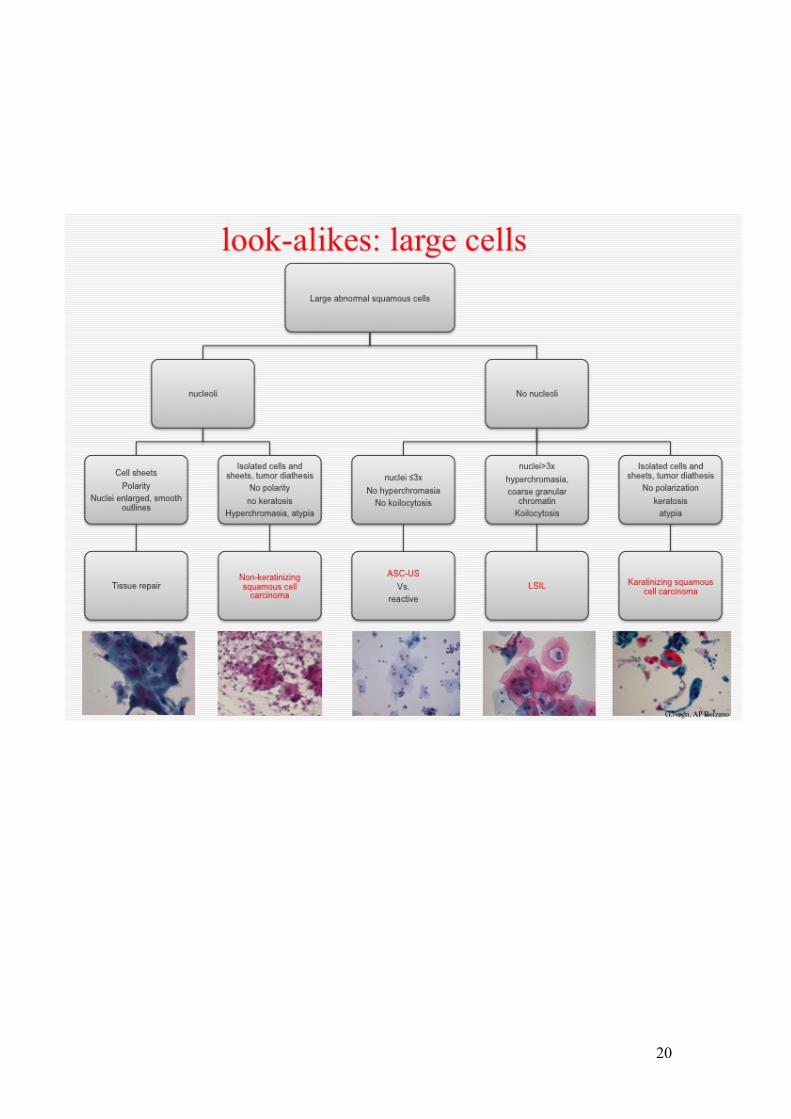
classification systems that are used in Europe divide precancerous squamous lesions in two categories, including HPV-induced changes and mild dysplasia in "low grade intraepithelial lesions", whereas moderate and severe dysplasia are included together in "high grade intraepithelial lesions". More substantial differences between classification systems may be found in the terminology of glandular lesions (6), which in part reflect the difficulties in the interpretation of both histology and biology of these lesions. Basically, in cervicovaginal cytology one has to distinguish three main cell patterns: large cells, small cells and glandular epithelia. If the cells are abnormal, each of these patterns calls for a limited number of differential diagnoses. Squamous cell lesions of the cervix Atypical large cells may be found particularly in LSIL, invasive cancer, tissue repair and reactive changes on squamous cells. Most cases with abnormal large cells include superficial squamous epithelia with nuclear enlargement and hyperchromasia. Some cases may also show distinct perinuclear halos (koilocytosis). When no tumor diathesis or nucleoli are evident, one may classify these lesions as LSIL. Invasive cancer usually also includes large atypical cells with high grade nuclear atypia, nucleoli and tumor diathesis. Tissue repair typically shows sheets of epithelia with evident nucleoli while isolated atypical cell and diathesis are absent. Reactive changes may be observed in some inflammatory smears and usually lack hyperchromasia and nucleoli while the nuclear enlargement is usually less than 2x. Borderline changes may be classified as ASC-US according to TBS. Atypical small cells may be found particularly in HSIL, invasive cancer, immature metaplasia and endometrial cells. HSIL typically show small squamous cells with definite hyperchromasia, variability of nuclear size and shape, abnormal n/c ratio and indentations of the nuclear membrane. Conventional smears may show a typical "Indian filing", which is lost in LBCs. When small cells show high-grade nuclear atypia and nucleoli or tumor diathesis is present, an invasive cancer should be ruled out. Reactive changes on metaplastic cells usually lack hyperchromasia and show a modest nuclear enlargement. HSIL may occasionally mimic a glandular lesion, but the typical criteria for AIS (sheets with crowded, elongated nuclei and feathering) are not fulfilled. Endometrial cells may

19
mimic a HSIL, but are mostly arranged in typical three-dimensional clusters and often show some degenerative changes. Borderline changes, particularly in smears with atypical metaplastic cells, may be classified as ASC-H according to TBS. Glandular lesions of the cervix Atypical glandular cells may be found in adenocarcioma in situ (AIS), endocervical adenocarcinoma, endometrial adenocarcinoma and metastasis of extrauterine malignancy. Particularly in LBC specimens, AIS is often characterized by a high cellularity. Already at screening magnification, several hyperchromatic sheets are mostly readily evident. At higher magnification, the sheets consist of abnormally pseudostratified cylindrical cells with crowded, enlarged, elongated and often hyperchromatic nuclei. The cytoplasm is typically reduced due to the nuclear enlargement, and nucleoli are mostly inconspicuous or small. Feathering is often observed at the periphery of the strips and pseudorosettes may be also present. Since artefactual cell overlapping is reduced in LBC, the presence of brushing artefact-like features at screening magnification in LBC may raise suspicion of an endocervical neoplasia. Tumor diathesis is not a feature of AIS, and its presence, as well as the loss of cell cohesion and highly atypical, polymorphic cells, strongly indicates the possibility of infiltration. Since in about 50% of endocervical lesions a SIL may coexist, atypical squamous cells are often found. Borderline findings that are suspicious but not definite for glandular neoplasia may be classified as AGC according to TBS.
20

21

22

23
References 1. Ronco G, Cuzick J, Pierotti P et al. Accuracy of liquid based versus
conventional cytology: overall results of new technologies for cervical cancer screening: randomised controlled trial. BMJ. 2007 Jul 7;335(7609):28.
2. Harrison WN, Teale AM, Jones SP, Mohammed MA. The impact of the introduction of liquid based cytology on the variation in the proportion of inadequate samples between GP practices. BMC Public Health. 2007 Aug 1;7:191.
3. Negri G, Menia E, Egarter-Vigl E et al.. ThinPrep versus conventional Papanicolaou smear in the cytologic follow-up of women with equivocal cervical smears. Cancer. 2003 Dec 25;99(6):342-5.
4. Burnley C, Dudding N, Parker M, Parsons P, Whitaker CJ, Young W. Glandular neoplasia and borderline endocervical reporting rates before and after conversion to the SurePath (TM) liquid-based cytology (LBC) system. Diagn Cytopathol. 2010 Nov 2.
5. Herbert A, Bergeron C, Wiener H et al. European guidelines for quality assurance in cervical cancer screening: recommendations for cervical cytology terminology. Cytopathology. 2007 Aug;18(4):213-9.
6. Negri G. Atypical glandular cells in cervical cytology: what are we talking about? Terminology and the impact of molecular techniques. Cytopathology. 2009 Dec;20(6):347-50.

24
Cytological characteristics of squamous lesions and their mimics Professor Luigi Di Bonito Ospedale di Cattinara, Strada Di Fiume 447, Trieste, Italy. Email: [email protected] ___________________________________________________________
Introduction Cervical cancer represents the second most common cancer among women and a leading cause of mortality worldwide, with 275,000 deaths estimated in 2008. Eighty-three percent of cases occur in the developing world, where cervical cancer accounts for 15% of female cancers, as compared to just 3.6% in developed countries (1). The large decline in cervical cancer incidence and mortality in high-income countries is largely credited to effective screening programs (2-3) and Pap test. Pap test’s main aim is to detect the lesions that precede the development of invasive cancer; thanks to this method, which is minimally invasive and can be easily performed in apparently normal women so we can get a diagnostic anticipation when no symptoms are present. In theory, the Pap test should be able to prevent cervical cancer onset, but there are several factors that can interfere with the correct final results leading to false negatives. Cytology of HPV infection and LSIL Several studies in the seventies assessed human papillomavirus (HPV) responsibility in the onset of cervical lesions, outlining the main cytomorphological aspects, which include koilocytosis and some nuclear changes, such as binucleation. Koilocytes occur in a great number of low grade squamous intraepithelial lesions (LSILs) confirming the close relationship of these lesions with HPV infection. They are basophilic or eosinophilic superficial squamous cells, and sometimes large intermediate type, with abnormal, enlarged and hyperchromatic single or double nuclei surrounded by large sharply demarcated perinuclear clear zones or halos. Presence of nuclear abnormalities is essential for koilocytes recognition; their nuclei contain viral particles whereas the perinuclear halos are due to a collapse of cytoplasmic filaments or cytoplasmic necrosis. Koilocytes must be differentiated from inflammatory cellular changes, such as those seen in infection with Trichomonas vaginalis and other organisms that may be responsible for slight nuclear abnormalities and narrow

25
perinuclear clear zones. Sometimes even glycogen storage in normal squamous cells could resemble viral alterations, but the halo is softer and there are no nuclear alterations. It is also important to have a correct setting of the smear, because the fixative spray held too close can create similar perinuclear halos. Koilocytes always express HPV presence but HPV does not always cause koilocytosis, which is not detectable in a small number of LSILs. In such cases great attention must be paid to the nuclei, which are hyperchromatic and their contour irregular (dyskaryotic cells). In samples from some low-grade lesions, the superficial and intermediate dyskaryotic cells and koilocytes are accompanied by smaller, parabasal dysplastic squamous cells. The classification of the material as to low- or high-grade squamous lesions depends on the proportion of the smaller cells: if only a few smaller dysplastic cells are present, the lesion should still be judged to be low-grade; if the population of smaller dyskaryotic cells is 10% or more of the abnormal cells, it becomes likely that the low-grade lesion is accompanied by a high-grade lesion in the adjacent epithelium. Cytology of HSIL Also high-grade squamous intraepithelial lesions (HSIL) morphologically show a variety of patterns that may be source of diagnostic controversy. They are characterized by a population of moderately sized parabasal (CIN2) or basal (CIN3) dyskaryotic cells with marked nuclear abnormalities occurring singly or in clusters. Rarely, dyskaryotic cells have not hyperchromatic nuclei (pale dyskaryosis) and their identification can become challenging. Basal dysplastic cells usually display scanty, barely visible, basophilic cytoplasm and large hyperchromatic, coarsely granular nuclei with irregular contours. These elements can be detected singly or in clusters also depending on the technique of cytologic sampling, and if the centres of clusters are way too dense for analysis, the study of their periphery will help to disclose the nuclear abnormalities of dysplastic cells. It has to be stressed that basal dysplastic cells must be carefully analysed, especially in their nuclear features, as they can be mistaken for lymphocytes (follicular cervicitis), macrophages (because of the similar arrangements in strings or files) or for small benign metaplastic or endometrial cells (due to their similar sizes). Regressive changes in endocervical cells can be also confused with HSIL. Repair cells can be source of confusion, but they are characterized by active nuclei with nucleoli and even distributed chromatin. High-grade

26
dysplastic lesions involving glandular structures may exfoliate in sheets and aggregates arising diagnostic doubts with glandular lesions. High grade keratinizing squamous lesions are characterized by keratin-forming dysplastic cells of different shapes with abundant orange or yellow thick cytoplasm. ‘Tadpole’ (caudate) cells, spindly shaped cells and squamous ‘pearls’ can be also observed. Nuclei are often pyknotic or sometimes they can be completely or partially replaced by keratin (‘ghost cells’). Excessive anucleated keratin material coming from the superficial part of lesions could hide either high-grade lesions or benign leukoplakia. Because of the high chances that invasive lesions may be present in these cases, an adequate colposcopic evaluation with biopsies becomes extremely important for this group of lesions. High-grade squamous intraepithelial lesions may also have some special morphologic presentations in particular conditions such as postmenopausal atrophy. Interpretation of atrophic smears is often a source of diagnostic challenges because atrophy and dryness have several effects on dysplastic cells. They appear enlarged, their nuclei appear pale and the chromatin texture is not well discernible; diagnosis can be performed by careful comparison of abnormal cells with adjacent dry, but normal cells. On the other side a markedly atrophic benign smear with enlarged or elongated spindly squamous cells (as may happen in cases of bad spreading on smears) may suggest the presence of a dysplastic lesion even to experienced observers. If nuclear sizes and texture of suspect cells are closely similar to those of benign cells, HSIL diagnosis is not suggested. If doubts persist, the simplest solution will be to refer patient for colposcopy with ASC diagnosis. Cytology suspicious of invasion In high-grade squamous lesions, suspicion of invasion should always be reported, as there could be a microinvasive carcinoma that precedes the fully invasive lesion. The lesions may occasionally show smear patterns that are intermediate between a high-grade intraepithelial lesion and an invasive carcinoma, often containing a large number of dysplastic and cancer cells and evidence of necrosis and inflammation. However, these aspects are not always present and histologic findings in such cases are variable, including CIN3 without invasion and rare fully invasive carcinomas. Thus, the cytologic concept of microinvasive carcinoma has limited validity in the day-to-day practice of diagnostic cytology and smears of this type should be classified as ‘squamous carcinoma – cannot rule out invasion’.

27
Cytology of invasive squamous cell carcinoma In conventional cytology, the diagnosis of fully invasive squamous cell carcinoma is often more difficult than that of intraepithelial lesions, because the preparations may contain necrotic material, blood and debris (‘cancer diathesis’), obscuring the often poorly preserved cancer cells. In such instances, a careful examination of the preparation with frequent use of a high-power objective may be necessary to avoid errors of omission. Not all invasive cancers show cancer diathesis, particularly those in early stages without surface necrosis. In such instances, the cell sample may be difficult to interpret as invasive tumour. Although by definition the cancer cells should reflect the presence of an invasive carcinoma, this is not always the case, because such cells may also occur in squamous intraepithelial lesions. Dysplastic cells, including koilocytes, may be present in cytological preparations from invasive squamous carcinomas and are sometimes the only evidence of disease. These cells are derived from areas of intraepithelial lesions on the margin of invasive cancer but may also originate from the surface of a well-differentiated invasive keratinizing carcinoma. However, the predominant abnormal cells are differentiated and undifferentiated cancer cells, displaying marked aberrations of the nucleus and the cytoplasm. This heterogeneous population of cancer cells is very characteristic of invasive cervical cancer. Cell necrosis, apoptosis, and aberrant mitotic figures may also be observed in cytological preparations. The cytological identification of the three principal patterns of invasive carcinoma in smears is sometimes possible but is of limited clinical value. However, certain basic differences in cell types may be observed:
Keratinizing squamous carcinomas shed mainly differentiated, keratinized, squamous cancer cells, some of which may closely resemble dysplastic cells. Koilocytes may be present. In such tumours, squamous ‘pearls’, spindly cancer cells, tadpole cells and other bizarre cell types are common. Large cell non-keratinizing squamous carcinomas show a heterogeneous population of cancer cells isolated or arranged in large sheets with markedly nuclear abnormalities, sometimes with elongated shapes and cytoplasmic projections similar to those of a keratinizing carcinoma, but they lack of cytoplasmic keratin. Small cell carcinomas usually shed a relatively monotonous population of undifferentiated, small malignant cells, occurring singly or in small clusters. Sometimes small cell cancers may shed very few cells which may be missed or misinterpreted, resembling histiocytes or lymphocytes; these cancers may also show small

28
columnar-shaped cells with cytoplasmic vacuoles that, when numerous, can mimic those of an adenocarcinoma (4).
The classification of cancer cells is arbitrary and its purpose didactic. Transitional cell forms exist between dysplastic and differentiated cancer cells and between differentiated and undifferentiated cancer cells. For this reason, it is often impossible to classify accurately a single cell, nor would such a procedure be essential or desirable. Most cytologic diagnoses are established on patterns of abnormal cells and none of the previous features is diagnostic alone, but considered all together they will allow the correct diagnosis.
References:
1. International Agency for Research on Cancer. Available: http://www.globocan.iarc.fr
2. US Centers for Disease Control and Prevention. 2006/2007 National Breast and Cervical Cancer Early Detection Program fact sheet. 2006. Available: http://www.cdc.gov/cancer/nbccedp/about.htm.
3. Farnsworth A; Mitchell HS. Prevention of cervical cancer. Med J Aust. 2003;178:653–654
4. Koss' Diagnostic Cytology And Its Histopathologic Bases Vol 2. Leopold G. Koss, MD; Myron R. Melamed. 5th edition 15 th October 2005

29
1. Koilocytes in LSIL 2. Perinuclear halos in
Trichomomas
3. HSIL (CIN3) 4. Histiocytes
5. Intraepithelial keratinizing carcinoma 5. Necrotic material and blood in invasive carcinoma

30
New technologies in cervical screening: the template of p16
Christine Bergeron Laboratoire Cerba, 95066 Cergy Pontoise Cedex 9, France Email : [email protected] ___________________________________________________________
Three different phases of HPV infections are characterized by distinct viral gene expression patterns and reflect the nature of clinical lesions provoked by the respective HPV infections i) the latent phase ii) the productive phase that is characterized by some expression of the viral E6 & E7 genes in basal cells, just enough to permit lateral expansion of the HPV-infected cell clone. However, the expression of the E6 & E7 genes in this phase is well controlled apparently by the viral E2 gene that initially activates early gene expression, however inhibits the expression of E6 and E7 if the E2 gene product accumulates with increasing HPV early gene expression. If these basal cells start differentiating and progress during the normal differentiation pathway upwards to the intermediate cell layer, the squamous cells lose their capacity to proliferate and exit irreversibly the cell cycle. In these differentiated cells the papillomavirus genes become expressed at high rates and trigger the replication of the episomal viral genomes within the nuclei of the infected cells. The late gene products permit packaging of the replicated viral genomes and the newly produced viral particles are being released from the disintegrating keratinocytes at the very surface of the infected squamous epithelium. iii) the transforming phase of HPV infection, that is characterized by marked overexpression of the E6&E7 genes in the basal squamous cells that apparently succeeded to evade control by the E2 protein. Due to the interaction of the E7 protein with the retinoblastoma gene product pRB1 overexpression of the E6&E7 genes results in inactivation of pRB and release of the cell cycle from control by pRB. This induces inappropriate progression of the cell cycle and chromosomal instability. Since pRB is directly involved in the control of the cyclin dependent kinase inhibitor p16INK4a, increased expression of E7 in proliferation competent cells also results in substantial overexpression of p16INK4a in all cells that over-express the viral oncogenes and retain the capacity to proliferate. p16INK4a is a cyclin dependent kinase inhibitor that blocks the phosphorylation of

31
various cyclins and counteracts the phosphorylation and inactivation of pRB. Its overexpression usually occurs in cells of aged organisms and p16INK4a is increasingly expressed in aging tissues. Here, overexpression of p16INK4a results in immediate cell cycle arrest and irreversible chromatin condensation that finally results in cell death. The p16INK4a thus protects cells with genomic damages from further proliferation and expansion. However, in HPV-transformed cells the HPV E7 protein triggers inactivation of pRB. Since p16INK4a requires active pRB for its function, cells that express the HPV E7 protein become resistant towards p16INK4a mediated growth repression. Thus virus transformed squamous epithelial cells express exceedingly high levels of p16INK4a and still retain their full proliferative capacity. Due to the strong association of p16INK4a over-expression and cervical neoplasia, numerous clinical research studies have evaluated the clinical utility diagnostic value of immunochemical staining for p16INK4a as a diagnostic adjunct in the evaluation of cervical histology and cytology specimens. The p16 in histology A recent consensus conference aimed to unify the terminology of HPV-associated squamous lesions of the lower anogenital tract considering the biological aspects of the various stages of HPV-infections and their relation to biomarker expression. (Lower Anogenital Squamous Terminology (LAST) Project) (Darragh et al., 2012). A two tiered classification system was proposed, differentiating low grade squamous epithelial lesions (LSIL) from high grade squamous intraepithelial lesions (HSIL), LSIL mostly represents the productive phase of an oncogenic HPV-infection, whereas HSIL represent the transforming phase of the infections. In histology, strong and diffuse staining of the basal and parabasal squamous cell compartment with p16INK4a is considered as a positive staining. The application of p16INK4a immunohistochemistry with these criteria substantially improves the inter-observer reproducibility of histopathology diagnoses of cervical lesions (Bergeron et al 2010). The p16 IHC is recommended for the differential diagnosis between HSIL and a mimic of precancer such as immature metaplasia, atrophy, reparative epithelial changes. The p16 IHC is also recommended if the pathologist is entertaining an H&E morphologic interpretation of CIN2 between a LSIL and HSIL.

32
Finally p16 IHC is also recommended for use as an adjudication tool for cases in which there is a professional disagreement in interpretation, with the caveat that the differential diagnosis includes a HSIL. The p16 is not recommended in case of unequivocal CIN1 (LSIL). The p16 IHC is not recommended in case of normal and unequivocal CIN3 (HSIL). The p16 by immunocytochemistry The availability of a marker that provides a similar sensitivity as HPV testing, but with a significantly higher specificity would be desirable to improve current triage testing of ASC-US cytology results and to allow for reducing the colposcopy referral rates after LSIL on cytology. In a recent clinical study using cytology specimen on women referred to colposcopy because of a previous abnormal cytology result, the sensitivity and specificity profiles of several tests using including p16INK4a were compared to different HPV-detection assays. The clinical performance of the individual tests has showed a sensitivity of p16INK4a less than HPV –DNA tests but a much better specificity (Szarewski et al., 2008). A retrospective analysis on a large cohort of Pap cytology cases categorized as ASC-US or LSIL and using adjudicated histology of cervical biopsy as reference standard has showed a similar sensitivity as HPV testing, but a substantially higher specificity for p16 cytology (Denton et al., 2010). This effect was even higher in women aged less than 30 years, which is due to the high prevalence rates of HPV infections in the younger age groups. However, p16INK4a single-staining immuno-cytochemistry protocols required the morphologic interpretation of immuno-reactive cells to distinguish between p16INK4a-positive cells showing intraepithelial lesions and those cervical cells occasionally over-expressing p16INK4a as it is seen in squamous metaplastic cells or rare endocervical cells. Simultaneous detection of p16 and Ki67 expression Then the clinical performance of a novel approach, i.e. the simultaneous detection of p16 and Ki-67 expression within the same cervical epithelial cell called p16/ki67 dual stained cytology as a morphology-independent marker of cell-cycle deregulation protocol has been evaluated. It was hypothesized that the combination of antibodies detecting p16INK4a and the cell cycle progression marker Ki67 may allow to unequivocally identifying truly HPV-transformed cervical cells.
33
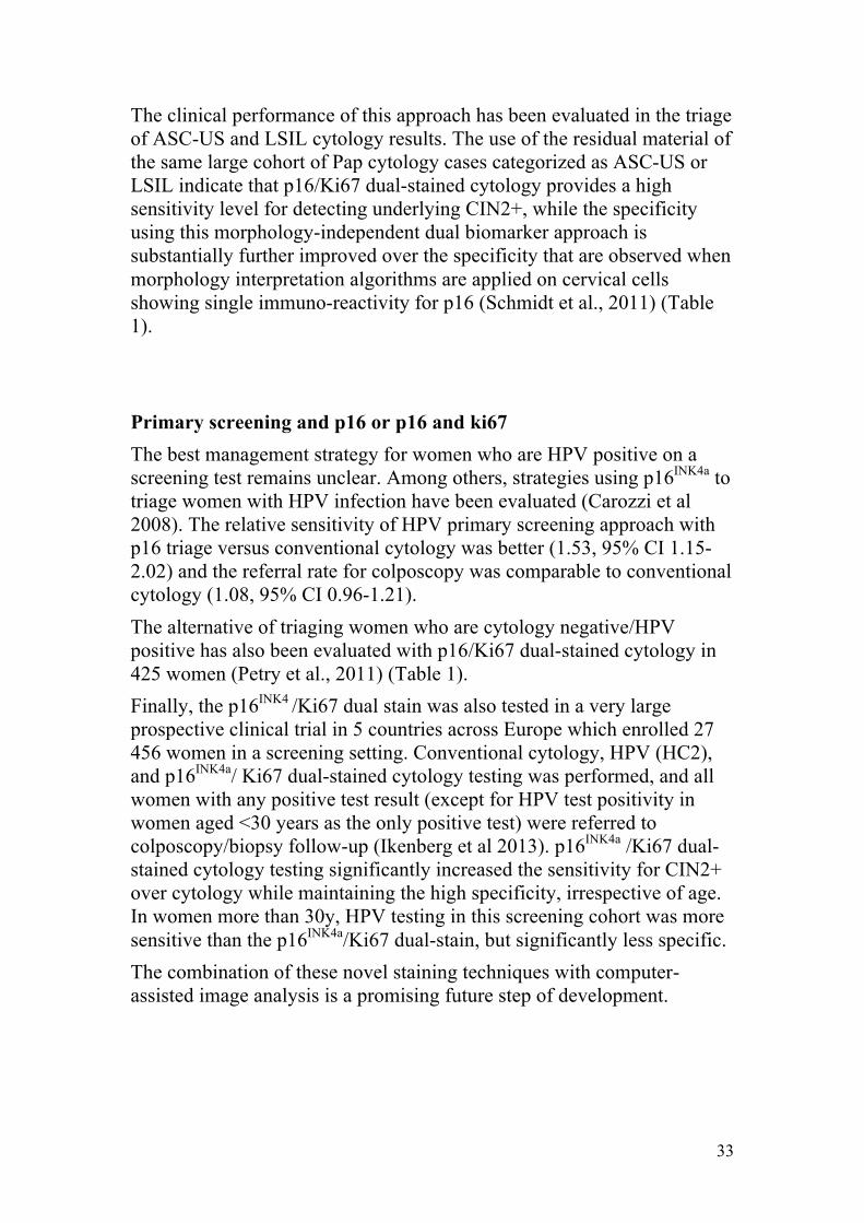
The clinical performance of this approach has been evaluated in the triage of ASC-US and LSIL cytology results. The use of the residual material of the same large cohort of Pap cytology cases categorized as ASC-US or LSIL indicate that p16/Ki67 dual-stained cytology provides a high sensitivity level for detecting underlying CIN2+, while the specificity using this morphology-independent dual biomarker approach is substantially further improved over the specificity that are observed when morphology interpretation algorithms are applied on cervical cells showing single immuno-reactivity for p16 (Schmidt et al., 2011) (Table 1). Primary screening and p16 or p16 and ki67 The best management strategy for women who are HPV positive on a screening test remains unclear. Among others, strategies using p16INK4a to triage women with HPV infection have been evaluated (Carozzi et al 2008). The relative sensitivity of HPV primary screening approach with p16 triage versus conventional cytology was better (1.53, 95% CI 1.15-2.02) and the referral rate for colposcopy was comparable to conventional cytology (1.08, 95% CI 0.96-1.21). The alternative of triaging women who are cytology negative/HPV positive has also been evaluated with p16/Ki67 dual-stained cytology in 425 women (Petry et al., 2011) (Table 1). Finally, the p16INK4 /Ki67 dual stain was also tested in a very large prospective clinical trial in 5 countries across Europe which enrolled 27 456 women in a screening setting. Conventional cytology, HPV (HC2), and p16INK4a/ Ki67 dual-stained cytology testing was performed, and all women with any positive test result (except for HPV test positivity in women aged <30 years as the only positive test) were referred to colposcopy/biopsy follow-up (Ikenberg et al 2013). p16INK4a /Ki67 dual-stained cytology testing significantly increased the sensitivity for CIN2+ over cytology while maintaining the high specificity, irrespective of age. In women more than 30y, HPV testing in this screening cohort was more sensitive than the p16INK4a/Ki67 dual-stain, but significantly less specific. The combination of these novel staining techniques with computer-assisted image analysis is a promising future step of development.

34
References Bergeron,C., Ordi,J., Schmidt,D., Trunk,M.J., Keller,T., Ridder,R. Conjunctive p16INK4a testing significantly increases accuracy in diagnosing high-grade cervical intraepithelial neoplasia. Am. J. Clin. Pathol. 2010, 133, 395-406. Carozzi, F., Confortini, M., Dalla Palma, P., Del Mistro, A., Gillio-Tos, A., De Marco, L., Giorgi-Rossi, P., Pontenani, G., Rosso, S., Sani, C., et al. Use of p16-INK4A overexpression to increase the specificity of human papillomavirus testing: a nested substudy of the NTCC randomised controlled trial. The Lancet Oncology 2008, 9, 937-945. Darragh TM, Colgan TJ, Thomas Cox J et al. The Lower Anogenital Squamous Terminology Standardization project for HPV-associated lesions: background and consensus recommendations from the College of American pathologists and the American Society for Colposcopy and cervical pathology. Int J Gynecol Pathol 2013, 32, 76-115. Denton K, Bergeron C, Klement P, Trunk T, Keller T, Ridder R, European CINtec Cytology Study Group. The sensitivity and specificity of p16INK4a cytology versus HPV testing for detecting High-Grade cervical disease in the triage of ASC-US and LSIL Pap cytology results. Am J Clin Pathol 2010,134, 12-21. Ikenberg H, Bergeron C, Schmidt D, Griesser H, Alameda F, Angeloni C, Bogers J, Dachez R, Denton K, Hariri J, Keller T, von Knebel Doeberitz M, Neumann HH, .Puig-Tintore L,Sideri M, Rehm S, Ridder R, for the PALMS Study Group. Screening for Cervical Cancer Precursors with p16/Ki-67 Dual-Stained Cytology: Results of the PALMS Study. JNCI 2013 in press Petry, K.U., Schmidt, D., Scherbring, S., Luyten, A., Reinecke-Luthge, A., Bergeron, C., Kommoss, F., Loning, T., Ordi, J., Regauer, S., et al. (2011). Triaging Pap cytology negative, HPV positive cervical cancer screening results with p16/Ki-67 Dual-stained cytology. Gynecologic Oncology 2011,121, 505-509. Szarewski A, Ambroisine L, Cadman L, Austin J, Ho L, Terry G, Liddle S, Dina R, McCarthy J, Buckley H, Bergeron C, Soutter WP, Lyons D, Cuzick J. Comparison of predictors for high-grade cervical intraepithelial neoplasia in women with abnormal smears. Cancer Epidemiol Biomarkers Prev 2008, 17, 3033-42. Schmidt,D., Bergeron,C., Denton,K.J., and Ridder,R. p16/ki-67 dual-stain cytology in the triage of ASCUS and LSIL papanicolaou cytology: results from the european equivocal or mildly abnormal papanicolaou cytology study. Cancer Cytopathol 2011,119, 158-166.

35
Table 1 Sensitivity and specificity of p16/Ki67 and HPV testing after a diagnosis of ASC-US and LSIL for diagnosing a CIN 2+ in primary screening above and below 30 years of age
p16/Ki67 HPV
N
Sensitivity%
Specificity%
Sensitivity%
Specificity%
ASC-US* 1 361 92 81 91 36
LSIL**1 415 94 68 96 19
Primary
screening <30
yrs2
6798 89 92 - -
Primary
screening >30
yrs2
20450 85 96 93 93
HPV+cytology normal 3
425 92 82 - -
*Atypical squamous cells of undetermined significance
**Low-grade squamous intraepithelial lesion
1 Schmidt et al 2011
2 Ikenberg et al 2013,
3 Petry et al 2011

36
Invasive cervical cancer audit and EU guidelines for quality assurance Amanda Herbert Guys & St Thomas’ NHS Foundation Trust. Email: [email protected] ___________________________________________________________ Workshop cases Each workshop case provides a brief case history on the front of the packet, slides that include conventional slides taken before or at the time of diagnosis, and in some cases histology sections. Examine the slides and make up your own mind as to how they should have been reported. A summary of the screening history and slide review is given on the back. European guidelines for quality assurance in cervical cancer screening The European guidelines for QA in cervical cancer screening recommend:
“Rescreening of smears from patients with negative or low-grade test results less than 3-5 years before the diagnosis of invasive cancer forms an important part of quality control but should be taken in the context of all components of the screening history, including cytological screening errors, sampling errors, non-compliance with follow-up recommendations, incomplete treatment and whether or not the cancer was screen-detected.”(1)
EU guidelines: all systems should be translatable into TBS The EU guidelines recommend that all systems should be translatable into the 2001 Bethesda system (TBS), that the CIN system should be retained for histology and that a statement on adequacy should be provided on reports (2). The NHS Cervical Screening Programme (NHSCSP) uses a three-tier system (mild, moderate and severe dyskaryosis), which has been modified and is now closer to TBS (3). This terminology has been adopted by the NHSCSP in their latest guidelines (ABC3), which are available on the NHSCSP website: http://www.cancerscreening.nhs.uk/cervical/publications/nhscsp01.pdf

37
Invasive cervical cancer audit Aims of cervical cancer audit: to identify areas where procedures have gone wrong, not necessarily through anyone’s fault, and to identify where local or national procedures could be improved. Cancer audit demonstrates the importance of these seemingly demanding quality assurance recommendations. Paradoxically, cancer audit demonstrates the effectiveness of screening as a process, since many of the cases might have been prevented if the guidelines had been followed. In order to place invasive cancers in the context of laboratory performance, it helps to consider them along with cases of high-grade CIN detected and treated during the same period of time: Figure 1 is taken from a Guy’s & St Thomas’ audit (4), using a population-based audit in Southampton as a baseline (5,6). CIN3 was 10-fold more frequent than cancer. This ratio and age distribution of invasive/in situ cancer is similar to England as a whole (7). Unlike other national audits (8,9), which assessed the relative risks of cancer in screened and unscreened women, our audits did not include control cases and aimed to identify reasons why cancers had not been prevented in individual women.
The relevance of screen-detected cancers in asymptomatic women We recorded whether the cancers were found on investigation of symptoms (symptomatic cancers) or investigation of abnormal cytology in asymptomatic women (screen-detected cancers). Screen-detected cancers were significantly more likely to be FIGO stage IA or IB1 (99%) compared with symptomatic cancers (42%) and were more likely to be seen in younger age groups (4,5,6).

38
Women who had not been screened within 5 years All women in England aged 20-64 years (from age 25 years since 2004) have been personally invited for cytology screening tests without charge every 3-5 years since 1988; yet in our audits (4,6), about half of the women with cancer had not been screened within five years (Figure 2). These women could be divided into those who had never been screened (around 1% of women in the UK) and those who are overdue for tests.
• Most had symptomatic cancers • Some in their early 30s had been screened when younger, in their
early 20s, and had not responded to reminders • Around 10% were known to have had previous cytological
abnormalities: previous treatment of CIN or low-grade cytology not followed up.
These cases justify the recommendations in the guidelines for cytological surveillance of previous abnormalities and reminders for repeat tests.
Interval cancers (women screened within 0.5-5.0 years before diagnosis) Why were more than half of cancers developing in women screened within 5 years? (These are described as ‘interval cancers’ in our audits.) How could compliance with guidelines have avoided those cancers? What had we done wrong and what are the reasons for women developing cancers in well-screened populations?
• The higher the screening coverage, the greater will be the percentage of cancers that are in previously screened women. If all women were screened, all cancers would be ‘interval cancers’ even though there would be very few of them.
• In Southampton, there was a significant trend towards interval cancers as screening coverage and quality control improved, and incidence fell (6).
• Half of all invasive cancers were screen-detected. Nearly two-thirds of these were microinvasive (stage IA).
• Women with previous cytology were more likely to be stage 1. • The most clinically important group of women with interval
cancers were those who had previous negative cytology, who were more likely to be symptomatic than screen-detected if they had only had negative cytology.

39
Women with previous negative cytology within 5 years Screening is not perfect and cancer may develop in women previously screened as negative. In the Guy’s & St Thomas’ audit (4):
• About 10% of women with cancer had at least two negative tests within 10 years; similar to other audits (9,11).
• Others had been screened infrequently, were overdue for 3-year recall or only had one previous test.
• About 60% of slides were confirmed as negative on review; about 20% showed HSIL and 33% showed borderline (atypical) changes (often ASC-H or AGC). A similar balance was recorded in other audits (10,12) between slides confirmed as negative or found to be abnormal on review.
• Most had very few abnormal cells, known to be at risk as false negatives (13).
• Although 49 (37%) of 133 had previous negative cytology within 5 years, almost half of these also had abnormal cytology; and these 49 women were among around 38,000 reported as negative each year.
These cases show the importance of accurate screening, which is improved by procedures recommended in the guidelines such as rapid re-screening of negative and inadequate tests (or rapid pre-screening of all slides), calculating and comparing screeners’ sensitivity, comparing abnormal reporting rates between laboratories and reviewing previous negative cytology in women with cancer and high-grade CIN - in order to find out what types of abnormalities may be missed and use them for teaching. Accuracy of screening could also be improved by introducing automated screening (14) especially if used for quality control alongside careful routine screening (15).

40
Women with previous repeats for low-grade cytology Prior to the introduction of low-grade HPV triage, the NHSCSP recommended colposcopy on the first or second occurrence of mild dyskaryosis and third borderline result; or on the first borderline result if ‘high-grade dyskaryosis is not excluded’ (i.e. ASC-H). Three annual negative tests were recommended before return to routine recall.
• There were very few women in this category • It was very rare for women with cancer to have had three negative
repeats after a low-grade cytology report. • Several had severe dyskaryosis on repeat cytology and were found
to have stage IA1 squamous cell carcinomas. • Most were overdue for follow-up or had not had repeat smears
Review of these slides showed that one-third should have been reported as HSIL+ and one-third as ASC-H or AGC with a recommendation for colposcopy; one-third were confirmed as ASC-US or LSIL. Low-grade cytology is common and most regresses spontaneously, but follow-up is needed to identify the minority that either progress or were under-called on cytology. Referral on first occurrence of LSIL, ASC-H and AGC, HPV triage, comparing low-grade rates between laboratories and monitoring their outcome at colposcopy all help avoid these cases. Even if referred for colposcopy, women with low-grade changes need follow-up to find the minority of lesions that persist or progress; as recommended in all the guidelines. Women referred for colposcopy more than 6 months before diagnosis A significant proportion of women in both audits had been recommended for colposcopy during the preceding 5 years, but had either not had colposcopy within 6 months of the cytology test, or had colposcopy but cancer was not detected or prevented. Reasons for delay were related to patient compliance or management:
• Patients refused treatment or did not attend • Waiting list delays between cytology and biopsy or biopsy and
treatment • Failures in ‘failsafe’ of women who changed address

41
Delays are now avoided by direct referral from the laboratory, waiting times being monitored and controlled, and ‘see-and-treat’ protocols (although the risks of over-treatment must also be considered). We concluded that prompt referral for high-grade dyskaryosis (HSIL) is an important recommendation, especially for severe dyskaryosis (the referral diagnosis in most of these cases).
Women who had previous treatment of CIN A similar percentage of cancers (10-12%) in the Southampton and London audits were in women who had previous treatment of CIN, which was usually (but not always) CIN3.
• Most had high-grade cytology or biopsy after CIN treatment • Some had defaulted from treatment or follow-up • Rare to have had regular negative follow-up smears after treatment • Women with cancer following CIN treatment were significantly
more likely to have initially been treated when older (35-64 years) compared with routine practice (20-34 years) (Figure 3)
These cases were rare: 16 in the Guy’s & St Thomas’ audit compared with 3027 (0.5%) treated for CIN2+ (Figure 1); the percentage would be higher if women with CIN3 aged 35-64 years were taken as the denominator. The results support screening programmes that start in younger age groups and show the importance of follow-up and treatment of any persistent high-grade abnormalities.
!"#$%&'('
)#&'*+',-&'./'0%&1".$2'+%&*+-&3+'./'456'

42
Summary Despite all these errors and omissions most cancers in previously screened women were screen-detected stage 1A1 or 1B1 cancers in younger age groups; and CIN3 was 10-fold more frequent than invasive cancer. Invasive cervical cancer audit, which is recommended by the NHSCSP for all cases of invasive cancer (16), shows the importance of accurate cytology, compliance with recommendations for repeat tests and investigation, effective failsafe procedures (all recommended in the guidelines) and, perhaps as important as anything, advice to women that regular cytological screening prevents the vast majority of cancers, especially the ones that kill.
References
1. Wiener HG, Klinkhamer P, Schenck U et al. European guidelines for quality assurance in cervical cancer screening: recommendations for cytology laboratories. Cytopathology 2007;18:67-78.
2. Herbert A, Bergeron C, Wiener H et al. European guidelines for quality assurance in cervical cancer screening: recommendations for cervical cytology terminology. Cytopathology 2007;18:213-9.
3. Denton KJ, Herbert A, Turnbull LS et al. The revised BSCC terminology for abnormal cervical cytology. Cytopathology 2008;19:137-57.
4. Herbert A, Anshu -, Culora G et al. Invasive cancer audit: why cancers developed in a high-risk population with an organised screening programme. Br J Obstet Gynaecol 2010;117:736-45.
5. Herbert A, Anshu, Gregory M, Gupta SS, Singh N. Screen-detected invasive cervical cancer and its clinical significance during the introduction of organized screening. BJOG 2009;116:854–9.
6. Herbert A, Anshu, Gregory M, Gupta SS, Singh N. Invasive cervical cancer audit: a relative increase in interval cancers while coverage increased and incidence declined. BJOG 2009;116: 845–53.

43
7. Office for National Statistics. Cancer Statistics Registrations, England (Series MB1) No. 1 (1992) to 40 (2010). Look up on google as above; No.2 (1993) etc. Or find on: www.ons.gov.uk/ons/taxonomy/index.html?nscl+Cancer+Registrations
8. Sasieni P, Adams J, Cuzick J. Benefit of cervical screening at different ages: evidence from the UK audit of screening histories. Br J Cancer 2003;89:88–93.
9. Andrae B, Kemetli L, Sparen P et al. Screening-Preventable Cervical Cancer Risks: Evidence From a Nationwide Audit in Sweden. J Natl Cancer Inst 2008;100:622-9.
10. Repše-Fokter A, Pogačnik A, Snoj V, Primic-Žakelj M, Strojan Fležar M. Review of negative and low-grade cervical smears in women with invasive cervical cancer after the first three years of the national cervical screening programme in Slovenia. Cytopathology 2012;23:23-9.
11. Priest P, Sadler L, Peters J et al. Pathways to diagnosis of cervical cancer:screening history, delay in follow up, and smear reading. BJOG 2007;114:398–407.
12. Castanon A, Ferryman S, Patnick J, Sasieni P. Review of cytology and histopathology as part of the NHS Cervical Screening Programme audit of invasive cervical cancers. Cytopathology 2012;23:13-22.
13. Mitchell H, Medley G. Differences between Papanicolaou smears with correct and incorrect diagnoses. Cytopathology 1995;6:368-75.
14. Davey E, Irwig L, Macaskill P et al. Cervical cytology reading times: a comparison between ThinPrep Imager and conventional methods. Diagn Cytopathol 2007;35:550-4.
15. Heard T, Chandra A, Culora G et al. Use of the ThinPrep Imaging System for internal quality control of cervical cytology. Cytopathology 2013;24:246-53.
16. Audit of Invasive Cervical Cancers. NHSCSP Publication no. 28, 2006.