9.4 search for better health - tsfx.com.au who has lived their whole life in a wheelchair may...
TRANSCRIPT
RED: write syllabus dot point on other side in red. (with question that you make up in blue – for flash cards) and one word heading on this side in red. Purple in Bold: Key information Green: Equations, Scientists & Definitions or Headings
9.4 Search for Better Health
9.4 Assumed Knowledge
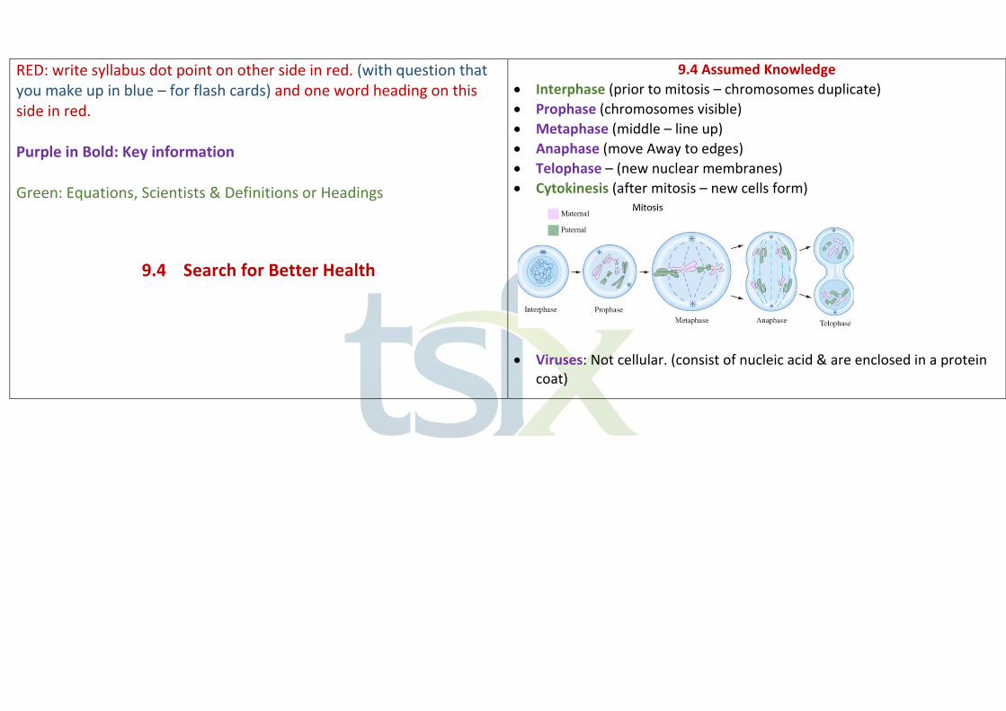
• Interphase (prior to mitosis – chromosomes duplicate)
• Prophase (chromosomes visible)
• Metaphase (middle – line up)
• Anaphase (move Away to edges)
• Telophase – (new nuclear membranes)
• Cytokinesis (after mitosis – new cells form)
• Viruses: Not cellular. (consist of nucleic acid & are enclosed in a protein coat)

9.4.1 What is a Healthy Organism? 9.4.1.1 Discuss the difficulties of defining the terms ‘health’ & ‘disease’
• Health: “The state of physical, mental & social well-being & not merely the absence of disease of infirmity” – WHO
• Disease: “state of impaired functioning or poor health”
• Issues: These are very broad. It is difficult to achieve a healthy status in all these areas as our health is also constantly changing. The definition of disease is also imprecise as it could infer that pregnancy is a disease. These definitions are very subjective (different standards) depending on:
o Their expectations of quality of life & their normal level of functioning.
• An individual may be healthy & have a disease at the same time. An individual who has lived their whole life in a wheelchair may consider themselves healthy when they feel “well”. Whilst a physically fit person may consider themselves unhealthy when they have a cold.
- Tumour suppressor genes: code for proteins that slow down or stop cell growth & mitosis. Mutated tumour suppressor genes lead to uncontrolled cell replication which:
o Doesn’t allow cells to differentiate & perform specialised function o Causes formation of tumours, or uncontrolled cell death.
Thus role of genes in control of mitosis is vital for the maintenance of health.
• Differentiation & Specialisation: maintains the health of an organism by ensuring complex functions are co-ordinated & controlled. If cells don’t differentiate & specialise body processes would not be co-ordinated, resulting in disease.
o Cell differentiation: (shape) Each cell has the potential code to change into different cell types (determines specialisation in structure & function).
9.4.1.2 Function of genes, mitosis, cell differentiation & specialisation assist in the maintenance of health.
• Gene Expression: Is essential in maintaining good health. - If a gene switch is not turned on (i.e. protein is not being produced). Then this protein’s role will not be performed, resulting in abnormal cell processes or structures which result in disease. - Thus, the correct amounts of proteins must be produced at the right time.
• Mitosis: allows growth, repair, replacement & genetic stability. Thus maintains health through replacing damaged cells & ensuring growth of an organism occurs.
- Cyclins & cyclin-dependent kinases proteins regulate mitosis. - DNA Repair genes: Produce enzymes to repair DNA damage. Eg. p53 gene produces the p53 protein that stops the cell cycle to allow DNA to be repaired. If this gene mutates, disease will occur. - Proto-oncogenes: Stimulate cell growth & mitosis at the right time. Mutated proto-oncogenes produces oncogenes which cause uncontrolled cell production & prevents cell death.
• Evidence from the study of the human genome reveals the link between BRCA gene mutations (inherited) with breast & ovarian cancers.
• BRCA gene expression occurs when the protein to repair the PTEN gene is produced. This PTEN gene produces a protein which in turn controls cell division leading to normal cells & tissues & absence of breast cancer.
o Cell Specialisation: (function) Each cell type carries out different functions.
9.4.1.3 Evidence of the link between gene expression & maintenance & repair of body tissues.
• BRCA genes: tumour suppressor genes produce proteins to help repair damaged DNA, thus help maintain our health.
9.4.2 Over 3000 years ago the Chinese & Hebrews were advocating cleanliness in food, water & personal hygiene
9.4.2.1/2 Distinguish between infectious & non-infectious disease & the conditions under which an organism is described as a pathogen
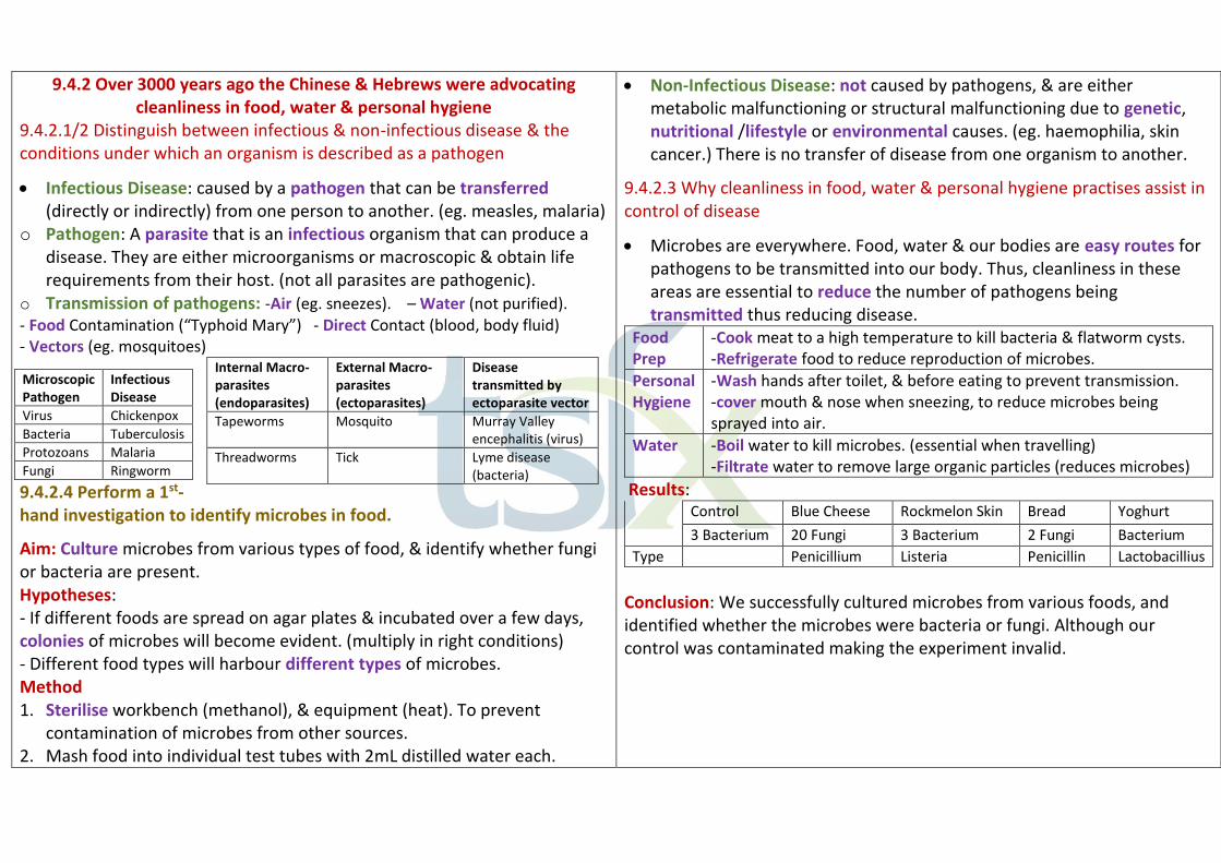
• Infectious Disease: caused by a pathogen that can be transferred (directly or indirectly) from one person to another. (eg. measles, malaria)
o Pathogen: A parasite that is an infectious organism that can produce a disease. They are either microorganisms or macroscopic & obtain life requirements from their host. (not all parasites are pathogenic).
o Transmission of pathogens: -Air (eg. sneezes). – Water (not purified). - Food Contamination (“Typhoid Mary”) - Direct Contact (blood, body fluid) - Vectors (eg. mosquitoes)
9.4.2.4 Perform a 1st-hand investigation to identify microbes in food.
Aim: Culture microbes from various types of food, & identify whether fungi or bacteria are present. Hypotheses: - If different foods are spread on agar plates & incubated over a few days, colonies of microbes will become evident. (multiply in right conditions) - Different food types will harbour different types of microbes. Method 1. Sterilise workbench (methanol), & equipment (heat). To prevent
contamination of microbes from other sources. 2. Mash food into individual test tubes with 2mL distilled water each.
Microscopic Pathogen
Infectious Disease
Virus Chickenpox
Bacteria Tuberculosis
Protozoans Malaria
Fungi Ringworm
Internal Macro-parasites (endoparasites)
External Macro-parasites (ectoparasites)
Disease transmitted by ectoparasite vector
Tapeworms Mosquito Murray Valley encephalitis (virus)
Threadworms Tick Lyme disease (bacteria)
• Non-Infectious Disease: not caused by pathogens, & are either metabolic malfunctioning or structural malfunctioning due to genetic, nutritional /lifestyle or environmental causes. (eg. haemophilia, skin cancer.) There is no transfer of disease from one organism to another.
9.4.2.3 Why cleanliness in food, water & personal hygiene practises assist in control of disease
• Microbes are everywhere. Food, water & our bodies are easy routes for pathogens to be transmitted into our body. Thus, cleanliness in these areas are essential to reduce the number of pathogens being transmitted thus reducing disease.
Food Prep
-Cook meat to a high temperature to kill bacteria & flatworm cysts. -Refrigerate food to reduce reproduction of microbes.
Personal Hygiene
-Wash hands after toilet, & before eating to prevent transmission. -cover mouth & nose when sneezing, to reduce microbes being sprayed into air.
Water -Boil water to kill microbes. (essential when travelling) -Filtrate water to remove large organic particles (reduces microbes)
Results: Control Blue Cheese Rockmelon Skin Bread Yoghurt
3 Bacterium
20 Fungi 3 Bacterium 2 Fungi Bacterium
Type Penicillium Listeria Penicillin Lactobacillius
Conclusion: We successfully cultured microbes from various foods, and identified whether the microbes were bacteria or fungi. Although our control was contaminated making the experiment invalid.

3. Dip inoculating loop into food type, and wipe over surface of agar plate. 4. Close & seal each agar plate with sticky tape, & label. & repeat steps 1-3.
5. Place each agar plate (+ controls) into an incubator of 30C. 6. Examine plates every 3 days. Count number of fungal colonies (larger,
fluffy) & bacterial colonies (small, shiny, round & coloured). 7. NOTE: don’t open plates, & dispose safely as microbes are harmful.
Note: Controls show whether the agar plates have been contaminated by other microbes that are not sourced from the food.
9.4.2.5 Drinking Water Treatment – & reduction of risk of infection
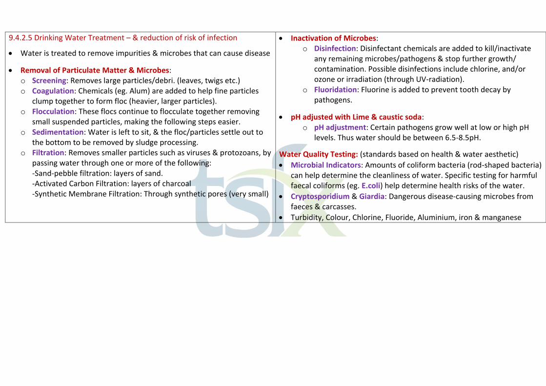
• Water is treated to remove impurities & microbes that can cause disease
• Removal of Particulate Matter & Microbes: o Screening: Removes large particles/debri. (leaves, twigs etc.) o Coagulation: Chemicals (eg. Alum) are added to help fine particles
clump together to form floc (heavier, larger particles). o Flocculation: These flocs continue to flocculate together removing
small suspended particles, making the following steps easier. o Sedimentation: Water is left to sit, & the floc/particles settle out to
the bottom to be removed by sludge processing. o Filtration: Removes smaller particles such as viruses & protozoans, by
passing water through one or more of the following: -Sand-pebble filtration: layers of sand. -Activated Carbon Filtration: layers of charcoal -Synthetic Membrane Filtration: Through synthetic pores (very small)
• Inactivation of Microbes: o Disinfection: Disinfectant chemicals are added to kill/inactivate
any remaining microbes/pathogens & stop further growth/ contamination. Possible disinfections include chlorine, and/or ozone or irradiation (through UV-radiation).
o Fluoridation: Fluorine is added to prevent tooth decay by pathogens.
• pH adjusted with Lime & caustic soda: o pH adjustment: Certain pathogens grow well at low or high pH
levels. Thus water should be between 6.5-8.5pH.
Water Quality Testing: (standards based on health & water aesthetic)
• Microbial Indicators: Amounts of coliform bacteria (rod-shaped bacteria) can help determine the cleanliness of water. Specific testing for harmful faecal coliforms (eg. E.coli) help determine health risks of the water.
• Cryptosporidium & Giardia: Dangerous disease-causing microbes from faeces & carcasses.
• Turbidity, Colour, Chlorine, Fluoride, Aluminium, iron & manganese
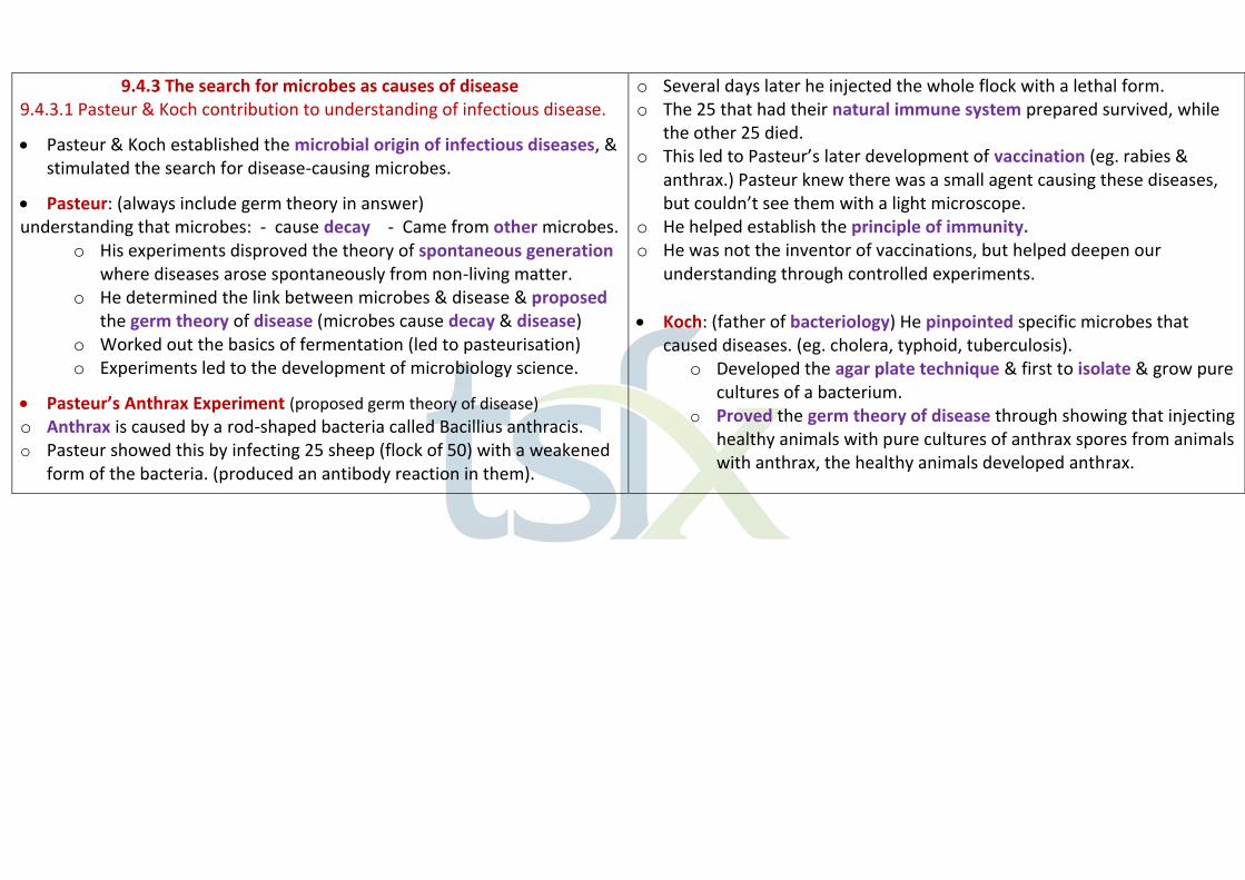
9.4.3 The search for microbes as causes of disease 9.4.3.1 Pasteur & Koch contribution to understanding of infectious disease.
• Pasteur & Koch established the microbial origin of infectious diseases, & stimulated the search for disease-causing microbes.
• Pasteur: (always include germ theory in answer) understanding that microbes: - cause decay - Came from other microbes.
o His experiments disproved the theory of spontaneous generation where diseases arose spontaneously from non-living matter.
o He determined the link between microbes & disease & proposed the germ theory of disease (microbes cause decay & disease)
o Worked out the basics of fermentation (led to pasteurisation) o Experiments led to the development of microbiology science.
• Pasteur’s Anthrax Experiment (proposed germ theory of disease) o Anthrax is caused by a rod-shaped bacteria called Bacillius anthracis. o Pasteur showed this by infecting 25 sheep (flock of 50) with a weakened
form of the bacteria. (produced an antibody reaction in them).
o Several days later he injected the whole flock with a lethal form. o The 25 that had their natural immune system prepared survived, while
the other 25 died. o This led to Pasteur’s later development of vaccination (eg. rabies &
anthrax.) Pasteur knew there was a small agent causing these diseases, but couldn’t see them with a light microscope.
o He helped establish the principle of immunity. o He was not the inventor of vaccinations, but helped deepen our
understanding through controlled experiments.
• Koch: (father of bacteriology) He pinpointed specific microbes that caused diseases. (eg. cholera, typhoid, tuberculosis).
o Developed the agar plate technique & first to isolate & grow pure cultures of a bacterium.
o Proved the germ theory of disease through showing that injecting healthy animals with pure cultures of anthrax spores from animals with anthrax, the healthy animals developed anthrax.
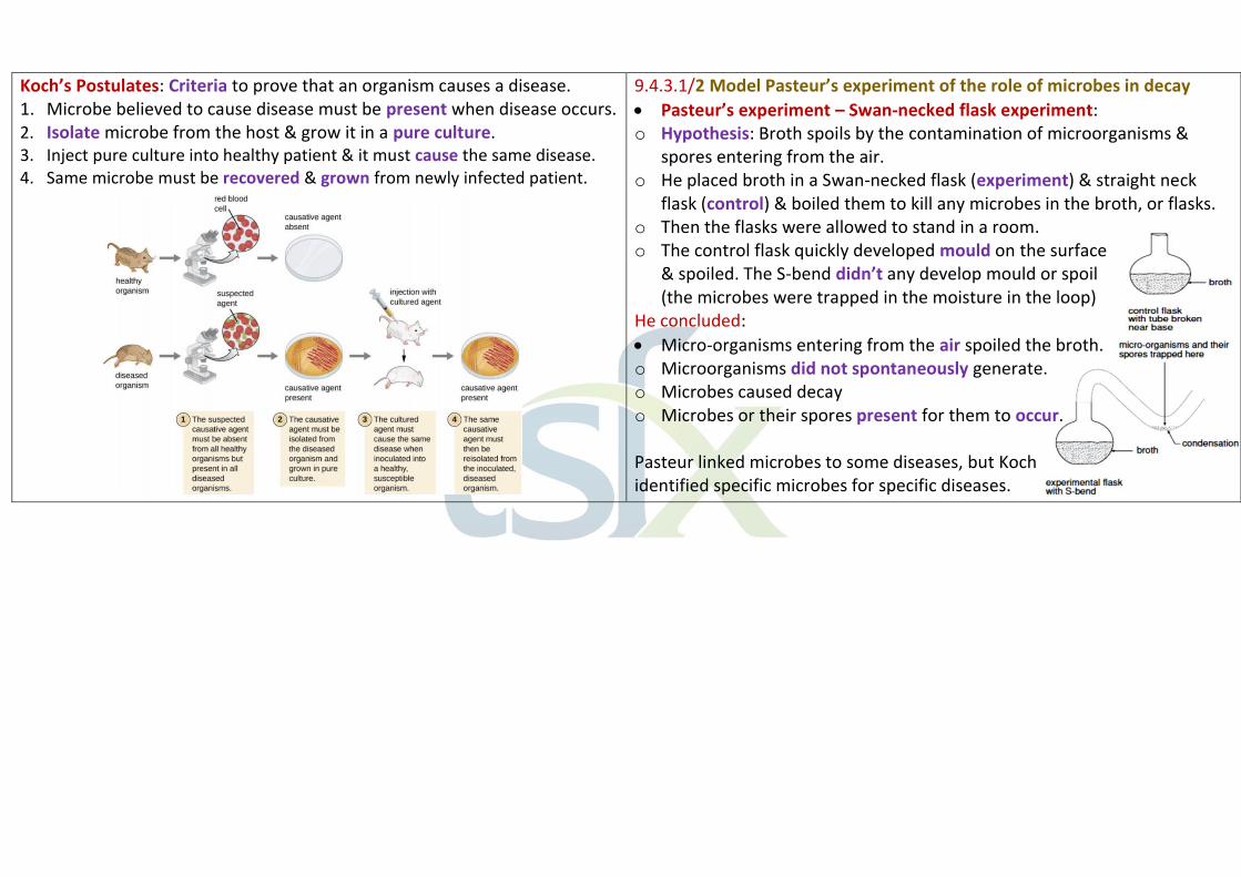
Koch’s Postulates: Criteria to prove that an organism causes a disease. 1. Microbe believed to cause disease must be present when disease occurs. 2. Isolate microbe from the host & grow it in a pure culture. 3. Inject pure culture into healthy patient & it must cause the same disease. 4. Same microbe must be recovered & grown from newly infected patient.
9.4.3.1/2 Model Pasteur’s experiment of the role of microbes in decay
• Pasteur’s experiment – Swan-necked flask experiment: o Hypothesis: Broth spoils by the contamination of microorganisms &
spores entering from the air. o He placed broth in a Swan-necked flask (experiment) & straight neck
flask (control) & boiled them to kill any microbes in the broth, or flasks. o Then the flasks were allowed to stand in a room. o The control flask quickly developed mould on the surface
& spoiled. The S-bend didn’t any develop mould or spoil (the microbes were trapped in the moisture in the loop)
He concluded:
• Micro-organisms entering from the air spoiled the broth. o Microorganisms did not spontaneously generate. o Microbes caused decay o Microbes or their spores present for them to occur. Pasteur linked microbes to some diseases, but Koch identified specific microbes for specific diseases.
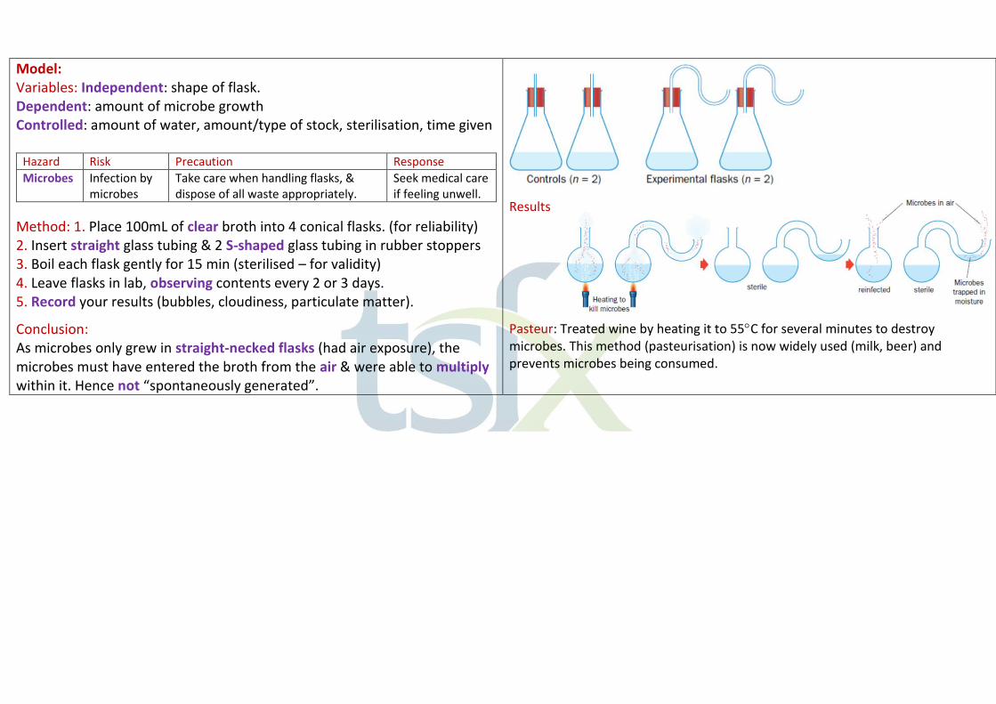
Model: Variables: Independent: shape of flask. Dependent: amount of microbe growth Controlled: amount of water, amount/type of stock, sterilisation, time given
Hazard Risk Precaution Response
Microbes Infection by microbes
Take care when handling flasks, & dispose of all waste appropriately.
Seek medical care if feeling unwell.
Method: 1. Place 100mL of clear broth into 4 conical flasks. (for reliability) 2. Insert straight glass tubing & 2 S-shaped glass tubing in rubber stoppers 3. Boil each flask gently for 15 min (sterilised – for validity) 4. Leave flasks in lab, observing contents every 2 or 3 days. 5. Record your results (bubbles, cloudiness, particulate matter).
Conclusion: As microbes only grew in straight-necked flasks (had air exposure), the microbes must have entered the broth from the air & were able to multiply within it. Hence not “spontaneously generated”.
Results
Pasteur: Treated wine by heating it to 55C for several minutes to destroy microbes. This method (pasteurisation) is now widely used (milk, beer) and prevents microbes being consumed.
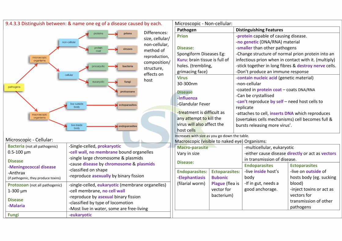
9.4.3.3 Distinguish between: & name one eg of a disease caused by each.
Differences: size, cellular/ non-cellular, method of reproduction, composition/ structure, effects on host
Microscopic - Cellular: Bacteria (not all pathogenic) 0.5-100 μm
Disease -Meningococcal disease -Anthrax (if pathogenic, they produce toxins)
-Single-celled, prokaryotic -cell wall, no membrane bound organelles -single large chromosome & plasmids -cause disease by chromosome & plasmids -classified on shape -reproduce asexually by binary fission
Protozoan (not all pathogenic)
1-300 μm
Disease -Malaria
-single-celled, eukaryotic (membrane organelles) -cell membrane, no cell wall -reproduce by asexual binary fission -classified by type of locomotion -Most live in water, some are free-living
Fungi -eukaryotic
Microscopic - Non-cellular: Pathogen Distinguishing Features
Prion Disease: Spongiform Diseases Eg: Kuru: brain tissue is full of holes. (trembling, grimacing face)
-protein capable of causing disease. -no genetic (DNA/RNA) material -smaller than other pathogens -Change structure of normal prion protein into an infectious prion when in contact with it. (multiply) -stick together in long fibres & destroy nerve cells. -Don’t produce an immune response
Virus 30-300nm
Disease -Influenza -Glandular Fever
-treatment is difficult as any attempt to kill the virus will also affect the host cells
-contain nucleic acid (genetic material) -non-cellular -coated in protein coat – coats DNA/RNA
-Can be crystallised -can’t reproduce by self – need host cells to replicate -attaches to cell, inserts DNA which reproduces (overtakes cells mechanisms) cell becomes full & bursts releasing more virus’.
Increases with size as you go down the table.
Macroscopic (visible to naked eye) Organisms:
Macro-parasite Vary in size
Disease:
-multicellular, eukaryotic -either cause disease directly or act as vectors in transmission of disease.
Endoparasites -live inside host’s body -If in gut, needs a good anchorage.
Ectoparasites -live on outside of hosts body (eg. sucking blood) -inject toxins or act as vectors for transmission of other pathogens
Endoparasites: -Elephantiasis (filarial worm)
Ectoparasites: Bubonic Plague (flea is vector for bacterium)

Microscopic or Macroscopic
Disease -Athlete’s foot -Candidiasis (Thrush)
-have cell wall (different to a plant’s cell wall) -no chlorophyll, can’t produce own food (not heterotrophic) -unicellular (yeast) or multicellular (mushrooms) -most have hyphae that spread to form mycelium -reproduce asexually (yeast) or both sexually & asexually
Virus: Fungi Note: some bacteria form
resistant spores that can’t be sterilised by heat.
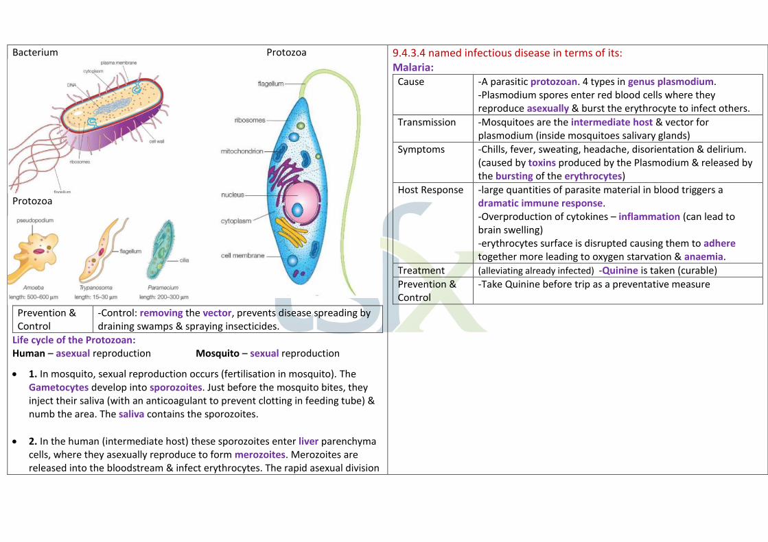
Bacterium Protozoa
Protozoa
Prevention & Control
-Control: removing the vector, prevents disease spreading by draining swamps & spraying insecticides.
Life cycle of the Protozoan: Human – asexual reproduction Mosquito – sexual reproduction
• 1. In mosquito, sexual reproduction occurs (fertilisation in mosquito). The Gametocytes develop into sporozoites. Just before the mosquito bites, they inject their saliva (with an anticoagulant to prevent clotting in feeding tube) & numb the area. The saliva contains the sporozoites.
• 2. In the human (intermediate host) these sporozoites enter liver parenchyma cells, where they asexually reproduce to form merozoites. Merozoites are released into the bloodstream & infect erythrocytes. The rapid asexual division
9.4.3.4 named infectious disease in terms of its: Malaria:
Cause -A parasitic protozoan. 4 types in genus plasmodium. -Plasmodium spores enter red blood cells where they reproduce asexually & burst the erythrocyte to infect others.
Transmission -Mosquitoes are the intermediate host & vector for plasmodium (inside mosquitoes salivary glands)
Symptoms -Chills, fever, sweating, headache, disorientation & delirium. (caused by toxins produced by the Plasmodium & released by the bursting of the erythrocytes)
Host Response -large quantities of parasite material in blood triggers a dramatic immune response. -Overproduction of cytokines – inflammation (can lead to brain swelling) -erythrocytes surface is disrupted causing them to adhere together more leading to oxygen starvation & anaemia.
Treatment (alleviating already infected) -Quinine is taken (curable)
Prevention & Control
-Take Quinine before trip as a preventative measure
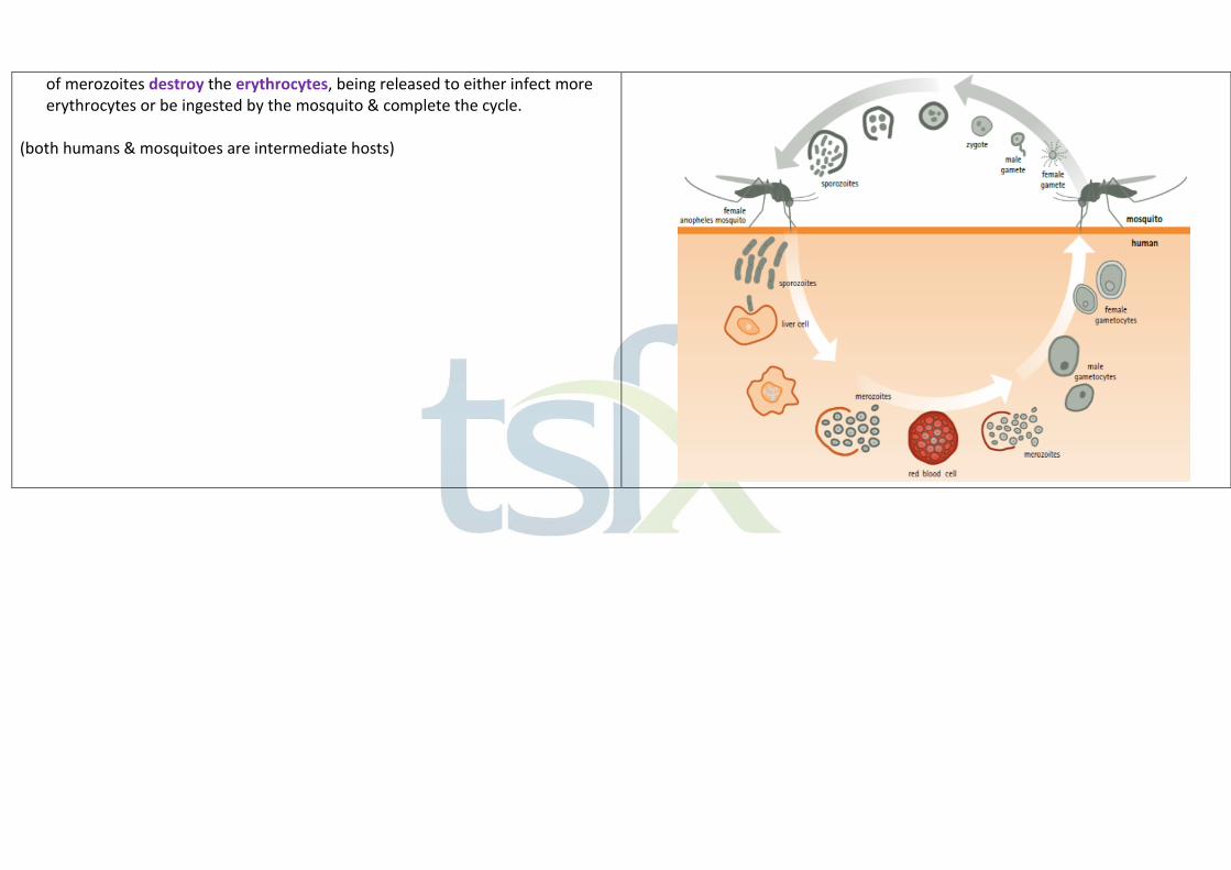
of merozoites destroy the erythrocytes, being released to either infect more erythrocytes or be ingested by the mosquito & complete the cycle.
(both humans & mosquitoes are intermediate hosts)
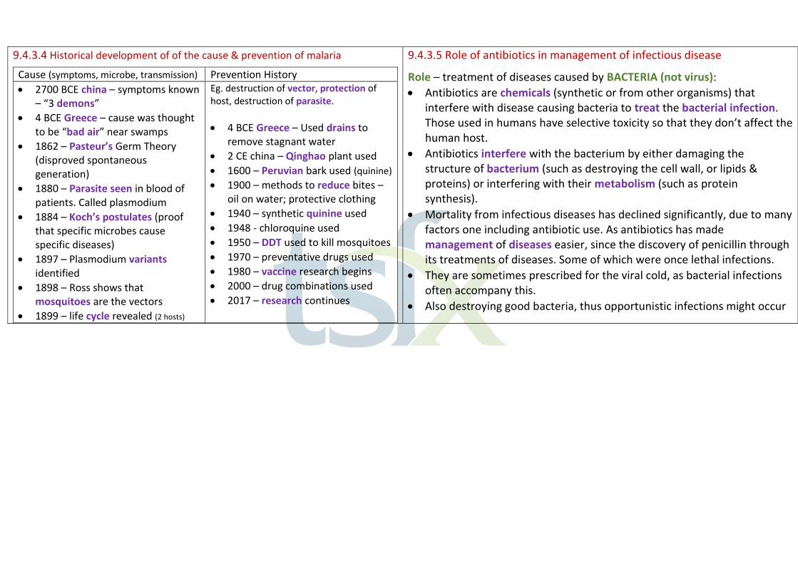
9.4.3.4 Historical development of of the cause & prevention of malaria
Cause (symptoms, microbe, transmission) Prevention History
• 2700 BCE china – symptoms known – “3 demons”
• 4 BCE Greece – cause was thought to be “bad air” near swamps
• 1862 – Pasteur’s Germ Theory (disproved spontaneous generation)
• 1880 – Parasite seen in blood of patients. Called plasmodium
• 1884 – Koch’s postulates (proof that specific microbes cause specific diseases)
• 1897 – Plasmodium variants identified
• 1898 – Ross shows that mosquitoes are the vectors
• 1899 – life cycle revealed (2 hosts)
Eg. destruction of vector, protection of host, destruction of parasite.
• 4 BCE Greece – Used drains to remove stagnant water
• 2 CE china – Qinghao plant used
• 1600 – Peruvian bark used (quinine)
• 1900 – methods to reduce bites – oil on water; protective clothing
• 1940 – synthetic quinine used
• 1948 - chloroquine used
• 1950 – DDT used to kill mosquitoes
• 1970 – preventative drugs used
• 1980 – vaccine research begins
• 2000 – drug combinations used
• 2017 – research continues
9.4.3.5 Role of antibiotics in management of infectious disease
Role – treatment of diseases caused by BACTERIA (not virus):
• Antibiotics are chemicals (synthetic or from other organisms) that interfere with disease causing bacteria to treat the bacterial infection. Those used in humans have selective toxicity so that they don’t affect the human host.
• Antibiotics interfere with the bacterium by either damaging the structure of bacterium (such as destroying the cell wall, or lipids & proteins) or interfering with their metabolism (such as protein synthesis).
• Mortality from infectious diseases has declined significantly, due to many factors one including antibiotic use. As antibiotics has made management of diseases easier, since the discovery of penicillin through its treatments of diseases. Some of which were once lethal infections.
• They are sometimes prescribed for the viral cold, as bacterial infections often accompany this.
• Also destroying good bacteria, thus opportunistic infections might occur
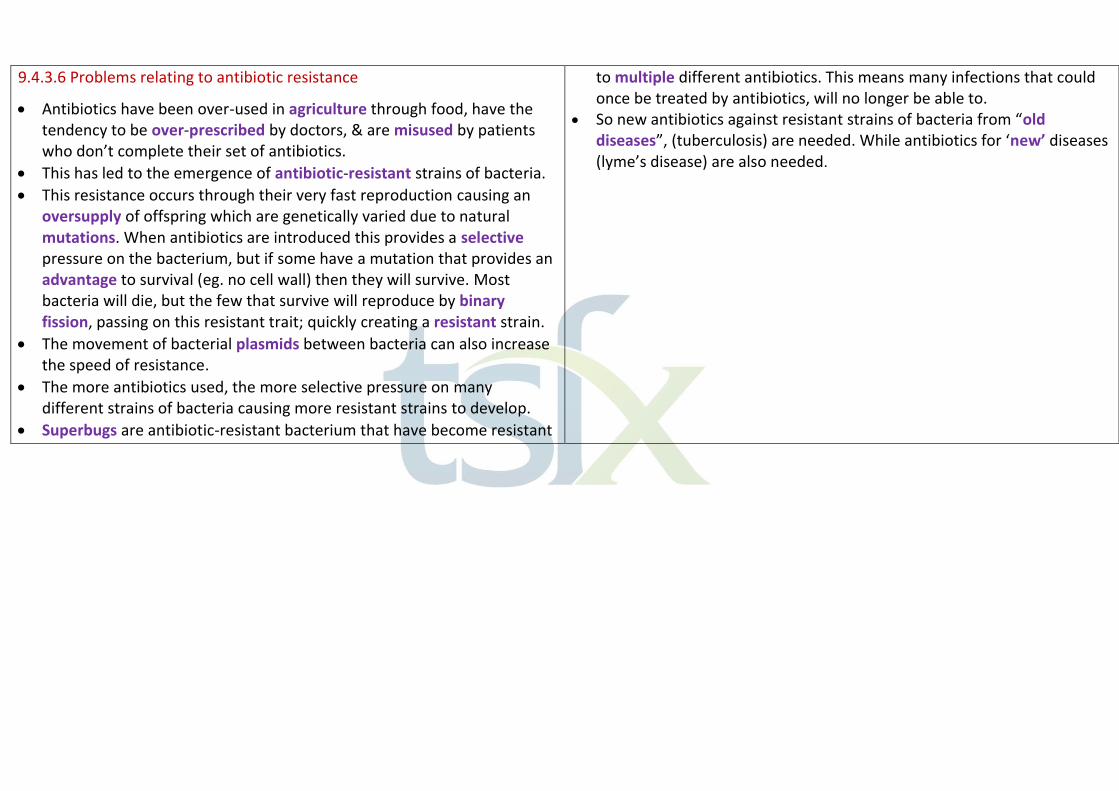
9.4.3.6 Problems relating to antibiotic resistance
• Antibiotics have been over-used in agriculture through food, have the tendency to be over-prescribed by doctors, & are misused by patients who don’t complete their set of antibiotics.
• This has led to the emergence of antibiotic-resistant strains of bacteria.
• This resistance occurs through their very fast reproduction causing an oversupply of offspring which are genetically varied due to natural mutations. When antibiotics are introduced this provides a selective pressure on the bacterium, but if some have a mutation that provides an advantage to survival (eg. no cell wall) then they will survive. Most bacteria will die, but the few that survive will reproduce by binary fission, passing on this resistant trait; quickly creating a resistant strain.
• The movement of bacterial plasmids between bacteria can also increase the speed of resistance.
• The more antibiotics used, the more selective pressure on many different strains of bacteria causing more resistant strains to develop.
• Superbugs are antibiotic-resistant bacterium that have become resistant
to multiple different antibiotics. This means many infections that could once be treated by antibiotics, will no longer be able to.
• So new antibiotics against resistant strains of bacteria from “old diseases”, (tuberculosis) are needed. While antibiotics for ‘new’ diseases (lyme’s disease) are also needed.

9.4.4 Often we recognise an infection by the symptoms it causes. The immune response is not so obvious, until we recover
9.4.4.1 Named disease results from an imbalance of microflora in humans
• Microflora: microbes that live symbiotically (mutualistic) in the body (eg. gastrointestinal tract). They use some digested food & produce compounds such as vitamins essential to our health. The unique balance of microflora helps to maintain/protect the epithelial barrier.
• At birth the alimentary canal is sterile, but different diet, nutrition, use of antibiotics & age leads to unique microflora for each person.
• Imbalance of microflora (caused by antibiotics, stress, infections…) means the intestinal barrier is not efficient & bacteria always in our body can become opportunistic pathogens.
• Thrush/Candidiasis:
• Caused by the overproduction of the yeast/fungus Candida albicans.
• There is always a low level of C. albicans in most of our mucous membranes. These are kept low by competition of other microbes
present in the body’s microflora. If the natural balance is upset the number of C. albicans can can multiply in an uncontrolled way in the gastrointestinal tract. • C. albicans then changes from a yeast-life form to a fungal mycelium with
hyphae that enter the mucous lining of the gastro wall. This weakens the lining & lets substances enter the bloodstream. (leaky gut)
• These substances (eg. partial digested protein) can cause an immune response. Resulting in depression, mood swings etc.
• Rapidly growing C. albicans produce toxins that can enter the bloodstream & relocate anywhere in the body. (causing thrush)
• Treatment: Antifungal medication. Prevention: balanced diet, good hygiene
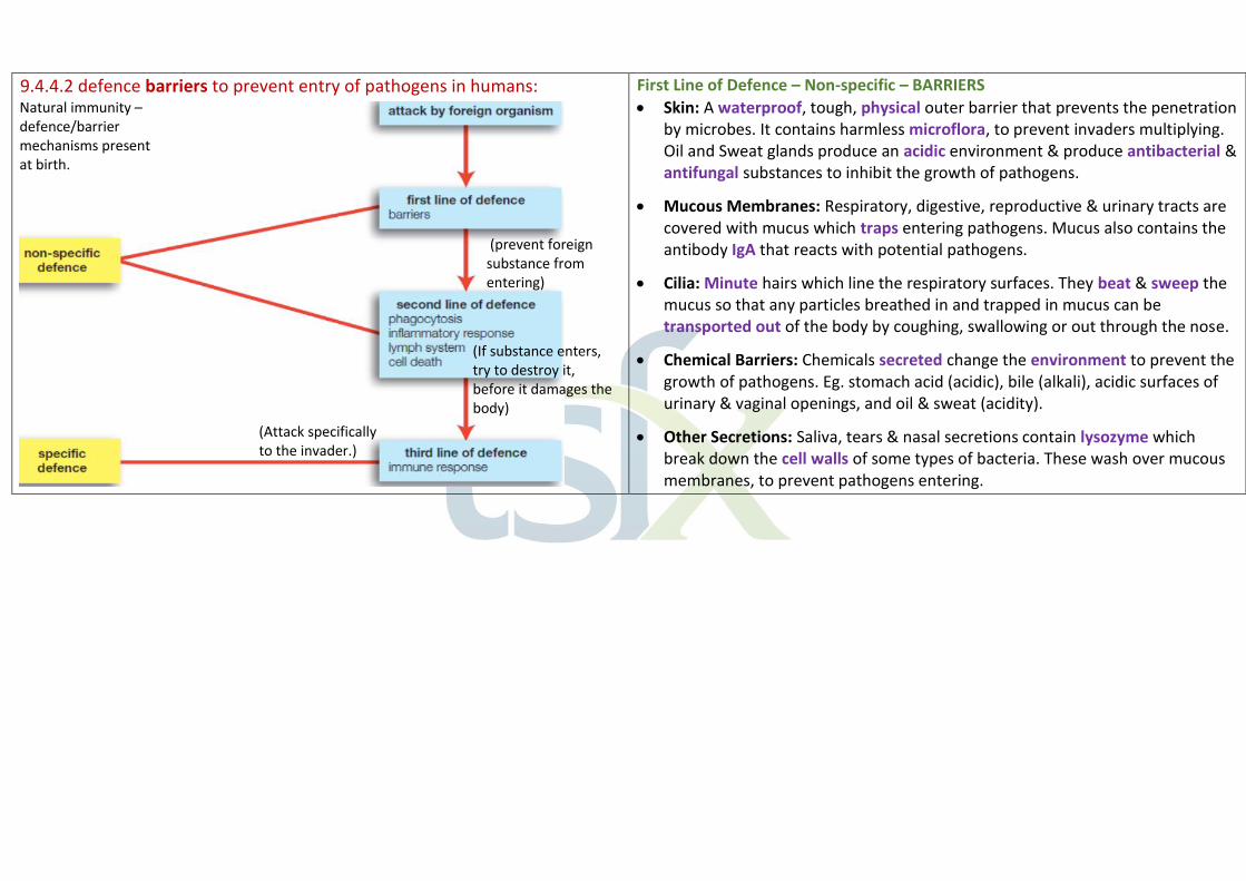
9.4.4.2 defence barriers to prevent entry of pathogens in humans: First Line of Defence – Non-specific – BARRIERS
• Skin: A waterproof, tough, physical outer barrier that prevents the penetration by microbes. It contains harmless microflora, to prevent invaders multiplying. Oil and Sweat glands produce an acidic environment & produce antibacterial & antifungal substances to inhibit the growth of pathogens.
• Mucous Membranes: Respiratory, digestive, reproductive & urinary tracts are covered with mucus which traps entering pathogens. Mucus also contains the antibody IgA that reacts with potential pathogens.
• Cilia: Minute hairs which line the respiratory surfaces. They beat & sweep the mucus so that any particles breathed in and trapped in mucus can be transported out of the body by coughing, swallowing or out through the nose.
• Chemical Barriers: Chemicals secreted change the environment to prevent the growth of pathogens. Eg. stomach acid (acidic), bile (alkali), acidic surfaces of urinary & vaginal openings, and oil & sweat (acidity).
• Other Secretions: Saliva, tears & nasal secretions contain lysozyme which break down the cell walls of some types of bacteria. These wash over mucous membranes, to prevent pathogens entering.
(prevent foreign substance from entering)
(If substance enters, try to destroy it, before it damages the body)
(Attack specifically to the invader.)
Natural immunity – defence/barrier mechanisms present at birth.
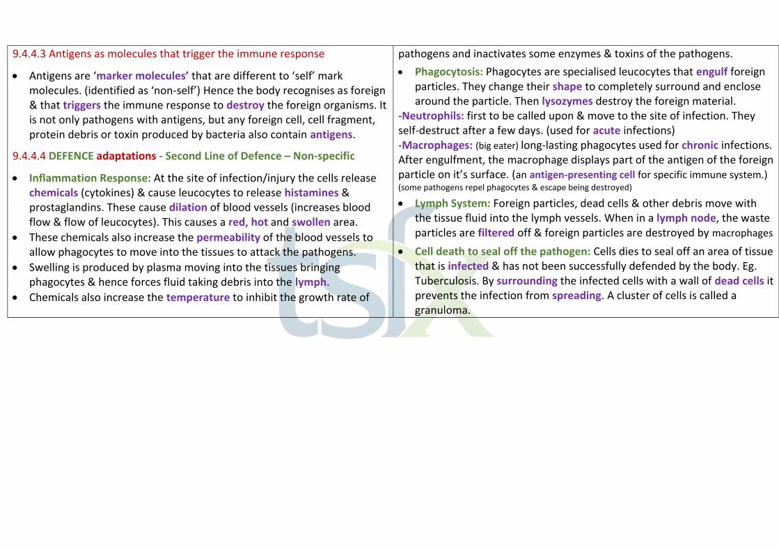
9.4.4.3 Antigens as molecules that trigger the immune response
• Antigens are ‘marker molecules’ that are different to ‘self’ mark molecules. (identified as ‘non-self’) Hence the body recognises as foreign & that triggers the immune response to destroy the foreign organisms. It is not only pathogens with antigens, but any foreign cell, cell fragment, protein debris or toxin produced by bacteria also contain antigens.
9.4.4.4 DEFENCE adaptations - Second Line of Defence – Non-specific
• Inflammation Response: At the site of infection/injury the cells release chemicals (cytokines) & cause leucocytes to release histamines & prostaglandins. These cause dilation of blood vessels (increases blood flow & flow of leucocytes). This causes a red, hot and swollen area.
• These chemicals also increase the permeability of the blood vessels to allow phagocytes to move into the tissues to attack the pathogens.
• Swelling is produced by plasma moving into the tissues bringing phagocytes & hence forces fluid taking debris into the lymph.
• Chemicals also increase the temperature to inhibit the growth rate of
pathogens and inactivates some enzymes & toxins of the pathogens.
• Phagocytosis: Phagocytes are specialised leucocytes that engulf foreign particles. They change their shape to completely surround and enclose around the particle. Then lysozymes destroy the foreign material.
-Neutrophils: first to be called upon & move to the site of infection. They self-destruct after a few days. (used for acute infections) -Macrophages: (big eater) long-lasting phagocytes used for chronic infections. After engulfment, the macrophage displays part of the antigen of the foreign particle on it’s surface. (an antigen-presenting cell for specific immune system.) (some pathogens repel phagocytes & escape being destroyed)
• Lymph System: Foreign particles, dead cells & other debris move with the tissue fluid into the lymph vessels. When in a lymph node, the waste particles are filtered off & foreign particles are destroyed by macrophages
• Cell death to seal off the pathogen: Cells dies to seal off an area of tissue that is infected & has not been successfully defended by the body. Eg. Tuberculosis. By surrounding the infected cells with a wall of dead cells it prevents the infection from spreading. A cluster of cells is called a granuloma.

9.4.5 MacFarlane Burnet’s work in the middle of the twentieth century contributed to a better understanding of the immune response & the
effectiveness of immunisation programs
9.4.5.1 Components of the immune response: antibodies; T cells; B cells 9.4.5.2 Explain/describe the immune response by interaction between B & T lymphocytes, the mechanisms that allow interaction between B & T cells, the range of T cells.
Immune Response – Third Line of Defence – Specific
Antibody-mediated Immunity (Humoral): – for blood & lymph B-lymphocytes: (detectives) – defend against bacteria, viruses & toxins outside cell
• Produced & matured in the bone marrow to have a specific receptor. Live in blood, lymph nodes etc. B-cells fight infection in the body fluids. Free antigens in body fluids can directly activate B-cells, by a B-cell’s receptor encountering a pathogen that it recognises & attaches to form an antigen-antibody complex.
• Or when a pathogen is engulfed by a macrophage it becomes an antigen-presenting cell that stimulates the helper T-cell. The Helper T-cell then activates the B-cell.
• Once the B-cell is activated it begins to clone itself. This cloning forms plasma (effector) cells which produce large amounts of antibodies. (the B-cells original receptor was specific to the antigen of the pathogen, hence the antibodies produced are specific to this antigen) to deactivate the pathogen.
• Or they clone into memory B-cells with the same antibody receptor to provide future immunity if the same pathogen is encountered.
Antibody:
• Produced by B-cell (plasma cells), they are specific to a specific antigen, and attach to form an antigen-antibody complex. Deactivate the antigen of the pathogen in extracellular fluids, & release chemical signals so that phagocytes can come & destroy the pathogen.
• Antibodies have a Y-shape structure complimentary to that of the antigen. To allow a highly specific binding.
• Antibodies are proteins in a class called immunoglobulins
(look at HSC question – interaction between B & T Cells)

Cell-mediated Immunity – for cells
T-lymphocytes: (directly or indirectly destroy antigens & their pathogen) (killers)
• Produced in the bone marrow, & matured in the thymus to have a specific role & have a specific receptor that can attach to the cell presenting antigen.
• Helper T-cells: activated by antigen-presenting cells. Eg. a macrophage who has engulfed a pathogen, & presents its antigen on it’s surface. It releases interleukin I to let the Helper cell know.
• The Helper T-cell releases interleukin II that activates the specific T-cells & B cells, these then clone. It also increases macrophage activity.
• Cytotoxic T-cells: move to the site of infection, find cells presenting antigens of a pathogen that has invaded it. It attaches to the antigen with it’s specific receptor and kills the cell & pathogen.
• Memory T-cells: keep a record of the antigen & remain in the body to respond to future infections by the same antigens.
• Suppressor T-cell: supress the immune response when the infection is defeated
• (look at Seccombe’s email for deeper understanding)
9.4.5.3 Organ Transplants should trigger an immune Response
• The surface of the cells on the organ transplant has MHC I molecules that are different from the MHC I molecules on the recipients own cells. These foreign molecules are seen as antigens & activated the immune response, where cytotoxic T-cells attack the organ. This causes the rejection of the organ.
• To prevent this, the tissue types are matched so that there are a higher number of matching MHC I molecules & fewer foreign MHC I molecules.
9.4.5.4 Suppression of the Immune response in organ transplant patients
• The MHC’s molecules might still not be fully matched, so immunosuppressant drugs are taken to suppress their immune system
Specificity: Only one type of T-cell targets the antigen & only one type of B-cell produces the specific antibody for the antigen. (B-cells, Helper & killer T-cells can all clone themselves
Active & Memory Cells: Active Cells: act immediately to destroy antigen. (Plasma & cytotoxic T-cells) Memory Cells: keep record of the antigen & remain in the body to respond to future exposure of the antigen.
Mechanisms that allow interaction between B-cells & T-cells
• MHC molecules (Glycoprotein) are on the surface of nucleated cells
• These allow the recognition of “self” & “non-self”.
• Body cells infected will signal the body’s cytotoxic T-cells by holding the antigen of the pathogen on its MHC I molecule.
• MHC II molecules are on macrophages & B-cells. MHC II are used by macrophages when they become an antigen-presenting cell. Or by B-cells when recognising an antigen.
• When a helper T-cell identifies antigens on an antigen presenting macrophage it releases interleukin II. (signals B & cytotoxic T-cells).
9.4.5.5 Way in which vaccinations prevent Infection:
Active Acquired Immunity (Development of secondary response) • Primary Response: Your body has to identify the antigen, & find the
specific B & T cells, which have to be activated and then have to clone & then attack. Memory cells are also produce to remember the pathogens that your body encounters.
• Then if the same antigen re-enters the body in the future, the secondary response occurs. This is quicker as with the memory cells the selection of which cell specifically fits the antigen occurs quicker. The memory cells then activate the production of cytotoxic T cells & B cells. Eg. when memory B-cells interact with an antigen they rapidly clone into plasma
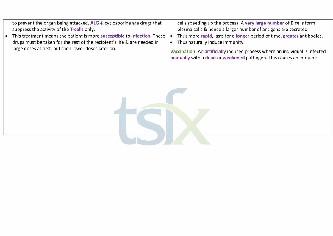
to prevent the organ being attacked. ALG & cyclosporine are drugs that suppress the activity of the T-cells only.
• This treatment means the patient is more susceptible to infection. These drugs must be taken for the rest of the recipient’s life & are needed in large doses at first, but then lower doses later on.
cells speeding up the process. A very large number of B cells form plasma cells & hence a larger number of antigens are secreted.
• Thus more rapid, lasts for a longer period of time, greater antibodies. • Thus naturally induce immunity.
Vaccination: An artificially induced process where an individual is infected manually with a dead or weakened pathogen. This causes an immune

response without causing symptoms of the disease. T & B cells are produced, that creates memory B & T cells so that an immunity is acquired.
• Vaccinations produces or improves immunity to a disease.
• Produced by either living microbes, dead microbes, or microbial toxins.
• Some need to be given periodically, while others are a one off.
Immunity: to resist pathogens & hence resist disease. Attenuated Vaccine: A vaccine created by reducing the virulence of a pathogen but still keeping it ‘live’.
Genetic Immunity
Acquired Immunity
Natural Artificial
An individual is born with immunity to a disease (pathogen) so never catches the disease; memory cells are present from birth.
Active (the body’s immune system is fighting the pathogens)
-An individual catches the disease, fights off the pathogen & develops immunity (memory cells remain in the system)
Active – A vaccine of dead or weakened pathogen is injected into the blood. A vaccination causes “Immunisation” (body’s response). It is not strong enough to cause the disease but it does cause the immune response (production of antibodies & the build-up of memory cells) & hence immunity.
Passive (introduction of antibodies into the body from another individual, to fight the disease)
-Antibodies pass across the placental membrane from mother to foetus.
Passive – A sample of antibodies (e.g. gamma globulin) from an immune individual is injected. The antibodies protect against the disease: the recipient’s immune system may act later but the administered antibodies provide the immunity for a short time period. (no memory cells formed)
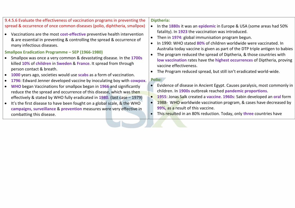
9.4.5.6 Evaluate the effectiveness of vaccination programs in preventing the spread & occurrence of once common diseases (polio, diphtheria, smallpox)
• Vaccinations are the most cost-effective preventive health intervention & are essential in preventing & controlling the spread & occurrence of many infectious diseases.
Smallpox Eradication Programme – SEP (1966-1980)
• Smallpox was once a very common & devastating disease. In the 1700s killed 10% of children in Sweden & France. It spread from through person contact & breath.
• 1000 years ago, societies would use scabs as a form of vaccination.
• 1796: Edward Jenner developed vaccine by inoculating boy with cowpox.
• WHO began Vaccinations for smallpox began in 1966 and significantly reduce the the spread and occurrence of this disease, which was then effectively & stated by WHO fully eradicated in 1980. (last case – 1979)
• It’s the first disease to have been fought on a global scale, & the WHO campaigns, surveillance & prevention measures were very effective in combatting this disease.
Diptheria:
• In the 1880s it was an epidemic in Europe & USA (some areas had 50% fatality). In 1923 the vaccination was introduced.
• Then in 1974: global immunisation program begun.
• In 1990: WHO stated 80% of children worldwide were vaccinated. In Australia today vaccine is given as part of the DTP triple antigen to babies
• The program reduced the spread of Diptheria, & those countries with low vaccination rates have the highest occurrences of Diptheria, proving vaccine effectiveness.
• The Program reduced spread, but still isn’t eradicated world-wide.
Polio:
• Evidence of disease in Ancient Egypt. Causes paralysis, most commonly in children. In 1900s outbreak reached pandemic proportions.
• 1955: Jonas Salk created a vaccine. 1960s: Sabin developed an oral form
• 1988- WHO worldwide vaccination program, & cases have decreased by 99%, as a result of this vaccine.
• This resulted in an 80% reduction. Today, only three countries have

• never stoped transmission of polio(endemic) & have had the lowest rates of vaccination. As long as a single child remains infected, children across all countries are at risk.
9.4.5 MacFarlane Burnet – A founder of Immunology
• Clonal Selection: A specific B cell (with required antibody) or T cell (with required receptor) is selected due to it’s unique match to the specific non-self antigen. These selected cells then clone themselves to produce large quantities of the required antibody/cell at the right time.
Steps: 1. Production of difference cells with different receptors. 2. Wait in lymph node until chosen for specific matching antigen. 3. Clone themselves to produce many specific effector & memory cells.
• MacFarlane Burnet: (Awarded Nobel Prize) His experimental work on viruses resulted in discoveries of their nature & replication. He pioneered the application of ecological principles to viral diseases.
• He proposed the concepts acquired immunological tolerance (bodies learn how to recognise self) & the clonal selection theory of antibody production.
9.4.6 Epidemiological studies involve the collection & careful statistical analysis of large quantities of data. Such studies assist the causal
identification of non-infectious diseases.
9.4.6.1 Describe the main features of epidemiology with eg. lung cancer
• Epidemiology: The study of diseases; including their distribution & frequency, patterns, influencing factors, & risk factors in a population for the purpose of developing control &/or preventative methods.
• Types: Descriptive, analytical or Intervention.
• Descriptive: Investigate the cause of a disease by looking at patterns of distribution, frequency, section of population (age, gender, occupation, socioeconomic status, environment, lifestyles, genetic history)
• His development of the cultivation of viruses in chick eggs, was significant, as viruses can’t reproduce outside a host cell (not on agar plates). This allowed a high concentration of the virus to be produced & vaccines later developed.
• Intervention Studies: Test the effectiveness of a treatment or a public health campaign to change the behaviour of the population as a whole.
• Valid Epidemiological Study Should:
• Conducted over a long time. • Study large sample size (thousand) • Range of RELEVANT data from a large group of both affected &
unaffected people (case-control), & other factors that might affect incidence of disease Eg. age, diet etc.
• Participants represent broad range of society/lifestyles but target study
• Use control groups who are not exposed to potential cause but similar in all other aspects (cohort studies)
• Data on incidence, prevalence, mortality, morbidity
• Statistically analyse data to identify patterns, trends & those most at risk
• Identify possible cause of the disease

• Analytical: (data on morbidity, mortality, prevalence (specific time) & incidence (new) to test specific hypotheses.
Case-control studies: compare people with the disease (case) to people without the disease (control). (differences in exposure to possible causes) Cohort Studies: Two or more groups of people who are FREE of the disease. The groups differ in their exposure to one potential cause of the disease.
• Develop a management plan & educate the public
• Evaluate effectiveness of control & treatment programs.
(long term, large sample size, extensive collection of data)

Lung Cancer Eg:
• Descriptive: data collected egs: age, sex, smoking habits, diet, occupation & drinking habits of both smokers & non-smokers.
• Analytical: Case-control studies: Compared patients with lung cancer to patients with other conditions. Information about many factors in their life were collected. The results of this study showed most individuals with lung cancer were smokers. Cohort Studies: Followed 40 ER doctors over a 10-year period. One group of doctors were smokers (test group) & the other were non-smokers (control). At the end of the study the test group had a higher incidence of lung cancer, & those who smoked the greater number of cigarettes daily had a greater chance of dying from lung cancer.
• Intervention Studies: effectiveness of campaigns such as the “Quit” campaign to decrease the number of people smoking is evaluated.
• All studies showed a strong correlation between smoking & lung cancer.
9.4.6.2 information to identify the cause & effect relationship of smoking & lung cancer (just roughly know)
• Lung Cancer: uncontrolled growth of tumours in the lung.
• Causes: carcinogenic chemicals in tobacco smoke.
• Effect: Carcinogenic chemicals damage cells & cause DNA to mutate – resulting in growth of tumours in lungs which destroy alveoli, making breathing difficult. It can then metastasis (spread to other organs). It can be a long illness.
• Epidemiological information: o 1930s – sudden lung cancer epidemic o 1950s-60s – studies showing cause & effect o Statistics now show that there is a direct correlation between
amount smoked & risk of lung cancer.
Note: Answer Eg. As the cigarette sales increased, so too did the number of deaths from lung cancer. (with a lag time).

Notes on Epidemiology HSC Questions: • Don’t forget judgments for Evaluate and Analyse (include both sides too)
• ALWAYS INCLUDE DATA FROM QUESTION!
• If asked to state limitations on data (explain why) o Eg. small sample size or data samples are from different places o No other factors included o Decrease in mortality could be health care treatment (hence should
include morbidity) o Not shown if smokers or non-smokers o Not showing large enough time period in data – lung cancer takes long
time to develop)
• Methodology: NEED TO REFER TO ALL PARTS OF QUESTION!!! o Include examples from question & examples of improvements o Larger sample space, & larger diversity of respondents o Shouldn’t remove respondents as this decreases sample size & reduces
scientific validity & introduces bias as who do you remove? & removes links to other possible sources for the cancers.
o More specific medical check (EG. lung health tests)
o Learn other important background information or possible risk factors (EG, age, history, living/working, genetic history etc)
o Don’t use a questionnaire – people more likely to lie. o Study peer reviewed in a scientific journal (not blog) o Follow over a long time period
• Areas of Study features needed o Be specific to question - Aged Care = control of variables (gender –
consistent across all homes & targeted for study (elderly) o Be specific – hygiene, location/environment (not just age, sex etc) o Broad range of participant backgrounds o Data analysis – compare common data with & without disease o –look for common data in people with disease

9.4.6.3 Causes of Non-Infectious disease using an example for inherited diseases, nutritional deficiencies & environmental diseases.
Non-Infectious Disease: Diseases that are not caused by a pathogen & are not contagious.
Classification About Example & Cause
Inherited Genetically transmitted & caused by errors in genetic information.
Down Syndrome: In the formation of gametes, non-disjunction of chromosome 21 occurs. Resulting in trisomy-21.
Nutritional Deficiency
Diets lacking proper balance & amount of nutrients.
Scurvy: Lack of vitamin C in diet (from fruits & vegetables). This weakens the blood capillary walls.
Environmental Caused by lifestyle, physical factors, or chemicals.
Lead Poisoning: High levels of lead exposure, which accumulates in the body, and disrupts mental development. Exposure from lead paint, lead batteries & leaded petrol.
9.4.6.4 Named non-infectious disease
• Down Syndrome: Inherited
Occurrence -1/1000 live births worldwide. (WHO) -associated with the mother’s age as non-disjunction is more likely
to occur in females over 35.
Symptoms Could include: -prominent forehead, round face, flattened nose, sagging mouth, skin folds at eye corners, wide set eyes. -Hand & fingers are both short. -joint weakness, heart defects, infertility -Main symptom is mild to severe mental retardation, & delayed mental development
Cause -extra chromosome 21. This is a chromosome mutation called trisomy-21. Non-disjunction of the members of pair 21, results in a gamete with two 21 chromosomes. Then after fertilisation the zygote has three 21 chromosomes.
Treatment/ Management
-There is no treatment but management includes educational programs to assist children in learning, & developing skills that helps them participate in the community. -Genetic Counselling is available for those at ‘risk’.

9.4.7 Increased understanding has led to the development of a wide range of strategies to prevent and control disease
9.4.7.1 Role of quarantine in preventing the spread of disease & plants & animals into Australia or across regions of Australia
• Quarantine: isolation of diseased organisms as a strategy to control (stops further spread) & prevent (stop occurrence of disease in individuals) the spread of disease. • Originally Australia remained free of the World’s pests & diseases due to it’s geographical isolation, but today with travel & trade it is the Australian Quarantine & Inspection Service (AQIS) that maintains this reputation.
Methods: • Border Control: with travel & global trade, at Australian border’s including airports, seaports, mail exchanges, & cargo clearance, there are many quarantine officers who inspect, risk analyse, test products, & treat if needed. There are also many policies in place for imports and exports. This also occurs for transport between states of Australia. • Animal Quarantine: If any animals come into Australia, they need to be in
Quarantine before being allowed into the country freely. This is to ensure no diseases affect Australian animals. Eg. Horses for Melbourne Cup.
• Plant Quarantine: All plants or plant materials including soil, fruit, cuttings etc. are examined & if required they are treated. Any living plants also need to be in quarantine for a required period. • Human Quarantine: A continuous check is kept on all arriving passengers to ensure they don’t have pathogens on their clothes/shoes etc, & aren’t carrying any infectious diseases. Aircrafts are sometimes sprayed with insecticides. If an individual has a highly infected disease, they may be quarantined within a hospital.
9.4.7.2 Evaluate the effectiveness of quarantine in preventing the spread of plant and animal disease into Australia or across regions of Australia
• PLANT: Black sigatoka is a fungal disease that destroys banana leaves, reducing the size/number of fruit & is especially in countries north of Aus. • Early detection of outbreaks in the Cape York Peninsula & North Queensland, led to eradication early on. It is due to the effectiveness of Australian quarantine methods that these outbreaks were prevented from
Spreading. • The strict laws on moving plants, fruit or soil out of countries north of Australia & Torre Strait island quarantine zones into mainland Australia, has created great success in preventing the introduction/spread of this disease • It also relies on responsible reporting of the yellow halo signs by farmers. • Prevention is always better than treatment which costs a lot & could greatly effect Australia’s banana economy.
• ANIMAL:
• The effectiveness of Australian Quarantine has been proven by the many diseases that have been kept out, of which could cause extreme damage to Australia’s natural biota. But the equine influenza outbreak was a time when Australian Quarantine wasn’t effective. • In 2007, 4 Japanese stallions arrive in Australia, were taken into Quarantine, & displayed symptoms & tested positive for equine influenza. • All horses (60) at this quarantine were isolated; i.e no movement allowed. • But on the same day, symptoms & positive testing, of other horses at Parklands (some distance a part) occurred, even though no horses moved. 9.4.7.3 Examine plant shoots/leaves & information of evidence of Black Spot
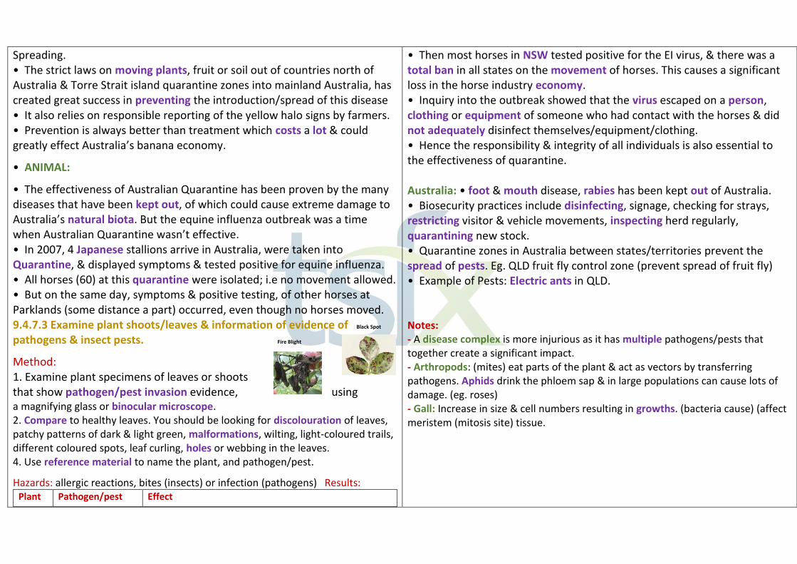
pathogens & insect pests. Fire Blight
Method:
1. Examine plant specimens of leaves or shoots that show pathogen/pest invasion evidence, using a magnifying glass or binocular microscope. 2. Compare to healthy leaves. You should be looking for discolouration of leaves, patchy patterns of dark & light green, malformations, wilting, light-coloured trails, different coloured spots, leaf curling, holes or webbing in the leaves. 4. Use reference material to name the plant, and pathogen/pest.
Hazards: allergic reactions, bites (insects) or infection (pathogens) Results: Plant Pathogen/pest Effect
• Then most horses in NSW tested positive for the EI virus, & there was a total ban in all states on the movement of horses. This causes a significant loss in the horse industry economy. • Inquiry into the outbreak showed that the virus escaped on a person, clothing or equipment of someone who had contact with the horses & did not adequately disinfect themselves/equipment/clothing. • Hence the responsibility & integrity of all individuals is also essential to the effectiveness of quarantine. Australia: • foot & mouth disease, rabies has been kept out of Australia. • Biosecurity practices include disinfecting, signage, checking for strays, restricting visitor & vehicle movements, inspecting herd regularly, quarantining new stock. • Quarantine zones in Australia between states/territories prevent the spread of pests. Eg. QLD fruit fly control zone (prevent spread of fruit fly) • Example of Pests: Electric ants in QLD. Notes: - A disease complex is more injurious as it has multiple pathogens/pests that together create a significant impact. - Arthropods: (mites) eat parts of the plant & act as vectors by transferring pathogens. Aphids drink the phloem sap & in large populations can cause lots of damage. (eg. roses) - Gall: Increase in size & cell numbers resulting in growths. (bacteria cause) (affect meristem (mitosis site) tissue.

Roses Fungus Black spot (on leaves): Cell death has occurred & disrupts photosynthesis (black/discoloured spots)
Lilly Pilly
Leftover eggs from insects (pest)
-Covers surface, disrupts photosynthesis & attracts other pathogens (fluffy eggs)
Apples Bacteria Fire Blight: affects ability to photosynthesise & economic fruit size (Burnt appearance & leaf curl)
9.4.7.4 ONE strategy has controlled &/or prevented disease: public health programs; pesticides; or GENETIC ENGINEERING to produce disease-resistant plants & animals
Genetic Engineering: Process that alters the genetic composition of an organism in some way. • It is now possible to make resistance to diseases, & prevent the disease from occurring & control the spread of the disease through the population
PLANTS: • Transgenic species Bt Cotton: Cotton that has a gene inserted from bacterium Bacillus Thuringiensis (Bt) which produces a protein that is a natural insecticide that kills specific pests (not all insects) such as Cotton Bollworm. (breaks down its gut wall causing death). Hence when the Bt Cotton produces this protein, it prevents disease from cotton bollworm who eat the cotton, & leave gouges that attracts pathogens. It also controls the spread by reducing the reproduction. • Potato plants with frog gene, to prevent fungal/bacterium infections (Late blight)
ANIMALS: • Technology CAR-T (Chimeric Antigen Receptor): ($600,000) - A girl with leukaemia was about to die, & this was the only option left - They took some of her blood, & genetically modified her cytotoxic T-cells. They were reprogramed & manipulated to have the perfect receptor for her leukaemia. When re-inserted they were specific to fight her cancer cells, & 5 years later she is healthy & cancer free. (Thus controlled her disease) • Genetically engineering Mice: - Are modified into a transgenic species with a gene from a human. (i.e a disease causing human gene is inserted into their germline cells, so that the offspring have this gene). - This allows the mice to be models that are prone to certain diseases such as cancer, heart disease, multiple sclerosis, diabetes etc. - Hence allowing research (unethical to perform human experiments) to be conducted to deeper understand human diseases & hopefully lead to preventions.
9.4.7.5 The changing methods of dealing with plant & animal diseases, including the shift in emphasis from treatment and control to management or prevention of disease.
Treatment: Cure or relieve symptoms of an already diseased organism. Control: Reduce the spread through population, once it’s already present. Prevention: Stops the occurrence of disease in organism before it has it. Management: Programs that improve the outcomes of chronic conditions & the quality of life of sufferers. (also reduces costs & use of health care)
• Shift from treatment to prevention, due to the reduction of costs, the better quality of life and the development of resistant strains. Eg: When penicillin was discovered, there was a large emphasis on treating bacterial infections and hence controlling them too, such as turberculosis. • But now many bacterial infections are resistant to antibiotics, & hence a shift towards prevention & management has occurred. • This is done through public health programs that educate about hygiene, covering your mouth & elbow when coughing/sneezing etc to help control the spread. Furthermore, vaccinations & quarantine further prevent &
Control the disease. Eg. vaccinations of smallpox has eradicated this disease throughout the world. • Pesticides have been used to treat & control the spread of pests, but a shift to prevention through genetic engineering such as BT cotton has reduced the costs & environmental impacts of pesticides.
Other Methods: Prevention (reduces cost) Treatment -public health programs for sun screen etc (slip, slop, slap program)
-chemotherapy
-Transgenic plant species (potatoes w frog gene) -Insecticide DDT for malaria
-Quarantine laws (prevents foot & mouth) -Antibiotics for bacterial infections
-Cervical cancer screening programs to detect -Medication of Fludrocortisol for Addison’s disease or for AIDS
-Prospective parents screened for presence of gene for the disease Thalassemia.
-Surgery for cardiovascular disease
-public health program “check your own skin” or “every cigarette is doing you damage” or lifestyle changes to prevent non-infectious disease.
